national services division pet-ct review of indications
TRANSCRIPT
1
National Services Division
PET-CT Review of Indications Report
V2
Published: July 2017
Review: July 2020
Executive Summary
Description of condition and service
- Positron Emission Tomography - Computed Tomography (PET-CT) is a unique imaging tool which shows pathology by using PET to detect derangement in tissue metabolism and CT to show structural changes. PET-CT is a key diagnostic service which provides information to allow informed clinical management decisions and more effective targeted care. This contributes to more individualised care and treatment of patients. The appropriate use of the examination in the patient pathway optimises the efficiency of the subsequent clinical interventions and treatment regimens.
Needs / activity
- The number of scans undertaken in Scotland was: o 5243 in 2013/14; o 6739 in 2014/15; and o 6725 in 2015/16.
- Existing capacity is estimated to be around 7,600 scans a year (if all scans were
[18F]-fluoro-deoxy-glucose (FDG PET-CT)). The actual capacity is less because a range of tracers is needed to support use of PET-CT in different conditions, and scans using alternative “non FDG” tracers take longer.
Clinical effectiveness
- The Royal Colleges of Radiology and Physicians published evidence based guidelines for the use of PET-CT in 2013 and has updated their guidance in February 2016 4,5. The guidelines describe the range of conditions for which there is evidence of effectiveness of PET-CT. The strength of evidence varies.
Outcomes - Four categories were defined in an audit of PET-CT in West of Scotland:
o High impact if PET-CT modified the decision to treat or mode of treatment; o Moderate if it partially modified treatment – eg location o Low if it did not determine any change in treatment o No impact when the information from PET-CT was considered inadequate by
the referring physician. - Outcomes have not been systematically recorded. The only information on outcomes
is from ad hoc audits. Cost
- The costs of PET-CT scanning in Scotland in 2015/16 were £6.76 million. Issues
- Capacity is limited in relation both to the skilled staffing available and the capacity of PET-CT scanners and cyclotrons (which produce the tracers required). Evidence exists of potential benefit of use of PET-CT in a wide range of indications. This report seeks to identify the indications for which current evidence suggests PET-CT offers the highest potential for patient benefit to guide the use of the limited capacity available and ensure optimum use is made of the resources available.
- The report examines the indications reported in the Royal Colleges 2016 Guidelines5 and makes recommendations for use of PET-CT in NHS Scotland.
3
Contents Executive Summary .............................................................................................................. 2
1.1 Brief description of service ........................................................................................... 4 1.2 Background to review .................................................................................................. 4 1.3 Approach to task .......................................................................................................... 5
Section 2: Assessed Needs .................................................................................................. 6 2.1 Summary of patient need ............................................................................................. 6 2.1.1 Introduction ............................................................................................................... 6 2.1.2 Current activity .......................................................................................................... 6 2.1.3 Estimate of unmet need ............................................................................................ 7 2.1.4 Cross Border activity ................................................................................................. 8 2.1.5 Summary .................................................................................................................. 8
Section 3: Current Provision .................................................................................................. 8 3.1 Description of current service ....................................................................................... 8 3.2 Accessibility and balanced geographic distribution ....................................................... 9 3.3 Service Risks and Issues ........................................................................................... 10
Section 4: Clinical Effectiveness / Clinical Outcomes .......................................................... 11 4.1 Clinical effectiveness & potential for health gain ........................................................ 11
Section 5: Costs .................................................................................................................. 12 5.1 Financial details of the current and proposed future provision .................................... 12 5.2 Average cost per patient ............................................................................................ 12 5.3 Value for money compared to alternatives ................................................................. 12
Section 6: Appraisal of evidence ......................................................................................... 13 6.1 SHTG Review of evidence for PET/CT ..................................................................... 13 6.2 Review by SCIN PET-CT review group .................................................................... 13 6.3 New Guidance under development by SCIN PET-CT review of indications group .... 14
6.3.1 Use of PET-CT in breast cancer ............................................................................. 14 6.3.3 Review evidence of PET-CT in Head and Neck ...................................................... 14 6.3.5 Review of evidence of PET-CT in Vasculitis ............................................................ 14 6.3.6 Non-FDG in Prostate Cancer .................................................................................. 15
Section 7: Conclusions and recommendations .................................................................... 15 Annex A – Membership ....................................................................................................... 20 Annex B – Dates of Guidelines ........................................................................................... 21 Annex C – Activity 2015/16 ................................................................................................. 22 Annex D - References ......................................................................................................... 23
4
Section 1: Introduction
1.1 Brief description of service Positron Emission Tomography - Computed Tomography (PET-CT) is a unique imaging tool which shows pathology by using PET to detect derangement in tissue metabolism and CT to show structural changes. PET-CT is a key diagnostic service which provides information to allow informed clinical management decisions and more effective targeted care. This contributes to more individualised care and treatment of patients. The appropriate use of the examination in the patient pathway optimises the efficiency of the subsequent clinical interventions and treatment regimens.
1.2 Background to review
Why was review undertaken, remit, aims In 2014, the Scottish Government Health and Social Care Directorates (SGHSCD) transferred responsibility for oversight of PET-CT development from the PET Advisory Group to the Scottish Clinical Imaging Network (SCIN). The SCIN network established a PET-CT subgroup to:
Develop protocols to inform clinical decision making on the use of PET-CT
Publish and monitor information on adherence to PET-CT protocols in Scotland
Examine the evidence for the use of PET-CT in new indications
Audit the provision of PET-CT in Scotland.
Current work involves gathering information on:
the indications for which PET-CT is currently being used in Scotland;
the number of referrals to England for PET-CT scanning for residents of Scotland;
the range of tracers in use in Scotland and how these are obtained;
the capacity of PET-CT facilities in the 4 centres in Scotland;
proposed changes such as the development of new tracers.
In May 2015 the SGHSCD Diagnostic Steering Group approved the establishment of a SCIN PET/CT Review of Indications Short Life Working Group with a remit “to develop consistent, evidence based PET/CT guidelines which would promote equity for patients”. Membership is set out in Annex A. The aim of the Short Life Working Group was therefore to review of the list of indications for which PET-CT provides evidence based, cost effective imaging and make recommendations for use of PET-CT in Scotland to the Diagnostic Steering Group. Situation in England on PET-CT Commissioning guidelines have been approved covering central funding for PET-CT in 29 indications – 23 cancer; 6 non cancer http://www.england.nhs.uk/commissioning/spec-services/npc-crg/group-b/b02/ The NHS England commissioning policy was developed to inform commissioning of oncology PET-CT indications and to normalise the commissioning of non-oncology PET-CT indications, and to ensure usage of PET-CT where there is good evidence that patients will benefit from improved disease assessment resulting in altered management and improved outcomes.
5
1.3 Approach to task
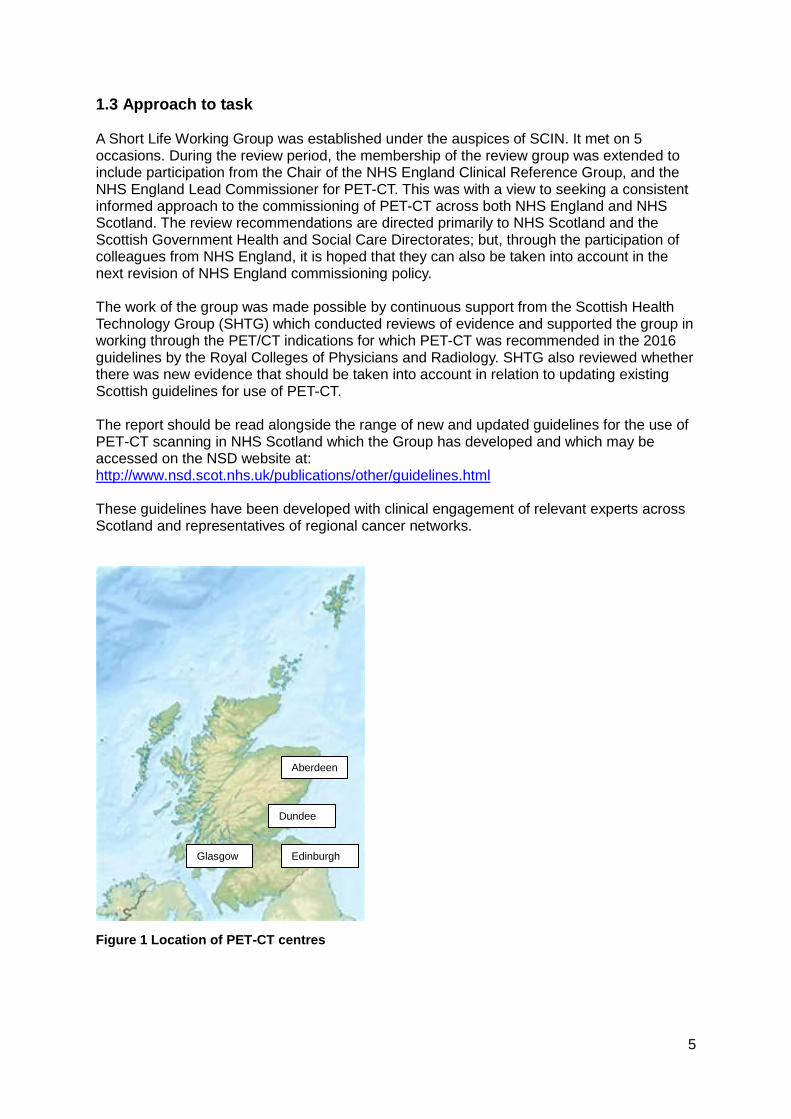
A Short Life Working Group was established under the auspices of SCIN. It met on 5 occasions. During the review period, the membership of the review group was extended to include participation from the Chair of the NHS England Clinical Reference Group, and the NHS England Lead Commissioner for PET-CT. This was with a view to seeking a consistent informed approach to the commissioning of PET-CT across both NHS England and NHS Scotland. The review recommendations are directed primarily to NHS Scotland and the Scottish Government Health and Social Care Directorates; but, through the participation of colleagues from NHS England, it is hoped that they can also be taken into account in the next revision of NHS England commissioning policy. The work of the group was made possible by continuous support from the Scottish Health Technology Group (SHTG) which conducted reviews of evidence and supported the group in working through the PET/CT indications for which PET-CT was recommended in the 2016 guidelines by the Royal Colleges of Physicians and Radiology. SHTG also reviewed whether there was new evidence that should be taken into account in relation to updating existing Scottish guidelines for use of PET-CT. The report should be read alongside the range of new and updated guidelines for the use of PET-CT scanning in NHS Scotland which the Group has developed and which may be accessed on the NSD website at: http://www.nsd.scot.nhs.uk/publications/other/guidelines.html These guidelines have been developed with clinical engagement of relevant experts across Scotland and representatives of regional cancer networks.
Figure 1 Location of PET-CT centres
Aberdeen
Dundee
Edinburgh Glasgow

6
Section 2: Assessed Needs
2.1 Summary of patient need
2.1.1 Introduction
Positron Emission Tomography - Computed Tomography (PET-CT) acquires Positron Emission Tomography (PET) data and X ray Computed Tomography (CT) data in one scan and combines the data into superimposed (co-registered) images. The technique allows for precise and accurate anatomical localisation of biochemical activity in the body.
PET-CT scanning is a non direct access, key clinical imaging tool. Patients are referred from secondary care. The investigation contributes directly to the management of cancer patients; it also aids the management of patients who suffer from other diseases including those with cardiac and neurological disease and clinical suspicion of inflammation of the arteries (vasculitis).
2.1.2 Current activity
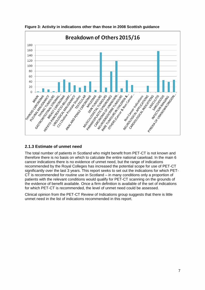
The target patient group who may benefit from PET-CT scanning are predominately patients with specific cancers. In 2008 SGHSCD issued guidance on the use of PET-CT in NHS Scotland. This guidance recommended PET-CT for use in 6 cancers – lung, colorectal, head and neck, lymphoma and oesophageal. The chart below (Figure 2) shows the trends in activity over the last 3 years. By far the highest use is in the management of lung cancer. A full breakdown of activity in 2015/16 is set out in Annex C.
Figure 2 – Trends in use of PET-CT in Scotland 2013-2016
The 2008 guidance permitted restricted use of PET-CT in other indications where there was less evidence of effectiveness. A breakdown on use in the other indications is shown in the chart below (Figure 3).
7
Figure 3: Activity in indications other than those in 2008 Scottish guidance
2.1.3 Estimate of unmet need
The total number of patients in Scotland who might benefit from PET-CT is not known and therefore there is no basis on which to calculate the entire national caseload. In the main 6 cancer indications there is no evidence of unmet need, but the range of indications recommended by the Royal Colleges has increased the potential scope for use of PET-CT significantly over the last 3 years. This report seeks to set out the indications for which PET-CT is recommended for routine use in Scotland – in many conditions only a proportion of patients with the relevant conditions would qualify for PET-CT scanning on the grounds of the evidence of benefit available. Once a firm definition is available of the set of indications for which PET-CT is recommended, the level of unmet need could be assessed.
Clinical opinion from the PET-CT Review of Indications group suggests that there is little unmet need in the list of indications recommended in this report.
8
2.1.4 Cross Border activity
PET-CT is not available in Scotland for the highly specialist scanning required prior to epilepsy surgery in children and, as a result, children requiring such scans are referred to Great Ormond Street in London. In addition, there is currently no provision for Gallium68 scans and dota-octreotide treatment for prostate and neuroendocrine tumours in Scotland.
The numbers of Scottish residents referred for PET-CT and dota-octreotide scans/ treatment in the last 3 years were: 2013/14 = 5 2014/15 = 6 2015/16 = 4 2016/17 = 6
2.1.5 Summary
The need for PET-CT scanning in Scotland is for around 6,500 scans a year based on the current list of recommended indications. This is within, but close to, full capacity available.
Section 3: Current Provision
3.1 Description of current service
NHS Scotland currently commissions oncology PET-CT using [18F]-fluoro-deoxy-glucose (FDG PET-CT) and non-FDG PET-CT radioactive tracers as recommended in the SGHSCD 2008 guidance. As such, FDG PET-CT is commissioned predominately for the 6 cancer conditions set out in the guidance but it is also available in exceptional circumstances for the investigation of selected patients with infection, pyrexia of unknown origin, suspected large vessel vasculitis, sarcoidosis, cardiac and neurological conditions.
In keeping with decisions in NHS England, NHS Scotland does not commission the use of amyloid radioactive tracers for brain imaging. This is because there is insufficient evidence available to demonstrate benefit. Specifically NHS Scotland commissions the following FDG PET – CT non-cancer indications on a non routine basis: Large Vessel Vasculitis • Evaluation of suspected vasculitis in selected cases; for example, to determine the extent and distribution of the disease activity or to exclude underlying malignancy which may be a paraneoplastic phenomenon, resulting in atypical presentations of vasculitis. • PET-CT would not be indicated in all patients with giant cell arteritis, but is of use in patients where conventional investigations are unhelpful and treatment would be altered if ongoing inflammatory disease is confirmed. Sarcoidosis • Assessment of activity and distribution of disease at baseline in highly selected cases where there is diagnostic uncertainty using conventional imaging (e.g. suspected cardiac sarcoidosis)
9
• Assessment of disease response where other measures to monitor response are unhelpful and/or in patients with disease resistant to treatment. Infection imaging • Detection of site of focal infection in immuno-compromised patients or problematic cases of infection • Evaluation of vascular graft infection in selected cases provided sufficient time has elapsed since surgery. Pyrexia of unknown origin (PUO) • To identify the cause of a PUO where conventional investigations have not revealed a source. Neurological applications • Pre-surgical assessment of medically refractory complex partial seizures where MR is normal, equivocal or conflicts with EEG localisation Cardiological indications • Assessment of myocardial viability in patients with ischaemic heart failure and poor left ventricular function being considered for revascularisation, usually in combination with perfusion imaging with sestamibi/tetrofosmin or ammonia/rubidium.
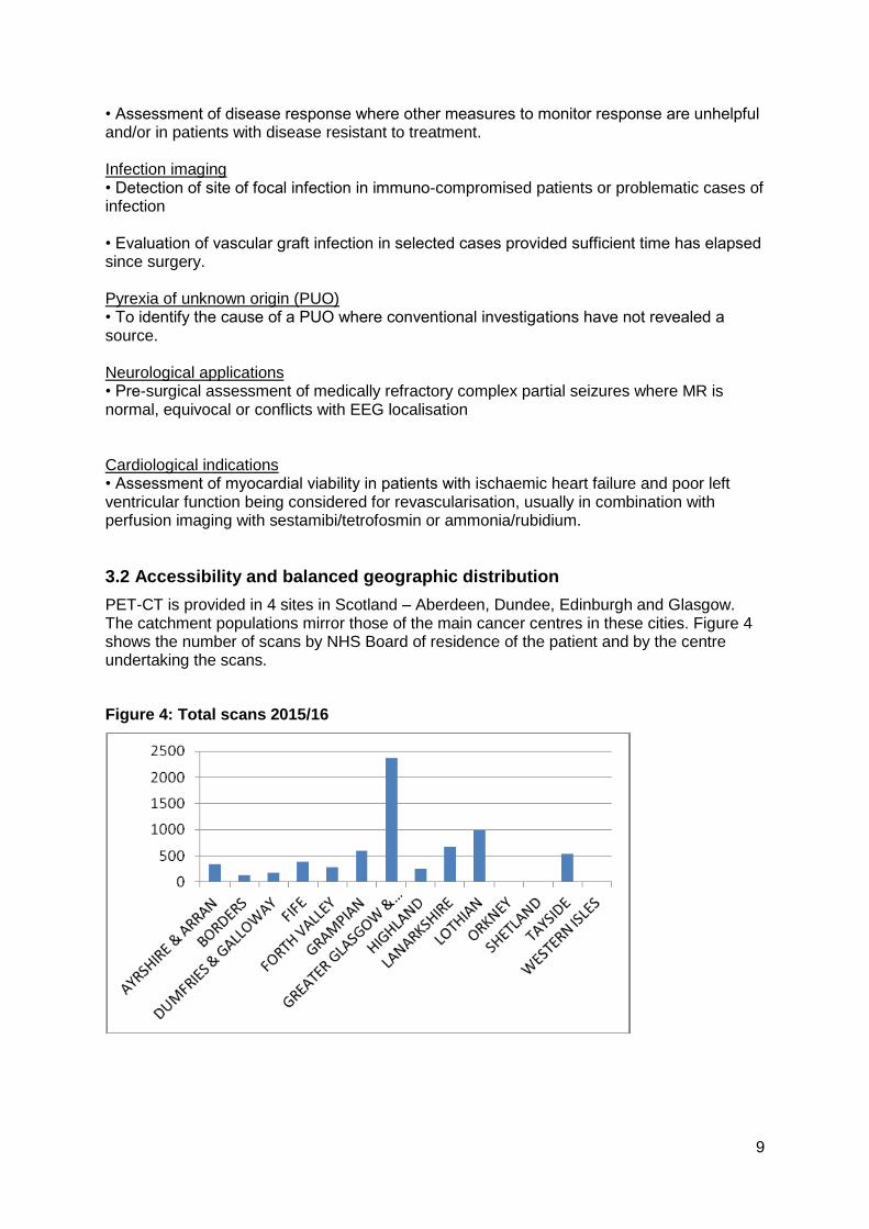
3.2 Accessibility and balanced geographic distribution
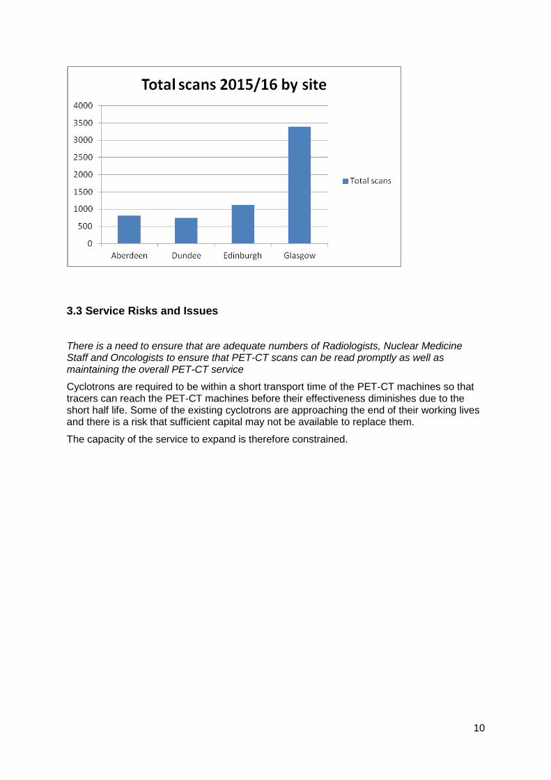
PET-CT is provided in 4 sites in Scotland – Aberdeen, Dundee, Edinburgh and Glasgow. The catchment populations mirror those of the main cancer centres in these cities. Figure 4 shows the number of scans by NHS Board of residence of the patient and by the centre undertaking the scans.
Figure 4: Total scans 2015/16
10
3.3 Service Risks and Issues
There is a need to ensure that are adequate numbers of Radiologists, Nuclear Medicine Staff and Oncologists to ensure that PET-CT scans can be read promptly as well as maintaining the overall PET-CT service
Cyclotrons are required to be within a short transport time of the PET-CT machines so that tracers can reach the PET-CT machines before their effectiveness diminishes due to the short half life. Some of the existing cyclotrons are approaching the end of their working lives and there is a risk that sufficient capital may not be available to replace them.
The capacity of the service to expand is therefore constrained.
11
Section 4: Clinical Effectiveness / Clinical Outcomes
4.1 Clinical effectiveness & potential for health gain
Since its introduction into clinical practice in the UK 26 years ago, PET followed by PET-CT has become a key investigative tool in the assessment of cancer and non-cancer medical conditions. The first version of the inter-collegiate ‘Evidence-based indications for the use of PET-CT in the United Kingdom 2012’3, provided a guide to the use of PET-CT in clinical practice and the evidence-base on which this was founded. It has been used to inform the commissioning of PET-CT services in the UK. Now in its 3rd edition, the 2016 version5 builds on the evidence cited in earlier editions providing an updated review with key references for the use of FDG and non-FDG PET-CT tracers in malignant and in non-malignant disease. The Royal Colleges guidance provides an up-to-date summary of relevant indications for the use of PET-CT, where there is good evidence that patients will benefit from improved disease assessment resulting in altered management and improved outcomes. Given its significance, it was used as the main basis for the SCIN PET-CT Review of Indications for PET-CT use in NHS Scotland.
The indications are divided into oncological and non-oncological applications and the publication explains that the list is not exhaustive and there are cases where PET-CT may be helpful in patients who have equivocal or definite abnormalities on other imaging where PET-CT may alter the management strategy if found to be ‘positive’ or ‘negative’; for example, radical or high-risk surgery. The guidance advocates that PET-CT would be appropriate in such patients at the discretion of the local Administration of Radioactive Substances Advisory Committee (ARSAC) certificate holder. In summary the guidance supports the use of PET-CT in: • Oncology: PET-CT may be helpful on an individual basis for the diagnosis, staging and
management of individual patients with rare malignancies in discussion with the specialist multidisciplinary team
• Non Oncology: PET-CT may be helpful on an individual case by case basis in the
diagnosis and management of individual patients in discussion with the specialist centre.
The evidence to support the benefits of PET-CT scanning is established by original research, expert opinion and professional and governmental bodies including National Institute of Clinical Excellence (NICE).
The table in Annex B sets out the dates of existing guidelines for the use of PET/CT based on the 2008/09 SGHSCD PET/CT protocols, NICE and SIGN guidelines, Scottish Health Technology Group scoping reports and Royal Colleges Guidelines.
12
Section 5: Costs
5.1 Financial details of the current and proposed future provision
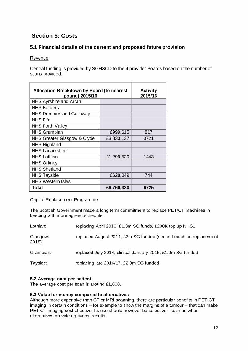
Revenue Central funding is provided by SGHSCD to the 4 provider Boards based on the number of scans provided.
Allocation Breakdown by Board (to nearest pound) 2015/16
Activity 2015/16
NHS Ayrshire and Arran
NHS Borders
NHS Dumfries and Galloway
NHS Fife
NHS Forth Valley
NHS Grampian £999,615 817
NHS Greater Glasgow & Clyde £3,833,137 3721
NHS Highland
NHS Lanarkshire
NHS Lothian £1,299,529 1443
NHS Orkney
NHS Shetland
NHS Tayside £628,049 744
NHS Western Isles
Total £6,760,330 6725
Capital Replacement Programme The Scottish Government made a long term commitment to replace PET/CT machines in keeping with a pre agreed schedule. Lothian: replacing April 2016, £1.3m SG funds, £200K top up NHSL Glasgow: replaced August 2014, £2m SG funded (second machine replacement 2018) Grampian: replaced July 2014, clinical January 2015, £1.9m SG funded Tayside: replacing late 2016/17, £2.3m SG funded. 5.2 Average cost per patient The average cost per scan is around £1,000.
5.3 Value for money compared to alternatives Although more expensive than CT or MRI scanning, there are particular benefits in PET-CT imaging in certain conditions – for example to show the margins of a tumour – that can make PET-CT imaging cost effective. Its use should however be selective - such as when alternatives provide equivocal results.

13
Section 6: Appraisal of evidence
6.1 SHTG Review of evidence for PET/CT
The Scottish Health Technology Group of Healthcare Improvement Scotland had previously produced 4 Advice Statements on PET-CT indications in 2013 6,7,8,9
For the current Review SHTG assessed current literature to inform the group if an update was required to previous guidance in:
o Non-FDG in Prostate Cancer o Head and Neck Cancer o Melanoma o Sarcoidosis o Paraneoplastic neurological syndrome o Pyrexia of unknown origin
The conclusion was that new guidance would be required on non-FDG in Prostate Cancer. The group agreed that the SHTG did not need to be consulted on the use of PET-CT in head and neck cancer and melanoma; these guidelines could be updated in the light of new evidence; and that existing advice on the last three indications remained up to date.
6.2 Review by SCIN PET-CT review group
In addition to the work by SHTG, individual members of the Review of Indications led work to review literature and to update guidelines – and appraise the Royal Colleges Guidelines with a view to the strength of evidence supporting the guideline. This work was undertaken in collaboration with Regional Cancer Networks and other stakeholders. The products of this work is a series of new and updated guidance designed for use in NHS Scotland to guide clinicians in the use of PET-CT. Specific outputs included updated guidance on lymphoma and head and neck cancer; proposed new guidance in breast cancer. The new lymphoma guidance is available on the NSD website, alongside the original 2008 guidance on 6 cancer indications, at: http://www.nsd.scot.nhs.uk/Documents/2016-07-12%20PET-CT%202016-03%20FINAL%20guidance%20on%20lymphoma.pdf Work continues on other guidance under the auspices of the PET-CT Working Group and will be published on the NSD website in due course on the guidelines page at: http://www.nsd.scot.nhs.uk/publications/other/guidelines.html
14
6.3 New Guidance under development by SCIN PET-CT review of indications group
6.3.1 Use of PET-CT in breast cancer The PET-CT ROI group heard that the three regional cancer networks were in agreement that PET/CT would not be routinely commissioned in breast cancer except where it was used in the following circumstances:
Assessment of multi-focal disease or suspected recurrence in patients with dense breasts in whom MRI is not available or is inconclusive
Differentiation of treatment-induced brachial plexopathy from tumour infiltration in symptomatic patients with an equivocal or normal MR.
Assessment of extent of disease in carefully selected patients (following MDT discussion) with disseminated breast cancer if aggressive therapy is being considered, e.g. metastatectomy
Assessment of response to chemotherapy in patients whose systemic disease is not well demonstrated using other techniques; for example, bone metastases.
Selected patients where conventional imaging is equivocal or conflicting.
Consider for patients with inflammatory breast cancer (in whom there is a significant incremental detection rate of distant metastases over and above conventional CT)
It was noted that PET/CT should not be used for staging or routine surveillance as there was insufficient evidence to justify its use. Breast cancer staging requires the detection of small <1cm tumours which were beyond the resolution of the technique. Low grade tumours might also be falsely negative. PET/CT has low sensitivity for nodal metastases and should not be used as a substitute for sampling and the sentinel node procedure. 6.3.2 Review evidence of PET-CT in Sarcoma
Clinical guidelines on PET-CT in sarcoma were reviewed by the Scottish Sarcoma Network and submitted.
6.3.3 Review evidence of PET-CT in Head and Neck The group noted that there had been recent evidence in PET-CT in Head and Neck Cancer NICE had looked at the evidence. The group amended the Scottish Government PET/CT protocol for head and neck to include response assessment 3-6 months post chemoradiotherapy. 6.3.4 Review of evidence of PET-CT in Brain/CNS Cancer The Scottish Adult Neuro-Oncology Network reviewed the evidence and recommended that PET-CT was not routinely commissioned.
6.3.5 Review of evidence of PET-CT in Vasculitis
The group considered draft guidelines and recommended that PET/CT should only be used for large vessel vasculitis and primarily for diagnosis, monitoring and/or response to medicine or treatment of disease. Small vessel vasculitis specialists were consulted and confirmed that PET-CT was not considered useful in small vessel vaculitis.

15
6.3.6 Non-FDG in Prostate Cancer SHTG prepared a scoping report on the use of Non-FDG PET-CT in Prostate Cancer for the Review Group. The review group noted that prostate specific membrane antigens (PSMAs) were a rapidly emergent technology which had largely replaced other tracers in England in imaging prostate cancer. PSMAs could be produced by a Gallium68 generator, and such generators provided opportunities in diagnosis as well as treatment. SHTG would in due course provide an evidence note and accompanying advice statement.
Section 7: Conclusions and recommendations
In concluding its appraisal, the SCIN PET-CT Review of Indications group reviewed the existing guidelines available (see Annexes B and D) alongside current usage of PET-CT (Annex C) in each indication listed in the Royal Colleges of Radiology and Physicians 2016 Guidelines5. The group agreed its recommendations for the use of PET – CT scanning in NHS Scotland, and for further work required, as follows. The report uses two classifications to differentiate between indications for PET-CT scanning which should be “routinely commissioned” (by which it is meant that PET-CT is an imaging option that is routinely considered in the specific indications identified) and those in which the use of PET-CT would be considered only in exceptional cases – “not routinely commissioned”. (This terminology matches that in common use in NHS England.) It should be noted that this report represents a snap shot in time and is correct only at the time it is being written. Work continues under the auspices of the SCIN PET-CT Working Group to maintain and update the suite of guidelines produced through the work of the PET-CT Review of Indications Short Life Working Group. The full suite of current guidelines to inform the use of PET-CT scanning in NHS Scotland is published on the NSD website at: http://www.nsd.scot.nhs.uk/publications/other/guidelines.html The recommended position as at September 2016 on each indication in the Royal Colleges of Radiology and Physicians 2016 Guidelines5 is as follows:
Brain/Central Nervous System: Given that there were currently <2 FDG scans a year reported in brain/CNS cancer in Scotland, the PET-CT ROI Group recommended continuation of current policy - “not routinely commissioned”. Consideration of use of PET-CT in this area in exceptional cases should be guided by the Royal Colleges 2016 Guidelines.
Colorectal Carcinoma: Definitive guidance was produced by SGHSCD in the 2008 protocols recommending use in specific indications. NICE guidelines were published in March 2011 and SIGN guidelines in December 2011. There were around 700 scans a year in Scotland. No evidence had been presented to the group to change existing policy as set out in the 2008 SGHSCD guidance – therefore Group recommended the position remains “Routinely commissioned for the indications set out in the 2008 SGHSCD Guidance”.
Gynaecological Malignancy: Definitive guidance was produced by SGHSCD in 2008 protocols recommending use in specific indications. There had been subsequent NICE guidelines in November 2015. There were around 300 scans a year in Scotland. No evidence had been presented to the group to change existing
16
policy – therefore Group recommended the position remains “Routinely commissioned for the indications set out in the 2008 SGHSCD Guidance”.
Head and Neck: Definitive guidance was produced by SGHSCD in 2008 protocols recommending use in specific indications. There were subsequent NICE guidelines in May 2015. There were around 400 scans a year in Scotland. The Group had been made aware that there was significant new evidence published in 2016 and agreed that changes to update the SGHSCD 2008 guidance. The group recommended that PET-CT should be “routinely commissioned” for the indications set out in the new 2016 NHS Scotland guidance.
Lung Carcinoma: Definitive guidance was produced by SGHSCD in the 2008 protocols recommending use in specific indications. There were subsequent NICE guidelines in April 2011 and a SIGN guideline in February 2014. Lung cancer accounted for the highest use of PET-CT in Scotland – around 3,000 scans a year. The group considered that the existing 2008 guidance should be reviewed in the light of the subsequent NICE / SIGN guidelines and the 2016 Royal Colleges Guidelines. The group recommended that PET-CT should be “routinely commissioned” for the indications set out in the updated 2016 NHS Scotland guidance.
Out with the scope of the group was Mesothelioma, at time of publication
Mesothelioma, “not routinely commissioned”.
Lymphoma: Definitive guidance was produced by SGHSCD in 2008 protocols recommending use in specific indications. There had been subsequent SIGN guidelines in February 2014 and NICE guidance in January 2016. There were around 800 scans a year in Scotland. The NHS Scotland PET-CT Working Group approved updated guidance in April 2016 and further revisions in September 2016. This is the current definitive guidance. The Group recommended “Routine Commissioning for indications set out in 2016 NHS Scotland PET-CT Working Group guideline.
Oesophageal (Upper GI)/ Oesophageal Gastric Carcinoma: Definitive guidance was produced by SGHSCD in 2008 recommending use in specific indications. There had been subsequent NICE guidance in December 2015. There were around 500 scans a year in Scotland. No evidence had been presented to the group to change existing policy as set out in the 2008 SGHSCD guidance – therefore Group recommended the position remains “Routinely Commissioned for the indications set out in the 2008 SGHSCD Guidance”.
Thyroid carcinoma: It was agreed that guidelines for the use of PET/CT in thyroid were covered under the extant Head and Neck guidelines for one indication. This was consistent with the low reported activity <3 a year – suggesting that activity was also reported under head and neck.
It was agreed that for the following indications SHTG would produce a brief literature search to assess the evidence available. If sufficient evidence was identified SHTG would produce a scoping report to inform guidance for NHS Scotland:
Pleural Malignancy (there were existing NICE and SIGN guidelines)
Thymic Tumors (existing SIGN guidelines)
Hepato-Pancreatco-Billiary Cancers (existing NICE guidelines)
Urological Malignancy (existing NICE guidelines)
17
Testicular (existing NICE and SIGN guidelines)
Myeleoma (existing NICE guidelines) Use in each indication was less than 30 scans a year. The group agreed that, until definitive guidance has been approved, the current policy of “not routinely commissioned” will continue to apply in all these indications. Consideration of use of PET-CT in these area in exceptional cases should be guided by the Royal Colleges 2016 Guidelines.
Breast Carcinoma: No previous guidance had been agreed for use of PET-CT in breast cancer in Scotland. There were NICE guidelines from June 2015 and SIGN guidelines from June 2013. There were around 50 scans a year in Scotland (and NHS England figures suggested only around 50 for all England). The group agreed a guideline based on the NOSCAN paper which is supported by SEAT and WOSCAN and is summarised in Section 6 of this report. The group’s recommendation is for “routine commissioning” for the very specific indications set out in Section 6 above.
Prostate cancer: No previous guidance had been agreed for use of PET-CT in prostate cancer in Scotland. There were NICE guidelines from January 2014. Ongoing work from SHTG due to report in late 2016 would inform the development of guidance for NHS Scotland. Effective imaging in prostate cancer requires alternative tracers to FDG. <20 scans a year were undertaken. The group agreed that, until definitive guidance has been approved, the current policy of “not routinely commissioned” should continue to apply – recognising that small numbers were currently being scanned in Scotland with F18 Choline and C11 Choline. Until Scottish guidelines are published in 2017, consideration of use of PET-CT in this area in exceptional cases should be guided by the Royal Colleges 2016 Guidelines.
Anal and Penile Carcinoma: No previous guidance had been agreed for use of PET-CT in anal and penile cancer in Scotland. Given that there were <10 scans undertaken each year, the PET-CT ROI Group recommended continuation of current policy - “not routinely commissioned”. Consideration of use of PET-CT in this area in exceptional cases should be guided by the Royal Colleges 2016 Guidelines.
Skin Tumours: Use of PET-CT in melanoma in Scotland is informed by the SHTG Advice Statement 003-139. There are around 150 scans a year in Scotland. The group segregated skin cancers into Melanoma, Basal cell carcinoma and Squamous cell carcinoma. The group identified that in the latter two indications, PET-CT should “not be routinely commissioned” whereas in melanoma it should “be routinely commissioned” for patients with indeterminate findings on CT or for patients who are being considered for major surgical resection, after discussion with the specialist multidisciplinary team.
Sarcoma - Gastrointestinal Stromal Tumours and Musculoskeletal Tumours Clinical guidance had been agreed for use of PET-CT in these two indications in Scotland by the Scottish Sarcoma Network. Current activity was <50 a year (in total). The Scottish Sarcoma Network reviewed the guidance and submitted it. The group agreed the current policy of “not routinely commissioned” will continue to apply. Consideration of use of PET-CT in this area in exceptional cases should be guided by the Royal Colleges 2016 Guidelines.
Rare Cancers in Children and Adults - No previous guidance had been agreed for use of PET-CT in rare cancers in Scotland. Current activity was around 50 a year.
18
The group agreed that the current policy of “not routinely commissioned” will continue to apply. Consideration of use of PET-CT in this area in exceptional cases should be guided by the Royal Colleges 2016 Guidelines.
Paraneoplastic Syndromes - Use of PET-CT in this indication in Scotland is guided by the 2013 STHG Advice Statement 010-137. Current activity is around 30 a year. In February 2016, SHTG assessed if there was new evidence that required an updating of established guidance and concluded that there was insufficient new evidence to merit an update the extant policy. The group agreed that the current policy of “not routinely commissioned” will continue to apply. Consideration of use of PET-CT in this area in exceptional cases should be guided by the SHTG 2013 Advice Statement.
Carcinoma of unknown primary - No previous guidance had been agreed for use of PET-CT in carcinoma of unknown primary in Scotland except in Head and Neck where this is “routinely commissioned” under the Head and Neck guidelines. Current activity was around 40 a year. The group agreed that the current policy of “not routinely commissioned” in other indication within this group should continue to apply. Consideration of use of PET-CT in this area in exceptional cases should be guided by the Royal Colleges 2016 Guidelines.
Neuroendocrine Tumours: No previous guidance had been agreed for use of PET-CT in this indication in Scotland. Current activity was <15 scans a year. Gallium68
generators could produce tracers for use in neuroendocrine tumours and future guidance should take into account possible use of Gallium68 in imaging. The group agreed that, until definitive guidance has been approved, the current policy of “not routinely commissioned” will continue to apply. Consideration of use of PET-CT in this area in exceptional cases should be guided by the Royal Colleges 2016 Guidelines.
Non cancer indications
Cardiological Indications: It was observed that there was no current guidance in Scotland. Members reported that PET-CT would only be considered where there was evidence of complex congenital cardiac care (multiple cardiac operations). Annual activity was <5. Therefore the group agreed that this should continue to be “not routinely commissioned”. Consideration of use of PET-CT in this area in exceptional cases should be guided by the Royal Colleges 2016 Guidelines.
Vasculitis: No guidance had been issued in Scotland on use of PET-CT in vasculitis. The PET-CT ROI group considered draft guidance prepared by a subgroup. Around 160 PET-CT scans take place each year for vasculitis. The group recommended “routine commissioning” in the very specific indication set out in the new 2016 guidance.
Sarcoidosis: Use of PET-CT in this indication in Scotland is informed by the SHTG 2013 Advice Statement – 002-13. Current activity was around 50 a year. SHTG trawled for any new evidence in February 2016 and established that there was insufficient new evidence to merit an update the extant policy. The group agreed that the current policy of “not routinely commissioned” should continue to apply. Consideration of use of PET-CT in this area in exceptional cases should be guided by the STHG 2013 Advice Statement.
19
Infection Imaging: No previous guidance had been agreed for use of PET-CT in this indication in Scotland. Current activity was around 40 a year. It was suggested that group consultation with cardiac and cardiovascular surgeons was needed to develop a guideline for Scotland. In the meantime, the group agreed that the current policy of “not routinely commissioned” should continue to apply. Consideration of use of PET-CT in this area in exceptional cases should be guided by the Royal Colleges 2016 Guidelines.
Pyrexia of Unknown Origin: Use of PET-CT in this indication in Scotland is informed by the SHTG 2013 Advice Statement 011-138. It was suggested that this may fall under the remit of infection imaging. Current activity was around 50 a year. SHTG assessed any new evidence in February 2016 and established that there was insufficient new evidence to merit an update the extant policy. The group agreed that the current policy of “not routinely commissioned” should continue to apply. Consideration of use of PET-CT in this area in exceptional cases should be guided by the STHG 2013 Advice Statement.
Radiotherapy Planning: The Review group considered the potential benefit, risks and impact of use of PET-CT in radiotherapy planning. It concluded that current practice for patients who would subsequently attend for radiotherapy, involved scanning in the position for radiotherapy. This maximised the information from a single PET-CT scan and provided valuable input to radiotherapy planning. The group concluded that use of PET-CT in radiotherapy planning was acceptable in current approved indications for PET-CT as long as it did not require an additional scan.
20
Annex A – Membership Professor Alan Denison Honorary Consultant Radiologist, Clinical Lead, Nuclear
Medicine/PET NHS Grampian (Initial Chair of Group) Mrs Deirdre Evans Director, National Services Division, NSS (Subsequent Chair) Ms Kate MacDonald Network Manager, South East Scotland Cancer Network Dr Sai Han Nuclear Medicine Physician, NHS Greater Glasgow & Clyde Dr Susan Myles Lead Health Economist, Scottish Health Technologies Group Dr Wai-lup Wong Consultant Radiologist, Chair, Clinical Reference Group for
Cancer Diagnostics, NHS England Dr Fergus Mckiddie PET Physicist, NHS Grampian Dr Anne Marie Sinclair SCIN Lead Clinician, NHS Greater Glasgow & Clyde Dr Dilip Patel Consultant Radiologist, NHS Lothian Dr Jonathon Hicks Clinical Oncologist, NHS Greater Glasgow & Clyde Mr Lindsay Campbell Network Manager for National Managed (Cancer) Clinical
Networks for sarcoma, hepatopancreatobiliary and brain/CNS Mrs Evelyn Thomson Regional Manager (Cancer) West of Scotland Cancer Network Dr Prasad Guntar Clinical Radiology Lead, NHS Tayside Dr Brian Neilly Nuclear Medicine Physician, NHS Greater Glasgow & Clyde Ms Aileen MacLennan Director of Diagnostics Directorate, NHS Greater Glasgow &
Clyde Dr Gerry Gillen PET Physicist, NHS Greater Glasgow & Clyde Ms Liz Porterfield Head of Strategic Planning/Clinical Priorities Team, SGHSCD Dr Al-Hasso Abdulla Consultant Oncologist, NHS Greater Glasgow & Clyde Ms Nicola McCulloch Senior Programme of Care Manager – Cancer, NHS England Dr John Davidson Nuclear Medicine Physician, NHS Tayside Dr John Shand Nuclear Medicine Physician, NHS GGC In support: Ms Karen MacPherson Lead Health Services Researcher, Scottish Health
Technologies Group Ms Sasha McKaig Health Economist, Scottish Health Technologies Group Secretariat: Mrs Alexandra Speirs Programme Manager, National Services Division, NSS Mr Liam Anderson Programme Support Officer, National Services Division, NSS
21
Annex B – Dates of Guidelines
Indication Scottish PET/CT
approved protocol NICE Guidelines SIGN Guidelines
SHTG
Scoping
Report
Royal Colleges
Guidelines
Cancer Indications
BRAIN 2016COLORECTAL
CARCINOMA 2008 Mar-11 Dec-11 2016
GYNAECOLOGICAL
MALIGNANCY 2008 Nov-15 Jan-08 2016HEAD AND NECK
TUMOURS 2008 (Reviewed 2016) May-15 Oct-06 2016
LUNG CARCINOMA 2008 Apr-11 Feb-14 2016
LYMPHOMA 2016 Jan-16 Feb-14 2016OESOPHAGEAL (upper
GI) / OESOPHAGO-
GASTRIC CARCINOMA 2008 Dec-15 Jun-06 2016
THYROID CARCINOMA 2016
PLEURAL MALIGNANCYApr-11 Feb-14 2016
THYMIC TUMOURS Feb-14 2016GASTROINTESTINAL
STROMAL TUMOURS Jun-06 2016
BREAST CARCINOMA Jun-15 Sep-13 2016
HEPATO-PANCREATCIO-
BILLIARY CANCERSDec-13 2016
UROLOGICAL
MALIGNANCY Feb-15 2016C11 Choline Prostate
Studies Jan-14 Jul-16 2016
TESTICULAR Mar-11 Mar-11 2016ANAL AND PENILE
CARCINOMA 2016
MYLEOMA Aug-15 2016
SKIN TUMOURS 2016MUSCULOSKETAL
TUMOURS Draft 2016 2016PARANEOPLASTIC
SYNDROMES Oct-13 2016CARCINOMA OF
UNKNOWN PRIMARY 2016NEUROENDOCRINE
TUMOURS 2016RARE TUMOURS IN
CHILDREN & ADULTS 2016
Non Cancer Indications
NEUROLOGICAL
APPLICATIONS 2016CARDIOLOGICAL
INDICATIONS 2016
VASCULITIS 2016
SARCDIDOSIS Oct-13 2016
INFECTION IMAGING 2016
PYREXIA OF UNKNOWN
ORIGION (PUO)Oct-13 2016
CHEST IMAGING 2016
ABDOMINAL IMAGING 2016
Others
RADTIOTHERAPY
PLANNING
22
Annex C – Activity 2015/16
Apr-15 May-15 Jun-15 Jul-15 Aug-15 Sep-15 Oct-15 Nov-15 Dec-15 Jan-16 Feb-16 Mar-16 Total
AYRSHIRE & ARRAN 21 22 24 36 27 31 21 28 24 30 37 42 343
BORDERS 10 9 11 12 12 13 11 9 9 8 10 15 129
DUMFRIES & GALLOWAY 20 13 18 10 14 14 15 12 11 7 25 17 176
FIFE 35 27 25 26 36 32 29 37 27 37 31 39 381
FORTH VALLEY 25 31 18 23 25 22 30 20 21 18 28 26 287
GRAMPIAN 62 41 60 42 46 57 40 42 71 44 46 51 602
GREATER GLASGOW & CLYDE 180 180 222 182 189 194 217 196 207 193 207 196 2363
HIGHLAND 17 29 21 18 14 19 30 23 20 19 18 19 247
LANARKSHIRE 44 67 60 70 48 51 62 54 51 48 60 60 675
LOTHIAN 79 98 79 69 64 75 72 103 84 91 81 84 979
ORKNEY 0 0 0 0 0 0 0 0 0 0 0 2 2
SHETLAND 0 0 0 0 0 0 0 0 0 0 0 1 1
TAYSIDE 44 43 39 41 32 52 47 49 48 47 42 54 538
WESTERN ISLES 0 0 0 0 0 0 0 0 0 0 1 1 2
TOTAL 6725
Apr-15 May-15 Jun-15 Jul-15 Aug-15 Sep-15 Oct-15 Nov-15 Dec-15 Jan-16 Feb-16 Mar-16 Total
BRAIN 0 0 0 0 0 0 0 1 0 0 0 1 2
COLORECTAL CARCINOMA 61 62 55 40 49 57 52 52 53 60 67 58 666
GYNAECOLOGICAL MALIGNANCY 31 23 20 20 17 20 26 27 23 20 27 32 286
HEAD AND NECK TUMOURS 36 25 33 27 18 31 42 36 28 40 31 33 380
LUNG CARCINOMA 233 243 279 251 233 263 247 254 254 226 245 254 2982
LYMPHOMA 66 73 66 72 78 68 65 74 71 67 81 73 854
OESOPHAGEAL (upper GI) / OESOPHAGO-GASTRIC CARCINOMA41 41 47 36 37 44 54 40 41 42 44 56 523
THYROID CARCINOMA 2 1 0 1 0 3 1 2 1 1 2 0 14
PLEURAL MALIGNANCY 0 0 0 1 0 0 1 1 2 2 2 1 10
THYMIC TUMOURS 0 2 0 0 1 0 0 0 0 0 0 0 3
GASTROINTESTINAL STROMAL TUMOURS 1 4 4 4 2 3 2 5 5 3 4 1 38
BREAST CARCINOMA 1 2 5 4 3 3 8 8 3 2 7 2 48
HEPATO-PANCREATCIO-BILLIARY CANCERS 0 4 1 1 2 2 1 5 8 5 5 3 37
UROLOGICAL MALIGNANCY 0 3 2 6 0 2 5 0 1 1 3 3 26
C11 Choline Prostate Studies 1 1 1 0 0 1 3 1 4 2 2 1 17
TESTICULAR 3 4 1 1 0 0 2 3 1 3 2 4 24
ANAL AND PENILE CARCINOMA 2 4 2 3 3 2 2 5 7 2 2 7 41
MYLEOMA 1 0 1 2 0 0 1 1 1 0 0 1 8
SKIN TUMOURS 11 12 14 8 12 9 17 12 13 14 10 20 152
MUSCULOSKETAL TUMOURS 0 1 0 0 2 1 0 0 2 3 4 4 17
PARANEOPLASTIC SYNDROMES 3 5 8 2 9 8 6 8 7 8 9 6 79
CARCINOMA OF UNKNOWN PRIMARY 15 9 12 8 7 12 12 9 8 11 9 7 119
NEUROENDOCRINE TUMOURS 1 2 1 2 1 0 1 2 3 1 0 0 14
RARE TUMOURS IN CHILDREN & ADULTS 2 4 3 3 4 4 3 0 6 3 7 6 45
OTHERS (Cancer & Non Cancer NHS Patients) 2 3 2 5 1 1 2 3 2 4 0 2 27
TOTAL 6412
Non Cancer Indications
NEUROLOGICAL APPLICATIONS 1 4 0 1 2 4 1 1 1 1 5 3 24
CARDIOLOGICAL INDICATIONS 0 0 0 0 0 0 0 0 0 0 0 0 0
VASCULITIS 15 8 11 17 14 9 10 12 20 12 12 17 157
SARCDIDOSIS 3 5 4 4 4 4 4 4 4 3 7 0 46
INFECTION IMAGING 2 4 4 1 4 4 8 0 2 2 0 8 39
PYREXIA OF UNKNOWN ORIGION (PUO) 4 3 2 3 4 6 4 8 4 3 2 4 47
CHEST IMAGING 0 0 0 0 0 0 0 0 0 0 0 0 0
ABDOMINAL IMAGING 0 0 0 0 0 0 0 0 0 0 0 0 0
TOTAL 313
BADGED STUDIES (included in above stats) 7 12 8 16 7 11 4 7 4 3 8 0 87
TOTAL 87
Tracers
18F-FDG (Fluorodeoxyglucose) 469 502 521 370 352 372 515 505 507 475 525 529 5642
18F-NaF (Sodium Fluoride) 0 0 0 0 0 0 0 0 0 4 3 5 12
15O-H2O (Water) 0 0 0 0 0 0 0 0 0 0 0 0 0
13N-Ammonia 0 0 0 0 0 0 0 0 0 0 0 0 0
11C-Choline 1 1 1 0 0 0 2 1 2 2 0 1 11
11C-Methionine 0 0 0 0 0 0 0 0 0 0 0 0 0
18F-FAZA (for tumour hypoxia): available in clinical grade,
currently used for clinical studies on gastro-oesophageal and
colon-rectal cancers. 1 0 0 2 0 1 2 0 1 0 1 2 10
18F-Fluciclatide (angiogenesis tracer licensed from GE
Healthcare) 0 0 0 0 0 0 0 0 0 0 0 0 0
18F Flutemetamol (beta-amyloid tracer licensed from GE
Healthcare) 0 0 0 0 0 0 0 0 0 0 0 0 0
18F-Fluorocholine (currently in validation) 0 0 2 0 0 0 3 1 3 4 0 2 15
18F-FLT (for oncology) 0 0 0 0 0 0 0 0 0 0 0 0 0
18F-FMISO (for tumour hypoxia) 0 0 0 0 0 0 0 0 0 0 0 0 0
18F-Florbetapir (beta-amloid tracer licensed from amyvid 0 0 0 0 1 2 0 1 1 1 0 2 8
TOTAL PET SCANS ACROSS SCOTLAND
APRIL 2015 - MARCH 2016
23
Annex D - References
1. NHS England. Clinical Commissioning Policy Statement: Positron Emission Tomography- Computed Tomography (PET-CT) Guidelines (all ages). July 2015. B02/P/a Positron Emission Tomography - Computed Tomography Guidelines Available at: https://www.england.nhs.uk/commissioning/wp-content/uploads/sites/12/2015/10/b02psa-emssn-tomogrphy-guids-oct15.pdf
2. NHS Commissioning Board. Clinical Commissioning Policy Statement: Positron Emission Tomography-Computerised Tomography. December 2012. NHSCB/B02. Available at: www.england.nhs.uk/wp-content/uploads/2013/09/b02-ps-a.pdf
3. Royal College of Physicians, The Royal College of Radiologists. Evidence based Indications for the use of PET-CT in the UK. 2012. Available at: www.rcr.ac.uk/publications.aspx
4. Royal College of Physicians, The Royal College of Radiologists. Evidence based
Indications for the use of PET-CT in the UK. 2013. Available at: www.rcr.ac.uk/publications.aspx
5. Royal College of Physicians, The Royal College of Radiologists. Evidence based Indications for the use of PET-CT in the UK. 2016. Available at: www.rcr.ac.uk/publications.aspx
6. Healthcare Improvement Scotland. Technologies scoping report 13 and Advice Statement 002-13 “Does the addition of positron emission tomography/computed tomography (PET/CT) to the routine investigation and assessment of patients with sarcoidosis yield clinical and economic benefits?” January 2013. Available at: http://www.healthcareimprovementscotland.org/our_work/technologies_and_medicines/earlier_scoping_reports/technologies_scoping_report_13.aspx PET-CT and the routine investigation and assessment of patients with sarcoidosis: Advice Statement 002-13
7. Healthcare Improvement Scotland. Technologies scoping report 19 and Advice
Statement 010-13 “What is the relative clinical and cost effectiveness of PET/CT compared with alternate diagnostic imaging modalities in the investigation of paraneoplastic syndromes (PNS)? Assuming diagnosis using PET/CT is clinically and cost effective, at what stage should it be used in the diagnostic pathway?” October 2013. Available at: http://www.healthcareimprovementscotland.org/our_work/technologies_and_medicines/shtg_scoping_reports/technologies_scoping_report_19.aspx PET-CT for the investigation of paraneoplastic syndromes (PNS): Advice Statement 010-13

24
8. Healthcare Improvement Scotland. Technologies scoping report 20 and Advice Statement 011-13 “What is the sensitivity and specificity of PET/CT compared with other diagnostic imaging modalities in determining the cause of pyrexia of unknown origin (PUO)? What is the clinical and cost effectiveness of PET/CT as a first line investigation in patients with PUO?” October 2013. Available at: http://www.healthcareimprovementscotland.org/our_work/technologies_and_medicines/shtg_scoping_reports/technologies_scoping_report_20.aspx The use of PET-CT in pyrexia of unknown origin (PUO): Advice Statement 011-13
9. Healthcare Improvement Scotland. Evidence note 48 and Advice statement 003-13 “Does the addition of positron emission tomography/computed tomography (PET/CT) to the routine investigation and assessment of patients with melanoma yield clinical and economic benefits?” February 2013. Available at: http://www.healthcareimprovementscotland.org/our_work/technologies_and_medicines/earlier_evidence_notes/evidence_note_48.aspx PET-CT and the routine investigation and assessment of patients with melanoma: Advice Statement 003-13
10. Healthcare Improvement Scotland. Unpublished evidence reviews produced for the
SCIN PET-CT Review of Indications 2016 on: o Non-FDG in Prostate Cancer o Head and Neck Cancer o Melanoma o Sarcoidosis o Paraneoplastic neurological syndrome o Pyrexia of unknown origin
11. Scottish Government. “PET/CT Scan Clinical Indications Protocols for NHS
Scotland”. 2008 Available at: http://www.nsd.scot.nhs.uk/Documents/2016-07 12%20MDICN%20PET_CT%20Paper%206.pdf