national diagnostic protocol for pierce’s disease, … · national diagnostic protocol for...
TRANSCRIPT

National Diagnostic Protocol for Pierce’s Disease, Xylella fastidiosa
PEST STATUS Not present in Australia
PROTOCOL NUMBER NDP 6
VERSION NUMBER V1.1
PROTOCOL STATUS Endorsed
ISSUE DATE 18 February 2010
REVIEW DATE December 2012 (Under Review)
ISSUED BY SPHDS

This version of the National Diagnostic Protocol (NDP) for Xylella fastidiosa is current as at the date
contained in the version control box on the front of this document.
NDPs are updated every 3 years or before this time if required (i.e. when new techniques become available).
The most current version of this document is available from the SPHDS website http://plantbiosecuritydiagnostics.net.au/resource-hub/protocols/national-diagnostic-protocols/
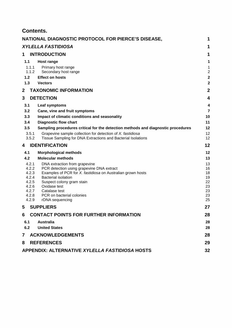
Contents. NATIONAL DIAGNOSTIC PROTOCOL FOR PIERCE’S DISEASE, 1
XYLELLA FASTIDIOSA 1
1 INTRODUCTION 1 1.1 Host range 1 1.1.1 Primary host range 1 1.1.2 Secondary host range 2
1.2 Effect on hosts 2 1.3 Vectors 2
2 TAXONOMIC INFORMATION 2
3 DETECTION 4 3.1 Leaf symptoms 4 3.2 Cane, vine and fruit symptoms 7 3.3 Impact of climatic conditions and seasonality 10 3.4 Diagnostic flow chart 11 3.5 Sampling procedures critical for the detection methods and diagnostic procedures 12 3.5.1 Grapevine sample collection for detection of X. fastidiosa 12 3.5.2 Tissue Sampling for DNA Extractions and Bacterial Isolations 12
4 IDENTIFICATION 12 4.1 Morphological methods 12 4.2 Molecular methods 13 4.2.1 DNA extraction from grapevine 13 4.2.2 PCR detection using grapevine DNA extract 16 4.2.3 Examples of PCR for X. fastidiosa on Australian grown hosts 18 4.2.4 Bacterial isolation 19 4.2.5 Suspect colony gram stain 22 4.2.6 Oxidase test 23 4.2.7 Catalase test 23 4.2.8 PCR on bacterial colonies 23 4.2.9 rDNA sequencing 25
5 SUPPLIERS 27
6 CONTACT POINTS FOR FURTHER INFORMATION 28 6.1 Australia 28 6.2 United States 28
7 ACKNOWLEDGEMENTS 28
8 REFERENCES 29
APPENDIX: ALTERNATIVE XYLELLA FASTIDIOSA HOSTS 32
1 Introduction Pierce’s disease is a lethal grapevine disease caused by the bacterium Xylella fastidiosa which infects the xylem tissue of grapevine. Bacterial aggregates and plant tyloses and gums, produced in response to infection, are thought to block the vessels which conduct water through the plant.
Xylella fastidiosa is a gram-negative bacterium confined to the xylem vessels of its host. The organism, designated Xylella fastidiosa was first described by Wells et al. (1987), and is the sole species belonging to this genus. X. fastidiosa has not been recorded in Australia.
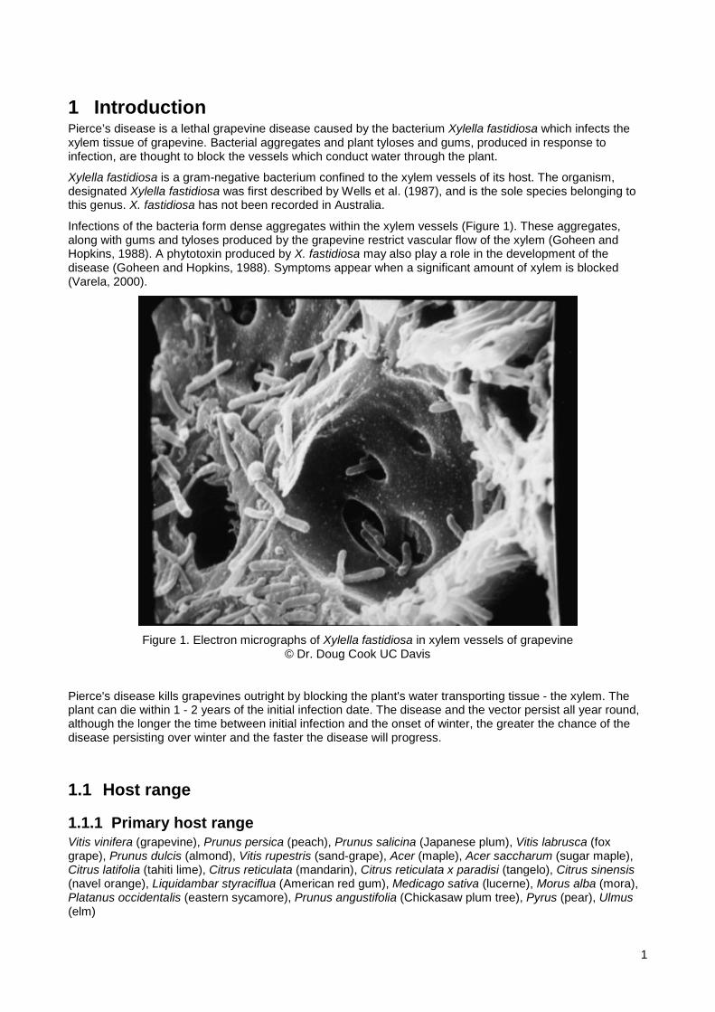
Infections of the bacteria form dense aggregates within the xylem vessels (Figure 1). These aggregates, along with gums and tyloses produced by the grapevine restrict vascular flow of the xylem (Goheen and Hopkins, 1988). A phytotoxin produced by X. fastidiosa may also play a role in the development of the disease (Goheen and Hopkins, 1988). Symptoms appear when a significant amount of xylem is blocked (Varela, 2000).
Figure 1. Electron micrographs of Xylella fastidiosa in xylem vessels of grapevine
© Dr. Doug Cook UC Davis
Pierce's disease kills grapevines outright by blocking the plant's water transporting tissue - the xylem. The plant can die within 1 - 2 years of the initial infection date. The disease and the vector persist all year round, although the longer the time between initial infection and the onset of winter, the greater the chance of the disease persisting over winter and the faster the disease will progress.
1.1 Host range
1.1.1 Primary host range Vitis vinifera (grapevine), Prunus persica (peach), Prunus salicina (Japanese plum), Vitis labrusca (fox grape), Prunus dulcis (almond), Vitis rupestris (sand-grape), Acer (maple), Acer saccharum (sugar maple), Citrus latifolia (tahiti lime), Citrus reticulata (mandarin), Citrus reticulata x paradisi (tangelo), Citrus sinensis (navel orange), Liquidambar styraciflua (American red gum), Medicago sativa (lucerne), Morus alba (mora), Platanus occidentalis (eastern sycamore), Prunus angustifolia (Chickasaw plum tree), Pyrus (pear), Ulmus (elm)
1
1.1.2 Secondary host range Poaceae (cereal), Brachiaria (signal grass), Conium maculatum (poison hemlock), Cynodon (quick grass), Cyperus (flatsedge), Digitaria, Echinochloa frumentacea (Japanese millet), Fragaria vesca (European strawberry), Lolium (ryegrass), Medicago (medic), Paspalum, Paspalum dilatatum (dallis grass), Rubus (blackberry, raspberry), Sambucus (elderberry), Salix (willow), Taraxacum officinale (dandelion), Trifolium (clovers), Vinca minor (common periwinkle), Coffea (coffee).
Alternative Xylella fastidiosa hosts are detailed in the Appendix.
1.2 Effect on hosts The main symptoms include scorched leaf margins, leaf abscission with petiole retention, irregular cane maturation, fruit raisining and delayed spring growth. Some of the symptoms of Pierce's disease can be confused with other syndromes such as salt toxicity, boron, copper or phosphorus deficiency and other diseases e.g. Eutypa.
1.3 Vectors All sucking insects that feed on xylem sap are potential vectors of X. fastidiosa, but all known vectors are limited to the Homoptera suborder (Purcell, 1999c). Vectors acquire the bacterium by feeding on infected plants. The bacteria adhere to the insect's foregut where they multiply and are then transmitted to healthy plants. Vectors remain infective indefinitely after acquiring the bacteria with the exception of nymphs which cannot transmit bacteria after they shed their external skeleton. After moulting, insects must feed again on an infected plant before they can acquire and transmit the bacterium (Purcell, 1999c). Insects currently known to be capable of transmitting X. fastidiosa all belong to the spittlebug/froghopper family (Cercopidae) and the ‘sharpshooter’ subfamily in the leafhopper family (Cicadellidae, subfamily Cicadellinae). . None of these genera have been reported in Australia. Of the 14 species of Cicadellidae in Australia, none have been recorded on Vitaceae. Within the Americas many genera of sharpshooters and spittlebugs serve as vectors of the bacterium (Goheen and Hopkins, 1988). However, in California, the major vectors are the blue-green sharpshooter (Graphocephala atropunctata), glassy-winged sharpshooter (Homalodisca coagulata), green sharpshooter (Draeculacephala minerva), and the red-headed sharpshooter (Carneocephala fulgida) (Gubler et al., 1999; Purcell, 1999b; Varela, 2000). Spittlebug vectors of Pierce's disease have been recorded in California (Delong and Severin, 1950), but none have been found on grapevines in California (Severin, 1950). Other sucking insects such as grape leafhoppers, are not vectors in California (Gubler et al., 1999). Cicadas (family Cicadidae) are also xylem feeders but there are no published reports of their being tested as vectors.
Prior to the introduction of H. coagulata, plants infected shortly before winter by other species of sharpshooter have recovered and been free of the bacteria in the following spring. This is partly because very cold winter weather helps cure vines of the bacterium and because other sharpshooters feed on and infect the tips of younger shoots, which are pruned during the summer. As H. coagulata feed much lower on the cane than other sharpshooters, late season infections are not removed by pruning and may survive the winter to cause chronic Pierce's disease the following season. This enables vine-to-vine spread of the disease rather than linear spread, as has been the case in the past.
Xylella fastidiosa can also be transmitted and dispersed by graft transmission. Propagative material is the pathway by which X. fastidiosa may spread (Smith et al., 1997). Xylella fastidiosa is not transmitted via contaminated pruning shears or by seed transmission (Smith et al., 1997; Varela, 2000).
Australia has no record of X. fastidiosa or sharpshooters.
2 Taxonomic Information
Kingdom: Bacteria
Phylum: Proteobacteria
2

Class: Gammaproteobacteria
Order: Xanthomonadales
Family: Xanthomonadaceae
Genus: Xylella
Species: Xylella fastidiosa
Scientific Name: Xyllela fastidiosa (Wells et al 1987)
Common Names: Pierce's disease, California vine disease, Anaheim disease (grapevine), leaf scorch (almond, coffee, elm, maple, mulberry, oak, oleander, sycamore), variegated chlorosis (citrus), phony peach disease (peach), leaf scald (plum), dwarf (lucerne), wilt (periwinkle).
3

3 Detection Xylella fastidiosa is mostly confined to the xylem tissue of its hosts (Figure 2). The major symptoms of Pierce’s disease include; leaf necrosis in concentric rings or in sections, leaf abscission with petiole retention, "green islands" on canes, fruit raisining, dieback, delayed growth in spring, and decline in vigour leading to death. The first evidence of Pierce’s disease infection usually is a drying or "scorching" of leaves. The best time to observe symptoms of Pierce’s disease is late summer through to autumn.
It takes about four-five months for the symptoms to appear, with only one or two canes showing symptoms in the first season. However, in young vines the symptoms may appear over the entire vine in a single season (Varela et al, 2001). In chronically infected vines new growth may be delayed by two weeks with interveinal chlorosis in the first four to eight leaves which may be small or distorted. The internodes are often shortened or zig-zagged. Delayed budbreak or bud failure may also occur (Varela et al, 2001).
Figure 2. Electron micrographs of Xylella fastidiosa in xylem vessels of grapevine
© Dr. Doug Cook UC Davis.
3.1 Leaf symptoms The leaves become slightly chlorotic along the margins before drying inwards, or the outer leaf may dry suddenly while still green. The leaf dries progressively over a period of days to weeks, leaving a series of concentric zones of discoloured and dead tissue.
On white varieties, a yellow chlorotic zone appears between the necrotic margin and the green interior of the leaf (Figure 3). The scorching develops inward from the margin and is continuous. On red varieties a dark-reddish to purple band appears between the green and necrotic tissue (Figure 4, Figure 5). There is a wide range of leaf symptoms ranging from highly regular, concentric zones of chlorosis followed by necrosis to discolouration and necrosis occurring in sectors of the leaf only (Varela et al, 2001).
Symptoms vary with the species and cultivar that is affected. Symptoms in muscadine and other native American grapes from the south eastern United States are milder than those in V. vinifera. Symptoms are usually more pronounced in vines that are stressed by high temperatures or drought conditions (Goheen and Hopkins, 1988).
The most characteristic symptom of X. fastidiosa infection is leaf scorch. An early sign is sudden drying of part of a green leaf, which then turns brown while adjacent tissue turns yellow or red. The desiccation spreads and the whole leaf may shrivel and drop, leaving only the petiole attached (Figure 6).
4

Leaf symptoms vary among grape varieties (Gubler et al., 1999). Grape varieties such as Pinot Noir and Cabernet Sauvignon have highly regular zones of progressive marginal discolouration and drying on blades. In the varieties Thompson seedless, Sylvaner, and Chenin Blanc (Figure 10), the discolouration and scorching may occur in sectors of the leaf rather than along the margins. Climatic differences between regions can affect the timing and severity of symptoms, but not the type of symptoms (Gubler et al., 1999). Hot climates accelerate symptom development, as moisture stress is more severe even with adequate soil moisture.
In later years, infected plants develop late and produce stunted chlorotic shoots. Highly susceptible cultivars rarely survive more than 2-3 years, despite any signs of recovery early in the growing season. Young vines succumb more quickly than older vines. More tolerant cultivars may survive chronic infection for more than 5 years.
Figure 3 Symptoms of X. fastidiosa on Chardonnay (© Regents, University of California 1999).
5
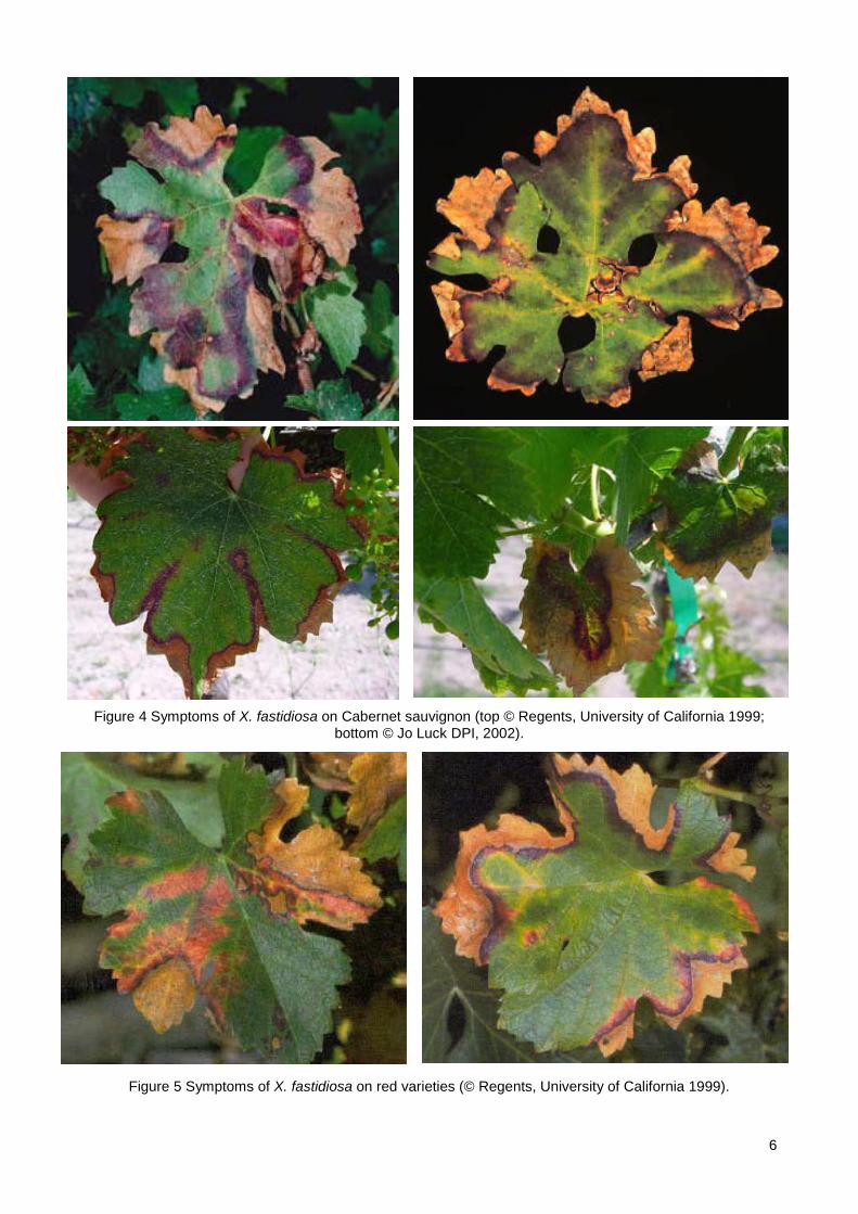
Figure 4 Symptoms of X. fastidiosa on Cabernet sauvignon (top © Regents, University of California 1999; bottom © Jo Luck DPI, 2002).
Figure 5 Symptoms of X. fastidiosa on red varieties (© Regents, University of California 1999).
6
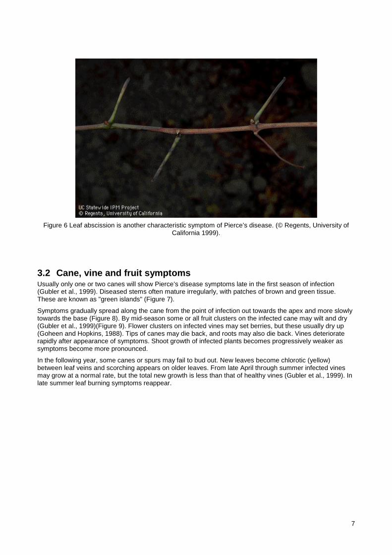
Figure 6 Leaf abscission is another characteristic symptom of Pierce’s disease. (© Regents, University of
California 1999).
3.2 Cane, vine and fruit symptoms Usually only one or two canes will show Pierce’s disease symptoms late in the first season of infection (Gubler et al., 1999). Diseased stems often mature irregularly, with patches of brown and green tissue. These are known as "green islands" (Figure 7).
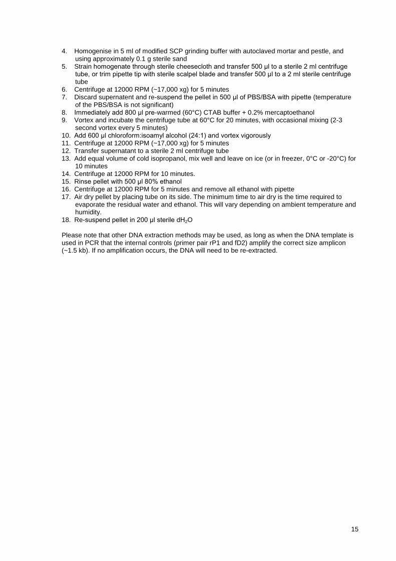
Symptoms gradually spread along the cane from the point of infection out towards the apex and more slowly towards the base (Figure 8). By mid-season some or all fruit clusters on the infected cane may wilt and dry (Gubler et al., 1999)(Figure 9). Flower clusters on infected vines may set berries, but these usually dry up (Goheen and Hopkins, 1988). Tips of canes may die back, and roots may also die back. Vines deteriorate rapidly after appearance of symptoms. Shoot growth of infected plants becomes progressively weaker as symptoms become more pronounced.
In the following year, some canes or spurs may fail to bud out. New leaves become chlorotic (yellow) between leaf veins and scorching appears on older leaves. From late April through summer infected vines may grow at a normal rate, but the total new growth is less than that of healthy vines (Gubler et al., 1999). In late summer leaf burning symptoms reappear.
7
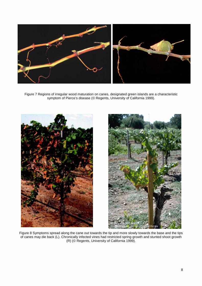
Figure 7 Regions of irregular wood maturation on canes, designated green islands are a characteristic symptom of Pierce’s disease (© Regents, University of California 1999).
Figure 8 Symptoms spread along the cane out towards the tip and more slowly towards the base and the tips of canes may die back (L). Chronically infected vines had restricted spring growth and stunted shoot growth
(R) (© Regents, University of California 1999).
8
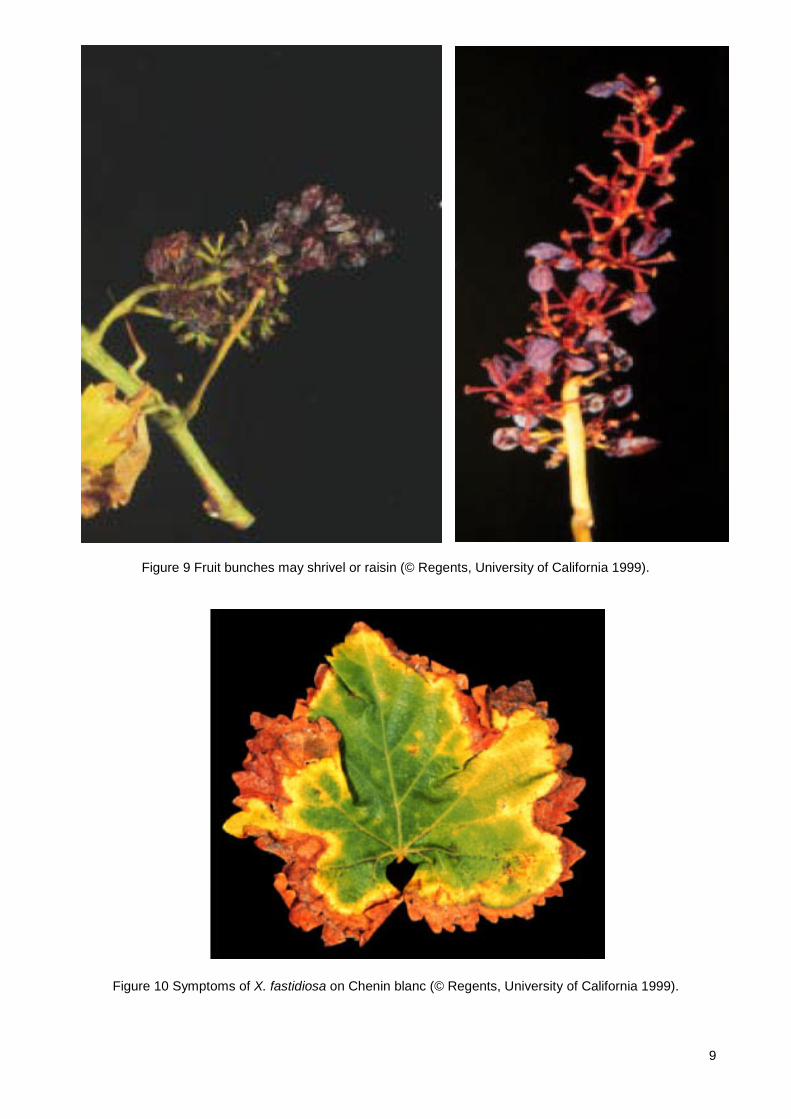
Figure 9 Fruit bunches may shrivel or raisin (© Regents, University of California 1999).
Figure 10 Symptoms of X. fastidiosa on Chenin blanc (© Regents, University of California 1999).
9

3.3 Impact of climatic conditions and seasonality Physiological changes in the vines induced by cold weather can cause death of the bacteria. The longer the time between initial infection and the onset of winter, the greater the chance of the disease persisting over winter and the faster the disease will progress. Plants infected shortly before winter have recovered and been free of the bacteria in the following spring. Laboratory observations from Purcell and Saunders (1995) work on harvested grape clusters as inoculum for Pierce’s disease showed that the number of viable X. fastidiosa decreased with time spent in cold storage and declined sharply after cold storage at 4°C. The bacterium was not recovered from infected grapes after 21 days of storage at this temperature. This data supports the observations made by Varela (2000). Further, experimental cold therapy of diseased grapevines suggests that freezing temperatures can eliminate the bacterium directly from plants (Purcell, 1980).
Winter weather conditions in Australia are not as severe as those experienced in the USA and in many areas vines are not considered to go dormant over the winter non-growing period. The effects of winter are not likely to affect survival of the bacterium in Australia.
Some vines infected during the season appear to recover from Pierce’s disease the first winter following infection (Varela, 2000). Recovery from Pierce’s disease depends on the grape variety. In Cabernet, recovery is high while in Barbera, Chardonnay and Pinot Noir it is low. In more tolerant cultivars, the bacterium spreads more slowly within the plant than in more susceptible cultivars (Varela, 2000). Once the vine has been infected for over a year (i.e. bacteria survive the first winter) recovery is much less likely (Varela, 2000). Young vines are more susceptible than mature vines, possibly because the bacteria can move more quickly through younger vines than through older vines. Rootstock species and hybrids vary greatly in susceptibility. Testing of rootstock plants show that V. riparia is rather susceptible; V. rupestris (St George) and 420A are very tolerant.
Rootstock does not confer resistance to susceptible V. vinifera varieties grafted on to it. Climate, variety and age determine how long a vine with Pierce’s disease can survive (Varela, 2000). One-year old Pinot Noir or Chardonnay can die the year they become infected, whereas chronically infected 10-year-old Chenin Blanc or Ruby Cabernet can live for more than five years. Long before that, however, these chronically infested vines will cease to bear a crop (Varela, 2000).
10

3.4 Diagnostic flow chart
3.5 Grapevine sample collection
3.1, 3.2 Symptom recognition
4.2.4 Bacterial Isolation
4.2.2 PCR detection using grapevine DNA extract
4.2.1 DNA extraction from grapevine
4.2.8 PCR on suspect bacterial colonies
4.2.9 rDNA sequencing (by suitable laboratory)
3.5.2 Tissue sampling for DNA extractions and bacterial isolations
Confirmation of results
(14-21 days)
11

3.5 Sampling procedures critical for the detection methods and diagnostic procedures
3.5.1 Grapevine sample collection for detection of X. fastidiosa 1. Late-summer to autumn is the best time to sample for Pierce's disease. In chronically infected vines,
bacteria do not move into the new season's growth until the middle of summer. Leaves attached to the cane generally give the most reliable result.
2. Collect leaf material which is showing symptoms characteristic of Xylella fastidiosa infection, and which is still attached to the cane
3. Collect 4-5 canes from the suspect plant.
4. Wrap the cane samples in damp newspaper and place inside a sealed plastic bag.
5. Ship to a diagnostic laboratory (for details see below) immediately after the material is collected.
NB. Negative test results, do not mean that Xylella fastidiosa is absent as the bacteria may be unevenly distributed through the vine. It is important to sample symptomatic material.
3.5.2 Tissue Sampling for DNA Extractions and Bacterial Isolations The most optimum tissue to sample for the detection of X. fastidiosa is the mid-rib and petiole from symptomatic leaves. Select five leaves from affected canes and treat as one sample. Replicate sampling.
Further detection and identification methods are outlined in Section 4.
4 Identification Positive identification of X. fastidiosa can be obtained by three methods: culturing the bacterium on selective media, serological test such as ELISA (enzyme linked immunosorbent assay) or PCR (polymerase chain reaction) (Varela, 2000).
4.1 Morphological methods For cultural diagnosis a specialised media (section 4.2.4.2) has been developed for isolating and growing the Pierce’s disease bacterium. Petioles are used to isolate the bacteria. Using this technique, 100 bacterial cells per gram of plant tissue are able to be detected (Hill and Purcell, 1995). The disadvantages are that it is time consuming, colonies may require 32 days to develop, microbial contaminants cloud or obscure results and the bacteria can only be isolated from petioles during the summer and early fall (Varela, 2000). Colonies of X. fastidiosa on most selective media are convex, smooth, entire or rough with finely undulate margins (Bradbury 1991).
The morphological and biochemical characteristics of X. fastidiosa are as follows (Davis et al 1978):
Single aflagellate straight rods, 0.25-0.35 X 0.9-3.5 μm, with filamentous strands under some cultural conditions. Colonies are of two types: convex to pulvinate smooth opalescent with entire margins and umbonate rough with finely undulated margins. Cells stain Gram negative. Non-motile. Oxidase negative and catalase positive. Strictly aerobic, non-fermentative, non-halophilic, non-pigmented. Nutritionally fastidious, requiring a specialised medium such as BC-YE containing charcoal or glutamine-peptone medium (PW) containing serum albumin. Optimal temperature for growth is 26-28°C. Optimum pH is 6.5-6.9. Habitat is exclusively in the xylem of plant tissue.
Hydrolyses gelatin and utilises hippurate. Most strains produce β-lactamase. Glucose is not fermented. Negative in tests for indole, H2S, β-galactosidase, lipase, amylase, coagulase, and phosphatase. The species has been isolated as a phytopathogen from tissues of a number of host plants. The type strain was isolated from grapevine with Pierce’s disease (Wells et al 1987).
12

Figure 11 Xylella fastidiosa © Jose Lima (1996) Citrolima, Brazil
4.2 Molecular methods PCR enzymatically amplifies specific parts of the bacterium's DNA. This is the most sensitive technique to detect small numbers of bacteria in plants. It is specific for X. fastidiosa but has the disadvantages that it is expensive, cannot determine if the bacteria are dead or alive or how many bacteria are present in the sample (Varela, 2000). The X. fastidiosa diagnostic PCR is rapid, with a result within 24 hours using plant DNA extracts from suspected hosts, whether the host is symptomatic or asymptomatic. This test can also be used on boiled preparations from bacterial colonies, bacterial DNA extracts and plant tissue extract. The likelihood of a false positive result occurring is low, providing the correct internal controls are used. There is however, a possibility of getting a false negative result due to extremely low bacterial numbers. The possibility of a false negative result occurring due to template inhibition is eliminated by including an additional set of PCR primers that amplify the 16S ribosomal DNA gene from a wide range of bacteria. If this fails then the template contains inhibitors and should be re-extracted.
4.2.1 DNA extraction from grapevine The following protocol utilises a fume hood (for handling chloroform:isoamyl alcohol) and as such DNA extraction kits such as the Qiagen Plant Tissue Mini Kit, which do not require a hood, may be easier to use for some laboratories.
4.2.1.1 Equipment 1. 2 ml centrifuge tubes 2. 20-200 μL and 200-1000 μL pipettes and tips 3. Autoclave 4. Autoclaved mortar and pestles 5. Balance 6. Centrifuge 7. Distilled water unit 8. Ice machine or freezer 9. Sterile cheesecloth 10. Sterile sand 11. Sterile scalpel blades 12. Vortex 13. Water bath at 60oC
13

4.2.1.2 Reagents
Modified SCP For 500 ml For 1000 ml
Disodium succinate C4H4Na2O7 (Sigma S2378) 0.5 g 1 g Trisodium citrate C6H5Na3O7 (Sigma S4641) 0.5 g 1 g K2HPO4 (Ajax A2221-500g) 0.75 g 1.5 g KH2PO4 (Ajax A391-500g) 0.5 g 1 g PVP40 (Sigma PVP-40) 25 g 50 g Autoclave. Add ascorbic acid (0.02M final concentration) and adjust to pH 7 just prior to use. The stock buffer (without ascorbic acid) can be stored frozen (-20C) for up to 6 months. The buffer with ascorbic acid shouldn’t be frozen once mixed but should be used immediately.
PBS/BSA a) 10X PBS For 1000 ml
NaCl (BDH Analar #10241.AP) 80 g KH2PO4 2 g Na2HPO4 (Ajax 478 or 621) 11.5 g KCl (Ajax 382-500g) 2 g Autoclave. Store at room temperature.
b) PBS/BSA
1x PBS plus 0.2% BSA. Store at 4oC.
CTAB buffer + 0.2% mercaptoethanol For 100 ml
1M Tris, pH 7.5 H2NC(CH2OH)3 (Amresco 0234) 20 ml 5M NaCl 28 ml 500mM EDTA, pH 8.0 [CH2.N(CH2.COOH).CH2COON9]2.2H2O 4 ml CTAB C19H42NBr (Sigma H6269) 2 g β-Mercaptoethanol (Sigma M3148) 200 μl Mix and make up to 100 ml with dH2O. Store at room temperature.
Choloroform:isoamyl alcohol 24:1 mix of choloroform (BDH 152835F) to isoamyl alcohol (Sigma I9392). Store at room temperature.
Isopropanol 100% isopropanol stored at 4oC.
Ethanol 80% ethanol. Store at room temperature.
Water Sterile dH2O.
4.2.1.3 Method 1. Place CTAB buffer + 0.2% mercaptoethanol in 60oC water bath 2. Select 5 symptomatic grapevine leaves from sample (repeat for duplication of test) 3. Weigh approximately 700 mg midrib and petiole tissue (combined from all 5 leaves)
14
4. Homogenise in 5 ml of modified SCP grinding buffer with autoclaved mortar and pestle, and using approximately 0.1 g sterile sand
5. Strain homogenate through sterile cheesecloth and transfer 500 μl to a sterile 2 ml centrifuge tube, or trim pipette tip with sterile scalpel blade and transfer 500 μl to a 2 ml sterile centrifuge tube
6. Centrifuge at 12000 RPM (~17,000 xg) for 5 minutes 7. Discard supernatent and re-suspend the pellet in 500 μl of PBS/BSA with pipette (temperature
of the PBS/BSA is not significant) 8. Immediately add 800 μl pre-warmed (60°C) CTAB buffer + 0.2% mercaptoethanol 9. Vortex and incubate the centrifuge tube at 60°C for 20 minutes, with occasional mixing (2-3
second vortex every 5 minutes) 10. Add 600 μl chloroform:isoamyl alcohol (24:1) and vortex vigorously 11. Centrifuge at 12000 RPM (~17,000 xg) for 5 minutes 12. Transfer supernatant to a sterile 2 ml centrifuge tube 13. Add equal volume of cold isopropanol, mix well and leave on ice (or in freezer, 0°C or -20°C) for
10 minutes 14. Centrifuge at 12000 RPM for 10 minutes. 15. Rinse pellet with 500 μl 80% ethanol 16. Centrifuge at 12000 RPM for 5 minutes and remove all ethanol with pipette 17. Air dry pellet by placing tube on its side. The minimum time to air dry is the time required to
evaporate the residual water and ethanol. This will vary depending on ambient temperature and humidity.
18. Re-suspend pellet in 200 μl sterile dH2O
Please note that other DNA extraction methods may be used, as long as when the DNA template is used in PCR that the internal controls (primer pair rP1 and fD2) amplify the correct size amplicon (~1.5 kb). If no amplification occurs, the DNA will need to be re-extracted.
15
4.2.2 PCR detection using grapevine DNA extract
4.2.2.1 Equipment 1. 0-2 μl, 2-20 μl, 20-200 μl, and 200-1000 μl pipettes and tips 2. 0.2 or 0.5 ml PCR tubes 3. 1.5 or 2 ml centrifuge tubes to store reagents 4. Bulb spinner or centrifuge 5. Freezer 6. Gel tanks, rigs and racks 7. Ice machine 8. Latex, and leather gloves 9. Microwave 10. Power pack 11. Thermocycler 12. UV transilluminator with camera
4.2.2.2 Reagents
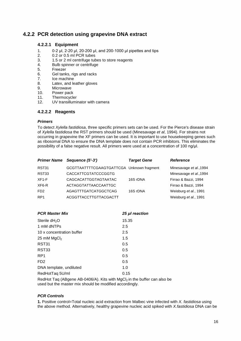
Primers To detect Xylella fastidiosa, three specific primers sets can be used. For the Pierce's disease strain of Xylella fastidiosa the RST primers should be used (Minesavage et al, 1994). For strains not occurring in grapevine the XF primers can be used. It is important to use housekeeping genes such as ribosomal DNA to ensure the DNA template does not contain PCR inhibitors. This eliminates the possibility of a false negative result. All primers were used at a concentration of 100 ng/μl.
Primer Name Sequence (5'-3') Target Gene Reference
RST31 GCGTTAATTTTCGAAGTGATTCGA Unknown fragment Minesavage et al.,1994 RST33 CACCATTCGTATCCCGGTG Minesavage et al.,1994 XF1-F CAGCACATTGGTAGTAATAC 16S rDNA Firrao & Bazzi, 1994 XF6-R ACTAGGTATTAACCAATTGC Firrao & Bazzi, 1994 FD2 AGAGTTTGATCATGGCTCAG 16S rDNA Weisburg et al., 1991
RP1 ACGGTTACCTTGTTACGACTT Weisburg et al., 1991
PCR Master Mix 25 μl reaction
Sterile dH2O 15.35 1 mM dNTPs 2.5 10 x concentration buffer 2.5 25 mM MgCl2 1.5 RST31 0.5 RST33 0.5 RP1 0.5 FD2 0.5 DNA template, undiluted 1.0 RedHotTaq 5U/ml 0.15 RedHot Taq (ABgene AB-0406/A). Kits with MgCl2 in the buffer can also be used but the master mix should be modified accordingly.
PCR Controls 1. Positive control=Total nucleic acid extraction from Malbec vine infected with X. fastidiosa using the above method. Alternatively, healthy grapevine nucleic acid spiked with X.fastidiosa DNA can be
16

used where X. fastidiosa infected material cannot be maintained in the laboratory. 2. Negative control is the master mix (24 μl) with 1.0 of RNAase/DNAase free water instead of DNA template.
5 x TBE 1L
Tris H2NC(CH2OH)3 54.0 g Boric acid H3BO3 27.5 g 0.5 M EDTA [CH2.N(CH2.COOH).CH2COON9]2.2H2O 20 ml Store at room temperature.
1% Agarose gel with SYBR Safe stain 1. Agarose gel is 1 g DNA grade agarose per 100 ml 1 x TBE. 2. Melt in the microwave. 3. Use SYBR Safe stain as per the manufacturers instructions.
Store at room temperature.
100 x TE solution 100 ml
Tris-Cl pH 8.0 50 mL 0.5M EDTA pH 8.0 20 mL dH2O 30 mL Store at room temperature.
Loading dye Loading dye should be purchased rather than made to ensure consistency. One suitable option is QIAGEN GelPilot Loading Dye 5x (239901).
4.2.2.3 Method 1. Label sterile 100 μl centrifuge tubes 2. Prepare "master mix" in sterile 1 ml centrifuge as described above 3. Add 2 μl sdH2O to the negative control tube, 2 μl test template to each tube, and 2 μl grapevine
DNA infected with X. fastidosa into positive control tube. 4. Cycle the tubes with the following PCR conditions:1 cycle 95°C 1 min, 30 cycles (94°C for 45
secs, 55°C for 30 secs, 72°C for 30 secs), 1 cycle 72°C, 10 mins and 1 cycle 25°C, 1 min. (The PCR conditions were adapted for duplex PCR using conditions described in Minesavage et al., 1994, Firrao and Bazzi, 1994 and Weisburg et al., 1991)
5. Mix 10 μl each PCR sample with 5 μl running dye 6. Load samples onto a 1% agarose gel containing SYBR Safe stain as per manufacturer’s
instructions. 7. Electrophorese in 1 X TBE at 100V for approximately 40 minutes 8. Visualise and photograph gel on UV transilluminator.
17
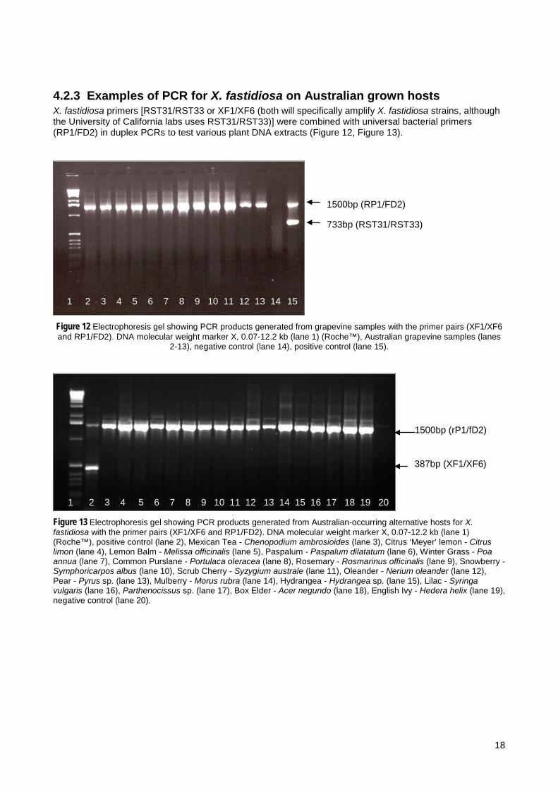
4.2.3 Examples of PCR for X. fastidiosa on Australian grown hosts X. fastidiosa primers [RST31/RST33 or XF1/XF6 (both will specifically amplify X. fastidiosa strains, although the University of California labs uses RST31/RST33)] were combined with universal bacterial primers (RP1/FD2) in duplex PCRs to test various plant DNA extracts (Figure 12, Figure 13).
Figure 12 Electrophoresis gel showing PCR products generated from grapevine samples with the primer pairs (XF1/XF6 and RP1/FD2). DNA molecular weight marker X, 0.07-12.2 kb (lane 1) (Roche™), Australian grapevine samples (lanes
2-13), negative control (lane 14), positive control (lane 15).
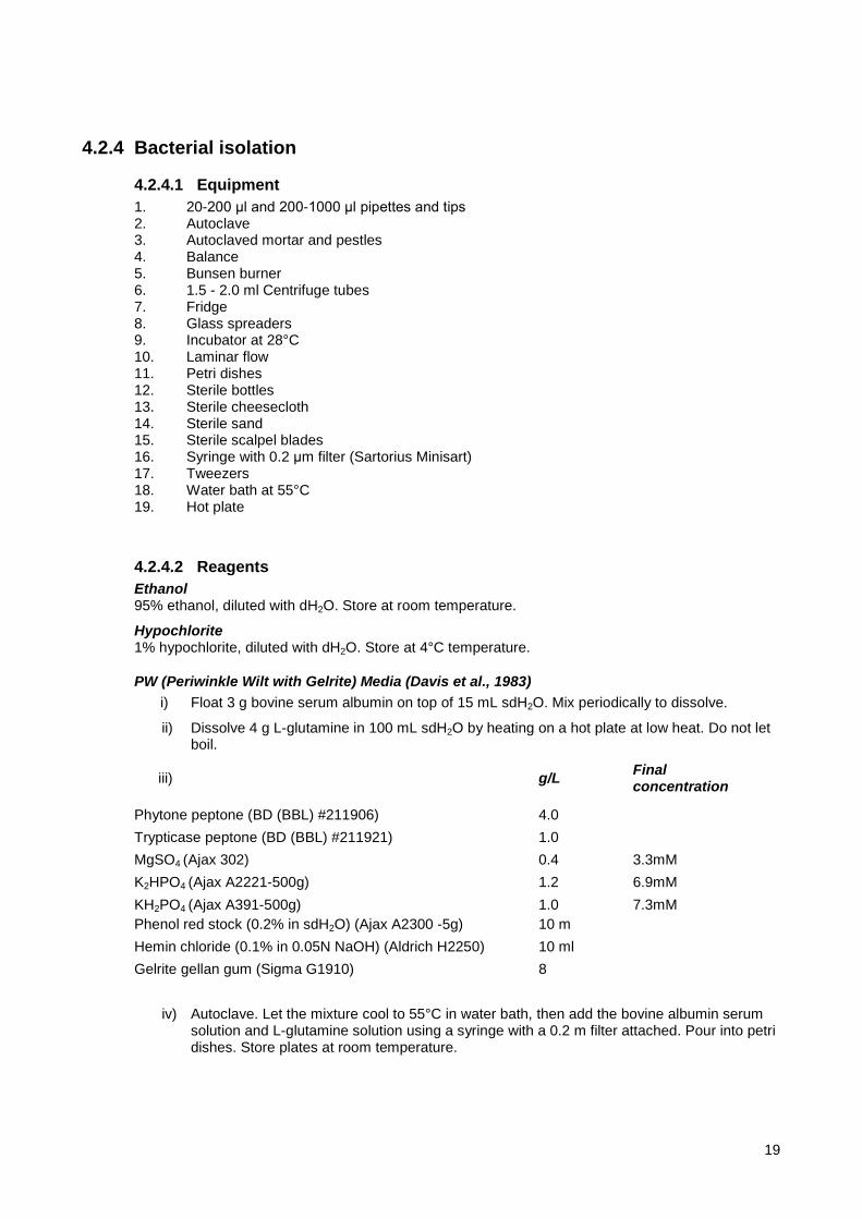
Figure 13 Electrophoresis gel showing PCR products generated from Australian-occurring alternative hosts for X. fastidiosa with the primer pairs (XF1/XF6 and RP1/FD2). DNA molecular weight marker X, 0.07-12.2 kb (lane 1) (Roche™), positive control (lane 2), Mexican Tea - Chenopodium ambrosioides (lane 3), Citrus ‘Meyer’ lemon - Citrus limon (lane 4), Lemon Balm - Melissa officinalis (lane 5), Paspalum - Paspalum dilatatum (lane 6), Winter Grass - Poa annua (lane 7), Common Purslane - Portulaca oleracea (lane 8), Rosemary - Rosmarinus officinalis (lane 9), Snowberry - Symphoricarpos albus (lane 10), Scrub Cherry - Syzygium australe (lane 11), Oleander - Nerium oleander (lane 12), Pear - Pyrus sp. (lane 13), Mulberry - Morus rubra (lane 14), Hydrangea - Hydrangea sp. (lane 15), Lilac - Syringa vulgaris (lane 16), Parthenocissus sp. (lane 17), Box Elder - Acer negundo (lane 18), English Ivy - Hedera helix (lane 19), negative control (lane 20).
1 2 3 4 5 6 7 8 9 10 11 12 13 14 15
1500bp (RP1/FD2)
733bp (RST31/RST33)
1 2 3 4 5 6 7 8 9 10 11 12 13 14 15 16 17 18 19 20
1500bp (rP1/fD2)
387bp (XF1/XF6)
18
4.2.4 Bacterial isolation
4.2.4.1 Equipment 1. 20-200 μl and 200-1000 μl pipettes and tips 2. Autoclave 3. Autoclaved mortar and pestles 4. Balance 5. Bunsen burner 6. 1.5 - 2.0 ml Centrifuge tubes 7. Fridge 8. Glass spreaders 9. Incubator at 28°C 10. Laminar flow 11. Petri dishes 12. Sterile bottles 13. Sterile cheesecloth 14. Sterile sand 15. Sterile scalpel blades 16. Syringe with 0.2 μm filter (Sartorius Minisart) 17. Tweezers 18. Water bath at 55°C 19. Hot plate
4.2.4.2 Reagents Ethanol 95% ethanol, diluted with dH2O. Store at room temperature.
Hypochlorite 1% hypochlorite, diluted with dH2O. Store at 4°C temperature.
PW (Periwinkle Wilt with Gelrite) Media (Davis et al., 1983) i) Float 3 g bovine serum albumin on top of 15 mL sdH2O. Mix periodically to dissolve.
ii) Dissolve 4 g L-glutamine in 100 mL sdH2O by heating on a hot plate at low heat. Do not let boil.
iii) g/L Final concentration
Phytone peptone (BD (BBL) #211906) 4.0 Trypticase peptone (BD (BBL) #211921) 1.0 MgSO4 (Ajax 302) 0.4 3.3mM K2HPO4 (Ajax A2221-500g) 1.2 6.9mM KH2PO4 (Ajax A391-500g) Phenol red stock (0.2% in sdH2O) (Ajax A2300 -5g)
1.0 10 m
7.3mM
Hemin chloride (0.1% in 0.05N NaOH) (Aldrich H2250) 10 ml Gelrite gellan gum (Sigma G1910) 8
iv) Autoclave. Let the mixture cool to 55°C in water bath, then add the bovine albumin serum solution and L-glutamine solution using a syringe with a 0.2 m filter attached. Pour into petri dishes. Store plates at room temperature.
19
PD3 Media (Hopkins and Adlerz, 1988) g/L Final concentration
Tryptone (Oxoid LP0042) 4.0 Soytone (Difco 243620) 2.0 Trisodium citrate (Sigma S4641) 1.0 3.9 mM Disodium succinate (Sigma S2378) 1.0 3.7 mM Hemin chloride (0.1% in 0.05N NaOH) (Aldrich H2250) 10 ml Potato starch (soluble) (Mallinckrodt #8188) 2.0 MgSO4. 7H2O (Ajax 302) 1.0 4.06 mM K2HPO4 (Ajax A2221-500g) 1.5 8.6 mM KH2PO4 (Ajax A391-500g) 1.0 7.3 mM Adjust the pH to 6.8, add agar Agar (Oxoid LP0013) 15.0 Autoclave. Pour into petri dishes. Store plates at room temperature.
4.2.4.3 Methods 1. Weigh approximately 100 mg midrib and petiole tissue combined from 5 symptomatic
grapevine leaves 2. Surface sterilise material as follows: 1 min in 95% ethanol, 2 mins in 1% hypochlorite and
rinse 3 times in sterile water 3. The ex-plant is aseptically cut into 1 mm pieces 4. Homogenise using a mortar and pestle with approximately 0.1 g of sterile sand in 2 ml of
sterile distilled H2O 5. Filter through sterile cheesecloth 6. Prepare serial dilutions to 10-4 by adding 100 ul to 900 ul sterile distilled water in sterile
eppendorf tubes 7. Spread plate 100 μl of undiluted, 1:10 and 1:100, 1:1000 and 1:10000 dilutions onto
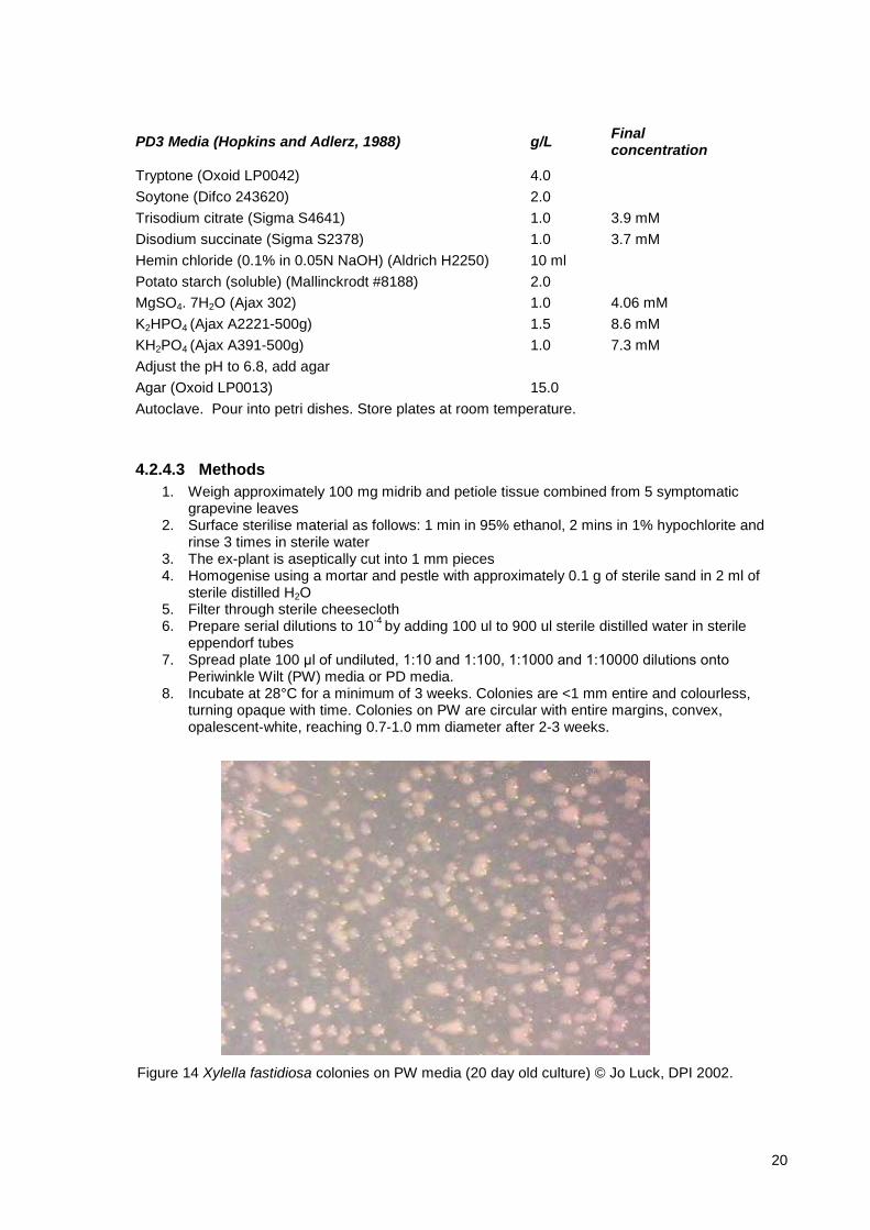
Periwinkle Wilt (PW) media or PD media. 8. Incubate at 28°C for a minimum of 3 weeks. Colonies are <1 mm entire and colourless,
turning opaque with time. Colonies on PW are circular with entire margins, convex, opalescent-white, reaching 0.7-1.0 mm diameter after 2-3 weeks.
Figure 14 Xylella fastidiosa colonies on PW media (20 day old culture) © Jo Luck, DPI 2002.
20
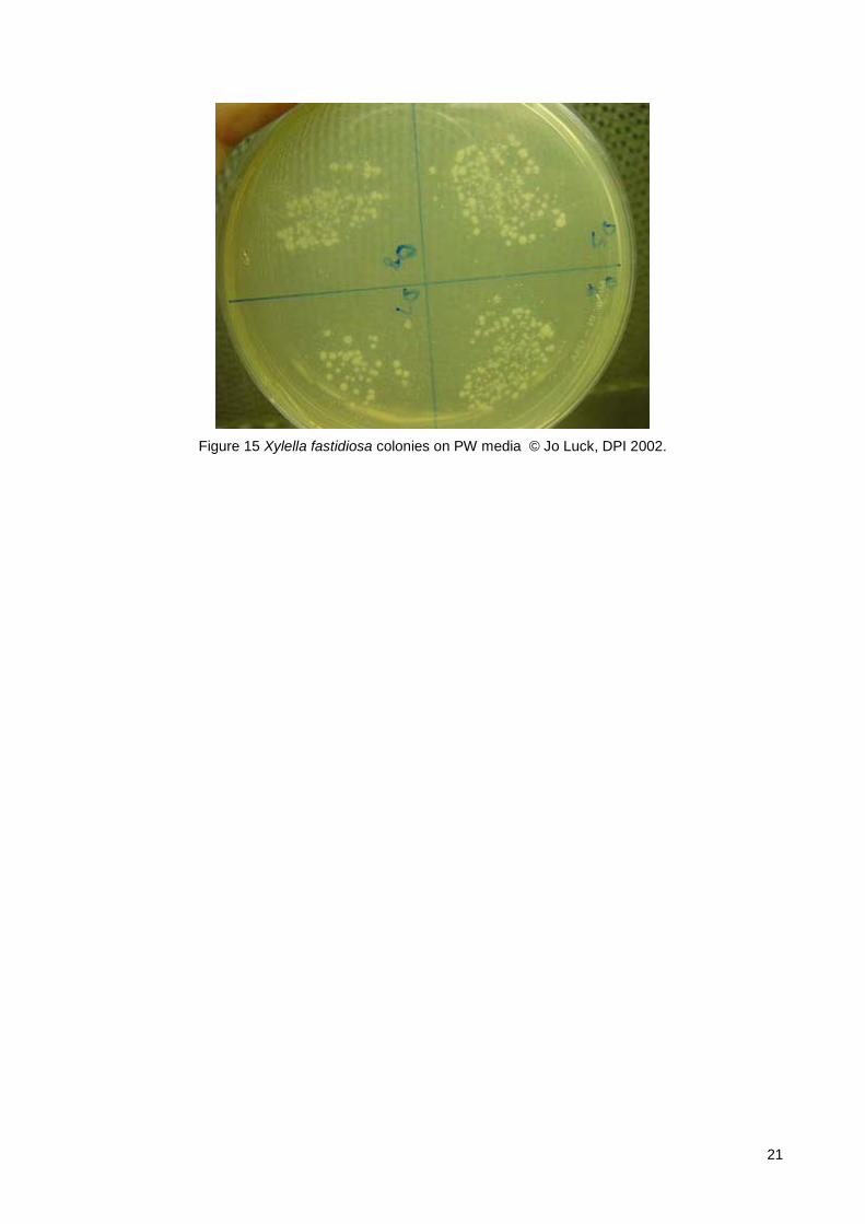
Figure 15 Xylella fastidiosa colonies on PW media © Jo Luck, DPI 2002.
21
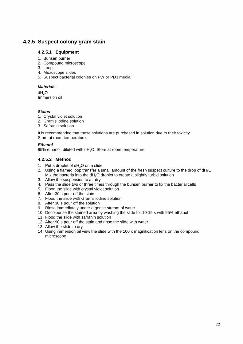
4.2.5 Suspect colony gram stain
4.2.5.1 Equipment 1. Bunsen burner 2. Compound microscope 3. Loop 4. Microscope slides 5. Suspect bacterial colonies on PW or PD3 media
Materials dH2O Immersion oil
Stains 1. Crystal violet solution 2. Gram's iodine solution 3. Safranin solution
It is recommended that these solutions are purchased in solution due to their toxicity. Store at room temperature.
Ethanol 95% ethanol, diluted with dH2O. Store at room temperature.
4.2.5.2 Method 1. Put a droplet of dH2O on a slide 2. Using a flamed loop transfer a small amount of the fresh suspect culture to the drop of dH2O.
Mix the bacteria into the dH2O droplet to create a slightly turbid solution 3. Allow the suspension to air dry 4. Pass the slide two or three times through the bunsen burner to fix the bacterial cells 5. Flood the slide with crystal violet solution 6. After 30 s pour off the stain 7. Flood the slide with Gram's iodine solution 8. After 30 s pour off the solution 9. Rinse immediately under a gentle stream of water 10. Decolourise the stained area by washing the slide for 10-15 s with 95% ethanol 11. Flood the slide with safranin solution 12. After 90 s pour off the stain and rinse the slide with water 13. Allow the slide to dry 14. Using immersion oil view the slide with the 100 x magnification lens on the compound
microscope
22
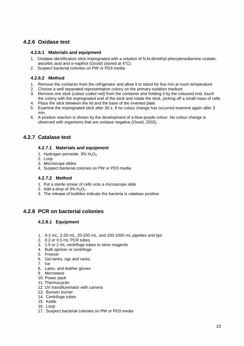
4.2.6 Oxidase test
4.2.6.1 Materials and equipment 1. Oxidase identification stick impregnated with a solution of N,N-dimethyl-phenylenediamine oxalate,
ascorbic acid and α-napthol (Oxoid) (stored at 4°C). 2. Suspect bacterial colonies on PW or PD3 media
4.2.6.2 Method 1. Remove the container from the refrigerator and allow it to stand for five min at room temperature 2. Choose a well separated representative colony on the primary isolation medium 3. Remove one stick (colour coded red) from the container and holding it by the coloured end, touch
the colony with the impregnated end of the stick and rotate the stick, picking off a small mass of cells 4. Place the stick between the lid and the base of the inverted plate 5. Examine the impregnated stick after 30 s. If no colour change has occurred examine again after 3
min. 6. A positive reaction is shown by the development of a blue-purple colour. No colour change is
observed with organisms that are oxidase negative (Oxoid, 2002).
4.2.7 Catalase test
4.2.7.1 Materials and equipment 1. Hydrogen peroxide, 3% H2O2 2. Loop 3. Microscope slides 4. Suspect bacterial colonies on PW or PD3 media
4.2.7.2 Method 1. Put a sterile smear of cells onto a microscope slide 2. Add a drop of 3% H2O2 3. The release of bubbles indicate the bacteria is catalase positive.
4.2.8 PCR on bacterial colonies
4.2.8.1 Equipment
1. 0-2 mL, 2-20 mL, 20-200 mL, and 200-1000 mL pipettes and tips 2. 0.2 or 0.5 mL PCR tubes 3. 1.5 or 2 mL centrifuge tubes to store reagents 4. Bulb spinner or centrifuge 5. Freezer 6. Gel tanks, rigs and racks 7. Ice 8. Latex, and leather gloves 9. Microwave 10. Power pack 11. Thermocycler 12. UV transilluminator with camera 13. Bunsen burner 14. Centrifuge tubes 15. Kettle 16. Loop 17. Suspect bacterial colonies on PW or PD3 media
23
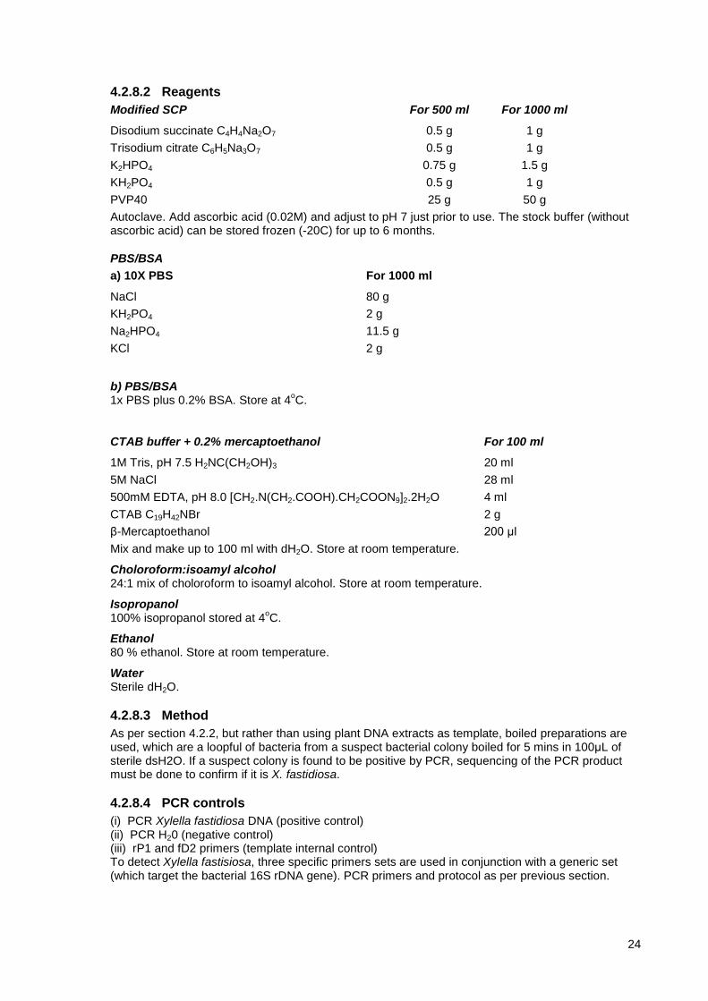
4.2.8.2 Reagents Modified SCP For 500 ml For 1000 ml
Disodium succinate C4H4Na2O7 0.5 g 1 g Trisodium citrate C6H5Na3O7 0.5 g 1 g K2HPO4 0.75 g 1.5 g KH2PO4 0.5 g 1 g PVP40 25 g 50 g Autoclave. Add ascorbic acid (0.02M) and adjust to pH 7 just prior to use. The stock buffer (without ascorbic acid) can be stored frozen (-20C) for up to 6 months.
PBS/BSA a) 10X PBS For 1000 ml
NaCl 80 g KH2PO4 2 g Na2HPO4 11.5 g KCl 2 g
b) PBS/BSA 1x PBS plus 0.2% BSA. Store at 4oC.
CTAB buffer + 0.2% mercaptoethanol For 100 ml
1M Tris, pH 7.5 H2NC(CH2OH)3 20 ml 5M NaCl 28 ml 500mM EDTA, pH 8.0 [CH2.N(CH2.COOH).CH2COON9]2.2H2O 4 ml CTAB C19H42NBr 2 g β-Mercaptoethanol 200 μl Mix and make up to 100 ml with dH2O. Store at room temperature.
Choloroform:isoamyl alcohol 24:1 mix of choloroform to isoamyl alcohol. Store at room temperature.
Isopropanol 100% isopropanol stored at 4oC.
Ethanol 80 % ethanol. Store at room temperature.
Water Sterile dH2O.
4.2.8.3 Method As per section 4.2.2, but rather than using plant DNA extracts as template, boiled preparations are used, which are a loopful of bacteria from a suspect bacterial colony boiled for 5 mins in 100μL of sterile dsH2O. If a suspect colony is found to be positive by PCR, sequencing of the PCR product must be done to confirm if it is X. fastidiosa.
4.2.8.4 PCR controls (i) PCR Xylella fastidiosa DNA (positive control) (ii) PCR H20 (negative control) (iii) rP1 and fD2 primers (template internal control) To detect Xylella fastisiosa, three specific primers sets are used in conjunction with a generic set (which target the bacterial 16S rDNA gene). PCR primers and protocol as per previous section.
24

4.2.9 rDNA sequencing
4.2.9.1 Equipment 1. 0-2 μl, 2-20 μl, 20-200 μl, and 200-1000 μl pipettes and tips 2. 0.2 or 0.5 ml PCR tubes 3. 1.5 or 2 ml centrifuge tubes to store reagents 4. Bulb spinner or centrifuge 5. Freezer 6. Ice machine 7. Latex gloves 8. PC with internet access 9. Thermocycler 10. UV illuminator
4.2.9.2 Reagents
QIAQuick PCR Purification Kit - Available from Qiagen, Catalogue Number 28104
ABI Prism BigDye Terminator Cycle Sequencing Ready Reaction Kits - Available from Applied Biosystems www.appliedbiosystems.com
Forward and Reverse primers
Sterile dH2O
4.2.9.3 Method PCR products are cleaned using the QIAquick Spin kit (Qiagen) as per manufacturer's instructions. The cleaned PCR products are prepared for sequencing with ABI Big Dye (Roche), as per the manufacturer's instructions. Sequencing is outsourced. The raw sequences are compared against all sequences posted on the GenBank database using the program BlastN (Altschul et al., 1997), to determine if the sequence is X. fastidiosa, and which strain. Please note: GenBank data is not always reliable and should not be used as a diagnostic method alone.
25
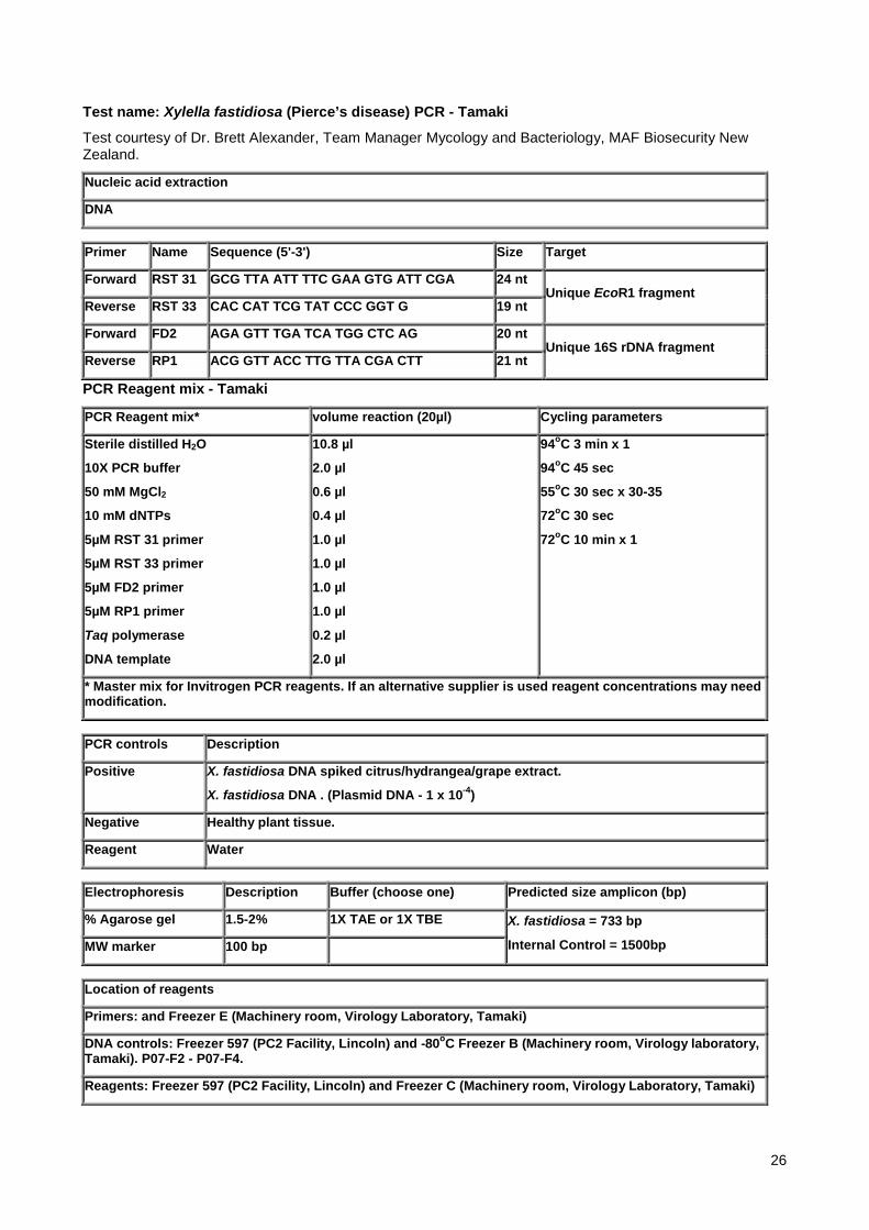
Test name: Xylella fastidiosa (Pierce’s disease) PCR - Tamaki
Test courtesy of Dr. Brett Alexander, Team Manager Mycology and Bacteriology, MAF Biosecurity New Zealand.
Nucleic acid extraction
DNA
Primer Name Sequence (5'-3') Size Target
Forward RST 31 GCG TTA ATT TTC GAA GTG ATT CGA 24 nt Unique EcoR1 fragment
Reverse RST 33 CAC CAT TCG TAT CCC GGT G 19 nt
Forward FD2 AGA GTT TGA TCA TGG CTC AG 20 nt Unique 16S rDNA fragment
Reverse RP1 ACG GTT ACC TTG TTA CGA CTT 21 nt
PCR Reagent mix - Tamaki
PCR Reagent mix* volume reaction (20µl) Cycling parameters
Sterile distilled H2O
10X PCR buffer
50 mM MgCl2
10 mM dNTPs
5µM RST 31 primer
5µM RST 33 primer
5µM FD2 primer
5µM RP1 primer
Taq polymerase
DNA template
10.8 µl
2.0 µl
0.6 µl
0.4 µl
1.0 µl
1.0 µl
1.0 µl
1.0 µl
0.2 µl
2.0 µl
94oC 3 min x 1
94oC 45 sec
55oC 30 sec x 30-35
72oC 30 sec
72oC 10 min x 1
* Master mix for Invitrogen PCR reagents. If an alternative supplier is used reagent concentrations may need modification.
PCR controls Description
Positive X. fastidiosa DNA spiked citrus/hydrangea/grape extract.
X. fastidiosa DNA . (Plasmid DNA - 1 x 10⁻⁴)
Negative Healthy plant tissue.
Reagent Water
Electrophoresis Description Buffer (choose one) Predicted size amplicon (bp)
% Agarose gel 1.5-2% 1X TAE or 1X TBE X. fastidiosa = 733 bp
Internal Control = 1500bp MW marker 100 bp
Location of reagents
Primers: and Freezer E (Machinery room, Virology Laboratory, Tamaki)
DNA controls: Freezer 597 (PC2 Facility, Lincoln) and -80oC Freezer B (Machinery room, Virology laboratory, Tamaki). P07-F2 - P07-F4.
Reagents: Freezer 597 (PC2 Facility, Lincoln) and Freezer C (Machinery room, Virology Laboratory, Tamaki)
26
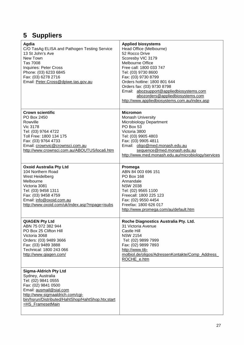
5 Suppliers Agdia C/O TasAg ELISA and Pathogen Testing Service 13 St John’s Ave New Town Tas 7008 Inquiries: Peter Cross Phone: (03) 6233 6845 Fax: (03) 6278 2716 Email: [email protected]
Applied biosystems Head Office (Melbourne) 52 Rocco Drive Scoresby VIC 3179 Melbourne Office Free call: 1800 033 747 Tel: (03) 9730 8600 Fax: (03) 9730 8799 Orders hotline: 1800 801 644 Orders fax: (03) 9730 8798 Email: [email protected] [email protected] http://www.appliedbiosystems.com.au/index.asp
Crown scientific PO Box 2450 Rowville Vic 3178 Tel: (03) 9764 4722 Toll Free: 1800 134 175 Fax: (03) 9764 4733 Email: [email protected] http://www.crownsci.com.au/ABOUTUS/loca6.htm
Micromon Monash University Microbiology Department PO Box 53 Victoria 3800 Tel: (03) 9905 4803 Fax: (03) 9905 4811 Email: [email protected] [email protected] http://www.med.monash.edu.au/microbiology/services
Oxoid Australia Pty Ltd 104 Northern Road West Heidelberg Melbourne Victoria 3081 Tel: (03) 9458 1311 Fax: (03) 9458 4759 Email: [email protected] http://www.oxoid.com/uk/index.asp?mpage=isubs
Promega ABN 84 003 696 151 PO Box 168 Annandale NSW 2038 Tel: (02) 9565 1100 Freecall: 1800 225 123 Fax: (02) 9550 4454 Freefax: 1800 626 017 http://www.promega.com/au/default.htm
QIAGEN Pty Ltd ABN 75 072 382 944 PO Box 25 Clifton Hill Victoria 3068 Orders: (03) 9489 3666 Fax: (03) 9489 3888 Technical: 1800 243 066 http://www.qiagen.com/
Roche Diagnostics Australia Pty. Ltd. 31 Victoria Avenue Castle Hill NSW 2154 Tel: (02) 9899 7999 Fax: (02) 9899 7893 http://www.tib-molbiol.de/oligos/AdressenKontakte/Comp_Address_ROCHE_e.htm
Sigma-Aldrich Pty Ltd Sydney, Australia Tel: (02) 9841 0555 Fax: (02) 9841 0500 Email: [email protected] http://www.sigmaaldrich.com/cgi-bin/hsrun/Distributed/HahtShop/HahtShop.htx;start=HS_FramesetMain
27
6 Contact points for further information
6.1 Australia Jo Luck Plant Pathologist Exotic Diseases Institute for Horticultural Development Department of Natural Resources and Environment Private Mail Bag 15 Ferntree Gully Delivery Centre Victoria Phone: 03 9210 9222 [email protected]
6.2 United States Bruce Kirkpatrick-(Biology, genetics and detection of Xylella fastidiosa) Plant Pathology University of California 452 Hutchison Hall Phone: (530) 752-2831 [email protected] Donald Hopkins-Xylella fastidiosa Central Florida Research and Education Centre University of Florida PO Box 111578 Gainesville FL 32611-1578 Phone: (352) 360-6686 [email protected] Alexander (Sandy) Purcell-Sharpshooters Division of Insect Biology University of California Berkeley, California 94720-3112 Phone: [email protected] Matthew Blua (PD and GWSS, biology and ecology) Entomologist UC Riverside Phone (909) 787-6301 [email protected] Douglas Cook (vector/pathogen relations, plant genomics) Plant Pathologist UC Davis Phone (530) 754-6561; [email protected]
7 Acknowledgements The information in this document was sourced from PaDIL (www.padil.gov.au), Pierce’s Disease Draft Diagnostic Manual (Luck, J., Mann, R., Van Rijswijk, R., Moran, J. and Merriman, P. (2006)) and the Pierce's Disease Pest Risk Review (2004). These documents were kindly provided by Office of the Chief Plant Protection Officer and Plant Health Australia.
Authors:
J. Luck, R. Mann, B. van Rijswijk, Jane Moran and Peter Merriman
28

Department of Natural Resources and Environment Institute for Horticultural Development Plant Health Private Mail Bag 15 Ferntree Gully Delivery Centre 3156 Victoria AUSTRALIA http://www.nre.vic.gov.au/agvic/ihd/ ph 61+3+9210 9222 fax 61+3+9800 3521 Additional Molecular tests were kindly provided by: Dr. Brett Alexander Team Manager, Mycology and Bacteriology Plant Health & Environment Laboratory Investigation and Diagnostic Centre MAF Biosecurity New Zealand 231 Morrin Road St Johns, PO Box 2095, Auckland 1140 New Zealand http://www.biosecurity.govt.nz/about-us/structure/phel
8 References Altschul S. F., Madden T. L., Scäffer, A. A., Zhang J., Zhang Z., Miller W. and Lipman D. J. (1997) Gapped
BLAST and PSI-BLAST: a new generation of protein database search programs. Nucleic Acids Research 25, 3389-3402.
Anonymous (1999). Crop Protection Compendium Global Module - 1999 Edition. CAB INTERNATIONAL: Wallingford, UK.
Anonymous (2000a). An Introduction to Pierce's Disease. http://www.cnr.berkeley.edu/xylella/page2.html
Banks, D, Albibi, R. Chen, J, Lamikanra, O, Jarret, R.L. and Smith, B. (1999) Specific detection of Xylella fastidiosa Pierce's Disease Strains. Curr. Microbiol. 39 85-88.
Berisha, B., Chen, Y.D. ,Zhang, G.Y., et al. (1998). Isolation of Pierce's Disease bacteria from grapevines in Europe. European Journal of Plant Pathology 104(5): 427- 433.
Beretta, M.J.G., Harakawa, R. and Chagas, C.M. (1996). First report of Xylella fastidiosa in coffee. Plant disease 80, 821.
Boubals, D. (1989) Pierce’s disease reaches the European vineyards (La maladie de Pierce arrive dans les vignobles d’Europe). Bulletin de l’OIV 62 (699-700) 309-314.
Bradbury, J.F. (1991) IMI descriptions of fungi and bacteria. Set 105, Nos 1041-1050. Mycopathologia 115 (1) 45-64.
Chang, C.J. and Donaldson, R.C. (1993). Xylella fastidiosa: cultivation in chemically defined medium. Phytopathology 83, 192-194.
Davis, M.J., French, W.J. and Shaad, N.W. (1981) Axenic culture of the bacteria associated with phony disease of peach and plum leaf scald. Curr. Microbiol. 6 309- 314.
Davis MJ, Purcell AH, Thomson SV (1978) Pierce's disease of grapevines: Isolation of the causal bacterium. Science 199:75-77.
Davis, M.J, Raju, B.C., Brlansky, R.H., Lee, R.F., Timmer L.W., Norris, R.C., McCoy, R.E. (1983) Periwinkle wilt bacterium - axenic culture, pathogenicity, and relationships to other gram-negative, xylem-inhabiting bacteria. Phytopathology 73 (11) 1510-1515
Day and Fletcher, 1994 (from WA ag PDS no reference)
Delong, D.M. and Severin, H.H.P. (1950). Spittle-insect vectors of Pierce's disease virus. I. Characters, distribution, and food plants. Hilgardia 19(11): 339-356.
Firrao, G. and Bazzi, C. (1994) Specific identification of Xylella fastidiosa using the polymerase chain reaction Phytopath. Medit. 33 90-92.
29

Freitag, J.H. (1951). Host range of the Pierce's Disease virus of grapes as determined by insect transmission. Phytopathology 41, 920 - 934.
Goheen, A.C. and Hopkins D.L. (1988). Pierce's Disease. pp. 44-45. In: Pearson, R.C. and Goheen, A.C. (eds). Compendium of Grape Diseases. The American Phytopathological Society (APS) Press: St Paul, Minnesota, USA 93 pp.
Goheen, A.C., Nyland, G. and Lowe, S.K. (1973). Association of rickettsia-like organism with Pierce's disease of grapevines and alfalfa dwarf and heat therapy of the disease in grapevines. Phytopathology 63: 341-345.
Goodwin, P., Purcell, A.H. (1992). Pierce's Disease. Grape Pest Management, 2nd Edition. Oakland, University of California, Division of Agriculture and Natural Resources: 76-84.
Gubler, D., Stapleton, J., Leavitt, G., Purcell, A., Varela, L. and Smith, R.J. (1999). UC Pest Management Guidelines. http://www.ipm.ucdavis.edu/PMG/r302101211.html
Hartman, J.R., Eshnaur, B.C. and Jarlfos, U.E. (1992). Shingle oak, a new host for bacterial leaf scorch caused by Xylella fastidiosa. Phytopathology 82, 498.
Hernandez, G.L. and Ochoa, C.F. (1994). Diagnosis of Xylella fastidiosa in grape and weeds associated with this crop. Manejo Integrado de Plagas 33, 7-10.
Hewitt, W.B., Frazier, N.W. and Freitag, J.H. (1949). Pierce's disease investigations. Hilgardia 19(7): 207-264.
Hill, B.L. and Purcell, A.H. (1995). Multiplication and movement of Xylella fastidiosa within grapevine and four other plants. Phytopathology 85, 1368 - 1372.
Hopkins, D.L. (1988) Production of diagnostic symptoms of blight in citrus inoculated with Xylella fastidiosa. Plant Dis. 72 (5) 432-435.
Hopkins, D.L. and Adlerz W.C. (1988) Natural hosts of Xylella fastidiosa in Florida. Plant Dis. 72 429-431.
Lopes, S.A., Ribeiro, D.M., Roberto, P.G., Franca, S.C. and Santos, J.M. (2000). Nicotiana tabaccum as an experimental host for the study of plant - Xylella fastidiosa interactions. Plant Disease 84, 827-830.
McElrone, A.J., Sherald, J.L. and Pooler, M.R. (1999). Identification of alternative hosts of Xylella fastidiosa in the Wasington DC area using nested polymerase chain reaction (PCR). Journal of Arboriculture 25, 258-263.
Medley, J.C. (1998). Pierce's Disease. http://aesrg.tamu.edu/Grapes/PierceDis.htm
Minesavage, G.V., Thompson, C.M., Hopkins, D.L., Leite, M.V.B.C. and Stall, R.E. (1994) Development of a Polymerase Chain Reaction protocol for detection of Xylella fastisiosa in plant tissue. Phyopathology 84 456-461.
Oxoid (2002) Identification stick oxidase BR64A. Oxoid limited, Hampshire, England.
Pierce, N.B. (1892) The California vine disease. U.S. Dept. Agric., Div. of Veg. Pathol. Bull. No. 2.
Purcell, A.H. (1980). Environmental therapy for Pierce's disease of grapevines. Plant Disease 64(4): 388-390.
Purcell, A.H. (1981). Vector preference and inoculation efficiency as components of resistance to Pierce's disease in European grape cultivars. Phytopathology 71(4): 429-435.
Purcell, A. H. 1989. Homopteran transmission of xylem-inhabiting bacteria. Advances in Disease Vector Research, Vol. 6. K. F. Harris. New York, Springer-Verlag: 243-266.
Purcell, A.H. (1999a). Blue-green Sharpshooter. http://www.cnr.berkeley.edu/xylella/bgss.html
Purcell, A.H. (1999b). Central Valley Guidelines for Pierce's Disease. http://www.cnr.berkeley.edu/xylella/central-valley-guidelines.html
Purcell, A.H. (1999c). General insect Category. http://www.cnr.berkeley.edu/xylella/geninsct.html
Purcell, A.H. and Hopkins, D.L. (1996) Fastidious xylem-limited bacterial plant pathogens. Annual Review of Phytopathology 34: 131-151.
Purcell, A.H. and Saunders, S. (1995). Harvested grape clusters as inoculum for Pierce's Disease. Plant Disease 79: 190-192.
30
Purcell, A.H. and Saunders, S.R. (1999) Fate of Pierce's Disease strains of Xylella fastidiosa in common riparian plants in California. Plant Disease 83 (9) 825-830.
Purcell, A. H., Finlay, A. H., McClean, D. L. (1979). Pierce's disease bacterium: Mechanism of transmission by leafhopper vectors. Science 206: 839-841.
Raju, B.C., Goheen, A.C., Teliz, D., Docampo, D.M. and Nyland, G. (1980). Pierce's Disease of grapevines in Mexico. Plant Disease 64, 280 -282.
Severin, H.H.P. (1950). Spittle-insect vectors of Pierce's disease virus. II. Life history and virus transmission. Hilgardia 19(11): 357-382.
Sherald, J.L., Wells, J.M., Hurtt, S.S. and Kostka, S.J. (1987). Association of fastidious xylem-inhabiting bacteria with leaf scorch in red maple. Plant Disease 71, 930 - 933.
Sherald, J.L. (1993). Pathogenicity of Xylella fastidiosa in American elm and failure of reciprocal transmission between strains from elm and sycamore. Plant Disease 77, 190 - 193.
Smith, I.M., McNamara, D.G., Scott, P.R., Holderness, M. and Burger, B. (eds). (1997). Quarantine Pests for Europe (2nd edition). CAB International: Wallingford, UK 1425 pp.
Sivapalan, S. (2001). Pest data sheet - bacteria. (unpublished).
Varela, L.G. (2000). Pierce's Disease in the North Coast. http://www.cnr.berkeley.edu/xylella/pd97.html
Varela, L.G., Smith, R.J. and Phillips, P.A. (2001) Pierce's Disease. University of California, Division of Agriculture and Natural Resources, USA. 2-14.
Weisburg, W.G., Barns, S., Pelletier, D.A. and Lane, D.J. (1991) 16S Ribosomal DNA Amplification for Phylogenetic Study. J. Bacteriol. 173 697-703.
Wells, J.M., Raju, B.C., Hung, H.Y., et al. (1987). Xylella fastidiosa gen. nov., sp. nov: gram-negative, xylem-limited, fastidious plant bacteria related to Xanthomonas spp. International Journal of Systematic Bacteriology 37: 136-143.
Yonce, C.E. and Chang, C.J. (1987). Detection of xylem limited bacteria from sharpshooter leafhoppers and their feeding hosts in peach environs monitored by culture isolations and ELISA techniques. Environmental Entomology 16, 68-71
31
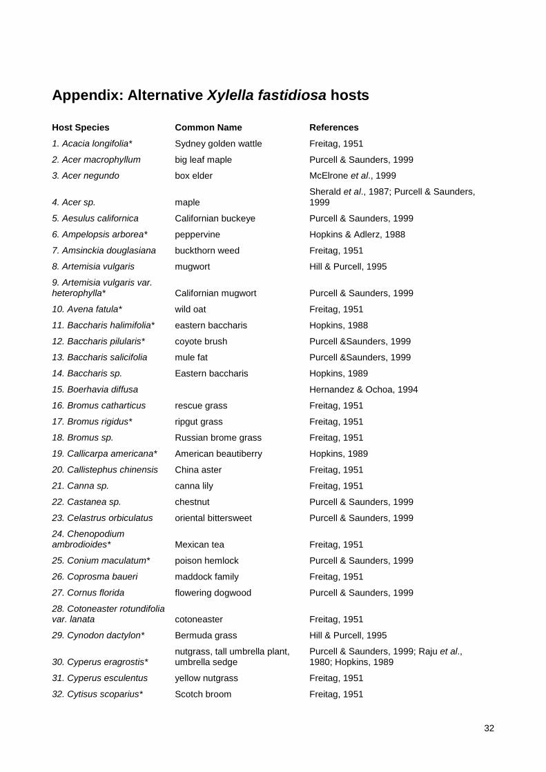
Appendix: Alternative Xylella fastidiosa hosts
Host Species Common Name References
1. Acacia longifolia* Sydney golden wattle Freitag, 1951
2. Acer macrophyllum big leaf maple Purcell & Saunders, 1999
3. Acer negundo box elder McElrone et al., 1999
4. Acer sp. maple Sherald et al., 1987; Purcell & Saunders, 1999
5. Aesulus californica Californian buckeye Purcell & Saunders, 1999
6. Ampelopsis arborea* peppervine Hopkins & Adlerz, 1988
7. Amsinckia douglasiana buckthorn weed Freitag, 1951
8. Artemisia vulgaris mugwort Hill & Purcell, 1995
9. Artemisia vulgaris var. heterophylla* Californian mugwort Purcell & Saunders, 1999
10. Avena fatula* wild oat Freitag, 1951
11. Baccharis halimifolia* eastern baccharis Hopkins, 1988
12. Baccharis pilularis* coyote brush Purcell &Saunders, 1999
13. Baccharis salicifolia mule fat Purcell &Saunders, 1999
14. Baccharis sp. Eastern baccharis Hopkins, 1989
15. Boerhavia diffusa Hernandez & Ochoa, 1994
16. Bromus catharticus rescue grass Freitag, 1951
17. Bromus rigidus* ripgut grass Freitag, 1951
18. Bromus sp. Russian brome grass Freitag, 1951
19. Callicarpa americana* American beautiberry Hopkins, 1989
20. Callistephus chinensis China aster Freitag, 1951
21. Canna sp. canna lily Freitag, 1951
22. Castanea sp. chestnut Purcell & Saunders, 1999
23. Celastrus orbiculatus oriental bittersweet Purcell & Saunders, 1999
24. Chenopodium ambrodioides* Mexican tea Freitag, 1951
25. Conium maculatum* poison hemlock Purcell & Saunders, 1999
26. Coprosma baueri maddock family Freitag, 1951
27. Cornus florida flowering dogwood Purcell & Saunders, 1999
28. Cotoneaster rotundifolia var. lanata cotoneaster Freitag, 1951
29. Cynodon dactylon* Bermuda grass Hill & Purcell, 1995
30. Cyperus eragrostis* nutgrass, tall umbrella plant, umbrella sedge
Purcell & Saunders, 1999; Raju et al., 1980; Hopkins, 1989
31. Cyperus esculentus yellow nutgrass Freitag, 1951
32. Cytisus scoparius* Scotch broom Freitag, 1951
32

33. Daucus carota var. sativa short white carrot Freitag, 1951
34. Digitaria sanguinalis* hairy crabgrass Freitag, 1951
35. Duranta repens* pigeon-berry Freitag, 1951
36. Echinochloa crusgalli* water grass Hill & Purcell, 1995
37. Epilobium californicum willow herb Freitag, 1951
38. Epilobium panniculatum panicled willow herb Freitag, 1951
39. Eragrostis diffusa diffuse love grass Freitag, 1951
40. Erodium cicutarium red-stem filaree Freitag, 1951
41. Escallonia montevidensis* saxifrage family Raju et al., 1980
42. Eugenia myrtifolia* Australian bushberry Freitag, 1951
43. Festuca megalura foxtail fescue Freitag, 1951
44. Fragaria californica wild strawberry Raju et al., 1980
45. Fragaria vesca* wild strawberry Purcell & Saunders, 1999
46. Franseria acanthicarpa annual burr weed Freitag, 1951
47. Fraxinus dipetala* foothill ash Freitag, 1951
48. Fraxinus latifolia Oregan ash Purcell and Saunders, 1999
49. Fuschia magellanica* Fuschia Freitag, 1951
50. Genista monspessulanus French broom Purcell and Saunders, 1999
51. Godetia grandiflora Godetia Freitag, 1951
52. Hedera helix ivy Freitag, 1951
53. Heliotropium fruticosum Hernandez & Ochoa, 1994
54. Heliotropium indicum Hernandez & Ochoa, 1994
55. Holcus sudanensis Sudan grass Freitag, 1951
56. Hordeum murinum common foxtail Freitag, 1951
57. Hordeum vulgare barley Freitag, 1951
58. Hydrangea panniculata* hydrangea Freitag, 1951
59. Ipomoea crassicaulis Hernandez & Ochoa, 1994
60. Juglans nigra black walnut Purcell & Saunders, 1999
61. Lactuca scariola prickly lettuce Freitag, 1951
62. Lathyrus ciceria pea family Freitag, 1951
63. Lathyrus clymenium pea family Freitag, 1951
64. Lathyrus sativa grass pea Freitag, 1951
65. Lolium multiflorum* Italian ryegrass Freitag, 1951
66. Lolium temulentum darnel Freitag, 1951
67. Lonicera japonica Japanese honeysuckle Freitag, 1951
68. Marjorana hortensis* sweet marjoram Freitag, 1951
69. Medicago hispida* bur clover Freitag, 1951
70. Meliotus alba white meilot Freitag, 1951
71. Meliotus alba var. annua hubum clover Freitag, 1951
33

72. Meliotus indica annual yellow sweet clover Freitag, 1951
73. Meliotus officinalis yellow sweet clover Freitag, 1951
74. Meliotus sp.* sweet clover Freitag, 1951
75. Melissa officinalis* garden balm Freitag, 1951
76. Mentha sp. mint Freitag, 1951
77. Merremia glabra Hernandez & Ochoa, 1994
78. Montia linearis* miner's lettuce Raju et al., 1980
79. Nicotiana tabaccum tobacco Lopes et al., 2000
80. Oenanthe sarmetosa water parsley Freitag, 1951
81. Oenothera hookeri evening primrose Freitag, 1951
82. Parthenocissus quinquefolia* Virginia creeper Hopkins, 1988
83. Parthenocissus tricuspidata* ivy Freitag, 1951
84. Paspalum dilatum* dallis grass Hopkins, 1989
85. Passiflora foetida Hernandez & Ochoa, 1994
86. Pelargonium hortorum fish geranium Freitag, 1951
87. Pennisetum clandestimum ikuyu grass Freitag, 1951
88. Phalaris minor Mediterranean Canary grass Freitag, 1951
89. Phalaris paradoxa gnawed Canary grass Freitag, 1951
90. Phleum pretense timothy Freitag, 1951
91. Photinia arbutifolia oyon/christmas berry Freitag, 1951
92. Pittosporum crassifolium karo Freitag, 1951
93. Plantago lanceolata ribgrass Purcell & Saunders, 1999
94. Platanus occidentalis sycamore Hartman et al., 1992
95. Poa annua* annual bluegrass Freitag, 1951
96. Portulaca oleracea Hernnandez & Ochoa, 1994
97. Ploygonum convolvulus black bindweed Freitag, 1951
98. Polygonum persicaria* lady's thumb Freitag, 1951
99. Populus fremonti freemont cottonwood Purcell & Saunders, 1999
100.Prunus serotina blackcherry Purcell & Saunders, 1999
101.Reseda odorata common mignonette Freitag, 1951
102.Rhamnus californica coffeeberry Purcell & Saunders, 1999
103.Rheum rhaponicum rhubarb Freitag, 1951
104.Rhus diversiloba* poison oak Purcell & Saunders, 1999
105.Rhus sp.* sumac Hopkins, 1988
106.Rosa californica* Californian rose Purcell & Saunders, 1999
107.Rosemarinus officinalis* rosemary Freitag, 1951
108.Rubus discolor Himalayan blackberry Purcell & Saunders, 1999
34

109.Rubus procerus* blackberry Hopkins, 1989
110.Rubus ursinus California blackberry Purcell & Saunders, 1999
111.Rubus vitifolius* Californian blackberry Freitag, 1951
112.Rumex crispus* curly dock Freitag, 1951
113.Salix sp.* willow Freitag, 1951; Purcell & Saunders, 1999
114.Sambucus caerulea* blue elder Freitag, 1951
115.Sambucus canadensis* American elder Hopkins, 1989
116.Sambucus mexicana blue elderberry Purcell & Saunders, 1999
117.Setaria lutescens yellow bristle grass Freitag, 1951
118.Solidago fistulosa* goldenrod Hopkins, 1989
119.Sonchus asper prickly sawthistle Freitag, 1951
120.Sorghum halepense* Johnson grass Yonce & Chang, 1987
121.Symphoricarpos albus snowberry Purcell & Saunders, 1999
122.Syringa vulgaris lilac Freitag, 1951
123.Toxicodendron diversilobum poison oak Purcell & Saunders, 1999
124.Trifolium fragerum Strawberry clover Freitag, 1951
125.Trifolium hybridum alsike clover Freitag, 1951
126.Trifolium incarnatum Crimson clover Freitag, 1951
127.Trifolium pratense red clover Freitag, 1951
128.Trifolium repens* white clover Freitag, 1951
129.Trifolium repens var. latum* Ladino clover Freitag, 1951
130.Ulnus sp elm Sherald, 1993
131.Umbellularia californica bay laurel Purcell & Saunders, 1999
132.Urtica dioica Stinging nettle Purcell & Saunders, 1999
133.Urtica gracilis* creek nettle Freitag, 1951
134.Veronica sp.* Speedwell Freitag, 1951
135.Vicia monanthus Vetch Freitag, 1951
136.Vinca major* greater periwinkle Purcell & Saunders, 1999
137.Vinca minor* lesser periwinkle Purcell & Saunders, 1999
138.Vitis californica* wild grape Freitag, 1951
139.Vitis munsoniana* wild grape Hopkins, 1989
140.Vitis riparia wild grape Hopkins, 1989
141.Xanthium canadense Cocklebur Freitag, 1951
* natural host
35