national diagnostic protocol for potato ring rot, caused...
TRANSCRIPT
National Diagnostic Protocol for Potato Ring Rot, caused by
Clavibacter michiganensis subsp. sepedonicus
PEST STATUS Not present in Australia
PROTOCOL NUMBER NDP 8
VERSION NUMBER V1.2
PROTOCOL STATUS Endorsed
ISSUE DATE November 2011
REVIEW DATE 2016
ISSUED BY SPHDS
Prepared for the Subcommittee on Plant Health Diagnostic Standards (SPHDS)
This version of the National Diagnostic Protocol (NDP) for Clavibacter michiganensis subsp. sepedonicus is current as at the date contained in the version control box on the front of this
document.
NDPs are updated every 5 years or before this time if required (i.e. when new techniques become available).
The most current version of this document is available from the SPHDS website: http://plantbiosecuritydiagnostics.net.au/resource-hub/priority-pest-diagnostic-resources/
NDP Clavibacter Potato Ring rot 2
Contents 1 Introduction ............................................................................................................................ 4
1.1 Host range ......................................................................................................................... 4 1.2 Transmission ...................................................................................................................... 4
2 Taxonomic Information .......................................................................................................... 5
3 Detection ................................................................................................................................. 6
3.1 Sampling techniques .......................................................................................................... 6 3.2 Symptom description .......................................................................................................... 6
3.2.1 Foliar and Stem symptoms .......................................................................................... 6 3.2.2 Tuber symptoms .......................................................................................................... 6 3.2.3 Symptom variability ...................................................................................................... 7
4 Identification ........................................................................................................................... 9
4.1 Isolation/culture techniques ................................................................................................ 9 4.1.1 Equipment and Media .................................................................................................. 9 4.1.2 NCP-88; Semiselective agar media (1 L) ..................................................................... 9 4.1.3 Method ...................................................................................................................... 10
4.2 Eggplant bioassay ............................................................................................................ 11 4.2.1 Preparation of samples for Immunofluorescence staining (IF) and eggplant test ....... 11 4.2.2 Eggplant propagation ................................................................................................. 12 4.2.3 Eggplant Assay .......................................................................................................... 12
4.3 Immunofluorescence (IF) testing ...................................................................................... 12 4.3.1 Procedure .................................................................................................................. 13
5 Polymerase chain reaction (PCR) detection using potato extracts .................................. 14
5.1 Potato tissue DNA extraction for PCR .............................................................................. 14 5.1.1 Modified SCP ............................................................................................................. 14 5.1.2 PBS/BSA ................................................................................................................... 14 5.1.3 CTAB buffer (+ 0.2% mercaptoethanol) (100 ml) ....................................................... 15 5.1.4 Other reagents ........................................................................................................... 15 5.1.5 Extraction Method ...................................................................................................... 15
5.2 PCR ................................................................................................................................. 15 5.2.1 PCR Reagents ........................................................................................................... 15 5.2.2 PCR Method .............................................................................................................. 16
6 Contact points for further information ................................................................................ 18
7 Acknowledgements .............................................................................................................. 18
8 References ............................................................................................................................ 18
9 Appendix 1. ........................................................................................................................... 22
9.1 Visual examination for ring rot symptoms ......................................................................... 22 9.2 Tissue sampling for DNA extractions and bacterial isolations ........................................... 22
9.2.1 Sampling methods ..................................................................................................... 22
NDP Clavibacter Potato Ring rot 3
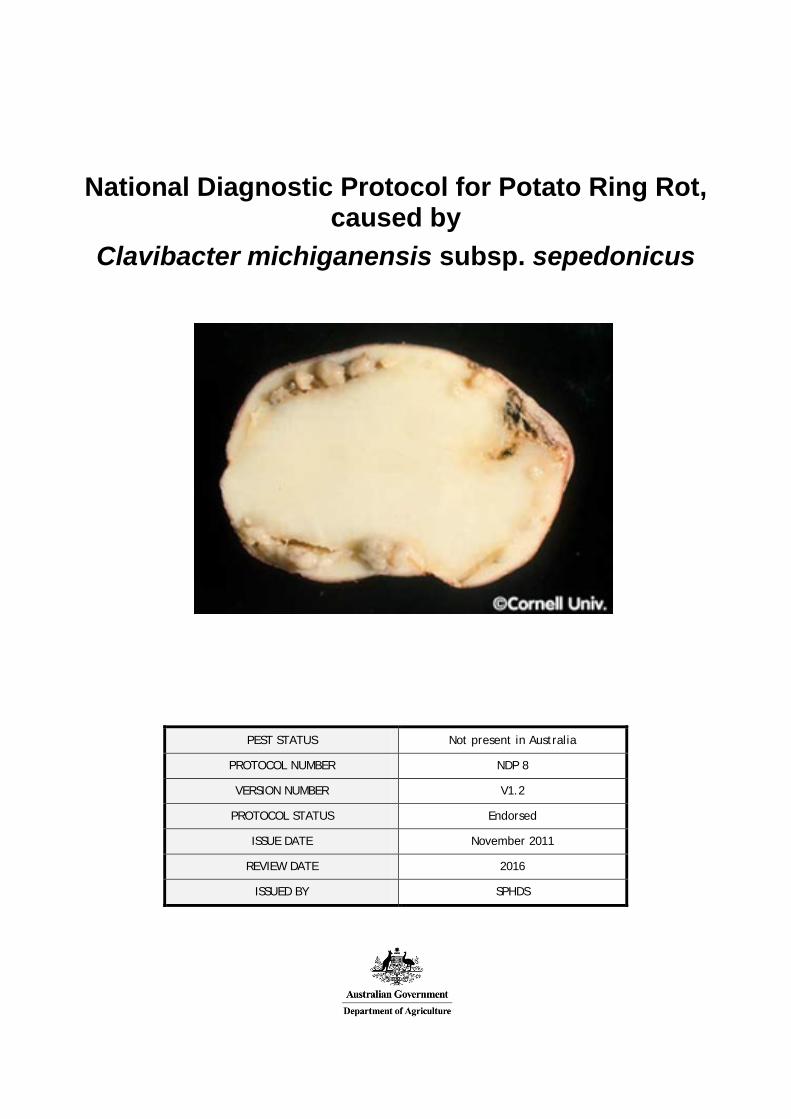
1 Introduction Clavibacter michiganensis subsp. sepedonicus (Spieck & Kotth) Davis (Cms) is the causal agent of the destructive potato disease, bacterial ring rot (Davis et al., 1997; Franc, 1999). The disease derives its name from the characteristic internal breakdown in the vascular ring of an infected tuber (Manzer and Genereux, 1981). This can be seen as a brown, cheesy decay of the vascular tissue. Above ground the disease is usually seen as a progressive wilt (Lelliott and Stead, 1987).
Cms is highly infectious and can cause extensive losses to infected crops (Rich 1983; Rowe et al. 1995). The disease causes even greater economic losses to seed potato growers.
Infected seed potatoes produce infected plants. After planting, bacteria multiply and spread to the vascular tissue of stems, petioles, roots and developing tubers. Symptoms rarely develop quickly and infections usually remain latent for long periods. Some cultivars tolerate infection so that symptoms may not develop for several plant generations, even though the bacteria can multiply in both plants and tubers. The latent period may encompass almost the entire period of host growth from vegetative propagule to mature plant (Bishop and Slack, 1987).
1.1 Host range Potatoes (Solanum tuberosum)
1.2 Transmission Important means of spread are the planting of infected seed potatoes and contamination of containers, equipment and premises. When an infected seed piece is planted, bacteria move from the seed through the vascular tissue into the stem and lower leaves of the growing plant (Babadoost, 1990). The plant will start to show foliar and stem symptoms mid season or later (Davis et al., 1997). Late in the season, bacteria migrate from the stem down into the stolons, infecting the new tubers (Babadoost, 1990). Internal symptoms may be present within tubers at harvest but are more commonly observed toward the end of the storage period (Lelliott and Stead, 1987).
Planters, graders and knives which have been contaminated by bacteria from diseased potatoes are also a potent infection source. Disease spread in the field from plant to plant is usually poor, but there is experimental evidence that some insects, including the potato flea beetle, Epitrix cucumeris (Harris), the Colorado potato beetle, Leptinotarsa decemlineata (Say), the green peach aphid, Myzus persicae (Sulzer) and the fruit fly are possible vectors of Cms (Christie et al., 1991; Christie et al., 1993; De Boer et al., 1988).
Bacteria can also survive and remain infectious for several years on potato bags, bulk bins, store walls and other surfaces that have been contaminated by rotting ooze. The bacterium is able to overwinter in the soil, usually in association with “groundkeepers” (unharvested potatoes from the previous crop) and debris from infected crops. Infected groundkeepers lifted with an otherwise clean seed or ware crop can infect that crop (DEFRA, 2002).
The pathogen can survive in water for more than a month but there is no known aquatic weed host to build up inoculum levels. Contaminated wash water from infected tuber lots can transmit the pathogen to subsequent lots washed in the same water (DEFRA 2002).
NDP Clavibacter Potato Ring rot 4
2 Taxonomic Information Monera
› Eubacteria
› Actinobacteria
› Actinobacteria (class)
› Actinobacteridae
› Actinomycetales
› Micrococcineae
› Microbacteriaceae
› Clavibacter
› Clavibacter michiganensis
› Clavibacter michiganensis subsp. sepedonicus
Name: Clavibacter michiganensis subsp. sepedonicus (Spieckermann and Kotthoff) Davis et al.
Synonyms: Corynebacterium sepedonicum, Pseudobacterium sepedonicum, Mycobacterium sepedonicum, Phytomonas sepedonica, Aplanobacter sepedonicum, Bacterium sepedonicum, Corynebacterium michiganense subsp. Sepedonicum, "Pseudobacterium sepedonicum" (Spieckermann and Kotthoff 1914) Krasil'nikov 1949, "Mycobacterium sepedonicum" (Spieckermann and Kotthoff 1914) Krasil'nikov 1949, "Phytomonas sepedonica" (Spieckermann and Kotthoff 1914) Magrou 1937, "Aplanobacter sepedonicum" (sic) (Spieckermann and Kotthoff 1914) Smith 1920, "Bacterium sepedonicum" Spieckermann and Kotthoff 1914, Corynebacterium michiganense subsp. sepedonicum (Spieckermann and Kotthoff 1914) Carlson and Vidaver 1982, Clavibacter michiganensis subsp. sepedonicus corrig.(Spieckermann and Kotthoff 1914) Davis et al 1984, Corynebacterium sepedonicum (Spieckermann and Kotthoff 1914) Skaptason and Burkholder 1942 (AL1980).
Equivalent name: Clavibacter michiganense sepedonicum
Common names: Bacterial ring rot (English), Bactériose annulaire, flétrissement bactérien (French), Bakterienringfäule (German), Podredumbre anular (Spanish)
NDP Clavibacter Potato Ring rot 5
3 Detection
3.1 Sampling techniques Formal identification is based on tuber symptoms and tests on the bacterial exudates from the affected tubers. While the bacteria can be detected in other plant parts (Appendix 1) these are usually sampled only during surveillance.
3.2 Symptom description
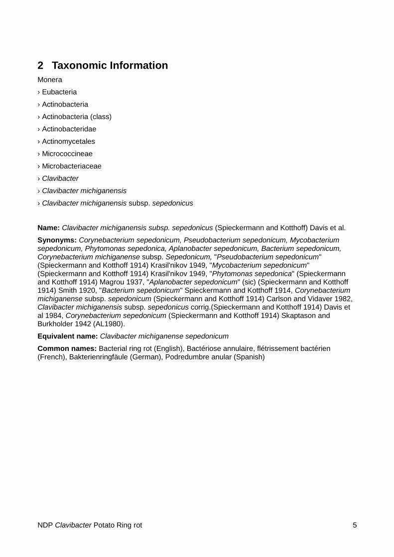
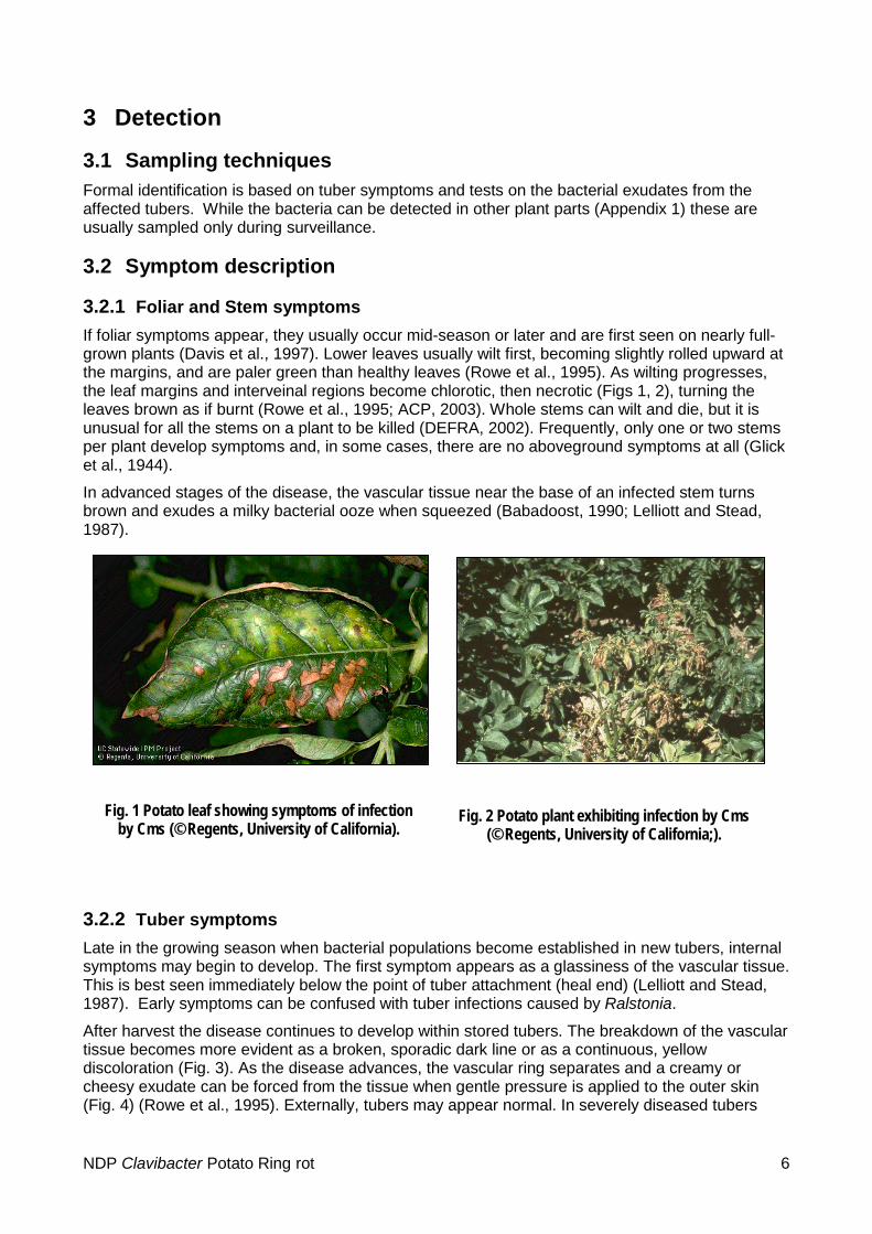
3.2.1 Foliar and Stem symptoms If foliar symptoms appear, they usually occur mid-season or later and are first seen on nearly full-grown plants (Davis et al., 1997). Lower leaves usually wilt first, becoming slightly rolled upward at the margins, and are paler green than healthy leaves (Rowe et al., 1995). As wilting progresses, the leaf margins and interveinal regions become chlorotic, then necrotic (Figs 1, 2), turning the leaves brown as if burnt (Rowe et al., 1995; ACP, 2003). Whole stems can wilt and die, but it is unusual for all the stems on a plant to be killed (DEFRA, 2002). Frequently, only one or two stems per plant develop symptoms and, in some cases, there are no aboveground symptoms at all (Glick et al., 1944).
In advanced stages of the disease, the vascular tissue near the base of an infected stem turns brown and exudes a milky bacterial ooze when squeezed (Babadoost, 1990; Lelliott and Stead, 1987).
3.2.2 Tuber symptoms Late in the growing season when bacterial populations become established in new tubers, internal symptoms may begin to develop. The first symptom appears as a glassiness of the vascular tissue. This is best seen immediately below the point of tuber attachment (heal end) (Lelliott and Stead, 1987). Early symptoms can be confused with tuber infections caused by Ralstonia.
After harvest the disease continues to develop within stored tubers. The breakdown of the vascular tissue becomes more evident as a broken, sporadic dark line or as a continuous, yellow discoloration (Fig. 3). As the disease advances, the vascular ring separates and a creamy or cheesy exudate can be forced from the tissue when gentle pressure is applied to the outer skin (Fig. 4) (Rowe et al., 1995). Externally, tubers may appear normal. In severely diseased tubers
Fig. 1 Potato leaf showing symptoms of infection by Cms (© Regents, University of California).
Fig. 2 Potato plant exhibiting infection by Cms (© Regents, University of California;).
NDP Clavibacter Potato Ring rot 6
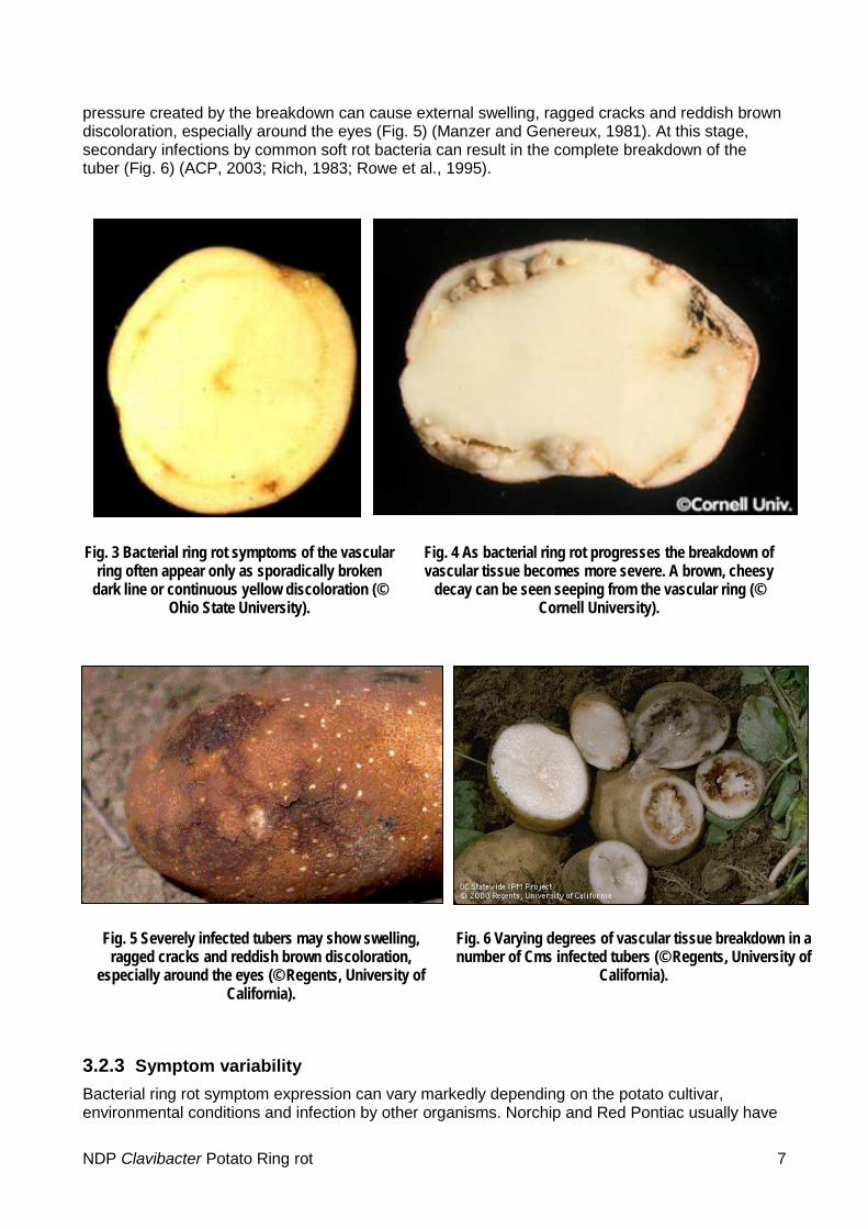
pressure created by the breakdown can cause external swelling, ragged cracks and reddish brown discoloration, especially around the eyes (Fig. 5) (Manzer and Genereux, 1981). At this stage, secondary infections by common soft rot bacteria can result in the complete breakdown of the tuber (Fig. 6) (ACP, 2003; Rich, 1983; Rowe et al., 1995).
Fig. 3 Bacterial ring rot symptoms of the vascular ring often appear only as sporadically broken
dark line or continuous yellow discoloration (© Ohio State University).
Fig. 4 As bacterial ring rot progresses the breakdown of vascular tissue becomes more severe. A brown, cheesy
decay can be seen seeping from the vascular ring (© Cornell University).
Fig. 5 Severely infected tubers may show swelling, ragged cracks and reddish brown discoloration,
especially around the eyes (© Regents, University of California).
Fig. 6 Varying degrees of vascular tissue breakdown in a number of Cms infected tubers (© Regents, University of
California).
3.2.3 Symptom variability Bacterial ring rot symptom expression can vary markedly depending on the potato cultivar, environmental conditions and infection by other organisms. Norchip and Red Pontiac usually have
NDP Clavibacter Potato Ring rot 7
easily recognised symptoms, Russet Burbank has moderate symptom expression and Desiree and Belrus have less apparent disease symptoms. Cultivars such as Teton and Urgenta rarely exhibit any symptoms when infected, but the bacterium can readily be recovered from infected symptomless tubers of both varieties (De Boer & McCann, 1990; ACP, 2003). Some varieties develop atypical symptoms such as dwarf rosette foliage (Nelson et al., 1992). Symptom expression is generally favoured by warm growing conditions, while under cool conditions; few or no symptoms may develop (ACP, 2003). Symptoms can also be confusing when other disorders such as early blight, late blight, blackleg, brown rot, freezing injury or water damage are present. Viruses, such as Potato leaf roll virus, can mask the effects of bacterial ring rot (Babadoost, 1990; Nelson & Torfason, 1974). Disease symptoms can be further complicated by the latent phase of the bacterium, and may be absent altogether, although bacterial population in the tubers is high (Lelliott & Stead, 1987). Bacterial ring rot can be confused in the early stages with brown rot caused by Ralstonia solanacearum.
NDP Clavibacter Potato Ring rot 8

4 Identification Many of the identification procedures in this protocol have been sourced from the EPPO protocol PM 7/59 (EPPO 2006), drafted by D. Stead, Central Science Laboratory, York (GB) and revised by P. Müller, Biologische Bundesanstalt für Land- und Forstwirtschaft, Kleinmachnow (DE).
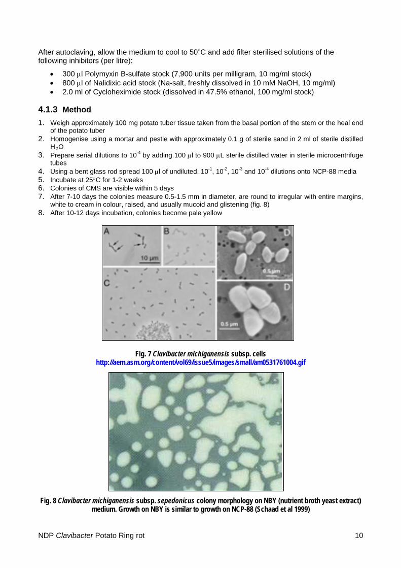
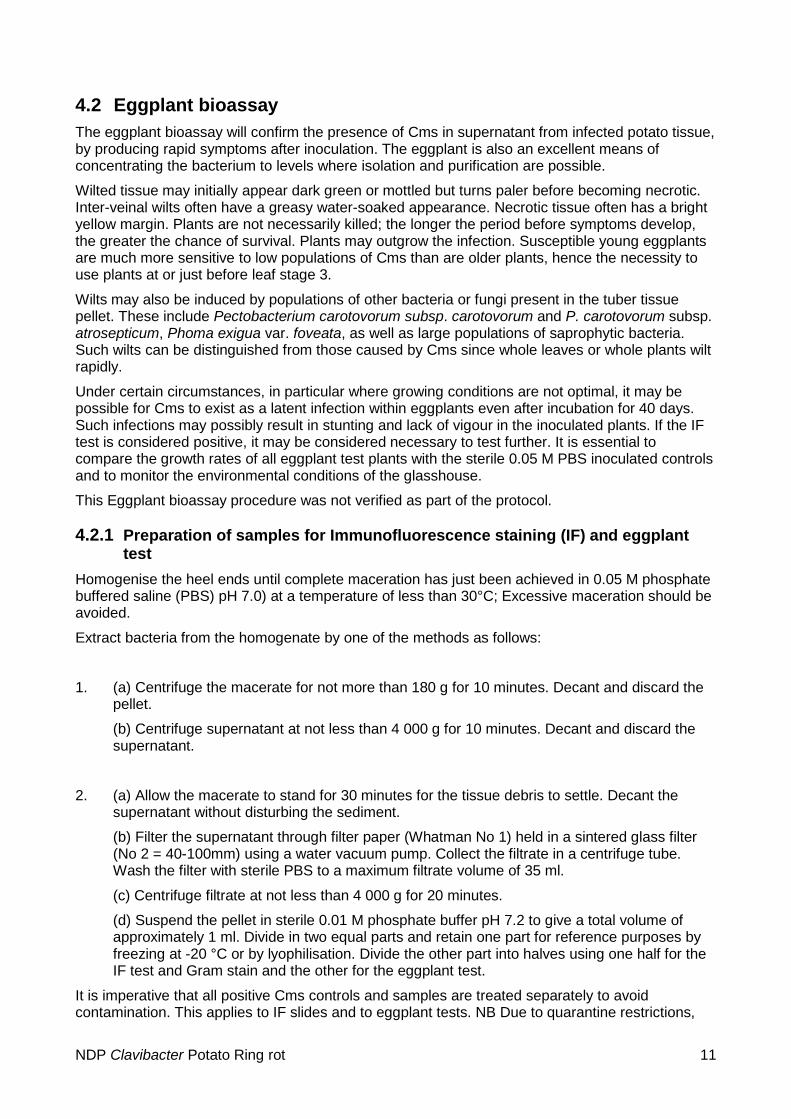
The bacterium is Gram positive, non-motile, non spore-forming and approximately 0.4-0.6 μm x 0.8-1.2 μm. Cells are coryneform rods, slightly curved and club shaped, arranged singly or in pairs in L or V formations (Fig 15). It is strictly aerobic and nutritionally fastidious, requiring a specialised media such as NCP-88. When plated on to NCP-88, colonies of CMS are visible within 5 days at 25°C, and measure 0.5-1.5 mm in diameter after 7-10 days. The colonies are round to irregular with entire margins, white to cream in colour, raised, and usually mucoid and glistening (Fig 16). After 10-12 days incubation, colonies become pale yellow (de la Cruz et al., 1992).
The bacterium can hydrolyse soluble starch, and utilise acetate, citrate and succinate. It can produce acid from the oxidation of mannitol and sorbitol. It cannot produce H2S from the decomposition of organic sulphur compounds or from the reduction of sulphate under anaerobic conditions. It is unable to produce and maintain stable acid end products from glucose fermentation (de la Cruz et al., 1992).
4.1 Isolation/culture techniques
4.1.1 Equipment and Media • 20-200 μl & 200-1000 μl pipettes and tips • Autoclave • Autoclaved mortar and pestles • Balance • Bunsen burner • Centrifuge tubes • Fridge • Glass spreaders • Incubator at 25oC • Lamina flow • Petri dishes • Sterile sand • Sterile scalpel blades • Syringe with 22 μm filter
4.1.2 NCP-88; Semiselective agar media (1 L)
Nutrient agar 23.0 g Yeast extract 2.0 g K2HPO4 MW = 174.18 2.0 g KH2PO4 MW = 136.09 0.5 g MgSO4
.7H2O MW = 246.48 0.25 g D-mannitol (C6H14O6) MW = 182.17 5.0 g Autoclave the media at 121oC at 110kPa for 25 minutes.
NDP Clavibacter Potato Ring rot 9
After autoclaving, allow the medium to cool to 50oC and add filter sterilised solutions of the following inhibitors (per litre):
• 300 µl Polymyxin B-sulfate stock (7,900 units per milligram, 10 mg/ml stock) • 800 µl of Nalidixic acid stock (Na-salt, freshly dissolved in 10 mM NaOH, 10 mg/ml) • 2.0 ml of Cycloheximide stock (dissolved in 47.5% ethanol, 100 mg/ml stock)
4.1.3 Method 1. Weigh approximately 100 mg potato tuber tissue taken from the basal portion of the stem or the heal end
of the potato tuber 2. Homogenise using a mortar and pestle with approximately 0.1 g of sterile sand in 2 ml of sterile distilled
H2O 3. Prepare serial dilutions to 10-4 by adding 100 µl to 900 µL sterile distilled water in sterile microcentrifuge
tubes 4. Using a bent glass rod spread 100 µl of undiluted, 10-1, 10-2, 10-3 and 10-4 dilutions onto NCP-88 media 5. Incubate at 25°C for 1-2 weeks 6. Colonies of CMS are visible within 5 days 7. After 7-10 days the colonies measure 0.5-1.5 mm in diameter, are round to irregular with entire margins,
white to cream in colour, raised, and usually mucoid and glistening (fig. 8) 8. After 10-12 days incubation, colonies become pale yellow
Fig. 7 Clavibacter michiganensis subsp. cells http://aem.asm.org/content/vol69/issue5/images/small/am0531761004.gif
Fig. 8 Clavibacter michiganensis subsp. sepedonicus colony morphology on NBY (nutrient broth yeast extract) medium. Growth on NBY is similar to growth on NCP-88 (Schaad et al 1999)
NDP Clavibacter Potato Ring rot 10
4.2 Eggplant bioassay The eggplant bioassay will confirm the presence of Cms in supernatant from infected potato tissue, by producing rapid symptoms after inoculation. The eggplant is also an excellent means of concentrating the bacterium to levels where isolation and purification are possible.
Wilted tissue may initially appear dark green or mottled but turns paler before becoming necrotic. Inter-veinal wilts often have a greasy water-soaked appearance. Necrotic tissue often has a bright yellow margin. Plants are not necessarily killed; the longer the period before symptoms develop, the greater the chance of survival. Plants may outgrow the infection. Susceptible young eggplants are much more sensitive to low populations of Cms than are older plants, hence the necessity to use plants at or just before leaf stage 3.
Wilts may also be induced by populations of other bacteria or fungi present in the tuber tissue pellet. These include Pectobacterium carotovorum subsp. carotovorum and P. carotovorum subsp. atrosepticum, Phoma exigua var. foveata, as well as large populations of saprophytic bacteria. Such wilts can be distinguished from those caused by Cms since whole leaves or whole plants wilt rapidly.
Under certain circumstances, in particular where growing conditions are not optimal, it may be possible for Cms to exist as a latent infection within eggplants even after incubation for 40 days. Such infections may possibly result in stunting and lack of vigour in the inoculated plants. If the IF test is considered positive, it may be considered necessary to test further. It is essential to compare the growth rates of all eggplant test plants with the sterile 0.05 M PBS inoculated controls and to monitor the environmental conditions of the glasshouse.
This Eggplant bioassay procedure was not verified as part of the protocol.
4.2.1 Preparation of samples for Immunofluorescence staining (IF) and eggplant test
Homogenise the heel ends until complete maceration has just been achieved in 0.05 M phosphate buffered saline (PBS) pH 7.0) at a temperature of less than 30°C; Excessive maceration should be avoided.
Extract bacteria from the homogenate by one of the methods as follows:
1. (a) Centrifuge the macerate for not more than 180 g for 10 minutes. Decant and discard the pellet.
(b) Centrifuge supernatant at not less than 4 000 g for 10 minutes. Decant and discard the supernatant.
2. (a) Allow the macerate to stand for 30 minutes for the tissue debris to settle. Decant the supernatant without disturbing the sediment.
(b) Filter the supernatant through filter paper (Whatman No 1) held in a sintered glass filter (No 2 = 40-100mm) using a water vacuum pump. Collect the filtrate in a centrifuge tube. Wash the filter with sterile PBS to a maximum filtrate volume of 35 ml.
(c) Centrifuge filtrate at not less than 4 000 g for 20 minutes.
(d) Suspend the pellet in sterile 0.01 M phosphate buffer pH 7.2 to give a total volume of approximately 1 ml. Divide in two equal parts and retain one part for reference purposes by freezing at -20 °C or by lyophilisation. Divide the other part into halves using one half for the IF test and Gram stain and the other for the eggplant test.
It is imperative that all positive Cms controls and samples are treated separately to avoid contamination. This applies to IF slides and to eggplant tests. NB Due to quarantine restrictions,
NDP Clavibacter Potato Ring rot 11
some diagnostic labs may not be able to use a viable culture of Cms for control inoculations. However, heat-killed cells may be used as an IF control.
4.2.2 Eggplant propagation 1. Sow seeds of eggplant (Solanum melongena cv. Black Beauty) in pasteurized potting mix.
Transplant seedlings with fully expanded cotyledons (10 to 14 days) into pasteurized potting mix.
2. Use eggplants at “leaf stage 3” when two to three leaves are fully uncurled. Eggplants should be grown in a glasshouse with the following environmental conditions:
a. day length- 14 hours or natural day length if greater;
b. temperature- day: 21 to 24°C, night: 15°C.
NB: Cms will not grow at temperatures >30°C.
4.2.3 Eggplant Assay Distribute the potato pellet suspension (from 4.2.1) between at least 25 eggplants at leaf stage 3 (4.2.2) by one of the methods given below.
4.2.3.1 Slit inoculation 1 1. Support each pot horizontally (a block of expanded polystyrene with a piece 5 cm deep × 10
cm wide × 15 cm long, removed from one surface is adequate for a 10 cm pot). A strip of sterile aluminium foil should be placed between the stem and the block for each sample tested. The plant may be held in place by a rubber band around the block.
2. Using a scalpel, make a longitudinal or slightly diagonal cut 0.5 to 1.0 cm long and approximately three quarters of the stem diameter deep, between the cotyledons and the first leaf.
3. Hold the slit open with the scalpel blade point and paint the inoculum into it using a fine artist's brush charged with the pellet. Distribute the remainder of the pellet between the eggplants.
4. Seal the cut with sterile Vaseline from a 2 ml syringe barrel.
4.2.3.2 Slit inoculation 2 1. Holding the plant between two fingers, pipette a drop (approximately 5 to 10 μl) of the
suspended pellet on the stem between the cotyledons and the first leaf.
2. Using a sterile scalpel, make a diagonal (at an angle of approximately 5°) slit, 1.0 cm long and approximately 2/3 of the stem thickness deep, starting the cut from the pellet drop.
3. Seal the cut with sterile Vaseline from a syringe barrel.
4.2.3.3 Syringe inoculation 1. Do not water eggplants for one day prior to inoculation to reduce turgor pressure.
2. Inoculate 25 eggplant stems with approx 40 µl of potato pellet suspension (from 4.2.1) per plant, just above the cotyledons using a syringe fitted with a hypodermic needle (not less than 23G).
4.3 Immunofluorescence (IF) testing Use antiserum to a known strain of Cms - ATCC 33113 (NCPPB 2137), or NCPPB 2140. Include one PBS control on the test slide to determine whether the fluorescein isothiocyanate anti-rabbit immunoglobulin conjugate (FITC) combines non-specifically with bacterial cells. Cms (ATCC
NDP Clavibacter Potato Ring rot 12

33113 (NCPPB 2137), NCPPB 2140) (heat-killed) should be used as homologous antigen controls on a separate slide.
The IF procedure was not verified as part of this protocol.
4.3.1 Procedure 1. Prepare three serial ten fold dilutions (101, 102, 103) of the final pellet in distilled water.
2. Pipette a measured standard volume of each pellet dilution sufficient to cover the window (approximately 25 μl ) or Cms suspension (approximately 106 cells/ml) to windows of a multi-spot slide.
3. Cover appropriate windows with Cms antiserum at the recommended dilutions, 0.01 M PBS pH 7.2 (Use PBS for the FITC control). The working dilution of the antiserum should be approximately half that of the IF titre. If other antiserum dilutions are to be included, separate slides should be prepared for each dilution to be used.
4. Incubate in a humid chamber at ambient temperature for 30 minutes.
5. Rinse with 0.01 M PBS pH 7.2. Wash for five minutes in three changes of 0.01 M PBS pH 7.2.
6. Carefully remove excess moisture.
7. Cover each window with FITC conjugate at the same dilution (and volume of antibody applied) used to determine the titre and incubate in a dark humid chamber at ambient temperature for 30 minutes.
8. Rinse and wash as before.
9. Apply approximately 5 to 10 μl of 0.1 M phosphate buffered glycerine pH 7.6 (or a similar mountant with a pH not less than 7.6) to each window and cover with a coverglass.
10. Examine with a microscope fitted with an epifluorescent light source and filters suitable for working with FITC A magnification of 400 to 1,000 X is suitable. Scan replicated windows across two diameters at right angles and around the window perimeters.
Observe for fluorescing cells in the positive controls and determine the titre. Observe for fluorescing cells in the FITC/PBS control window and, if absent, proceed to the test windows. Determine in a minimum of 10 microscope fields the mean number of morphologically typical fluorescing cells per field and calculate the number per ml of undiluted pellet.
NB. There are several problems which may be encountered with the immunofluorescence test. Background populations of fluorescing cells with atypical morphology and cross-reacting saprophytic bacteria with size and morphology similar to Cms are likely to occur in potato pellets. Consider only fluorescing cells with typical size and morphology. Because of the possibility of cross-reactions, samples with a positive IF test should be retested using a different antiserum.
The technical limit of detection of this method is between 103 and 104
cells per ml of undiluted pellet. Samples with counts of IF typical cells at the detection limit are usually negative for Cms but should be confirmed with the eggplant assay. A negative immunofluorescence test is identified for any sample where morphologically typical fluorescing cells are not found and the eggplant test is not required. A positive immunofluorescence test is identified for any sample where morphologically typical fluorescing cells are found. Samples for which a positive immunofluorescence test have been identified with both antisera shall be considered as 'potentially positive' for Cms. The eggplant test is required for all samples considered as potentially positive.
NDP Clavibacter Potato Ring rot 13
5 Polymerase chain reaction (PCR) detection using potato extracts
The two primer pairs used for the detection of Cms are listed below in Table 1. The primer pair, PSA-1/PSA-R (Pastrik, 2000) is used to detect Cms, whereas the generic bacterial primer pair fD2/rD1 amplifies the bacterial 16SrDNA gene (Weisburg et al., 1991)as an internal control which tests the integrity of the extracted DNA template. It is recommended that all primers are used at a concentration of 100 ng/μl. Using the optimized multiplex PCR protocol, Pastrik was able to detect artificially added C. michiganensis subsp. sepedonicus in potato core fluid in the range of 2–20 CFU per PCR reaction mixture. Note Pastrik used internal control primers targeting the 18S rRNA gene which amplified host plant DNA rather than bacterial DNA.
Table 1Primers used in the detection of Cms
Primer Sequence (5'-3') Target PCR product
PSA-1 CTC CTT GTG GGG TGG GAA AA CMS intergenic
spacer region (16S-
23S rDNA) (Pastrik, 2000)
502 bp
PSA-R TAC TGA GAT GTT TCA CTT CCC C
fD2 AGA GTT TGA TCA TGG CTC AG 16S rDNA gene(Weisberg et al., 1991) ~1600 bp
rD1 AAG GAG GTG ATC CAG CC
5.1 Potato tissue DNA extraction for PCR Alternatively a Promega Wizard DNA extraction kit, Qiagen DNeasy extraction kit or similar can be used according to manufacturer’s instructions.
5.1.1 Modified SCP For 1000 ml Final Concentration
• Disodium succinate (C4H4Na2O7) 1 g 3.7mM • Trisodium citrate (C6H5Na3O7) 1 g 3.9mM • Dibasic potassium phosphate (K2HPO4) 1.5 g 8.6mM • Monobasic potassium phosphate (KH2PO4) 1 g 7.3mM • PVP40 50 g 1.25mM
Autoclave and add ascorbic acid (0.02M) and adjust to pH 7 just prior to use. The stock buffer, without ascorbic acid, can be stored frozen (-20oC) for up to 6 months. The buffer with ascorbic acid can be stored at room temperature.
5.1.2 PBS/BSA a) 10X PBS (1000 ml) Final Concentration
• Sodium chloride (NaCl) 80 g 1.4M • Monobasic potassium phosphate (KH2PO4) 2 g 14.7mM • Disodium phosphate (Na2HPO4) 11.5 g 81.0mM • Potassium chloride (KCl) 2 g 26.8mM
Autoclave. Store at room temperature.
b) PBS/BSA - 1x PBS plus 0.2% BSA. Store at 4oC.
NDP Clavibacter Potato Ring rot 14
5.1.3 CTAB buffer (+ 0.2% mercaptoethanol) (100 ml) • 1M Tris, pH 7.5 (H2NC(CH2OH)3) 20 ml • 5M Sodium chloride (NaCl) 28 ml • 500mM EDTA, pH 8.0 ([CH2.N(CH2.COOH).CH2COON9]2.2H2O) 4 ml • CTAB (C19H42NBr) 2 g • β-Mercaptoethanol (optional) 200 µl
Mix and make up to 100 ml with dH2O. Store at room temperature.
5.1.4 Other reagents • Chloroform:isoamyl alcohol - 24:1 mix of chloroform to isoamyl alcohol. Store at room
temperature. • Isopropanol - 100% isopropanol stored at 4oC. • Ethanol - 80% ethanol. Store at room temperature. • Water - Sterile distilled H2O. Store at room temperature.
5.1.5 Extraction Method 1. Place CTAB buffer in 60oC water bath
2. Weigh approximately 700 mg of plant tissue (tuber, stem and lower leaf mid vein)
3. Homogenise in 5 ml of modified SCP grinding buffer with autoclaved mortar and pestle, and using approximately 0.1 g sterile sand
4. Strain homogenate through sterile cheesecloth and transfer 500 μl to a sterile 2 ml centrifuge tube, or trim pipette tip with sterile scalpel blade and transfer 500 μl to a 2 ml sterile centrifuge tube (repeat for duplication of test)
5. Centrifuge at 12000 RPM for 5 minutes
6. Discard supernatant and re-suspend the pellet in 500 μl of PBS/BSA with gentle pipetting
7. Immediately add 800 μl pre-warmed CTAB buffer + 0.2% mercaptoethanol
8. Vortex and incubate the centrifuge tube at 60°C for 20 minutes, with occasional mixing
9. Add 600 μl chloroform:isoamyl alcohol (24:1), vortex vigorously, then centrifuge at 12000 RPM for 5 minutes
10. Transfer supernatant to a sterile 2 ml centrifuge tube
11. Add equal volume of cold isopropanol, mix well and leave on ice (or in freezer) for 10 minutes
12. Centrifuge at 12000 RPM for 10 minutes
13. Rinse pellet with 500 μl 80% ethanol
14. Centrifuge at 12000 RPM for 5 minutes, remove all ethanol with pipette, and air dry pellet by placing tube on its side.
15. Re-suspend pellet in 200 μl sterile dH2O
5.2 PCR
5.2.1 PCR Reagents
5.2.1.1 PCR Controls • Positive control - DNA extract from potato infected with CMS
NDP Clavibacter Potato Ring rot 15
• Alternatively a “plasmid control” that has the appropriate region of the CMS genome cloned into a plasmid
• Negative plant control - DNA extract from uninfected plant tissue of the same species as that used for the positive control.
• Negative buffer control - an aliquot of the PCR Master Mix without template.
5.2.1.2 5 x TBE buffer Per 1 litre
• Tris (C4H11NO3) 54 g • Boric acid (H3BO3) 27.5 g • 0.5M EDTA ([CH2.N(CH2.COOH).CH2COONa]2.2H2O) pH 8.0 20 ml
Store at room temperature.
5.2.1.3 100 x TE buffer Per 1 litre
• Tris (C4H11NO3) 21.14 g • 0.5M EDTA ([CH2.N(CH2.COOH).CH2COONa]2.2H2O) 37.22 g
Adjust pH to 8.0± 0.2. Store at room temperature.
• 1% Agarose gel with ethidium bromide
• Use a 1% DNA grade agarose (w/v) gel made with 0.5x TBE solution, and stained with 0.03 μg/ml ethidium bromide.
5.2.1.4 6x loading dye Final volume 100 ml
• 1 x TE 10 ml • Glycerol 50 ml • Bromophenol blue 100mg
5.2.2 PCR Method 1. Label sterile 0.2 ml centrifuge tubes
2. Prepare "Master Mix" on ice in a sterile microcentrifuge tube
3. The “Master Mix” usually contains buffer, forward and reverse primers, dNTPs, Taq polymerase and nuclease-free water
4. Prepare the “Master Mix” according to the Taq polymerase manufacturer’s recommendations
5. Ensure that the final volume for each reaction is 24 μl
6. Add 24 μl of Master Mix to each PCR tube
7. Add 2 μl sdH2O to the negative control tube, 2 μl test template to each sample’s respective tube, and 2 μl potato DNA infected with CMS into positive control tube.
8. PCR reaction under these conditions: 95°C 1 min, 30 cycles of [94°C 45 sec, 50°C 30 sec, 72°C 30 sec], 72°C 10 min], hold at 25°C
9. Mix 10 μl each PCR sample with 5 μl loading dye
10. Load samples onto a 1% agarose gel containing ethidium bromide
11. Electrophorese in 1 X TBE at 100V
NDP Clavibacter Potato Ring rot 16
12. Visualise and photograph gel on UV transilluminator
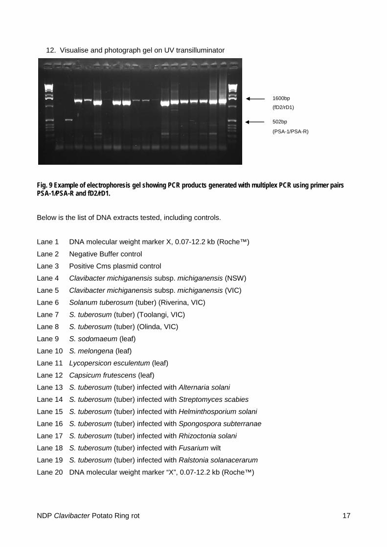
Fig. 9 Example of electrophoresis gel showing PCR products generated with multiplex PCR using primer pairs PSA-1/PSA-R and fD2/rD1.
Below is the list of DNA extracts tested, including controls.
Lane 1 DNA molecular weight marker X, 0.07-12.2 kb (Roche™)
Lane 2 Negative Buffer control
Lane 3 Positive Cms plasmid control
Lane 4 Clavibacter michiganensis subsp. michiganensis (NSW)
Lane 5 Clavibacter michiganensis subsp. michiganensis (VIC)
Lane 6 Solanum tuberosum (tuber) (Riverina, VIC)
Lane 7 S. tuberosum (tuber) (Toolangi, VIC)
Lane 8 S. tuberosum (tuber) (Olinda, VIC)
Lane 9 S. sodomaeum (leaf)
Lane 10 S. melongena (leaf)
Lane 11 Lycopersicon esculentum (leaf)
Lane 12 Capsicum frutescens (leaf)
Lane 13 S. tuberosum (tuber) infected with Alternaria solani
Lane 14 S. tuberosum (tuber) infected with Streptomyces scabies
Lane 15 S. tuberosum (tuber) infected with Helminthosporium solani
Lane 16 S. tuberosum (tuber) infected with Spongospora subterranae
Lane 17 S. tuberosum (tuber) infected with Rhizoctonia solani
Lane 18 S. tuberosum (tuber) infected with Fusarium wilt
Lane 19 S. tuberosum (tuber) infected with Ralstonia solanacerarum
Lane 20 DNA molecular weight marker “X”, 0.07-12.2 kb (Roche™)
1600bp
(fD2/rD1)
502bp
(PSA-1/PSA-R)
NDP Clavibacter Potato Ring rot 17
6 Contact points for further information Dr Dolf De Boer ([email protected])
Dr Jo Luck ([email protected])
Dr. John Elphinstone ([email protected])
7 Acknowledgements Many of the identification procedures in this protocol have been sourced from the EPPO protocol PM 7/59 (EPPO 2006), drafted by D. Stead, Central Science Laboratory, York (GB) and revised by P. Müller, Biologische Bundesanstalt für Land- und Forstwirtschaft, Kleinmachnow (DE).
The protocol was developed from the scholarship report of Dr Jo Luck and Bonny van Rijswijk, Department of Primary Industries Victoria. Additional material was sourced via the PaDIL Plant Biosecurity Toolbox (http://www.padil.gov.au/pbt/), which used the Potato Ring Rot Diagnostic Manual authored by Bonny Rowles-van Rijswijk, Dr Joanne Luck, Ross Mann, Jane Moran and Dr Peter Merriman (Department of Primary Industries, Victoria), as well as the Bacterial Ring Rot of Potatoes Diagnostic Manual authored by Eric Cother, Christine McKenzie (NSW Department of Primary Industries), kindly provided by Plant Health Australia.
Thanks to Dr John Elphinstone, Central Science Laboratories, now the Food and Environment Agency (FERA) Sand Hutton, UK for technical advice in the detection of this organism and to the Department of Agriculture Fisheries and Forestry for funding the travel to complete the scholarship.
8 References ACP (Atlantic Committee on Potatoes) (2003) Bacterial Ring Rot. Atlantic Provinces Agricultural
Services Coordinating Committee. http://www.gnb.ca/0029/00290033-e.asp
Babadoost M (1990) Bacterial Ring Rot of Potato. University of Illinois, Department of Crop Sciences, Extension.
Bishop AL and Slack SA (1987) Effect of inoculum dose and preparation, strain variation, and plant growth conditions on the eggplant assay for bacterial ring rot. American Potato Journal 64, 227-234.
Christie RD, Schulz JT and Gudmestad NC (1993) Potato flea beetle (Coleoptera: Chrysomelidae) evaluated as a possible vector of ring rot bacterium in potatoes. Journal of Economic Entomology 86(4), 1223-1227.
Christie RD, Sumalde AC, Schulz JT and Gudmestad NC (1991) Insect transmission of the bacterial ring rot pathogen. American Potato Journal 68, 363 – 372.
Davis RM, Nunez J and Smart C (1997) UC IPM pest management guidelines: potato. The University of California, USA. http://www.ipm.ucdavis.edu/PMG/r607100211.html
De Boer SH and McCann M (1990) Detection of Corynebacterium sepedonicum in potato cultivars with different propensities to express ring rot symptoms. American Potato Journal 67, 685-694
De Boer SH, Wieczorek A and Kummer A (1988) An ELISA test for bacterial ring rot of potato with a new monoclonal antibody. Plant Disease 72 (10), 874-878.
de la Cruz AR, Wiese MV and Schaad NW (1992) A semi selective agar medium for isolation of Clavibacter michiganensis subsp. sepedonicus from potato tissues. Plant Disease 76(8), 830-834.
NDP Clavibacter Potato Ring rot 18
DEFRA (2002) Potato ring rot. (Department of Environment, Food and Rural Affairs), United Kingdom.http://www.fera.defra.gov.uk/plants/plantHealth/pestsDiseases/potatoRingRot.cfm. Accessed Feb 2008.
EPPO (2006) Diagnostic protocol for Clavibacter michiganensis subsp. sepedonicus. OEPP/EPPO Bulletin 36, 99–109.
Franc GD (1999) Persistence and Latency of Clavibacter michiganensis subsp. sepedonicus in Field-Grown Seed Potatoes. Plant Disease 83, 247-250.
Glick DP, Ark PA and Racicot HN (1944) Outline of procedure for the diagnosis of bacterial ring rot of potatoes – report of the committee of the potato association of America. The American Potato Journal 21, 311-314.
Lansade, M. (1950) Recherches sur le flétrissement bactérien de la pomme de terre en France, Corynebacterium sepedonicum. Annales de l'Institut National de Recherches Agronomiques Series C (Annales des Epiphyties) 1, 69-156.
Lelliott RA and Stead DE (1987) Methods in Plant Pathology Volume 2, Methods for the Diagnosis of Bacterial Diseases of Plants, Chapter 3 Diagnostic procedures; 3.5.7. Bacterial ring rot of potatoes (Clavibacter michiganese subsp. sepedonicum syn. Corynebacterium sepedonicum). Blackwell Scientific Publications. p 97-101.
Manzer FE and Genereux H (1981) Compendium of Potato Diseases, Part I Disease in the Presence of Infectious Pathogens, Bacterial Ring Rot. The American Phytopathological Society. p 9-10.
Nelson GA and Torfason WE (1974) Associative effects of leaf roll and ring rot on disease expression and yield of potatoes. American Potato Journal 51, 12-15.
Pastrik KH (2000) Detection of Clavibacter michiganensis subsp. sepedonicus in potato tubers by multiplex PCR with co amplification of host DNA. European Journal of Plant Pathology 106, 155-165.
Rich AE (1983) Potato Diseases, Chapter 2 Bacterial Diseases, Ring Rot. Academic Press Inc. p 20-25.
Rowe RC, Miller SA and Riedel RM (1995) Bacterial Ring Rot of Potatoes. Extension Fact Sheet, Ohio State University. http://ohioline.osu.edu/hyg-fact/3000/pdf/3103.pdf
Schaad NW, Berther-Schaad Y, Sechler A and Knorr D (1999) Detection of Clavibacter michiganensis subsp. sepedonicus in potato tubers by BIO-PCR and an automated real-time fluorescence detection
Weisburg WG, Barns SM, Pelletier DA and Lane DJ (1991) 16S ribosomal DNA amplification for phylogenetic study. Journal of Bacteriology 173, 697-703.
Related articles
Baer D, Mitzel E, Pasche J and Gudmestad NC (2000) PCR detection of Clavibacter michiganensis subsp. sepedonicus-infected tuber samples in a plate capture assay. American Journal of Potato Research 78, 269-277.
De Boer, SH McNaughton, ME (1986) Evaluation of immunofluorescence with monoclonal antibodies for detecting latent bacterial ring rot infections. American Potato Journal 63, 533-543.
De Boer SH and Slack SA (1984) Current Status and Prospects for Detecting and Controlling Bacterial Ring Rot of Potatoes in North America, Plant Disease, 841-844.
De Boer SH (1987) The relationship between bacterial ringrot and North American seed potato export markets. American Potato Journal 64, 683-693.
NDP Clavibacter Potato Ring rot 19
De Boer SH (1991) Current status and future prospects of bacterial ring rot testing. Proceedings of Pathogen Testing Symposium. American Potato Journal 68, 107-113.
De Boer SH and Hall JW (2000) Proficiency testing in a laboratory accreditation program for the bacterial ring rot pathogen of potato. Plant Disease 84, 649-653.
DEFRA (1998) Ring Rot and Brown Rot of Potato. (Department of Environment, Food and Rural Affairs), United Kingdom.http://www.defra.gov.uk/planth/ring.htm. Accessed Feb 2008.
Easton, G.D. (1979) The biology and epidemiology of potato ring rot. American Potato Journal 56, 459-460.
EPPO (2001) 39th meeting of the working party on phytosanitary regulations (Irkutsk, RU, 2001-06-25/28). http://www.eppo.org/50ans/programme/irkutsk.html
EPPO Secretariat (1999) Potato and tomato diseases – Europe.
Evans I, Yarsh C, Schaupmeyer W, Wolff G and Duplessis P (1998) Understanding bacterial ring rot in potatoes. Alberta Agriculture, Food and Rural Development, Canada. http://www.agric.gov.ab.ca/agdex/200/258_635-5.htm
Holt JG, Krieg NR, Sneath PHA, Staley JT and Williams ST (1994) Bergey's Manual of Determinative Bacteriology (9th ed.). Lippincott Williams and Wilkins, Maryland, USA. p. 575, 583, 591.
Khan MA and Slack SA (1978) Studies on the sensitivity of a latex agglutination test for the serological detection of potato virus S and potato virus X in Wisconsin. The American Potato Journal 55, 627-638.
Lynch, D.R.; Nelson, G.A.; Kulcsar, F. (1989) Elimination of bacterial ring rot (Corynebacterium sepedonicum) by in vitro culture of sprout tissue. Potato Research 32, 341-345.
Manzer, F.E.; Gudmestad, N.C.; Nelson, G.A. (1987) Factors affecting infection, disease development and symptom expression of bacterial ring rot. American Potato Journal 64, 641- 676.
Manzer, F.E.; McKenzie, A.R. (1988) Cultivar response to bacterial ring rot infection in Maine. American Potato Journal 65, 333-339.
Martin J and Beaumanoir N (2001) Comparison of the effectiveness of five extraction methods for Clavibacter michiganensis subsp. sepedonicus and Ralstonia solanacearum from potato tubers. Bulletin OEPP/EPPO Bulletin 31, 153-157.
Mills D, Russell BW and Hanus JW (1997) Specific detection of Clavibacter michiganensis subsp. sepedonicus by amplification of three unique DNA sequences isolated by subtraction hybridisation. Phytopathology 87(8), 853-861.
Mills, D and Russell, BW (2003) Parameters for specific detection of Clavibacter michiganensis subsp. sepedonicus in potato stems and tubers by multiplexed PCR-ELISA American Journal of Potato Research 80: 223-234.
Mosley A, Yilma S and Charlton B (2000) Mission and propagation profile. http://www.css.orst.edu/EXTENSIO/crops/fsps/psf5.htm#MISSION
Muller, H.J.; Ficke, W. (1974) [Bacterial ring rot (Corynebacterium sepedonicum) a dangerous quarantine disease for potato cultivation.] Nachrichtenblatt für den Pflanzenschutz in der DDR 28, 159-160.
Nelson GA, Lynch DR and Kozub GC (1992) Ring rot symptom development on potato cultivars and lines in southern Alberta. Potato Research 35, 133-142.
Plant Research International (2000) Epidemiological studies for control of Clavibacter michiganensis subspecies sepedonicus, the causal agent of bacterial ring rot in potato (ring rot). http://europa.eu.int/comm/research/agro/fair/en/nl4366.html
NDP Clavibacter Potato Ring rot 20
Rousson J (2002) Seed Potato Certification Program - Bacterial Ring Rot testing program for field grown seed potatoes. Canadian Food Inspection Agency. http://www.inspection.gc.ca/english/plaveg/protect/dir/d-97-12e.pdf
Schneider BJ, Zhao J and Orser CS (1993) Detection of Clavibacter michiganensis subsp. sepedonicus by DNA amplification. FEMS Microbiol Lett 109, 207-212.
Secor, G.A.; De Buhr, L.; Gudmestad, N.C. (1987) Chemical sanitation for bacterial ring rot control. American Potato Journal 64, 699-700.
Slack SA, Drennan JL and Westra AAG (1996) Comparison of PCR, ELISA, and DNA hybridisation for the detection of Clavibacter michiganensis subsp. sepedonicus in field-grown potatoes. Plant Disease 80 (5), 519-524.
Slack SA, Sanford HA and Manzer FE (1979) The latex agglutination test as a rapid serological assay for Corynebacterium sepedonicum. American Potato Journal 56, 441-446.
Slack SA and Westra AAG (1998) Evaluation of flusulfamide for the control of bacterial ring rot of potato. American Journal of Potato Research 75 (5), 225-230.
Sorensen KA (1995) Flea beetles on vegetables. Insect note #27 (revised). Department of Entomology, North Carolina State University. http://www.ces.ncsu.edu/depts/ent/notes/Vegetables/veg27.html
Summers CG and Godfrey LD (2001) Sugarbeet flea beetles - UC Pest Management Guidelines. University of California. http://www.ipm.ucdavis.edu/PMG/r735301811.html
Vidaver AK and Davis MJ (1994) Coryneform plant pathogens, In: Schaad N.W. (ed) Laboratory guide for identification of plant pathogenic bacteria 2nd edition. American Phytopathological Society, Minnesota. 104-113.
Resources http://eur-lex.europa.eu/LexUriServ/LexUriServ.do?uri=CONSLEG:1993L0085:20060707:EN:PDF http://www.padil.gov.au/viewPest.aspx?id=587 http://www.fera.defra.gov.uk/plants/plantHealth/pestsDiseases/documents/protocols/clavibacter.pdf http://web.aces.uiuc.edu/vista/pdf_pubs/937.pdf http://ohioline.osu.edu/hyg-fact/3000/3103.html http://library.wur.nl/file/wurpubs/LUWPUBRD_00338085_A502_001.pdf
NDP Clavibacter Potato Ring rot 21
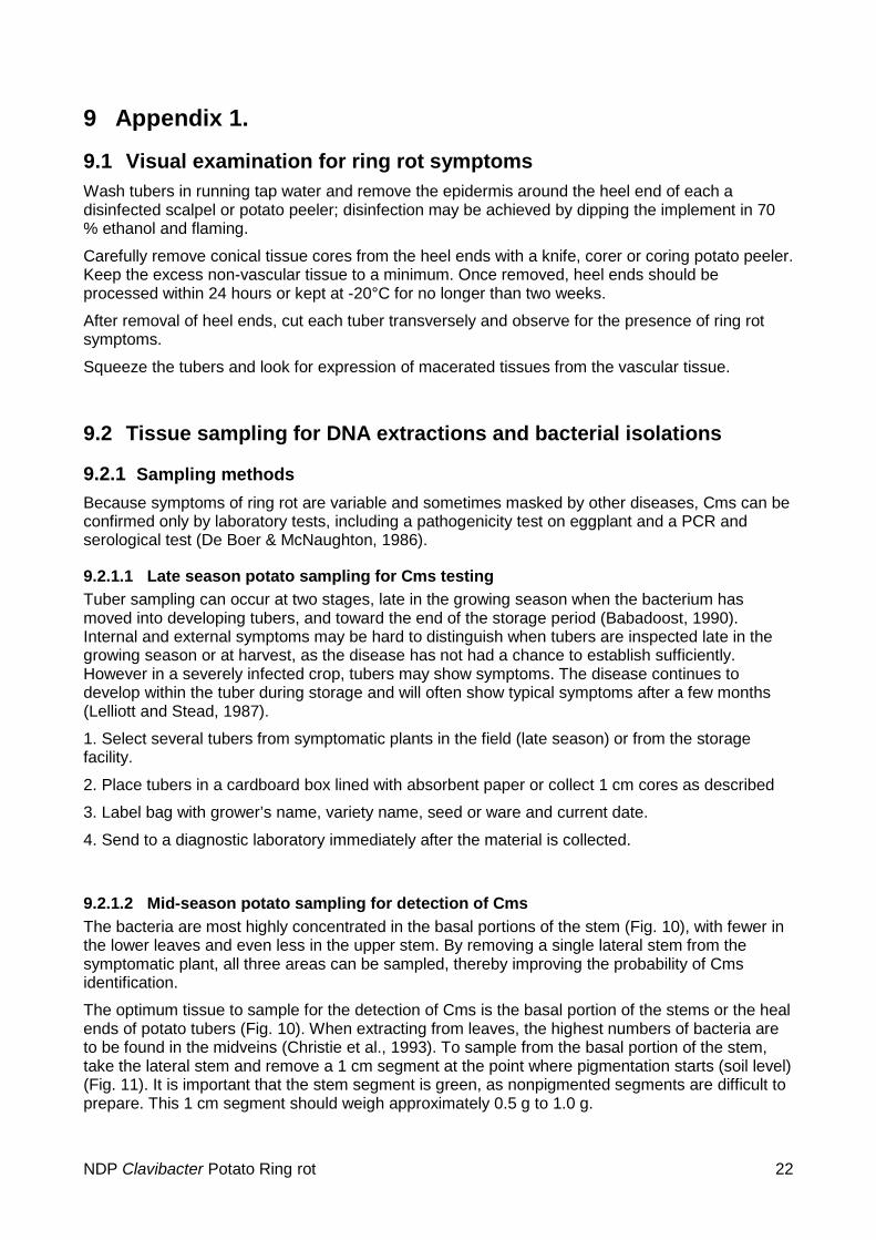
9 Appendix 1.
9.1 Visual examination for ring rot symptoms Wash tubers in running tap water and remove the epidermis around the heel end of each a disinfected scalpel or potato peeler; disinfection may be achieved by dipping the implement in 70 % ethanol and flaming.
Carefully remove conical tissue cores from the heel ends with a knife, corer or coring potato peeler. Keep the excess non-vascular tissue to a minimum. Once removed, heel ends should be processed within 24 hours or kept at -20°C for no longer than two weeks.
After removal of heel ends, cut each tuber transversely and observe for the presence of ring rot symptoms.
Squeeze the tubers and look for expression of macerated tissues from the vascular tissue.
9.2 Tissue sampling for DNA extractions and bacterial isolations
9.2.1 Sampling methods Because symptoms of ring rot are variable and sometimes masked by other diseases, Cms can be confirmed only by laboratory tests, including a pathogenicity test on eggplant and a PCR and serological test (De Boer & McNaughton, 1986).
9.2.1.1 Late season potato sampling for Cms testing Tuber sampling can occur at two stages, late in the growing season when the bacterium has moved into developing tubers, and toward the end of the storage period (Babadoost, 1990). Internal and external symptoms may be hard to distinguish when tubers are inspected late in the growing season or at harvest, as the disease has not had a chance to establish sufficiently. However in a severely infected crop, tubers may show symptoms. The disease continues to develop within the tuber during storage and will often show typical symptoms after a few months (Lelliott and Stead, 1987).
1. Select several tubers from symptomatic plants in the field (late season) or from the storage facility.
2. Place tubers in a cardboard box lined with absorbent paper or collect 1 cm cores as described
3. Label bag with grower’s name, variety name, seed or ware and current date.
4. Send to a diagnostic laboratory immediately after the material is collected.
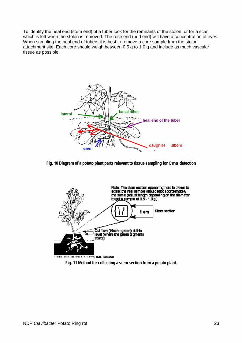
9.2.1.2 Mid-season potato sampling for detection of Cms The bacteria are most highly concentrated in the basal portions of the stem (Fig. 10), with fewer in the lower leaves and even less in the upper stem. By removing a single lateral stem from the symptomatic plant, all three areas can be sampled, thereby improving the probability of Cms identification.
The optimum tissue to sample for the detection of Cms is the basal portion of the stems or the heal ends of potato tubers (Fig. 10). When extracting from leaves, the highest numbers of bacteria are to be found in the midveins (Christie et al., 1993). To sample from the basal portion of the stem, take the lateral stem and remove a 1 cm segment at the point where pigmentation starts (soil level) (Fig. 11). It is important that the stem segment is green, as nonpigmented segments are difficult to prepare. This 1 cm segment should weigh approximately 0.5 g to 1.0 g.
NDP Clavibacter Potato Ring rot 22
To identify the heal end (stem end) of a tuber look for the remnants of the stolon, or for a scar which is left when the stolon is removed. The rose end (bud end) will have a concentration of eyes. When sampling the heal end of tubers it is best to remove a core sample from the stolon attachment site. Each core should weigh between 0.5 g to 1.0 g and include as much vascular tissue as possible.
Fig. 10 Diagram of a potato plant parts relevant to tissue sampling for Cms detection
Fig. 11 Method for collecting a stem section from a potato plant.
NDP Clavibacter Potato Ring rot 23

Fig. 12 Sampling a 1cm core from a potato tuber.
NDP Clavibacter Potato Ring rot 24