nanoparticles from biowastes and microbes: focus on · pdf filenanoparticles from biowastes...
TRANSCRIPT

1
Nanoparticles from biowastes and microbes: Focus on role in water purification
and food preservation
Samir Mahgoub1,2 and Petros Samaras2
1Microbiology Department, Faculty of Agriculture, Zagazig University, Zagazig 44511, Egypt 2Laboratory of Water and Wastewater Technology, Department of Food Technology, School of Food Technology and Nutrition, Technological Educational Institute of Thessaloniki, P.O.Box 141,57400 Sindos, Thessaloniki, Greece
*Corresponding author: E-mail: [email protected], Tel/Fax: +20552287567

2
Abstract
Biowasts are eco-friendly to produce typical nanoparticles with well-defined chemical
composition, size, and morphology. This paper highlights the recent developments of
the produce nanoparticles from biowastes e.g. eggs and shrimp peels etc…. Bio-
processing of nanoparticles from such resources with microbes such as bacteria, fungi
and yeasts are being increasingly explored to meet the twin objectives of resource
recycling and pollution mitigation. This review focuses on all the available information
on the preparation methodologies of bio-nanoparticles, and highlighted the inherent
advantages and disadvantages involved in each method. The second objective was to
appear how microorganisms could be used for bio-extracting bionanoparticles.
Recently, increasing public concern about microbial pathogens has been driving many
investigations for anti-microbial modification of food and water. This review is also
concerned with the application of nanoparticles in water and food stuff areas for
enhanced filtrating of water contaminated by microbial pathogenic and industrial
activities of food systems.
Highlights
Review focuses on extraction of nanobarticles from biowastes.
Bio-processing of certain nanoparticles from peel eggs and shrimps is explained.

3
Morphology and characterization of nanoparticles from biowastes is highlighted
for recovering eco-friendly material.
Bio-recovery of nanoparticles using bacteria, fungi and yeasts is also included
with briefly.
Methodology/mechanism of nanoparticles in water purification and against
foodborne pathogens is discussed.
Keywords: Biowastes; Nanoparticles; Microbes; Bio-extraction; Water, Food
Introduction
Biowaste refers to any organic waste such as cow dung ash, corn cob ash, mango stone
ash, lemon peel, pomegranates peel, orange peel, eggs and shrimp peel as well as the
organic fraction of municipal solid wastes and animal manure. Biowaste is often
interpreted as being the organic biodegradable fraction of the municipal waste stream,
including garden waste, food waste and other biodegradable materials such as paper
cardboard, some textiles and wood. The rapid expansion of poultry production has
caused an increased production of poultry biowaste, i.e. chicken manure (Axtell 1999).
Improper use of chicken manure can result in pollution of air, soil and groundwater
(Yetilmezsoy &Sakar 2008). Food waste is a growing problem in many parts of the
world, but discarded fruit peel, in the case of pomegranates, lemon could be put to good
use in the burgeoning field of nanotechnology according to (Ahmad & Sharma 2012,
4
Nisha et al. 2014). Environmental concern about the management of these materials is
increasing at the same time that legislation is becoming more restrictive.
Plants, garden waste, remnants of food industries, fruit seeds, carbohydrates, bacteria,
actinomycetes, fungi, yeasts, and viruses have used for the biosynthesis of gold, silver,
gold–silver alloy, selenium, tellurium, platinum, palladium, silica, titania, zirconia,
quantum dots, magnetite and uraninite nanoparticles (Narayanan & Sakthivel 2010).
Microbial route for the synthesis of nanoparticles has one the most exciting process as
several factors such as microbial cultivation methods on organic and/or inorganic
wastes and the extraction techniques can be optimized for the fast synthesis of
monodisperse nanoparticles. It has been, therefore, of increasing interest to develop
efficient green synthesis of gold nanoparticles. In recent years, there is lot of interest
shown in the environmentally benign synthesis of nanoparticles that do not use any
toxic chemicals or extreme conditions in the synthesis process. Many biological systems
such as that of fungi (Jain et al 2011, Mohammadian et al. 2007), algae (Singaravelu et
al. 2007), bacteria (Reddy et al. 2010), actinomycetes (Otari et al. 2014), and plants
(Mohanpuria et al. 2008, Choi, et al. 2011, Gan et al., 2012, Vijayakumara et al. 2013)
have been studied for biosynthesis of gold and silver nanoparticles.
Production of metal nanoparticles (NPs) by physical and chemical methods are too
harsh and non-ecofriendly. Also such nano metals have limited shelf life. Metallic
nanoparticles are traditionally synthesized by wet chemical techniques, where the

5
chemicals used are quite often toxic and flammable (Nisha et al. 2014). In addition most
of these physical and chemical methods need extreme conditions like temperature,
pressure etc. The most widespread and common synthesis of gold nanoparticles is the
chemical reduction of an ionic gold in aqueous phase by a chemical reducing agent such
as NaBH4, citrate, and ascorbate. But such reducing agents may be associated with
environmental toxicity or biological hazards. Thus, nanobiotechnology is the branch of
biotechnology which deals with synthesis and fabrication of nanoparticles by biological
systems and their applications especially in biological systems. Nanoparticles possess at
least one dimension in the size range of 1 to 100 nm. A nanometer is one billionth (10-9)
of a meter, roughly the width of three or four atoms. The principal properties of
nanoparticles include size, shape and sub-surface of the substance. Nanoparticles can be
classified as organic (for e.g. carbon nanoparticles) or inorganic (for e.g. magnetic and
noble metal nanoparticles). This recent concept has proved to be a promising
technology enabling the study of immensely efficient biological systems at the
molecular level. The field of nanotechnology has gained attention in the recent past
owing to a broad spectrum of applications in diagnostics, therapeutics, medicine,
delivery system, agriculture, consumer goods, and cosmetics. Biological approaches
using microorganisms and plant extracts for synthesis of metal nanoparticles have been
suggested as valuable alternatives to traditional methods (Rassaei et al. 2008, Krumov
et al. 2009).

6
Biological methods for synthesis of nanoparticles and their role in biotransformation
process on formation of different bio-products, such as bioethanol, biohydrogen,
biodiesel, enzymes and bioplastics is reported by Mohapatra, et al. (2011) because there
is an increasing commercial demand for bio-nanoparticles due to their wide
applicability in various areas and the other bio-products. The nanoparticles are going to
prove revolutionary in the field of biotransformation by improving the efficiency and
yield and often widening the application range (Mohapatra et al. 2011). Additionally,
the possibility to recover H2 from waste organic streams in biorefineries using
photocatalytic approaches is an attractive option to enhance process sustainability and
produce valuable energy products. With respect to the overall photoreforming to obtain
H2 and CO2, the photo-dehydrogenation of bioethanol leads to the co-production of a
valuable chemical (acetaldehyde) together with H2 (Ampelli et al. 2013).
The metal nanoparticles obtained after bioleaching of waste can find a range of
applications especially in the field of medicine, food and water. These applications
include drug delivery, gene therapy, antimicrobials, medical prosthetics and tissue
engineering. Thus, it is possible to ‘marry’ the two diverse fields as ‘waste to nano’ for
biomedical sciences, thereby providing an active area of research in nanotechnology
(Majumder 2013).
Biological and chemical contaminants present in water have threatened its drinking
quality. Water treatment for drinking purposes is gaining attraction in the developing

7
world due to increasing trend in water borne infectious diseases. These diseases are
caused by microorganisms such as bacteria, viruses, protozoan etc. Nanotechnology is
applying to disinfect these infectious microorganisms present in water because these
microorganisms can multiply very rapidly in the body causing health and environmental
concerns. The utilization of nanoparticles to remove these biological infectious
microorganisms from water and food systems is an efficient and promising technique
used to improve the water drinking quality. This review focuses on 1) extraction of
nanobarticles from biowastes and microorganisms 2) recovering eco-friendly material
and 3) role of nanoparticles in water purification and against foodborne in food systems.
Nanoparticles from biowastes
Green chemistry started for the search of benign methods for the development
nanoparticles and searching antibacterial, antioxidant, and antitumor activity of natural
products. Biosynthetic processes of nanoparticles have received much attention as a
viable alternative for the development of metal nanoparticles where by products of
factories and plant extract is used for the synthesis of nanoparticles without any
chemical ingredients (Shankar et al. 2003, Narayanan & Sakthivel 2008, Vijayakumara
et al. 2013). Recently weeds, popularly known as ‘enemies of the farmer’, have also
been used successfully for the synthesis of gold, copper and silver nanoparticles.
Silver nanoparticles (AgNPs)

8
The use of environmentally benign and renewable plant material offers enormous
benefits of eco-friendliness biosynthesis of silver nanoparticles (AgNPs) using lemon or
pomegranate peel extract as the reducing agent. The biosynthesis of AgNPs using lemon
peel extract is very simple and economic. The extract of lemon peel was prepared and
mixed with 1mM AgNO3 solution for the effective synthesize of AgNPs. The
bioreduction of Ag+ ion in solution was monitored using UV-visible spectrometer,
Fourier-Transform IR spectroscopy (FTIRS) and X-ray diffraction (EDAX) analysis.
The AgNPs produced from lemon peels showed good activity against the isolated
dermatophytes. The use of lemon peels for the effective synthesize of AgNPs are found
to develop drug resistant towards broad-spectrum antibiotics (Nisha, et al. 2014).
Pomegranate peel extract was challenged with AgNO3 solution for the production of
AgNPs. The reaction process was simple for the formation of highly stable silver
nanoparticles at room temperature by using the biowaste of the fruit. The morphology
and crystalline phase of the NPs was determined from UV-Vis spectroscopy,
transmission electron microscopy (TEM), selected area electron diffraction (SAED), X-
ray diffraction (XRD) spectra and FTIRS. TEM studies showed that the silver
nanoparticles obtained were of sizes 5 ± 1.5 nm. Presumably biosynthetic products or
reduced cofactors play an important role in the reduction of respective salts to
nanoparticles (Ahmad & Sharma 2012). Additionally, preparation of chitosan
nanoparticles include emulsion cross-linking, emulsion-droplet coalescence,
9
coacervation/precipitation, ionotropic gelation, reverse micelles, template
polymerization, and molecular self-assembly. All these methods have their own
advantages as well as drawbacks, in relation to the properties of the nanoparticles.
However, careful preparation of chitosan nanoparticles could provide a higher affinity
for negatively charged biological membranes and site-specific targeting in vivo,
enabling their application as encapsulating materials of drugs, enzymes, and DNA, used
in controlled release systems and as coatings of wound dressings to accelerate healing
(Perera & Rajapakse 2014). Bovine femur bone hydroxyapatite (HA) containing silver
nanoparticles was synthesized by thermal decomposition method and subsequent
reduction of silver nitrate with N, N-dimethylformamide (DMF) in the presence of poly
(vinylacetate) (PVAc). The structural, morphological, and chemical properties of the
HA–Ag nanoparticles were characterized using X-ray diffraction (XRD), scanning
electron microscopy (SEM), field-emission scanning electron microscopy (FE-SEM),
transmission electron microscopy (TEM), and X-ray photoelectron spectroscopy (XPS).
TEM images showed that the Ag nanoparticles with size ranging from 8 to 20 nm and
were arranged at the periphery of HA crystals (Fig. 1). Bactericidal activity of HA–Ag
with different concentration of Ag nanoparticles immobilized on the surface of HA was
investigated against Gram-positive Staphylococcus aureus (non-MRSA), Methicillin
resistant S. aureus (MRSA) and Gram-negative Escherichia coli by the disc diffusion
susceptibility test. The HA–Ag nanoparticles showed that broad spectrum activity

10
against non-MRSA, MRSA, and E. coli bacterial strains (Nirmala et al. 2011). Natural
HA bioceramics has been extracted by normal calcination of biowastes (Barakat et al.,
2009) and extraction of HA from biowaste is economically and environmentally
preferable, compared to the other procedures (Chen et al. 2008). The utilization of the
reductive potency of a common byproduct of food processing industry i.e. orange peel
is reported here to prepare biopolymertemplated “green” silver nanoparticles. Aqueous
extract of orange peel at basic pH was exploited to prepare starch supported
nanoparticles under ambient conditions. The compositional abundance of pectins,
flavonoids, ascorbic acid, sugars, carotenoids and myriad other flavones may be
envisaged for the effective reductive potential of orange peel to generate silver
nanoparticles. The nanoparticles were distributed within a narrow size spectrum of (312
nm) with characteristic Bragg’s reflection planes offcc structure, and surface plasmon
resonance peak at 404 nm. Anti-lipid peroxidation assay using goat liver homogenate
and free radical scavenging “green” silver nanoparticles test established the anti-oxidant
potency. Their synergy with rifampicin against Bacillus subtilis MTCC 736 and
cytocompatibility with the human leukemic monocytic cell line, THP-1 were also
investigated (Konwarh et al. 2011). Biosynthesis of silver AgNPs from seaweed extracts
is currently under exploitation. Seaweed extracts are cost effective and eco-friendly and
thus can be an economic and efficient alternative for large-scale synthesis of
nanoparticles. The synthesis of AgNPs from silver precursor, silver nitrate using
11
aqueous extract of seaweed Gracilaria corticata. The organic compounds present in the
filtrate of G. corticata were mainly responsible for reduction of silver ions to AgNPs.
The filtrate when added to 1 mM aqueous silver nitrate solution at 121°C changed to
dark brown colour solution within ten minutes, which confirms the bioreduction. These
extremely stable AgNPs were characterised by UV-Vis spectrophotometer, FTIR, XRD,
TEM, and EDAX analysis. The nanoparticles exhibited maximum absorbance at 424
nm in the UV spectrum. The presence of proteins was identified by FTIR. TEM
micrograph revealed the formation of polydispersed and spherical shaped nanoparticles
with the size range of 10-50 nm and the presence of elemental silver were confirmed by
EDAX analysis. These nanoparticles showed cytotoxic activity against Hep2 cells (Devi
and Bhimba, 2013). Lantana camara, a weed commonly found in Maharashtra was also
screened for leaching copper. The characteristics of the copper nanoparticles obtained
were studied using X-ray diffraction analysis, energy-dispersive spectroscopy, scanning
electron microscopy, Fourier Tranform Infrared analysis, Transmission electron
microscopy, Thermogravimetric analysis and Cyclic Voltammetry. Copper
nanoparticles were found to be effective against hospital strain Escherichia coli 2065
(Majumder 2012). Green synthesis of nanoparticles from other several plants and their
used in biological application has been reported by several authors (Mohanpuria et al.
2008, Choi et al. 2011, Gan et al. 2012, Vijayakumara et al. 2013).
Gold nanoparticles (AuNPs)

12
Peel extract of pomegranate was challenged with chloroauric acid (HAuCl4) solution for
the production of gold nanoparticles (AuNPs). The reaction process was simple for the
formation of highly stable gold nanoparticles at room temperature by using the biowaste
of the fruit. The morphology and crystalline phase of the NPs were determined from
UV-Vis spectroscopy, TEM, SAED and XRD spectra. TEM studies showed that the
average gold nanoparticles were found to be 10 ± 1.5 nm (Ahmad et al. 2012). A facile
green biosynthesis method has been successfully developed to prepare gold
nanoparticles (AuNPs) of various core sizes (25+/-7 nm) using a natural biomaterial,
eggshell membrane (ESM) at ambient conditions. In situ synthesis of AuNPs-
immobilized ESM is conducted in a simple manner by immersing ESM in a pH 6.0
aqueous solution of HAuCl4 without adding any reductant. The formation of AuNPs on
ESM protein fibers is attributed to the reduction of Au(III) ions to Au(0) by the
aldehyde moieties of the natural ESM fibers. Energy dispersive X-ray spectroscopy,
scanning electron microscopy, X-ray photoelectron spectroscopy, and X-ray powder
diffraction unambiguously identify the presence of AuNPs on ESM. This works by
Zheng et al. (2010) a very simple, non-toxic, convenient, and green route to synthesize
AuNPs on ESM which is potentially useful in the biosensing field. The synthesis of
AuNps from gold precursor using palm oil without adding external surfactant, capping
agent or template has been studied by Gan et al. (2012). Silica powder at nanoscale was
obtained by heat treatment of Vietnamese rice husk following the sol–gel method. The

13
rice husk ash (RHA) is synthesized using rice husk which was thermally treated at
optimal condition at 600°C for 4 h. The silica from RHA was extracted using sodium
hydroxide solution to produce a sodium silicate solution and then precipitated by adding
H2SO4 at pH = 4 in the mixture of water/butanol with cationic presence. In order to
identify the optimal condition for producing the homogenous silica nanoparticles, the
effects of surfactant surface coverage, aging temperature, and aging time were
investigated. By analysis of X-ray diffraction, scanning electron microscopy, and
transmission electron microscopy, the silica product obtained was amorphous and the
uniformity of the nanosized sample was observed at an average size of 3 nm, and the
BET result showed that the highest specific surface of the sample was about 340 m2/g.
The results obtained in the mentioned method prove that the rice husk from agricultural
wastes can be used for the production of silica nanoparticles (Le, et al., 2013). Large
amount of silica recovered from waste rice husk silica, hence the silica materials derived
from waste product can be the low cost and used for potential application such as low-k
dielectric material development. Gold doping on ceria-mixed silica derived from rice
husk is carried out by in situ method (Au/Ce-silica-A) and deposition-co precipitation
method (Au/Ce-silica-B). Au/Ce-silica-A and Au/Ce-silica-B shown the low-k
dielectric constant compared to bulk ceria-silica (Shanmugan et al. 2013).
Nanocomposite fillers

14
Biowastes of rice husk and wheat straw are value-engineered to carbonaceous structures
in a single-step process under ambient conditions. By controlling the laser fluence,
structures with a variety of different morphologiesfrom nanostructures to
microstructures lead to influences the chemical composition of the synthesized
structures. This sustainable approach presents an important step towards synthesizing 3-
D micro/nanofibrous compounds from biowaste materials. These structures, as-
synthesized or as nanocomposite fillers, can have practical uses in electronic, sensing,
biological, and environmental applications (Tavangar et al. 2013). Other study
presented a laser-based approach to synthesize carbonaceous micro/nanofibrous
structures from rice husks and wheat straws. This research is the first time that
synthesizing 3-D micro/ nanofibrous structures generated from rice husks and wheat
straws using femtosecond laser. The morphological analyses by SEM confirmed that
fabricated structures were composed of approximately uniform 3-D structure at micro
and nano sizes. Also, by altering the laser pulse energy and the number of laser pulses,
different structures from micro- to nanoarchitectures with different porosities and
features could be achieved. The EDAX analyses confirmed that laser irradiation
affected the chemical composition as well; part of the organic matter is believed to be
burned away owing to the laser irradiation. This approach suggests a promising step
towards engineering green 3-D platforms from sustainable materials. The as-engineered
carbonaceous materials would have very broad practical applications in a variety of

15
areas, such as environmental, catalytic, electronic, sensing, and biological applications.
They can also be utilized to form biodegradable nanocomposites with other materials,
e.g., polymers (Tavangar et al. 2013).
Nanoparticles from microorganisms
The use of natural eco-friendly sources such as microorganisms for the production of
nanomaterials is a promising approach, owing to the feasibility and cost-effectiveness of
the process. Bacteria, actinomycetes and fungi have been known for ages for their
potential to leach out metals from their surrounding over the last couple of decades. The
biological agents in the form of algae and microbes have emerged as an efficient
candidate for the synthesis of nanoparticles. These biogenic nanoparticles are cost
efficient, simpler to synthesize, and focus toward a greener approach. But the exact
mechanism of synthesis of biogenic nanoparticles needs to be worked out (Méndez-
Vilas 2011). Synthesis of nanoparticles may be intracellular or extracellular of which
the later is more preferred as it makes the downstream processing less laborious and
also is effective in cost-cutting of the entire process during industrial applications. The
biosynthesis of AgNPs was studied from bacteria such as Bacillus subtilis (Reddy et al.
2010), actinomycetes i.e. Rhodococcus sp. (Otari et al. 2014), fungi e. g. Aspergillus
flavus, Fusarium oxysporum (Jain et al 2011, Mohammadian et al. 2007) and from
marine alga, Sargassum wightii Greville for gold nanoparticles (Singaravelu et al.
2007). Howevere, the intracellular accumulation of gold nanoparticles with a dimension

16
of 5–15 nm by an alkalotolerant actinomycete, Rhodococcus sp., has been demonstrated
by Ahmad et al. (2003). Fusarium oxysporum and Pseudomonas sp. were able to leach
copper (84-130 nm) from integrated circuits present on electronic boards (e-waste)
under ambient conditions. The characteristics of the copper nanoparticles obtained were
studied using X-ray diffraction analysis, energy-dispersive spectroscopy, scanning
electron microscopy, Fourier Tranform Infrared analysis, Transmission electron
microscopy, Thermogravimetric analysis and Cyclic Voltammetry. Copper
nanoparticles were found to be effective against hospital strain Escherichia coli 2065
(Majumder 2012).
Although the market of nanoparticles (NPs) is rapidly expanding, the environmental and
health impact of manufactured NPs and nanomaterials is still poorly understood and
predictable. The ecological impact of metallic and metal oxide NPs span from the
surface atoms of NPs, to unicellular organisms, such as bacteria, and organism levels
have been studied by Thié et al. (2012). On multicellular organisms, the authors
focused on biomarkers reporting on stress, central nervous system endpoints and
antioxidative balance assessment. On bacteria, which are key players in NPs transfer,
they study not only the impact of NPs on cells, at the microbial community, cell and
molecular level, but also the effect of cells on NPs ( Thié et al. 2012). A novel approach
for the green synthesis of silver nanoparticles (AgNPs) from aqueous solution of
AgNO3 using culture supernatant of phenol degraded broth has been reported by Otari et

17
al. (2014). The synthesis by phenol degraded Rhodococcus sp. was observed within 10
h, and AgNPs showed characteristic surface plasmon resonance around 410 nm.
Spherical nanoparticles of size less than 30 nm were observed in transmission electron
microscopy. X-ray diffraction pattern corresponding to111, 200, 220, and 311 revealed
the crystalline nature of the as-formed nanoparticles (Fig. 2). It was found that the
colloidal solution of AgNP suspensions exhibited excellent stability over a wide range
of ionic strength, pH, and temperature. The effect of pH and ionic strength indicated
that stabilization is due to electrostatic repulsion arising from the negative charge of the
conjugate proteins. The AgNPs showed highly potent antimicrobial activity against
Gram-positive, Gram-negative, and fungal microorganisms. The as-prepared AgNPs
showed excellent catalytic activity in reduction of 4-nitrophenol to 4-aminophenol by
NaBH4. By manufacturing magnetic alginate beads, the reusability of the AgNPs for the
catalytic reaction has been demonstrated.
The active and inactive cells/biomass (AB and IB) and their corresponding cell-free
extracts (ACE and ICE) of Aspergillus oryzae var. viridis were found to be suitable
agents for the synthesis of gold nanoparticles from gold chloride solution (Binupriya et
al. 2010) and showed that A. oryzae var. viridis is a suitable candidate for the synthesis
of gold nanoparticles. The formation of gold nanoparticles (Au NPs) was visually
confirmed by the change in the color of reaction medium from colorless to purple. The
Au NPs synthesized were monitored via UV–vis spectrophotometer and characterized.

18
The SPR (Surface Plasmon Resonance) showed the formation of Au NPs with respect to
time, pH and initial biomaterial concentration. The peak area of UV–vis spectrum
showed that the IB and ICE were found to synthesize more nanoparticles compared to
their counterparts, which is believed to be due to presence of more organics in the
autoclaved cells due to cell-rupture. High organic content in ICE was confirmed through
TOC analysis. The TEM images of formed Au NPs showed that the particles were
aggregated and entrapped insomeregions possibly an organic matrix of fungal origin.
The TEM micrographs of gold nanoparticles nanoparticles formed in cell-free extracts
showed polydiversity in shape and size (Fig. 3). The particles formed were of different
sizes as well as shapes. Particles of very small size of 10 nm as well of large triangular
nanoplates of nearly 400nm were formed in ACE. High pH favored more number of
spherical particles, whereas acidic pH did not favor the synthesis of Au NPs. The
presence of zero valent gold nanoparticles was confirmed by EDX and XRD
measurements. Other interesting result was the formation of high amount of Au NPs in
autoclaved cells and extracts which indicate the role of organics other than enzymes in
the reduction reaction. The autoclaved fungal-mediated green chemistry approach
towards the synthesis of nanoparticles has many advantages such as ease with which the
process can be scaled up, economic viability, simple downstream processing and easy
handling of biomass. Compared to bacterial fermentations, in which the process
technology involves the use of sophisticated equipment for getting clear filtrates from

19
the colloidal broths, fungal broths can be easily filtered by filter press or similar simple
equipment, thus saving considerable investment costs for equipment. The capacity of
fungi to produce high amounts of biomass than bacteria make them preferred candidates
for nanoparticle synthesis studies.
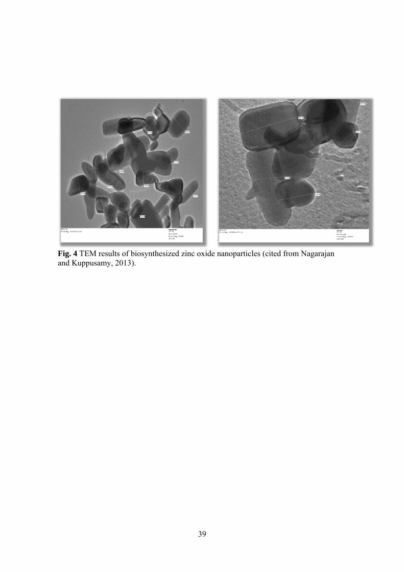
Seaweeds constitute one of the commercially important marine living renewable
resources. Seaweeds such as green Caulerpa peltata, red Hypnea Valencia and brown
Sargassum myriocystum were used for synthesis of Zinc oxide nanoparticles. Seaweed
S. myriocystum were able to synthesize zinc oxide nanoparticles. It was confirmed
through the, initial colour change of the reaction mixture and UV visible
spectrophotometer. The extracellular biosynthesized clear zinc oxide nanoparticles size
36 nm (Fig. 4) through characterization technique such as DLS, AFM, SEM –EDX,
TEM, XRD and FTIR. The biosynthesized ZnO nanoparticles are effective antibacterial
agents against Gram-positive than the Gram-negative bacteria. Based on the FTIR
results, fucoidan water soluble pigments present in S. myriocystum leaf extract is
responsible for reduction and stabilization of zinc oxide nanoparticles (Nagarajan &
Kuppusamy 2013).
Application of nanoparticles in food systems
Among various metals, silver has been known since ancient times as an effective
antimicrobial agent for the treatment of diseases and food preservation (Jain et al.
2008). Silver nano materials exhibit broad spectrum biocidal activity toward bacteria,

20
fungi, viruses, and algae. This motivates its use in food applications. However, there is
mounting evidence that silver nanoparticles exhibit an array of cytotoxic and genotoxic
effects in higher organisms. This raises concern about possible impacts to higher
organisms including humans. Although significant progress has been made to elucidate
the mechanisms of silver nano material toxicity, further research is required to fully
understand the processes involved and to safely exploit the tremendous antimicrobial
properties of silver without jeopardizing human health, critical infrastructure, and the
environment. Future in vivo, in vitro and environmental studies should consider more
systematically the various effects of aquatic chemistry on nano-scaled silver fate,
transport, and toxicity (Méndez-Vilas 2011). Although, there is a need to develop and
implement more economical delivery approaches for multiple-hurdle antimicrobial
interventions that can be applied to food matrices such as retail meats. Recently,
potential opportunities have emerged to use nanoscience and nanoengineering principles
to develop antimicrobial carriers for controlling the major foodborne pathogens such as
Salmonella in meat and food preservation systems (Ricke & Hanning 2013). Several
studies explore the potential of nanoparticle-based composite systems for practical and
economical antimicrobial interventions to inhibit and decontaminate such pathogens on
cooked ready-to-eat (RTE) poultry and red-meat products. The opportunities for
specific systems such as chitosan-nanoparticle-based nanocomposite systems containing
ɛ-polylysine peptide dispersed in organic acids and the potential health hazards that

21
arise from the use of nanoparticles (Ricke & Hanning 2013). The methods of
preparation of chitosan nanoparticles are significantly responsible for their bioactivities
and behavioral characteristics in different systems and applications. Chitosan
nanoparticles-based films are used in the food industry to control microorganisms in
food and to enhance shelf life while strengthening the mechanical properties and
stability of the food-packing materials. Although the chitosan nanoparticles appear to be
safe in some of their applications, knowledge on the risks imposed in these food and
pharmaceutical applications needs to be strengthened further (Perera & Rajapakse
2014).
Application of nanoparticles in water systems
In the area of water purification, nanotechnology offers the possibility of an efficient
removal of pollutants and germs. Today nanoparticles, nanomembrane and nanopowder
used for detection and removal of chemical and biological substances include metals
(e.g. cadmium, copper, lead, mercury, nickel, and zinc), nutrients (e.g. phosphate,
ammonia, nitrate and nitrite), cyanide, organics, algae (e.g. cyanobacterial toxins),
viruses, bacteria, parasites and antibiotics. If the amount of nano-scaled silver entering
sewage becomes higher than the tolerable levels for microbial communities in
wastewater treatment plants, critical environmental infrastructure might be impacted
(Méndez-Vilas 2011). Silver nanoparticles have been known as an effective
antimicrobial agent and water purification (Jain et al. 2008). The comparison of some

22
nanoparticles, such as metallic and non- metallic oxide nanoparticles, especially
Titanium Dioxide, Nanoscale Zero-Valent Iron and Carbon Nanotubes to remove
organic impurities from the water has been studied by Ghauri et al. (2012). Biogenic Pd
nanoparticles produced by Desulfovibrio vulgaris have been reported to efficiently
catalyze the reduction of chromate (Mabbett et al. 2002), the degradation of perchlorate
and nitrate by NPs produced by Shewanella oneidensis (De Windt et al. 2006), and the
dehalogenation of chlorinated aromatic compounds by NPs produced by sulphate-
reducing bacteria (Baxter-Plant et al. 2003), trichloroethylene (Hennebel et al. 2009a),
trichloroethylene ( Hennebel et al. 2009b) and chlorophenols (Baxter-Plant et al. 2003).
So, a new biological inspired method to produce nanopalladium is the precipitation of
Pd on a bacterium, i.e., bio-Pd. This bio-Pd can be applied as catalyst in dehalogenation
reactions. However, large amounts of hydrogen are required as electron donor in these
reactions resulting in considerable costs (Boon & Verstraete 2011). This research
demonstrates how bacteria, cultivated under fermentative conditions, can be used to
reductively precipitate bio-Pd catalysts and generate the electron donor hydrogen. In
these reduction and dehalogenation reactions, the Pd nanoparticles were activated by
adding an external electron donor such as hydrogen or formate. Hydrogen was
consistently identified as the most reactive among possible electron donors, but its use
was found to be prohibitively expensive and presented significant safety and supply
issues (Hennebel et al. 2009b).

23
Several reactor types were constructed to limit the hydrogen supply (Hennebel et al.
2010) or to produce hydrogen in a sustainable way (Yong et al. 2003, Hennebel et al.
2011). Humphries et al. (2007) used hydrogen produced by E. coli during the
fermentation of sugar waste streams. In another recent study, Pd(0) nanoparticle
formation by C. pasteurianum BC1 was coupled with the microbial generation of
hydrogen to reductively immobilize aqueous chromate using biohydrogen as the
electron donor (Chidambaram et al. 2010). In this way, one could avoid the costs
coupled to hydrogen supply. The catalytic activities of Pd(0) nanoparticles produced by
different strains of bacteria (bio-Pd) cultivated under fermentative conditions were
compared in terms of their ability to dehalogenate the recalcitrant aqueous pollutants
diatrizoate and trichloroethylene. While all of the fermentative bio-Pd preparations
followed first order kinetics in the dehalogenation of diatrizoate, the catalytic activity
differed systematically according to hydrogen production and starting Pd(II)
concentration in solution. Batch reactors with nanoparticles formed by Citrobacter
braakii showed the highest diatrizoate dehalogenation activity with first order constants
of 0.45±0.02 h−1 and 5.58±0.6 h−1 in batches with initial concentrations of 10 and 50 mg
L−1 Pd, respectively. Nanoparticles on C. braakii, used in a membrane bioreactor
treating influent containing 20 mg L−1 diatrizoate, were capable of dehalogenating 22
mg diatrizoate mg−1 Pd over a period of 19 days before bio-Pd catalytic activity was
exhausted. This study demonstrate the possibility to use the combination of Pd(II),
24
acarbon source and bacteria under fermentative conditions for the abatement of
environmental halogenated contaminants (Hennebel et al. 2011).
Tiwari et al. (2008) evaluated four classes of nanoscale materials that are being applied
as functional materials for water purification e.g. metal-containing nanoparticles,
carbonaceous nanomaterials, zeolites and dendrimers. Carbon nanotubes and nanofibers
also show some positive result. Nanomaterials reveal good result than other techniques
used in water treatment because of its high surface area (surface/volume ratio). It is
suggested that these may be used in future at large scale water purification. It is also
found that the coliform bacteria treated with ultrasonic irradiation for short time period
before Ag-nanoparticle treatment at low concentration, enhanced antibacterial effect. In
future, combination of both may be the best option for treatment of wastewater. The
CoxCu1-xTiO3 nanoparticles were used for removal of congo red (CR) from aqueous
solutions. The effect of parameters such as contact time, pH and temperature on the
adsorption of CR was investigated. Tow common kinetic models, pseudo-first-order and
pseudo-second-order were employed to describe the adsorption kinetics. The adsorption
of CR followed pseudo-first-order kinetic model. Thermodynamic results revealed that
the adsorption of CR onto CoxCu1-xTiO3 particles is endothermic and spontaneously
process. The nanoparticles of CoxCu1-xTiO3 can be conveniently regenerated by
chemical and physical methods after adsorption. Two common isotherm models, the
Langmuir and Freundlich were used to investigate the interaction of CR onto CoxCu1-

25
xTiO3 nanoparticles. The equilibrium adsorption of CuTiO3 was best described by the
Langmuir isotherm model. The reused sorbent can be employ after 5cycle. The results
showed that the adsorption capacity of Ilmenite type nanoparticles for CR is as
followed: CoTiO3<Co0.2Cu0.8TiO3< Co0.5Cu0.5TiO3< Co0.8 Cu0.2TiO3<CuTiO3
(Hashemian& Foroghimoqhadam 2014). Magnetic MnFe2O4/chitosan nanocomposites
(MCNCs) were prepared by a simple method. The as-prepared sample was
characterized by various technologies. The MnFe2O4 nanoparticles were coated by
chitosan without phase change, and the as-prepared MCNCs displayed
superparamagnetic properties with a high saturation magnetization (39.5 emu g-1). The
as-prepared MCNCs were used to remove Cr(VI) from low concentration solutions
(0.6-1.0 mg L-1). And it was found that the MCNCs showed a high Cr(VI) removal
capacity of 35.2 mg g-1. The effects of various parameters, such as pH, temperature,
agitation time, and initial concentration on the adsorption performance were
investigated. The experimental data were well described by the pseudo-second-order
kinetics and Freundlich isotherm model. Taking advantages of the high adsorption
capacity and quick magnetic separation from treated water, the MCNCs can be regarded
as an efficient magnetic adsorbent for Cr(VI) removal from aqueous solution (Xiao, et
al., 2013). Chitosan–MAA nanoparticles (CS–MAA) with an average size of 10–70 nm
had ability to remove Cd(II), Ni(II) and Pb(II) from aqueous solution and were
conducted as function of the pH, adsorbent dose and initial metal ions concentration.

26
The morphology and the composition of the biosorbent were characterized by using
SEM images, NMR spectroscopy, and Zetasizer analyzer and FT-IR spectra. The
presence of amine and carboxylic groups in the CS–MAA nanoparticle composition
provided binding sites for the metal ions. The Langmuir adsorption and Freundlich
models were used for analyzing the efficiency of adsorption of Cd(II), Pb(II) and Ni(II)
ions onto CS–MAA nanoparticles. For CS–MAA nanoparticles and for the three metal
ions studied, the value of the adsorption capacity increased in the following order:
Pb(II) > Cd(II) > Ni(II). The aforementioned results suggest that the nanoparticles,
developed as a natural biopolymer based biodegradable packaging material, can be used
selectively for the elimination of heavy metal pollution from wastewater. Therefore, the
CS–MAA nanoparticles could be successfully applied as adsorbent for the recovery of
Ni(II), Cd(II) and Pb(II) ions from water and wastewater (Heidari, et al. 2013).
Consumer products such as clothing and medical products are increasingly integrating
AgNPs into base materials to serve as an antimicrobial agent. Thus, it is critical to
assess the effects of AgNPs on wastewater microorganisms essential to biological
nutrient removal. Pulse and continuous additions of 0.2 and 2 ppm gum arabic and
citrate coated AgNPs as well as Ag as AgNO3 were fed into sequencing batch reactors
(SBRs) inoculated with nitrifying sludge. Treatment efficiency (chemical oxygen
demand (COD) and ammonia removal, Ag dissolution measurements, and 16S rRNA
bacterial community analyses (terminal restriction fragment length polymorphism, T-

27
RFLP) were performed to evaluate the response of the SBRs to Ag addition. Results
suggest that the AgNPs may have been precipitating in the SBRs. While COD and
ammonia removal decreased by as much as 30% or greater directly after spikes, SBRs
were able to recover within 24 h hydraulic retention times (HRTs) and resume removal
near 95%. T-RFLP results indicate Ag spiked SBRs were similar in a 16S rRNA
bacterial community. The results shown in this study indicate that wastewater treatment
could be impacted by Ag and AgNPs in the short term but the amount of treatment
disruption will depend on the magnitude of influent Ag (Christina et al. 2014).
References
Ahmad, A., Senapati S., Khan, M.I., Kumar, R., Ramani, R., Srinivas, V. & Sastry, M.
(2003) Extracellular synthesis of gold nanoparticles by novel alkalotolerant
actinomycete Rhodococcus species. Nanotechnology, 14, 824–828
Ahmad, N. & Sharma, S. (2012) Biosynthesis of silver nanoparticles from biowaste
pomegranate peels. International Journal of Nanoparticles, 5 (3): 185 – 195
Ahmad, N., Sharma, S. & Rai, R. (2012) Rapid green synthesis of silver and gold
nanoparticles using peels of Punica granatum. Advanced Materials Letters, VBRI
Press, DOI: 10.5185/amlett.2012.5357
Axtell, R.C. (1999) Poultry integrated pest management: Status and future Integrated
Pest Management Reviews, 4, 53–73

28
Barakat, N.A.M., Khil, M.S., Omran, A.M., Sheik, F.A. & Kim, H.Y. (2009) Extraction
of pure natural hydroxyapatite from the bovine bones bio waste by three different
methods. Journal of Materials Processing Technology, 209, 3408–3415
Baxter-Plant, V., Mikheenko, I.P. & Macaskie, L.E. (2003) Sulphate-reducing bacteria,
palladium and the reductive dehalogenation of chlorinated aromatic compounds.
Biodegradation, 14(2): 83–90
Binupriya, A.R., Sathishkumar, M., Vijayaraghavan, K. & Yun, S.-I. (2010)
Bioreduction of trivalent aurum to nano-crystalline gold particles by active and
inactive cells and cell-free extract of Aspergillus oryzae var. viridis. Journal of
Hazardous Materials, 177, 539–545
Boon, N. & Verstraete, W. (2011) Palladium nanoparticles produced by fermentatively
cultivated bacteria as catalyst for diatrizoate removal with biogenic hydrogen.
Applied Microbiology and Biotechnology, 91(5): 1435-45
Chen, Y., Zheng, X., Xie, Y., Ding, C., Ruan, H. & Fan, C. (2008) Antibacterial and
cytotoxic properties of plasma sprayed silver- containing HA coatings. Journal of
Materials Science: Materials in Medicine, 19, 3603–3609
Cheung, W.H., Lee, V.K.C. & McKay, G. (2007) Minimizing dioxin emissions from
integrated MSW thermal treatment. Environmental Science &Technology, 41,
2001–2007

29
Christina, L. A. & Gunsch, C.K. (2014) Assessing the Effects of Silver Nanoparticles
on Biological Nutrient Removal in Bench-Scale Activated Sludge Sequencing
Batch Reactors. Environmental Science and Technology, 48 (2): 970–976
De Windt, W., Boon, N., Van den Bulcke, J., Rubberecht, L., Prata, F., Mast, J.,
Hennebel, T.& Verstraete, W. (2006) Biological control of the size and reactivity
of catalytic Pd(0) produced by Shewanella oneidensis. Antonie van Leeuwenhoek
International Journal of General and Molecular Microbiology, 90(4), 377–389
Devi, J. S. & Bhimba, B. V. (2013) Biogenic synthesis by Gracilaria corticata for
efficient production of biocompatible silver nanoparticles and its applications.
International Journal of Nanoparticles, 6 (4): 312 – 323
Gan, P.P., Ng S.H., Huang, Y. & Li, S.F.Y. (2012) Green synthesis of gold
nanoparticles using palm oil mill effluent (POME): A low-cost and eco-friendly
viable approach. Bioresource Technology, 113, 132–135
García-Albacete, M., Martín, A. & Cartagena, M. C. (2012) Fractionation of
phosphorus biowastes: Characterisation and environmental risk. Waste
Management, 32 (6): 1061–1068
Ghauri, M., Ilyas S.U. & Ahmad, I. (2012) Application of nanoparticles in the removal
of micro- organisms from water. Science International (Lahore), 24(4): 147-151
Harrad, S., Robson, M., Hazrati, S., Baxter-Plant, V.S., Deplanche, K., Redwood,
M.D.& Macaskie, L.E. (2007) Dehalogenation of polychlorinated biphenyls and

30
polybrominated diphenyl ethers using a hybrid bioinorganic catalyst.
Journal of Environmental Monitoring, 9(4): 314–318
Heidari, A., Younesi, H., Mehraban, Z. & Heikkinen, H. (2013) Selective adsorption of
Pb(II), Cd(II), and Ni(II) ions from aqueous solution using chitosan–MAA
nanoparticles. International Journal of Biological Macromolecules, 61, 251– 263
Hennebel, T., De Corte, S., Vanhaecke, L., Vanherck, K., Forrez I., De Gusseme, B.,
Verhagen, P., Verbeken, K., Van der Bruggen, B., Vankelecom, I., Boon, N. &
Verstraete, W. (2010) Removal of diatrizoate with catalytically active membranes
incorporating microbially produced palladium nanoparticles. Water Research, 44
(5): 1498–1506
Hennebel, T., Simoen, H., De Windt, W., Verloo, M., Boon, N. & Verstraete, W.
(2009a) Biocatalytic dechlorination of trichloroethylene with bio- Pd in a pilot-
scale membrane reactor. Biotechnology and Bioengineering, 102 (4): 995–1002
Hennebel, T., Van Nevel, S., Verschuere, S.,& De Corte, S., Jain, P.K., Huang, X., El-
Sayed, I.H. & El-Sayed, M.A. (2008) Noble metals on the nanoscale: optical and
photothermal properties. Accounts of Chemical Research, 41(12): 1578–1586
Hennebel, T., Verhagen, P., Simoen, H., De Gusseme, B., Vlaeminck, S.E., Boon, N. &
Verstraete, W. (2009b) Remediation of trichloroethylene by bio-precipitated and
encapsulated palladium nanoparticles in a fixed bed reactor. Chemosphere, 76(9):
1221–1225

31
Jain, N., Bhargava, A., Majumdar, S., Tarafdar, J.C. & Panwar, J. (2011) Extracellular
biosynthesis and characterization of silver nanoparticles using Aspergillus flavus
NJP08: a mechanism perspective. Nanoscale, 3, 635–641
Konwarh, R., Gogoi, B., Philip, R., Laskar, M.A. & Karak, N. (2011) Biomimetic
preparation of polymer-supported free radical scavenging, cytocompatible and
antimicrobial “green” silver nanoparticles using aqueous extract of Citrus sinensis
peel. Colloids and Surfaces B: Biointerfaces 84, 338–345
Krumov, N., Nochta, I. P., Oder, S., Gotcheva, V., Angelov, A. & Posten, C. (2009)
Production of Inorganic Nanoparticles by Microorganisms. Chemical
Engineering & Technology, 32(7): 1026–1035
Le, V.H., Thuc, C.N.H. & Thuc, H.H. (2013) Synthesis of silica nanoparticles from
Vietnamese rice husk by sol–gel method. Nanoscale Research Letters, 8:58
Mabbett, A.N., Lloyd, J.R. & Macaskie, L.E. (2002) Effect of complexing agents on
reduction of Cr(VI) by Desulfovibrio vulgaris ATCC 29579. Biotechnology and
Bioengineering, 79(4): 389 397
Majumder, D.R. (2012) Bioremediation: Copper Nanoparticles from Electronic-waste.
International Journal of Engineering Science and Technology, 4 (10), 4380-4389
Majumder, D.R. (2013) Waste to health: Bioleaching of nanoparticles from e-waste and
their medical applications. Indian Journal of Applied Research, 3 (2): 277- 286

32
Méndez-Vilas, A. (Ed.) (2011). Science against microbial pathogens: communicating
current research and technological advances .Sahayaraj K. and S. Rajesh (2011)
Bionanoparticles: synthesis and antimicrobial applications, pp 228-244,
©FORMATEX 2011.
Mohammadian, A, Shojaosadati, S.A. & Rezaee, M.H. (2007) Fusarium oxysporum
mediated photogeneration of silver nanoparticles. Scientia Iranica, 14, 323–326
Mohanpuria, P., Rana, N. K. & Yadav, S. K. (2008) Biosynthesis of nanoparticles:
technological concepts and future applications. Journal of Nanoparticles Research,
10, 507–517
Mohapatra, D.P., Gassara, F. & Brar, S.K. (2011) Nanoparticles-production and role in
biotransformation. Journal of Nanoscience and Nanotechnology, 11 (2), 899-918
Nagarajan, S. & Kuppusamy, K.A. (2013) Extracellular synthesis of zinc oxide
nanoparticle using seaweeds of Gulf of Mannar, India. Journal of
Nanobiotechnology, 11-39
Narayanan, K. B. & Sakthivel, N. (2008) Coriander leaf mediated biosynthesis of gold
nanoparticles. Materials Letters, 62, 4588–4590
Narayanan, K.B. & Sakthivel, N. (2010) Biological synthesis of metal nanoparticles by
microbes. Advances in Colloid and Interface Science, 156, 1–13
Nirmala, R., Faheem, A., Kanjwal, S.M.A., Lee, J.H., Park, S-J., Navamathavan, R. &
Kim, H.Y. (2011) Synthesis and characterization of bovine femur bone

33
hydroxyapatite containing silver nanoparticles for the biomedical applications.
Journal of Nanoparticles Research, 13, 1917–1927
Nisha, S. N., Aysha, O.S., Rahaman, J. S. N., Kumar, P. V., Valli, S., Nirmala, P. &
Reena, A. (2014) Lemon peels mediated synthesis of silver nanoparticles and its
antidermatophytic activity. Spectrochimica Acta - Part A: Molecular and
Biomolecular Spectroscopy, 124 (24): 194-198
Otari, S.V., Patil, R.M., Nadaf, N.H., Ghosh, S.J. & Pawar, S.H. (2014) Green synthesis
of silver nanoparticles by microorganism using organic pollutant: its antimicrobial
and catalytic application. Environmental Science and Pollution Research, 21,
1503–1513
Perera, U.P. & Rajapaks, N. (2014) Chitosan Nanoparticles: Preparation,
Characterization, and Applications. Seafood Processing By-Products, 371-387
Rassaei, L., Sillanpaa M., French R.W., Compton R.G. & Markenv F. (2008) Arsenite
determination in the presence of phosphate at electro aggregated gold nanoparticle
deposits. Electro analysis, 20, 1286-1292
Reddy, A.S., Chen, C.Y., Chen, C.C., Jean, J.S., Chen, H.R., Tseng, M.J., Fan, C.W. &
Wang, J.C. (2010) Biological synthesis of gold and silver nanoparticles mediated
by the bacteria Bacillus subtilis. Journal of Nanoscience and Nanotechnology, 10,
6567–6574
34
Ricke, S.C. & Hanning, I. (2013) Chapter 9 – Food Safety Applications of
Nanoparticles. Nanotechnology Safety, Pages 115–125
Shankar, S.S., Ahmad, A. & Sastry, M. (2003) Geranium leaf assisted biosynthesis of
silver nanoparticles. Biotechnology Progress, 19, 1627–1631
Shanmugan, S., Radhika, T., Jothiramalingam, R. & Mutharasu, D. (2013) Synthesis
and characterisation of gold nanoparticle doped ceria-rice husk silica
nanocomposites derived from biomass. International Journal of Nanoparticles, 6
(4): 350 – 357
Singaravelu, G., Arockiamary, J.S., Kumar, V.G. & Govindaraju, K.A. (2007) A novel
extracellular synthesis of monodisperse gold nanoparticles using marine alga,
Sargassum wightii Greville. Colloids Surf B, 57, 97–101
Tavangar, A., Tan, B. & Venkatakrishnan, K. (2013) Sustainable approach toward
synthesis of green functional carbonaceous 3-D micro/nanostructures from
biomass. Nanoscale Research Letters, 8, 348
Thié , A., De Jong, L., Issartel, J., Moreau, X., Saez, G., Barthé, lé P., Bestel, I.,
Santaella, C., Achouak, W., Auffan, M., Rose, J. & Bottero, J-Y. (2012) Effects
of metallic and metal oxide nanoparticles in aquatic and terrestrial food chains.
Biomarkers responses in invertebrates and bacteria. International Journal of
Nanotechnology, 9, 181 – 203

35
Tiwari, D.K., Behari, J. & Sen, P. (2008) Application of Nanoparticles in Waste Water
Treatment. World Applied Sciences Journal 3 (3): 417-433
Vijayakumara, M., Priya, K., Nancy, F.T., Noorlidah, A. & Ahmed, A.B.A. (2013)
Biosynthesis, characterisation and anti-bacterial effect of plant-mediated silver
nanoparticles using Artemisia nilagirica. Industrial Crops and Products, 41, 235–
240
Xiao, Y., Liang, H. & Wang, Z. (2013) MnFe2O4/chitosan nanocomposites as a
recyclable adsorbent for the removal of hexavalent chromium. Materials Research
Bulletin, 48 (10): 3910-3915
Yetilmezsoy, K. & Sakar, K. (2008) Development of empirical models for performance
evaluation of UASB reactors treating poultry manure wastewater under different
operational conditions. Journal of Hazardous Materials, 153, 532–543
Zheng, B., Qian, L., Yuan, H., Xiao, D., Yang, X., Paau, M.C., Choi, M.M. (2010)
Preparation of gold nanoparticles on eggshell membrane and their biosensing
application. Talanta, 82(1): 177-83

36
Fig. 1 SEM images of HA-Ag nanoparticles with various Ag concentrations of a 0%, b 1%, c 3%, and d 5% (cited from Nirmala, et al., 2011).

37
Fig. 2 a UV–Vis spectroscopy of AgNPs, b XRD of AgNPs, c TEM image of AgNPs (inset, SAED pattern), d fluorescence spectroscopy of AgNPs from phenol degrading Rhodococcus sp. (cited from Otari, et al., 2014).

38
Fig. 3. TEM images of the gold nanoparticles synthesized by Aspergillus oryzae var. viridis �(A) ICE, (B) IB, (C) ACE and (D) AB (bar scale: 0.2 m for A–C; 100nm for D) (cited from Binupriya, et al., 2010).
39
Fig. 4 TEM results of biosynthesized zinc oxide nanoparticles (cited from Nagarajan and Kuppusamy, 2013).