microbes facilitate mineral deposition in ... · microbes facilitate mineral deposition in...
TRANSCRIPT
Microbes Facilitate Mineral Deposition in BioelectrochemicalSystemsA. Gartman,*,†,§ A. Picard,† H. C. Olins,† N. Sarode,† D. R. Clarke,‡ and P. R. Girguis*,†
†Department of Organismic and Evolutionary Biology, Harvard University, 16 Divinity Avenue, Cambridge, Massachusetts 02138,United States‡John A. Paulson School of Engineering and Applied Sciences, Harvard University, 29 Oxford Street, Cambridge, Massachusetts02138, United States
*S Supporting Information
ABSTRACT: Hydrothermal chimneys are striking, characteristicfeatures of marine hydrothermal vents. These chimneys are dynamicenvironments occupied by a diversity of microbes whose distribution istypically concordant with mineralogy and temperature. Recent studiesindicate that these chimney assemblages are conductive and present thepossibility that microbial extracellular electron transfer may occurthrough these minerals, linking spatially separated electron donors andacceptors. Here we explore the relationships among biology,mineralogy, and electric potential in hydrothermal systems usingcrushed hydrothermal chimney as inoculum in high (75 °C) and low(30 °C) temperature bioelectrical systems. All experiments with livemicrobial communities incubated in the presence of a poised electroderesulted in enhanced mineral deposition relative to (A) a live, opencircuit (not poised) electrode, or (B) dead microbial communities in the presence of a poised electrode. Microbial abundanceincreased in both high- and low-temperature treatments, dominated by taxa allied to the Deferribacterales on the high-temperature treatment electrode, and Chromatiales and Campylobacterales on the low-temperature treatment electrodes. Herewe discuss the results of these experiments and consider the implications of these observations for the role that microorganismsmay play in the formation of metal-rich hydrothermal chimneys.
KEYWORDS: Microbe-mineral interactions, hydrothermal chimney, exoelectrogen, biofilm, metal sulfide
■ INTRODUCTION
Hydrothermal vent chimneys are conspicuous structures thatare common around deep-sea hydrothermal vents. Thesefeatures are combinations of minerals including pyrite,chalcopyrite, sphalerite (metal sulfides), barite, and anhydrite(barium and calcium sulfate1), assembled in various grain sizes,pore spaces, and connectivities.2 Vent chimney “walls” exhibitstrong redox gradients, as hot, reduced metal-rich fluid in theinterior mixes with cold, oxygenated seawater, with the twofluids separated by actively forming porous mineral precipitates.Many metal sulfides are known semiconductors, and resistivitiesof seawater-immersed hydrothermal chimneys from diversesystems vary from 10−7 to 7 Ωm and trend with mineralogy,porosity, and likely mineral texture; broadly, Cu- and Fe-richsulfides exhibit the lowest resistivities (highest conductivities).3
Metal-like resistivities measured from the interior to theexterior of a chimney collected from Mariner, Lau Basin, aresuggested to occur as a result of nanoparticulate inclusions ormineral doping to the host mineral.4
The microbial communities inhabiting these chimneys arediverse and have been observed to zone with mineralogy.5,6 Inactive, high temperature chimneys, cells are most abundanttoward the exterior (where temperatures are habitable) and can
reach densities of >108 cells/g.5 Broadly speaking, bacteria aremore abundant in the cooler, exterior sections, whereas archaeaare more prevalent in the warmer habitable zones. Zonationalso occurs even when the systems are inactive.6 Abiogeochemical reaction-transport model of a chimneyindicates that microbial activity is likely to be highest in thehottest-habitable central regions and suggests that peakmetabolic rates occur where the temperature is greater than50 °C, often between one and two centimeters from the innerchimney wall.7 For example, in chimneys in Middle Valley, Juande Fuca, the highest rates of microbial sulfate reduction havebeen measured at 90 °C, and different rates of sulfate reductionbetween sites were not easily explained by geochemistry orbiomass.8 However, the relationships between temperature andmicrobial activity are complex and other factors (e.g., oxidantavailability and organic carbon load in the bottom water as wellas competition for space and resources) can influence thedistribution of microbes and their rates of activity.8,9
Received: April 18, 2017Revised: June 5, 2017Accepted: June 9, 2017Published: June 9, 2017
Article
http://pubs.acs.org/journal/aesccq
© 2017 American Chemical Society 277 DOI: 10.1021/acsearthspacechem.7b00042ACS Earth Space Chem. 2017, 1, 277−287
This is an open access article published under an ACS AuthorChoice License, which permitscopying and redistribution of the article or any adaptations for non-commercial purposes.
In recent years, an increasing number of bacteria in a varietyof environments have been characterized as capable oftransferring electrons extracellularly through several mecha-nisms10,11 including soluble redox shuttles, direct contact withthe electrode, and microbial “nanowires”. Extracellular electrontransfer (EET) enables microbes to access electron donors oracceptors that they may be spatially separated from, or that maybe insoluble. Shewanella oneidensis, Geobacter sulfurreducens, andTherminocola potens are three of the best-studied exoelec-trogens11 and are genetically and metabolically quite different,demonstrating that extracellular electron transfer occurs acrossphyla (γ- and δ- Proteobacteria; Firmicutes) and throughdiverse mechanisms. Bioelectrical systems have been used tostudy these taxa, through presenting an electrode as either anelectron donor or acceptor, and mimicking the minerals thatmay be encountered in situ.Although the external electron transferring capability of
exoelectrogens has been suggested to be linked to mineraloxidation and reduction, and electrodes have been used toenrich for lithotrophs from marine environments,12 theprocesses of lithotrophy and EET with an electrode aredifferent enough from one another that natural “geobatteries13”have been proposed as a more direct analog to a laboratoryelectrode, as they present a more sustained source of current.11
In nature, sustained current flow has been measured in coldseep sediments,14,15 as well as hydrothermal vents.16 Whilesome studies have considered the role of minerals in thezonation of hydrothermal vent microbes, the converse, the roleof microbes in the mineral deposition and potential zonation ofhydrothermal chimneys, has not been as thoroughly examined.For example, the role of electrical conductivity in the microbe−mineral zonation of hydrothermal chimneys has not beenexplored and whether biofilms enhance conductivity ofchimneys is also unknown. To better understand the interplaybetween chimney mineralogy, electrical interactions, and ventmicrobial activity, we conducted a series of experiments inwhich we examined the nature and rate of mineral growth anddeposition on poised and open-circuit electrodes (no imposedpotential) in the presence of live and dead vent microbialcommunities. The poised electrodes are aimed at mimickingthe electrical potentials observed during in situ measurements.Post-treatment, we conducted geochemical, mineralogical, andmicrobial phylogenetic analyses to examine differences amongthe treatments. Here we discuss these findings and theirimplications for the role of microbes in hydrothermal ventchimney deposition.
■ EXPERIMENTAL SECTIONEquipment and Supplies Used for Bioelectrochemical
Systems. Our experiments were designed to assess microbe−mineral−electrochemical reactions relevant to hydrothermalvents. To assess the diversity of regimes occurring in thesesystems, we chose to test two temperatures (30 and 75 °C) andtwo potentials that could occur in the hydrothermal vent zone(195−218 mV vs Ag/AgCl (403−428 mV vs standardhydrogen electrode (SHE)), with a natural mineral/microbialassemblage. We set up three-electrode systems across a two-chambered borosilicate glass reactor using a graphite rod as theworking electrode, a saturated Ag/AgCl reference electrode,and a graphite cloth and titanium wire counter electrode (SIFigure 1). The reactor was divided by a Nafion membranepurchased from the Fuel Cell Store (part no. 591239). Theworking electrode was a high-temperature conductive graphite
rectangular cuboid (6 mm/side × 50 mm length immersed influid (surface area 1.24 × 10⁻³ m2); McMaster Carr Inc.1763T31) and was epoxied (WestMarine Inc.) into a butylrubber stopper, which was used to seal the anoxic, workingchamber of the reactor. An Ag/AgCl reference electrode filledwith saturated KCl, a graphite cloth (AvCarb Materials), andtitanium wire counter electrode constructed with 1 mmdiameter titanium wire were threaded through a second rubberstopper and placed in the oxic counter chamber. GamryInterface 1000 potentiostats were used to poise the electrodesat a fixed potential and log current as a function of time.
Sample Collection and Preparation. The inoculum usedfor both experiments was collected from Bio9, EPR 9°N(9°50′18.75″ N, 104°17′29.03″W) in Nov 2014 by HOV Alvin,dive D4768, during cruise AT 26-23 on board the RV Atlantis.Samples were collected by breaking off a piece of hightemperature chimney from an active black smoker using theAlvin manipulator. Upon recovery to the surface vessel, wenoted that the chimney exhibited a typical gradient fromcopper−iron rich sulfides in the inner wall, through zinc andiron rich sulfides, and an anhydrite dominated outer wall (SIFigure 2). Recovered chimney samples were placed in anoxicseawater with ∼5 mM ∑H2S at pH ∼ 7 and stored in glassgastight jars (Mason Inc.) at 4 °C until experiments werebegun. For the 30 °C experiment, approximately two monthselapsed between the time of sample collection and the time ofexperiment; for the 75 °C experiment, approximately fivemonths elapsed between collection and the start of theexperiment.Prior to the experiment, pieces of hydrothermal chimney
were removed from the sulfidic seawater jar and crushed with asterile mortar and pestle in an anaerobic chamber (CoyLaboratories Inc.). For the “live” treatments, a ∼ 3 mL scoop ofhydrothermal chimney was introduced to the working cell in ananaerobic chamber. For the “control” treatments, which areintended to assess the abiotic processes in the system, thecrushed chimney scoop was autoclaved for >30 min prior tobeing added to the reactors. After addition of the mineral slurry,the working chamber was closed, and within an hour ofassembly was hooked up to its relevant potentiostat.
Experimental Design. Individual bioelectrochemical sys-tems (or BESs) were operated at both 30 and 75 °C. The 30 °Ctemperature experiments were maintained in a 30 °C room,whereas the 75 °C experiment was immersed in a 5 L metalbead bath (Lab Armor). Two potentials, 195 and 218 mV, weretested at 30 °C (all potentials discussed here are vs. Ag/AgCl).These potentials were chosen to be similar to the midpointpotential of pyrite at pH values relevant at hydrothermal vents(between 5 and 617) and were chosen to be slightly different inorder to acknowledge that potential values occurring in naturalsystems are not static. The temperatures represent two possiblethermal regimes that are common at hydrothermal vents. Afterobserving similar mineralogical results for both potentials at 30°C (Results), we chose to solely test the 195 mV in the 75 °Cexperiments. At each potential, one BES was inoculated withchimney subsamples that were replete with living hydrothermalvent microbes, while the other BES was inoculated withchimney subsamples that had been killed by autoclaving. Eachworking cell was filled with anaerobic medium DSMZ 195C,18
which contained 21 mM Na2SO4, 1.5 mM KH2PO4, 5.6 mMNH4Cl, 360 mM NaCl, 15.3 mM MgCl2, 6.7 mM KCl, 1 mMCaCl2, 47 mM NaHCO3, and 28 mM lactate. The volume offluid in the reactors for all experiments was 160 mL.
ACS Earth and Space Chemistry Article
DOI: 10.1021/acsearthspacechem.7b00042ACS Earth Space Chem. 2017, 1, 277−287
278
Trace Mineral Supplement MD-TMS and Vitamin StocksMD-VS, (both by ATCC Inc.) were added at concentrations of1 mL per liter of medium. At the start of the experiment, thesolution was brought to a concentration of 1.5 mM Fe, 40 μM
Cu, 100 μM Zn, and 120 μM Mn, and 5 mM Na2S toapproximate the high metal and sulfide concentrations athydrothermal vents. Additional sulfide was added every 3−8days in all treatments to maintain the sulfide concentration at
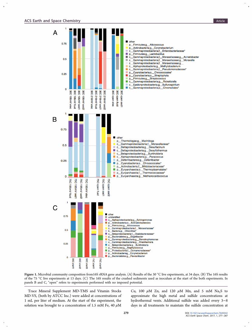
Figure 1.Microbial community composition from16S rRNA gene analysis. (A) Results of the 30 °C live experiments, at 34 days. (B) The 16S resultsof the 75 °C live experiments at 13 days. (C) The 16S results of the crushed sediments used as inoculum at the start of the both experiments. Inpanels B and C, “open” refers to experiments performed with no imposed potential.
ACS Earth and Space Chemistry Article
DOI: 10.1021/acsearthspacechem.7b00042ACS Earth Space Chem. 2017, 1, 277−287
279
∼5 mM, ensuring that the working chamber remained anoxic.The counter chamber was filled with filter-sterilized seawaterand saturated with air via bubbling, resulting in oxygenconcentrations of ∼195 and ∼110 μM in the 30° and 75 °Ctreatments, respectively. The pH of the medium prior to theaddition of sulfide was 6.9−7.1. At the end of the experiments,pH values were ∼7.9−8. The treatments at 30 °C ran for 34days and 75 °C experiments ran for 13 days and were stirredcontinually over the course of the experiment.X-ray Diffraction (XRD).Mineralogy of the chimney sulfide
slurries, prior to and upon completion of the treatments, wasmeasured via XRD at the Harvard Department of Chemistryand Chemical Biology X-ray Laboratory. Powdered chimneysubsamples were dried under nitrogen then subjected to XRDusing a Bruker D2 Phaser with Cu kα radiation. Scans werecollected from 5 to 70° 2θ with an increment of 0.01 and 5 sper step. Mineralogical deposits on the electrode were assessedat the end of the experiment using a Bruker D8 Discover usinga 2D detector in XRD 2 mode. XRD 2 scans were collectedfrom 10 to 100° 2θ. EVA D3 software was used to search forphases and perform semiquantitative analysis. The XRDanalysis prior to the start of the experiment was performedon a sample that had not been autoclaved.Scanning Electron Microscopy and Energy Dispersive
X-ray Spectroscopy (SEM/EDS). SEM was performed using aZeiss SupraVP55 FE-SEM at the Harvard Center for NanoscaleSystems. High-resolution imaging was performed at 5 kV usingan Everhart-Thornley detector. Elemental analysis performed at20 kV using an EDAX detector. For analysis of mineralmorphology, samples were dried in a glovebox under nitrogen.For analysis of microbial growth, samples were fixed withglutaraldehyde (final concentration of 2.5%), ethanol dehy-drated, and critical point dried (CPD). All samples were coatedwith 5 nm Pd/Pt prior to microscopy. EDAX Genesis was usedfor elemental analysis.Confocal Laser Scanning Microscopy (CLSM). Fluo-
rescent probes (Invitrogen Inc.) were used to label nucleic acids(Sybr Green ), peptidoglycan (wheat germ agglutinin AlexaFluor 555), and lipids (FM4-64) on the electrode. Theelectrode piece was stained with the fluorescent probes for ∼30min and rinsed with phosphate-buffered saline (1× PBS). It wasthen immersed in 1× PBS in a Nunc Lab-Tek II chamberedcoverglass and imaged using an inverted confocal laser scanningmicroscope (LSM 880, Zeiss) at the Harvard Center forBiological Imaging. Z-stacks of images were acquired with a100× oil immersion lens. The fluorescence signal was excitedusing lasers (488 nm for nucleic acids and 561 nm forpeptidoglycan and lipids) and recorded sequentially in separatechannels. The reflection signal of the mineral surface (at 405
nm) was also recorded. Alexa Fluor 555 specifically targets theresidues of N-acetylglucosamine and N-acetyl muramic acid.Only the poised electrode from the 75 °C trial was investigatedusing CLSM.
X-ray Fluorescence (XRF). XRF of the electrodes wasperformed at the Harvard Center for Nanoscale Systems usinga SPECTRO XEPOS XRF. Molybdenum was used as thesecondary target. Synthesized and natural pyrite, naturalchalcopyrite, natural sphalerite, and natural anhydrite wereused to calibrate elemental ratios and XRF results are presentedin terms of relative intensities based on normalizing theabsolute intensities to these calibrations.
16S rRNA Extraction, Sequencing, and Analysis. DNAextractions were performed using MoBio PowerSoil DNAisolation kit. The sample used to represent the inoculum (T =0) was crushed chimney material frozen at −80 °C at the timeof inoculation for each experiment (called “start” in Figure 1, SIFigure 3). The postincubation material consisted of scrapingsfrom the electrode (represented as “elec” in Figure 1, SI Figure3), the fluid in the chamber at the end of the experiment(“fluid” in Figure 1, SI Figure 3), and the mineral precipitateremaining at the bottom of the chamber at the end of theexperiment (“sed” in Figure 1, SI Figure 3), collected at the endof each experiment, 34 days for the 30 °C trials and 13 days forthe 75 °C trials. All were frozen at −80 °C immediately aftersampling until the time of extraction.The extraction procedure is briefly described as follows:
Either crushed hydrothermal sulfide chimney, up to 1 mL offluid from the reactor, or scrapings from one face of theworking electrode was added to a PowerSoil bead-beating tube.DNA was then extracted using a modified MoBio PowerSoilextraction protocol.19 Where material was sufficient, duplicateor triplicate extractions were performed and sequencedseparately (these are indicated in Figure 1 and SI Figure 3 bysequential numbers). After extraction, DNA was sent toResearch and Testing Laboratories (RTLGenomics) for 16SrRNA gene amplification using Earth Microbiome Project(EMP) primers (universal bacteria/archaeal primers 515F/806R). The amplified gene regions were then sequenced onIllumina MiSeq to generate paired end 250bp reads.20
RTLGenomics also completed taxonomic identification,including chimera checking using their standard protocol.Briefly, merged sequences were clustered into OTUs using theUPARSE algorithm21 and OTUs were identified with theUSEARCH algorithm using NCBI database as reference.Chimera checking was performed using a de novo method inUCHIME.For further confidence in taxonomic identification, post- QC
sequences generated by RTL’s standard protocol were
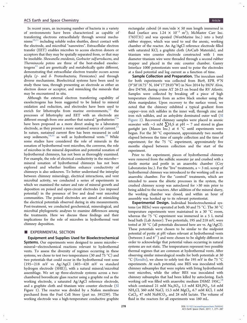
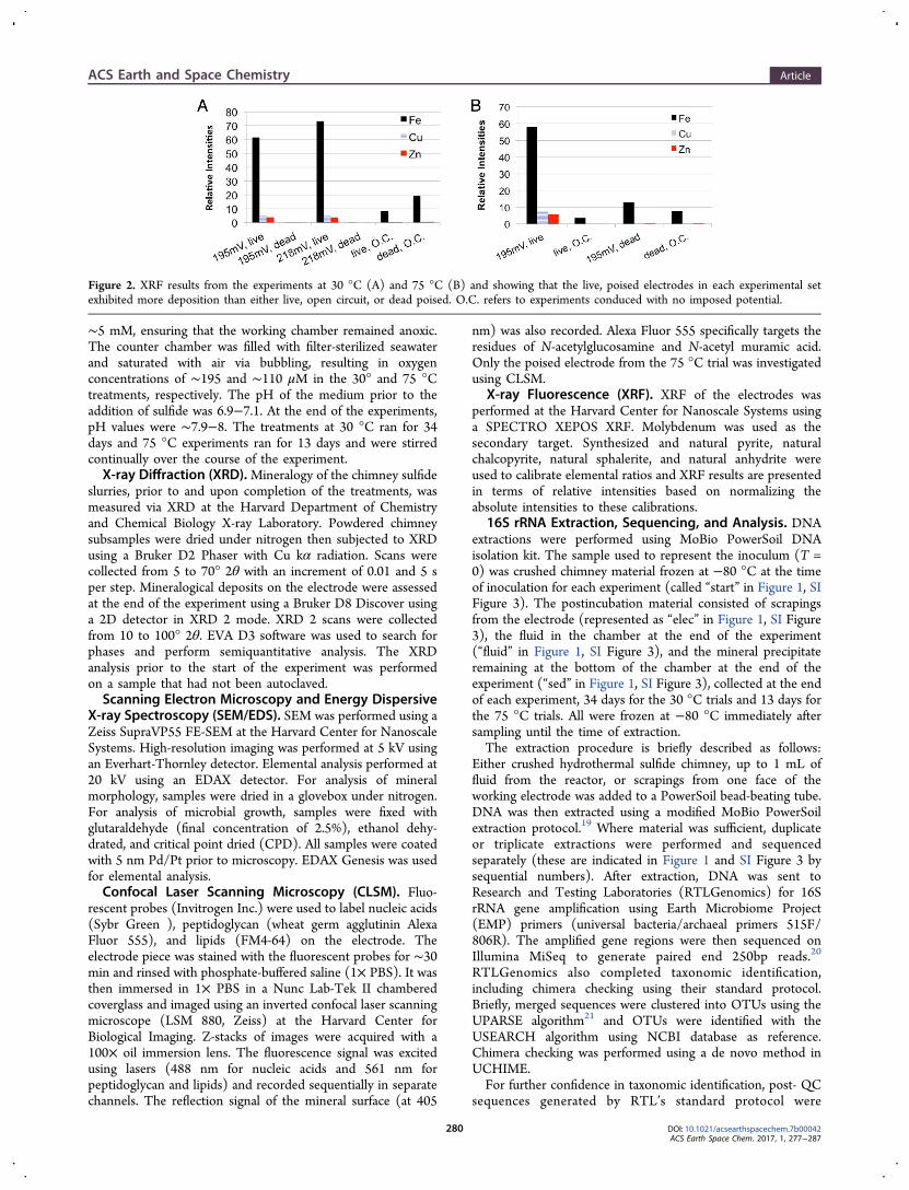
Figure 2. XRF results from the experiments at 30 °C (A) and 75 °C (B) and showing that the live, poised electrodes in each experimental setexhibited more deposition than either live, open circuit, or dead poised. O.C. refers to experiments conduced with no imposed potential.
ACS Earth and Space Chemistry Article
DOI: 10.1021/acsearthspacechem.7b00042ACS Earth Space Chem. 2017, 1, 277−287
280
processed through QIIME pipeline v1.9.0.22 The merged readswere first screened for additional chimeras using Usearch6.1,23
which performs both de novo and reference-based detection.An additional 19 500 chimeras were detected and removedfrom further analysis. The nonchimeric sequences were thenclustered into OTUs at 97% sequence similarity with openreference OTU picking method implementing Uclusteralgorithim23 and PyNast for alignment.22 The referencedatabase used was the August 2013 release of Greengenes.24
For all samples, the taxonomy assigned by QIIME is discussedunless otherwise indicated. Nonmetric multidimensional scalingwas performed using Mothur25 and was used to visualize betadiversity patterns in microbial community structure over spatialand temporal scales for the experiments (SI Figure 4).
■ RESULTSSummary. In all experiments, treatments in which the
inoculum was live and the electrode was poised resulted in thegreatest metal sulfide deposition on the electrode, which wasdetermined quantitatively through XRF with respect to iron,copper, and zinc (Figure 2) with minerals identified throughXRD (Table 1). For all three experiments, phylogenetic
community analyses revealed strong enrichment on theworking electrode; >97% Campylobacterales at 30 °C and218 mV, >95% Chromatiales sp. at 30 °C and 195 mV, and>97% Deferribacterales at 75 °C (these percentages are theproportion of 16 rRNA gene fragments attributable to thesetaxa). SEM results confirm the mineral deposition in the liveexperiments and the presence of a biofilm in the 75 °Cexperiment (Figures 3 and 4, respectively). The resultsobtained from CLSM further demonstrate the spatial relation-ship of lipids, DNA, and polysaccharides in the biofilm in the75 °C experiment (Figure 5). The high-temperature experi-ment resulted in the continuous and sustained production ofcurrent, which is not typical of solely abiotic interactions14 butrather is consistent with an active community of exoelectro-
genic microbes associated with the electrode and, potentially,the deposited minerals. For the experiments conducted at 30°C, both poised, live electrodes (195 and 218 mV) had moremetal sulfide deposition, observed via SEM and confirmed viaXRF (Figures 2 and 3) than dead or open circuit controls, butcurrent generation was inconclusive at both potentials (Figure6). At both temperatures, the metal sulfide minerals depositedupon the electrodes appeared to consist of metal sulfideminerals that were added to the BES in the initial inoculum, asopposed to new sulfide mineral formation.
The 30 °C Experiment: Sediment and SolutionReactions. XRD results revealed that the starting material inthese systems was primarily anhydrite (>80%) with gypsum asthe next most abundant mineral (∼11%), followed bychalcopyrite (SI Figure 5). At the end of the experiment, allof the gypsum had dissolved, along with some of the anhydrite.Sediments in the two dead, poised systems precipitatedmonohydrocalcite.Beginning on day 24 of the experiment, a yellow color was
visible in all of the working cells with the exception of the opencircuit, dead system. On the basis of UV−vis spectrometry,which indicated an absorbance at 375 nm, this yellow color istentatively ascribed to polysulfides. Although this was notdemonstrated conclusively, the high concentration of sulfideadded to the system, the oxidizing nature of the electrode, andthe lack of organics to which the color could be attributed areall consistent with the presence of polysulfides.
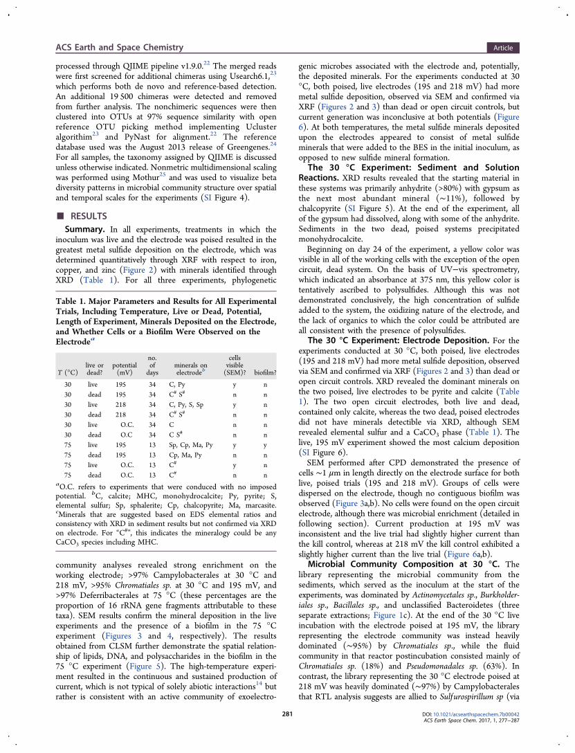
The 30 °C Experiment: Electrode Deposition. For theexperiments conducted at 30 °C, both poised, live electrodes(195 and 218 mV) had more metal sulfide deposition, observedvia SEM and confirmed via XRF (Figures 2 and 3) than dead oropen circuit controls. XRD revealed the dominant minerals onthe two poised, live electrodes to be pyrite and calcite (Table1). The two open circuit electrodes, both live and dead,contained only calcite, whereas the two dead, poised electrodesdid not have minerals detectible via XRD, although SEMrevealed elemental sulfur and a CaCO3 phase (Table 1). Thelive, 195 mV experiment showed the most calcium deposition(SI Figure 6).SEM performed after CPD demonstrated the presence of
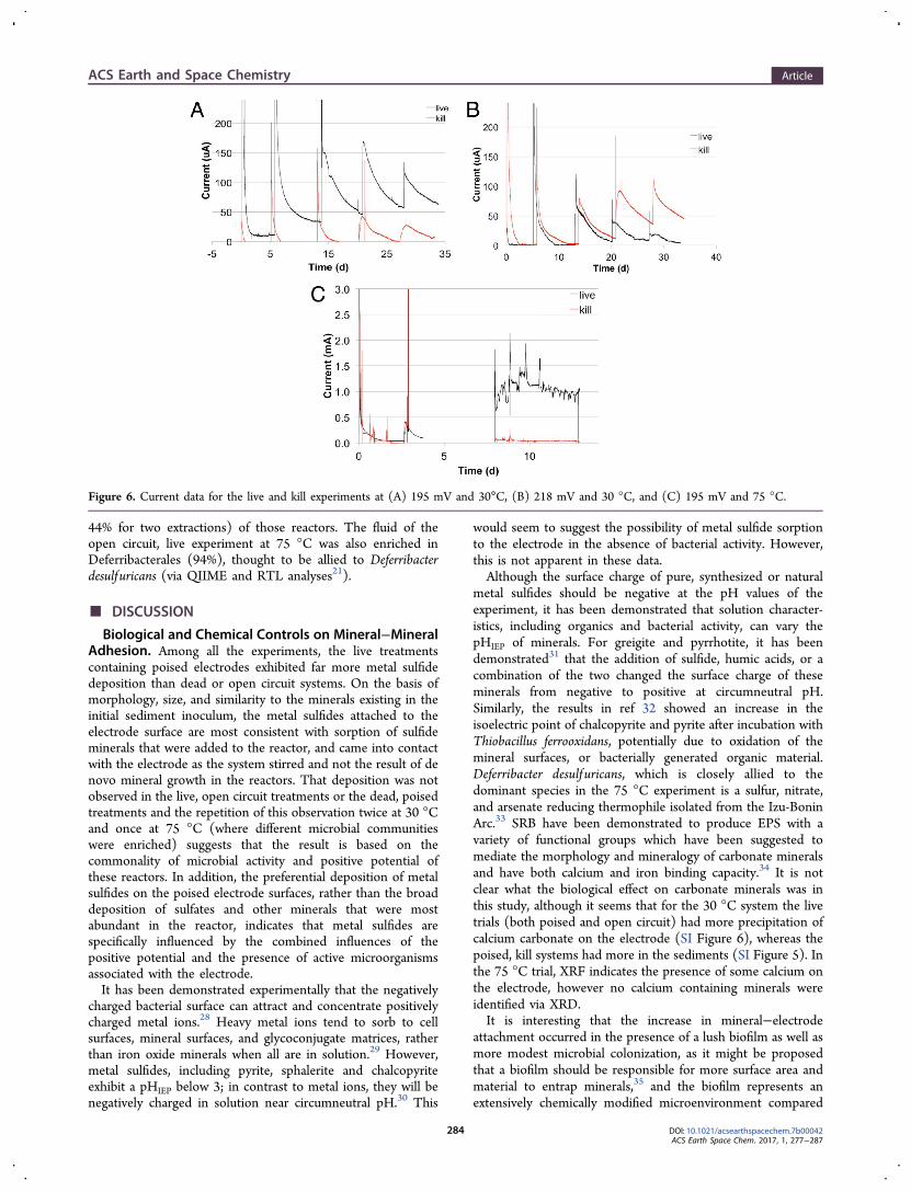
cells ∼1 μm in length directly on the electrode surface for bothlive, poised trials (195 and 218 mV). Groups of cells weredispersed on the electrode, though no contiguous biofilm wasobserved (Figure 3a,b). No cells were found on the open circuitelectrode, although there was microbial enrichment (detailed infollowing section). Current production at 195 mV wasinconsistent and the live trial had slightly higher current thanthe kill control, whereas at 218 mV the kill control exhibited aslightly higher current than the live trial (Figure 6a,b).
Microbial Community Composition at 30 °C. Thelibrary representing the microbial community from thesediments, which served as the inoculum at the start of theexperiments, was dominated by Actinomycetales sp., Burkholder-iales sp., Bacillales sp., and unclassified Bacteroidetes (threeseparate extractions; Figure 1c). At the end of the 30 °C liveincubation with the electrode poised at 195 mV, the libraryrepresenting the electrode community was instead heavilydominated (∼95%) by Chromatiales sp., while the fluidcommunity in that reactor postincubation consisted mainly ofChromatiales sp. (18%) and Pseudomonadales sp. (63%). Incontrast, the library representing the 30 °C electrode poised at218 mV was heavily dominated (∼97%) by Campylobacteralesthat RTL analysis suggests are allied to Sulfurospirillum sp (via
Table 1. Major Parameters and Results for All ExperimentalTrials, Including Temperature, Live or Dead, Potential,Length of Experiment, Minerals Deposited on the Electrode,and Whether Cells or a Biofilm Were Observed on theElectrodea
T (°C)live ordead?
potential(mV)
no.ofdays
minerals onelectrodeb
cellsvisible(SEM)? biofilm?
30 live 195 34 C, Py y n30 dead 195 34 C# S# n n30 live 218 34 C, Py, S, Sp y n30 dead 218 34 C# S# n n30 live O.C. 34 C n n30 dead O.C 34 C S# n n75 live 195 13 Sp, Cp, Ma, Py y y75 dead 195 13 Cp, Ma, Py n n75 live O.C. 13 C# y n75 dead O.C. 13 C# n n
aO.C. refers to experiments that were conduced with no imposedpotential. bC, calcite; MHC, monohydrocalcite; Py, pyrite; S,elemental sulfur; Sp, sphalerite; Cp, chalcopyrite; Ma, marcasite.cMinerals that are suggested based on EDS elemental ratios andconsistency with XRD in sediment results but not confirmed via XRDon electrode. For “C#”, this indicates the mineralogy could be anyCaCO3 species including MHC.
ACS Earth and Space Chemistry Article
DOI: 10.1021/acsearthspacechem.7b00042ACS Earth Space Chem. 2017, 1, 277−287
281
the Krona pipeline;26 Figure 1a). The postincubation fluidsfrom this reactor were also dominated by Campylobacterales(87%). The library from the open circuit electrode communitywas also dominated (89%) by Chromatiales sp. postincubation,whereas those from open circuit sediment and fluidcommunities were predominantly Legionellales sp. (88%) andPseudomonadales sp. (50%) and Lactobacillales sp. (22%),respectively.Sediment and Solution Reactions in the 75 °C
Treatment. In the higher temperature experiment, XRD onsediments revealed the main change from the start of theexperiment was the dissolution of gypsum (SI Figure 5).Although by the end of the experiment the relative abundanceof minerals had changed, there was no evidence that newminerals were present.The production of sulfide was observed in both the higher
temperature “live” treatments, starting around day 8 of each
experiment. The sulfide production persisted in the poised trialuntil the end of the experiment, while the sulfide in theunpoised trial returned to abiotic levels by the point ofsampling on day nine.
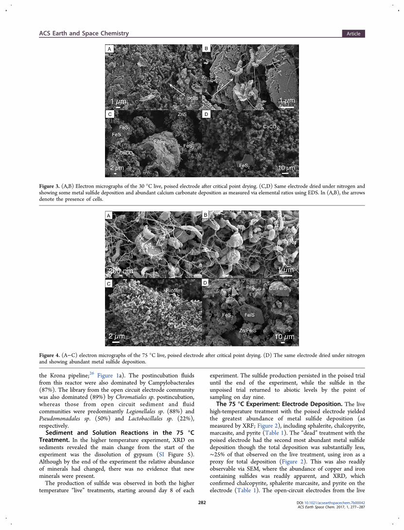
The 75 °C Experiment: Electrode Deposition. The livehigh-temperature treatment with the poised electrode yieldedthe greatest abundance of metal sulfide deposition (asmeasured by XRF; Figure 2), including sphalerite, chalcopyrite,marcasite, and pyrite (Table 1). The “dead” treatment with thepoised electrode had the second most abundant metal sulfidedeposition though the total deposition was substantially less,∼25% of that observed on the live treatment, using iron as aproxy for total deposition (Figure 2). This was also readilyobservable via SEM, where the abundance of copper and ironcontaining sulfides was readily apparent, and XRD, whichconfirmed chalcopyrite, sphalerite marcasite, and pyrite on theelectrode (Table 1). The open-circuit electrodes from the live
Figure 3. (A,B) Electron micrographs of the 30 °C live, poised electrode after critical point drying. (C,D) Same electrode dried under nitrogen andshowing some metal sulfide deposition and abundant calcium carbonate deposition as measured via elemental ratios using EDS. In (A,B), the arrowsdenote the presence of cells.
Figure 4. (A−C) electron micrographs of the 75 °C live, poised electrode after critical point drying. (D) The same electrode dried under nitrogenand showing abundant metal sulfide deposition.
ACS Earth and Space Chemistry Article
DOI: 10.1021/acsearthspacechem.7b00042ACS Earth Space Chem. 2017, 1, 277−287
282
and dead treatments had even less mineral deposition, with theopen-circuit electrode from the dead treatment exhibitingmostly calcium carbonate deposition (SI Figure 6), and theopen-circuit electrode from the live treatment exhibiting theleast deposition of all trials. No diffraction patterns (other thangraphite) were observed on the two open circuit electrodes.Current production in the poised, live experiment began
around day five (data not shown until day eight due to acomputer logging malfunction) and persisted at milliamp levelsuntil the end of the experiment. Current in the poised, deadtreatment remained around 50 microamps for the entirety ofthe experiment, with the exception of brief spikes during sulfideadditions (Figure 6). When converted to current per surfacearea, the maximum current produced in the live experiment is∼1100 mA/m2 ; this is within the range previously reported formicrobial fuel cells, which ranged from 44 to 6000 mA/m2.27
Electrode Biofilm and Microbial Community Compo-sition at 75 °C. The poised electrode from the live high-
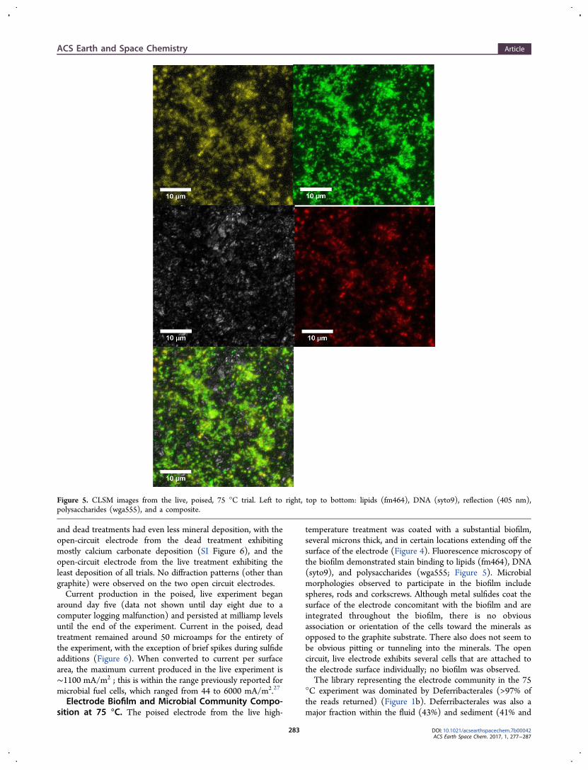
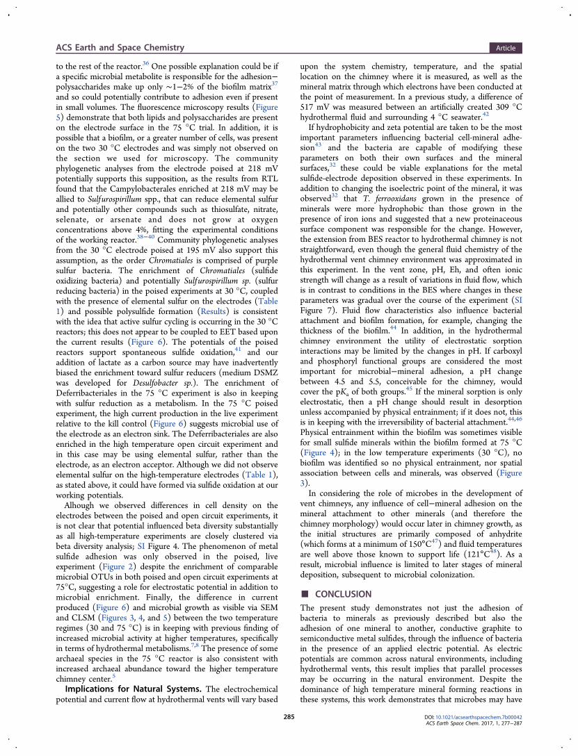
temperature treatment was coated with a substantial biofilm,several microns thick, and in certain locations extending off thesurface of the electrode (Figure 4). Fluorescence microscopy ofthe biofilm demonstrated stain binding to lipids (fm464), DNA(syto9), and polysaccharides (wga555; Figure 5). Microbialmorphologies observed to participate in the biofilm includespheres, rods and corkscrews. Although metal sulfides coat thesurface of the electrode concomitant with the biofilm and areintegrated throughout the biofilm, there is no obviousassociation or orientation of the cells toward the minerals asopposed to the graphite substrate. There also does not seem tobe obvious pitting or tunneling into the minerals. The opencircuit, live electrode exhibits several cells that are attached tothe electrode surface individually; no biofilm was observed.The library representing the electrode community in the 75
°C experiment was dominated by Deferribacterales (>97% ofthe reads returned) (Figure 1b). Deferribacterales was also amajor fraction within the fluid (43%) and sediment (41% and
Figure 5. CLSM images from the live, poised, 75 °C trial. Left to right, top to bottom: lipids (fm464), DNA (syto9), reflection (405 nm),polysaccharides (wga555), and a composite.
ACS Earth and Space Chemistry Article
DOI: 10.1021/acsearthspacechem.7b00042ACS Earth Space Chem. 2017, 1, 277−287
283
44% for two extractions) of those reactors. The fluid of theopen circuit, live experiment at 75 °C was also enriched inDeferribacterales (94%), thought to be allied to Deferribacterdesulfuricans (via QIIME and RTL analyses21).
■ DISCUSSION
Biological and Chemical Controls on Mineral−MineralAdhesion. Among all the experiments, the live treatmentscontaining poised electrodes exhibited far more metal sulfidedeposition than dead or open circuit systems. On the basis ofmorphology, size, and similarity to the minerals existing in theinitial sediment inoculum, the metal sulfides attached to theelectrode surface are most consistent with sorption of sulfideminerals that were added to the reactor, and came into contactwith the electrode as the system stirred and not the result of denovo mineral growth in the reactors. That deposition was notobserved in the live, open circuit treatments or the dead, poisedtreatments and the repetition of this observation twice at 30 °Cand once at 75 °C (where different microbial communitieswere enriched) suggests that the result is based on thecommonality of microbial activity and positive potential ofthese reactors. In addition, the preferential deposition of metalsulfides on the poised electrode surfaces, rather than the broaddeposition of sulfates and other minerals that were mostabundant in the reactor, indicates that metal sulfides arespecifically influenced by the combined influences of thepositive potential and the presence of active microorganismsassociated with the electrode.It has been demonstrated experimentally that the negatively
charged bacterial surface can attract and concentrate positivelycharged metal ions.28 Heavy metal ions tend to sorb to cellsurfaces, mineral surfaces, and glycoconjugate matrices, ratherthan iron oxide minerals when all are in solution.29 However,metal sulfides, including pyrite, sphalerite and chalcopyriteexhibit a pHIEP below 3; in contrast to metal ions, they will benegatively charged in solution near circumneutral pH.30 This
would seem to suggest the possibility of metal sulfide sorptionto the electrode in the absence of bacterial activity. However,this is not apparent in these data.Although the surface charge of pure, synthesized or natural
metal sulfides should be negative at the pH values of theexperiment, it has been demonstrated that solution character-istics, including organics and bacterial activity, can vary thepHIEP of minerals. For greigite and pyrrhotite, it has beendemonstrated31 that the addition of sulfide, humic acids, or acombination of the two changed the surface charge of theseminerals from negative to positive at circumneutral pH.Similarly, the results in ref 32 showed an increase in theisoelectric point of chalcopyrite and pyrite after incubation withThiobacillus ferrooxidans, potentially due to oxidation of themineral surfaces, or bacterially generated organic material.Deferribacter desulf uricans, which is closely allied to thedominant species in the 75 °C experiment is a sulfur, nitrate,and arsenate reducing thermophile isolated from the Izu-BoninArc.33 SRB have been demonstrated to produce EPS with avariety of functional groups which have been suggested tomediate the morphology and mineralogy of carbonate mineralsand have both calcium and iron binding capacity.34 It is notclear what the biological effect on carbonate minerals was inthis study, although it seems that for the 30 °C system the livetrials (both poised and open circuit) had more precipitation ofcalcium carbonate on the electrode (SI Figure 6), whereas thepoised, kill systems had more in the sediments (SI Figure 5). Inthe 75 °C trial, XRF indicates the presence of some calcium onthe electrode, however no calcium containing minerals wereidentified via XRD.It is interesting that the increase in mineral−electrode
attachment occurred in the presence of a lush biofilm as well asmore modest microbial colonization, as it might be proposedthat a biofilm should be responsible for more surface area andmaterial to entrap minerals,35 and the biofilm represents anextensively chemically modified microenvironment compared
Figure 6. Current data for the live and kill experiments at (A) 195 mV and 30°C, (B) 218 mV and 30 °C, and (C) 195 mV and 75 °C.
ACS Earth and Space Chemistry Article
DOI: 10.1021/acsearthspacechem.7b00042ACS Earth Space Chem. 2017, 1, 277−287
284
to the rest of the reactor.36 One possible explanation could be ifa specific microbial metabolite is responsible for the adhesion−polysaccharides make up only ∼1−2% of the biofilm matrix37
and so could potentially contribute to adhesion even if presentin small volumes. The fluorescence microscopy results (Figure5) demonstrate that both lipids and polysaccharides are presenton the electrode surface in the 75 °C trial. In addition, it ispossible that a biofilm, or a greater number of cells, was presenton the two 30 °C electrodes and was simply not observed onthe section we used for microscopy. The communityphylogenetic analyses from the electrode poised at 218 mVpotentially supports this supposition, as the results from RTLfound that the Campylobacterales enriched at 218 mV may beallied to Sulfurospirillum spp., that can reduce elemental sulfurand potentially other compounds such as thiosulfate, nitrate,selenate, or arsenate and does not grow at oxygenconcentrations above 4%, fitting the experimental conditionsof the working reactor.38−40 Community phylogenetic analysesfrom the 30 °C electrode poised at 195 mV also support thisassumption, as the order Chromatiales is comprised of purplesulfur bacteria. The enrichment of Chromatiales (sulfideoxidizing bacteria) and potentially Sulfurospirillum sp. (sulfurreducing bacteria) in the poised experiments at 30 °C, coupledwith the presence of elemental sulfur on the electrodes (Table1) and possible polysulfide formation (Results) is consistentwith the idea that active sulfur cycling is occurring in the 30 °Creactors; this does not appear to be coupled to EET based uponthe current results (Figure 6). The potentials of the poisedreactors support spontaneous sulfide oxidation,41 and ouraddition of lactate as a carbon source may have inadvertentlybiased the enrichment toward sulfur reducers (medium DSMZwas developed for Desulfobacter sp.). The enrichment ofDeferribacteriales in the 75 °C experiment is also in keepingwith sulfur reduction as a metabolism. In the 75 °C poisedexperiment, the high current production in the live experimentrelative to the kill control (Figure 6) suggests microbial use ofthe electrode as an electron sink. The Deferribacteriales are alsoenriched in the high temperature open circuit experiment andin this case may be using elemental sulfur, rather than theelectrode, as an electron acceptor. Although we did not observeelemental sulfur on the high-temperature electrodes (Table 1),as stated above, it could have formed via sulfide oxidation at ourworking potentials.Alhough we observed differences in cell density on the
electrodes between the poised and open circuit experiments, itis not clear that potential influenced beta diversity substantiallyas all high-temperature experiments are closely clustered viabeta diversity analysis; SI Figure 4. The phenomenon of metalsulfide adhesion was only observed in the poised, liveexperiment (Figure 2) despite the enrichment of comparablemicrobial OTUs in both poised and open circuit experiments at75°C, suggesting a role for electrostatic potential in addition tomicrobial enrichment. Finally, the difference in currentproduced (Figure 6) and microbial growth as visible via SEMand CLSM (Figures 3, 4, and 5) between the two temperatureregimes (30 and 75 °C) is in keeping with previous finding ofincreased microbial activity at higher temperatures, specificallyin terms of hydrothermal metabolisms.7,8 The presence of somearchaeal species in the 75 °C reactor is also consistent withincreased archaeal abundance toward the higher temperaturechimney center.5
Implications for Natural Systems. The electrochemicalpotential and current flow at hydrothermal vents will vary based
upon the system chemistry, temperature, and the spatiallocation on the chimney where it is measured, as well as themineral matrix through which electrons have been conducted atthe point of measurement. In a previous study, a difference of517 mV was measured between an artificially created 309 °Chydrothermal fluid and surrounding 4 °C seawater.42
If hydrophobicity and zeta potential are taken to be the mostimportant parameters influencing bacterial cell-mineral adhe-sion43 and the bacteria are capable of modifying theseparameters on both their own surfaces and the mineralsurfaces,32 these could be viable explanations for the metalsulfide-electrode deposition observed in these experiments. Inaddition to changing the isoelectric point of the mineral, it wasobserved32 that T. ferrooxidans grown in the presence ofminerals were more hydrophobic than those grown in thepresence of iron ions and suggested that a new proteinaceoussurface component was responsible for the change. However,the extension from BES reactor to hydrothermal chimney is notstraightforward, even though the general fluid chemistry of thehydrothermal vent chimney environment was approximated inthis experiment. In the vent zone, pH, Eh, and often ionicstrength will change as a result of variations in fluid flow, whichis in contrast to conditions in the BES where changes in theseparameters was gradual over the course of the experiment (SIFigure 7). Fluid flow characteristics also influence bacterialattachment and biofilm formation, for example, changing thethickness of the biofilm.44 In addition, in the hydrothermalchimney environment the utility of electrostatic sorptioninteractions may be limited by the changes in pH. If carboxyland phosphoryl functional groups are considered the mostimportant for microbial−mineral adhesion, a pH changebetween 4.5 and 5.5, conceivable for the chimney, wouldcover the pKa of both groups.45 If the mineral sorption is onlyelectrostatic, then a pH change should result in desorptionunless accompanied by physical entrainment; if it does not, thisis in keeping with the irreversibility of bacterial attachment.44,46
Physical entrainment within the biofilm was sometimes visiblefor small sulfide minerals within the biofilm formed at 75 °C(Figure 4); in the low temperature experiments (30 °C), nobiofilm was identified so no physical entrainment, nor spatialassociation between cells and minerals, was observed (Figure3).In considering the role of microbes in the development of
vent chimneys, any influence of cell−mineral adhesion on themineral attachment to other minerals (and therefore thechimney morphology) would occur later in chimney growth, asthe initial structures are primarily composed of anhydrite(which forms at a minimum of 150°C47) and fluid temperaturesare well above those known to support life (121°C48). As aresult, microbial influence is limited to later stages of mineraldeposition, subsequent to microbial colonization.
■ CONCLUSIONThe present study demonstrates not just the adhesion ofbacteria to minerals as previously described but also theadhesion of one mineral to another, conductive graphite tosemiconductive metal sulfides, through the influence of bacteriain the presence of an applied electric potential. As electricpotentials are common across natural environments, includinghydrothermal vents, this result implies that parallel processesmay be occurring in the natural environment. Despite thedominance of high temperature mineral forming reactions inthese systems, this work demonstrates that microbes may have
ACS Earth and Space Chemistry Article
DOI: 10.1021/acsearthspacechem.7b00042ACS Earth Space Chem. 2017, 1, 277−287
285
a role in assembling natural mineral composites, such ashydrothermal vent chimneys, through mineral entrainment andadhesion, and extend the possibility that previously unconsid-ered biological processes influence otherwise abiotic mineralstructures.
■ ASSOCIATED CONTENT*S Supporting InformationThe Supporting Information is available free of charge on theACS Publications website at DOI: 10.1021/acsearthspace-chem.7b00042.
Photograph of the experimental setup (SI Figure 1),SEM (SI Figure 2) of initial inoculum; full microbialcommunity composition from 16S rRNA gene analysis(SI Figure 3); beta diversity analysis for all experiments(SI Figure 4); XRD results of sediments (SI Figure 5);XRF results of calcium (SI Figure 6); a schematiccontrasting BES with natural hydrothermal chimneys (SIFigure 7) (PDF)
■ AUTHOR INFORMATIONCorresponding Authors*E-mail: [email protected].*E-mail: [email protected]. Gartman: 0000-0001-9307-3062Present Address§(A.G.) U.S. Geological Survey, 2885 Mission Street, SantaCruz, CA 95060, U.S.A.NotesThe authors declare no competing financial interest.
■ ACKNOWLEDGMENTSThis work was performed in part at the Center for NanoscaleSystems (CNS), a member of the National NanotechnologyCoordinated Infrastructure Network (NNCI), which issupported by the National Science Foundation under NSFAward No. 1541959. CNS is part of Harvard University.Optical microscopy was performed at the Harvard Center forBiological Imaging (HCBI). XRD was performed at theHarvard Department of Chemistry and Chemical Biology X-ray Laboratory We would like to thank the staff of the CHNSand the HCBI and Shao-Liang Zheng at the X-ray Laboratoryfor training and support. We would also like to thank the HOVAlvin and Atlantis crew of AT26-23 for sample collection.Mahalo nui to Dr. Kiana Frank for providing the beta-diversityanalysis. This work was supported by the National ScienceFoundation Grant 1344241.
■ REFERENCES(1) Haymon, R. H. Growth history of hydrothermal black smokerchimneys. Nature 1983, 301, 695−698.(2) Tivey, M. K.; Singh, S. Nondestructive imagining of fragile sea-floor vent deposit samples. Geology 1997, 25 (10), 931−934.(3) Spagnoli, G.; Hannington, M.; Bairlein, K.; Hordt, A.; Jegen, M.;Petersen, S.; Laurila, T. Electrical properties of seafloor massivesulfides. Geo-Mar. Lett. 2016, 36, 235−245.(4) Nakamura, R.; Takashima, T.; Kato, S.; Takai, K.; Yamamoto, M.;Hashimoto, K. Electrical Current Generation across a Black SmokerChimney. Angew. Chem. 2010, 122, 7858−7860.(5) Schrenk, M. O.; Kelley, D. S.; Delaney, J. R.; Baross, J. A.Incidence and Diversity of Microorganisms within the Walls of an
Active Deep-Sea Sulfide Chimney. Appl. Environ. Microbiol. 2003, 69(6), 3580−3592.(6) Toner, B. M.; Lesniewski, R. A.; Marlow, J. J.; Briscoe, L. J.;Santelli, C. M.; Bach, W.; Orcutt, B. N.; Edwards, K. J. MineralogyDrives Bacterial Biogeography of Hydrothermally Inactive SeafloorSulfide Deposits. Geomicrobiol. J. 2013, 30 (4), 313−326.(7) LaRowe, D. E.; Dale, A. W.; Aguilera, D. R.; L’Heureux, I.;Amend, J. P.; Regnier, P. Modeling microbial reaction rates in asubmarine hydrothermal vent chimney wall. Geochim. Cosmochim. Acta2014, 124, 72−97.(8) Frank, K. L.; Rogers, D. R.; Olins, H. C.; Vidoudez, C.; Girguis,P.R. Characterizing the distribution and rates of microbial sulfatereduction at Middle Valley hydrothermal vents. ISME J. 2013, 7,1391−1401.(9) Olins, H. C.; Rogers, D. R.; Frank, K. L.; Vidoudez, C.; Girguis, P.R. Assessing the influence of physical, geochemical and biologicalfactors on anaerobic microbial primary productivity within hydro-thermal vent chimneys. Geobiology. 2013, 11, 279−293.(10) Gralnick, J. A.; Newman, D. K. Extracellular respiration. Mol.Microbiol. 2007, 65 (1), 1−11.(11) Lovley, D. R. Electromicrobiology. Annu. Rev. Microbiol. 2012,66, 391−409.(12) Rowe, A. R.; Chellamuthu, P.; Lam, B.; Okamoto, A.; Nealson,K. H. Marine sediments microbes capable of electrode oxidation as asurrogate for lithotrophic insoluble substrate metabolism. Front.Microbiol. 2015, 5, 784.(13) Sato, M.; Mooney, H. M. The electrochemical mechanism ofsulfide self-potentials. Geophysics 1960, 25 (1), 226−249.(14) Reimers, C. E.; Girguis, P.; Stecher, H. A., III.; Tender, L. M.;Ryckelynck, N. Whaling, P. Microbial fuel cell energy from an oceancold seep. Geobiology 2006, 4, 123−126.(15) Nielsen, M. E.; Reimers, C. E.; White, H. K.; Sharma, S.;Girguis, P. R. Sustainable energy from deep ocean cold seeps. 2008.Energy Environ. Sci. 2008, 1, 584−593.(16) Girguis, P. R.; Holden, J. F. On the potential for bioenergy andbiofuels from hydrothermal vent microbes. Oceanography 2012, 25(1), 213−217.(17) Hamilton, I. C.; Woods, R. An investigation of surface oxidationof pyrite and pyrrhotite by linear potential sweep voltammetry. J.Electroanal. Chem. Interfacial Electrochem. 1981, 118, 327−343.(18) Widdel, F.; Bak, F. Gram-negative mesophilic sulfate-reducingbacteria. In The Prokaryotes; Balows, A., Truper, H., Dworkin, M.,Harder, W., Schleifer, K-H., Eds.; Springer: New York, 1992; pp3352−3378.(19) Santelli, C. S.; Orcutt, B. N.; Banning, E.; Bach, W.; Moyer, C.L.; Sogin, M. L.; Staudigel, H.; Edwards, K. J. Abundance and diversityof microbial life in ocean crust. Nature 2008, 453, 653−657.(20) Caporaso, J. G.; Lauber, C. L.; Walters, W. A.; Berg-Lyons, D.;Huntley, J.; Fierer, N.; Owens, S. M.; Betley, J.; Fraser, L.; Bauer, M.;Gormley, N.; Gilbert, J. A.; Smith, G.; Knight, R. Ultra-high-throughput microbial community analysis on the Illumina HiSeqand MiSeq platforms. ISME J. 2012, 6, 1621−1624.(21) Edgar, R. C. UPARSE: highly accurate OTU sequences frommicrobial amplicon reads. Nat. Methods 2013, 10, 996−998.(22) Caporaso, J. G.; Bittinger, K.; Bushman, F. D.; DeSantis, T. Z.;Andersen, G. L.; Knight, R. PyNAST: a flexible tool for aligningsequences to a template alignment. Bioinformatics 2010, 26, 266−267.(23) Edgar, R. C. Search and clustering orders of magnitude fasterthan BLAST. Bioinformatics 2010, 26 (19), 2460−2461.(24) DeSantis, T. Z.; Hugenholtz, P.; Larsen, N.; Rojas, M.; Brodie,E. L.; Keller, K.; Huber, T.; Dalevi, D.; Hu, P.; Andersen, G. L.Greengenes, a chimera-checked 16S rRNA gene database andworkbench compatible with ARB. Appl. Environ. Microbiol. 2006, 72(7), 5069−5072.(25) Schloss, P. D.; Westcott, S. L.; Ryabin, T.; Hall, J. R. A.;Hartmann, M.; Hollister, E. B.; Lesniewski, R. A.; Oakley, B. B.; Parks,D. H.; Robinson, C. J.; Sahl, J. W.; Stres, B.; Thallinger, G. G.; VanHorn, D. J.; Weber, C. F. Introducing mothur: open source, platform-independent, community-supported software for describing and
ACS Earth and Space Chemistry Article
DOI: 10.1021/acsearthspacechem.7b00042ACS Earth Space Chem. 2017, 1, 277−287
286
comparing microbial communities. Appl. Environ. Microbiol. 2009, 69,3580−3592.(26) Ondov, B. D.; Bergman, N. H.; Phillippy, A. M. 2011.Interactive metagenomic visualization in a Web browser. BMCBioinformatics 2011, 385 (12), 1471−2105.(27) Nevin, K. P.; Richter, H.; Covalla, S. F.; Johnson, J. P.;Woodard, T. L.; Orloff, A. L.; Jia, H.; Zhang, M.; Lovley, D. R. Poweroutput and columbic efficiencies from biofilms of Geobactersulfurreducens comparable to mixed community microbial fuel cells.Environ. Microbiol. 2008, 10 (10), 2505−2514.(28) Beveridge, T. J. Role of cellular design in bacterial metalaccumulation and mineralization. Annu. Rev. Microbiol. 1989, 43, 147−171.(29) Hao, L.; Guo, Y.; Byrne, J. M.; Zeitvogel, F.; Schmid, G.; Ingino,P.; Li, J.; Neu, T. R.; Swanner, E. D.; Kappler, A.; Obst, M. Binding ofheavy metal ions in aggregates of microbial cells, EPS and biogeniciron minerals measured in-situ using metal- and glycoconjugates-specific fluorophores. Geochim. Cosmochim. Acta 2016, 180, 66−96.(30) Bebie, J.; Schoonen, M. A. A.; Fuhrmann, M.; Strongin, D. R.Surface charge development on transition metal sulfides: Anelectrokinetic study. Geochim. Cosmochim. Acta 1998, 62 (4), 633−642.(31) Dekkers, M. J.; Schoonen, M. A. A. An electrokinetic study ofsynthetic greigite and pyrrhotite. Geochim. Cosmochim. Acta 1994, 58(19), 4147−4153.(32) Devasia, P.; Natarajan, K. A.; Sathyanarayana, D. N.;Ramandana, R. G. Surface Chemistry of Thiobacillus ferrooxidansRelevant to Adhesion on Mineral Surfaces. Appl. Environ. Microbiol.1993, 59 (12), 4051.(33) Takai, K.; Kobayashi, H.; Nealson, K. H.; Horikoshi, K.Deferribacter desulfuricans sp. nov., a novel sulfur-, nitrate- andarsenate-reducing thermophile isolated from a deep-sea hydrothermalvent. Int. J. Syst. Evol. Microbiol. 2003, 53, 839−836.(34) Braissant, O.; Decho, A. W.; Dupraz, C.; Glunk, C.; Przekop, K.M.; Visscher, P. T. Exopolymeric substances of sulfate-reducingbacteria: Interactions with calcium at alkaline pH and implication forformation of carbonate minerals. Geobiology 2007, 5, 401−411.(35) Sutherland, I. W. Biofilm exopolysaccharides: a strong and stickyframework. Microbiology 2001, 147, 3−9.(36) Decho, A. W. Overview of biopolymer-induced mineralization:What goes on in biofilms? Ecol. Eng. 2010, 36, 137−144.(37) Sutherland, I. W. The biofilm matrix- an immobilized butdynamic microbial environment. Trends Microbiol. 2001, 9 (5), 222−227.(38) Stolz, J. F.; Ellis, D. J.; Switzer Blum, J.; Ahmann, D.; Lovley, D.R.; Oremland, R. S. Sulfurospirillum barnesii sp. mov. AndSulfurospirillum arsenophilum sp. nov., new members of theSulfurospirilllum clade of the ε Proteobacteria. Int. J. Syst. Bacteriol.1999, 49, 1177−1180.(39) Stolz, J. F.; et al. Sulfurospirillum. Bergey’s Manual of Systematicsof Archaea and Bacteria 2015, 1−7.(40) Rabus, R.; Hansen, T. A.; Widdel, F. Dissimilatory Sulfate- andSulfur-Reducing Prokaryotes. In Prokaryotes; Falkow, S., Rosenberg, E.,Schleifer, K. H., Stackebrandt, E., Eds.; Springer: New York, 2006; pp659−768.(41) Rabaey, K.; Van de Sompel, K.; Maignien, L.; Boon, N.;Aelterman, P.; Clauwaert, P.; De Schamphelaire, L.; Pham, H. T.;Vermeulen, J.; Verhaege, M.; Lens, P.; Verstraete, W. Microbial FuelCells for Sulfide Removal. Environ. Sci. Technol. 2006, 40 (17), 5218−5224.(42) Yamamoto, M.; Nakamura, R.; Oguri, K.; Kawagucci, S.; Suzuki,K.; Hashimoto, K.; Takai, K. Generation of Electricity and Illuminationby an Environmental Fuel Cell in Deep-Sea Hydrothermal Vents.Angew. Chem., Int. Ed. 2013, 52, 10758−10761.(43) van Loosdrecht, M. C. M.; Lyklema, J.; Norde, W.; Schraa, G.;Zehnder, A. J. B. Electrophoretic Mobility and Hydrophobicity as aMeasure To Predict the Initial Steps of Bacterial Adhesion. Appl.Environ. Microbiol. 1987, 53 (8), 1898−1901.
(44) Katsikogianni, M.; Missirlis, Y. F. Concise review of mechanismsof bacterial adhesion to biomaterials and of techniques used inestimating bacteria-material interactions. Eur. Cell. Mater. 2004, 8, 37−57.(45) Stumm, W., Morgan, J. J. Aquatic Chemistry: Chemical Equilibriaand Rates in Natural Waters, 3rd, ed.; Wiley: New York, 1996.(46) Rijnaarts, H. H. M.; Norde, W.; Bouwer, E. J.; Lyklema, J.;Zehnder, A. J. B. Reversibility and mechanism of bacterial adhesion.Colloids Surf., B 1995, 4, 5−22.(47) Bischoff, J. L.; Seyfried, W. E. Hydrothermal Chemistry ofSeawater from 25° to 350°C. Am. J. Sci. 1978, 278, 838−860.(48) Kashefi, K.; Lovley, D. R. Extending the Upper TemperatureLimit for Life. Science 2003, 301 (5635), 934.
ACS Earth and Space Chemistry Article
DOI: 10.1021/acsearthspacechem.7b00042ACS Earth Space Chem. 2017, 1, 277−287
287