nanomedicine for treating spinal cord injury
DESCRIPTION
Reparacion del cordon espinalTRANSCRIPT

Nanoscale
REVIEW
Publ
ishe
d on
16
July
201
3. D
ownl
oade
d by
Uni
vers
ity o
f Il
linoi
s -
Urb
ana
on 1
9/09
/201
3 10
:38:
23.
View Article OnlineView Journal | View Issue
aWeldon School of Biomedical Engineering
47907, USA. E-mail: [email protected] Cord and Brain Injury Research Grou
and Department of Neurological Surgery,
Indianapolis, IN 46202, USA. E-mail: xu26@cDepartment of Chemistry, Purdue Universit
Cite this: Nanoscale, 2013, 5, 8821
Received 24th February 2013Accepted 9th July 2013
DOI: 10.1039/c3nr00957b
www.rsc.org/nanoscale
This journal is ª The Royal Society of
Nanomedicine for treating spinal cord injury
Jacqueline Y. Tyler,a Xiao-Ming Xu*b and Ji-Xin Cheng*ac
Spinal cord injury results in significantmortality andmorbidity, lifestyle changes, and difficult rehabilitation.
Treatment of spinal cord injury is challenging because the spinal cord is both complex to treat acutely and
difficult to regenerate. Nanomaterials can be used to provide effective treatments; their unique properties
can facilitate drug delivery to the injury site, enact as neuroprotective agents, or provide platforms to
stimulate regrowth of damaged tissues. We review recent uses of nanomaterials including nanowires,
micelles, nanoparticles, liposomes, and carbon-based nanomaterials for neuroprotection in the acute
phase. We also review the design and neural regenerative application of electrospun scaffolds, conduits,
and self-assembling peptide scaffolds.
1 Spinal cord injury: current outlook
Spinal cord injury (SCI) is a widespread problem affecting about250 000 people living in the United States, with an estimated13 400 new cases each year.1 SCI tragically oen affects theyoungest and most active segment of our society, with 60% ofinjuries occurring in those under the age of 30. The mostcommon cause, making up greater than 40% of SCI, is motorvehicle accidents. Other common causes include recreation-related accidents, work-related accidents, falls, and acts ofviolence.2 The pathological progression of SCI is oen separatedinto two categories: primary injury and secondary injury.
Primary injury involves initial trauma and local tissue injurycaused by bone fracture and stretching, exion, rotation,laceration, compression, or displacement of the spinal cord.3
Initial injury aer a contusive SCI mainly damages the greymatter of the spinal cord, resulting in hemorrhage anddisruption of the blood ow.
The secondary injury denotes the spread of damage from theoriginal site to adjacent tissue through a cascade of deleteriousreactions to the trauma.4 The extent of secondary injury isproportional in magnitude to the primary injury. Secondaryinjury includes many different mechanisms, including threekey pathophysiological events. First, damage to blood vessels isespecially prevalent in small vessels and results in ischemia,thrombosis, and hypoxia; starving the tissue of nutrients.Second, reactive oxygen species are produced during ischemiaand contribute to oxidative stress. Once the ability of cells toprotect themselves from oxidative stress with antioxidants has
, Purdue University, West Lafayette, IN
p, Stark Neurosciences Research Institute
Indiana University School of Medicine,
iupui.edu
y, West Lafayette, IN 47907, USA
Chemistry 2013
been exceeded, the oxidation of proteins, nucleic acids, andlipids will occur and perpetuate the damage. Third, membranedisruption and depolarization of the cells from primary damagecauses voltage dependent channels in the cells to open, result-ing in a mass release of ions, edema, and intracellular Ca2+
overload. Calcium overload contributes to damage by inhibitingcellular respiration and stimulating calcium dependent lipasesand proteases, which subsequently degrade important proteinstructures in the central nervous system (CNS). This chain ofevents eventually results in recruitment of immune cells,apoptosis, disruption of synaptic connections, and also axonaldegradation, contraction, and demyelination.3,5–7
The progression of primary injury and secondary injury ishighlighted in Fig. 1 adopted from GhoshMitra et al.7 In thechronic phase, damaged tissue is cleared away by microglia andmacrophages, leaving a uid-lled cavity and an astrocyte-populated glial scar. Molecules that inhibit axon growth areexpressed, and the glial scar and cyst remain as barriers toreconnection.3,8,9
Victims who survive SCI can expect to live long lives, but theyface extensive rehabilitation and long-term disability. Rehabil-itation prospects depend on the severity of the damage. Indi-viduals with injury at or below T-6, may be candidates forwalking.10 These patients are re-taught how to balance and learna modied “swing-to” gait. Braces or crutches may be used towalk and energy expenditures are much higher, 800%, for thisgait as compared to a normal stride.10
Currently, the drug used clinically to acutely treat SCI is anextremely large dose (30 mg kg�1 I.V. for the rst hour, 5.4 mgkg�1 h�1 drip for 24 hours) of methylprednisolone (MP)administered within the rst 8 hours post-injury.11 MP is aglucocorticoid, and is thought to work through several mecha-nisms, including inhibition of lipid peroxidation and suppres-sion of inammation by reducing cytokine release andexpression.12 The efficacy of MP treatment is highly controver-sial.13,14 The dose prescribed in the case of SCI is the highest
Nanoscale, 2013, 5, 8821–8836 | 8821

Fig. 1 Pathophysiology of spinal cord injury, demonstrating pathological events during primary SCI, secondary SCI, and recovery phases. Mechanical trauma leads todisruption in blood flow, hemorrhage, ischemia, hypoxia, membrane damage, edema, glutamate release, and inflammation. These are often followed by glutamatemediated cytotoxicity, calcium mediated injury, lipid peroxidation, electrolyte imbalance and apoptosis. Dysfunction during recovery resulting from injuries occursbecause of neuron loss and an environment that inhibits regeneration. Modified with permission from S. GhoshMitra, D. R. Diercks, N. C. Mills, D. L. Hynds and S. Ghosh,Adv. Drug Delivery Rev., 2012, 64, 110–125. Copyright 2012 Elsevier.
Nanoscale Review
Publ
ishe
d on
16
July
201
3. D
ownl
oade
d by
Uni
vers
ity o
f Il
linoi
s -
Urb
ana
on 1
9/09
/201
3 10
:38:
23.
View Article Online
dose of any steroid in a 24–48 hour period11 and is associatedwith serious side effects.15,16 Moreover, MP is only effective ifgiven in the rst 8 hours post-injury, aer which MP treatmentmay do more harm than good.17 Indeed, it is debated whetherthe small improvements are worth risking many serious sideeffects which may include myopathy, infections, and gastricbleeding.13
The inefficacy of MP treatment is partly associated with thespecial environment of the CNS and the spatio-temporal proleof SCI. The CNS has a limited capacity for regeneration due toinhibitory factors.18 Additionally, the blood–spinal cord barrier(BSCB) protects and regulates the parenchyma and provides aspecialized microenvironment for the cellular constituents ofthe spinal cord. This functional equivalent of the blood–brainbarrier provides special challenges of its own; endothelial cellsthat line the capillaries form tight junctions that keep mostdrugs from entering the parenchyma.19 In order for drugs likeMP to reach therapeutic levels at the injury site, an extremelyhigh systemic dose is required. These high doses are undesir-able, as they can result in toxicity and systemic side effects.20
Systemic delivery also faces challenges from renal clearance ofdrug, limited drug circulation time, and drug degradation.21 Tocombat these shortcomings, local delivery methods like bolusinjection into the intrathecal space and osmotic minipumpshave been endeavored. Clearance of the cerebrospinal uid intothe lymph and venous system, cellular barriers, and diffusionalbarriers limit these local delivery methods to some extent.These methods may also disrupt the tissue and prevent recoveryof the BSCB aer injury.22,23 Osmotic minipumps face addi-tional challenges with blockage and infection, and the pumpshave not been widely accepted.24 An additional problem
8822 | Nanoscale, 2013, 5, 8821–8836
confronting SCI treatment stems from the limited treatmenttime window. Secondary injury progresses rapidly aer initialtrauma and continues for days or months.25 Subsequentdamage is difficult to overcome,8 as aer secondary injury hasoccurred the local environment is not conducive to regenera-tion. Inhibitory factors are produced that keep neurons fromgrowing, and the lesion and glial scarring create a physicalbarrier that blocks reconnection. Subsequently, early interven-tion is very important.
Research for treating SCI can be broadly divided into twomain areas: neuroprotection and regeneration. Neuro-protection focuses on preventing the spread of secondaryinjury, reducing the subsequent damage. Because secondaryinjury involves many different injury mechanisms, manydifferent neuroprotective drugs or therapies may be applied tomitigate the damage. Neuroprotective agents can prevent thespread of secondary injury through many different methods,which can include reducing edema, relieving inammation,reducing excitotoxicity, preventing apoptosis and necrosis,scavenging free radicals, repairing damaged membranes, orrestoring the ionic balance.5,26,27 Some neuroprotective thera-pies that have been tested include delivery of antibodies againsta cell adhesion molecule present on immune cells28; erythro-poietin;29,30 minocycline,31,32 an antibiotic used for its ability toenter the CNS; and steroids like MP.12,33 The efficacy of thesemethods has been limited so far. Neuroprotective treatmentsmust contend with the challenging spinal cord environment.Because of the BSCB, neuroprotective therapy faces difficultiesdelivering therapeutic agents effectively.
Regenerative therapy focuses on regaining neural circuitryand functionality in the damaged tissue. Regeneration must
This journal is ª The Royal Society of Chemistry 2013

Review Nanoscale
Publ
ishe
d on
16
July
201
3. D
ownl
oade
d by
Uni
vers
ity o
f Il
linoi
s -
Urb
ana
on 1
9/09
/201
3 10
:38:
23.
View Article Online
overcome both intrinsic (e.g., lack of intrinsic capacity toregenerate) and extrinsic (e.g., glial scarring and production ofinhibitory factors) environmental challenges.34 Althoughnatural regeneration is difficult, many different methods forregenerating the injured spinal cord have been investigated.Three main areas of research include neural implantation,electrical stimulation, and environmental modication.35
Although there has been some success in recovering spinal cordfunctions through these methods,36–41 problems still persist indirecting axonal growth and re-knitting tissue to support func-tional conduction. To summarize, the challenging pathophysi-ology of SCI has prevented development of effective clinicaltreatment. The advent of nanomedicine may provide new toolsfor tackling this problem.
2 Nanomedicine: new hope for overcomingbarriers to treatment
Nanomaterials have unique benets that can be applied to solvethe multifaceted and challenges facing neuroprotective andregenerative therapies. Nanomaterials can be used as carrierswhich provide particular advantages for neuroprotection. First,nanocarriers have the potential to increase the bioavailability ofneuroprotective drugs through targeted delivery and extendedcirculation times.42 Second, because of their size, nanocarriershave the potential to cross barriers like the BSCB and cellmembrane walls.43 Furthermore, the large surface area to massratio of nanocarriers allows for compounds, such as targetingmoieties or drugs, to be bound to the surface. Some kinds ofnanocarriers may have other desirable benets on their own,such as the ability to self-assemble, scavenge reactive oxygenspecies, or act as imaging probes.44
Nanomaterials can also aid regeneration. The goal of neural-regeneration is to re-establish conduction in damaged spinalcords by promoting axonal re-growth. A growth permissiveenvironment can be provided by blocking inhibitory factors,promoting neurotrophic factors, aligning axons, or circum-venting glial scarring.45–49 Scaffolds composed of nanomaterialscan mimic the natural cell environment and inuence cellulargrowth, differentiation, and proliferation.50 These nanomaterialscaffolds can be easily functionalized with molecules thatsupport attachment or axonal growth, and thus provide asubstrate that promotes and guides new tissue growth.
Although nanomedicine for treating spinal cord injury is ayoung eld, great progress has already been made in bothneuroprotection and regeneration areas, as highlighted in thefollowing sections.
3 Nanomaterials for neuroprotection
Nanomaterials can be used as a carrier for various pharma-ceutical agents by providing targeting capability, greaterdelivery efficiency, or protection of drug from degradation.Nanomaterials can also be used as for neuroprotective treat-ment, performing functions such as scavenging free radicals, orpatching the cell membranes. The main applications to datehave been focused on membrane integrity, immune response,
This journal is ª The Royal Society of Chemistry 2013
and oxidative stress. Specically, nanowires, nanoparticles(NPs), micelles, liposomes, and carbon-based nanostructureshave all been investigated for their respective neuroprotective ordrug delivery capabilities. Table 1 summarizes the neuro-protective treatments to date. Details are discussed in thefollowing subsections.
3.1 TiO2 nanowires
Nanowires have been applied in SCI treatment with limitedsuccess. In recent years nanowires have been explored forapplication in sensors, electronics, and optics due to theirunique properties.51,52 Compared to other nanostructures,nanowires have not been as widely investigated for applicationsin drug delivery, although there have been several recentstudies.53,54 Even though the mechanism is unknown, nano-wires have been postulated to improve the bioavailability ofneuroprotective compounds to which they are conjugated.55
Sharma et al. tested this hypothesis, and found that innocuousTiO2 nanowires were able to improve the efficacy of neuro-protective Acure compounds to which they are attached.56,57 Inan in vivo right dorsal horn incision rat model of SCI, thenanowired compounds were locally applied to the injury site at5 minutes and 60 minutes post-injury. Functional recovery,BSCB permeability, edema, and pathology were tested at 5hours post-injury. The nanowired compounds performedsignicantly better than the un-wired compound and no-treat-ment controls, although the benets were greatly reduced withdelayed application. While timeliness is important for treatingSCI, it is important that the drug is effective within a clinicallyrelevant time frame, as patients are unlikely to get instanta-neous treatment for their injury. Furthermore, as secondaryinjury continues to progress for several days or weeks post-injury,25 the use of such a short evaluation time frame is ques-tionable as the injury is incomplete at the time of evaluation.The authors clarify that the incision model was chosen forinjury consistency and for monitoring the spread of secondaryinjury, although the model is not as clinically relevant asother injury models.58 While the improved efficacy of the wiredcompounds is demonstrated, the diminished capabilities withdelayed application, the short evaluation time frame (5 hours),and the model of SCI need to be considered when evaluatingthis treatment for practical usage.
3.2 Micelles
Micelles have been used for many years to deliver drugs, andhave found applications in drug delivery to the spinal cord.59,60
Micelles are formed from self-assembling amphiphilic mole-cules, consisting of a hydrophobic core and a hydrophilic shell.Hydrophobic drug can be encapsulated in the core, whichprotects it from degradation and improves the drug's circula-tion half-life.61 Due to their size and exibility, micelles areresistant to glomerular ltration, which extends their retentiontime in blood.42 Micelles are also easily adapted; their size,chemical composition, and surface modications can bealtered to suit a specic application. This allows for micelles tohold various drugs and markers, such as imaging agents.
Nanoscale, 2013, 5, 8821–8836 | 8823

Table 1 Nanomedicine for Neuroprotection
Model Methods Mechanism
NanowiresTiO2 Acure
56,57 In vivo – rat Permeability, edema, pathology,motor recovery – 5 hours
Improve compound deliverySharma et al. � Dorsal horn incision
MicellesPEO–PPO–PEO MP59 In vivo – rabbit, mice Release characteristics,
bioavailability, anti-apoptoticprotein and mRNA levels – 24hours
Improve MP deliveryChen et al. � Crush
mPEG–PDLLA60 Ex vivo – rat spinal cord Ex vivo: CAP, myelin imaging Seal cell membranes� Crush
Shi et al. In vivo – rat In vivo: BBB, toxicity analysis, Ca2+
inux, lesion volume, immunereactivity – 4 weeks
� Crush
NanoparticlesMP-NPs71 In vivo – rat Protein expression – 24 h Improve MP deliveryKim et al. � Dorsal hemisection Cellular reactivity, lesion volume,
functional recovery – 2 & 4 weeksPEO–PPO–PEO magneticNPs72
In vivo – rat Immunohistochemistry, neuriteoutgrowth, toxicity via bodyweight and mortality – 4 weeks
Improve delivery of GM-1
Chen et al. � TransectionPSiNPs75,76 Ex vivo – guinea pig spinal cord Ex vivo: LDH, ROS, LPO assays,
CAP, TMR uorescenceSeal cell membranes
� TransectionIn vivo – guinea pig In vivo: SSEP – 24 h, 1 & 2 weeks
Cho et al. � CrushMSN–hy–PEG84 In vitro – PC12 LDH, MTT, ATP, and glutathione
assaysSeal membrane, scavenge acrolein
Cho et al. � Acrolein challengeSOD-NR1-PBCA NPs91 In vitro – cerebellar neuronal cells Fluorescent microscopy, live/dead
assayProtect from glutamergic toxicity,scavenge reactive oxygen speciesReukov et al. � Superoxide xanthine/xanthine
oxidase challengeCeria NPs92 In vitro – adult rat spinal cord cell
cultureLive/dead assay, patch clamping Scavenge reactive oxygen species
Das et al. � H2O2 challenge
LiposomesPEG–TAT–MPLs94,95 In vivo – rat MRI, staining, electron
microscopy, ame atomicabsorption spectroscopy – 72 h
Improve delivery andbioavailabilityLiu et al, Wang et al. � Contusion
Carbon-based nanomaterialsC60(OH)n
104 In vitro – neuronal cells LDH, GABA, and Taurine assays,Ca2+ inux, morphology
Block glutamate receptors,lowering intracellular Ca2+Jin et al. � Glutamate and H2O2/Fe
2+
challengesC60–ebselen
105 In vitro – cortical neuronal cells LDH and MTT assays Scavenge reactive oxygen speciesLiu et al. � H2O2 challengeSWNT–PEG109 In vivo – rat BBB, behavioral analysis,
immunohistochemistry, lesionvolume neurite outgrowth – 5weeks
Seal membraneRoman et al. � Transection Promote outgrowth
Nanoscale Review
Publ
ishe
d on
16
July
201
3. D
ownl
oade
d by
Uni
vers
ity o
f Il
linoi
s -
Urb
ana
on 1
9/09
/201
3 10
:38:
23.
View Article Online
Poly(ethylene glycol) (PEG) is commonly used as the hydrophilicmoiety of micelles due to its solubility, ability to extend circu-lating time, efficacy as a steric protector, and ability to preventopsonization and clearance by macrophages.62–64 The micellesurface can also be modied to permit crossing of the BSCB.65
There have been several reports of micelles for SCI treat-ment. Chen et al. improved the bioavailability of MP in thespinal cord using Poly(ethylene oxide)–poly(propylene oxide)–poly(ethylene oxide) (PEO–PPO–PEO, Pluronic) polymeric
8824 | Nanoscale, 2013, 5, 8821–8836
micelles as a delivery vehicle.59 Like PEG, Pluronic is a popularcomponent of drug delivery systems and has been shown tocross the blood–brain barrier. Furthermore, it has been shownto be temperature-responsive and forms micelles at bodytemperature. Using in vivo rabbit and mice models, releasecharacteristics and bioavailability of MP were tested, andmRNAand protein levels of Bcl-xl anti-apoptotic protein were moni-tored. The micelle increased the bioavailability of MP in thespinal cord to levels 2–3 times higher than that with standard
This journal is ª The Royal Society of Chemistry 2013

Review Nanoscale
Publ
ishe
d on
16
July
201
3. D
ownl
oade
d by
Uni
vers
ity o
f Il
linoi
s -
Urb
ana
on 1
9/09
/201
3 10
:38:
23.
View Article Online
systemic delivery, and the plasma half-life was increased 7times. At 7 hours post-injury, the mRNA and protein levels ofBcl-xl were also signicantly increased over controls. Althoughin this particular study it is not clear whether this increase inbioavailability was due to improved crossing of the BSCB ormerely improved circulation time, the micelles were able tosignicantly improve bioavailability to the spinal cord.
Shi et al. explored polymeric micelles as a direct means oftreating SCI.60 In extensive in vivo and ex vivo testing, mono-methoxy PEG–poly(D,L-lactic acid) (mPEG–PDLLA) di-blockcopolymer micelles were evaluated. Fig. 2 highlights someresults of this study. The mPEG–PDLLA micelle's neuro-protective effects stem from the amphiphilic polymer compo-nents acting to seal the damaged cell membranes. In this study,Ca2+ inux, lesion volume, immune-reactivity, ex vivocompound action potential, functional recovery, toxicity, andmyelin degradation were analyzed to give a complete overviewof the treatment effects. Presence of the polymer micelles at theinjury was conrmed with confocal microscopy using FITC-conjugated micelles. Signicant improvements over bothsaline-treated and PEG-treated controls were found in all areasinvestigated. Notably, the compound action potential, which isa measurement of what proportion of axons are conductingaction potentials, was signicantly restored. Aer 20 minutes,without treatment the compound action potential recoveredonly to about 18.5%, whereas with treatment it recovered to
Fig. 2 Neuroprotection from mPEG–PDLLA micelles. Calcium influx into axons.(a–c) TPEF images of OG 488 (green) and coherent anti-Stokes Raman scatteringimages of myelin (red) show intra-axonal free Ca2+ levels in compression-injured(a), healthy (b), and compression-injured and micelle-treated (c) spinal cords.Images were acquired 1 h after compression injury. (d) Statistical analysis. Withoutmicelle treatment, the TPEF intensity from OG inside the injured axons was 10times greater than intact axons. The intensity was only twice that of intact axonswhen 0.67 mg mL�1 micelles were added immediately after compression injury.Reprinted with permission from Y. Shi, S. Kim, T. B. Huff, R. B. Borgens, K. Park, R.Shi and J. X. Cheng, Nat. Nanotechnol., 2009, 5, 80–87. Copyright 2009 NaturePublishing Group.
This journal is ª The Royal Society of Chemistry 2013
about 66.5%. Treatment with mPEG–PDLLA micelles was alsoable to improve functional recovery, measured by the BassoBeattie Bresnahan (BBB) locomotor scale. At 4 weeks post-injurytreatment animals recovered to about 12.5, which was signi-cantly different than both the saline control group (7.1) and the30% PEG group (7.0). This difference is noteworthy, consideringthat a BBB score of 12 signies axonal transduction through thelesion site.66 These results are striking, especially since no drugwas delivered in this experiment and there was no apparenttoxicity. Although recovery was not complete, this treatmentcould be expanded upon, for example, by encapsulation orconjugation of a drug, to possibly achieve even greater results.This study demonstrates a unique, simplistic, and effective useof micelles in treating SCI.
While micelles are useful carriers, they do have limitations.Micelles can be unstable in the blood and can dump their drugpayload soon aer injection. Studies performed using Forsterresonant energy transfer between hydrophobic uorescentprobes entrapped in the core of polymeric micelles show thatthe hydrophobic probes in the core are quickly released fromthe micelles.67 Forster resonant energy transfer efficiency wassignicantly reduced within 15 minutes of injection, indicatingthat the micelles were becoming dissociated and were losingtheir payload. This instability stems from interactions of themicelle with blood lipoproteins, a- and b-globulins.68 To combatthis type of dissociation during circulation, stable cross-linkedmicelles have been developed for cancer treatment.69 Similarcross-linking strategies could be employed in the developmentof nanocarriers for treating spinal cord injury.
3.3 Nanoparticles (NPs)
The most extensively tested NPs for drug delivery to the spinalcord have been polymeric NPs and silica NPs, although otherNPs are also being investigated. Like micelles, NPs can becoated or functionalized with targeting peptides to improvedelivery efficacy.65 Polymeric NPs are typically solid and biode-gradable, which allows drugs to be adsorbed, entrapped,encapsulated, or chemically linked to the particle throughsurface modication.70 In experiments performed by Kim et al.,poly[lactic-co-glycolide] (PLGA) NPs were loaded with MP forlocal delivery in an in vivo dorsal over hemisection rat model ofSCI.71 MP loaded NPs (MP-NPs) were compared to equivalentlocal dose of MP, clinical systemic dose of MP, and salineloaded NPs. The MP-NPs were topically applied to the injury siteand embedded in an agarose gel. In these experiments,expression of secondary injury indicators (Calpain, iNOS, Bcl-2,and Bax3,5) was quantied at 24 hours post-injury. Functionalrecovery was measured by beam and grid walking tests at 1, 2,and 4 weeks post-injury. Lesion volume and cellular reactivitywere also assessed. Animals treated with the MP-NPs demon-strated reduced immune response, reduced pro-apoptoticprotein reactivity, and reduced lesion volume. MP-NP treatedrats recovered earlier than control rats, but the early differencesbetween groups dwindled with time, and at 4 weeks the grid-walking results were not signicantly different. Beam walkingresults showed signicant differences between all groups at all
Nanoscale, 2013, 5, 8821–8836 | 8825

Nanoscale Review
Publ
ishe
d on
16
July
201
3. D
ownl
oade
d by
Uni
vers
ity o
f Il
linoi
s -
Urb
ana
on 1
9/09
/201
3 10
:38:
23.
View Article Online
measured times, with MP-NP rats recovering more function.While, as noted by the authors, the dorsal over hemisectioninjury model is not representative of most SCI cases, theseresults showed relationships between functional recovery,protein expression, and pathophysiology. These studies alsodemonstrated some benet associated with MP-NP treatment.Ideally, studies of systemic toxicity would have been performedto give an indication of the reduction in toxicity that could beexpected with NP treatment, as reducing toxicity compared toconventional MP treatment is a signicant goal. The use ofagarose gel and local application in this treatment is worthnoting, as hydrogels are extensively researched for treating SCI.This local agarose treatment may have additional advantagesrelated to sustained and targeted release that are not associatedwith NP delivery. This agarose delivery system does not,however, avoid pitfalls related to local treatment.
A less typical application of polymeric NPs is demonstratedby Chen et al. in their extension of Pluronic, also used inmicelles, in a temperature responsive, magnetic, controlled-dosing drug delivery vehicle.72 Pluronic chains, which containboth hydrophobic poly(propylene oxide) and hydrophilicpoly(ethylene oxide) segments, assemble on modied anionicsuperparamagnetic iron oxide NPs through strong ionic inter-actions. At low temperatures the copolymer chains are fullyextended and the polymer shell is open and hydrated, allowingfor loading of therapeutic agents. At higher temperatures thecopolymer dehydrates and contracts, inhibiting the diffusion ofmolecules out of the shell. As the NPs are magnetic, they can bedirected to their destination through application of an externalmagnetic eld. Monosialotetrahexosylganglioside (GM-1),which is reported to re-establish function of the damagedCNS,73,74 was loaded into the NPs. The loaded NPs were tested ina complete transection rat model of SCI. The NPs were appliedand sealed with brin glue post-injury, and their efficacy wasevaluated four weeks later using immunohistochemistrymethods. No behavioral or functional recovery testing wasperformed, as the focus of this study was primarily on synthesisand characterization. Rats treated with GM-1 loaded NPsdemonstrated signicant histological improvement of thespinal cord; many nerve bers regenerated in treated animals,while the no-treatment and unloaded NP control animalsshowed no evidence of regeneration. Although the deliverymechanism for this NP system was fascinating albeit complex,the topical means through which they were delivered in thisstudy did not demonstrate the full targeting and non-invasivepotential of the system. Magnetic directing of the NPs was nottested in vivo, nor were the pharmacokinetics characterizedin vivo.
Silica NPs (SiNPs), which have been demonstrated to be non-toxic in vivo, also have been studied in depth for treatment ofSCI. Cho et al. demonstrated the effectiveness of PEG decoratedSiNPs (PSiNPs) in ex vivo and in vivo contusion guinea pigmodels of SCI.75,76 In this case the NPs do not carry drug, butfunction instead to increase the bioavailability of PEG, whichhas well documented neuroprotective effects77–79 and sealsdamaged cell membranes.80,81 Using NPs the effective concen-tration of PEG was lowered by 2 orders of magnitude as
8826 | Nanoscale, 2013, 5, 8821–8836
compared to treatment with PEG alone. This is signicant, asthe use of PEG for treatment has been found to be effective, butdelivery is limited by the viscosity and by the concentration ofPEG monomers, which can be toxic at high doses.82,83 PSiNPswere compared to PEG alone, SiNPs alone, a no injury control,and a treatment control. In ex vivo transection assays, PSiNPsreduced lactate dehydrogenase loss to control levels, indicatingrestoredmembrane integrity; reduced reactive oxygen species tocontrol levels; and reduced lipid peroxidase production. PEGwas also shown to selectively target the damaged areas of thecord. In vivo somatosensory evoked potential measurement wasused to demonstrate conduction through the injury site. In thistest, 14 out of 15 animals treated with PSiNPs recoveredsomatosensory evoked potential, whereas no controls showedany somatosensory evoked potential recovery by 9 days post-injury. Furthermore, compound action potential measurementswith marked amplitudes were recovered in all treated animals.The electrical recovery in this study is an impressive indicationof recovery.
In another study by Cho et al., the efficacy of hydralazine-loaded mesoporous silica NPs functionalized with PEG (MSN–hy–PEG) was investigated in an in vitro acrolein-challengedneuron cell model.84 Acrolein, a well-known aldehyde, isproduced during secondary injury as a byproduct of lipid per-oxidation and is toxic to spinal tissue.85 Hydralazine combatsthis toxicity by binding acrolein.86 PEG serves several purposesin this design; it reduces free-radical-mediated injury, sealsmembranes, and targets damaged regions of the CNS. PEG canalso be used to control release of hydralazine from the NPs,since the large PEG molecules slow the drug's escape. Theauthors demonstrate that MSN–hy–PEG NPs restore cellmembrane function and rescue cells challenged with acrolein.Lactate dehydrogenase, MTT, ATP, and glutathione assays wereused to evaluate membrane integrity, mitochondrian function,metabolic state, and oxidative stress, respectively. MSN–hy–PEGalleviated acrolein toxicity in all assays, and lactate dehydroge-nase release was actually lower in the NP treated group than theunchallenged control. This delivery and treatment systemshows promise in vitro, but animal testing will be necessary forvalidation.
Several other lesser-known NPs are under early stage inves-tigation for treating SCI because of their desirable properties,which include free radical scavenging or capability of crossingthe BSCB. Past studies have shown that poly(butyl cyanoacry-late) NPs (PBCA-NPs) coated with the surfactant polysorbate-80are able to penetrate the blood–brain barrier.87–89 Upon injec-tion these particles are coated with adsorbed plasma proteins,notably apolipoprotein E, and it is believed that they aremistaken for low-density lipoprotein particles and internalizedby the low density lipoprotein uptake system, allowing them tocross the blood–brain barrier.90 In a study by Reukov et al.,PBCA-NPs were conjugated with superoxide dismutase and anti-glutamate N-methyl D-aspartate receptor 1 (NR1) antibody inorder to achieve a dual neuroprotective effect;91 glutamergictoxicity is combated with NR1 antibody, and oxidative injury isaddressed with superoxide dismutase. Protein modied PBCA-NPs were cultured with neurons and cellular uptake was tracked
This journal is ª The Royal Society of Chemistry 2013
Review Nanoscale
Publ
ishe
d on
16
July
201
3. D
ownl
oade
d by
Uni
vers
ity o
f Il
linoi
s -
Urb
ana
on 1
9/09
/201
3 10
:38:
23.
View Article Online
through confocal microscopy. Neuroprotective efficacy wasmonitored via superoxide challenge and live/dead assay. PBCA-NPs were taken up by neurons, and no dead neurons were foundin treated cultures, with or without superoxide challenge. Nolive cells were found in the untreated, challenged cells. Thisstudy is preliminary, and more work, particularly animalstudies, will need to be done to assess the full potential of thistreatment.
Another interesting NP under evaluation for its neuro-protective properties is auto-catalytic nano-ceria particles.92
These ceria NPs have the ability to harvest reactive oxygenspecies and undergo catalytic oxidative recovery, refreshingthemselves. Neuroprotection and general biocompatibility weregauged in an in vitro adult rat nerve model. Ceria NPs wereincubated with neural cells harvested from enzymaticallydigested adult rat spinal cords and were assessed in a hydrogenperoxide injury model by culture assays, UV-vis spectroscopy,and patch clamping. Compared with controls, cells treated withceria NPs had signicantly more live cells, fewer dead cells, andmore neurons aer the challenge. They were also able todemonstrate voltage dependent inward and outward currents,and to generate single action potentials. UV shi resultsdemonstrate that the NPs have the capacity for catalytic oxida-tive recovery, which indicates that they have a pseudo-innitehalf-life for antioxidant activity. To assess treatment possibili-ties, more studies need to be performed on these particles.These studies may include bioavailability, targeting, toxicity,and in vivo functional recovery tests.
3.4 Liposomes
Liposomes have long been a popular subset of nanoscale drugcarriers. Liposomes are easy to prepare, biocompatible, non-toxic, and hydrophilic drug can be easily loaded into theaqueous inner core.93 Multifunctional transactivating-trans-duction protein and PEG modied magnetic polymeric lipo-somes (TAT–PEG–MPLs) were tested for their bioavailabilityand delivery capabilities in an in vivo rat SCI model.94,95 Theseliposomes possess several interesting characteristics. As theliposomes have an iron core, they can be used as a contrastagent for MRI. Additionally, conjugation with transactivating-transduction protein, which is derived from HIV and canpenetrate cell membranes, facilitates transfer across theBSCB.19,96 PEG is effective in both targeting damaged areas inthe cord and sealing damaged membranes.77 In this study, nodrug was loaded into the TAT–PEG–MPLs. Subsequently, func-tional recovery and neuroprotection were not evaluated and thefocus was on the efficacy of delivery. Rats suffering impactinjury to the spinal cord were dosed with TAT–PEG–MPLs,which were injected into the caudal vein. Animals were sacri-ced 72 hours later. Accumulation of iron at the lesion site wasevaluated via staining, MRI, electron microscopy, and ameatomic absorption spectrophotometry. A low signal wasobserved from T2-weighted MRI images. Flame absorptionspectrophotometry demonstrated that signicantly more ironaccumulated around the lesion site, indicating successfuldelivery of the liposome payload. The data suggests that this
This journal is ª The Royal Society of Chemistry 2013
delivery system is effective in crossing the BSCB and delivering apayload preferentially to the damaged spinal cord. As delivery iseffective, it would be interesting to see results of animal testingthat evaluates recovery. Although this delivery system seems tobe effective, liposomes do have some limitations. Liposomescan be quickly removed from the system by the reticuloendo-thelial system. A second limitation is that liposomes, likemicelles, have also been known to destabilize and drop theirpayload in the blood due to interactions with plasma proteins.97
Careful design of the liposome with attention to the size, lipidcontent, or surface of the liposome can help somewhat tomitigate these problems.98
3.5 Carbon-based nanomaterials
In addition to the previously discussed carriers, carbon-basednanomaterials have been explored for applications in neuro-protection. Both carbon nanotubes and fullerenes have beenexplored for SCI treatment. Fullerenes are three-dimensionalmolecules completely composed of carbon and offer severalbenets for neuroprotection; fullerenes can scavengemore thanone free-radical per molecule, they have active sites for easyfunctionalization, and they also have structural and chemicalexibility.99,100 Although insolubility in water has been limitingin the past, several different fullerene derivatives have beendeveloped that are soluble.101,102 Carboxyfullerenes, C60, havedemonstrated potent free-radical scavenging properties andneuroprotective effects against two forms of apoptosis throughreduction of hydroxy radical and superoxide radical concen-trations.103 Several studies with implications in SCI have beenperformed to better characterize the neuroprotective potentialof fullerenes. In one study, C60 derivative fullerenols wereshown to be effective neuro-protectors by blocking glutamatepathways and reducing intracellular Ca2+.104 Another studydemonstrated that covalently bonded C60–ebselen derivativeswere more effective than C60 alone, ebselen alone, and acombination of the two agents in preventing cell injury in anH2O2 challenge model of cell injury.105 While fullerenes haveinteresting neuroprotective effects, to our knowledge they havenot yet been applied to an in vivo model of SCI.
Carbon nanotubes are another carbon-based nanomaterialthat has been applied to SCI treatment. Carbon nanotubes areelectrically conductive, and also have a similar size scale toneuronal processes. They are also exible, strong, durable, andeasy tomodify. Testing has also shown that carbon nanotubes canpromote outgrowth of neurites in cell culture.106–108 Roman et al.tested single-walled carbon nanotubes functionalized with PEG(SWNT–PEG) in vivo in a rat transection model of SCI.109 Animalswere either treated with 25 mL of a saline control or SWNT–PEG(1 mg mL�1, 10 mg mL�1, or 100 mg mL�1), injected into the lesionepicenter one week aer the spinal cord transection. Functionalrecovery of the animals was assessed by behavioral analysis, andimmunohistochemistry was used to detect lesion volume, glialscarring, and axonal morphology. The authors found that theSWNT–PEG treatment modestly improved locomotor recovery;animals receiving 100 mg mL�1 SWNT–PEG had statisticallysignicant recovery compared to the control group by 35 days
Nanoscale, 2013, 5, 8821–8836 | 8827

Nanoscale Review
Publ
ishe
d on
16
July
201
3. D
ownl
oade
d by
Uni
vers
ity o
f Il
linoi
s -
Urb
ana
on 1
9/09
/201
3 10
:38:
23.
View Article Online
post-injury, scoring approximately 3 as compared to approxi-mately 0.5. This means that treated animals had spontaneousextensive movement of two joints, and control animals had eitherno observable joint movement or only slight movement of one ortwo joints.66 Animals receiving SWNT–PEG treatment wereobserved to have decreased lesion volume and increased neuro-lament-positive bers and corticospinal tract bers in the lesion.Higher doses of SWNT–PEG contributed to more signicantresults. Although the carbon nanotubes are not biodegradable, itdid not appear that they increased reactive gliosis or causedtoxicity. The authors did not look deeply into the mechanismsbehind the repair, but pondered that the observed recovery couldbe the result of either carbon nanotubes promoting outgrowth bydirect contact with neurons, or, citing Shi et al. and their study ofPSiNPs, protective effects from bound PEG interacting withdamaged cell membranes. It is important to note that thecomplete transection model induces a sizable lesion and there-fore contributions from spared tissue towards recovery areextremely limited. For this reason, recovery in all groups wasmuch lower. Additionally, treatment was given very late followinginjury, spanning the injury phase between secondary injury andchronic injury. This may contribute to the modest nature of theresults. Because the treatment was given one week post-injury andsince the mechanisms of repair may be due to either protective orregenerative effects, some may choose to characterize this treat-ment as regenerative rather than neuroprotective. Due to theformulation, delivery, and application of PEG in the treatment, wehave chosen to include it in the neuroprotective section.
To summarize, effects of secondary injury can be mitigatedthrough neuroprotective treatment, and nanomedicine shows agreat potential for targeting and treating various causes ofdamage. The human spinal cord provides a unique and chal-lenging environment for drug delivery, but with clever designthese obstacles and barriers can be maneuvered. Well-designedcarriers can be used to prolong circulation or cross the BSCB toimprove bioavailability of drug to the spinal cord. It is impor-tant to understand the limitations of NPs and carriers. As dis-cussed earlier, micelles and liposomes can be unstable in theblood and drop their payload soon aer injection.67,68,97 Thislimitation is necessitating the development of alternativecarrier designs. Nanocarriers and nanoparticles must alsocontend with clearance or cellular uptake related to size, shape,surface charge, and exibility.110–112 Careful attention pertainingto these factors during design and surface modication canhelp to mitigate these issues.
Because an understanding of toxicity, delivery, targeting,specicity, and efficacy is crucial for any drug, a great need stillexists for in vivo testing to investigate the neuroprotectivecapacity of newer treatments. A major challenge in this area iscomparing treatment results. Comparison of treatments acrosslaboratories is difficult; many different injury models, treat-ment schedules, dosing schemes, and analysis methods areused to study SCI. Because of these differences, it is very chal-lenging to ascertain which treatment may provide the bestresults. For example, prognosis following a contusion injuryand a transection injury are very different, and subsequently,recovery looks different for these models. Treatment given at
8828 | Nanoscale, 2013, 5, 8821–8836
different post-injury time points will have different effects; itmay be more effective when given at a certain time post-injury(e.g., within 8 hours). At this early investigative stage, moststudies apply the drug at only one time point post-injury, in onedosing scheme, and in one animal injury model; a limitationwhich perhaps confounds effects. Since it is not possible to testall viable methods, results must be analyzed critically.
Great progress has been made in identifying and developingnanomedicine with the capacity tomitigate harm caused by SCI,but there is still a great need to repair the remaining damagethat could not be avoided and rebuild the disrupted neuralnetworks. Chronic injury is another issue that can only beaddressed through regrowth of neural networks. For thosesuffering from paralysis related to SCI it is already too late toallay damage. To re-knit spinal tissue and awaken new growthwe must rely on regeneration techniques.
4 Nanomaterials for neural regeneration
Regeneration of the CNS is much more difficult than that of theperipheral nervous system (PNS), which is capable of sponta-neous regeneration. In the PNS, damaged axons can overcomelarge gaps to reconnect and promote recovery with help fromguidance tubes and nerve gras.113,114 In PNS injury, axons at thedistal end degenerate and axons at the proximal end elongate,develop growth cones and can reconnect, forming synapses tonerves or muscles. Schwann cells assist in the process byremyelinating axons and by producing growth factors and anextracellular matrix (ECM) that guide axon growth.115 Regenera-tion of the CNS is more difficult for several reasons. In the CNS,oligodendrocytes that myelinate the axons make up a muchsmaller proportion of the cells than Schwann cells do in the PNS.When oligodendrocytes are damaged or die, a greater number ofaxons are affected by the loss, reducing support for regeneration.Degraded myelin poses another complication; it contains growthinhibitors that are cleared slowly in the CNS.116 Furthermore, cystand glial scar formation are signicant physical and chemicalbarriers to regeneration.117 Although these hurdles to regenera-tion are disheartening, only a small number of tracts need to bepreserved or regenerated in order to maintain function.118
A signicant goal in neural regeneration is to provide anenvironment that is permissive to axon growth. This can be doneby promoting neurotrophic factors, blocking inhibitory factors,and through pharmacological intervention or cell introduction.48
To bridge a physiological gap in tissue caused by lesion forma-tion or to prevent a lesion from forming, a scaffold can beincorporated into the damaged portion of the spinal cord. Thiscan be done either through surgical implantation, or in the caseof self-assembling scaffold and hydrogels, through injection.
Scaffolds provide structural support for the damaged spinalcord and also a physical surface for regeneration, guiding andsupporting cell growth from migration or transplantation.Several different nanomaterial approaches to scaffolding havebeen explored: nanober scaffolds, self-assembled peptidesystems, and nanober conduits. Regenerative methods can becombinedwith drug or cell therapy for a combinatorial approach.A summary of the regenerative approaches discussed in this
This journal is ª The Royal Society of Chemistry 2013
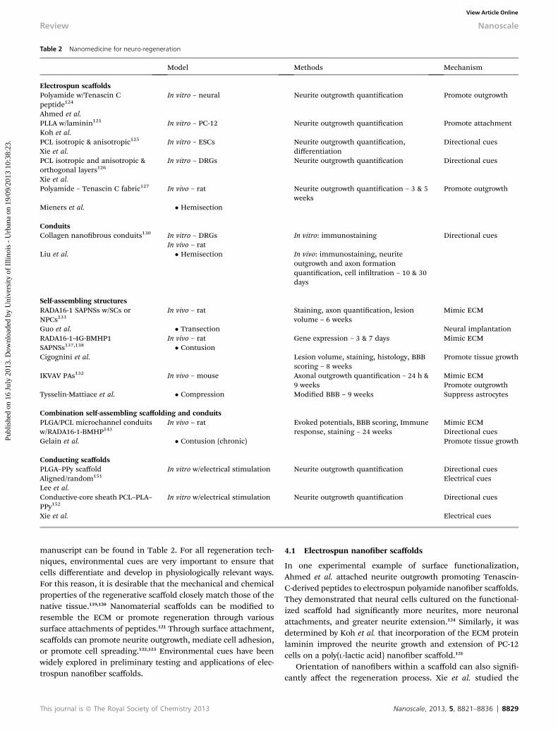
Table 2 Nanomedicine for neuro-regeneration
Model Methods Mechanism
Electrospun scaffoldsPolyamide w/Tenascin Cpeptide124
In vitro – neural Neurite outgrowth quantication Promote outgrowth
Ahmed et al.PLLA w/laminin121 In vitro – PC-12 Neurite outgrowth quantication Promote attachmentKoh et al.PCL isotropic & anisotropic125 In vitro – ESCs Neurite outgrowth quantication,
differentiationDirectional cues
Xie et al.PCL isotropic and anisotropic &orthogonal layers126
In vitro – DRGs Neurite outgrowth quantication Directional cues
Xie et al.Polyamide – Tenascin C fabric127 In vivo – rat Neurite outgrowth quantication – 3 & 5
weeksPromote outgrowth
Mieners et al. � Hemisection
ConduitsCollagen nanobrous conduits130 In vitro – DRGs In vitro: immunostaining Directional cues
In vivo – ratLiu et al. � Hemisection In vivo: immunostaining, neurite
outgrowth and axon formationquantication, cell inltration – 10 & 30days
Self-assembling structuresRADA16-1 SAPNSs w/SCs orNPCs133
In vivo – rat Staining, axon quantication, lesionvolume – 6 weeks
Mimic ECM
Guo et al. � Transection Neural implantationRADA16-1-4G-BMHP1 In vivo – rat Gene expression – 3 & 7 days Mimic ECMSAPNSs137,138 � ContusionCigognini et al. Lesion volume, staining, histology, BBB
scoring – 8 weeksPromote tissue growth
IKVAV PAs132 In vivo – mouse Axonal outgrowth quantication – 24 h &9 weeks
Mimic ECMPromote outgrowthSuppress astrocytesTysselin-Mattiace et al. � Compression Modied BBB – 9 weeks
Combination self-assembling scaffolding and conduitsPLGA/PCL microchannel conduitsw/RADA16-1-BMHP143
In vivo – rat Evoked potentials, BBB scoring, Immuneresponse, staining – 24 weeks
Mimic ECMDirectional cues
Gelain et al. � Contusion (chronic) Promote tissue growth
Conducting scaffoldsPLGA–PPy scaffold In vitro w/electrical stimulation Neurite outgrowth quantication Directional cuesAligned/random151 Electrical cuesLee et al.Conductive-core sheath PCL–PLA–PPy152
In vitro w/electrical stimulation Neurite outgrowth quantication Directional cues
Xie et al. Electrical cues
Review Nanoscale
Publ
ishe
d on
16
July
201
3. D
ownl
oade
d by
Uni
vers
ity o
f Il
linoi
s -
Urb
ana
on 1
9/09
/201
3 10
:38:
23.
View Article Online
manuscript can be found in Table 2. For all regeneration tech-niques, environmental cues are very important to ensure thatcells differentiate and develop in physiologically relevant ways.For this reason, it is desirable that the mechanical and chemicalproperties of the regenerative scaffold closely match those of thenative tissue.119,120 Nanomaterial scaffolds can be modied toresemble the ECM or promote regeneration through varioussurface attachments of peptides.121 Through surface attachment,scaffolds can promote neurite outgrowth, mediate cell adhesion,or promote cell spreading.122,123 Environmental cues have beenwidely explored in preliminary testing and applications of elec-trospun nanober scaffolds.
This journal is ª The Royal Society of Chemistry 2013
4.1 Electrospun nanober scaffolds
In one experimental example of surface functionalization,Ahmed et al. attached neurite outgrowth promoting Tenascin-C-derived peptides to electrospun polyamide nanober scaffolds.They demonstrated that neural cells cultured on the functional-ized scaffold had signicantly more neurites, more neuronalattachments, and greater neurite extension.124 Similarly, it wasdetermined by Koh et al. that incorporation of the ECM proteinlaminin improved the neurite growth and extension of PC-12cells on a poly(L-lactic acid) nanober scaffold.121
Orientation of nanobers within a scaffold can also signi-cantly affect the regeneration process. Xie et al. studied the
Nanoscale, 2013, 5, 8821–8836 | 8829

Nanoscale Review
Publ
ishe
d on
16
July
201
3. D
ownl
oade
d by
Uni
vers
ity o
f Il
linoi
s -
Urb
ana
on 1
9/09
/201
3 10
:38:
23.
View Article Online
differentiation of embryonic stem cells seeded on both isotropicand anisotropic biodegradable poly(3-caprolactone) (PCL)nanober scaffolds prepared via electrospinning. They discov-ered that scaffolds with aligned bers not only discouraged thedifferentiation of embryonic stem cells into astrocytes, whichare prevalent in glial scarring; but also promoted outgrowth ofneurites parallel to the direction of ber alignment.125 Lookinginto this phenomenon further, they found that dorsal rootganglia cells grown on the border between random and alignedbers grew simultaneously radially and directionally, depend-ing on the underlying ber orientation.126 In the same study,dorsal root ganglia cells were grown on double layer scaffolds inwhich bers were aligned in different directions. This resultedin a biaxial growth pattern, which suggests that different layersof the scaffold can inuence neurite outgrowth. Meiners et al.reached a supporting conclusion regarding neurite outgrowthduring an in vivo experiment in which polyamide nanoberfabric was implanted into a rat hemi-section model. Althoughaxonal growth was supported, the random orientation of thebers in the fabric impeded the forward movement of theneurites and subsequently, regeneration was not verysuccessful.127
4.2 Conduits
Another option for directing neuronal growth is the use ofconduits. Conduits are tubes that facilitate communicationbetween the proximal and distal ends of the nerve gap andprovide physical guidance for regrowth. Conduits have beensuccessful in peripheral nerve regeneration, as demonstrated byfeats like the regeneration of sciatic nerve over an 80 mm gap ina beagle model using a polyglycolic acid–collagen tube lledwith laminin coated collagen bers.128 Nanobrous conduitscan be formed via electrospinning and have been used forperipheral nerve regeneration. In one recent study, PCL nano-brous conduits were able to close a 15 mm gap in a rat sciaticnerve model and were also able to generate signicantly moremyelinated axons with thicker myelin sheaths than microberconduits and lm conduits.129 Clearly, nanobers have signi-cant benets in neural regeneration. However, despite thesuccess of collagen and nanobrous conduits in peripheralnerve regeneration, nanoscale conduits have been lesssuccessful in repairing SCI. In one study by Liu et al., tubularconduits were formed from either random or aligned electro-spun collagen nanobers and implanted in a short-term rathemisection model of SCI.130 Regardless of ber orientation,neurolament sprouting was observed at 10 days post-implan-tation, although the orientation of these regenerated axons wasnot obvious. There were a limited number of neural bersobserved in the center of the conduit, even at 30 days post-implantation. Further improvements on these nanobrousconduit systems can be made through surface functionaliza-tion, and perhaps future designs will be more successful.Although these electrospun scaffolds and conduits have theorientation benets of aligned bers and directional guidance,a limitation of these systems is that they must be invasivelyimplanted into the subject. This can cause further damage to
8830 | Nanoscale, 2013, 5, 8821–8836
the spinal cord by disrupting spared tissue, and lead to infec-tion or other surgical complications.131 To overcome theselimitations associated with implantation there has beenincreasing interest in injectables, such as self-assemblingscaffolds and hydrogels, which solidify under in vivo conditions.
4.3 Self-assembling systems
Self-assembling peptide systems are synthetic amino acid basedsystems that transition from a solution to a gel within secondsunder in vivo pH and ion concentration conditions. The resultingmaterial is a nanobrous mesh similar to the native ECM thatbiodegrades naturally several weeks aer implantation.132 Thegels are generally biocompatible and non-cytotoxic, although insome cases pH must be buffered prior to implantation. The gelscan also safely encapsulate cells or drugs for combination ther-apies.133 Self-assembling peptide systems are generally dividedinto several categories; self-assembling peptide amphiphiles(PAs), self-assembling peptide nanober scaffolds (SAPNSs),Amphiphilic diblock copolypeptide hydrogels (DCHs), and mix-ing induced two component protein gels (MITCHs).134 DCHs aresynthetic polymers, which, through hydrophobic association inwater, form into gel structures with porous bril-like nano-structures. DCHs have been successfully injected withoutsignicant immune response or toxicity into the mouse fore-brain, and were able to integrate with tissue, support the in-growth of blood vessels, glia, and some nerve bers.135 MITCHSare synthetic protein gels and can be formed to gel upon themixing of its two protein components. MITCHs have been shownto support neural stem cells, which were able differentiate,replicate, and sprout neurites.136 Neither DCHs nor MITCHs havebeen successfully applied yet in an in vivo SCI model.
SAPNSs have been used in several in vivo models withpromising results. SAPNSs are synthetic biomaterials formed ofionic self-complementary peptides that form into a nano-lamentous, hydrated scaffold under in vivo pH. Guo et al. usedRADA16-1 SAPNS loaded with either Schwann cells or neuralprogenitor cells in an in vivo rat transection model of SCI.133
Because of the low pH when untreated, the SAPNSs wereneutralized before transplantation; otherwise the treatmentdamaged the host tissue. When evaluated aer 6 weeks theneutralized SAPNSs had integrated well with the host tissue andhad greatly decreased inammation at the lesion site. SAPNSsseeded with cells, especially Schwann cells, showed many axonsinltrating the implant. Furthermore, host cells had migratedinto the implant and there was robust growth of blood vessels,indicating the potential for repairing damaged tissue andproviding the necessary supporting vasculature.
A similar study was performed by Cigognini et al. usingRADA16-1 SAPNSs modied with bone marrow homing motif(BMHP1), which has previously demonstrated the ability topromote nerve tissue regrowth, and a linker (4G).137,138 In an invivo rat contusion model, RADA16-1-4G-BMHP1 was injectedimmediately following injury. Assessment of gene expression at 7days demonstrated that in treated animals there was a generalupregulation of GAP-43, which correlates with axonal regenera-tion;139 trophic factors, which suggests synaptic formation;140 and
This journal is ª The Royal Society of Chemistry 2013

Review Nanoscale
Publ
ishe
d on
16
July
201
3. D
ownl
oade
d by
Uni
vers
ity o
f Il
linoi
s -
Urb
ana
on 1
9/09
/201
3 10
:38:
23.
View Article Online
ECM remodeling proteins, indicating restructuring of the ECMand subsequent axonal growth.141 Together, these results suggesttissue regeneration. BBB scoring was performed for 8 weeks totrack locomotor recovery, and it was found that there was a verysmall but statistically signicant improvement in motor perfor-mance and coordination. Additionally, the SAPNS was compat-ible with surrounding nervous tissue and was able to ll thecavity. Within the lesion there was increased cellular inltration,basement membrane deposition, and axon regeneration andsprouting. This study conrms the potential uses of SAPNSs as aless invasive means of treating SCI.
Self-assembling PAs have also been successfully tested in vitroand in vivo. PA molecules assemble into supramolecular nano-bers, which at in vivo ion concentration form a continuous,random, mesh-like network. In one study by Tysselin-Mattiaceet al., PAs were designed to display the laminin epitope IKVAVupon assembly.132 The IKVAV epitope is known to promoteoutgrowth of neurites and suppress formation of astrocytes, twoproperties that are desirable for preventing glial scarring.142 Inthis study, IKVAV PAs were injected into a compression injuredmouse model of SCI 24 hours post-injury. Several differentbenecial effects were observed with this treatment. IKVAV PAssuccessfully reduced astrogliosis and cell death. At 11 weeks post-injury it was observed that almost 80% of all labeledmotor axonsin the IKVAV PA group entered the lesion compared with 50% inthe control group. Furthermore, while no bers in the controlgroup made it even a quarter of the way across the lesion, 50% ofthe bers in the treated group penetrated half way through, and35% traversed the entire lesion. Similar results were found withthe sensory axons at 11 weeks, as 60% of axons entered the injurysite in the IKVAV PA group, and only 20% did in the controlgroup. Of these axons, none in the control group penetratedhalfway, while 25% of the bers in the treated group made it halfthe way across the lesion and 10% crossed the whole gap.Although these axons took indirect routes through the lesion,indicating a lack of directional cues, crossing the injury site is avery noteworthy accomplishment. A small but statisticallysignicant behavioral improvement was also observed using amodied BBB scoring system. The IKVAV PAs demonstrateda good starting point for noninvasively repairing SCI within aclinically relevant treatment window.
4.4 Combination of self-assembling and scaffolding systems
In order to glean the benets of both self-assembling systems anddirectional cues provided by scaffolding, combination approacheshave been developed. Gelain et al. developed composite guidancechannels constructed from electrospun PLGA/PCL nanoberslled with RADA16-1-BMHP1 self-assembling peptides in order totackle the substantial challenge of chronic SCI.143 This “neuro-prosthesis” was implanted at the injury site 1 month aercontusion injury in a ratmodel, and was evaluated 6months later.At the time of evaluation, while cysts persisted in control andsham animals, there was neo-formed tissue in the animalsreceiving the microconduit implants. This neo-tissue lackedinammation and contained regenerating axons andmyelin, well-developed vascular structures, cell-deposited ECM, and stromal
This journal is ª The Royal Society of Chemistry 2013
cells. An example of these results can be seen in Fig. 3.143
Furthermore, there was a signicant improvement in the motorfunction of the animals as evaluated by BBB scoring, and also inevoked responses in the ascending tracts. This strong evidencedemonstrates that it is possible to reconstruct the anatomical andhistological framework and restore signicant motor and elec-trical function in a chronically injured spinal cord. Despite thesuccesses observed, this method does not solve the problem ofinvasive surgery and many complications were observed; 25–33%of animals died due to postsurgical complications. However, itstill is an incredible step in treating chronic injury, which somanypatients currently suffer from.
4.5 Combination of nanober scaffolding and electricalstimulation
Another interesting combination method under investigationfor regenerating the spinal cord is the combination of nanoberscaffolding and electrical stimulation. Electrical stimulationhas long been investigated and used in treatment ofSCI.36,37,144,145 It is known that in the absence of topographicalcues neurons align and extend in the presence of electricalstimulation.146,147 Electrically conductive synthetic polymers,such as polypyrrol (PPy) or polyaniline, have been combinedwith biodegradable polymers and investigated as scaffoldingmaterials for repairing conducting tissue. These polymers haveeven been used for in vivo testing as an interior coating onconduits for nerve repair.148–150 In one study, a nanobrousmesh-like electrospun PLGA scaffold coated with a nanothicklayer of PPy was tested for potential application as neural scaf-fold material.151 Neurons were cultured on both aligned andrandom scaffolds, and electrical stimulation was applied to thecells. It was found that in the presence of stimulation 40–90%more neurites were generated and neurites were 40–50% longer.Furthermore, there were more and longer neurites on thealigned scaffolds than on the random scaffolds, suggestingcombined benets. Conductive-core sheath PCL–PPy andpoly(L-lactide)–PPy (PLA–PPy) nanobers have found similarsuccess. In a study by Xie et al., it was found that dorsal rootganglion cells incubated with uniaxially aligned core-sheathPLA–PPy and PCL–PPy nanober scaffolds had a 1.82-foldincrease in neurite extension compared with randomly alignedscaffolds.152 With electrical stimulation there was an increase inneurite length of 1.83-fold and 1.47-fold for aligned and randomscaffolds respectively, again demonstrating the synergisticeffects of electrical stimulation and ber alignment. Carbonnanostructures also have the ability to conduct electricity. Asdetailed earlier, carbon nanotubes have been tested previouslyfor a neuroprotective and regenerative SCI treatment109 and alsohave been investigated for regenerative nerve repair.107,153–155
Carbon nanotubes have also been applied in multifunctionalneural interfaces that provide electrical and topographicalcues.156 Given their unique properties, carbon nanotubesprovide a wealth of options, including functionalization,incorporation into scaffolding, and electrical stimulation; all ofwhich are interesting and promising directions for regenerativetreatment.
Nanoscale, 2013, 5, 8821–8836 | 8831

Fig. 3 Nerve regeneration in chronically injured rat spinal cord using PLGA/PCL microchannel conduits with RADA16-1-BMHP. Dashed lines outline implanted channelwalls in (A)–(C). In transverse sections (A), GAP-43 positive fibers were found inside and between transplanted tubes (arrows; coronal section). (B) NF200 positive fiverswere seen crossing the top rostral interface of the lesion. (C) bIII-tubulin positive fasciculi stretched through the lumen of the conduits in a longitudinal spinal cordsection. (D) Quantitative analysis (mean� SEM) of the percentage of channels showing positivity for neural markers throughout the entire tube length (N¼ 5 for group1, N ¼ 8 for group 8). Statistical significance (*) p ¼ 0.03; (**) p ¼ 0.003. NF200 (green) and GAP43 (red) did not colocalize inside the same fiver (E). Myelinated (green,arrow) and unmyelinated (red, arrowhead) bIII-tubulin positive fibers (F) were observed (enlargement in G). Both SMI-32 (H) and SMI-31 (I) positive fibers were detectedwithin guidance conduits, in adjacent coronal sections. In the longitudinal reconstruction of transplanted cord in (J) (group 2), NF200 positive fibers cover the wholelength (approx. 2 mm) of the lesion, crossing both the rostral and the caudal channels–tissue interfaces (dotted line). Reprinted with permission from F. Gelain, S.Panseri, S. Antonini, C. Cunha, M. Donega, J. Lowery, F. Taraballi, G. Cerri, M. Montagna and F. Baldissera, ACS Nano, 2011. Copyright 2010 American Chemical Society.
Nanoscale Review
Publ
ishe
d on
16
July
201
3. D
ownl
oade
d by
Uni
vers
ity o
f Il
linoi
s -
Urb
ana
on 1
9/09
/201
3 10
:38:
23.
View Article Online
Although testing has been limited to in vitro testing so far,electrically stimulated nanoscaffolding is an intriguing optionfor spinal cord repair and regeneration, although in vivo inte-gration of stimulation in a safe and effective manner may proveto be a substantial challenge.
5 Discussion
Although SCI will never disappear entirely, perhaps futurevictims of SCI will not share quite the same fate as those whosuffer today. Treatment of both acute and chronic SCI injury
8832 | Nanoscale, 2013, 5, 8821–8836
remains a multi-faceted medical challenge, but with continuedinvestigations of nanomedicine, the future looks bright. Therehas been general consensus157–159 that in order to achieve thebest effects a combination of both neuroprotection and regen-eration treatments should be employed. This dual prongedapproach would work to decrease the amount of regenerationnecessary by sparing the maximum amount of tissue, then workto repair this latent damage, resulting in a maximally healedand functional spinal cord. As both elds continue to advance, acombined approach could help us realize the goal of functionalrecovery much faster than either eld could on its own. NPs,75,76
This journal is ª The Royal Society of Chemistry 2013

Review Nanoscale
Publ
ishe
d on
16
July
201
3. D
ownl
oade
d by
Uni
vers
ity o
f Il
linoi
s -
Urb
ana
on 1
9/09
/201
3 10
:38:
23.
View Article Online
liposomes,94,95 and micelles59 have shown the ability to increasebioavailability of neuroprotective therapies to the spinal cordaer injection. Such improvements in delivery have the capa-bility of improving clinical outcomes by reducing the requireddosages and systemic toxicity of current treatments. Thesedelivery vehicles also have the exibility to potentially deliverother drugs that may improve upon current standards. Besidesimproving the bioavailability of neuroprotective treatment,nanomaterials with innate neuroprotective attributes, such asmPEG–PDLLA micelles,60 ceria NPs,92 fullerenes,104,105 andcarbon nanotubes,109 have been developed and tested in vivo orin vitro with varying degrees of success. While in vitro testingcan give an indication of the abilities of the nanomaterial, invivo testing is very important for establishing clinical value.Neuroprotective treatments like mPEG–PDLLA micelles,60
PSiNPs,75,76 and MP-NPs,71 have had successful in vivo tests, anddemonstrate exciting and signicant behavioral or electricalfunctional recovery. Treatments, such as SWNT–PEG,109
temperature responsive Pluronic NPs,72 and TAT–PEG–MPLs94,95
have been tested in vivo, but may consider changing the focus ofthe study (e.g., from delivery or characterization to recovery) orusing alternative animal models (e.g., contusion instead ofcomplete transection) that would permit a better comparisonwith other treatments. Still other treatments, such as ceria NPs92
and PBCA-NPs,91 are intriguing prospects but have not beentested in vivo will require a great deal of validation.
Neural regenerative strategies have also had success withnanomedicine, specically nanostructures. Nanober scaffolds,self-assembling systems, conduits, and combination systemshave demonstrated promising results in both in vivo and in vitrotesting. Self-assembling peptides132,133,137,138 were able to showformation of blood vessels, signicant progression of motorbers through the lesion site, recovery of function and asignicant reduction in astrocytes production. These results arepromising steps towards developing full vascular support,eliminating glial scar formation, and recovering conduction;important building blocks towards full recovery. Successfulrepair of chronic injury is especially of notice, particularlybecause there are so many patients currently suffering fromparalysis. The combination microchannel guidance and self-assembling peptide scaffolding neuroprosthetic implant143
showed exciting results in treating chronic SCI. While thisdevice was able to successfully develop neo-tissue and havefunctional motor and electrical recovery, there were manyproblems related to the invasive application of the device thatmust be resolved before possible application in humans. Ifresults like these can be achieved in a chronic case with non-invasive techniques, the implications would be enormous.Although there is a great deal of work to be done in this area,especially with successful integration of directional, chemical,and structural cues in a non-invasive manner, a great deal ofprogress has been realized in a few short years and thisadvancement will only continue with development and appli-cation of new nanotechnologies.
For both regenerative and neuroprotective strategies,comparative studies between treatments involving severaldifferent markers for functional recovery may be an important
This journal is ª The Royal Society of Chemistry 2013
next step in identifying treatments to move forward with forfurther development and clinical testing. The best treatmentmay not yet have been developed, but some the current researchexamined has shown signicant improvement over the currentstandards of care. With the progression of nanomedicine, weare entering a new stage in the evolution of SCI treatment.Although there are still many obstacles ahead of us, the future isbright. Nanomedicine, with its multifunctional and combina-torial capabilities can allow these treatment approaches to beblended, providing avenues through which the dream of pre-venting or reversing paralysis caused by SCI may be attained.
Acknowledgements
This work was supported by a Translation Research Grant fromthe Coulter Foundation and by the Indiana Clinical and Trans-lational Sciences Institute funded, in part by Grant # RR025761,from the National Institutes of Health, National Center forResearch Resources, Clinical and Translational Sciences Award.J.Y.T. acknowledges the Charles C. Chappelle fellowship.
References
1 J. Lasfargues, D. Custis and F. Morrone, Spinal Cord, 1995,33, 62–68.
2 M. J. De Vivo, J. Stuart Krause and D. P. Lammertse, Arch.Phys. Med. Rehabil., 1999, 80, 1411–1419.
3 C. H. Tator, Brain Pathol., 1995, 5, 407–413.4 C. E. Hulsebosch, Adv. Physiol. Educ., 2002, 26, 238–255.5 B. K. Kwon, W. Tetzlaff, J. N. Grauer, J. Beiner andA. R. Vaccaro, Spine J., 2004, 4, 451–464.
6 T. Hagg and M. Oudega, J. Neurotrauma, 2006, 23, 263–280.7 S. GhoshMitra, D. R. Diercks, N. C. Mills, D. L. Hynds andS. Ghosh, Adv. Drug Delivery Rev., 2012, 64, 110–125.
8 A. P. Amar and M. L. Levy, Neurosurgery, 1999, 44, 1027.9 E. D. Hall and J. E. Springer, NeuroRx, 2004, 1, 80–100.10 E. C. Field-Fote, Spinal Cord Injury Rehabilitation, FA Davis
Company, Philadelphia, 2009.11 M. B. Bracken, M. J. Shepard, T. R. Holford, L. Leo-
Summers, E. F. Aldrich, M. Fazl, M. Fehlings, D. L. Herr,P. W. Hitchon and L. F. Marshall, JAMA, J. Am. Med.Assoc., 1997, 277, 1597–1604.
12 J. Xu, G. Fan, S. Chen, Y. Wu, X. M. Xu and C. Y. Hsu, Mol.Brain Res., 1998, 59, 135–142.
13 R. J. Hurlbert, J. Neurosurg., 2000, 93, 1–7.14 S. Gerndt, J. Rodriguez, J. Pawlik, P. Taheri, W. Wahl,
A. Micheals and S. Papadopoulos, J. Trauma, 1997, 42, 279.15 B. Suberviola, A. Gonzalez-Castro, J. Llorca, F. Ortiz-Melon
and E. Minambres, Injury, 2008, 39, 748–752.16 T. Qian, X. Guo, A. Levi, S. Vanni, R. Shebert and M. Sipski,
Spinal Cord, 2004, 43, 199–203.17 M. B. Bracken and T. R. Holford, J. Neurosurg., 1993, 79,
500–507.18 B. S. Bregman, Curr. Opin. Neurobiol., 1998, 8, 800–807.19 D. J. Begley, Pharmacol. Ther., 2004, 104, 29–45.20 P. Ballabh, A. Braun and M. Nedergaard, Neurobiol. Dis.,
2004, 16, 1–13.
Nanoscale, 2013, 5, 8821–8836 | 8833

Nanoscale Review
Publ
ishe
d on
16
July
201
3. D
ownl
oade
d by
Uni
vers
ity o
f Il
linoi
s -
Urb
ana
on 1
9/09
/201
3 10
:38:
23.
View Article Online
21 T. M. Allen and P. R. Cullis, Science, 2004, 303, 1818–1822.22 P. S. Hodgson, J. M. Neal, J. E. Pollock and S. S. Liu, Anesth.
Analg., 1999, 88, 797.23 R. G. Thorne and W. Frey, Clin. Pharmacokinet., 2001, 40,
907–946.24 C. E. Kang, P. C. Poon, C. H. Tator andM. S. Shoichet, Tissue
Eng., Part A, 2008, 15, 595–604.25 J. Lu, K. W. S. Ashwell and P. Waite, Spine, 2000, 25, 1859.26 A. P. Amar and M. L. Levy, Neurosurgery, 1999, 44, 1027–
1039.27 B. K. Kwon, C. G. Fisher, M. F. Dvorak and W. Tetzlaff,
Spine, 2005, 30, S3–S13.28 D. Gris, D. R. Marsh, M. A. Oatway, Y. Chen, E. F. Hamilton,
G. A. Dekaban and L. C. Weaver, J. Neurosci., 2004, 24, 4043–4051.
29 E. Kaptanoglu, I. Solaroglu, O. Okutan, H. S. Surucu,F. Akbiyik and E. Beskonakli, Neurosurg. Rev., 2004, 27,113–120.
30 M. Celik, N. Gokmen, S. Erbayraktar, M. Akhisaroglu,S. Konakc, C. Ulukus, S. Genc, K. Genc, E. Sagiroglu andA. Cerami, Proc. Natl. Acad. Sci. U. S. A., 2002, 99, 2258–2263.
31 S. M. Lee, T. Y. Yune, S. J. Kim, D. W. Park, Y. K. Lee,Y. C. Kim, Y. J. Oh, G. J. Markelonis and T. H. Oh, J.Neurotrauma, 2003, 20, 1017–1027.
32 J. E. Wells, R. J. Hurlbert, M. G. Fehlings and V. W. Yong,Brain, 2003, 126, 1628–1637.
33 D. Short, W. El Masry and P. Jones, Spinal Cord, 2000, 38,273–286.
34 J. D. Houle and A. Tessler, Exp. Neurol., 2003, 182, 247–260.35 S. Hamid and R. Hayek, Eur. Spine J., 2008, 17, 1256–1269.36 S. Shapiro, R. Borgens, R. Pascuzzi, K. Roos, M. Groff,
S. Purvines, R. B. Rodgers, S. Hagy and P. Nelson, J.Neurosurg., 2005, 2, 3–10.
37 K. Ragnarsson, Spinal Cord, 2007, 46, 255–274.38 H. S. Keirstead, G. Nistor, G. Bernal, M. Totoiu, F. Cloutier,
K. Sharp and O. Steward, J. Neurosci., 2005, 25, 4694–4705.39 A. Iwanami, S. Kaneko, M. Nakamura, Y. Kanemura,
H. Mori, S. Kobayashi, M. Yamasaki, S. Momoshima,H. Ishii and K. Ando, J. Neurosci. Res., 2005, 80, 182–190.
40 B. S. Bregman, J. V. Coumans, H. N. Dai, P. L. Kuhn,J. Lynskey, M. McAtee and F. Sandhu, Prog. Brain Res.,2002, 137, 257–273.
41 P. Lu, H. Yang, L. L. Jones, M. T. Filbin andM. H. Tuszynski,J. Neurosci., 2004, 24, 6402–6409.
42 N. Daum, C. Tscheka, A. Neumeyer and M. Schneider,WileyInterdiscip. Rev.: Nanomed. Nanobiotechnol., 2011, 4, 52–65.
43 L. Juillerat-Jeanneret, Drug Discovery Today, 2008, 13, 1099–1106.
44 H. S. Sharma, S. Ali, Z. R. Tian, R. Patnaik, S. Patnaik,P. Lek, A. Sharma and T. Lundstedt, J. Nanosci.Nanotechnol., 2009, 9, 5014–5037.
45 M. S. Ramer, J. V. Priestley and S. B. McMahon, Nature,2000, 403, 312–316.
46 J. W. Fawcett, J. Neurotrauma, 2006, 23, 371–383.47 J. Silver and J. H. Miller, Nat. Rev. Neurosci., 2004, 5, 146–
156.48 P. J. Horner and F. H. Gage, Nature, 2000, 963–970.
8834 | Nanoscale, 2013, 5, 8821–8836
49 C. E. Schmidt and J. B. Leach, Annu. Rev. Biomed. Eng., 2003,5, 293–347.
50 L. Zhang and T. J. Webster, Nano Today, 2009, 4, 66–80.51 R. Agarwal and C. Lieber, Appl. Phys. A: Mater. Sci. Process.,
2006, 85, 209–215.52 A. Javey, S. W. Nam, R. S. Friedman, H. Yan and
C. M. Lieber, Nano Lett., 2007, 7, 773–777.53 K. E. Fischer, B. J. Aleman, S. L. Tao, R. H. Daniels, E. M. Li,
M. D. Bunger, G. Nagaraj, P. Singh, A. Zettl and T. A. Desai,Nano Lett., 2009, 9, 716–720.
54 V. Uskokovifa, P. P. Lee, L. A. Walsh, K. E. Fischer andT. A. Desai, Biomaterials, 2012, 33, 1663–1672.
55 H. S. Sharma, Nanomedicine, 2007, 2, 753–758.56 H. S. Sharma, S. F. Ali, Z. R. Tian, R. Patnaik, S. Patnaik,
A. Sharma, A. Boman, P. Lek, E. Seifert and T. Lundstedt,Brain Edema XIV, 2010, pp. 343–350.
57 H. S. Sharma, S. F. Ali, W. Dong, Z. Tian, R. Patnaik,S. Patnaik, A. Sharma, A. Boman, P. Lek and E. Seifert,Ann. N. Y. Acad. Sci., 2007, 1122, 197–218.
58 A. E. M. Mautes, M. R. Weinzierl, F. Donovan andL. J. Noble, Phys. Ther., 2000, 80, 673–687.
59 C. L. Chen, S. F. Chang, D. Lee, L. Y. Yang, Y. H. Lee,C. Y. Hsu, S. J. Lin and J. Liaw, Pharm. Res., 2008, 25, 39–47.
60 Y. Shi, S. Kim, T. B. Huff, R. B. Borgens, K. Park, R. Shi andJ. X. Cheng, Nat. Nanotechnol., 2009, 5, 80–87.
61 K. Kataoka, A. Harada and Y. Nagasaki, Adv. Drug DeliveryRev., 2012, 64, 37–48.
62 D. Bazile, C. Prud'homme, M. T. Bassoullet, M. Marlard,G. Spenlehauer and M. Veillard, J. Pharm. Sci., 1995, 84,493–498.
63 T. M. Allen, J. Liposome Res., 1992, 2, 289–305.64 G. He, L. L. Ma, J. Pan and S. Venkatraman, Int. J. Pharm.,
2007, 334, 48–55.65 G. Orive, E. Anitua, J. L. Pedraz and D. F. Emerich, Nat. Rev.
Neurosci., 2009, 10, 682–692.66 D.M. Basso, M. S. Beattie and J. C. Bresnahan, J. Neurotrauma,
1995, 12, 1–21.67 H. Chen, S. Kim, L. Li, S. Wang, K. Park and J.-X. Cheng,
Proc. Natl. Acad. Sci. U. S. A., 2008, 105, 6596–6601.68 H. Chen, S. Kim, W. He, H. Wang, P. S. Low, K. Park and
J.-X. Cheng, Langmuir, 2008, 24, 5213–5217.69 S.-Y. Lee, S. Kim, J. Y. Tyler, K. Park and J.-X. Cheng,
Biomaterials, 2013, 34, 552–561.70 K. S. Soppimath, T. M. Aminabhavi, A. R. Kulkarni and
W. E. Rudzinski, J. Controlled Release, 2001, 70, 1–20.71 Y. Kim, J. M. Caldwell and R. V. Bellamkonda, Biomaterials,
2009, 30, 2582–2590.72 S. Chen, Y. Li, C. Guo, J. Wang, J. Ma, X. Liang, L. R. Yang
and H. Z. Liu, Langmuir, 2007, 23, 12669–12676.73 B. Bose, J. L. Osterholm and M. Kalia, Neurosci. Lett., 1986,
63, 165–169.74 A. L. R. Oliveira and F. Langone, Neurosci. Lett., 2000, 293,
131–134.75 Y. Cho, R. Shi, A. Ivanisevic and R. Ben Borgens, Journal of
neuroscience research, 2010, 88, 1433–1444.76 Y. Cho, R. Shi, R. Borgens and A. Ivanisevic, Small, 2008, 4,
1676–1681.
This journal is ª The Royal Society of Chemistry 2013
Review Nanoscale
Publ
ishe
d on
16
July
201
3. D
ownl
oade
d by
Uni
vers
ity o
f Il
linoi
s -
Urb
ana
on 1
9/09
/201
3 10
:38:
23.
View Article Online
77 J. Luo, R. Borgens and R. Shi, J. Neurochem., 2002, 83, 471–480.
78 J. Luo, R. Borgens and R. Shi, J. Neurotrauma, 2004, 21, 994–1007.
79 J. Luo and R. Shi, Brain Res., 2007, 1155, 10–16.80 R. B. Borgens and R. Shi, FASEB J., 2000, 14, 27–35.81 R. B. Borgens, R. Shi and D. Bohnert, J. Exp. Biol., 2002, 205,
1–12.82 Y. Cho, R. Shi and R. B. Borgens, J. Exp. Biol., 2010, 213,
1513–1520.83 J. Brent, Drugs, 2001, 61, 979–988.84 Y. Cho, R. Shi, R. B. Borgens and A. Ivanisevic,
Nanomedicine, 2008, 3, 507–519.85 K. Hamann, A. Durkes, H. Ouyang, K. Uchida, A. Pond and
R. Shi, J. Neurochem., 2008, 107, 712–721.86 P. Liu-Snyder, R. B. Borgens and R. Shi, J. Neurosci. Res.,
2006, 84, 219–227.87 J. Kreuter, R. N. Alyautdin, D. A. Kharkevich and
A. A. Ivanov, Brain Res., 1995, 674, 171–174.88 R. N. Alyautdin, V. E. Petrov, K. Langer, A. Berthold,
D. A. Kharkevich and J. Kreuter, Pharm. Res., 1997, 14,325–328.
89 R. N. Alyaudtin, A. Reichel, R. Lobenberg, P. Ramge,J. Kreuter and D. J. Begley, J. Drug Targeting, 2001, 9, 209–221.
90 P. Ramge, R. E. Unger, J. B. Oltrogge, D. Zenker, D. Begley,J. Kreuter and H. Von Briesen, Eur. J. Neurosci., 2000, 12,1931–1940.
91 V. Reukov, V. Maximov and A. Vertegel, Biotechnol. Bioeng.,2011, 108, 243–252.
92 M. Das, S. Patil, N. Bhargava, J. F. Kang, L. M. Riedel, S. Sealand J. J. Hickman, Biomaterials, 2007, 28, 1918–1925.
93 A. Sharma and U. S. Sharma, Int. J. Pharm., 1997, 154, 123–140.
94 Y. Liu, C. Y. Wang, X. H. Kong, H. J. Wang, J. Chang,D. P. Zhang, D. X. Ban and S. Q. Feng, J. Drug Targeting,2010, 18, 420–429.
95 H. Wang, S. Zhang, Z. Liao, C. Wang, Y. Liu, S. Feng,X. Jiang and J. Chang, Biomaterials, 2010, 31, 6589–6596.
96 S. R. Schwarze, K. A. Hruska and S. F. Dowdy, Trends CellBiol., 2000, 10, 290–295.
97 S. C. Semple, A. Chonn and P. R. Cullis, Adv. Drug DeliveryRev., 1998, 32, 3–17.
98 T. Hernandez-Caselles, J. Villalain and J. Gomez-Fernandez,Mol. Cell. Biochem., 1993, 120, 119–126.
99 I. C. Wang, L. A. Tai, D. D. Lee, P. Kanakamma,C. K. F. Shen, T. Y. Luh, C. H. Cheng and K. C. Hwang, J.Med. Chem., 1999, 42, 4614–4620.
100 S. Bosi, T. Da Ros, G. Spalluto and M. Prato, Eur. J. Med.Chem., 2003, 38, 913–923.
101 M. Brettreich and A. Hirsch, Tetrahedron Lett., 1998, 39,2731–2734.
102 A. Bar-Shir, Y. Engel and M. Gozin, J. Org. Chem., 2005, 70,2660–2666.
103 L. L. Dugan, D. M. Turetsky, C. Du, D. Lobner, M. Wheeler,C. R. Almli, C. K. F. Shen, T. Y. Luh, D. W. Choi andT. S. Lin, Proc. Natl. Acad. Sci. U. S. A., 1997, 94, 9434.
This journal is ª The Royal Society of Chemistry 2013
104 H. Jin, W. Chen, X. Tang, L. Chiang, C. Yang, J. Schloss andJ. Wu, J. Neurosci. Res., 2000, 62, 600–607.
105 X. F. Liu, W. C. Guan and W. S. Ke, Can. J. Biochem., 2007,85, 157–163.
106 Y. Ni, H. Hu, E. B. Malarkey, B. Zhao, V. Montana,R. C. Haddon and V. Parpura, J. Nanosci. Nanotechnol.,2005, 5, 1707–1712.
107 H. Hu, Y. Ni, V. Montana, R. C. Haddon and V. Parpura,Nano Lett., 2004, 4, 507–511.
108 E. Malarkey and V. Parpura, in Brain Edema XIV, ed.Z. Czernicki, A. Baethmann, U. Ito, Y. Katayama, T.Kuroiwa and D. Mendelow, Springer, Vienna, 2010, vol.106, ch. 62, pp. 337–341.
109 J. A. Roman, T. L. Niedzielko, R. C. Haddon, V. Parpura andC. L. Floyd, J. Neurotrauma, 2011, 28, 2349–2362.
110 F. Alexis, E. Pridgen, L. K. Molnar and O. C. Farokhzad,Mol.Pharm., 2008, 5, 505–515.
111 X. Huang, L. Li, T. Liu, N. Hao, H. Liu, D. Chen and F. Tang,ACS Nano, 2011, 5, 5390–5399.
112 B. Wang, X. He, Z. Zhang, Y. Zhao and W. Feng, Acc. Chem.Res., 2012, 46, 761–769.
113 S. Y. Fu and T. Gordon, Mol. Neurobiol., 1997, 14, 67.114 E. Huebner and S. Strittmatter, Cell biology of the axon,
2009, pp. 339–351.115 B. A. Tucker and K. M.Mearow, Can. J. Neurol. Sci., 2008, 35,
551–566.116 A. Buss, G. Brook, B. Kakulas, D. Martin, R. Franzen,
J. Schoenen, J. Noth and A. Schmitt, Brain, 2004, 127, 34–44.117 G. Yiu and Z. He, Nat. Rev. Neurosci., 2006, 7, 617–627.118 M. G. Fehlings and C. H. Tator, Exp. Neurol., 1995, 132, 220–
228.119 K. S. Straley, C. W. P. Foo and S. C. Heilshorn, J. Neurotrauma,
2010, 27, 1–19.120 C. Miller, S. Jeinija and S. Mallapragada, Tissue Eng., 2002,
8, 367–378.121 H. Koh, T. Yong, C. Chan and S. Ramakrishna, Biomaterials,
2008, 29, 3574–3582.122 N. N. Madigan, S. McMahon, T. O'Brien, M. J. Yaszemski
and A. J. Windebank, Respir. Physiol. Neurobiol., 2009,169, 183–199.
123 L. Ghasemi-Mobarakeh, M. P. Prabhakaran, M. Morshed,M. H. Nasr-Esfahani and S. Ramakrishna, Biomaterials,2008, 29, 4532–4539.
124 I. Ahmed, H. Y. Liu, P. C. Mamiya, A. S. Ponery, A. N. Babu,T. Weik, M. Schindler and S. Meiners, J. Biomed. Mater. Res.,Part A, 2006, 76, 851–860.
125 J. Xie, S. M. Willerth, X. Li, M. R. Macewan, A. Rader,S. E. Sakiyama-Elbert and Y. Xia, Biomaterials, 2009, 30,354–362.
126 J. Xie, M. R. MacEwan, X. Li, S. E. Sakiyama-Elbert andY. Xia, ACS Nano, 2009, 3, 1151–1159.
127 S. Meiners, I. Ahmed, A. S. Ponery, N. Amor, S. L. Harris,V. Ayres, Y. Fan, Q. Chen, R. Delgado-Rivera andA. N. Babu, Polym. Int., 2007, 56, 1340–1348.
128 K. Matsumoto, K. Ohnishi, T. Kiyotani, T. Sekine, H. Ueda,T. Nakamura, K. Endo and Y. Shimizu, Brain Res., 2000,868, 315–328.
Nanoscale, 2013, 5, 8821–8836 | 8835

Nanoscale Review
Publ
ishe
d on
16
July
201
3. D
ownl
oade
d by
Uni
vers
ity o
f Il
linoi
s -
Urb
ana
on 1
9/09
/201
3 10
:38:
23.
View Article Online
129 X. Jiang, R. Mi, A. Hoke and S. Y. Chew, J. Tissue Eng.Regener. Med., 2012, DOI: 10.1002/term.1531.
130 T. Liu, J. D. Houle, J. Xu, B. P. Chan and S. Y. Chew, TissueEng., 2011, 18, 1057–1066.
131 M. Tuszynski, J. Steeves, J. Fawcett, D. Lammertse,M. Kalichman, C. Rask, A. Curt, J. Ditunno, M. Fehlingsand J. Guest, Spinal Cord, 2006, 45, 222–231.
132 V. M. Tysseling-Mattiace, V. Sahni, K. L. Niece, D. Birch,C. Czeisler, M. G. Fehlings, S. I. Stupp and J. A. Kessler,J. Neurosci., 2008, 28, 3814–3823.
133 J. Guo, H. Su, Y. Zeng, Y. X. Liang, W. M. Wong, R. G. Ellis-Behnke, K. F. So and W. Wu, Nanomedicine:Nanotechnology, Biology and Medicine, 2007, 3, 311–321.
134 D. Macaya and M. Spector, Biomed. Mater., 2012, 7, 012001.135 C. Y. Yang, B. Song, Y. Ao, A. P. Nowak, R. B. Abelowitz,
R. A. Korsak, L. A. Havton, T. J. Deming andM. V. Sofroniew, Biomaterials, 2009, 30, 2881–2898.
136 C. T. S. W. P. Foo, J. S. Lee, W. Mulyasasmita, A. Parisi-Amon and S. C. Heilshorn, Proc. Natl. Acad. Sci. U. S. A.,2009, 106, 22067–22072.
137 F. Gelain, D. Bottai, A. Vescovi and S. Zhang, PLoS One,2006, 1, e119.
138 D. Cigognini, A. Satta, B. Colleoni, D. Silva, M. Donega,S. Antonini and F. Gelain, PLoS One, 2011, 6, e19782.
139 C. Woolf, M. Reynolds, C. Molander, C. O'brien andR. Lindsay, Neuroscience, 1990, 34, 465–478.
140 M. A. Woodin, D. W. Munno and N. I. Syed, J. Neurosci.,2002, 22, 505–514.
141 Y. Duchossoy, J. C. Horvat and O. Stettler, Mol. Cell.Neurosci., 2001, 17, 945–956.
142 G. A. Silva, C. Czeisler, K. L. Niece, E. Beniash,D. A. Harrington, J. A. Kessler and S. I. Stupp, Science,2004, 303, 1352–1355.
143 F. Gelain, S. Panseri, S. Antonini, C. Cunha, M. Donega,J. Lowery, F. Taraballi, G. Cerri, M. Montagna andF. Baldissera, ACS Nano, 2011, 5, 227–236.
8836 | Nanoscale, 2013, 5, 8821–8836
144 J. S. Petrofsky and C. A. Phillips, Cent. Nerv. Syst. Trauma,1984, 1, 57–74.
145 M. Granat, A. Ferguson, B. Andrews and M. Delargy, SpinalCord, 1993, 31, 207–215.
146 K. Kimura, Y. Yanagida, T. Haruyama, E. Kobatake andM. Aizawa, Med. Biol. Eng. Comput., 1998, 36, 493–498.
147 G. Ming, J. Henley, M. Tessier-Lavigne, H. Song andM. Poo,Neuron, 2001, 29, 441–452.
148 M. Li, Y. Guo, Y. Wei, A. G. MacDiarmid and P. I. Lelkes,Biomaterials, 2006, 27, 2705–2715.
149 H. Durgam, S. Sapp, C. Deister, Z. Khaing, E. Chang,S. Luebben and C. E. Schmidt, J. Biomater. Sci., Polym.Ed., 2010, 21, 1265–1282.
150 P. Moroder, M. B. Runge, H. Wang, T. Ruesink, L. Lu,R. J. Spinner, A. J. Windebank and M. J. Yaszemski, ActaBiomater., 2011, 7, 944–953.
151 J. Y. Lee, C. A. Bashur, A. S. Goldstein and C. E. Schmidt,Biomaterials, 2009, 30, 4325–4335.
152 J. Xie, M. R. MacEwan, S. M. Willerth, X. Li, D. W. Moran,S. E. Sakiyama-Elbert and Y. Xia, Adv. Funct. Mater., 2009,19, 2312–2318.
153 X. Zhang, S. Prasad, S. Niyogi, A. Morgan, M. Ozkan andC. S. Ozkan, Sens. Actuators, B, 2005, 106, 843–850.
154 Y. Cho and R. B. Borgens, J. Biomed. Mater. Res., Part A,2010, 95, 510–517.
155 G. Z. Jin, M. Kim, U. S. Shin and H. W. Kim, Neurosci. Lett.,2011, 501, 10–14.
156 T. D. B. Nguyen-Vu, H. Chen, A. M. Cassell, R. J. Andrews,M. Meyyappan and J. Li, IEEE Trans. Biomed. Eng., 2007,54, 1121–1128.
157 S. Thuret, L. D. F. Moon and F. H. Gage, Nat. Rev. Neurosci.,2006, 7, 628–643.
158 L. Anderberg, H. Aldskogius and A. Holtz, Upsala J. Med.Sci., 2007, 112, 259–288.
159 F. Scholtes, G. Brook and D. Martin, Adv. Tech. Stand.Neurosurg., 2012, 29–56.
This journal is ª The Royal Society of Chemistry 2013