radioisotopes and nanomedicine
TRANSCRIPT
3
Radioisotopes and Nanomedicine
Nathan C. Sheets and Andrew Z. Wang University of North Carolina – Chapel Hill
United States of America
1. Introduction
Nanomedicine, the medical application of nanotechnology, is poised to make a major impact on the diagnosis and treatment of many diseases. It is a relatively new branch of science that involves harnessing the unique properties of particles that are nanometers in scale (nanoparticles) for both diagnostic and therapeutic applications. Nanoparticles (NPs) can be engineered with precise sizes, shapes, compositions and surface chemistries for specific applications. In particular, nanoparticles are uniquely suited for the treatment of cancers and cardiovascular diseases. NPs can be engineered with surface targeting ligands against cancer markers or atherosclerosis markers, which in turn allow the specific detection of as well as specific drug delivery to these diseases. In the case of cancer, nanoparticles also preferentially accumulate in tumors due to the enhanced permeability and retention (EPR) effect (Noguchi et al. 1998). Although nanomedicine is a relatively new field, it has been quickly translated into clinical medicine and has led to a number of clinically-approved agents. Today, there are more than twenty nanoparticle therapeutics in clinical use. Furthermore, a 2006 global survey conducted by the European Science and Technology Observatory (Duconge et al.) revealed that more than 150 companies are developing nanoscale therapeutics, highlighting the high enthusiasm for nanoparticle-based therapeutics (Wagner et al. 2006). While nanomedicine has many facets, radioisotopes have been an integral component. Radioisotopes have been extensively utilized in the development of nanoparticle-based therapeutics. Radiolabeling has been the predominant method for the accurate assessment of biodistribution, circulation half-life, and pharmacokinetics of nanoparticles. In addition, there has been great interest in developing nanoparticles with dual diagnostic and therapeutic functionalities (theranostics) and radioisotopes have been a natural fit for the diagnostic component. Lastly, therapeutic radioisotopes can also be delivered by nanoparticles, especially for the treatment of cancer. In this chapter, we will review the various applications of radioisotopes in nanomedicine in detail. We also plan to discuss the methods of incorporating radioisotopes into nanoparticles as well as preclinical and clinical data on radioisotope containing nanoparticle diagnostics and therapeutics.
2. Nanoparticle platforms
Nanomedicine is a fairly new and rapidly developing branch of science. First described in 1965, liposomes are one of the first structures manufactured on a nanometer scale to be applied in medicine (Bangham 1993). Over the past 15 years, there has been a rapid growth
www.intechopen.com
Radioisotopes – Applications in Bio-Medical Science 48
of interest in NPs probably due in large part to the inherent flexibility in the construction and design of the particles. Liposomal agents were the first NP platform to make it into the clinic and have since been joined by a number of other nanoparticle classes. These various platforms include polymer-therapeutic conjugates, polymeric micelles, dendrimers, nanoshells, nucleic acid-based nanoparticles and carbon nanotubes (Wang et al. 2008). Liposomes are spherical lipid containers enclosing an aqueous space. Depending on design, they can range in size from tens of nanometers up to micrometers in size (Torchilin 2005). The outer sphere is composed of a phospholipid bilayer which acts a hydrophobic shell around the aqueous core. This allows for entrapment of hydrophilic compounds within the liposomes. Conversely, hydrophobic drugs can be incorporated into the lipid bilayer itself. The outside surface of the bilayer is exposed and can therefore act as a point of interaction between a liposome and a biosystem. The liposomal surface can be functionalized with targeting agents such as antibodies, protective polymers such as polyethylene glycol to prevent opsonization by the immune system, or diagnostic labels such as radioisotopes (Torchilin 2005). Most of the next generation nanoparticle therapeutics are biodegradable polymer nanoparticles, which have been extensively investigated as therapeutic carriers (Kim et al. 2005). Polymeric nanoparticles are generally formulated by the self-assembly of block-copolymers consisting of two or more polymer chains with different hydrophobicity. These copolymers spontaneously assemble into a core-shell structure in an aqueous environment through the hydrophobic hydrophilic interactions. Specifically, the hydrophobic blocks form the core to minimize their exposure to aqueous surroundings while the hydrophilic blocks form the shell to stabilize the core (Farokhzad et al. 2004). This results in a structure ideally suited for drug delivery. The hydrophobic core is capable of carrying therapeutics with high loading capacity (5-25 % weight). The hydrophilic shell not only provides a steric protection for the micelle, but also provides functional groups for further particle surface modifications. Polymeric nanoparticles have been formulated to encapsulate either hydrophilic or hydrophobic small drug molecules, and macromolecules such as proteins and nucleic acids (Gu et al. 2008). Fullerene carriers are another well-studied nanoparticle platform which includes both spherical carbon “buckyballs” and carbon nanotubes (CNTs). Both have potential as carriers for radioisotopes, in particular because of their ability to shield the reactive radioisotopes from the biosystem while at the same time allowing for modification of the outside of the nanotube for targeted therapy (Mackeyev et al. 2005). CNTs have cylindrical architecture with an extremely high length-to-diameter ratio. Until recently this system had not been successfully tested in vivo. Researchers at Oxford carried out the first experiments in a mouse model with I-125-filled, single-walled carbon nanotubes. Though iodine is rapidly taken up by thyroid tissue, researchers were able to completely redirect the CNTs to lung. This suggests that the iodine was entirely shielded from the circulation. This particular platform also set a record for radiation ionizing dose per gram of carrier at 800% (70% represents a reasonable prior in vivo benchmark). By surface functionalizing this platform, researchers hope to show versatility of targeting which may allow for extremely high doses of radiation to be delivered very selectively (Hong et al. 2010; Strano 2010). Other platforms of nanoparticle delivery have been extensively studied including dendrimers, metal nanoparticles, nanoemulsions and nucleic acid based nanoparticles (Wang et al. 2008). The interest in these other nanoparticle platforms is growing and several dendrimer-based antiviral therapies are currently in clinical trials. Investigators have also
www.intechopen.com
Radioisotopes and Nanomedicine 49
developed metallic nanoshells used for controlled release of chemotherapy. Each of these platforms has unique properties and holds promise for almost every branch of medicine. Much of this work continues in the preclinical stages with a few drugs now making it to clinical trials. By improving the stealth properties and optimizing the payload capacities of these various nanomolecules, there is great hope and high expectations that this branch of science can continue to make wide-ranging contributions to medicine.
3. Common isotopes for nanomedicine and incorporation strategies
Selection of an isotope for imaging or therapy depends on a number of parameters. For imaging, radioisotopes should have decay properties conducive to detection. In positron emission tomography (PET) imaging, the energy is invariably 511 keV due to positron decay; however, for single-photon emission computed tomography (SPECT) imaging ideal energy is 100 keV to 200 keV(Phillips et al. 2009). The photons must be of sufficient energy to penetrate the body while still allowing for adequate collimation for spatial localization. The half-life of the isotope is also important to consider, particularly in relation to the duration of expected nanoparticle circulation. For instance, fluorine-18 as a component of fluorodeoxyglucose-positron emission tomography (FDG-PET) is most commonly used in clinical practice. With a half-life of 110 minutes, this allows for adequate time to allow uptake of the FDG while it retains enough activity to be clinically detected after getting a patient to the scanner, but not so much as to require long periods of detection time or as to retain substantial in vivo activity for more than a day. The same principles guide selection of laboratory isotope half-life, particularly when selecting PET-agents which have a wider range of half-life. Commercially available PET agents range in half-life from 2 minutes up to 4 days (Table 1) (Delbeke & Martin 2001; 2004). Naturally, nanoparticles with longer expected circulation time will require PET agents with longer half-life while those with relatively short circulation times should be tracked using isotopes with shorter half-life to improve photon yield over time. This selection process is complicated by variation in stability amongst different isotopes-nanoparticle combinations (Phillips et al. 2009). It is important to consider both in vitro and in vivo stability because unstable labeling can lead to incorrect measurements of biodistribution via isotope loss. Many methods have been described for labeling nanoparticles, most commonly liposomal platforms. The method of labeling can also greatly affect stability of the conjugate (Phillips et al. 2009). To be useful in the laboratory setting, radiolabeled nanoparticles ideally need to meet certain criteria. Important factors include ease and efficiency of preparation as well as stability of isotope-nanoparticle conjugate without in vitro or in vivo release of the radioisotope. Steady advancements in this area have led to increasing stability of isotope labeled liposomes, some of which are being translated into the clinic for diagnostic molecular imaging (Hahn et al. 2011). Due to the relatively short half-life of the commonly used radioisotopes, nanoparticles should be labeled shortly before being used. This is referred to as “after loading” or “remote-labeling.” Though not ideal, a very simple strategy for radiolabeling liposomes is to incubate the premanufactured liposomes with a lipophilic isotope (Phillips et al. 2009). This leads to emulsion of the isotope into the lipid bilayer; however, while this method is fairly simplistic, it results in a very unstable conjugation. Several more effective “after loading” techniques have been described that yield more stable isotope-liposome conjugations (Harrington et al. 2001).
www.intechopen.com
Radioisotopes – Applications in Bio-Medical Science 50
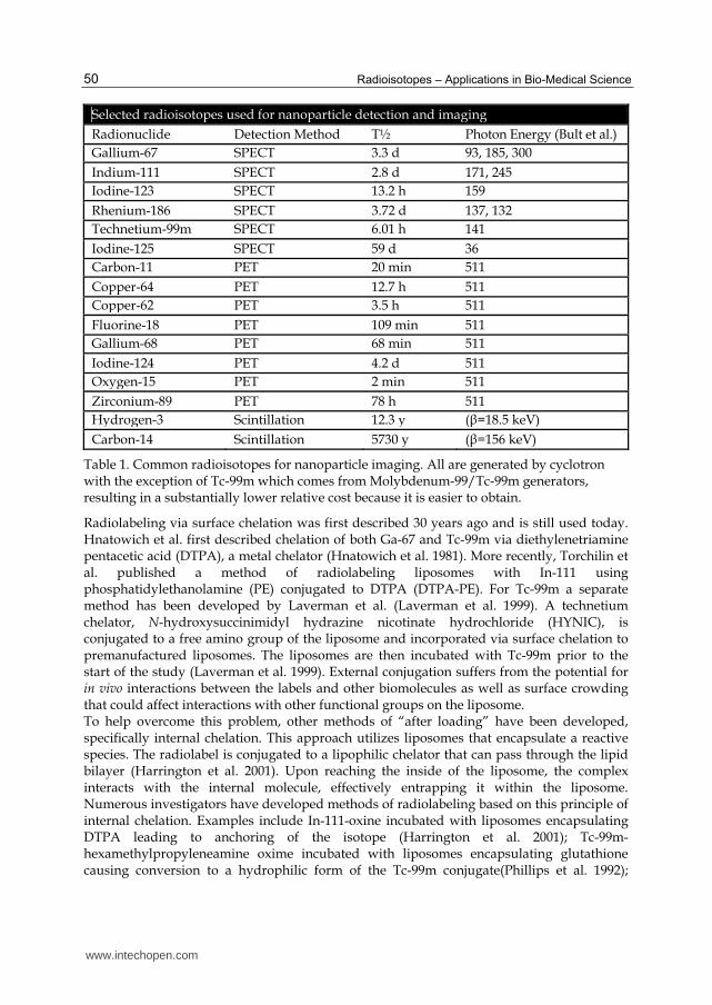
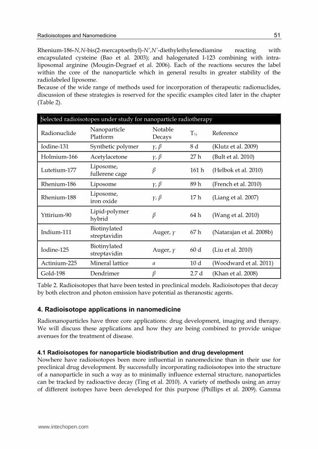
Selected radioisotopes used for nanoparticle detection and imaging
Radionuclide Detection Method T½ Photon Energy (Bult et al.)
Gallium-67 SPECT 3.3 d 93, 185, 300
Indium-111 SPECT 2.8 d 171, 245
Iodine-123 SPECT 13.2 h 159
Rhenium-186 SPECT 3.72 d 137, 132
Technetium-99m SPECT 6.01 h 141
Iodine-125 SPECT 59 d 36
Carbon-11 PET 20 min 511
Copper-64 PET 12.7 h 511
Copper-62 PET 3.5 h 511
Fluorine-18 PET 109 min 511
Gallium-68 PET 68 min 511
Iodine-124 PET 4.2 d 511
Oxygen-15 PET 2 min 511
Zirconium-89 PET 78 h 511
Hydrogen-3 Scintillation 12.3 y (┚=18.5 keV)
Carbon-14 Scintillation 5730 y (┚=156 keV)
Table 1. Common radioisotopes for nanoparticle imaging. All are generated by cyclotron with the exception of Tc-99m which comes from Molybdenum-99/Tc-99m generators, resulting in a substantially lower relative cost because it is easier to obtain.
Radiolabeling via surface chelation was first described 30 years ago and is still used today. Hnatowich et al. first described chelation of both Ga-67 and Tc-99m via diethylenetriamine pentacetic acid (DTPA), a metal chelator (Hnatowich et al. 1981). More recently, Torchilin et al. published a method of radiolabeling liposomes with In-111 using phosphatidylethanolamine (PE) conjugated to DTPA (DTPA-PE). For Tc-99m a separate method has been developed by Laverman et al. (Laverman et al. 1999). A technetium chelator, N-hydroxysuccinimidyl hydrazine nicotinate hydrochloride (HYNIC), is conjugated to a free amino group of the liposome and incorporated via surface chelation to premanufactured liposomes. The liposomes are then incubated with Tc-99m prior to the start of the study (Laverman et al. 1999). External conjugation suffers from the potential for in vivo interactions between the labels and other biomolecules as well as surface crowding that could affect interactions with other functional groups on the liposome. To help overcome this problem, other methods of “after loading” have been developed, specifically internal chelation. This approach utilizes liposomes that encapsulate a reactive species. The radiolabel is conjugated to a lipophilic chelator that can pass through the lipid bilayer (Harrington et al. 2001). Upon reaching the inside of the liposome, the complex interacts with the internal molecule, effectively entrapping it within the liposome. Numerous investigators have developed methods of radiolabeling based on this principle of internal chelation. Examples include In-111-oxine incubated with liposomes encapsulating DTPA leading to anchoring of the isotope (Harrington et al. 2001); Tc-99m-hexamethylpropyleneamine oxime incubated with liposomes encapsulating glutathione causing conversion to a hydrophilic form of the Tc-99m conjugate(Phillips et al. 1992);
www.intechopen.com
Radioisotopes and Nanomedicine 51
Rhenium-186-N,N-bis(2-mercaptoethyl)-N’,N’-diethylethylenediamine reacting with encapsulated cysteine (Bao et al. 2003); and halogenated I-123 combining with intra-liposomal arginine (Mougin-Degraef et al. 2006). Each of the reactions secures the label within the core of the nanoparticle which in general results in greater stability of the radiolabeled liposome. Because of the wide range of methods used for incorporation of therapeutic radionuclides,
discussion of these strategies is reserved for the specific examples cited later in the chapter
(Table 2).
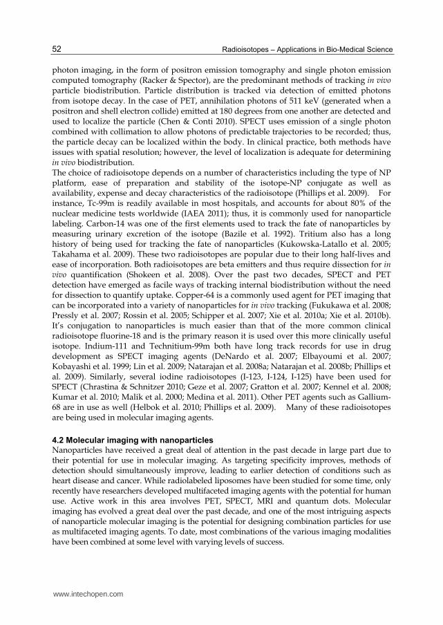
Selected radioisotopes under study for nanoparticle radiotherapy
Radionuclide Nanoparticle Platform
Notable Decays
T½ Reference
Iodine-131 Synthetic polymer ┛, ┚ 8 d (Klutz et al. 2009)
Holmium-166 Acetylacetone ┛, ┚ 27 h (Bult et al. 2010)
Lutetium-177 Liposome, fullerene cage
┚ 161 h (Helbok et al. 2010)
Rhenium-186 Liposome ┛, ┚ 89 h (French et al. 2010)
Rhenium-188 Liposome, iron oxide
┛, ┚ 17 h (Liang et al. 2007)
Yttirium-90 Lipid-polymer hybrid
┚ 64 h (Wang et al. 2010)
Indium-111 Biotinylated streptavidin
Auger, ┛ 67 h (Natarajan et al. 2008b)
Iodine-125 Biotinylated streptavidin
Auger, ┛ 60 d (Liu et al. 2010)
Actinium-225 Mineral lattice ┙ 10 d (Woodward et al. 2011)
Gold-198 Dendrimer ┚ 2.7 d (Khan et al. 2008)
Table 2. Radioisotopes that have been tested in preclinical models. Radioisotopes that decay by both electron and photon emission have potential as theranostic agents.
4. Radioisotope applications in nanomedicine
Radionanoparticles have three core applications: drug development, imaging and therapy.
We will discuss these applications and how they are being combined to provide unique
avenues for the treatment of disease.
4.1 Radioisotopes for nanoparticle biodistribution and drug development
Nowhere have radioisotopes been more influential in nanomedicine than in their use for preclinical drug development. By successfully incorporating radioisotopes into the structure of a nanoparticle in such a way as to minimally influence external structure, nanoparticles can be tracked by radioactive decay (Ting et al. 2010). A variety of methods using an array of different isotopes have been developed for this purpose (Phillips et al. 2009). Gamma
www.intechopen.com
Radioisotopes – Applications in Bio-Medical Science 52
photon imaging, in the form of positron emission tomography and single photon emission computed tomography (Racker & Spector), are the predominant methods of tracking in vivo particle biodistribution. Particle distribution is tracked via detection of emitted photons from isotope decay. In the case of PET, annihilation photons of 511 keV (generated when a positron and shell electron collide) emitted at 180 degrees from one another are detected and used to localize the particle (Chen & Conti 2010). SPECT uses emission of a single photon combined with collimation to allow photons of predictable trajectories to be recorded; thus, the particle decay can be localized within the body. In clinical practice, both methods have issues with spatial resolution; however, the level of localization is adequate for determining in vivo biodistribution. The choice of radioisotope depends on a number of characteristics including the type of NP platform, ease of preparation and stability of the isotope-NP conjugate as well as availability, expense and decay characteristics of the radioisotope (Phillips et al. 2009). For instance, Tc-99m is readily available in most hospitals, and accounts for about 80% of the nuclear medicine tests worldwide (IAEA 2011); thus, it is commonly used for nanoparticle labeling. Carbon-14 was one of the first elements used to track the fate of nanoparticles by measuring urinary excretion of the isotope (Bazile et al. 1992). Tritium also has a long history of being used for tracking the fate of nanoparticles (Kukowska-Latallo et al. 2005; Takahama et al. 2009). These two radioisotopes are popular due to their long half-lives and ease of incorporation. Both radioisotopes are beta emitters and thus require dissection for in vivo quantification (Shokeen et al. 2008). Over the past two decades, SPECT and PET detection have emerged as facile ways of tracking internal biodistribution without the need for dissection to quantify uptake. Copper-64 is a commonly used agent for PET imaging that can be incorporated into a variety of nanoparticles for in vivo tracking (Fukukawa et al. 2008; Pressly et al. 2007; Rossin et al. 2005; Schipper et al. 2007; Xie et al. 2010a; Xie et al. 2010b). It’s conjugation to nanoparticles is much easier than that of the more common clinical radioisotope fluorine-18 and is the primary reason it is used over this more clinically useful isotope. Indium-111 and Technitium-99m both have long track records for use in drug development as SPECT imaging agents (DeNardo et al. 2007; Elbayoumi et al. 2007; Kobayashi et al. 1999; Lin et al. 2009; Natarajan et al. 2008a; Natarajan et al. 2008b; Phillips et al. 2009). Similarly, several iodine radioisotopes (I-123, I-124, I-125) have been used for SPECT (Chrastina & Schnitzer 2010; Geze et al. 2007; Gratton et al. 2007; Kennel et al. 2008; Kumar et al. 2010; Malik et al. 2000; Medina et al. 2011). Other PET agents such as Gallium-68 are in use as well (Helbok et al. 2010; Phillips et al. 2009). Many of these radioisotopes are being used in molecular imaging agents.
4.2 Molecular imaging with nanoparticles
Nanoparticles have received a great deal of attention in the past decade in large part due to their potential for use in molecular imaging. As targeting specificity improves, methods of detection should simultaneously improve, leading to earlier detection of conditions such as heart disease and cancer. While radiolabeled liposomes have been studied for some time, only recently have researchers developed multifaceted imaging agents with the potential for human use. Active work in this area involves PET, SPECT, MRI and quantum dots. Molecular imaging has evolved a great deal over the past decade, and one of the most intriguing aspects of nanoparticle molecular imaging is the potential for designing combination particles for use as multifaceted imaging agents. To date, most combinations of the various imaging modalities have been combined at some level with varying levels of success.
www.intechopen.com
Radioisotopes and Nanomedicine 53
Single modality carriers
There is good rationale for functionalizing nanoparticles with PET or SPECT agents for imaging. While glucose analog agents such as FDG show tumor uptake through the Warburg effect (Gatenby & Gillies 2004), specificity may be increased via the enhanced permeability and retention (EPR) effect (Noguchi et al. 1998). Additionally, nanocarriers may be functionalized with targeting ligands to further improve specificity for their target (Wang et al. 2008). Despite its widespread use in the clinic for imaging, fluorine-18 is difficult to efficiently functionalize to targeted nanoparticles for this purpose. Due to its long half-life relative to other isotopes (carbon-11, nitrogen-13, oxygen-15), F-19 is a F-19 is a better candidate candidate for PET nanoparticles as there is sufficient time to functionalize the NP with radioisotope prior to administration. In 2008, Matson et al. were the first to demonstrate efficient synthesis of F-19 labeled micelle nanoparticles (Matson & Grubbs 2008). This may pave the way for better imaging agents using NPs. Elements such as copper-64 are much easier to functionalize to NP because of their longer half-life (12.7 h), and thus to this point it has been used more frequently than fluorine. Chakrabarti et al. designed a peptide nucleic acid probe radiolabeled with copper-64 to target mutated KRAS mRNA. KRAS is mutated at a very high level in pancreatic cancer and mutated KRAS mRNA is expressed at a high level in these tumors suggesting it may be a good target for early detection or targeted therapy. To achieve cellular uptake, peptide nanoparticles were combined with an IGF1-analog peptide to allow for internalization by IGF1 receptor-mediated endocytosis. IGF1 receptor is commonly upregulated in human cancers, including pancreatic cancer. Authors designed a series of radiohybridization probes linked with copper-64 to target various single nucleotide polymorphisms in KRAS mRNA. The probe had good selectivity for KRAS-mutated in mice engrafted with AsPC1, a pancreatic cancer cell line (Chakrabarti et al. 2007). Similarly, Petersen et al. designed copper-64 labeled liposomes for PET imaging in a mouse model (Petersen et al. 2011). Authors have described the targeting of tumor neovasculature with both SPECT and PET
nanoparticles. Hu et al. designed an indium-111 labeled perfluorocarbon nanoparticle targeted
to ┙v┚3, an integrin characteristically overexpressed in tumor neovasculature. In a rabbit
model, the authors demonstrated a high sensitivity, low-resolution signal for tumor
vasculature (Hu et al. 2007). Perfluorocarbons NP may also be used for MRI (Schmieder et al.
2005) allowing for a dual modality probe in the future that would increase this resolution
while maintaining sensitivity. Ruggiero et al. imaged tumor vasculature using zirconium-89
labeled carbon nanotubes to target the monomeric vascular-endothelial cadherin epitobe that
is frequently overexpressed in tumor vasculature (Ruggiero et al. 2010). The versatile carbon
nanotube may prove to be both an efficient imaging and therapeutic nanoparticle platform.
These dual modality (theranostic) particles will be discussed in detail later. More recently, Sun
et al. published imaging data on the clinically available nanoparticle albumin-bound paclitaxel
(Abraxane, Celegene). To judge early response to therapy, researchers attached either the PET
agent 18F-FPPRGD2, a novel radiopharmaceutical shown to target ┙v┚3(Mittra et al. 2010), or
standard 18F-FDG to Abraxane. The authors concluded that the novel agent was more
sensitive than 18F-FDG for early detection of tumor response, likely due to down-regulation of
┙v┚3 in tumor endothelial cells when responding to Abraxane therapy (Sun et al. 2011).
The utility of nanoparticle SPECT versus PET agents was directly compared by Cheng et al. Tc-99m and F-18 were each conjugated individually to an anti-HER-2 nanoparticle, and the
www.intechopen.com
Radioisotopes – Applications in Bio-Medical Science 54
agents were tested in a mouse model with HER-2-positive xenografts. Despite the better resolution of Tc-99m, F-18 was 15 times more sensitive for in vivo tumor detection. Unfortunately, covalent attachment of F-18 is a much more difficult process than chelation of Tc-99m to the nanoparticle platform (Cheng et al. 2010); however, due to its much higher sensitivity, at this time, PET appears to be the choice modality for clinical applications.
Dual modality carriers
One of the exciting features of nanoparticle platforms is their versatility. Researchers are actively investigating almost every permutation of the various imaging modalities in combination with nanoparticle carriers. The list of potential modalities includes PET, SPECT, MRI and quantum dots (QD). While a full description of investigations into nanoparticle MRI and QD is beyond the scope of this chapter, each will be described as they pertain to multifaceted imaging nanoparticles involving PET and SPECT. QD are nano-sized semiconductors with unique optical properties specific to the size and possibly the shape of the semiconductor crystal. In medical imaging, quantum dots are being studied as fluorophores, particles that absorb and emit energy of characteristic wave lengths. QD have been used in vitro for applications such as cell labeling and fluorescence in situ hybridization (Jiang et al. 2007; Wu et al. 2003). To date, in vivo use has been mostly limited to non-specific applications such as qualitative imaging of lymph nodes and vasculature in animal models (Hikage et al. 2010). Progress for medical applications has been hindered by technical challenges such as toxicity and characterization of the in vivo fluorescence signal (Aillon et al. 2009; Zhang & Badea 2011). Despite these challenges, several researchers have described multifaceted nanoparticles for imaging with either PET or SPECT in combination with QD. Cai et al. designed cadmium QD functionalized with RGD peptide, a ┙v┚3 antagonist, and labeled with copper-64. This allowed for simultaneous near-infrared fluorescence (NIRF) and PET imaging. The probe was tested in a murine glioblastoma model and correlation between the in vivo PET signal and ex vivo QD fluorescence was excellent. On histologic examination, the nanoparticle exhibited specificity for tumor neovasculature. This study allowed for accurate determination of the probe biodistribution. A major advantage of this probe may be the gain in sensitivity with PET. This allows for the use of lower levels of cadmium QD to achieve accurate imaging. In the future, this will be of key importance for quantum dots to reduce the potential for toxicity from metals used in the semiconductors (Cai et al. 2007). Similarly, Duconge et al. reported the first F-18-labeled QD using a PEG-phospholipid QD micelles. Pegylation allowed for prolonged circulation (T1/2 of 2 h) and reduced RES uptake. Researchers were able to study the biodistribution both by whole body PET and by QD fluorescence using in vivo fibered confocal fluorescence microscopy. The authors conclude that this system effectively utilizes two molecular imaging modalities simultaneously: PET for whole body imaging and fluorescence imaging for subcellular localization of tumor cells (Duconge et al. 2008). SPECT-QD combinations have also been described (Liang et al. 2010). While one can envision clinical applications of these probes for invasive procedures, they still suffer from the inability to assess the fluorescence signal in deep tissue without direct surgical intervention. This is not the case for dual MRI/PET probes which both allow for non-invasive imaging in
vivo. While several researchers have looked at MRI/SPECT (Lijowski et al. 2009; Park et al.
2011; Torres Martin de Rosales et al. 2011; Zielhuis et al. 2006), the much improved
sensitivity of positron emitting probes makes PET a much more enticing option for dual
www.intechopen.com
Radioisotopes and Nanomedicine 55
modality imaging. MRI allows for excellent spatial resolution that PET is lacking while PET
improves upon the suboptimal sensitivity of MRI for functional imaging. The combination is
appealing because of this complimentary nature (Cheon & Lee 2008).
Several investigators have designed iron oxide nanoparticles with copper-64 for dual modality imaging (Glaus et al. 2010; Jarrett et al. 2008; Patel et al. 2011). Jarrett et al. had previously demonstrated the use of an MRI compatible PET scanner to obtain simultaneous images using separate imaging probes (Catana et al. 2006; Judenhofer et al. 2007; Wu et al. 2009). Preclinical results are awaited using these untargeted nanoparticles in a model system for dual modality imaging. Several authors have developed and tested a similar iron oxide nanoparticles labeled with copper-64 and functionalized the carriers with integrin–binding ┙v┚3 RGD peptide (Lee et al. 2008; Yang et al. 2011). Researchers have tested the particles in vivo in a mouse model; however, the MRI characterization was performed without copper-64 due to limitations of the MR scanner (Lee et al. 2008). Despite this, imaging results between the two modalities were comparable.
Trimodality imaging agents and beyond
With clinical application of multifunctional imaging nanoparticles on the horizon, researchers have continued to push the limits of what is achievable with a single nanoparticle agent. Several authors have described trimodality imaging nanoparticles used for PET, MR and fluorescence imaging (Devaraj et al. 2009; Ko et al. 2011; Nahrendorf et al. 2008; Zhou et al. 2011), and at least one group has published on the synthesis of a tetramodality particle that allows for bioluminescence and fluorescence imaging as well as PET and MRI using a cobalt-ferrite nanoparticle (Hwang do et al. 2009).
4.3 Therapeutic radionanoparticles
While much of the early work on therapeutic nanoparticles focused on drug delivery, there is increasing interest in targeting diseases, specifically cancer, via therapeutic doses of internal radiation. Improvements in functionality and streamlining of manufacturing are leading to multidimensional particles capable of delivering multiple therapies via a single carrier. Internal radiation therapy already has a track record as a useful modality in the clinic. Iodine-131 therapy has long been a standard ablative therapy for both non-malignant thyroid diseases as well as thyroid cancer. In the past 5 years several radioimmunotherapies (Davies et al.) using monoclonal antibodies have been approved for clinical use. Radiolabeled anti-CD-20-antibody therapies Y-90-ibritumomab tiuxetan (Zevalin, Spectrum Pharmaceuticals) and I-131-tositumomab (Bexxar, GlaxoSmithKline) have shown promising results in clinical trials for the treatment of relapsed/refractory non-Hodgkin’s lymphoma (Davies et al. 2004; Vose et al. 2005; Vose et al. 2000) and are now being tested as part of initial treatment in clinical trials (Fisher 2005; NA 2006; Press et al. 2006). A limitation of RIT is that only one radioisotope can be attached per antibody (Jestin et al. 2007). Yttrium-90-microspheres, an agent in clinical use for ablation of hepatic malignancies, overcome this first problem but due to their size must be injected directly into the hepatic arterial supply to result in meaningful tumor ablation without substantial toxicity (Cosimelli et al. 2010; Hendlisz et al. 2010; Kennedy & Salem 2010). Other transarterial embolic agents tagged with Lu-177, Rh-188 and Ho-166 have been developed (Chakraborty et al. 2008; Hamoudeh et al. 2007; Kim et al. 2006), but all range in size from 300 nm up to 60 μm. Nanoparticles may be able to overcome both the size and payload problems and provide a systemic route to selectively deliver a larger radioisotope and/or drug payload.
www.intechopen.com
Radioisotopes – Applications in Bio-Medical Science 56
Radioisotopes with various decay properties are being utilized for therapeutic
nanoparticles. With penetration ranges of 1-10 mm and energies 0.1-2.2 MeV, beta-emitting
isotopes such as those in clinical use for RIT may be ideal candidates for nanoparticle
radiation. The range of beta particles allows the so-called “cross-fire” effect as it is greater
than several cell diameters. However, the decay property should match the goal of therapy
(Sofou 2008). For instance, particles that decay through alpha emission may be attractive
alternatives for targeting intravascular micrometastases due to high linear energy transfer.
These particles are limited by a penetration range of 50-100 μm making them less well suited
for bulky tumor deposits. Auger electrons, with a path length of less than 10 nm such as
those produced by Indium-111, have been suggested as potential therapeutic agents for
small volume disease if carriers can be internalized in high enough numbers to result in
meaning radiation dose deposition to the tumor cell DNA. One theoretical appeal of auger-
mediated therapy is that the short path length also suggests a low potential for toxicity
(Tavares & Tavares 2010).
Many of the studies involving therapeutic radionanoparticles have been theoretical and
qualitative in nature. Such studies have looked at modeling of radio-loaded nanoparticles
with various decay properties with mixed theoretical results. Bouchat et al. modeled Y-90
labeled antibody biodistribution by Monte Carlo code and showed that RIT would not result
in tumor doses sufficient to treat solid tumors such as lung cancer (biological effective dose
< 60 Gy). Using a similar modeling system, they showed that radioactive nanoparticles with
~100 Y-90 atoms per nanoparticle can substantially increase the biologic effective dose
deposited to a solid tumor (Bouchat et al. 2010). Additionally, radioisotopes that are usually
insoluble in body fluids can be encapsulated within nanoparticles, allowing them to be
sequestered until delivery at the target (Khan et al. 2008). Hrycushko et al. have modeled
liposomes tagged with both beta emitters Re-186 and Re-188 for the post-surgical treatment
of breast cancer, showing this to be a potential method of delivering focal radiation to the
locations at highest risk for harboring residual cancer cells while sparing normal tissue
(Hrycushko et al. 2011a; Hrycushko et al. 2011b).
Preclinical development of therapeutic radionanoparticles has been primarily directed at the
treatment of cancer often involving concurrent beta decay (for therapy) and gamma decay
(for imaging). Rapid advances in the ability to functionalize nanoparticle platforms for
diagnostic and therapeutic purposes have led investigators to develop dual purpose
particles, a field now known as theranostics. Many of the therapeutic nanoparticles fall into
this category owing to multiple decay pathways of the incorporated radioisotopes, most
often involving concurrent both beta decay for therapy and gamma decay for imaging.
While some authors consider dual imaging nanoparticles to be theranostic agents (Ma et al.
2006), this section will deal only with those agents that have been considered for therapeutic
use. To date, the study of therapeutic radionanoparticles has involved a wide range of
nanoparticle platforms combined with an even vaster array of radioisotopes. The section is
organized by type of radiation decay (beta, alpha, and auger) as each has its own unique
applications and shortcomings when used for internal radiation therapy (Sofou 2008).
Beta decay radionanoparticles
Radioisotopes decaying through beta emission are perhaps the best studied agents for
nanoparticle radiotherapy. With a path length of 1-10 mm, beta particles traverse multiple
cells while interacting, leading to a “cross fire” effect. Therefore, tagged-NPs do not require
www.intechopen.com
Radioisotopes and Nanomedicine 57
cellular internalization to provide effective cell kill. The particle range may also allow for
potential cell kill in areas of tumor lacking good vascular access.
Significant research has gone into gold nanoparticles for imaging and enhancement of
external beam irradiation, but until recently, little successful work had been published using
beta-emitting gold nanoparticles for internal radiation therapy. Though first described for
this purpose several decades ago, nanoparticle technology was not as robust at that time,
and efforts to deliver internal radiation with gold were discontinued (Hainfeld et al. 1990).
Recently, Khan et al. demonstrated the antitumor effect of dendrimer nanoparticles carrying
therapeutic loads of Au-198. Researchers generated the radioactive composite nanodevice
(CND) by simultaneous neutron activation and radiation polymerization of dendrimers
tagged with Au-197. Neutron capture formed Au-198 which could then decay by beta
emission. In a melanoma model, tumors of mice injected with the Au-198 CND decreased in
size by 45% compared to the untagged CND with no observed toxicity (Khan et al. 2008).
Chanda et al. described a similar Au-198 NP with good therapeutic efficacy and low toxicity
in a murine prostate cancer model (Chanda et al. 2010).
Several other platforms have been developed for delivery of internal beta irradiation.
Schultz et al. described a fullerene cage nanoparticle tagged with Lu-177 for targeting Il-13,
an interleukin characteristically overexpressed on glioblastoma cells (Shultz et al. 2010).
Diener et al. devised a similar fullerene system incorporating Pb-212, a beta emitter with an
alpha-emitting daughter radionuclide, Bi-212 (Diener et al. 2007). Reactive ionic
radioisotopes such as Lu and Pb must be shielded from the microenvironment to prevent
interaction. Fullerene cages are attractive nanoparticles for this purpose because the ions are
completely enclosed within the platform. Ho-166 is another appealing candidate
radioisotope due to its concurrent gamma and beta decays as well as its high attenuation
coefficient and paramagnetic properties. This combination suggests it to have wide-ranging
potential as a true theranostic agent. With this in mind, Bult et al. successfully developed an
acetylacetone nanoparticle labeled with Ho-166 (Bult et al. 2010); though, the particle has yet
to be tested as a therapeutic agent in an animal model.
Alpha decay radionanoparticles
While beta-emitters have received the bulk of the attention for radionanoparticles, alpha
emitters possess unique qualities that make them viable therapy candidates. One such
radioisotope is Ac-225, which has received attention as an in vivo radiation generator. Ac-225
decays through two chains of short-lived radioisotopes, yielding 4 alpha particles and
ending in stable Bi-209. A challenge of these generators is to prevent leakage of the daughter
radionuclides during circulation, and nanoparticles are being studied as a way to retain
decay chain products for delivery at the target site. Two groups have developed
nanoparticles capable of daughter particle retention to a limited extent. Sofou et al. designed
a multivesicular liposomal carrier of Ac-225 to enhance retention of daughter radionuclides.
Though retention was not as high as hoped, when the particles were targeted to an ovarian
cell line in vitro using immunolabeling with anti-HER2/neu, they were internalized at an
impressive rate (Sofou et al. 2007). More recently, Woodward et al.developed a lanthum
phosphate nanoparticle (LaPO4 NP) for Ac-225 encapsulation. The particles were conjugated
with antibodies to lung endothelium. This system showed better retention of the daughter
nuclides in the LaPO4 NP and excellent dose deposition in mouse lung tissue (Woodward et
www.intechopen.com
Radioisotopes – Applications in Bio-Medical Science 58
al. 2011). Effective targeting and daughter retention suggest this model may have
therapeutic potential in humans.
Auger decay radionanoparticles
Due to their short range, auger electrons rely on high selective cellular uptake and incorporation in cellular DNA to result in meaning tumoricidal effect. To date, In-111, I-123 and I-125 have been studied as potential agents for this purpose (Chow et al. 2008; Liu et al. 2010; Liu et al. 2009; Mamede et al. 2003; Reske et al. 2007).
Combined modality particles for cancer treatment
Radiation is a major treatment modality for cancer along with surgery and chemotherapy. Over the past 20 years, a large body of evidence has shown that chemotherapy can synergistically enhance the tumoricidal effects of radiation. Many cancers are now treated with concomitant chemotherapy and radiation with absolute benefits in overall survival on the order of 5-10%, but at the cost of increased toxicity of treatment (Auperin et al. 2010; Green et al. 2001; Pignon et al. 2009). Advancements in disease targeting and release properties make nanoparticles an intriguing choice for combining these two therapies in vivo with the hope of reducing toxicity and enhancing efficacy, similar to what is seen with nanoparticle chemotherapy agents already on the market such as Doxil and Abraxane. Wang et al. described such a combined modality nanoparticle, ChemoRad NP (Wang et al.
2010). Using a lipid-polymer hybrid nanoparticle platform, researchers synthesized a
particle containing docetaxel chemotherapy within the core and used metal chelation with
DMPE-DPTA to attach Yttrium-90, a beta-emitter that has been effectively used in the clinic
when conjugated to anti-CD-20 monoclonal antibodies for treatment of Hodgkin’s
lymphoma. Both agents were attached in therapeutic quantities with 5% docetaxel by
weight in the NP and 100 μCi Y-90/mg NP. This activity of Yttrium is enough to deliver 6
Gy of radiation to a tumor target based on pharmacokinetic studies of the NP. The surface
of the NP was conjugated via DSPE-PEG with the aptamer A10, a ligand that targets
prostate specific membrane antigen (PSMA). In a preliminary study of the molecule, in vitro
profiling showed the NP to have excellent stability and controlled release properties while
successfully targeting tumor cells using the aptamer ligand. Comparison of the combined
modality NP to single-agent NP and non-targeted NP as controls showed that the
ChemoRad NP has higher therapeutic efficacy than the control options. ChemoRad NP is
believed to be the first targeted nanoparticle for delivery of chemoradiation therapy.
Chow et al. report on a second combined modality particle combining Indium-111 and
vinorelbine in a pegylated liposome carrier (Chow et al. 2008). This nanoparticle platform
did not have a targeting ligand, but showed tumor selectively by the EPR effect on mice
with colorectal carcinoma xenografts. The agent showed antitumor efficacy both when
chemotherapy and radiation were included and when the single agents alone were used.
Because Indium-111 produces both a gamma photon and auger electron, this suggests the
auger electron may contribute meaningfully to tumor kill. Because of this decay feature, In-
111 has potential as a dual diagnostic and therapeutic (theranostic) agent.
5. Summary
Advances in nanotechnology have inspired the development of nanoparticles for medical applications. Radioisotopes have grown to be an integral part of this field both for purposes
www.intechopen.com
Radioisotopes and Nanomedicine 59
of drug development and now diagnostic and therapeutic applications. The flexibility of NPs enables the utilization of radioisotopes in novel applications. The wide range of ongoing nanoparticle research and the explosion of interest in the field are both testaments to the versatility and potential of this expanding branch of science in which radioisotopes promise to continue playing an important role.
6. References
Aillon, K. L., Y. Xie, et al. (2009). "Effects of nanomaterial physicochemical properties on in vivo toxicity." Advanced drug delivery reviews 61(6): 457-466.
Auperin, A., C. Le Pechoux, et al. (2010). "Meta-analysis of concomitant versus sequential radiochemotherapy in locally advanced non-small-cell lung cancer." Journal of clinical oncology : official journal of the American Society of Clinical Oncology 28(13): 2181-2190.
Bangham, A. D. (1993). "Liposomes: the Babraham connection." Chem Phys Lipids 64(1-3): 275-285.
Bao, A., B. Goins, et al. (2003). "186Re-liposome labeling using 186Re-SNS/S complexes: in vitro stability, imaging, and biodistribution in rats." Journal of nuclear medicine : official publication, Society of Nuclear Medicine 44(12): 1992-1999.
Bazile, D. V., C. Ropert, et al. (1992). "Body distribution of fully biodegradable [14C]-poly(lactic acid) nanoparticles coated with albumin after parenteral administration to rats." Biomaterials 13(15): 1093-1102.
Bouchat, V., V. E. Nuttens, et al. (2010). "Radioimmunotherapy with radioactive nanoparticles: biological doses and treatment efficiency for vascularized tumors with or without a central hypoxic area." Med Phys 37(4): 1826-1839.
Bult, W., R. Varkevisser, et al. (2010). "Holmium nanoparticles: preparation and in vitro characterization of a new device for radioablation of solid malignancies." Pharm Res 27(10): 2205-2212.
Cai, W., K. Chen, et al. (2007). "Dual-function probe for PET and near-infrared fluorescence imaging of tumor vasculature." J Nucl Med 48(11): 1862-1870.
Catana, C., Y. Wu, et al. (2006). "Simultaneous acquisition of multislice PET and MR images: initial results with a MR-compatible PET scanner." Journal of nuclear medicine : official publication, Society of Nuclear Medicine 47(12): 1968-1976.
Chakrabarti, A., K. Zhang, et al. (2007). "Radiohybridization PET imaging of KRAS G12D mRNA expression in human pancreas cancer xenografts with [(64)Cu]DO3A-peptide nucleic acid-peptide nanoparticles." Cancer Biol Ther 6(6): 948-956.
Chakraborty, S., T. Das, et al. (2008). "Preparation and preliminary studies on 177Lu-labeled hydroxyapatite particles for possible use in the therapy of liver cancer " Nucl Med Biol 35(5): 589-597.
Chanda, N., P. Kan, et al. (2010). "Radioactive gold nanoparticles in cancer therapy: therapeutic efficacy studies of GA-198AuNP nanoconstruct in prostate tumor-bearing mice." Nanomedicine 6(2): 201-209.
Chen, K. & P. S. Conti (2010). "Target-specific delivery of peptide-based probes for PET imaging." Advanced drug delivery reviews 62(11): 1005-1022.
Cheng, D., Y. Wang, et al. (2010). "Comparison of 18F PET and 99mTc SPECT imaging in phantoms and in tumored mice." Bioconjugate chemistry 21(8): 1565-1570.
www.intechopen.com
Radioisotopes – Applications in Bio-Medical Science 60
Cheon, J. & J. H. Lee (2008). "Synergistically integrated nanoparticles as multimodal probes for nanobiotechnology." Accounts of chemical research 41(12): 1630-1640.
Chow, T. H., Y. Y. Lin, et al. (2008). "Diagnostic and therapeutic evaluation of 111In-vinorelbine-liposomes in a human colorectal carcinoma HT-29/luc-bearing animal model." Nucl Med Biol 35(5): 623-634.
Chrastina, A. & J. E. Schnitzer (2010). "Iodine-125 radiolabeling of silver nanoparticles for in vivo SPECT imaging." Int J Nanomedicine 5: 653-659.
Cosimelli, M., R. Golfieri, et al. (2010). "Multi-centre phase II clinical trial of yttrium-90 resin microspheres alone in unresectable, chemotherapy refractory colorectal liver metastases." British journal of cancer 103(3): 324-331.
Davies, A. J., A. Z. Rohatiner, et al. (2004). "Tositumomab and iodine I 131 tositumomab for recurrent indolent and transformed B-cell non-Hodgkin's lymphoma." Journal of clinical oncology : official journal of the American Society of Clinical Oncology 22(8): 1469-1479.
Delbeke, D. & W. H. Martin (2001). "Positron emission tomography imaging in oncology." Radiologic clinics of North America 39(5): 883-917.
Delbeke, D. & W. H. Martin (2004). "Metabolic imaging with FDG: a primer." Cancer journal 10(4): 201-213.
DeNardo, S. J., G. L. DeNardo, et al. (2007). "Thermal dosimetry predictive of efficacy of 111In-ChL6 nanoparticle AMF--induced thermoablative therapy for human breast cancer in mice." J Nucl Med 48(3): 437-444.
Devaraj, N. K., E. J. Keliher, et al. (2009). "18F labeled nanoparticles for in vivo PET-CT imaging." Bioconjug Chem 20(2): 397-401.
Diener, M. D., J. M. Alford, et al. ( 2007). "(212)Pb@C(60) and its water-soluble derivatives: synthesis, stability, and suitability for radioimmunotherapy." Journal of the American Chemical Society 129(16): 5131-5138.
Duconge, F., T. Pons, et al. (2008). "Fluorine-18-labeled phospholipid quantum dot micelles for in vivo multimodal imaging from whole body to cellular scales." Bioconjug Chem 19(9): 1921-1926.
Elbayoumi, T. A., S. Pabba, et al. (2007). "Antinucleosome antibody-modified liposomes and lipid-core micelles for tumor-targeted delivery of therapeutic and diagnostic agents." J Liposome Res 17(1): 1-14.
Farokhzad, O. C., S. Jon, et al. (2004). "Nanoparticle-aptamer bioconjugates: a new approach for targeting prostate cancer cells." Cancer Res 64(21): 7668-7672.
Fisher, R. I. (2005). "Overview of Southwest Oncology Group Clinical Trials in non-Hodgkin Lymphoma. S0016. A phase III trial of CHOP vs CHOP + rituximab vs CHOP + iodine131-labeled monoclonal anti-B1 antibody (tositumomab) for treatment of newly diagnosed follicular NHL." Clinical advances in hematology & oncology : H&O 3(7): 544-546.
French, J. T., B. Goins, et al. (2010). "Interventional therapy of head and neck cancer with lipid nanoparticle-carried rhenium 186 radionuclide." J Vasc Interv Radiol 21(8): 1271-1279.
Fukukawa, K., R. Rossin, et al. (2008). "Synthesis and characterization of core-shell star copolymers for in vivo PET imaging applications." Biomacromolecules 9(4): 1329-1339.
www.intechopen.com
Radioisotopes and Nanomedicine 61
Gatenby, R. A. & R. J. Gillies (2004). "Why do cancers have high aerobic glycolysis?" Nature reviews. Cancer 4(11): 891-899.
Geze, A., L. T. Chau, et al. (2007). "Biodistribution of intravenously administered amphiphilic beta-cyclodextrin nanospheres." Int J Pharm 344(1-2): 135-142.
Glaus, C., R. Rossin, et al. (2010). "In vivo evaluation of (64)Cu-labeled magnetic nanoparticles as a dual-modality PET/MR imaging agent." Bioconjug Chem 21(4): 715-722.
Gratton, S. E., P. D. Pohlhaus, et al. (2007). "Nanofabricated particles for engineered drug therapies: a preliminary biodistribution study of PRINT nanoparticles." J Control Release 121(1-2): 10-18.
Green, J. A., J. M. Kirwan, et al. (2001). "Survival and recurrence after concomitant chemotherapy and radiotherapy for cancer of the uterine cervix: a systematic review and meta-analysis." Lancet 358(9284): 781-786.
Gu, F., L. Zhang, et al. (2008). "Precise engineering of targeted nanoparticles by using self-assembled biointegrated block copolymers." Proc Natl Acad Sci U S A 105(7): 2586-2591.
Hahn, M. A., A. K. Singh, et al. (2011). "Nanoparticles as contrast agents for in-vivo bioimaging: current status and future perspectives." Analytical and bioanalytical chemistry 399(1): 3-27.
Hainfeld, J. F., C. J. Foley, et al. (1990). "Radioactive gold cluster immunoconjugates: potential agents for cancer therapy." Int J Rad Appl Instrum B 17(3): 287-294.
Hamoudeh, M., H. Salim, et al. (2007). "Preparation and characterization of radioactive dirhenium decacarbonyl-loaded PLLA nanoparticles for radionuclide intra-tumoral therapy." Eur J Pharm Biopharm 67(3): 597-611.
Harrington, K. J., S. Mohammadtaghi, et al. (2001). "Effective targeting of solid tumors in patients with locally advanced cancers by radiolabeled pegylated liposomes." Clinical cancer research : an official journal of the American Association for Cancer Research 7(2): 243-254.
Helbok, A., C. Decristoforo, et al. (2010). "Radiolabeling of lipid-based nanoparticles for diagnostics and therapeutic applications: a comparison using different radiometals." J Liposome Res 20(3): 219-227.
Hendlisz, A., M. Van den Eynde, et al. (2010). "Phase III trial comparing protracted intravenous fluorouracil infusion alone or with yttrium-90 resin microspheres radioembolization for liver-limited metastatic colorectal cancer refractory to standard chemotherapy." Journal of clinical oncology : official journal of the American Society of Clinical Oncology 28(23): 3687-3694.
Hikage, M., K. Gonda, et al. (2010). "Nano-imaging of the lymph network structure with quantum dots." Nanotechnology 21(18): 185103.
Hnatowich, D. J., B. Friedman, et al. (1981). "Labeling of preformed liposomes with Ga-67 and Tc-99m by chelation." J Nucl Med 22(9): 810-814.
Hong, S. Y., G. Tobias, et al. (2010). "Filled and glycosylated carbon nanotubes for in vivo radioemitter localization and imaging." Nat Mater 9(6): 485-490.
Hrycushko, B. A., A. N. Gutierrez, et al. (2011a). "Radiobiological characterization of post-lumpectomy focal brachytherapy with lipid nanoparticle-carried radionuclides." Phys Med Biol 56(3): 703-719.
www.intechopen.com
Radioisotopes – Applications in Bio-Medical Science 62
Hrycushko, B. A., S. Li, et al. (2011b). "Postlumpectomy focal brachytherapy for simultaneous treatment of surgical cavity and draining lymph nodes." Int J Radiat Oncol Biol Phys 79(3): 948-955.
Hu, G., M. Lijowski, et al. (2007). "Imaging of Vx-2 rabbit tumors with alpha(nu)beta3-integrin-targeted 111In nanoparticles." Int J Cancer 120(9): 1951-1957.
Hwang do, W., H. Y. Ko, et al. (2009). "Development of a quadruple imaging modality by using nanoparticles." Chemistry 15(37): 9387-9393.
IAEA (2011). "Radiopharmaceuticals: Production and Availability." Jarrett, B. R., B. Gustafsson, et al. (2008). "Synthesis of 64Cu-labeled magnetic nanoparticles
for multimodal imaging." Bioconjug Chem 19(7): 1496-1504. Jestin, E., M. Mougin-Degraef, et al. (2007). "Radiolabeling and targeting of lipidic
nanocapsules for applications in radioimmunotherapy." Q J Nucl Med Mol Imaging 51(1): 51-60.
Jiang, Z., R. Li, et al. (2007). "Detecting genomic aberrations by fluorescence in situ hybridization with quantum dots-labeled probes." Journal of nanoscience and nanotechnology 7(12): 4254-4259.
Judenhofer, M. S., C. Catana, et al. (2007). "PET/MR images acquired with a compact MR-compatible PET detector in a 7-T magnet." Radiology 244(3): 807-814.
Kennedy, A. S. & R. Salem (2010). "Radioembolization (yttrium-90 microspheres) for primary and metastatic hepatic malignancies." Cancer journal 16(2): 163-175.
Kennel, S. J., J. D. Woodward, et al. (2008). "The fate of MAb-targeted Cd(125m)Te/ZnS nanoparticles in vivo." Nucl Med Biol 35(4): 501-514.
Khan, M. K., L. D. Minc, et al. (2008). "Fabrication of {198Au0} radioactive composite nanodevices and their use for nanobrachytherapy." Nanomedicine 4(1): 57-69.
Kim, J. K., K. H. Han, et al. (2006). "Long-term clinical outcome of phase IIb clinical trial of percutaneous injection with holmium-166/chitosan complex (Milican) for the treatment of small hepatocellular carcinoma." Clinical cancer research : an official journal of the American Association for Cancer Research 12(2): 543-548.
Kim, S. H., J. H. Jeong, et al. (2005). "Target-specific cellular uptake of PLGA nanoparticles coated with poly(L-lysine)-poly(ethylene glycol)-folate conjugate." Langmuir 21(19): 8852-8857.
Klutz, K., V. Russ, et al. (2009). "Targeted radioiodine therapy of neuroblastoma tumors following systemic nonviral delivery of the sodium iodide symporter gene." Clin Cancer Res 15(19): 6079-6086.
Ko, H. Y., K. J. Choi, et al. (2011). "A multimodal nanoparticle-based cancer imaging probe simultaneously targeting nucleolin, integrin alphavbeta3 and tenascin-C proteins." Biomaterials 32(4): 1130-1138.
Kobayashi, H., C. Wu, et al. (1999). "Evaluation of the in vivo biodistribution of indium-111 and yttrium-88 labeled dendrimer-1B4M-DTPA and its conjugation with anti-Tac monoclonal antibody." Bioconjug Chem 10(1): 103-111.
Kukowska-Latallo, J. F., K. A. Candido, et al. (2005). "Nanoparticle targeting of anticancer drug improves therapeutic response in animal model of human epithelial cancer." Cancer Res 65(12): 5317-5324.
Kumar, R., I. Roy, et al. (2010). "In vivo biodistribution and clearance studies using multimodal organically modified silica nanoparticles." ACS Nano 4(2): 699-708.
www.intechopen.com
Radioisotopes and Nanomedicine 63
Laverman, P., E. T. Dams, et al. (1999). "A novel method to label liposomes with 99mTc by the hydrazino nicotinyl derivative." J Nucl Med 40(1): 192-197.
Lee, H. Y., Z. Li, et al. (2008). "PET/MRI dual-modality tumor imaging using arginine-glycine-aspartic (RGD)-conjugated radiolabeled iron oxide nanoparticles." J Nucl Med 49(8): 1371-1379.
Liang, M., X. Liu, et al. (2010). "Multimodality nuclear and fluorescence tumor imaging in mice using a streptavidin nanoparticle." Bioconjugate chemistry 21(7): 1385-1388.
Liang, S., Y. Wang, et al. (2007). "Surface modified superparamagnetic iron oxide nanoparticles: as a new carrier for bio-magnetically targeted therapy." J Mater Sci Mater Med 18(12): 2297-2302.
Lijowski, M., S. Caruthers, et al. (2009). "High sensitivity: high-resolution SPECT-CT/MR molecular imaging of angiogenesis in the Vx2 model." Investigative radiology 44(1): 15-22.
Lin, Y. Y., J. J. Li, et al. (2009). "Evaluation of pharmacokinetics of 111In-labeled VNB-PEGylated liposomes after intraperitoneal and intravenous administration in a tumor/ascites mouse model." Cancer Biother Radiopharm 24(4): 453-460.
Liu, X., K. Nakamura, et al. (2010). "Auger-mediated cytotoxicity of cancer cells in culture by an 125I-antisense oligomer delivered as a three-component streptavidin nanoparticle." J Biomed Nanotechnol 6(2): 153-157.
Liu, X., Y. Wang, et al. (2009). "Auger radiation-induced, antisense-mediated cytotoxicity of tumor cells using a 3-component streptavidin-delivery nanoparticle with 111In." J Nucl Med 50(4): 582-590.
Ma, D., Z. J. Jakubek, et al. (2006). "A new approach towards controlled synthesis of multifunctional core-shell nano-architectures: luminescent and superparamagnetic." Journal of nanoscience and nanotechnology 6(12): 3677-3684.
Mackeyev, Y. A., J. W. Marks, et al. (2005). "Stable containment of radionuclides on the nanoscale by cut single-wall carbon nanotubes." The journal of physical chemistry. B 109(12): 5482-5484.
Malik, N., R. Wiwattanapatapee, et al. (2000). "Dendrimers: relationship between structure and biocompatibility in vitro, and preliminary studies on the biodistribution of 125I-labelled polyamidoamine dendrimers in vivo." J Control Release 65(1-2): 133-148.
Mamede, M., T. Saga, et al. (2003). "Radiolabeling of avidin with very high specific activity for internal radiation therapy of intraperitoneally disseminated tumors." Clin Cancer Res 9(10 Pt 1): 3756-3762.
Matson, J. B. & R. H. Grubbs (2008). "Synthesis of fluorine-18 functionalized nanoparticles for use as in vivo molecular imaging agents." J Am Chem Soc 130(21): 6731-6733.
Medina, O. P., N. Pillarsetty, et al. (2011). "Optimizing tumor targeting of the lipophilic EGFR-binding radiotracer SKI 243 using a liposomal nanoparticle delivery system." Journal of controlled release : official journal of the Controlled Release Society 149(3): 292-298.
Mittra, E., M. Goris, et al. (2010). "First in man studies of [18F]FPPRGD2: A novel PET radiopharmaceutical for imaging {alpha}v{beta}3 integrin levels." J NUCL MED MEETING ABSTRACTS 51(2_MeetingAbstracts): 1433-.
Mougin-Degraef, M., E. Jestin, et al. (2006). "High-activity radio-iodine labeling of conventional and stealth liposomes." Journal of liposome research 16(1): 91-102.
www.intechopen.com
Radioisotopes – Applications in Bio-Medical Science 64
NA (2006). "A phase III trial of CHOP vs CHOP + rituximab vs CHOP + 1-131 tositumomab for newly diagnosed follicular non-Hodgkin lymphoma." Clinical advances in hematology & oncology : H&O 4(7): 536-538.
Nahrendorf, M., H. Zhang, et al. (2008). "Nanoparticle PET-CT imaging of macrophages in inflammatory atherosclerosis." Circulation 117(3): 379-387.
Natarajan, A., C. Gruettner, et al. (2008a). "NanoFerrite particle based radioimmunonanoparticles: binding affinity and in vivo pharmacokinetics." Bioconjug Chem 19(6): 1211-1218.
Natarajan, A., C. Y. Xiong, et al. (2008b). "Development of multivalent radioimmunonanoparticles for cancer imaging and therapy." Cancer Biother Radiopharm 23(1): 82-91.
Noguchi, Y., J. Wu, et al. (1998). "Early phase tumor accumulation of macromolecules: a great difference in clearance rate between tumor and normal tissues." Japanese journal of cancer research : Gann 89(3): 307-314.
Park, S. I., B. J. Kwon, et al. (2011). "Synthesis and characterization of 3-[131I]iodo-L-tyrosine grafted Fe3O4@SiO2 nanocomposite for single photon emission computed tomography (SPECT) and magnetic resonance imaging (MRI)." Journal of nanoscience and nanotechnology 11(2): 1818-1821.
Patel, D., A. Kell, et al. (2011). "The cell labeling efficacy, cytotoxicity and relaxivity of copper-activated MRI/PET imaging contrast agents." Biomaterials 32(4): 1167-1176.
Petersen, A. L., T. Binderup, et al. (2011). "64Cu loaded liposomes as positron emission tomography imaging agents." Biomaterials 32(9): 2334-2341.
Phillips, W. T., B. A. Goins, et al. (2009). "Radioactive liposomes." Wiley Interdiscip Rev Nanomed Nanobiotechnol 1(1): 69-83.
Phillips, W. T., A. S. Rudolph, et al. (1992). "A simple method for producing a technetium-99m-labeled liposome which is stable in vivo." International journal of radiation applications and instrumentation. Part B, Nuclear medicine and biology 19(5): 539-547.
Pignon, J. P., A. le Maitre, et al. (2009). "Meta-analysis of chemotherapy in head and neck cancer (MACH-NC): an update on 93 randomised trials and 17,346 patients." Radiotherapy and oncology : journal of the European Society for Therapeutic Radiology and Oncology 92(1): 4-14.
Press, O. W., J. M. Unger, et al. (2006). "Phase II trial of CHOP chemotherapy followed by tositumomab/iodine I-131 tositumomab for previously untreated follicular non-Hodgkin's lymphoma: five-year follow-up of Southwest Oncology Group Protocol S9911." Journal of clinical oncology : official journal of the American Society of Clinical Oncology 24(25): 4143-4149.
Pressly, E. D., R. Rossin, et al. (2007). "Structural effects on the biodistribution and positron emission tomography (PET) imaging of well-defined (64)Cu-labeled nanoparticles comprised of amphiphilic block graft copolymers." Biomacromolecules 8(10): 3126-3134.
Racker, E. & M. Spector (1981). "Warburg effect revisited: merger of biochemistry and molecular biology." Science 213(4505): 303-307.
Reske, S. N., S. Deisenhofer, et al. (2007). "123I-ITdU-mediated nanoirradiation of DNA efficiently induces cell kill in HL60 leukemia cells and in doxorubicin-, beta-, or gamma-radiation-resistant cell lines." J Nucl Med 48(6): 1000-1007.
www.intechopen.com
Radioisotopes and Nanomedicine 65
Rossin, R., D. Pan, et al. (2005). "64Cu-labeled folate-conjugated shell cross-linked nanoparticles for tumor imaging and radiotherapy: synthesis, radiolabeling, and biologic evaluation." J Nucl Med 46(7): 1210-1218.
Ruggiero, A., C. H. Villa, et al. (2010). "Imaging and treating tumor vasculature with targeted radiolabeled carbon nanotubes." International journal of nanomedicine 5: 783-802.
Schipper, M. L., Z. Cheng, et al. (2007). "microPET-based biodistribution of quantum dots in living mice." J Nucl Med 48(9): 1511-1518.
Schmieder, A. H., P. M. Winter, et al. (2005). "Molecular MR imaging of melanoma angiogenesis with alphanubeta3-targeted paramagnetic nanoparticles." Magnetic resonance in medicine : official journal of the Society of Magnetic Resonance in Medicine / Society of Magnetic Resonance in Medicine 53(3): 621-627.
Shokeen, M., N. M. Fettig, et al. (2008). "Synthesis, in vitro and in vivo evaluation of radiolabeled nanoparticles." The quarterly journal of nuclear medicine and molecular imaging : official publication of the Italian Association of Nuclear Medicine 52(3): 267-277.
Shultz, M. D., J. C. Duchamp, et al. (2010). "Encapsulation of a radiolabeled cluster inside a fullerene cage, (177)Lu(x)Lu((3-x))N@C(80): an interleukin-13-conjugated radiolabeled metallofullerene platform." J Am Chem Soc 132(14): 4980-4981.
Sofou, S. (2008). "Radionuclide carriers for targeting of cancer." Int J Nanomedicine 3(2): 181-199.
Sofou, S., B. J. Kappel, et al. (2007). "Enhanced retention of the alpha-particle-emitting daughters of Actinium-225 by liposome carriers." Bioconjugate chemistry 18(6): 2061-2067.
Strano, M. S. (2010). "Biomedical materials: Nanoscale radiosurgery." Nat Mater 9(6): 467-468. Sun, X., Y. Yan, et al. (2011). "18F-FPPRGD2 and 18F-FDG PET of response to Abraxane
therapy." Journal of nuclear medicine : official publication, Society of Nuclear Medicine 52(1): 140-146.
Takahama, H., T. Minamino, et al. (2009). "Prolonged targeting of ischemic/reperfused myocardium by liposomal adenosine augments cardioprotection in rats." J Am Coll Cardiol 53(8): 709-717.
Tavares, A. A. & J. M. Tavares (2010). "Evaluating 99mTc Auger electrons for targeted tumor radiotherapy by computational methods." Med Phys 37(7): 3551-3559.
Ting, G., C. H. Chang, et al. (2010). "Nanotargeted radionuclides for cancer nuclear imaging and internal radiotherapy." J Biomed Biotechnol 2010.
Torchilin, V. P. (2005). "Recent advances with liposomes as pharmaceutical carriers." Nature reviews. Drug discovery 4(2): 145-160.
Torres Martin de Rosales, R., R. Tavare, et al. (2011). "(99m)Tc-Bisphosphonate-Iron Oxide Nanoparticle Conjugates for Dual-Modality Biomedical Imaging." Bioconjugate chemistry 22(3): 455-465.
Vose, J. M., P. J. Bierman, et al. (2005). "Phase I trial of iodine-131 tositumomab with high-dose chemotherapy and autologous stem-cell transplantation for relapsed non-Hodgkin's lymphoma." Journal of clinical oncology : official journal of the American Society of Clinical Oncology 23(3): 461-467.
Vose, J. M., R. L. Wahl, et al. (2000). "Multicenter phase II study of iodine-131 tositumomab for chemotherapy-relapsed/refractory low-grade and transformed low-grade B-cell
www.intechopen.com
Radioisotopes – Applications in Bio-Medical Science 66
non-Hodgkin's lymphomas." Journal of clinical oncology : official journal of the American Society of Clinical Oncology 18(6): 1316-1323.
Wagner, V., A. Dullaart, et al. (2006). "The emerging nanomedicine landscape." Nature biotechnology 24(10): 1211-1217.
Wang, A. Z., F. Gu, et al. (2008). "Biofunctionalized targeted nanoparticles for therapeutic applications." Expert Opin Biol Ther 8(8): 1063-1070.
Wang, A. Z., K. Yuet, et al. (2010). "ChemoRad nanoparticles: a novel multifunctional nanoparticle platform for targeted delivery of concurrent chemoradiation." Nanomedicine (Lond) 5(3): 361-368.
Woodward, J., S. J. Kennel, et al. (2011). "LaPO(4) Nanoparticles Doped with Actinium-225 that Partially Sequester Daughter Radionuclides." Bioconjug Chem.
Wu, X., H. Liu, et al. (2003). "Immunofluorescent labeling of cancer marker Her2 and other cellular targets with semiconductor quantum dots." Nature biotechnology 21(1): 41-46.
Wu, Y., C. Catana, et al. (2009). "PET Performance Evaluation of an MR-Compatible PET Insert." IEEE transactions on nuclear science 56(3): 574-580.
Xie, H., Z. J. Wang, et al. (2010a). "In vivo PET imaging and biodistribution of radiolabeled gold nanoshells in rats with tumor xenografts." Int J Pharm 395(1-2): 324-330.
Xie, J., K. Chen, et al. (2010b). "PET/NIRF/MRI triple functional iron oxide nanoparticles." Biomaterials 31(11): 3016-3022.
Yang, X., H. Hong, et al. (2011). "cRGD-functionalized, DOX-conjugated, and (64)Cu-labeled superparamagnetic iron oxide nanoparticles for targeted anticancer drug delivery and PET/MR imaging." Biomaterials 32(17): 4151-4160.
Zhang, X. & C. T. Badea (2011). "Highly efficient detection in fluorescence tomography of quantum dots using time-gated acquisition and ultrafast pulsed laser." Proceedings - Society of Photo-Optical Instrumentation Engineers 7896.
Zhou, J., M. Yu, et al. (2011). "Fluorine-18-labeled Gd3+/Yb3+/Er3+ co-doped NaYF4 nanophosphors for multimodality PET/MR/UCL imaging." Biomaterials 32(4): 1148-1156.
Zielhuis, S. W., J. H. Seppenwoolde, et al. (2006). "Lanthanide-loaded liposomes for multimodality imaging and therapy." Cancer biotherapy & radiopharmaceuticals 21(5): 520-527.
www.intechopen.com
Radioisotopes - Applications in Bio-Medical ScienceEdited by Prof. Nirmal Singh
ISBN 978-953-307-748-2Hard cover, 320 pagesPublisher InTechPublished online 21, November, 2011Published in print edition November, 2011
InTech EuropeUniversity Campus STeP Ri Slavka Krautzeka 83/A 51000 Rijeka, Croatia Phone: +385 (51) 770 447 Fax: +385 (51) 686 166www.intechopen.com
InTech ChinaUnit 405, Office Block, Hotel Equatorial Shanghai No.65, Yan An Road (West), Shanghai, 200040, China
Phone: +86-21-62489820 Fax: +86-21-62489821
The book Radioisotopes - Applications in Bio-Medical Science contains two sections: Radioisotopes andRadiations in Bioscience and Radioisotopes and Radiology in Medical Science. Section I includes chapters onmedical radioisotope production, radio-labeled nano-particles, radioisotopes and nano-medicine, use ofradiations in insects, drug research, medical radioisotopes and use of radioisotopes in interdisciplinary fieldsetc. In Section II, chapters related to production of metal PET (positron emission tomography) radioisotopes,3-dimensional and CT (computed tomography) scan, SS nuclear medicine in imaging, cancer diagnose andtreatments have been included. The subject matter will by highly useful to the medical and paramedical staff inhospitals, as well as researchers and scholars in the field of nuclear medicine medical physics and nuclear bio-chemistry etc.
How to referenceIn order to correctly reference this scholarly work, feel free to copy and paste the following:
Nathan C. Sheets and Andrew Z. Wang (2011). Radioisotopes and Nanomedicine, Radioisotopes -Applications in Bio-Medical Science, Prof. Nirmal Singh (Ed.), ISBN: 978-953-307-748-2, InTech, Availablefrom: http://www.intechopen.com/books/radioisotopes-applications-in-bio-medical-science/radioisotopes-and-nanomedicine