nanobody‐mediated resistance to grapevine fanleaf virus in
TRANSCRIPT
Nanobody-mediated resistance to Grapevine fanleaf virusin plantsCaroline Hemmer1,2, Samia Djennane2, L�ea Ackerer1,2,3, Kamal Hleibieh1, Aur�elie Marmonier2, Sophie Gersch2,Shahinez Garcia2, Emmanuelle Vigne2, V�eronique Komar2, Mireille Perrin2, Claude Gertz2, Lor�ene Belval2,Franc�ois Berthold1, Baptiste Monsion1, Corinne Schmitt-Keichinger1, Olivier Lemaire2, Bernard Lorber4,Carlos Guti�errez5, Serge Muyldermans6, G�erard Demangeat2,* and Christophe Ritzenthaler1,*
1Institut de biologie mol�eculaire des plantes du CNRS, Universit�e de Strasbourg, Strasbourg, France2SVQV, INRA, Universit�e de Strasbourg, Colmar, France3Institut franc�ais de la vigne et du vin, Domaine de l’Espiguette, Le Grau du Roi, France4Institut de biologie mol�eculaire et cellulaire du CNRS, Strasbourg Cedex, France5Research Institute of Biomedical and Health Sciences, University of Las Palmas de Gran Canaria, Arucas, Las Palmas, Spain6Cellular and Molecular Immunology, Vrije Universiteit Brussel, Brussels, Belgium
Received 8 June 2017;
revised 16 July 2017;
accepted 4 August 2017.
*Correspondence (Tel +333 89 22 49 76;
fax +333 89 22 49 33;
Christophe Ritzenthaler
Tel +333 67 15 53 32;
Fax +333 67 15 53 00; email:
[email protected] (C.R.) and
[email protected] (G.D.))
Keywords: nanobodies, plant virus,
transgenic plant, grapevine, nepovirus,
single-chain antibodies, GMO.
SummarySince their discovery, single-domain antigen-binding fragments of camelid-derived heavy-chain-
only antibodies, also known as nanobodies (Nbs), have proven to be of outstanding interest as
therapeutics against human diseases and pathogens including viruses, but their use against
phytopathogens remains limited. Many plant viruses including Grapevine fanleaf virus (GFLV), a
nematode-transmitted icosahedral virus and causal agent of fanleaf degenerative disease, have
worldwide distribution and huge burden on crop yields representing billions of US dollars of
losses annually, yet solutions to combat these viruses are often limited or inefficient. Here, we
identified a Nb specific to GFLV that confers strong resistance to GFLV upon stable expression in
the model plant Nicotiana benthamiana and also in grapevine rootstock, the natural host of the
virus. We showed that resistance was effective against a broad range of GFLV isolates
independently of the inoculation method including upon nematode transmission but not against
its close relative, Arabis mosaic virus. We also demonstrated that virus neutralization occurs at an
early step of the virus life cycle, prior to cell-to-cell movement. Our findings will not only be
instrumental to confer resistance to GFLV in grapevine, but more generally they pave the way for
the generation of novel antiviral strategies in plants based on Nbs.
Introduction
With well over 60 different viruses identified, grapevine (Vitis spp)
is the crop with the highest number of infecting viruses (Martelli,
2014). Although the pathogenicity of all these viruses has not
been established, a number of them are considered as severe
grapevine pathogens such as the emerging Red blotch virus or
Grapevine pinot gris virus or the well-described viruses respon-
sible for rugose wood, leafroll and fanleaf degenerative diseases
(Basso et al., 2017; Maliogka et al., 2015). The latter is often
considered to be the most detrimental and widespread grapevine
viral disease as it affects vineyards worldwide, in particular those
of high-added value in which grapevine has been cultivated for
centuries (Basso et al., 2017). Fanleaf degenerative disease is
characterized by a range of symptoms that include yellow
mottling and distortion of the leaves that can resemble a fan,
malformed canes with exceedingly short internodes, smaller than
normal clusters and overall stunted vines of reduced vigour
(Schmitt-Keichinger et al., 2016).
Grapevine fanleaf virus (GFLV) and to a lesser extent Arabis
mosaic virus (ArMV) are the major causal agents of fanleaf
degenerative disease. As members of the genus Nepovirus within
the family Secoviridae, these viruses are transmitted in nature by
ectoparasitic dagger nematode vectors of the genus Xiphinema
that primarily feed on root tips (Andret-Link et al., 2017). GFLV
and ArMV possess a bipartite positive-strand RNA genome. Their
icosahedral capsid with T = pseudo3 symmetry is composed of 60
copies of the 54 kDa coat protein (CP) that plays essential
functions in transmission by nematodes (Lai-Kee-Him et al., 2013;
Schellenberger et al., 2010, 2011b).
Breeding is commonly used and remains the most efficient and
practical way for the effective management of diseases in
cultivated plants. In this manner, resistance to Plasmopara
viticola, the causal agent of downy mildew, or to phylloxera has
been widely deployed in grapevine (Collinge et al., 2010).
However, so far, plant breeding has remained ineffective against
grapevine viruses due to the absence of known sources of viral
resistance in the Vitis germplasm (Oliver and Fuchs, 2011). As an
alternative, genetic engineering has served to incorporate resis-
tance to grapevine viruses and various strategies have been
employed including pathogen-derived resistance, RNA-mediated
resistance and plantibodies (Gottula and Fuchs, 2009; Laimer
et al., 2009; Safarnejad et al., 2011; Simon-Mateo and Garcia,
2011). The full spectrum of these strategies has been deployed
660 ª 2017 The Authors. Plant Biotechnology Journal published by Society for Experimental Biology and The Association of Applied Biologists and John Wiley & Sons Ltd.This is an open access article under the terms of the Creative Commons Attribution License, which permits use,
distribution and reproduction in any medium, provided the original work is properly cited.
Plant Biotechnology Journal (2018) 16, pp. 660–671 doi: 10.1111/pbi.12819
for GFLV (Gambino et al., 2010; Jardak-Jamoussi et al., 2009;
Jelly et al., 2012; Krastanova et al., 1995; Mauro et al., 1995;
N€olke et al., 2009; Vigne et al., 2004b; Xue et al., 1999), but
their efficacy has only rarely been addressed in grapevine.
Probably the most advanced study is provided by Vigne et al.
(2004b) with transgenic grapevine rootstocks expressing the CP
gene of GFLV, but resistance observed was insufficient for
commercial use.
Recently, Ghannam et al. (2015) reported about nanobodies
(Nbs) derived from heavy-chain-only antibodies found in camelids
(Muyldermans, 2013) to confer resistance against viruses. More
specifically, they showed that Nbs directed against Broad bean
mottle virus (BBMV) displayed neutralizing activity against the
cognate virus upon transient expression in Vicia faba, suggesting
that Nbs could represent promising tools to immunomodulate
plant resistance against viruses. Here, we report about the
identification of a Nb specific to GFLV able to confer strong
resistance to GFLV upon stable expression in both Nicotiana
benthamiana and grapevine. We demonstrate that resistance is
effective against a wide range of GFLV isolates but not against
ArMV and is due to the neutralization of the virus at the initial
stage of the virus life cycle, prior to cell-to-cell movement.
Results
Nb23 recognizes a broad range of GFLV isolates but notArMV
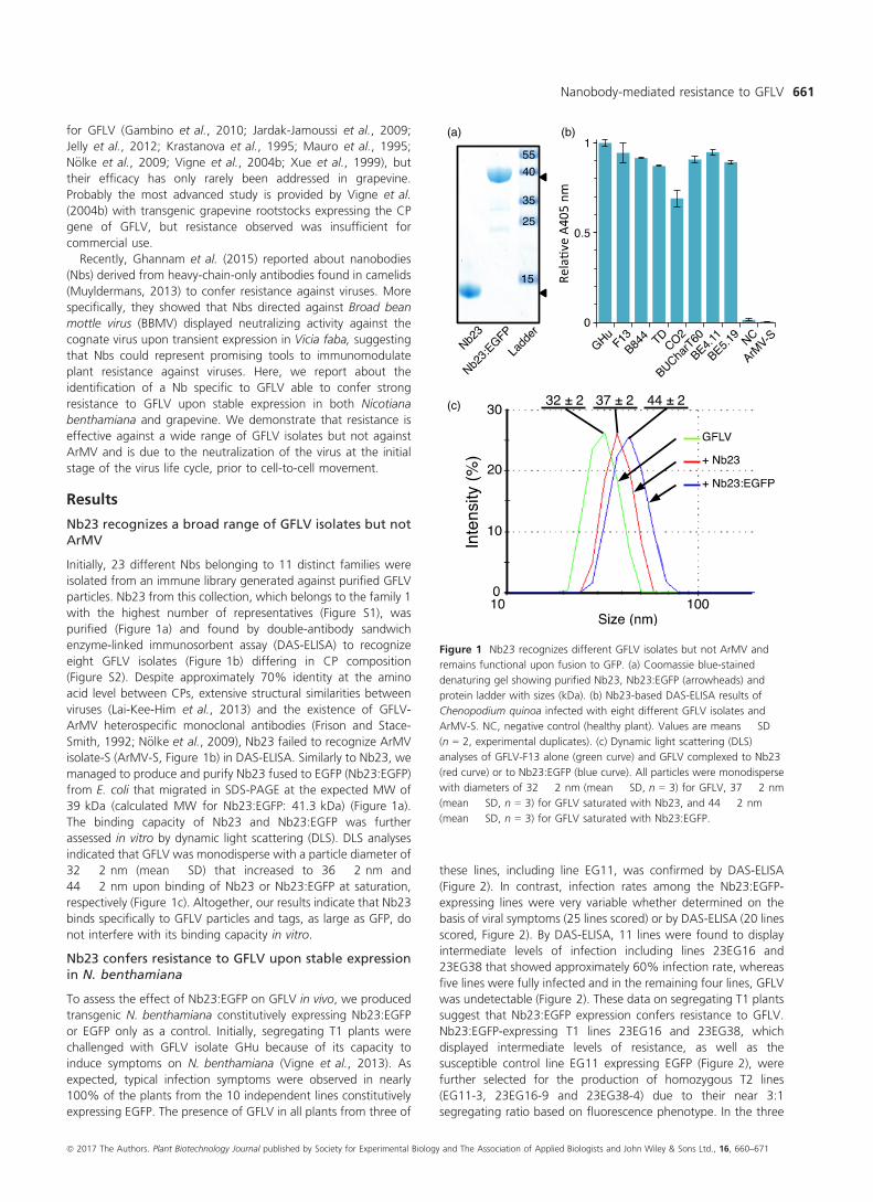
Initially, 23 different Nbs belonging to 11 distinct families were
isolated from an immune library generated against purified GFLV
particles. Nb23 from this collection, which belongs to the family 1
with the highest number of representatives (Figure S1), was
purified (Figure 1a) and found by double-antibody sandwich
enzyme-linked immunosorbent assay (DAS-ELISA) to recognize
eight GFLV isolates (Figure 1b) differing in CP composition
(Figure S2). Despite approximately 70% identity at the amino
acid level between CPs, extensive structural similarities between
viruses (Lai-Kee-Him et al., 2013) and the existence of GFLV-
ArMV heterospecific monoclonal antibodies (Frison and Stace-
Smith, 1992; N€olke et al., 2009), Nb23 failed to recognize ArMV
isolate-S (ArMV-S, Figure 1b) in DAS-ELISA. Similarly to Nb23, we
managed to produce and purify Nb23 fused to EGFP (Nb23:EGFP)
from E. coli that migrated in SDS-PAGE at the expected MW of
39 kDa (calculated MW for Nb23:EGFP: 41.3 kDa) (Figure 1a).
The binding capacity of Nb23 and Nb23:EGFP was further
assessed in vitro by dynamic light scattering (DLS). DLS analyses
indicated that GFLV was monodisperse with a particle diameter of
32 � 2 nm (mean � SD) that increased to 36 � 2 nm and
44 � 2 nm upon binding of Nb23 or Nb23:EGFP at saturation,
respectively (Figure 1c). Altogether, our results indicate that Nb23
binds specifically to GFLV particles and tags, as large as GFP, do
not interfere with its binding capacity in vitro.
Nb23 confers resistance to GFLV upon stable expressionin N. benthamiana
To assess the effect of Nb23:EGFP on GFLV in vivo, we produced
transgenic N. benthamiana constitutively expressing Nb23:EGFP
or EGFP only as a control. Initially, segregating T1 plants were
challenged with GFLV isolate GHu because of its capacity to
induce symptoms on N. benthamiana (Vigne et al., 2013). As
expected, typical infection symptoms were observed in nearly
100% of the plants from the 10 independent lines constitutively
expressing EGFP. The presence of GFLV in all plants from three of
these lines, including line EG11, was confirmed by DAS-ELISA
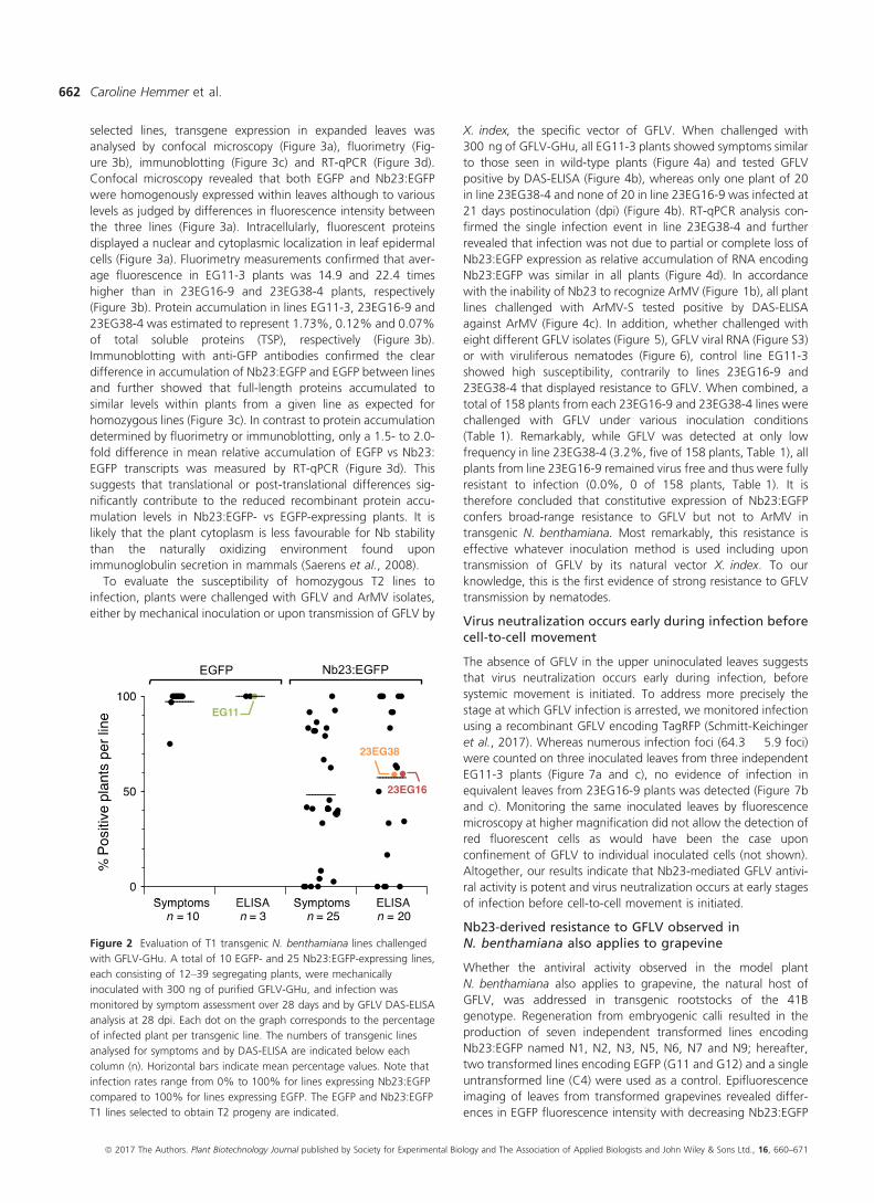
(Figure 2). In contrast, infection rates among the Nb23:EGFP-
expressing lines were very variable whether determined on the
basis of viral symptoms (25 lines scored) or by DAS-ELISA (20 lines
scored, Figure 2). By DAS-ELISA, 11 lines were found to display
intermediate levels of infection including lines 23EG16 and
23EG38 that showed approximately 60% infection rate, whereas
five lines were fully infected and in the remaining four lines, GFLV
was undetectable (Figure 2). These data on segregating T1 plants
suggest that Nb23:EGFP expression confers resistance to GFLV.
Nb23:EGFP-expressing T1 lines 23EG16 and 23EG38, which
displayed intermediate levels of resistance, as well as the
susceptible control line EG11 expressing EGFP (Figure 2), were
further selected for the production of homozygous T2 lines
(EG11-3, 23EG16-9 and 23EG38-4) due to their near 3:1
segregating ratio based on fluorescence phenotype. In the three
(a)
(c)
(b)
Figure 1 Nb23 recognizes different GFLV isolates but not ArMV and
remains functional upon fusion to GFP. (a) Coomassie blue-stained
denaturing gel showing purified Nb23, Nb23:EGFP (arrowheads) and
protein ladder with sizes (kDa). (b) Nb23-based DAS-ELISA results of
Chenopodium quinoa infected with eight different GFLV isolates and
ArMV-S. NC, negative control (healthy plant). Values are means � SD
(n = 2, experimental duplicates). (c) Dynamic light scattering (DLS)
analyses of GFLV-F13 alone (green curve) and GFLV complexed to Nb23
(red curve) or to Nb23:EGFP (blue curve). All particles were monodisperse
with diameters of 32 � 2 nm (mean � SD, n = 3) for GFLV, 37 � 2 nm
(mean � SD, n = 3) for GFLV saturated with Nb23, and 44 � 2 nm
(mean � SD, n = 3) for GFLV saturated with Nb23:EGFP.
ª 2017 The Authors. Plant Biotechnology Journal published by Society for Experimental Biology and The Association of Applied Biologists and John Wiley & Sons Ltd., 16, 660–671
Nanobody-mediated resistance to GFLV 661
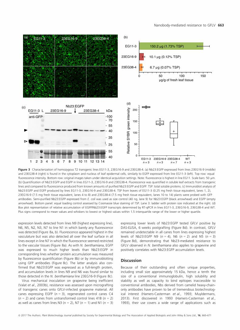
selected lines, transgene expression in expanded leaves was
analysed by confocal microscopy (Figure 3a), fluorimetry (Fig-
ure 3b), immunoblotting (Figure 3c) and RT-qPCR (Figure 3d).
Confocal microscopy revealed that both EGFP and Nb23:EGFP
were homogenously expressed within leaves although to various
levels as judged by differences in fluorescence intensity between
the three lines (Figure 3a). Intracellularly, fluorescent proteins
displayed a nuclear and cytoplasmic localization in leaf epidermal
cells (Figure 3a). Fluorimetry measurements confirmed that aver-
age fluorescence in EG11-3 plants was 14.9 and 22.4 times
higher than in 23EG16-9 and 23EG38-4 plants, respectively
(Figure 3b). Protein accumulation in lines EG11-3, 23EG16-9 and
23EG38-4 was estimated to represent 1.73%, 0.12% and 0.07%
of total soluble proteins (TSP), respectively (Figure 3b).
Immunoblotting with anti-GFP antibodies confirmed the clear
difference in accumulation of Nb23:EGFP and EGFP between lines
and further showed that full-length proteins accumulated to
similar levels within plants from a given line as expected for
homozygous lines (Figure 3c). In contrast to protein accumulation
determined by fluorimetry or immunoblotting, only a 1.5- to 2.0-
fold difference in mean relative accumulation of EGFP vs Nb23:
EGFP transcripts was measured by RT-qPCR (Figure 3d). This
suggests that translational or post-translational differences sig-
nificantly contribute to the reduced recombinant protein accu-
mulation levels in Nb23:EGFP- vs EGFP-expressing plants. It is
likely that the plant cytoplasm is less favourable for Nb stability
than the naturally oxidizing environment found upon
immunoglobulin secretion in mammals (Saerens et al., 2008).
To evaluate the susceptibility of homozygous T2 lines to
infection, plants were challenged with GFLV and ArMV isolates,
either by mechanical inoculation or upon transmission of GFLV by
X. index, the specific vector of GFLV. When challenged with
300 ng of GFLV-GHu, all EG11-3 plants showed symptoms similar
to those seen in wild-type plants (Figure 4a) and tested GFLV
positive by DAS-ELISA (Figure 4b), whereas only one plant of 20
in line 23EG38-4 and none of 20 in line 23EG16-9 was infected at
21 days postinoculation (dpi) (Figure 4b). RT-qPCR analysis con-
firmed the single infection event in line 23EG38-4 and further
revealed that infection was not due to partial or complete loss of
Nb23:EGFP expression as relative accumulation of RNA encoding
Nb23:EGFP was similar in all plants (Figure 4d). In accordance
with the inability of Nb23 to recognize ArMV (Figure 1b), all plant
lines challenged with ArMV-S tested positive by DAS-ELISA
against ArMV (Figure 4c). In addition, whether challenged with
eight different GFLV isolates (Figure 5), GFLV viral RNA (Figure S3)
or with viruliferous nematodes (Figure 6), control line EG11-3
showed high susceptibility, contrarily to lines 23EG16-9 and
23EG38-4 that displayed resistance to GFLV. When combined, a
total of 158 plants from each 23EG16-9 and 23EG38-4 lines were
challenged with GFLV under various inoculation conditions
(Table 1). Remarkably, while GFLV was detected at only low
frequency in line 23EG38-4 (3.2%, five of 158 plants, Table 1), all
plants from line 23EG16-9 remained virus free and thus were fully
resistant to infection (0.0%, 0 of 158 plants, Table 1). It is
therefore concluded that constitutive expression of Nb23:EGFP
confers broad-range resistance to GFLV but not to ArMV in
transgenic N. benthamiana. Most remarkably, this resistance is
effective whatever inoculation method is used including upon
transmission of GFLV by its natural vector X. index. To our
knowledge, this is the first evidence of strong resistance to GFLV
transmission by nematodes.
Virus neutralization occurs early during infection beforecell-to-cell movement
The absence of GFLV in the upper uninoculated leaves suggests
that virus neutralization occurs early during infection, before
systemic movement is initiated. To address more precisely the
stage at which GFLV infection is arrested, we monitored infection
using a recombinant GFLV encoding TagRFP (Schmitt-Keichinger
et al., 2017). Whereas numerous infection foci (64.3 � 5.9 foci)
were counted on three inoculated leaves from three independent
EG11-3 plants (Figure 7a and c), no evidence of infection in
equivalent leaves from 23EG16-9 plants was detected (Figure 7b
and c). Monitoring the same inoculated leaves by fluorescence
microscopy at higher magnification did not allow the detection of
red fluorescent cells as would have been the case upon
confinement of GFLV to individual inoculated cells (not shown).
Altogether, our results indicate that Nb23-mediated GFLV antivi-
ral activity is potent and virus neutralization occurs at early stages
of infection before cell-to-cell movement is initiated.
Nb23-derived resistance to GFLV observed inN. benthamiana also applies to grapevine
Whether the antiviral activity observed in the model plant
N. benthamiana also applies to grapevine, the natural host of
GFLV, was addressed in transgenic rootstocks of the 41B
genotype. Regeneration from embryogenic calli resulted in the
production of seven independent transformed lines encoding
Nb23:EGFP named N1, N2, N3, N5, N6, N7 and N9; hereafter,
two transformed lines encoding EGFP (G11 and G12) and a single
untransformed line (C4) were used as a control. Epifluorescence
imaging of leaves from transformed grapevines revealed differ-
ences in EGFP fluorescence intensity with decreasing Nb23:EGFP
Figure 2 Evaluation of T1 transgenic N. benthamiana lines challenged
with GFLV-GHu. A total of 10 EGFP- and 25 Nb23:EGFP-expressing lines,
each consisting of 12–39 segregating plants, were mechanically
inoculated with 300 ng of purified GFLV-GHu, and infection was
monitored by symptom assessment over 28 days and by GFLV DAS-ELISA
analysis at 28 dpi. Each dot on the graph corresponds to the percentage
of infected plant per transgenic line. The numbers of transgenic lines
analysed for symptoms and by DAS-ELISA are indicated below each
column (n). Horizontal bars indicate mean percentage values. Note that
infection rates range from 0% to 100% for lines expressing Nb23:EGFP
compared to 100% for lines expressing EGFP. The EGFP and Nb23:EGFP
T1 lines selected to obtain T2 progeny are indicated.
ª 2017 The Authors. Plant Biotechnology Journal published by Society for Experimental Biology and The Association of Applied Biologists and John Wiley & Sons Ltd., 16, 660–671
Caroline Hemmer et al.662
expression levels detected from lines N9 (highest expressing line),
N6, N5, N2, N3, N7 to line N1 in which barely any fluorescence
was detected (Figure 8a, b). Fluorescence appeared highest in the
vasculature but was also detected all over the leaf surface in all
lines except in line N7 in which the fluorescence seemed restricted
to the vascular tissues (Figure 8a). As with N. benthamiana, EGFP
was expressed to much higher levels than Nb23:EGFP in
corresponding lines whether protein accumulation was measured
by fluorescence quantification (Figure 8b) or by immunoblotting
using GFP antibodies (Figure 8c). The latter analysis also con-
firmed that Nb23:EGFP was expressed as a full-length protein,
and accumulation levels in lines N9 and N6 was found similar to
those detected in the N. benthamiana line 23EG16-9 (Figure 8c).
Virus mechanical inoculation on grapevine being inefficient
(Valat et al., 2003b), resistance was assessed upon micrografting
of transgenic canes onto GFLV-infected grapevine material. All
canes expressing EGFP (n = 3), regenerated control canes C4
(n = 2) and canes from untransformed control lines 41B (n = 2)
as well as canes from lines N3 (n = 2), N7 (n = 1) and N1 (n = 3)
expressing lower levels of Nb23:EGFP tested GFLV positive by
DAS-ELISA, 6 weeks postgrafting (Figure 8d). In contrast, GFLV
remained undetectable in all canes from lines expressing highest
levels of Nb23:EGFP N9 (n = 4), N6 (n = 2) and N5 (n = 3)
(Figure 8d), demonstrating that Nb23-mediated resistance to
GFLV observed in N. benthamiana also applies to grapevine and
positively correlates with Nb23:EGFP accumulation levels.
Discussion
Because of their outstanding and often unique properties,
including small size approximately 15 kDa, hence a tenth the
size of a conventional immunoglobulin, high solubility and
stability as well as capacity to bind epitopes inaccessible to
conventional antibodies, Nbs derived from camelid heavy-chain-
only antibodies have proven to be of tremendous biotechnolog-
ical interest (Hamers-Casterman et al., 1993; Muyldermans,
2013). First discovered in 1993 (Hamers-Casterman et al.,
1993), their use covers a wide range of applications such as
(a) (b)
(c)(d)
Figure 3 Characterization of homozygous T2 transgenic lines EG11-3, 23EG16-9 and 23EG38-4. (a) Nb23:EGFP expressed from lines 23EG16-9 (middle)
and 23EG38-4 (right) is found in the cytoplasm and nucleus of leaf epidermal cells, similarly to EGFP expressed from line EG11-3 (left). Top row: equal
fluorescence intensity. Bottom row: original images taken under identical acquisition settings. Note: fluorescence is highest in line EG11. Scale bars: 50 lm.
(b) Quantification of Nb23:EGFP and EGFP in lines EG11-3, 23EG16-9 and 23EG38-4. Fluorescence was quantified in soluble leaf extracts from transgenic
lines and compared to fluorescence produced from known amounts of purified Nb23:EGFP and EGFP. TSP: total soluble proteins. (c) Immunoblot analysis of
Nb23:EGFP and EGFP produced by lines EG11-3, 23EG16-9 and 23EG38-4. TSP from leaves of EG11-3 (0.25 mg fresh tissue equivalent, lanes 1, 2),
23EG16-9 (7.5 mg fresh tissue equivalent, lanes 4 to 8) and 23EG38-4 (7.5 mg fresh tissue equivalent, lanes 10 to 14) plants were probed with GFP
antibodies. Semi-purified Nb23:EGFP expressed from E. coli was used as size control (40 ng, lane 9) for Nb23:EGFP (black arrowhead) and EGFP (empty
arrowhead). Bottom panel: equal loading control assessed by Coomassie blue staining of TSP. Lane 3: ladder with protein size indicated at the right. (d)
Box plot representation of relative accumulation of EGFP/Nb23:EGFP transcripts determined by RT-qPCR in lines EG11-3, 23EG16-9, 23EG38-4 and WT.
Plus signs correspond to mean values and whiskers to lowest or highest values within 1.5 interquartile range of the lower or higher quartile.
ª 2017 The Authors. Plant Biotechnology Journal published by Society for Experimental Biology and The Association of Applied Biologists and John Wiley & Sons Ltd., 16, 660–671
Nanobody-mediated resistance to GFLV 663
bioimaging, prophylactic or therapeutic vaccines, diagnosis for
various human and animal diseases such as cancer, Alzheimer or
viral diseases (De Meyer et al., 2014; Wang et al., 2016). In
contrast, their use in plant biology is rather limited (Wang et al.,
2016). It is only recently that Ghannam et al. (2015) suggested
their potential as antiviral molecules against plant viruses by
showing that Nbs directed against BBMV attenuated viral
spreading upon transient expression in Vicia faba leaves. Here,
we confirm this hypothesis by showing that a single Nb with
specificity to GFLV is able to confer strong resistance to the
cognate virus when stably expressed in transgenic plants. This
conclusion applies not only to the model plant N. benthamiana
but also to the economically relevant crop grapevine for which
GFLV is a very serious pest. To the best of our knowledge, it is the
first report of Nb-mediated resistance in transgenic plants and
perhaps more importantly, the first compelling evidence of
resistance to GFLV in Vitis (Basso et al., 2017).
The use of antibodies to confer resistance against plant
pathogens has been reported previously (for review see Safarnejad
et al. (2011) including for GFLV and ArMV in N. benthamiana
0.0
0.1
1.0
10.0
100.0
1000.0
#01 #02 #03 #04 #05 #06 #07 #08 #09 #10 #12 #13 #14 #15 #16 #17 #18 #19 #20
Rel
ativ
e S
YB
R g
reen
in
tens
ity
RNA1 EGFP
0.0
0.5
1.0
Rel
ativ
e A
405
nm
EG11-3n = 10 n = 10 n = 10100%
23EG16-9
100%
23EG38-4
100%
NCn = 40%
0.0
0.5
1.0
Rel
ativ
e A
40
5 n
m
EG11-3n = 20100%
23EG16-9n = 200%
23EG38-4n = 20
5%
WTn = 10100%
NCn = 80%
EG11-3
23EG16-9
WT
23EG38-4
(a)
(b)
(d)
(c)
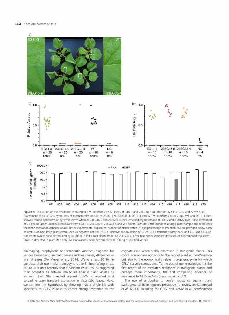
Figure 4 Evaluation of the resistance of transgenic N. benthamiana T2 lines 23EG16-9 and 23EG38-4 to infection by GFLV-GHu and ArMV-S. (a)
Assessment of GFLV-GHu symptoms of mechanically inoculated 23EG16-9, 23EG38-4, EG11-3 and WT N. benthamiana at 7 dpi. WT and EG11-3 lines
showed mosaic symptoms on systemic leaves whereas 23EG16-9 and 23EG38-4 lines remained asymptomatic. (b) GFLV and c, ArMV DAS-ELISA performed
at 21 dpi on upper uninoculated leaves from EG11-3, 23EG16-9, 23EG38-4 and WT plants. Each dot corresponds to a single plant sample and represents
the mean relative absorbance at 405 nm of experimental duplicates. Number of plants tested (n) and percentage of infection (%) are provided below each
column. Noninoculated plants were used as negative control (NC). d, Relative accumulation of GFLV RNA1 transcripts (grey bars) and EGFP/Nb23:EGFP
transcripts (white bars) determined by RT-qPCR in individual plants from line 23EG38-4. Error bars show standard deviation of experimental triplicates.
RNA1 is detected in plant #17 only. All inoculations were performed with 300 ng of purified viruses.
ª 2017 The Authors. Plant Biotechnology Journal published by Society for Experimental Biology and The Association of Applied Biologists and John Wiley & Sons Ltd., 16, 660–671
Caroline Hemmer et al.664
(N€olke et al., 2009). In general, best results were obtained with
single-chain variable fragments (scFvs) rather than full-length
immunoglobulins (Schillberg et al., 1999). Here, we show that
Nb23 is functional and accumulates to concentrations of 0.12%
(line 23EG16-9) and 0.07% (line 23EG38-4) of TSP in the
cytoplasm of resistant N. benthamiana cells and to similar levels
in resistant grapevine lines. Similar expression levels (up to 0.1% of
TSP) were achieved in N. benthamiana expressing scFvGFLVcp-55
directed against GFLV (N€olke et al., 2009), suggesting that Nb23 is
as potent as scFvGFLVcp-55 to confer resistance. Considering our
results with GFLV and those of Ghannam et al. (2015) with BBMV,
it is tempting to speculate that antiviral activity is a common
property of Nbs directed against plant viruses and they should
therefore be considered as a reliable source of resistance. In
addition, considering the difficulties often encountered to gener-
ate functional scFvs (Fiedler et al., 1997; Safarnejad et al., 2011),
which are circumvented with Nbs due to their intrinsic single-
domain origin and structural specificities (Muyldermans, 2013),
Nbs should be considered superior to other antibodies-derived
products when aiming at generating virus-resistant plants.
Here, we describe the production of 23 different Nbs
directed against GFLV belonging to 11 different families and
thus potentially able to recognize up to 11 different epitopes
(Muyldermans, 2013). The choice of Nb23 (family 1, Figure S1)
was essentially driven by its capacity to recognize numerous
GFLV isolates in vitro (Figure 1) and hence for its potential to
confer broad-range resistance to GFLV, which we demon-
strated. Whether antiviral activity is equivalent for all GFLV-
specific Nbs is unknown at this stage. However, considering
Nbs from family 1 are likely recognizing the same epitope
suggests that they all possess similar antiviral activity. In
contrast to N€olke et al. (2009) in which scFvGFLVcp-55 was
shown to cross-react with ArMV and to confer enhanced
tolerance to ArMV, no cross-reactivity was detected for Nb23
with ArMV and consequently transgenic lines expressing Nb23
displayed full susceptibility to ArMV.
In this work, we also describe the production of a total of 25
lines expressing Nb23:EGFP (Figure 2) of which only lines
23EG16-9 and 23EG38-4 were characterized up to T2 stage
contrarily to the other lines that presented complex segregation
profiles probably as a consequence of multiples insertions of the
transgene. It is remarkable to notice that the vast majority of lines
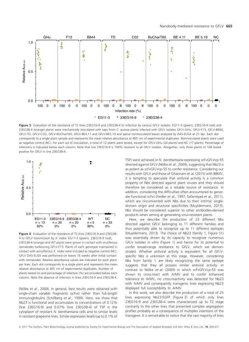
Figure 5 Evaluation of the resistance of T2 lines 23EG16-9 and 23EG38-4 to infection by various GFLV isolates. EG11-3 (green), 23EG16-9 (red) and
23EG38-4 (orange) plants were mechanically inoculated with saps from C. quinoa plants infected with GFLV isolates GFLV-GHu, GFLV-F13, GFLV-B844,
GFLV-TD, GFLV-CO2, GFLV-BUCharT60, GFLV-BE4.11 and GFLV-BE5.19 and apical noninoculated leaves analysed by DAS-ELISA at 21 dpi. Each dot
corresponds to a single plant sample and represents the mean relative absorbance at 405 nm of experimental duplicates. Noninoculated plants were used
as negative control (NC). For each set of inoculation, a total of 12 plants were tested, except for GFLV-GHu (24 plants) and NC (17 plants). Percentage of
infections is indicated below each column. Note that line 23EG16-9 is 100% resistant to all GFLV isolates. Altogether, only three plants of 108 tested
positive for GFLV in line 23EG38-4.
Figure 6 Evaluation of the resistance of T2 lines 23EG16-9 and 23EG38-
4 to GFLV transmission by X. index. EG11-3 (green), 23EG16-9 (red),
23EG38-4 (orange) and WT plants were grown in contact with viruliferous
nematodes harbouring GFLV-F13. Plants of each genotype maintained in
contact with aviruliferous X. index were included as negative control (NC).
GFLV DAS-ELISA was performed on leaves 16 weeks after initial contact
with nematodes. Relative absorbance values are indicated for each plant
per lines. Each dot corresponds to a single plant and represents the mean
relative absorbance at 405 nm of experimental duplicates. Number of
plants tested (n) and percentage of infection (%) are provided below each
column. Note the absence of infection in lines 23EG16-9 and 23EG38-4.
ª 2017 The Authors. Plant Biotechnology Journal published by Society for Experimental Biology and The Association of Applied Biologists and John Wiley & Sons Ltd., 16, 660–671
Nanobody-mediated resistance to GFLV 665
were resistant to GFLV, some up to 100% already at the T1
generation. Considering transgenic plants were selected on the
basis of their fluorescence and therefore Nb23:EGFP expression,
we assume that resistance is directly linked to the expression level
of the transgene. However, contrarily to most other studies with
scFvs (Boonrod et al., 2004; N€olke et al., 2009; Schillberg et al.,
2000; Villani et al., 2005), we were unable to correlate the
degree of virus resistance (i.e. virus load or delay in symptom
appearance) to Nb23 accumulation levels, plant being either
symptomatic and ELISA positive or asymptomatic and ELISA
negative. Best example is provided by line 23EG38-4 that showed
high degree although not complete resistance to GFLV (3.2%
infection rate, 158 plants challenged, Table 1). It is likely that the
five plants of line 23EG38-4 in which GFLV was detected despite
appropriate expression of the transgene (Figure 4d) correspond
to cases where the virus succeeded in overcoming resistance as a
consequence of a mutation affecting the Nb23 epitope. Struc-
tural studies aiming at precisely identifying the Nb23 epitope and
sequencing of the CP from putative GFLV escape variants for the
presence of mutations explaining the loss of resistance shall be
addressed elsewhere.
Finally, our results raise also the question of the resistance
mechanism. As resistant transgenic plants showed no evidence of
systemic spread of the virus, neither the occurrence of infection
foci nor the presence of single infected cells on inoculated leaves
indicates that Nb23 blocks GFLV very early during the infection
process, before cell-to-cell movement of the virus. Virus neutral-
ization may be the consequence of a stabilizing effect exerted by
Nb23 upon binding to the capsid, preventing its disassembly and
thereby the release of the viral RNA and consequently the
initiation of the replication cycle. Nbs with stabilizing activity have
been reported for poliovirus, but strict correlation between virus
neutralization and stabilizing activity could not be established
(Schotte et al., 2014). Another possibility may consist in inhibition
of capsid formation due to early CP tethering prior to virion
assembly. Considering that GFLV moves from cell to cell as entire
virions via tubules that form within plasmodesmata (Laporte
et al., 2003; Ritzenthaler et al., 1995), the interference of Nb23
with capsid assembly would ineluctably prevent the spread of the
virus to neighbouring cells. Finally, one cannot rule out the
possibility that Nb23 may interfere with CP folding resulting in the
cytosolic accumulation of aggregated or misfolded proteins that
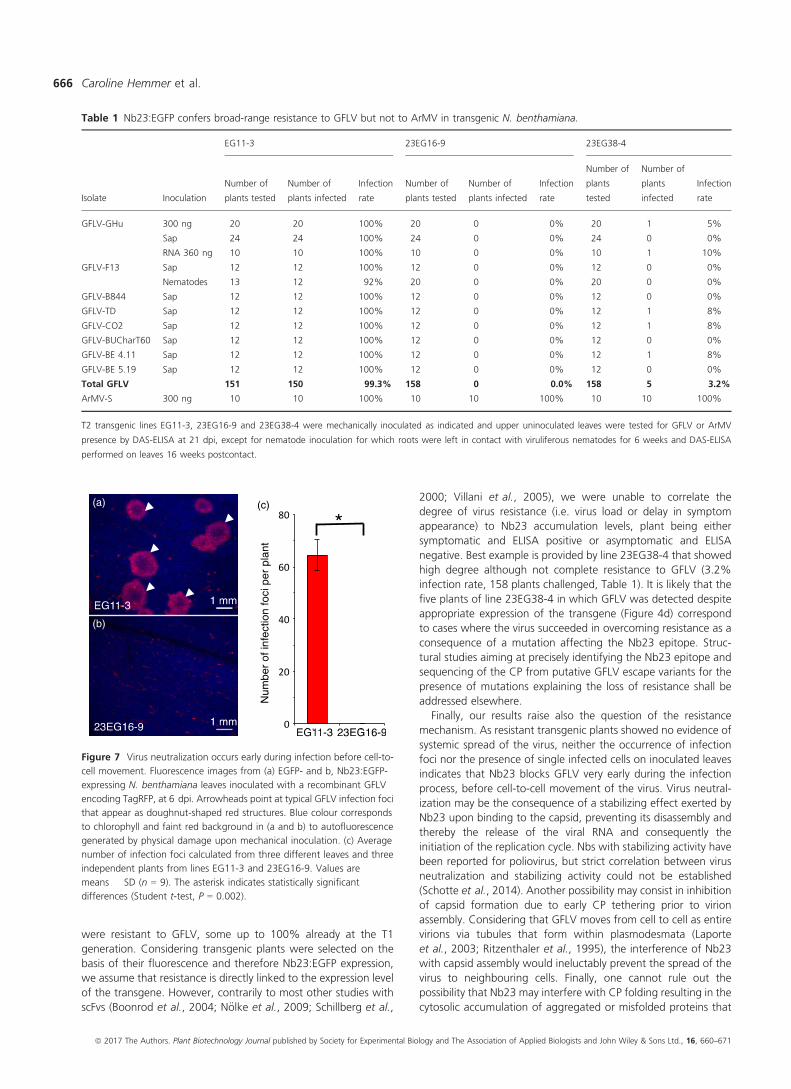
Table 1 Nb23:EGFP confers broad-range resistance to GFLV but not to ArMV in transgenic N. benthamiana.
Isolate Inoculation
EG11-3 23EG16-9 23EG38-4
Number of
plants tested
Number of
plants infected
Infection
rate
Number of
plants tested
Number of
plants infected
Infection
rate
Number of
plants
tested
Number of
plants
infected
Infection
rate
GFLV-GHu 300 ng 20 20 100% 20 0 0% 20 1 5%
Sap 24 24 100% 24 0 0% 24 0 0%
RNA 360 ng 10 10 100% 10 0 0% 10 1 10%
GFLV-F13 Sap 12 12 100% 12 0 0% 12 0 0%
Nematodes 13 12 92% 20 0 0% 20 0 0%
GFLV-B844 Sap 12 12 100% 12 0 0% 12 0 0%
GFLV-TD Sap 12 12 100% 12 0 0% 12 1 8%
GFLV-CO2 Sap 12 12 100% 12 0 0% 12 1 8%
GFLV-BUCharT60 Sap 12 12 100% 12 0 0% 12 0 0%
GFLV-BE 4.11 Sap 12 12 100% 12 0 0% 12 1 8%
GFLV-BE 5.19 Sap 12 12 100% 12 0 0% 12 0 0%
Total GFLV 151 150 99.3% 158 0 0.0% 158 5 3.2%
ArMV-S 300 ng 10 10 100% 10 10 100% 10 10 100%
T2 transgenic lines EG11-3, 23EG16-9 and 23EG38-4 were mechanically inoculated as indicated and upper uninoculated leaves were tested for GFLV or ArMV
presence by DAS-ELISA at 21 dpi, except for nematode inoculation for which roots were left in contact with viruliferous nematodes for 6 weeks and DAS-ELISA
performed on leaves 16 weeks postcontact.
(a)
(b)
(c)
Figure 7 Virus neutralization occurs early during infection before cell-to-
cell movement. Fluorescence images from (a) EGFP- and b, Nb23:EGFP-
expressing N. benthamiana leaves inoculated with a recombinant GFLV
encoding TagRFP, at 6 dpi. Arrowheads point at typical GFLV infection foci
that appear as doughnut-shaped red structures. Blue colour corresponds
to chlorophyll and faint red background in (a and b) to autofluorescence
generated by physical damage upon mechanical inoculation. (c) Average
number of infection foci calculated from three different leaves and three
independent plants from lines EG11-3 and 23EG16-9. Values are
means � SD (n = 9). The asterisk indicates statistically significant
differences (Student t-test, P = 0.002).
ª 2017 The Authors. Plant Biotechnology Journal published by Society for Experimental Biology and The Association of Applied Biologists and John Wiley & Sons Ltd., 16, 660–671
Caroline Hemmer et al.666
could act as a signal for CP targeting to degradation pathways
(Liu and Bassham, 2012) and thereby virus clearance. Of course,
these hypotheses should not be considered as exhaustive or
mutually exclusive and further studies are needed.
In conclusion, we have shown that Nb-mediated resistance can
be used to confer resistance to GFLV in N. benthamiana and
more importantly in grapevine where there is an urgent need to
find sustainable solutions to the increasing problem of fanleaf
degeneration in cultivated vineyards. The proof of concept being
established, our work shall pave the way for the creation of novel
virus-resistant varieties of agriculturally important crops. Whether
it will help to improve the public acceptance of genetically
modified crops is a long debate that extends far beyond science.
Experimental procedures
Virus isolates
GFLV and ArMV isolates used here are GFLV-GHu (Vigne et al.,
2013), GFLV-F13 (Pinck et al., 1988), GFLV-B844 (Legin et al.,
1993), GFLV-TD (Schellenberger et al., 2011b), GFLV-CO2 (Vigne
et al., 2004a), GFLV-BuCharT60, GFLV-BE 4.11, GFLV-BE 5.19
and ArMV-S (Loudes et al., 1995).
Immunization, Nbs library construction and screening
GFLV-specific Nbs were generated according to Muyldermans
et al. (2009). Briefly, a camel (Camelus dromedarius) was immu-
nized six times subcutaneously at weekly intervals with 100 lg of
purified GFLV-F13 according to standard immunization protocols.
After immunization, total RNA was extracted from isolated
peripheral blood lymphocytes and mRNAs reverse-transcribed to
cDNA. The regions encoding variable fragments of heavy-chain-
only antibodies were amplified in two subsequent PCR, cloned
into the pHEN4 phagemid vector and transformed into E. coli
TG1. The resulting Nbs library was screened by phage display for
GFLV-specific binders in three consecutive biopanning rounds
against 10 lg of purified GFLV-F13 each. Sequences of GFLV-
specific Nbs were obtained following the isolation of individual
clones from the enriched library by a phage ELISA approach
performed against 100 ng of purified GFLV-F13.
Expression and purification of Nbs from E. coli
GFLV-specific Nbs coding sequences were subcloned into the
pHEN6 (Conrath et al., 2001) expression vector as a BstEII/PstI
fragment adding a N-terminal PelB signal peptide sequence for
(a) (c)
(b) (d)
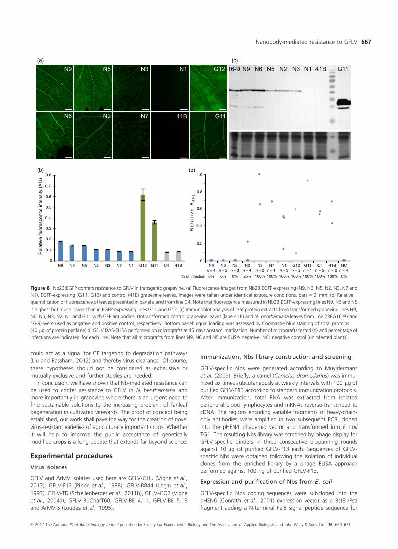
Figure 8 Nb23:EGFP confers resistance to GFLV in transgenic grapevine. (a) Fluorescence images from Nb23:EGFP-expressing (N9, N6, N5, N2, N3, N7 and
N1), EGFP-expressing (G11, G12) and control (41B) grapevine leaves. Images were taken under identical exposure conditions. bars = 2 mm. (b) Relative
quantification of fluorescence of leaves presented in panel a and from line C4. Note that fluorescencemeasured in Nb23:EGFP-expressing lines N9, N6 and N5
is highest but much lower than in EGFP-expressing lines G11 and G12. (c) Immunoblot analysis of leaf protein extracts from transformed grapevine lines N9,
N6, N5, N3, N2, N1 and G11 with GFP antibodies. Untransformed control grapevine leaves (lane 41B) and N. benthamiana leaves from line 23EG16-9 (lane
16-9) were used as negative and positive control, respectively. Bottom panel: equal loading was assessed by Coomassie blue staining of total proteins
(40 lg of protein per lane) d, GFLV DAS-ELISA performed on micrografts at 45 days postacclimatization. Number of micrografts tested (n) and percentage of
infections are indicated for each line. Note that all micrografts from lines N9, N6 and N5 are ELISA negative. NC: negative control (uninfected plants).
ª 2017 The Authors. Plant Biotechnology Journal published by Society for Experimental Biology and The Association of Applied Biologists and John Wiley & Sons Ltd., 16, 660–671
Nanobody-mediated resistance to GFLV 667
periplasmic targeting and a C-terminal 6-His-tag for purification.
Production of 6-His-tagged Nbs constructs was performed by
expression in E. coli WK6 grown in Terrific Broth medium and
induced overnight with 1 mM IPTG at 28 °C.An additional C-terminal Strep-tag II (Trp-Ser-His-Pro-Gln-Phe-
Glu-Lys) was added for Nbs used in ELISA. To do so, Nbs coding
sequences were amplified by PCR and amplicons were introduced
by Gateway cloning into the pDONR/Zeo vector (Thermo Fisher
Scientific, France) which was further recombined into the p0GWA
expression vector (Busso et al., 2005). Large-scale production of
Strep II-tagged Nbs constructs was performed by expression in
E. coli BL21 (DE3) grown overnight at 23 °C in auto-inducing
ZYP50502 medium.
Nbs were extracted from periplasm by osmotic shock (Habib
et al., 2013) and purified at 4 °C by immobilized metal ion
chromatography (IMAC) on a 1 mL Protino Ni-NTA column
(Macherey-Nagel, France) using 500 mM imidazole in running
buffer (50 mM Tris, 300 mM NaCl, 5% glycerol, pH 8.0) for
elution, followed by size exclusion chromatography (SEC) on a
Hiload 16/60 Superdex75 prep grade column (GE Healthcare Life
Science, France) in 1X phosphate buffer saline. Purity of eluted
proteins was assessed by Coomassie blue staining of denatured
Nbs in denaturing Tris–tricine polyacrylamide gel. Purification
yields were estimated from absorbance at 280 nm based on
extinction coefficients computed from Nbs composition.
DAS-ELISA assessment of Nb23 reactivity against GFLVstrains
Plants at the four- to six-leaf stage were mechanically inoculated
with crude saps from Chenopodium quinoa infected with various
GFLV isolates. Apical noninoculated leaves were ground in
extraction buffer (35 mM Na2HPO4, 15 mM KH2PO4, pH 7.0) in
a 1:5 w/v ratio. Virus detection was performed on clarified
extracts by DAS-ELISA using anti-GFLV or anti-ArMV polyclonals
(Bioreba AG, Switzerland) diluted 1000-fold in coating buffer
(15 mM Na2CO3, 35 mM NaHCO3, pH 9.6) as capture antibody
and the Strep II-tagged Nb23 at 1 lg/mL in conjugate buffer
(10 mM PBS, 0.1% w/v bovine serum albumin (BSA), 0.05% v/v
Tween-20, pH 7.4) as detection antibody. For development,
streptavidin–alkaline phosphatase (Jackson Immunoresearch, Suf-
folk, UK) at 50 ng/mL in conjugate buffer was used in conjunc-
tion with para-nitrophenyl phosphate (Interchim, France) at
1 mg/mL in substrate buffer (1 M diethanolamine, pH 9.8).
Negative control consisted of noninoculated healthy plants.
Absorbance at 405 nm (A405 nm) was recorded after one hour,
and samples with mean A405 nm values exceeding by a factor of
2.4 those of negative controls were considered positive. Results
are presented as mean absorbance of experimental dupli-
cates � standard error normalized against maximum assay value.
N. benthamiana transformation and production
Nb23 was cloned in frame to the N-terminus of EGFP with a
Gly3SerGly3 linker sequence into the pEAQ-HT-DEST3 plant
expression vector (Sainsbury et al., 2009). EGFP was first intro-
duced into the pHEN6-Nb23 vector as an EcoRI/BstEII fragment
following a PCR amplification. The Nb23:EGFP gene was then
reamplified by PCR and introduced by Gateway cloning into the
pDONR/Zeo vector, which was further recombined into the
pEAQ-HT-DEST3 vector. A control consisting of pEAQ-HT-DEST3-
EGFP was included.
The resulting vectors were transferred into Agrobac-
terium tumefaciens GV3101::pMP90 and used at A595 nm = 0.1
for agrotransformation of N. benthamiana leaves. After 3 days,
expression of fluorescence was checked for EGFP expression with
an Axio Zoom V16 macroscope (Zeiss, Germany) and sterilized
infiltrated leaf segments were placed in a growth chamber (16 h
light/8 h dark, 25 °C) onto Murashige and Skoog medium ((MS,
Duchefa, The Netherlands), 10 mM NH4NO3, 19 MS vitamin
solution (Sigma-Aldrich, France), 3% w/v sucrose, 0.05 lg/mL 1-
naphthalene acetic acid, 2 lg/mL 6-benzyl-aminopurine, 0.8%
w/v agar, 150 lg/mL kanamycin, 500 lg/mL carbenicillin, pH
5.8). Calli were subcultured every week onto fresh medium and
shoots excised 3–4 weeks later transferred onto rooting medium
(1/2 MS medium, 1.5% w/v sucrose, 0.59 MS vitamin solution,
0.8% w/v agar, 150 lg/mL kanamycin, 500 lg/mL carbenicillin,
pH 5.8) until plantlets could be acclimatized and established in
soil. Regenerated T0 plants were self-pollinated through T2
generation.
Nb23:EGFP purification
For Nb23:EGFP production, the whole Nb23:EGFP:6-His coding
sequence was cloned into the pET-22b(+) expression vector
(Novagen, Madison) as a NdeI/XhoI fragment subsequent to a
PCR amplification using pEAQ-HT-DEST3-Nb23:EGFP as template.
Expression was performed in E. coli SHuffle T7 Express (New
England Biolabs, France) grown in TB medium and induced
overnight with 0.1 mM IPTG at 20 °C. Pelleted cells resuspended
in PB-NaCl buffer (10 mM phosphate buffer, 300 mM NaCl, pH
7.4) were lysed by sonication (80% amplitude for 2 min with
13 mm diameter probe, Vibra-Cell VCX 500 (Sonics & Materials
Inc) before purification of cytoplasmic extract. The purity of the
eluted proteins was assessed by Coomassie blue staining after
denaturing Tris–tricine polyacrylamide gel electrophoresis.
Quantification of recombinant proteins inN. benthamiana by fluorimetry
Three youngest apical leaves of 6–7 weeks old N. benthamiana
were homogenized in extraction buffer (200 mM Tris-HCl,
300 mM NaCl, 100 mM ascorbic acid, 2.5% w/v
polyvinylpolypyrrolidone, complete protease inhibitor cocktail
(Roche, France), pH 7.0) at a 1:2 w/v ratio. Cell debris were
removed by centrifugation at 20 000 g for 20 min at 4 °C, andTSP concentrations were determined using the Bio-Rad protein
assay following manufacturer’s instructions with BSA as standard.
Fluorescence intensity was recorded in a FLUOstar Omega plate
reader (BMG Labtech, Germany) equipped with 485 � 12 nm
excitation and 520 � 25 nm emission filters, on 100 lL of
soluble extracts in a white flat-bottom polystyrene plate (Greiner
Bio One, Austria). Wild-type N. benthamiana extracts were used
as blank and known amount of purified Nb23:EGFP for fluores-
cent titration.
Fluorescence quantification of recombinant proteins ingrapevine leaves
Two leaves from each line were imaged under identical conditions
using a Axio Zoom V16 stereomicroscope (Zeiss, Germany)
equipped with appropriate excitation and emission filters for
the visualization of EGFP and chlorophyll. The fluorescence was
quantified using ImageJ (rsb.info.nih.gov/ij/) on six randomly
chosen areas of identical surface (8176636 pixels) for each leaf.
Immunoblotting
For N. benthamiana, acetone-precipitated TSP were used. For
grapevine, proteins were extracted according to Hurkman and
ª 2017 The Authors. Plant Biotechnology Journal published by Society for Experimental Biology and The Association of Applied Biologists and John Wiley & Sons Ltd., 16, 660–671
Caroline Hemmer et al.668
Tanaka (1986). Proteins were heated in denaturing buffer,
separated by Tris-glycine SDS-PAGE and transferred onto a
polyvinylidene difluoride membrane using the TransBlot Turbo
transfer system (Bio-Rad, France). After incubation in blocking
buffer (10 mM PBS, 0,1% v/v Tween-20, 5% w/v skim milk),
proteins were sequentially probed with rabbit anti-GFP IgGs
(Sigma-Aldrich, France) at 0.1 lg/mL in blocking buffer and goat
anti-rabbit IgGs conjugated to horseradish peroxidase (Thermo
Fisher Scientific, France) at 0.1 lg/mL in blocking buffer.
Immunolabelled proteins were detected by enhanced chemilumi-
nescence (ECL) using the Lumi-LightPLUS kit (Roche, France) and
the Fusion FX imaging system (Vilber Lourmat GmbH, Germany).
Dynamic Light Scattering (DLS)
Viral particles were purified as described previously (Schellen-
berger et al., 2011a). Mean particle diameters and polydispersity
of GFLV-F13 alone or complexed to Nb23 or to Nb23:EGFP was
estimated by DLS using a Zetasizer NanoZS (Malvern, France) and
Nanostar (Wyatt, CA). Five successive measurements were
performed using three independent virus and protein prepara-
tions with virus at 0.1 mg/mL in Tris buffer (50 mM Tris, 100 mM
NaCl, pH 8.3), Nb23 at 0.1 mg/mL and Nb23:EGFP at 0.9 mg/
mL. Scattered intensities were recorded at 20 °C and data were
processed with DTS software (www.dtssoftware.com, version
6.01) or DYMAMICS (www.wyatt.com/products/software/dyna
mics.html, version 7.1.8.93), respectively. All particles were
monodisperse.
Reverse Transcriptase-qPCR (RT-qPCR) analyses
GFLV RNA1 and EGFP/Nb23:EGFP transcripts were quantified by
RT-qPCR relatively to the expression of cyclin-dependent kinase
homolog (GI:849067, N. tabacum), elongation factor 1 alpha
(GI:37783254) and actin (GI:380505031) genes from N. ben-
thamiana used as internal controls due to their stability assessed
by GeneNorm (Vandesompele et al., 2002) and NormFinder
(Andersen et al., 2004) algorithms. Total RNA was isolated at
21 dpi from approximately 17 mg of noninoculated apical leaves
ground at a 1 : 30 w/v ratio in TLES buffer (100 mM Tris, 100 mM
LiCl, 10 mM EDTA, 0.1% w/v SDS, pH 8.0) followed by a water-
saturated phenol and phenol chloroform extraction before
precipitation with 2 M LiCl. cDNA was generated according to
manufacturers’ instructions from 1 lg of DNaseI-treated total
RNA using 2.5 lM Oligo(dT)18 primer (Thermo Fisher Scientific,
France) and SuperScript III Reverse Transcriptase (Thermo Fisher
Scientific, France). PCR was performed in triplicates using 0.5 lLof reverse transcription reaction and 2.5 lM gene specific primers
in a total volume of 10 lL LightCycler 480 SYBR Green Master I
mix on a LightCycler 480 system (Roche, France) with cycling
conditions of 5-min denaturation at 95 °C followed by 40 cycles
at 95 °C for 10 s, 60 °C for 15 s and 72 °C for 15 s. Relative
gene expression levels were calculated by means of the linear
regression of efficiency method using LinRegPCR software
(version 2013.0) (Ruijter et al., 2009). Primer sequences are
available upon request.
Fluorescence microscopy
For cellular-level scale observations, water-mounted leaf discs of
six- to 7-week-old N. benthamiana plants were imaged with a
LSM 780 laser scanning confocal microscope attached to an
observer Z1 microscope (Zeiss, Germany) equipped with a 209/
0.8 Plan-Apochromat objective and using excitation and emission
wavelength filters set to 488 nm and 499–521 nm.
For macroscopic observations, leaves were imaged with a
MacroFluo Z16 APO(A) macroscope (Leica, Germany) using
excitation and emission wavelength filters of 538–562 nm and
570–640 nm for red channel. Images were processed using Zen
2011 imaging software (Zeiss) and Photoshop CS5 (Adobe).
Inoculations
Plants were inoculated either mechanically or via nematodes.
Mechanical inoculations were performed with purified virus, with
saps of infected C. quinoa or with purified viral RNA.
Nematode inoculation was performed by growing plants for
6 weeks in soil (3 : 1 : 1 v/v ratio of sand, loess and clay pebbles)
containing ca. 300 viruliferous X. index per plant. GFLV ELISAs
were performed at 16 weeks postcontact with nematodes on
apical noninoculated leaves.
Grapevine transformation
A friable embryogenic callus originated from the 41B rootstock
cultivar was used for genetic transformation experiments as
described by Romon et al. (2013) using the same pEAQ
constructs than used for N. benthamiana transformation. After
a 9- to 12-month selection process, seven transgenic lines
harbouring the Nb23:EGFP construct (lines N1, N2, N3, N5, N6,
N7 and N9) and two transgenic lines harbouring solely the EGFP
construct (lines G11 and G12) were isolated on the basis of their
resistance to 50 lg/mL kanamycin. A wild-type 41B rootstock
and a regenerated plant named C4 issued from the same
embryogenic callus in the absence of kanamycin were used as
controls. All the lines were maintained in vitro as cuttings on
McCown woody plant medium (McCown and Lloyd, 1981).
In vitro micrografting
One-node cuttings of nine transgenic grapevine lines and two
control lines were in vitro-grafted as scion onto GFLV-F13-
infected Kober 5BB rootstock cultivar by cleft grafting under
sterile conditions as described by Valat et al. (2003a). After
3 months of in vitro culture, rooted grafts were acclimatized in a
growth chamber. The presence of GFLV in scions was assessed by
DAS-ELISA 45 days after acclimatization.
Statistical analyses
For infection foci measurement, statistical analysis was performed
by unequal variances two-tailed unpaired Student’s t-test (P
value = 0.002) and Shapiro–Wilk test for normality (W = 0.87. P
value = 0.12).
Acknowledgements
This work was supported by the Centre National de la Recherche
Scientifique (CNRS), the Institut National de la Recherche
Agronomique (INRA) ‘Plant Health and the Environment’ division,
the Agence Nationale de la Recherche (ANR) awards COMBiNiNG
(ANR-14-CE19-0022) and VinoBodies (ANR-14-CE19-0018), the
European Regional Development Fund (ERDF) in the framework of
the INTERREG V Upper Rhine programme Vitifutur, Transcending
borders with every project, the Conseil interprofessionel des vins
d’Alsace, the Comit�e interprofessionel du vin Champagne, the
Bureau interprofessionnel des vins de Bourgogne and the Conseil
interprofessionnel du vin de Bordeaux. C.H. was supported by a
fellowship from the R�egion Alsace. L.A. was supported by a CIFRE
grant from the Institut Franc�ais de la Vigne et du Vin, subsidized by
the ANRT (CIFRE convention number 2012/0929).
ª 2017 The Authors. Plant Biotechnology Journal published by Society for Experimental Biology and The Association of Applied Biologists and John Wiley & Sons Ltd., 16, 660–671
Nanobody-mediated resistance to GFLV 669
References
Andersen, C.L., Jensen, J.L. and Orntoft, T.F. (2004) Normalization of real-time
quantitative reverse transcription-PCR data: a model-based variance
estimation approach to identify genes suited for normalization, applied to
bladder and colon cancer data sets. Cancer Res. 64, 5245–5250.
Andret-Link, P., Marmonier, A., Belval, L., Hleibieh, K., Ritzenthaler, C. and
Demangeat, G. (2017) Nematode vectors for grapevine viruses: biology,
behavior and mechanisms of virus transmission. In Grapevine Viruses:
Molecular Biology, Diagnostics and Management (Meng, B., Martelli, G.P.,
Fuchs, M. and Golino, D. eds), pp. 505–529. Switzerland: Springer
International Publishing AG.
Basso, M.F., Fajardo, T.V. and Saldarelli, P. (2017) Grapevine virus diseases:
economic impact and current advances in viral prospection and management.
Rev. Bras. Fruticultura, 39, n. 1: (e-411).
Boonrod, K., Galetzka, D., Nagy, P.D., Conrad, U. and Krczal, G. (2004) Single-
chain antibodies against a plant viral RNA-dependent RNA polymerase confer
virus resistance. Nature Biotech. 22, 856–862.
Busso, D., Delagoutte-Busso, B. and Moras, D. (2005) Construction of a set
Gateway-based destination vectors for high-throughput cloning and
expression screening in Escherichia coli. Anal. Biochem. 343, 313–321.
Collinge, D.B., Jorgensen, H.J., Lund, O.S. and Lyngkjaer, M.F. (2010)
Engineering pathogen resistance in crop plants: current trends and future
prospects. Annu. Rev. Phytopathol. 48, 269–291.
Conrath, K.E., Lauwereys, M., Galleni, M., Matagne, A., Fr�ere, J.-M., Kinne, J.,
Wyns, L. et al. (2001) b-Lactamase inhibitors derived from single-domain
antibody fragments elicited in the camelidae. Antimicrob. Agents Chemother.
45, 2807–2812.
De Meyer, T., Muyldermans, S. and Depicker, A. (2014) Nanobody-based
products as research and diagnostic tools. Trends Biotechnol. 32, 263–270.
Fiedler, U., Phillips, J., Artsaenko, O. and Conrad, U. (1997) Optimization of scFv
antibody production in transgenic plants. Immunotechnology, 3, 205–216.
Frison, E.A. and Stace-Smith, R. (1992) Cross-reacting and heterospecific
monoclonal antibodies produced against arabis mosaic nepovirus. J. Gen.
Virol. 73, 2525–2530.
Gambino, G., Perrone, I., Carra, A., Chitarra, W., Boccacci, P., Torello
Marinoni, D., Barberis, M. et al. (2010) Transgene silencing in grapevines
transformed with GFLV resistance genes: analysis of variable expression of
transgene, siRNAs production and cytosine methylation. Transgenic Res. 19,
17–27.
Ghannam, A., Kumari, S., Muyldermans, S. and Abbady, A.Q. (2015) Camelid
nanobodies with high affinity for broad bean mottle virus: a possible
promising tool to immunomodulate plant resistance against viruses. Plant
Mol. Biol. 87, 355–369.
Gottula, J. and Fuchs, M. (2009) Toward a quarter century of pathogen-derived
resistance and practical approaches to plant virus disease control. Adv Virus
Res 75, 161–183.
Habib, I., Smolarek, D., Hattab, C., Grodecka, M., Hassanzadeh-Ghassabeh, G.,
Muyldermans, S., Sagan, S. et al. (2013) V(H)H (nanobody) directed against
human glycophorin A: a tool for autologous red cell agglutination assays.
Anal. Biochem. 438, 82–89.
Hamers-Casterman, C., Atarhouch, T., Muyldermans, S., Robinson, G., Hamers,
C., Songa, E., Bendahman, N. et al. (1993) Naturally-occurring antibodies
devoid of light-chains. Nature, 363, 446–448.
Hurkman, W.J. and Tanaka, C.K. (1986) Solubilization of plant membrane
proteins for analysis by two-dimensional gel electrophoresis. Plant Physiol. 81,
802–806.
Jardak-Jamoussi, R., Winterhagen, P., Bouamama, B., Dubois, C., Mliki, A.,
Wetzel, T., Ghorbel, A. et al. (2009) Development and evaluation of a GFLV
inverted repeat construct for genetic transformation of grapevine. Plant Cell
Tiss. Org. 97, 187–196.
Jelly, N.S., Schellenbaum, P., Walter, B. and Maillot, P. (2012) Transient
expression of artificial microRNAs targeting Grapevine fanleaf virus and
evidence for RNA silencing in grapevine somatic embryos. Transgenic Res. 21,
1319–1327.
Krastanova, S., Perrin,M., Barbier, P., Demangeat, G., Cornuet, P., Bardonnet, N.,
Otten, L. et al. (1995) Transformation of grapevine rootstocks with the coat
protein gene of grapevine fanleaf nepovirus. Plant Cell Rep. 14, 550–554.
Lai-Kee-Him, J., Schellenberger, P., Dumas, C., Richard, E., Trapani, S., Komar,
V., Demangeat, G. et al. (2013) The backbone model of the Arabis mosaic
virus reveals new insights into functional domains of Nepovirus capsid. J.
Struct. Biol. 182, 1–9.
Laimer, M., Lemaire, O., Herrbach, E., Goldschmidt, V., Minafra, A., Bianco, P.
and Wetzel, T. (2009) Resistance to viruses, phytoplasmas and their vectors in
the grapevine in Europe: a review. J. Plant Pathol. 91, 7–23.
Laporte, C., Vetter, G., Loudes, A.M., Robinson, D.G., Hillmer, S., Stussi-
Garaud, C. and Ritzenthaler, C. (2003) Involvement of the secretory pathway
and the cytoskeleton in intracellular targeting and tubule assembly of
Grapevine fanleaf virus movement protein in tobacco BY-2 cells. Plant Cell,
15, 2058–2075.
Legin, R., Bass, P., Etienne, L. and Fuchs, M. (1993) Selection of mild virus
strains of fanleaf degeneration by comparative field performance of infected
grapevines. Vitis, 32, 103–110.
Liu, Y. and Bassham, D.C. (2012) Autophagy: pathways for self-eating in plant
cells. Annu. Rev. Plant Biol. 63, 215–237.
Loudes, A.M., Ritzenthaler, C., Pinck, M., Serghini, M.A. and Pinck, L. (1995)
The 119 kDa and 124 kDa polyproteins of arabis mosaic nepovirus (isolate S)
are encoded by two distinct RNA2 species. J. Gen. Virol. 76, 899–906.
Maliogka, V.I., Martelli, G.P., Fuchs, M. and Katis, N.I. (2015) Control of viruses
infecting grapevine. Adv. Virus Res. 91, 175–227.
Martelli, G.P. (2014) Directory of virus and virus-like diseases of the grapevine
and their agents. J. Plant Pathol. 96, 1–136.
Mauro, M.C., Toutain, S., Walter, B., Pinck, L., Otten, L., Coutos-Thevenot, P.,
Deloire, A. et al. (1995) High efficiency regeneration of grapevine plants
transformed with the GFLV coat protein gene. Plant Sci. 112, 97–106.
McCown, B. and Lloyd, G. (1981) Woody plant medium (WPM)-a mineral
nutrient formulation for microculture of woody plant-species. HortScience.
16, 453.
Muyldermans, S. (2013) Nanobodies: natural single-domain antibodies. Annu.
Rev. Biochem. 82, 775–797.
Muyldermans, S., Baral, T.N., Retamozzo, V.C., De Baetselier, P., De Genst, E.,
Kinne, J., Leonhardt, H. et al. (2009) Camelid immunoglobulins and
nanobody technology. Vet. Immunol. Immunopathol. 128, 178–183.
N€olke, G., Cobanov, P., Uhde-Holzem, K., Reustle, G., Fischer, R. and Schillberg,
S. (2009) Grapevine fanleaf virus (GFLV)-specific antibodies confer GFLV and
Arabis mosaic virus (ArMV) resistance in Nicotiana benthamiana. Mol Plant
Pathol. 10, 41–49.
Oliver, J.E. and Fuchs, M. (2011) Tolerance and resistance to viruses and their
vectors in vitis sp.: A virologist’s perspective of the literature. Am. J. Enol.
Vitic. 62, 438–451.
Pinck, L., Fuchs, M., Pinck, M., Ravelonandro, M. and Walter, B. (1988) A
satellite RNA in Grapevine fanleaf virus strain F13. J. Gen. Virol. 69, 233–239.
Ritzenthaler, C., Schmit, A.C., Michler, P., Stussigaraud, C. and Pinck, L. (1995)
Grapevine Fanleaf Nepovirus P38 putative movement protein is located on
tubules in-vivo. Mol. Plant Microbe In. 8, 379–387.
Romon, M., Soustre-Gacougnolle, I., Schmitt, C., Perrin, M., Burdloff, Y.,
Chevalier, E., Mutterer, J. et al. (2013) RNA silencing is resistant to low-
temperature in grapevine. PLoS ONE, 8, e82652.
Ruijter, J.M., Ramakers, C., Hoogaars, W.M., Karlen, Y., Bakker, O., van den
Hoff, M.J. and Moorman, A.F. (2009) Amplification efficiency: linking
baseline and bias in the analysis of quantitative PCR data. Nucleic Acids
Res. 37, e45.
Saerens, D., Conrath, K., Govaert, J. and Muyldermans, S. (2008) Disulfide
bond introduction for general stabilization of immunoglobulin heavy-chain
variable domains. J. Mol. Biol. 377, 478–488.
Safarnejad, M.R., Jouzani, G.S., Tabatabaie, M., Twyman, R.M. and Schillberg,
S. (2011) Antibody-mediated resistance against plant pathogens. Biotechnol.
Adv. 29, 961–971.
Sainsbury, F., Thuenemann, E.C. and Lomonossoff, G.P. (2009) pEAQ: versatile
expression vectors for easy and quick transient expression of heterologous
proteins in plants. Plant Biotechnol. J. 7, 682–693.
Schellenberger, P., Andret-Link, P., Schmitt-Keichinger, C., Bergdoll, M.,
Marmonier, A., Vigne, E., Lemaire, O. et al. (2010) A stretch of 11 amino
acids in the ßB-ßC loop of the coat protein of grapevine fanleaf virus is
essential for transmission by the nematode Xiphinema index. J. Virol. 84,
7924–7933.
ª 2017 The Authors. Plant Biotechnology Journal published by Society for Experimental Biology and The Association of Applied Biologists and John Wiley & Sons Ltd., 16, 660–671
Caroline Hemmer et al.670
Schellenberger, P., Demangeat, G., Lemaire, O., Ritzenthaler, C., Bergdoll, M.,
Oli�eric, V., Sauter, C. et al. (2011a) Strategies for the crystallization of viruses:
Using phase diagrams and gels to produce 3D crystals of Grapevine fanleaf
virus. J. Struct. Biol. 174, 344–351.
Schellenberger, P., Sauter, C., Lorber, B., Bron, P., Trapani, S., Bergdoll, M.,
Marmonier, A. et al. (2011b) Structural insights into viral determinants of
nematode mediated Grapevine fanleaf virus transmission. PLoS Pathog. 7,
e1002034.
Schillberg, S., Zimmermann, S., Voss, A. and Fischer, R. (1999) Apoplastic and
cytosolic expression of full-size antibodies and antibody fragments in
Nicotiana tabacum. Transgenic Res. 8, 255–263.
Schillberg, S., Zimmermann, S., Findlay, K. and Fischer, R. (2000) Plasma
membrane display of anti-viral single chain Fv fragments confers resistance to
tobacco mosaic virus. Mol. Breeding, 6, 317–326.
Schmitt-Keichinger, C., Hemmer, C., Berthold, F. and Ritzenthaler, C. (2017)
Molecular, cellular and structural biology of Grapevine fanleaf virus. In
Grapevine Viruses: Molecular Biology, Diagnostics and Management
(Meng, B., Martelli, G., Golino, D. and Fuchs, M. eds), pp. 83–107.
Switzerland: Springer International Publishing AG.
Schotte, L., Strauss, M., Thys, B., Halewyck, H., Filman, D.J., Bostina, M., Hogle,
J.M. et al. (2014) Mechanism of action and capsid-stabilizing properties of
VHHs with an in vitro antipolioviral activity. J. Virol. 88, 4403–4413.
Simon-Mateo, C. and Garcia, J.A. (2011) Antiviral strategies in plants based on
RNA silencing. Biochim. Biophys. Acta, 1809, 722–731.
Valat, L., Burrus, M., Fuchs, M. and Mauro, M.-C. (2003a) Review of techniques
to inoculate grapevines with Grapevine fanleaf virus: Lessons and
perspectives. Am. J. Enol. Vitic. 54, 279–285.
Valat, L., Mode, F., Mauro, M.C. and Burrus, M. (2003b) Preliminary attempts
to biolistic inoculation of grapevine fanleaf virus. J. Virol. Methods, 108,
29–40.
Vandesompele, J., De Preter, K., Pattyn, F., Poppe, B., Van Roy, N., De Paepe, A.
and Speleman, F. (2002) Accurate normalization of real-time quantitative RT-
PCR data by geometric averaging of multiple internal control genes. Genome
Biol. 3(research0034), 0031.
Vigne, E., Bergdoll, M., Guyader, S. and Fuchs, M. (2004a) Population structure
and genetic variability within isolates of Grapevine fanleaf virus from a
naturally infected vineyard in France: evidence for mixed infection and
recombination. J. Gen. Virol. 85, 2435–2445.
Vigne, E., Komar, V. and Fuchs, M. (2004b) Field safety assessment of
recombination in transgenic grapevines expressing the coat protein gene of
Grapevine fanleaf virus. Transgenic Res. 13, 165–179.
Vigne, E., Gottula, J., Schmitt-Keichinger, C., Komar, V., Ackerer, L., Belval, L.,
Rakotomalala, L. et al. (2013) A strain-specific segment of the RNA-
dependent RNA polymerase of grapevine fanleaf virus determines
symptoms in Nicotiana species. J. Gen. Virol. 94, 2803–2813.
Villani, M.E., Roggero, P., Bitti, O., Benvenuto, E. and Franconi, R. (2005)
Immunomodulation of cucumber mosaic virus infection by intrabodies
selected in vitro from a stable single-framework phage display library. Plant
Mol. Biol. 58, 305–316.
Wang, Y., Fan, Z., Shao, L., Kong, X., Hou, X., Tian, D., Sun, Y. et al. (2016)
Nanobody-derived nanobiotechnology tool kits for diverse biomedical and
biotechnology applications. Int. J. Nanomed. 11, 3287–3303.
Xue, B., Ling, K.S., Reid, C.L., Krastanova, S., Sekiya, M., Momol, E.A., Sule, S.
et al. (1999) Transformation of five grape rootstocks with plant virus genes
and a virE2 gene from Agrobacterium tumefaciens. In Vitro Cell Dev-Pl. 35,
226–231.
Supporting information
Additional Supporting Information may be found online in the
supporting information tab for this article:
Figure S1 Phylogenetic tree of Nanobodies (Nbs) directed against
GFLV.
Figure S2 Unrooted phylogenetic tree reconstructed from the
amino acid sequence of the CP protein of the eight GFLV isolates
and ArMV-S.
Figure S3 Evaluation of the resistance of T2 lines 23EG16 9 and
23EG38 4 to infection by viral RNA.
ª 2017 The Authors. Plant Biotechnology Journal published by Society for Experimental Biology and The Association of Applied Biologists and John Wiley & Sons Ltd., 16, 660–671
Nanobody-mediated resistance to GFLV 671