myeloid lineage commitment from the hematopoietic stem cell
TRANSCRIPT

Immunity
Reviews
Myeloid Lineage Commitmentfrom the Hematopoietic Stem Cell
Hiromi Iwasaki1 and Koichi Akashi1,2,*1 Center for Cellular and Molecular Medicine, Kyushu University Hospital, Fukuoka 812-8582, Japan2 Department of Cancer Immunology and AIDS, Dana-Farber Cancer Institute, Harvard Medical School, Boston, MA 02115, USA*Correspondence: [email protected] 10.1016/j.immuni.2007.06.004
Prospective isolation of hematopoietic stem and progenitor cells has identified the lineal relation-ships among all blood-cell types and has allowed their developmental mechanisms to be assayedat the single-cell level. These isolated cell populations are used to elucidate the molecular mecha-nism of lineage fate decision and of its plasticity directly by stage-specific enforcement or repressionof lineage-instructive signaling in purified cells. With an emphasis on the myeloid lineage, this reviewsummarizes current concepts and controversies regarding adult murine hematopoietic developmentand discusses the potential mechanisms, operated by single or by multiple transcription factors, ofmyeloid lineage fate decision.
IntroductionOver the past four decades, much has been learned
regarding the hematopoietic hierarchy that ultimately
produces all mature blood-cell types from rare hemato-
poietic stem cells (HSCs). The identification of HSCs
with Lin�Thy1.1loSca-1+ phenotype of the mouse bone
marrow (Spangrude et al., 1988) paved the way for con-
structing the hierarchical lineage map based on the exis-
tence of prospectively isolatable lineage-restricted pro-
genitors downstream of HSCs. The Thy1.1loLin�Sca-1+
HSC is found within the ‘‘LSK’’ (Lin�Sca-1+c-Kit+) fraction
(Morrison and Weissman, 1994; Osawa et al., 1996) that
is now the prevailing definition for murine HSCs.
Mature blood cells are traditionally categorized into two
separate lineages: lymphoid and myeloid. The lymphoid
lineage consists of T, B, and natural killer (NK) cells. The
myeloid lineage includes a number of morphologically,
phenotypically, and functionally distinct cell types includ-
ing different subsets of granulocytes (neutrophils, eosino-
phils, and basophils), monocytes, macrophages, erythro-
cytes, megakaryocytes, and mast cells. Dendritic cells
(DCs) have a unique developmental program that can be
activated from either the myeloid or the lymphoid path-
ways (Manz et al., 2001; Traver et al., 2000). These two
classes have been believed to use separate differentiation
pathways (Traver and Akashi, 2004).
The successful isolation of the common lymphoid pro-
genitor (CLP) that can generate all lymphoid types but
not any myeloid cells (Kondo et al., 1997), and its counter-
part, the common myeloid progenitor (CMP) that can be
a source of all myeloid-cell types (Akashi et al., 2000), sup-
ports the concept that the myeloid and lymphoid develop-
mental programs independently operate downstream of
HSCs. Recent studies utilizing additional lineage-related
markers, however, have provided evidences that the
myeloid-versus-lymphoid divergence is more compli-
cated. Focusing on myeloid development, this review
will discuss recent progress and controversies in cellular
726 Immunity 26, June 2007 ª2007 Elsevier Inc.
and mechanistic aspects of lineage commitment in adult
murine hematopoiesis.
Subsetting Primitive HSCsIn murine hematopoiesis, the multipotent activity resides
in a small fraction of bone-marrow cells, which lacks the
expression of lineage-associated surface markers (Lin)
but expresses high Sca-1 and c-Kit (Ikuta and Weissman,
1992; Li and Johnson, 1995; Spangrude et al., 1988).
Within the LSK fraction, several criteria have been used
to isolate the most primitive self-renewing HSCs with
long-term reconstituting activity (LT-HSCs). LT-HSCs re-
side in the CD34�, CD38+, or Thy1.1lo fraction of the
LSK population (Morrison and Weissman, 1994; Osawa
et al., 1996; Randall et al., 1996). In contrast, the LSK pop-
ulation with CD34+, CD38�, or Thy1.1� phenotype is, as
a population, capable of only transient reconstitution,
thereby containing short-term HSCs (ST-HSCs) or multi-
potent progenitors (MMPs) (Osawa et al., 1996; Randall
et al., 1996). Several reports have tried to discriminate
ST-HSCs and MPPs by involving low or negative expres-
sion of CD4, CD11b, or Thy-1.1 (Morrison et al., 1997;
Morrison and Weissman, 1994). These studies showed
some difference in duration and magnitude of reconstitu-
tion. However, no clear-cut phenotypic or functional defi-
nition for the ST-HSC and the MPP has been proposed.
Furthermore, clonal multilineage differentiation activity of
ST-HSCs or MPPs has never been shown at the single-
cell level. Accordingly, the LSK ‘‘ST-HSC’’ or the ‘‘MPP’’
could be heterogeneous and contain a variety of transi-
tional intermediates between LT-HSCs and oligopotent
progenitors.
The Myeloid and Lymphoid DifferentiationPathwaysBecause the LSK population contains all multipotent
precursors including LT-HSCs, ST-HSCs, and MPPs, line-
age-restricted progenitor populations were first sought

Immunity
Reviews
Figure 1. Cellular Pathways in Adult Murine Hematopoiesis(A) Proposed developmental pathways based on prospective purification of lineage-restricted progenitors.(B) The Flt3+ LMPP population includes other defined progenitors such as the ELP (Igarashi et al., 2002), the VCAM-1�MPP (Lai and Kondo, 2006),and the putative GMLP. It might also include cells with a variety of myelolymphoid potential such as mpp and clp, which correspond to MPPs andCLPs, respectively. The following abbreviations are used: MPP, multipotential progenitor; CMP, common myeloid progenitor; CLP, common lym-phoid progenitor; MEP, megakaryocyte-erythrocyte progenitor; GMP, granulocyte-monocyte progenitor; LMPP, lymphoid-primed multipotent pro-genitor; and GMLP, granulocyte-monocyte-lymphoid progenitor.
outside the LSK fraction. A series of efforts focused on
the isolation of lineage-committed progenitors resulted
in the successful isolation of the CLP (Kondo et al.,
1997). The CLP is the earliest population that upregulates
the receptor for interleukin 7 (IL-7), an essential cytokine
for both T and B cell development (Bhatia et al., 1995;
Peschon et al., 1994; von Freeden-Jeffry et al., 1995).
The IL-7 receptor (IL-7R) is composed of the IL-7Ra chain
and the common cytokine receptor g chain (gc) (Kondo
et al., 1994; Noguchi et al., 1993). Its signaling plays a
critical role in thymocyte survival through maintenance
of Bcl-2 (Akashi et al., 1997) and in the rearrangement of
immunoglobulin heavy-chain V segments through the
activation of the Pax5 gene (Corcoran et al., 1998). The
IL-7Ra+ fraction concentrates lymphoid potential in
the bone marrow, and the IL-7Ra+c-KitloLin�Sca-1lo cells
have a strong CLP activity. The IL-7Ra+c-KitloLin�Sca-1lo
CLPs possess clonogenic T, B, and NK cell potential, but
lacks myelo-erythroid differentiation activity.
The CMP is IL-7Ra� and is not part of the LSK popula-
tion. The IL-7Ra�c-Kit+Lin�Sca-1� fraction, which pos-
sesses >98% of myeloerythroid colony-forming activity
in the bone marrow, can be further fractionated on the
basis of the expression of FcgRII and FcgRIII (FcgRII/III)
and CD34. Three distinct myeloid-progenitor subsets
are isolatable: FcgRII/IIIloCD34+ CMPs, FcgRII/IIIloCD34�
megakaryocyte-erythrocyte progenitors (MEPs), and
FcgRII/IIIhiCD34+ granulocyte-macrophage progenitors
(GMPs) (Akashi et al., 2000). The CMP differentiates into
the GMP and the MEP. The CMP can generate all types
of myeloid colonies, whereas the GMP or the MEP
produces only granulocyte macrophage (GM) or mega-
karyocyte erythrocyte (MegE) lineage cells, respectively.
Upon in vivo transfer, these populations display short-
term production of lineages consistent with their in vitro
activities, indicating that they do not appreciably self-
renew (Na Nakorn et al., 2002). The presence of the
CMP and the CLP beyond the LSK fraction suggests
that myeloid and lymphoid development start at the
CMP and the CLP stages, respectively (Figure 1A, left).
This simple model is widely used to analyze normal and
malignant hematopoiesis. Together with the GMP and
the MEP that were isolated downstream of the CMP, these
prospectively isolated stem and progenitor populations
have been used for targeted analysis and manipulation
of cells at specific hematopoietic stages.
Early GM and Lymphoid Commitmentwithin the LSK FractionRecent studies suggest that phenotypically distinct popu-
lations with skewed lymphoid potential have already
emerged within the LSK population. In mice carrying
GFP knocked into the Rag1 gene locus (Igarashi et al.,
2001; Kuwata et al., 1999), a fraction (�5%) of LSK cells
express GFP (Igarashi et al., 2002), and this population,
called the early lymphocyte precursor (ELP), displays
potent T, B, and NK differentiation potential with a weak
myeloid colony-forming activity (Igarashi et al., 2002).
Immunity 26, June 2007 ª2007 Elsevier Inc. 727

Immunity
Reviews
The ELP resides within the CD34+ LSK (ST-HSC or MPP)
population (Osawa et al., 1996), and almost 40% of
ELPs can form GM but not MegE colonies (unpublished
data). Despite the fact that clonal development of GM
and lymphoid cells from ELPs has not been formally
proven, these data strongly suggest that the ELP is
composed of cells with GM and lymphoid but not
MegE lineage potential. Additionally, toward lymphoid
lineage commitment, multipotent cells might lose MegE
potential prior to abrogating GM potential (Akashi
et al., 2005).
The existence of lymphoid progenitors retaining GM
potential in early hematopoiesis has also been proposed
by fractionation of the LSK population utilizing Flt3 (also
known as Flk2), a HSC-specific receptor tyrosine kinase
(Mackarehtschian et al., 1995), as an additional marker.
According to two reports by independent groups, Flt3 is
not expressed in long-term reconstituting HSCs but is
upregulated in the majority (�60%) of the LSK cells in-
capable of self-renewal (Adolfsson et al., 2001; Christen-
sen and Weissman, 2001). Flt3+ LSK cells are CD34+,
satisfying the criteria of ST-HSCs or MPPs (Osawa et al.,
1996). A follow-up report further showed that Flt3+ LSK
cells predominantly lack MegE differentiation, whereas
they have clonal and robust GM and lymphoid potential
and thus claimed that this population constitutes a critical
developmental stage where the GM and lymphoid lineage
commitment occurs (Figure 1A, middle) (Adolfsson et al.,
2005).
In murine hematopoiesis, Flt3 plays a critical role in lym-
phoid development because mice deficient for Flt3 dis-
play loss or reduction of early T, B, and NK cells and
DCs (Mackarehtschian et al., 1995; McKenna et al.,
2000; Sitnicka et al., 2002) and because mice deficient
for Flt3 ligand (FL) lack CLPs but possess normal numbers
of CMPs (Adolfsson et al., 2001). Flt3 signaling promotes
expression of IL-7R, at least in vitro (Borge et al., 1999).
Taking these lymphoid functions of Flt3 into account, the
Flt3+ LSK population was termed as the lymphoid-primed
multipotent progenitor (LMPP) (Adolfsson et al., 2005). On
the basis of the presence of the LMPP within the LSK
fraction, the model predicting the coupled loss of self-
renewal activity and MegE potential is proposed
(Figure 1A, middle) (Adolfsson et al., 2005). In this model,
the CMP does not have a suitable place, and the LMPP
needs to give rise to all GMPs and CLPs, whereas MEPs
need to directly develop from multipotent stages (i.e.,
LT-HSCs, ST-HSCs, or MPPs).
Further fractionation of the MPP population has been
performed (Lai and Kondo, 2006). The Thy1.1� LSK pop-
ulation, which is almost equal to the CD34+ LSK cells, con-
tains cells with the Flt3-VCAM-1+, the Flt3+VCAM-1+, and
the Flt3+VCAM-1� phenotypes. Only the Flt3-VCAM-1+
cells have substantial potential to give rise to CMPs,
whereas MegE and GM potential are gradually lost as cells
progress into the Flt3+VCAM-1+ and the Flt3+VCAM-1�
stages. Flt3+VCAM-1� cells mostly display CLP activity,
but similar to the ELP, approximately 10% of
Flt3+VCAM-1� cells give rise to GM but not MegE colonies
728 Immunity 26, June 2007 ª2007 Elsevier Inc.
(Lai and Kondo, 2006). Thus, like ELPs, the majority of
Flt3+VCAM-1� MPPs have committed to the lymphoid
lineage, but a fraction of them probably still possess GM
and lymphoid potential, although clonogenic studies
have not been performed. This study describes the het-
erogeneity of the LMPP, basically supporting the idea
that toward the lymphoid lineage development, the loss
of myeloid potential occurs first in the MegE and then in
the GM lineage (Akashi et al., 2005).
More recently, however, Forsberg et al. (2006) reported
that the LMPP still possesses robust MegE potential. It is
difficult to correctly evaluate donor-derived MegE cells
because the expression of CD45 subclass (i.e., Ly5.1 or
Ly5.2) is commonly used to label donor and recipient cells
and is downregulated in the early phase of MegE develop-
ment. In this paper, donor-derived platelets were visual-
ized utilizing the actin-GFP mouse. LMPPs generate
significant numbers of GFP+ platelets and spleen erythroid
cells at a relatively late phase after transplantation (day
12–15). These data were interpreted that the CMP and
the CLP could still constitute the major site for myeloid
versus lymphoid lineage decision (Forsberg et al., 2006).
The Forsberg et al. (2006) report clearly shows that the
LMPP contains cells with potent MegE potential. How-
ever, because only a small fraction of LMPPs could form
MegE colonies in vitro (Adolfsson et al., 2005), the MegE
potential of LMPPs could also be due to contaminants of
ST-HSCs or MPPs within the LMPP gate. Therefore, the
existence of MegE potential as a population does not
exclude possibility that the putative progenitor population
strictly committed to the GM-lymphoid lineage (GMLP)
exists within the LMPP population.
For delineating the hematopoietic developmental
pathway more clearly, it should be important to sub-
fractionate the LMPP to purify putative GMLP (Figure 1B).
The ELP is Flt3+ (Igarashi et al., 2001), satisfying the crite-
ria for the LMPP. The phenotype of ELPs substantially
overlaps that of Flt3+VCAM-1�Thy1.1� LSK cells. LMPPs,
ELPs, and the Flt3+VCAM-1�Thy1.1� cells are found in
approximately 30%, 5%, and 15% of the CD34+ or
Thy1.1� LSK population, respectively, and therefore the
ELP and the Flt3+VCAM-1�Thy1.1� cell should constitute
a minor fraction of the LMPP. Although MegE potential of
the LMPP in vivo may be unexpectedly robust when
carefully evaluated (Forsberg et al., 2006), on the basis
of the presence of the RAG1+ (Flt3+) ELP and the
Flt3+VCAM-1� cells within the LMPP population, it is
highly likely that the putative GMLP exists within the
Flt3+ LSK LMPP fraction. The LMPP might contain the
GMLP as a major population but could also contain cells
with lineage potential consistent with MPPs, ELPs (Igara-
shi et al., 2002), VCAM-1+ cells (Lai and Kondo, 2006), and
even CLPs (Figure 1B).
Because both the LMPP and the CMP independently
exist in normal hematopoiesis, they should constitute
a critical diverging point from the multipotent stage (Fig-
ure 1A, right). In this composite model, GMPs should be
derived from both CMPs and LMPPs. The role of Flt3 sig-
naling in myeloid development is noted for its constitutive
Immunity
Reviews
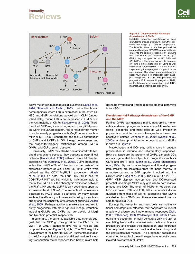
Figure 2. Developmental Pathwaysdownstream of GMPsIsolatable progenitor populations for eachgranulocyte lineage are shown. The GMP is di-vided into integrin b7� and b7lo populations.The latter is primed to the basophil and themast-cell lineages: b7lo GMPs presumably mi-grate into the spleen to become b7hi BMCPsthat give rise to b7hi intestinal MCPs. b7lo
GMPs may also give rise to b7lo BaPs andb7hi MCPs in the bone marrow. In contrast,b7� GMPs differentiate into b7- EoPs as wellas MDPs or putative NMPs. The lineal relation-ships among these progenitor populations re-main unclear. The following abbreviations areused: MCP, mast-cell progenitor; BaP, baso-phil progenitor; BMCP, basophil/mast-cellprogenitor; EoP, eosinophil progenitor; NMP,neutrophil-monocyte progenitor; and MDP,macrophage-dendritic-cell progenitor.
active mutants in human myeloid leukemias (Nakao et al.,
1996; Stirewalt and Radich, 2003), but unlike human
hematopoiesis where Flt3 is expressed in the entire LT-
HSC and GMP populations as well as in CLPs (unpub-
lished data), murine Flt3 is not expressed in GMPs or in
the vast majority of CMPs (Karsunky et al., 2003). There-
fore, the LMPP may include only a part of early GM poten-
tial within the LSK population. Flt3 is not a perfect marker
to exclude early progenitors with MegE potential such as
MPP or ST-HSCs. Furthermore, the relative contribution
of CMPs and LMPPs in GM lineage development and
the progenitor-progeny relationships among LMPPs,
GMPs, and CLPs remain obscure.
Conversely, CMPs may also be contaminated with lym-
phoid progenitors because they possess a weak B cell
potential (Akashi et al., 2000) within a minor CMP fraction
expressing Flt3 (Karsunky et al., 2003). CMPs are purified
within the c-Kit+Lin�Sca-1� fraction on the basis of the
expression pattern of CD34 and FcgRII/III: CMPs were
defined as the CD34+FcgRII/IIIlo population (Akashi
et al., 2000). Of note, the Flt3+ LSK LMPP has the
CD34+FcgRII/IIIlo profile, which is indistinguishable to
that of the CMP. Thus, the phenotypic distinction between
the Flt3+ CMP and the LMPP is only dependent upon the
expression level of Sca-1. The amounts of fluorescence
detected by FACS could be affected by many factors
such as the efficiency of fluorescent labeling for each an-
tibody and the sensitivity of fluorescent channels (Akashi
et al., 2005). Perhaps additional markers are required to
purify progenitors with more rigorous lineage restriction,
including GMLPs and CMPs that are devoid of MegE
and lymphoid potential, respectively.
In summary, the currently available data strongly sug-
gest that the MPP go through either the CMP or the
LMPP (or GMLP) stages to give rise to myeloid and
lymphoid lineages (Figure 1A, right). The CLP might be
downstream of the LMPP (or GMLP). Further fractionation
of the LSK population by use of additional markers includ-
ing transcription factor reporters (see below) might help
delineate myeloid and lymphoid developmental pathways
from HSCs.
Developmental Pathways downstream of the GMPand the MEPPurified GMPs can generate mainly neutrophils, mono-
cytes, and macrophages and a minor population of eosin-
ophils, basophils, and mast cells as well. Progenitor
populations restricted to such lineages have been pro-
spectively isolated (Arinobu et al., 2005; Iwasaki et al.,
2005a). A developmental scheme downstream of GMPs
is shown in Figure 2.
Macrophages and DCs play critical roles in antigen
presentation in immune and inflammatory responses.
Both cell types are the progeny of GMPs, although DCs
are also generated from lymphoid progenitors such as
CLPs and pro-T cells (Manz et al., 2001; Shigematsu
et al., 2004). Bipotent macrophage-dendritic-cell progen-
itors (MDPs) are isolatable from the bone marrow in
a mouse carrying a GFP reporter knocked into the
Cx3cr1 locus (Fogg et al., 2006). The Lin�c-KitloCX3CR1-
GFP+ MDP displays macrophage- and DC-restricted
potential, and single MDPs may give rise to both macro-
phages and DCs. The origin of MDPs is not clear, but
MDPs express CD34 and FcRgII/III at amounts indistin-
guishable from those of GMPs, suggesting that MDPs
are derived from GMPs and therefore represent precur-
sors for myeloid DCs.
Eosinophils, basophils, and mast cells are multifunc-
tional hematopoietic effectors that cooperate to mount
a variety of allergic and innate immune responses (Galli,
2000; Rothenberg, 1998; Wedemeyer et al., 2000). Eosin-
ophils and basophils normally constitute only 1%–2% of
circulating blood cells, whereas mast cells circulate as
progenitors and finalize their maturation after migration
into peripheral tissues such as the skin, heart, lung, and
the gastrointestinal mucosa. The progenitor populations
committed to each of these lineages have recently been
isolated downstream of GMPs.
Immunity 26, June 2007 ª2007 Elsevier Inc. 729

Immunity
Reviews
Eosinophil lineage-committed progenitors (EoPs) are
isolatable within the mouse bone marrow expressing
high IL-5 receptor a chain (Iwasaki et al., 2005a). EoPs
have the IL-5Ra+Lin�Sca-1�CD34+c-Kitlo phenotype.
Although EoPs constitute only 0.05% of the steady-
state bone-marrow cells, they can expand substantially
in response to helminth infection, suggesting that
the EoP stage is physiologically critical for eosinophil
production.
Mast cells and basophils share their origin at the baso-
phil-mast-cell bipotent progenitor (BMCP) stage (Arinobu
et al., 2005). In adult mice, the intestine is the main periph-
eral tissue harboring mast-cell colony-forming activity
(Gurish et al., 2001). The b7-integrin (b7) is an essential
molecule for formation of the intestinal mast-cell pool
and plays a role in tissue-specific homing of putative pre-
cursors for intestinal mast cells (Gurish et al., 2001).
BMCPs are present within the b7hi fraction of the spleen
and have the Lin�CD13loCD34+b7hiFc3RIa�FcgRII/III+c-
Kit+Thy-1+ phenotype. This population, consisting only
0.005% of the total spleen cells, gives rise to basophils
and mast cells at the single-cell level and can reconstitute
mucosal and tissue mast cells after transplantation into
mast-cell-deficient W/Wv mice. The monopotent mast-
cell progenitor (MCP) is also isolatable in the intestine.
Intestinal MCPs are Lin�CD34+b7hiFc3RIaloFcgRII/III+c-
KitloThy-1� cells. They bear blastic morphology with
a few scattered metachromatic granules and can give
rise exclusively to pure mast-cell colonies. The monopo-
tent basophil progenitor (BaP) is also present in the bone
marrow and has the Lin�CD34+Fc3RIahic-Kit� phenotype
(Arinobu et al., 2005).
In vitro, purified GMPs can give rise to EoPs, BMCPs,
MCPs, and BaPs, whereas BMCPs generate MCPs and
BaPs (Arinobu et al., 2005). The lineal relationship among
these populations is schematized in Figure 2. Differenti-
ation of spleen BMCPs into monopotent progeny may
lead to their selective migration, BaPs to the bone marrow
or MCPs to peripheral tissues. This progenitor allocation
may be critical for their distinct lineage functions and their
development. In the bone marrow, the basophil-mast-cell
potential was enriched in the b7lo GMPs (Arinobu et al.,
2005), and a fraction expressing high T1/ST2, a mast-
cell marker (Moritz et al., 1998), is mast-cell lineage com-
mitted (Chen et al., 2005). The spleen BMCP could be
derived from the b7lo GMPs. Relationships between the
progenitor allocation and the physiological pathway of
these cells are still unclear.
The monopotent megakaryocyte lineage-committed
progenitor (MKPs) has been isolated downstream of
MEPs by CD9, a megakaryocyte-associated surface
protein. MKPs have the CD9+IL-7Ra�Lin�Sca-1�c-Kit+
Thy1.1� phenotype and represent only 0.01% of the total
bone-marrow cells (Nakorn et al., 2003). MKPs give rise
exclusively to various sizes of megakaryocyte colonies.
MEPs represent the majority of day 8 CFU-S activity, but
MKPs do not have CFU-S activity, generate only mega-
karyocytes in vitro, and could give rise to platelets for
approximately 3 weeks when transplanted into mice.
730 Immunity 26, June 2007 ª2007 Elsevier Inc.
Erythroid-committed progenitors, a putative counterpart
of MKPs, however, have not been identified.
Granulocyte and Monocyte Lineage Commitmentby Single Transcription FactorsThese prospectively isolatable progenitor populations
emerge in normal hematopoiesis as a result of lineage
specification. Therefore, understanding the mechanism
of lineage commitment should in turn be useful to delin-
eate the hematopoietic developmental pathway. Lineage
commitment and subsequent differentiation of multipo-
tent cells is likely to involve the selective activation and
silencing of a set of genes. Such programs could be
triggered by extrinsic signals (Kondo et al., 2000; Metcalf,
1998), intrinsic signals (Akashi et al., 1997; Fairbairn
et al., 1993; Maraskovsky et al., 1997; Suda et al., 1983),
or by both at different developmental stages, which are
ultimately controlled by transcription factors. Transcrip-
tion factors can play a key role in activating lineage-
specific programs dependent upon their expression levels
(Dahl et al., 2003; Iwasaki et al., 2003; Laslo et al., 2006)
and timing (Iwasaki et al., 2006), and multiple transcription
factors exert collaborative or competitive actions for cell
fate decision (Laslo et al., 2006). The function of each
transcription factor is usually analyzed by the loss-
of-function and force-to-express studies. For detailed
function of each transcription factor, readers should refer
to recent reviews (Laiosa et al., 2006a; Rosenbauer and
Tenen, 2007).
The disappearance of specific myeloid progenitors by
disruption of single transcription factors is one of the
strongest piece of evidence that they are absolutely re-
quired to form and maintain a certain stage of hematopoi-
etic development. Such results have been clearly shown in
either PU.1- or C/EBPa-deficient mice. These factors co-
operate in the regulation of a number of GM-related genes
(Rosenbauer and Tenen, 2007; Tenen, 2003). Of note,
PU.1 also has a function for lymphoid development (Singh
and Pongubala, 2006; Warren and Rothenberg, 2003).
PU.1-deficient mice die at a late embryonic stage or
shortly after birth and have impaired GM and B cell devel-
opment (McKercher et al., 1996; Scott et al., 1994). T and
NK cell development is also severely impaired in PU.1-
deficient mice (Colucci et al., 2001; Spain et al., 1999),
but MegE development is intact (McKercher et al., 1996;
Scott et al., 1994). Conditional disruption of PU.1 in adult
bone marrow results in a complete loss of CMPs, GMPs,
and CLPs but retains a slightly increased number of
MEPs (Dakic et al., 2005; Iwasaki et al., 2005b). Thus,
PU.1 is necessary for multipotent progenitors to proceed
to the CMP and the CLP stages. In contrast, mice deficient
in C/EBPa lack neutrophils and eosinophils (Zhang et al.,
1997). Mice conditionally disrupted with C/EBPa do not
have granulocytes but have normal numbers of mature
lymphoid and MegE cells (Zhang et al., 2004). In the
bone marrow, GMPs disappear, but the development of
CMPs, MEPs, or CLPs is normal, indicating that C/EBPa
is necessary for CMPs (or LMPPs) to become GMPs
(Zhang et al., 2004).

Immunity
Reviews
C/EBPb-deficient mice show normal hematopoiesis un-
der steady-state conditions, suggesting that myeloid-
progenitor distribution should be normal. Mice having
C/EBPb knocked into the Cebpa gene locus show normal
hematopoietic development (Jones et al., 2002), indicating
that C/EBPb can replace C/EBPa function. The difference
between phenotype of C/EBPa- and C/EBPb-deficient he-
matopoiesis might be due to their distribution: C/EBPa is
gradually upregulated in the GM pathway during HSC,
CMP to GMP development, whereas the expression of
C/EBPb declines, particularly at the CMP stage (Hirai
et al., 2006). Therefore, CMPs could be maintained by
C/EBPa under steady-state hematopoiesis. C/EBPb is im-
portant in neutrophil production under stress hematopoie-
sis on the basis of the fact that C/EBPa-deficient mice can
develop neutrophils in the presence of a high concentra-
tion of cytokines in association with rapid upregulation
of C/EBPb in myeloid progenitors, whereas C/EBPb-
deficient mice are incapable of emergent neutrophil pro-
duction in response to exogenous cytokines or infection
(Hirai et al., 2006). The requirement for late GM lineage de-
velopment is also different between PU.1 and C/EBPs: The
GMP deficient for PU.1 disrupted by retrovirally trans-
duced Cre recombinase displays severe differentiation ar-
rest at the myeloblastic stage (Iwasaki et al., 2005b),
whereas the disruption of C/EBPa at the GMP stage
does not prevent their terminal differentiation (Zhang
et al., 2004). This may be because C/EBPa function is
substituted by C/EBPb (Jones et al., 2002) after the GMP
stage. Thus, PU.1 but not C/EBPa or C/EBPb is absolutely
required for GMPs to differentiate into mature GM cells.
Interestingly, these major myeloerythroid transcription
factors are expressed in the LSK HSC population (Miya-
moto et al., 2002). Although the expression of these genes
is considered to reflect myeloid lineage priming as dis-
cussed in the next section, the low expression of tran-
scription factors such as PU.1 and C/EBPa might also
control self-renewal activity of HSCs. The disruption of
PU.1 at birth induced a rapid loss of HSCs (Iwasaki
et al., 2005b), whereas that of C/EBPa induced expansion
of HSCs (Zhang et al., 2004). Although the mechanism of
these phenomena is still unclear, it has been suggested
that they could act as a positive or negative regulator for
HSC self-renewal, respectively, and these factors may
competitively interact with each other to control HSC
self-renewal, as they do at the granulocyte versus mono-
cyte divergence (Dahl et al., 2003).
Megakaryocyte and Erythroid LineageCommitment by Single Transcription FactorsFor MegE differentiation, GATA factors and their cofactors
such as a friend of GATA-1 (FOG-1) are indispensable
(Fujiwara et al., 1996; Tsai and Orkin, 1997; Tsang et al.,
1998). GATA-1-deficient mice die between embryonic
day 10.5 and 11.5 of gestation because of severe anemia.
Maturation of erythroid cells is arrested at an early proer-
ythroblast-like stage in GATA-1-deficient embryos (Fuji-
wara et al., 1996). In another mutant line with modification
of the DNase I-hypersensitive region upstream of the
GATA-1 locus (Shivdasani et al., 1997), GATA-1 expres-
sion is impaired specifically in the megakaryocyte lineage.
The platelet production of this mouse is markedly re-
duced, whereas in the bone marrow, megakaryocytes
with severely impaired cytoplasmic maturation are in-
creased (Shivdasani et al., 1997). Collectively, GATA-1 is
indispensable for the MegE differentiation, whereas the
MegE lineage commitment occurs in the absence of
GATA-1. GATA-1 possesses strong MegE lineage-
instructive effects (Iwasaki et al., 2003) as discussed in
the next section. This discrepancy might be explained
by a functional redundancy between GATA factors. The
enforced expression of either GATA-2 or GATA-3 trans-
genes can rescue the erythroid lineage defect in GATA-
1-deficientmice (Takahashi etal., 2000).Furthermore,prim-
itive erythropoiesis is completely absent in the GATA-1
and GATA-2 double-deficient mice (Fujiwara et al., 2004).
GATA-2-deficient mouse embryos also die at the early
stage of gestation because of severe anemia. Impaired
expansion of HSCs and progenitors is the cause of the
anemia, whereas the early erythroid maturation is normal
in the GATA-2-deficient yolk sac (Tsai and Orkin, 1997),
suggesting that GATA-1 but not GATA-2 is primarily criti-
cal for early erythroid differentiation. Interestingly, GATA
factors are expressed in granulocyte subclasses such as
eosinophils, basophils, and mast cells. Deletion of
a high-affinity GATA-binding site in the GATA-1 promoter,
an essential element for positive autoregulation of GATA-1
expression, leads to selective loss of the eosinophil line-
age (Yu et al., 2002), suggesting that GATA-1 is indispens-
able for eosinophil development. In the GATA-2-deficient
yolk sac, mast-cell development is severely impaired (Tsai
and Orkin, 1997).
FOG-1 binds to GATA factors and cooperatively regu-
lates transactivation of their target genes (Tsai and Orkin,
1997). FOG-1-deficient embryos also die between embry-
onic day 10.5 and 12.5 of gestation because of impaired
erythroid maturation comparable to that of GATA-
1-deficient mice (Tsang et al., 1998). FOG-1-deficient em-
bryos completely lack megakaryocytes (Tsang et al.,
1998), whereas GATA-1 deficiency leads to an increase
of megakaryocytes (Shivdasani et al., 1997). FOG-1 abso-
lutely requires the binding of GATA factor for its indispens-
able role in megakaryopoiesis because mice carrying
mutant GATA-1 and GATA-2, both of which are unable
to bind to FOG-1, display megakaryocyte deficiency, the
phenotype similar to FOG-1-deficient mice (Chang et al.,
2002). No conditional knockout systems have been devel-
oped for GATA factors or FOG-1. Therefore, requirement
of these factors in adult hematopoiesis has not been thor-
oughly evaluated. GATA-1, GATA-2, and FOG-1 are also
expressed in a fraction of LSK cells (Iwasaki et al., 2003;
Miyamoto et al., 2002), but it is unknown whether they
play a role in HSC function.
Plasticity of Lineage Determination via EctopicTranscription-Factor ExpressionThe myeloid transcription factors including GM-related
PU.1 and C/EBPa and MegE-related GATA factors play
Immunity 26, June 2007 ª2007 Elsevier Inc. 731

Immunity
Reviews
a role in cell fate decision of uncommitted progenitor cells
(Laiosa et al., 2006a; Traver and Akashi, 2004). Although
HSCs prime all of these factors at a low level, enforcement
of each factor by retroviral transduction into purified HSCs
results in their specification into a lineage. HSCs trans-
duced with PU.1 or C/EBPa formed GM but not MegE col-
onies, whereas those enforced with GATA-1 or GATA-2
mainly formed MegE or eosinophil colonies, respectively
(Iwasaki et al., 2006; Iwasaki et al., 2003). The enforced
FOG-1 does not change the colony-forming activity of
HSCs (Iwasaki et al., 2003), suggesting that FOG-1 can
play a role in the presence of an appropriate amount of
GATA-1. These data strongly suggest that the increment
of the expression of single factors is sufficient to abrogate
self-renewal activity of HSCs and to instruct them into
specific lineages.
In these experiments, it is difficult to exclude the possi-
bility that these effects result from a selection of a particu-
lar lineage, which is dependent upon ‘‘permissive’’ signals
that simply support survival or expansion of target cells.
The most clear-cut evidence for lineage instructive action
of transcription factors is to prove their ability to repro-
gram cells into a different lineage because this phenome-
non reflects two important components for lineage
instruction: perturbation of natural cell fate and reestab-
lishment of a new program. A number of reports have
shown that lymphoid-cell lines could be reprogrammed
into macrophages by a variety of external ectopic signals
(Graf, 2002). CLPs are one of the most useful cell types for
testing myeloid instructive signals: They are primary cells
purified from normal mice and do not have any myeloid
potential nor express any major myeloid genes even at
a genome-wide level (Akashi et al., 2000), whereas they
possess plasticity for all myeloerythroid lineages, which
could be triggered by ectopic transcription factors or cyto-
kine signaling (Hsu et al., 2006; Iwasaki et al., 2006;
Iwasaki et al., 2003; Iwasaki-Arai et al., 2003; King et al.,
2002; Kondo et al., 2000).
The enforced expression of C/EBPa reprograms CLPs
into the GM lineage (neutrophils and monocytes but not
eosinophils or basophils) (Hsu et al., 2006; Iwasaki et al.,
2006), whereas enforced GATA-1 or GATA-2 reprograms
CLPs into either the MegE or the mast-cell lineage,
respectively (Iwasaki et al., 2006; Iwasaki et al., 2003).
Importantly, the clonal efficiencies of single-plated cells
are R80%, and this reprogramming occurs at the ex-
pense of lymphoid potential of CLPs. CLPs with enforced
FOG-1 normally develop B cells in vitro (unpublished
data), again suggesting that FOG-1 has an effect only in
the presence of GATA factors.
Even more committed T or B cell precursors can be re-
programmed into the myeloid lineage by the enforced
expression of C/EBPa, C/EBPb, or PU.1. Graf and
colleagues have extensively analyzed the myeloid-
reprogramming processes from monopotent T or B cell
precursor stages (Laiosa et al., 2006b; Xie et al., 2004).
They used ROSA26-EYFP mice with lck-cre or CD19-cre
genes to perform in vivo labeling of cells having expressed
T cell-related lck or B cell-related CD19, respectively.
732 Immunity 26, June 2007 ª2007 Elsevier Inc.
Lck-EYFP+ cells are mostly DN3 and DN4 pre-T cells.
The retroviral expression of C/EBPa and C/EBPb or of
PU.1 reprograms EYFP+ pre-T cells into macrophages
or dendritic cells, respectively. CD19-EYFP+ pro B and
even spleen B cells are also converted into macrophages
by C/EBPs. Unlike CLPs, the lineage conversion from T or
B cell precursors appears to occur in a fraction of trans-
fected cells, suggesting that the lineage plasticity may
decline as cells become further committed. It is important
to note that at these committed T or B cell stages, cells are
converted only into macrophages or DCs but not into
other myeloid classes such as MegE cells or granulocytes.
When GATA-1 is enforced in proB cells, cells immediately
undergo apoptotic cell death, whose process cannot be
rescued by the enforced expression of Bcl-2 (Iwasaki
et al., 2003). Thus, plasticity for lineage specification is
preserved until monopotent T or B cell progenitor stages,
but their destination is predominantly limited to the macro-
phage lineage.
A series of force-to-express studies have thus shown
that single-myeloid transcription factors can establish
the myeloid developmental program in uncommitted pro-
genitors or even in lymphoid progenitors. It is critical to
understand how transcription factors override the ongoing
lymphoid program and ultimately reprogram cells into the
myeloid lineage in a cell-context-dependent manner. The
reprogramming event might at least be dependent upon
the ability of transcription factors to remodel chromatin
(Bonifer, 2005; Muller and Leutz, 2001). Reprogramming
from the myeloid into the lymphoid lineage, however,
has never been achieved. Lymphoid development may
require more complex regulatory processes governed by
multiple lymphoid transcription factors (Laiosa et al.,
2006a).
For lymphoid commitment, because HSCs naturally
prime myeloid transcription factors, some mechanisms
that can abrogate or repress preceding myeloid pro-
grams might be required. The OP9 expressing Notch
ligand (OP9-DL1) (Schmitt and Zuniga-Pflucker, 2002)
induces T cell differentiation from HSCs via CLPs, sug-
gesting that Notch signaling is critical for multipotent
cells to initiate the T cell program. Interestingly, the
myeloid potential as well as the PU.1- or C/EBPa-
dependent myeloid reprogramming of thymic precursors
(pro-T and pre-T cells) can be blocked by active Notch
signaling (Franco et al., 2006; Laiosa et al., 2006b;
Rothenberg, 2007 [this issue of Immunity]). In this con-
text, the myeloid fate could be a default commitment
pathway for multipotent HSC, and lymphoid lineage
commitment may be dependent upon successful pertur-
bation of the preceding myeloid program by myeloid-
repressing Notch signals.
Transcriptional Regulatory Network that GovernsHierarchical Hematopoietic DevelopmentIf hematopoietic development is dependent upon gradual
accumulation of sequentially activated lineage-promoting
signals, it is reasonable to assume that the primitive HSCs
or MPPs do not express any lineage-related genes.

Immunity
Reviews
Previous studies, however, have provided evidences sup-
porting the concept that the developmental potential is
engraved in the chromatin of cells at the multipotent stage.
For lineage-specific genetic programs to be activated, lo-
cal chromatin must become accessible to the transcrip-
tion machinery (Berger and Felsenfeld, 2001; Felsenfeld
et al., 1996). The activation of chromatin remodeling can
occur prior to substantial expression of genes in the region
of interest (Kontaraki et al., 2000; Weintraub, 1985). An
open chromatin structure is maintained in early hemato-
poietic progenitors, enabling multilineage differentiation
programs to be readily accessible (Cross and Enver,
1997), and multipotent cells ‘‘prime’’ multiple lineage-affil-
iated programs of gene activity (i.e., transcription factors,
cytokine receptors, and genes encoding lineage-exclu-
sive function) at a low level, prior to being specified into
each lineage (Hu et al., 1997). In fact, a number of GM-
and MegE-related genes are coexpressed in single
HSCs, MPPs, or CMPs (Miyamoto et al., 2002), even at
the genome-wide level (Akashi et al., 2003; Mansson
et al., 2007). Because most myeloid transcription factors
possess lineage-instructive functions as discussed, the
subtle change in the expression of these factors at the
HSC stage should lead to an early myeloid lineage fate de-
cision.
Uncommitted Progenitor and StemCells Primes Multiple Lineage-Affiliated Genesand Transcription FactorsThe first evidence for the hematopoietic lineage priming
was shown in a multipotent cell line, FDCP-mix (Hu
et al., 1997). This seminal study showed that single multi-
potent FDCP-mix cells coexpress GM- and MegE-related
and some lymphoid-related genes by single-cell RT-PCR
assays. The ability to prospectively isolate lineage-
restricted progenitor subsets has enabled the similar anal-
ysis at specific stages in normal hematopoiesis. In the
myeloid lineage, the majority (>60%) of CMPs coexpress
GM- (i.e., G-CSFR, myeloperoxidase, and PU.1) and
MegE-related genes (i.e., EpoR, b-globin, and GATA-2)
at the single-cell level, whereas virtually all GMPs and
MEPs express only GM- or MegE-related genes, respec-
tively (Miyamoto et al., 2002). In mice harboring a GFP re-
porter knocked into the murine lysozyme M (LysM) locus
(Faust et al., 2000), �60% of CMPs express substantial
amounts of LysMGFP. Both LysMGFP+ and LysMGFP�
CMPs express erythroid b-globin and can display MegE
lineage differentiation at equal efficiencies (Miyamoto
et al., 2002). Similarly, in the lymphoid pathway, single
CLPs coexpress genes that encode B (i.e., l5 and Pax-
5) and T lymphoid (GATA-3 and CD3d) cells (Miyamoto
et al., 2002). A fraction of CLPs express pTa receptor
mRNA, represented by a T cell-affiliated pTa reporter
(Reizis and Leder, 1999), but their frequency for coexpres-
sion of other B and T lymphoid genes and B cell potential
is equal irrespective of the pTa reporter expression. Col-
lectively, transcription of LysM or pTa at a low level does
not predict their GM or T cell fates. These studies suggest
that promiscuous gene priming is likely to play a key role in
maintaining flexibility in oligopotent precursors and that
the level of gene expression may fluctuate at multipotent
or oligopotent stages. The priming of genes affiliated
with multiple lineages would afford flexibility in cell fate
decisions and would allow multipotent precursors to rap-
idly respond to environmental cues (Hu et al., 1997). In this
context, CMPs and CLPs have the molecular signature to
assure their common myeloid and lymphoid potency,
respectively. Thus, lineage commitment and subsequent
differentiation of multipotent cells involve the upregulation
of genes associated with the appropriate lineage, whereas
the concomitant downregulation of inappropriate genes
makes such lineage decision stable (Enver and Greaves,
1998).
At the genome-wide level, HSCs express a large num-
ber of myeloid but not lymphoid genes (Akashi et al.,
2003; Mansson et al., 2007). The myeloid gene expression
in the HSC fraction might not be due to committed con-
taminants. This was confirmed by tracing of the fate of
HSCs with lysM activation (Ye et al., 2003). In mice harbor-
ing a LysM Cre-knockin together with ROSA-EYFP re-
porter alleles, cells activating LysM transcription are
permanently marked by EYFP. In this mouse, EYFP is ex-
pressed not only in GM cells but also in a minority of MegE
and lymphoid cells. A fraction of LT-HSCs also express
EYFP, and these cells are capable of long-term reconstitu-
tion after transplantation with nearly 100% of the reconsti-
tuted cells being EYFP+. This provides formal proof that
the lysM gene is physiologically primed at the HSC stage
(Ye et al., 2003). Thus, HSCs are ready to commit to the
myeloid lineage presumably as a default, although some
other internal or external cues might be necessary for
them to initiate the lymphoid developmental program. A
fraction of the LMPP coexpresses both GM and early lym-
phoid genes such as Rag1 and Il7r (encoding IL-7Ra),
again suggesting that progenitor cells at the stage com-
mon to GM and lymphoid lineages exist within the LMPP
population (Mansson et al., 2007). If the lineage priming
of transcription factors at low levels is a natural property
of multipotent cells such as hematopoietic stem and
progenitors, both the upregulation and downregulation
of single or multiple transcription factors should be critical
to turn on lineage-specification programs.
Transcription-Factor Dosage Determines LineageFate at Binary BranchpointsThe amount of transcription factor might play an important
role in binary lineage fate decision. B cell and macrophage
differentiation can be restored in PU.1 (encoded by Sfpi1)-
deficient fetal-liver hematopoietic progenitors by retroviral
transduction of Sfpi1 with a GFP expression reporter. After
culture on OP9 stromal layer, the Sfpi1�/� progenitor gives
rise mainly to macrophages with a high expression of GFP
and rare B cell progeny expressing only a low amount of
GFP. In contrast, Sfpi1�/+ fetal-liver progenitors naturally
give rise to B cells and rare macrophages on OP9. When
they are infected with the Sfpi1-GFP retrovirus, the
Sfpi1�/+ fetal-liver progenitor with a high expression of
GFP gives rise mainly to macrophages at the expense of
Immunity 26, June 2007 ª2007 Elsevier Inc. 733

Immunity
Reviews
Figure 3. Timing-Based Interplay ofC/EBPa and GATA-2 in Lineage Decisionof Granulocyte LineagesIf uncommitted cells upregulate C/EBPa first,cells became GMPs. Further upregulation ofC/EBPa should lead to the formation of puta-tive NMP that gives rise only to neutrophilsand monocytes that are the major products ofGMPs. If GATA-2 is upregulated at the GMPstage, cells commit into the eosinophil lineage.In contrast, if uncommitted cells upregulateGATA-2 first, they become the BMCP. Furtherupregulation of GATA-2 should result in thegeneration of MCP, whereas if C/EBPa is upre-gulated at the BMCP stage, cells commit intothe basophil lineage (Iwasaki et al., 2006).These results suggest that GATA-2 instructseosinophil lineage commitment in the pres-ence of a sufficient level of C/EBPa, whereasC/EBPa activates the basophil lineage pro-gram in the presence of GATA-2. Collectively,the order of the expression of C/EBPa andGATA-2 can be a critical lineage determinantfor the eosinophil, the basophil, the neutrophil,and the mast-cell lineages.
B cell differentiation (DeKoter and Singh, 2000). Thus, the
dosage of PU.1 might play a critical role in macrophage
versus B cell differentiation when the commitment pro-
cess is started at an experimental stage completely defi-
cient for PU.1: Cultured Sfpi1�/� fetal-liver cells are not
normal multipotent progenitors because they are incapa-
ble of differentiation into myeloerythroid cells except for
macrophages. This phenomenon was not evaluated at
the single-cell level, and therefore, it is still unclear
whether different concentrations of PU.1 can specify mac-
rophage or B cell fates or can differentially support matu-
ration of each lineage.
The quantity of the transcription factor is also important
at the site of mast-cell generation from GMPs. Graded re-
duction of C/EBPa by transduction of a set of anti-C/EBPa
RNAi shows that the frequency of mast-cell development
is inversely correlated with the amount of C/EBPa (Iwasaki
et al., 2006). In contrast, GMPs retrovirally transduced
with C/EBPa lose mast-cell (and basophil) potential, sug-
gesting that the amount of C/EBPa is critical also for
GMPs to choose neutrophil versus mast-cell fates (Iwa-
saki et al., 2006).
Interestingly, downstream of GMPs, C/EBPa is a critical
determinant for the mast-cell versus basophil lineage fate
decision. The depletion of C/EBPa in BMCPs results in
their exclusive differentiation into mast cells (Iwasaki
et al., 2006). In contrast to GMPs, retroviral transduction
of C/EBPa into BMCPs results in their exclusive genera-
tion of basophils, and MCPs enforced with C/EBPa are
reprogrammed into basophils (Arinobu et al., 2005). These
data collectively suggest that the dosage of single tran-
scription factors is able to specify the lineage in a cell-
context-dependent manner.
734 Immunity 26, June 2007 ª2007 Elsevier Inc.
Lineage Diversification by Timing-Based Interplayof Myeloid Transcription FactorsIf the expression oscillates at the priming stage, it is likely
to randomly reach the threshold at which it can activate
a lineage-specification program. Thus, the timing of acti-
vation of each transcription factor could be ‘‘stochastic.’’
Recent findings show that eosinophil versus basophil lin-
eage specification could be determined by the order of
expression of C/EBPa and GATA-2 (Iwasaki et al., 2006)
(Figure 3).
BaPs and EoPs expressed both GATA-2 and C/EBPa,
whereas upstream GMPs expressed only C/EBPa
(Arinobu et al., 2005; Iwasaki et al., 2005a). All single
GMPs with the enforced expression of GATA-2 generate
pure eosinophil colonies, indicating that GATA-2 can in-
struct GMPs to become EoPs (Iwasaki et al., 2006). In con-
trast, MCPs express GATA-2 but not C/EBPa, and all
single MCPs with the enforced expression of C/EBPa
form pure basophil colonies (Arinobu et al., 2005). These
results suggest that GATA-2 instructs eosinophil lineage
commitment in the presence of C/EBPa, whereas
C/EBPa activates the basophil lineage program in the
presence of GATA-2. Furthermore, C/EBPa and GATA-2
can reprogram CLPs differentially into eosinophil and ba-
sophil lineages by changing the timing of their expression
(Iwasaki et al., 2006). When CLPs are retrovirally intro-
duced with C/EBPa and then GATA-2 within a 24 hr inter-
val, CLPs transduced with C/EBPa alone generate neutro-
phil and monocyte/macrophage colonies, whereas those
transduced with C/EBPa plus GATA-2 give rise to eosino-
phil colonies. In contrast, when the order of retroviral in-
fection is switched, CLPs introduced with GATA-2 alone
or GATA-2 plus C/EBPa generate mast-cell or basophil
Immunity
Reviews
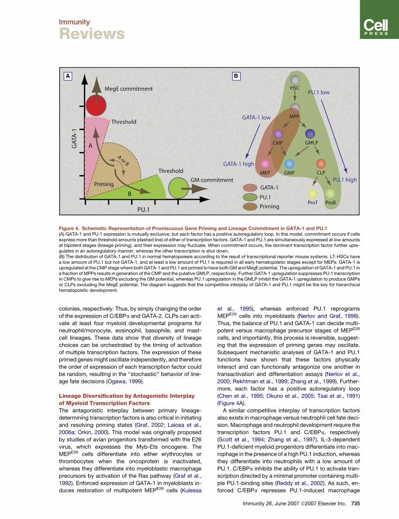
Figure 4. Schematic Representation of Promiscuous Gene Priming and Lineage Commitment in GATA-1 and PU.1(A) GATA-1 and PU.1 expression is mutually exclusive, but each factor has a positive autoregulatory loop. In this model, commitment occurs if cellsexpress more than threshold amounts (dashed line) of either of transcription factors. GATA-1 and PU.1 are simultaneously expressed at low amountsat bipotent stages (lineage priming), and their expression may fluctuate. When commitment occurs, the dominant transcription factor further upre-gulates in an autoregulatory manner, whereas the other transcription is shut down.(B) The distribution of GATA-1 and PU.1 in normal hematopoiesis according to the result of transcriptional reporter mouse systems. LT-HSCs havea low amount of PU.1 but not GATA-1, and at least a low amount of PU.1 is required in all early hematopoietic stages except for MEPs. GATA-1 isupregulated at the CMP stage where both GATA-1 and PU.1 are primed to have both GM and MegE potential. The upregulation of GATA-1 and PU.1 ina fraction of MPPs results in generation of the CMP and the putative GMLP, respectively. Further GATA-1 upregulation suppresses PU.1 transcriptionin CMPs to give rise to MEPs excluding the GM potential, whereas PU.1 upregulation in the GMLP inhibit the GATA-1 upregulation to produce GMPsor CLPs excluding the MegE potential. The diagram suggests that the competitive interplay of GATA-1 and PU.1 might be the key for hierarchicalhematopoietic development.
colonies, respectively. Thus, by simply changing the order
of the expression of C/EBPa and GATA-2, CLPs can acti-
vate at least four myeloid developmental programs for
neutrophil/monocyte, eosinophil, basophile, and mast-
cell lineages. These data show that diversity of lineage
choices can be orchestrated by the timing of activation
of multiple transcription factors. The expression of these
primed genes might oscillate independently, and therefore
the order of expression of each transcription factor could
be random, resulting in the ‘‘stochastic’’ behavior of line-
age fate decisions (Ogawa, 1999).
Lineage Diversification by Antagonistic Interplayof Myeloid Transcription FactorsThe antagonistic interplay between primary lineage-
determining transcription factors is also critical in initiating
and resolving priming states (Graf, 2002; Laiosa et al.,
2006a; Orkin, 2000). This model was originally proposed
by studies of avian progenitors transformed with the E26
virus, which expresses the Myb-Ets oncogene. The
MEPE26 cells differentiate into either erythrocytes or
thrombocytes when the oncoprotein is inactivated,
whereas they differentiate into myeloblastic macrophage
precursors by activation of the Ras pathway (Graf et al.,
1992). Enforced expression of GATA-1 in myeloblasts in-
duces restoration of multipotent MEPE26 cells (Kulessa
et al., 1995), whereas enforced PU.1 reprograms
MEPE26 cells into myeloblasts (Nerlov and Graf, 1998).
Thus, the balance of PU.1 and GATA-1 can decide multi-
potent versus macrophage precursor stages of MEPE26
cells, and importantly, this process is reversible, suggest-
ing that the expression of priming genes may oscillate.
Subsequent mechanistic analyses of GATA-1 and PU.1
functions have shown that these factors physically
interact and can functionally antagonize one another in
transactivation and differentiation assays (Nerlov et al.,
2000; Rekhtman et al., 1999; Zhang et al., 1999). Further-
more, each factor has a positive autoregulatory loop
(Chen et al., 1995; Okuno et al., 2005; Tsai et al., 1991)
(Figure 4A).
A similar competitive interplay of transcription factors
also exists in macrophage versus neutrophil cell fate deci-
sion. Macrophage and neutrophil development require the
transcription factors PU.1 and C/EBPa, respectively
(Scott et al., 1994; Zhang et al., 1997). IL-3-dependent
PU.1-deficient myeloid progenitors differentiate into mac-
rophage in the presence of a high PU.1 induction, whereas
they differentiate into neutrophils with a low amount of
PU.1. C/EBPa inhibits the ability of PU.1 to activate tran-
scription directed by a minimal promoter containing multi-
ple PU.1-binding sites (Reddy et al., 2002). As such, en-
forced C/EBPa represses PU.1-induced macrophage
Immunity 26, June 2007 ª2007 Elsevier Inc. 735

Immunity
Reviews
differentiation, simultaneously enhancing neutrophil dif-
ferentiation. Because G-CSF increases the C/EBPa:PU.1
ratio in IL-3-dependent PU.1-deficient myeloid progeni-
tors, G-CSF signaling could be an external cue for granu-
locyte differentiation by increasing the relative concentra-
tion of C/EBPa at the macrophage versus neutrophil
binary cell fate decision (Dahl et al., 2003).
It is well known that either a mutually inhibitory-
feedback or positive-feedback loop of two factors can
produce bistability that plays like a toggle switch between
two discrete states (Angeli et al., 2004). This might be the
case for both GATA-1 versus PU.1, and C/EBPa versus
PU.1 cross-antagonisms (Figure 4A). In addition to the
cross-antagonism, GATA-1 and PU.1 have positive autor-
egulatory loops. A mathematical model integrating both
cross antagonism and positive autoregulation shows
that in addition to classical bistable states, there is another
stable region corresponding to the priming stage (Fig-
ure 4A) (Enver and Huang, 2005, ASH, abstract). In this
model, the priming stage constitutes a stage where cells
can stably express a low level of two transcription factors.
For lineage-specific programs to be activated, cells need
to upregulate either of them beyond the threshold (Enver
and Huang, 2005, ASH, abstract). These mathematical
models represent a small network module, and regulation
of hematopoietic development might consist of multiple
modules that may further interact with one another. For
example, the secondary cross-antagonism composed of
Egr/Nab and Gfi-1 has been proposed downstream of
C/EBPa and PU.1 (Laslo et al., 2006). These models are
likely to capture essential features of the hematopoietic
hierarchy, and they should ultimately be useful to predict
the genome-scale behavior of multipotent cells in lineage
specification.
Lineage Tracing by UtilizingTranscription-Factor ReportersIf the hierarchical lineage fate decision is operated by sin-
gle or a collaboration of multiple transcription factors,
tracing the timing and the amount of transcription-factor
expression should in turn be useful to delineate early
hematopoietic developmental pathways.
In two mouse lines having a GFP reporter knocked into
the PU.1 locus (Back et al., 2004; Nutt et al., 2005), CD34�
LSK HSCs express low PU.1-GFP (Iwasaki et al., 2005b;
Nutt et al., 2005). A fraction of CD34+ MPPs begins to ex-
press a high PU.1-GFP, which is further upregulated in
CMPs, GMPs, and CLPs, but not MEPs (Iwasaki et al.,
2005b; Nutt et al., 2005). In contrast, in mice having
a transgenic GFP reporter for GATA-1 (Iwasaki et al.,
2005a), CD34� LSK HSCs do not express GATA-1-GFP,
but a fraction of CD34+ MPPs express GATA-1-GFP at
a low level. GATA-1-GFP is further upregulated in CMPs
and MEPs but not in GM or lymphoid progenitors. In
Figure 4B, the distribution of PU.1 and GATA-1 reporters
in normal hematopoiesis is overlaid on the cellular-
evidence-based developmental scheme. In this develop-
mental model, HSCs express low PU.1, perhaps to main-
tain self-renewal activity (Iwasaki et al., 2005b), and the
736 Immunity 26, June 2007 ª2007 Elsevier Inc.
subsequent upregulation of GATA-1 occurs in a fraction
of CD34+ MPPs and CMPs to establish the bipotent
GATA-1 versus PU.1 priming state. Then, when PU.1 be-
comes dominant, the CMP and the putative GMLP (or
the LMPP) give rise to GMPs plus CLPs, whereas if
GATA-1 becomes dominant, CMPs produce MEPs at
the expense of GMPs. In fact, CD34+ MPPs expressing
a high level of PU.1-GFP possess predominantly the
GM- and lymphoid-restricted developmental potential
(unpublished data). This diagram nicely fits the concept
that the competitive interplay of PU.1 and GATA-1 might
play a critical role in early hematopoietic lineage fate
decision (Graf, 2002; Laiosa et al., 2006a; Orkin, 2000).
Ikaros is a transcription factor that plays a critical role in
T and B cell development. Ikaros�/� mice lack all B cells
and have only a small number of T cell precursors in the
thymus (Wang et al., 1996) with an increased number of
myeloid cells (Nichogiannopoulou et al., 1999). In a trans-
genic mouse strain expressing the GFP reporter under
control of an Ikaros promoter-enhancer (Kaufmann et al.,
2003), the LSK population is subdivided into GFP�/lo and
GFP+ cells, and the latter is exclusively found within the
Flt3+ population (Yoshida et al., 2006) that is an equivalent
of the LMPP (Adolfsson et al., 2005). Ikaros-GFP+ LSK
cells differentiate mainly into GM cells as well as into T
and B cells but form only a few percent of mixed colonies
containing both GM and MegE cells (Yoshida et al., 2006).
These data suggest that the graded upregulation of Ikaros
is associated with progression of restriction into the GM
and lymphoid lineages. Thus, both Ikaros and PU.1-
reporter analyses again support the existence of the puta-
tive GMLP in early hematopoiesis (Figure 1).
By tracking the expression of transcription factors or
genes capable of lineage instruction, we should be able
to visualize the stage at which each developmental
program turns on. Accordingly, the lineage-instructive
signal-based fractionation studies utilizing transcription-
factor reporter systems should be useful to further under-
stand the developmental pathway and mechanisms in
early hematopoietic development in future studies.
ConclusionsThe ability to prospectively isolate lineage-restricted
progenitors has greatly helped us understand the mecha-
nism of lineage commitment from HSCs. It is now clear
that murine HSCs prime myeloid but not lymphoid genes
as their natural property. On a cellular basis, however,
we have not reached a general agreement concerning
where the myeloid and lymphoid branching occurs. On
a molecular basis, cooperative and antagonistic interplays
between transcription factors as well as timing of their ex-
pression might play a critical role in hematopoietic lineage
commitment. It is still unclear, however, how such
transcription factors are activated or repressed in a cell-
context-dependent manner. Future studies on epigenetic
and posttranscriptional regulation of these lineage
determinants are critical to further understand the mecha-
nism of hematopoietic development from multipotent
HSCs.

Immunity
Reviews
ACKNOWLEDGMENTS
We thank T. Graf and D.-D. Weiszhausz for critical comments on themanuscript.
REFERENCES
Adolfsson, J., Borge, O.J., Bryder, D., Theilgaard-Monch, K., Astrand-Grundstrom, I., Sitnicka, E., Sasaki, Y., and Jacobsen, S.E. (2001).Upregulation of Flt3 expression within the bone marrowLin(-)Sca1(+)c-kit(+) stem cell compartment is accompanied by lossof self-renewal capacity. Immunity 15, 659–669.
Adolfsson, J., Mansson, R., Buza-Vidas, N., Hultquist, A., Liuba, K.,Jensen, C.T., Bryder, D., Yang, L., Borge, O.J., Thoren, L.A., et al.(2005). Identification of flt3(+) lympho-myeloid stem cells lackingerythro-megakaryocytic potential a revised road map for adult bloodlineage commitment. Cell 121, 295–306.
Akashi, K., He, X., Chen, J., Iwasaki, H., Niu, C., Steenhard, B., Zhang,J., Haug, J., and Li, L. (2003). Transcriptional accessibility for genes ofmultiple tissues and hematopoietic lineages is hierarchically controlledduring early hematopoiesis. Blood 101, 383–389.
Akashi, K., Kondo, M., von Freeden-Jeffry, U., Murray, R., andWeissman, I.L. (1997). Bcl-2 rescues T lymphopoiesis in interleukin-7receptor-deficient mice. Cell 89, 1033–1041.
Akashi, K., Traver, D., Miyamoto, T., and Weissman, I.L. (2000). A clo-nogenic common myeloid progenitor that gives rise to all myeloidlineages. Nature 404, 193–197.
Akashi, K., Traver, D., and Zon, L.I. (2005). The complex cartography ofstem cell commitment. Cell 121, 160–162.
Angeli, D., Ferrell, J.E., Jr., and Sontag, E.D. (2004). Detection of multi-stability, bifurcations, and hysteresis in a large class of biological pos-itive-feedback systems. Proc. Natl. Acad. Sci. USA 101, 1822–1827.
Arinobu, Y., Iwasaki, H., Gurish, M.F., Mizuno, S., Shigematsu, H.,Ozawa, H., Tenen, D.G., Austen, K.F., and Akashi, K. (2005). Develop-mental checkpoints of the basophil/mast cell lineages in adult murinehematopoiesis. Proc. Natl. Acad. Sci. USA 102, 18105–18110.
Back, J., Dierich, A., Bronn, C., Kastner, P., and Chan, S. (2004). PU.1determines the self-renewal capacity of erythroid progenitor cells.Blood 103, 3615–3623.
Berger, S.L., and Felsenfeld, G. (2001). Chromatin goes global. Mol.Cell 8, 263–268.
Bhatia, S.K., Tygrett, L.T., Grabstein, K.H., and Waldschmidt, T.J.(1995). The effect of in vivo IL-7 deprivation on T cell maturation.J. Exp. Med. 181, 1399–1409.
Bonifer, C. (2005). Epigenetic plasticity of hematopoietic cells. CellCycle 4, 211–214.
Borge, O.J., Adolfsson, J., and Jacobsen, A.M. (1999). Lymphoid-restricted development from multipotent candidate murine stem cells:Distinct and complimentary functions of the c-kit and flt3-ligands.Blood 94, 3781–3790.
Chang, A.N., Cantor, A.B., Fujiwara, Y., Lodish, M.B., Droho, S.,Crispino, J.D., and Orkin, S.H. (2002). GATA-factor dependence ofthe multitype zinc-finger protein FOG-1 for its essential role in mega-karyopoiesis. Proc. Natl. Acad. Sci. USA 99, 9237–9242.
Chen, C.C., Grimbaldeston, M.A., Tsai, M., Weissman, I.L., and Galli,S.J. (2005). Identification of mast cell progenitors in adult mice. Proc.Natl. Acad. Sci. USA 102, 11408–11413.
Chen, H., Ray-Gallet, D., Zhang, P., Hetherington, C.J., Gonzalez,D.A., Zhang, D.E., Moreau-Gachelin, F., and Tenen, D.G. (1995).PU.1 (Spi-1) autoregulates its expression in myeloid cells. Oncogene11, 1549–1560.
Christensen, J.L., and Weissman, I.L. (2001). Flk-2 is a marker in hema-topoietic stem cell differentiation: A simple method to isolate long-termstem cells. Proc. Natl. Acad. Sci. USA 98, 14541–14546.
Colucci, F., Samson, S.I., DeKoter, R.P., Lantz, O., Singh, H., andDi Santo, J.P. (2001). Differential requirement for the transcription fac-tor PU.1 in the generation of natural killer cells versus B and T cells.Blood 97, 2625–2632.
Corcoran, A.E., Riddell, A., Krooshoop, D., and Venkitaraman, A.R.(1998). Impaired immunoglobulin gene rearrangement in mice lackingthe IL-7 receptor. Nature 391, 904–907.
Cross, M.A., and Enver, T. (1997). The lineage commitment of haemo-poietic progenitor cells. Curr. Opin. Genet. Dev. 7, 609–613.
Dahl, R., Walsh, J.C., Lancki, D., Laslo, P., Iyer, S.R., Singh, H., andSimon, M.C. (2003). Regulation of macrophage and neutrophilcell fates by the PU.1:C/EBPalpha ratio and granulocyte colony-stimulating factor. Nat. Immunol. 4, 1029–1036.
Dakic, A., Metcalf, D., Di Rago, L., Mifsud, S., Wu, L., and Nutt, S.L.(2005). PU.1 regulates the commitment of adult hematopoietic progen-itors and restricts granulopoiesis. J. Exp. Med. 201, 1487–1502.
DeKoter, R.P., and Singh, H. (2000). Regulation of B lymphocyte andmacrophage development by graded expression of PU.1. Science288, 1439–1441.
Enver, T., and Greaves, M. (1998). Loops, lineage, and leukemia. Cell94, 9–12.
Fairbairn, L.J., Cowling, G.J., Reipert, B.M., and Dexter, T.M. (1993).Suppression of apoptosis allows differentiation and development ofa multipotent hemopoietic cell line in the absence of added growthfactors. Cell 74, 823–832.
Faust, N., Varas, F., Kelly, L.M., Heck, S., and Graf, T. (2000). Insertionof enhanced green fluorescent protein into the lysozyme gene createsmice with green fluorescent granulocytes and macrophages. Blood96, 719–726.
Felsenfeld, G., Boyes, J., Chung, J., Clark, D., and Studitsky, V. (1996).Chromatin structure and gene expression. Proc. Natl. Acad. Sci. USA93, 9384–9388.
Fogg, D.K., Sibon, C., Miled, C., Jung, S., Aucouturier, P., Littman,D.R., Cumano, A., and Geissmann, F. (2006). A clonogenic bone mar-row progenitor specific for macrophages and dendritic cells. Science311, 83–87.
Forsberg, E.C., Serwold, T., Kogan, S., Weissman, I.L., and Passegue,E. (2006). New evidence supporting megakaryocyte-erythrocytepotential of flk2/flt3+ multipotent hematopoietic progenitors. Cell126, 415–426.
Franco, C.B., Scripture-Adams, D.D., Proekt, I., Taghon, T., Weiss,A.H., Yui, M.A., Adams, S.L., Diamond, R.A., and Rothenberg, E.V.(2006). Notch/Delta signaling constrains reengineering of pro-T cellsby PU.1. Proc. Natl. Acad. Sci. USA 103, 11993–11998.
Fujiwara, Y., Browne, C.P., Cunniff, K., Goff, S.C., and Orkin, S.H.(1996). Arrested development of embryonic red cell precursors inmouse embryos lacking transcription factor GATA-1. Proc. Natl.Acad. Sci. USA 93, 12355–12358.
Fujiwara, Y., Chang, A.N., Williams, A.M., and Orkin, S.H. (2004).Functional overlap of GATA-1 and GATA-2 in primitive hematopoieticdevelopment. Blood 103, 583–585.
Galli, S.J. (2000). Mast cells and basophils. Curr. Opin. Hematol. 7,32–39.
Graf, T. (2002). Differentiation plasticity of hematopoietic cells. Blood99, 3089–3101.
Graf, T., McNagny, K., Brady, G., and Frampton, J. (1992). Chicken‘‘erythroid’’ cells transformed by the Gag-Myb-Ets-encoding E26 leu-kemia virus are multipotent. Cell 70, 201–213.
Gurish, M.F., Tao, H., Abonia, J.P., Arya, A., Friend, D.S., Parker, C.M.,and Austen, K.F. (2001). Intestinal mast cell progenitors requireCD49dbeta7 (alpha4beta7 integrin) for tissue-specific homing.J. Exp. Med. 194, 1243–1252.
Immunity 26, June 2007 ª2007 Elsevier Inc. 737

Immunity
Reviews
Hirai, H., Zhang, P., Dayaram, T., Hetherington, C.J., Mizuno, S.,Imanishi, J., Akashi, K., and Tenen, D.G. (2006). C/EBPbeta is requiredfor ‘emergency’ granulopoiesis. Nat. Immunol. 7, 732–739.
Hsu, C.L., King-Fleischman, A.G., Lai, A.Y., Matsumoto, Y.,Weissman, I.L., and Kondo, M. (2006). Antagonistic effect of CCAATenhancer-binding protein-alpha and Pax5 in myeloid or lymphoidlineage choice in common lymphoid progenitors. Proc. Natl. Acad.Sci. USA 103, 672–677.
Hu, M., Krause, D., Greaves, M., Sharkis, S., Dexter, M., Heyworth, C.,and Enver, T. (1997). Multilineage gene expression precedes commit-ment in the hemopoietic system. Genes Dev. 11, 774–785.
Igarashi, H., Gregory, S.C., Yokota, T., Sakaguchi, N., and Kincade,P.W. (2002). Transcription from the RAG1 locus marks the earliest lym-phocyte progenitors in bone marrow. Immunity 17, 117–130.
Igarashi, H., Kuwata, N., Kiyota, K., Sumita, K., Suda, T., Ono, S.,Bauer, S.R., and Sakaguchi, N. (2001). Localization of recombinationactivating gene 1/green fluorescent protein (RAG1/GFP) expressionin secondary lymphoid organs after immunization with T-dependentantigens in rag1/gfp knockin mice. Blood 97, 2680–2687.
Ikuta, K., and Weissman, I.L. (1992). Evidence that hematopoietic stemcells express mouse c-kit but do not depend on steel factor for theirgeneration. Proc. Natl. Acad. Sci. USA 89, 1502–1506.
Iwasaki, H., Mizuno, S., Arinobu, Y., Ozawa, H., Mori, Y., Shigematsu,H., Takatsu, K., Tenen, D.G., and Akashi, K. (2006). The order ofexpression of transcription factors directs hierarchical specificationof hematopoietic lineages. Genes Dev. 20, 3010–3021.
Iwasaki, H., Mizuno, S., Mayfield, R., Shigematsu, H., Arinobu, Y.,Seed, B., Gurish, M.F., Takatsu, K., and Akashi, K. (2005a). Identifica-tion of eosinophil lineage-committed progenitors in the murine bonemarrow. J. Exp. Med. 201, 1891–1897.
Iwasaki, H., Mizuno, S., Wells, R.A., Cantor, A.B., Watanabe, S., andAkashi, K. (2003). GATA-1 converts lymphoid and myelomonocyticprogenitors into the megakaryocyte/erythrocyte lineages. Immunity19, 451–462.
Iwasaki, H., Somoza, C., Shigematsu, H., Duprez, E.A., Iwasaki-Arai,J., Mizuno, S., Arinobu, Y., Geary, K., Zhang, P., Dayaram, T., et al.(2005b). Distinctive and indispensable roles of PU.1 in maintenanceof hematopoietic stem cells and their differentiation. Blood 106,1590–1600.
Iwasaki-Arai, J., Iwasaki, H., Miyamoto, T., Watanabe, S., and Akashi,K. (2003). Enforced granulocyte/macrophage colony-stimulating fac-tor signals do not support lymphopoiesis, but instruct lymphoid tomyelomonocytic lineage conversion. J. Exp. Med. 197, 1311–1322.
Jones, L.C., Lin, M.L., Chen, S.S., Krug, U., Hofmann, W.K., Lee, S.,Lee, Y.H., and Koeffler, H.P. (2002). Expression of C/EBPbeta fromthe C/ebpalpha gene locus is sufficient for normal hematopoiesisin vivo. Blood 99, 2032–2036.
Karsunky, H., Merad, M., Cozzio, A., Weissman, I.L., and Manz, M.G.(2003). Flt3 ligand regulates dendritic cell development from Flt3+ lym-phoid and myeloid-committed progenitors to Flt3+ dendritic cellsin vivo. J. Exp. Med. 198, 305–313.
Kaufmann, C., Yoshida, T., Perotti, E.A., Landhuis, E., Wu, P., andGeorgopoulos, K. (2003). A complex network of regulatory elementsin Ikaros and their activity during hemo-lymphopoiesis. EMBO J. 22,2211–2223.
King, A.G., Kondo, M., Scherer, D.C., and Weissman, I.L. (2002). Line-age infidelity in myeloid cells with TCR gene rearrangement: A latentdevelopmental potential of proT cells revealed by ectopic cytokinereceptor signaling. Proc. Natl. Acad. Sci. USA 99, 4508–4513.
Kondo, M., Scherer, D.C., Miyamoto, T., King, A.G., Akashi, K.,Sugamura, K., and Weissman, I.L. (2000). Cell-fate conversion oflymphoid-committed progenitors by instructive actions of cytokines.Nature 407, 383–386.
Kondo, M., Takeshita, T., Higuchi, M., Nakamura, M., Sudo, T.,Nishikawa, S., and Sugamura, K. (1994). Functional participation of
738 Immunity 26, June 2007 ª2007 Elsevier Inc.
the IL-2 receptor gamma chain in IL-7 receptor complexes. Science263, 1453–1454.
Kondo, M., Weissman, I.L., and Akashi, K. (1997). Identification ofclonogenic common lymphoid progenitors in mouse bone marrow.Cell 91, 661–672.
Kontaraki, J., Chen, H.H., Riggs, A., and Bonifer, C. (2000). Chromatinfine structure profiles for a developmentally regulated gene: Reorgani-zation of the lysozyme locus before trans-activator binding and geneexpression. Genes Dev. 14, 2106–2122.
Kulessa, H., Frampton, J., and Graf, T. (1995). GATA-1 reprogramsavian myelomonocytic cell lines into eosinophils, thromboblasts, anderythroblasts. Genes Dev. 9, 1250–1262.
Kuwata, N., Igarashi, H., Ohmura, T., Aizawa, S., and Sakaguchi, N.(1999). Cutting edge: Absence of expression of RAG1 in peritonealB-1 cells detected by knocking into RAG1 locus with green fluorescentprotein gene. J. Immunol. 163, 6355–6359.
Lai, A.Y., and Kondo, M. (2006). Asymmetrical lymphoid and myeloidlineage commitment in multipotent hematopoietic progenitors. J.Exp. Med. 203, 1867–1873.
Laiosa, C.V., Stadtfeld, M., and Graf, T. (2006a). Determinants oflymphoid-myeloid lineage diversification. Annu. Rev. Immunol. 24,705–738.
Laiosa, C.V., Stadtfeld, M., Xie, H., de Andres-Aguayo, L., and Graf, T.(2006b). Reprogramming of committed T cell progenitors to macro-phages and dendritic cells by C/EBP alpha and PU.1 transcriptionfactors. Immunity 25, 731–744.
Laslo, P., Spooner, C.J., Warmflash, A., Lancki, D.W., Lee, H.J.,Sciammas, R., Gantner, B.N., Dinner, A.R., and Singh, H. (2006). Multi-lineage transcriptional priming and determination of alternate hemato-poietic cell fates. Cell 126, 755–766.
Li, C.L., and Johnson, G.R. (1995). Murine hematopoietic stem andprogenitor cells: I. Enrichment and biologic characterization. Blood85, 1472–1479.
Mackarehtschian, K., Hardin, J.D., Moore, K.A., Boast, S., Goff, S.P.,and Lemischka, I.R. (1995). Targeted disruption of the flk2/flt3 geneleads to deficiencies in primitive hematopoietic progenitors. Immunity3, 147–161.
Mansson, R., Hultquist, A., Luc, S., Yang, L., Anderson, K., Kharazi, S.,Al-Hashmi, S., Liuba, K., Thoren, L., Adolfsson, J., et al. (2007). Molec-ular evidence for hierarchical transcriptional lineage priming in fetaland adult stem cells and multipotent progenitors. Immunity 26,407–419.
Manz, M.G., Traver, D., Miyamoto, T., Weissman, I.L., and Akashi, K.(2001). Dendritic cell potentials of early lymphoid and myeloid progen-itors. Blood 97, 3333–3341.
Maraskovsky, E., O’Reilly, L.A., Teepe, M., Corcoran, L.M., Peschon,J.J., and Strasser, A. (1997). Bcl-2 can rescue T lymphocyte develop-ment in interleukin-7 receptor-deficient mice but not in mutantrag-1�/� mice. Cell 89, 1011–1019.
McKenna, H.J., Stocking, K.L., Miller, R.E., Brasel, K., De Smedt, T.,Maraskovsky, E., Maliszewski, C.R., Lynch, D.H., Smith, J., Pulendran,B., et al. (2000). Mice lacking flt3 ligand have deficient hematopoiesisaffecting hematopoietic progenitor cells, dendritic cells, and naturalkiller cells. Blood 95, 3489–3497.
McKercher, S.R., Torbett, B.E., Anderson, K.L., Henkel, G.W., Vestal,D.J., Baribault, H., Klemsz, M., Feeney, A.J., Wu, G.E., Paige, C.J., andMaki, R.A. (1996). Targeted disruption of the PU.1 gene results inmultiple hematopoietic abnormalities. EMBO J. 15, 5647–5658.
Metcalf, D. (1998). Lineage commitment and maturation in hematopoi-etic cells: The case for extrinsic regulation. Blood 92, 345–347.
Miyamoto, T., Iwasaki, H., Reizis, B., Ye, M., Graf, T., Weissman, I.L.,and Akashi, K. (2002). Myeloid or lymphoid promiscuity as a criticalstep in hematopoietic lineage commitment. Dev. Cell 3, 137–147.
Moritz, D.R., Rodewald, H.R., Gheyselinck, J., and Klemenz, R. (1998).The IL-1 receptor-related T1 antigen is expressed on immature and

Immunity
Reviews
mature mast cells and on fetal blood mast cell progenitors. J. Immunol.161, 4866–4874.
Morrison, S.J., Wandycz, A.M., Hemmati, H.D., Wright, D.E., andWeissman, I.L. (1997). Identification of a lineage of multipotent hema-topoietic progenitors. Development 124, 1929–1939.
Morrison, S.J., and Weissman, I.L. (1994). The long-term repopulatingsubset of hematopoietic stem cells is deterministic and isolatable byphenotype. Immunity 1, 661–673.
Muller, C., and Leutz, A. (2001). Chromatin remodeling in developmentand differentiation. Curr Opin Genet Dev. 11, 167–174.
Na Nakorn, T., Traver, D., Weissman, I.L., and Akashi, K. (2002).Myeloerythroid-restricted progenitors are sufficient to confer radiopro-tection and provide the majority of day 8 CFU-S. J. Clin. Invest. 109,1579–1585.
Nakao, M., Yokota, S., Iwai, T., Kaneko, H., Horiike, S., Kashima, K.,Sonoda, Y., Fujimoto, T., and Misawa, S. (1996). Internal tandem dupli-cation of the flt3 gene found in acute myeloid leukemia. Leukemia 10,1911–1918.
Nakorn, T.N., Miyamoto, T., and Weissman, I.L. (2003). Characteriza-tion of mouse clonogenic megakaryocyte progenitors. Proc. Natl.Acad. Sci. USA 100, 205–210.
Nerlov, C., and Graf, T. (1998). PU.1 induces myeloid lineage commit-ment in multipotent hematopoietic progenitors. Genes Dev. 12, 2403–2412.
Nerlov, C., Querfurth, E., Kulessa, H., and Graf, T. (2000). GATA-1 in-teracts with the myeloid PU.1 transcription factor and repressesPU.1-dependent transcription. Blood 95, 2543–2551.
Nichogiannopoulou, A., Trevisan, M., Neben, S., Friedrich, C., andGeorgopoulos, K. (1999). Defects in hemopoietic stem cell activity inIkaros mutant mice. J. Exp. Med. 190, 1201–1214.
Noguchi, M., Nakamura, Y., Russell, S.M., Ziegler, S.F., Tsang, M.,Cao, X., and Leonard, W.J. (1993). Interleukin-2 receptor gammachain: A functional component of the interleukin-7 receptor. Science262, 1877–1880.
Nutt, S.L., Metcalf, D., D’Amico, A., Polli, M., and Wu, L. (2005).Dynamic regulation of PU.1 expression in multipotent hematopoieticprogenitors. J. Exp. Med. 201, 221–231.
Ogawa, M. (1999). Stochastic model revisited. Int. J. Hematol. 69, 2–5.
Okuno, Y., Huang, G., Rosenbauer, F., Evans, E.K., Radomska, H.S.,Iwasaki, H., Akashi, K., Moreau-Gachelin, F., Li, Y., Zhang, P., et al.(2005). Potential autoregulation of transcription factor PU.1 by an up-stream regulatory element. Mol Cell Biol. 25, 2832–2845.
Orkin, S.H. (2000). Diversification of haematopoietic stem cells to spe-cific lineages. Nat. Rev. Genet. 1, 57–64.
Osawa, M., Hanada, K., Hamada, H., and Nakauchi, H. (1996). Long-term lymphohematopoietic reconstitution by a single CD34- low/neg-ative hematopoietic stem cell. Science 273, 242–245.
Peschon, J.J., Morrissey, P.J., Grabstein, K.H., Ramsdell, F.J.,Maraskovsky, E., Gliniak, B.C., Park, L.S., Ziegler, S.F., Williams,D.E., Ware, C.B., et al. (1994). Early lymphocyte expansion is severelyimpaired in interleukin 7 receptor-deficient mice. J. Exp. Med. 180,1955–1960.
Randall, T.D., Lund, F.E., Howard, M.C., and Weissman, I.L. (1996).Expression of murine CD38 defines a population of long-term reconsti-tuting hematopoietic stem cells. Blood 87, 4057–4067.
Reddy, V.A., Iwama, A., Iotzova, G., Schulz, M., Elsasser, A., Vangala,R.K., Tenen, D.G., Hiddemann, W., and Behre, G. (2002). Granulocyteinducer C/EBPalpha inactivates the myeloid master regulator PU.1:Possible role in lineage commitment decisions. Blood 100, 483–490.
Reizis, B., and Leder, P. (1999). Expression of the mouse pre-T cell re-ceptor alpha gene is controlled by an upstream region containinga transcriptional enhancer. J. Exp. Med. 189, 1669–1678.
Rekhtman, N., Radparvar, F., Evans, T., and Skoultchi, A.I. (1999).Direct interaction of hematopoietic transcription factors PU.1 andGATA-1: Functional antagonism in erythroid cells. Genes Dev. 13,1398–1411.
Rosenbauer, F., and Tenen, D.G. (2007). Transcription factors inmyeloid development: Balancing differentiation with transformation.Nat. Rev. Immunol. 7, 105–117.
Rothenberg, M.E. (1998). Eosinophilia. N. Engl. J. Med. 338, 1592–1600.
Rothenberg, E.V. (2007). Negotiation of the T lineage fate decision bytranscription-factor interplay and microenvironmental signals. Immu-nity 26, this issue, 690–702.
Schmitt, T.M., and Zuniga-Pflucker, J.C. (2002). Induction of T celldevelopment from hematopoietic progenitor cells by delta-like-1in vitro. Immunity 17, 749–756.
Scott, E.W., Simon, M.C., Anastasi, J., and Singh, H. (1994). Require-ment of transcription factor PU.1 in the development of multiple hema-topoietic lineages. Science 265, 1573–1577.
Shigematsu, H., Reizis, B., Iwasaki, H., Mizuno, S., Hu, D., Traver, D.,Leder, P., Sakaguchi, N., and Akashi, K. (2004). Plasmacytoid dendriticcells activate lymphoid-specific genetic programs irrespective of theircellular origin. Immunity 21, 43–53.
Shivdasani, R.A., Fujiwara, Y., McDevitt, M.A., and Orkin, S.H. (1997).A lineage-selective knockout establishes the critical role of transcrip-tion factor GATA-1 in megakaryocyte growth and platelet develop-ment. EMBO J. 16, 3965–3973.
Singh, H., and Pongubala, J.M. (2006). Gene regulatory networks andthe determination of lymphoid cell fates. Curr. Opin. Immunol. 18,116–120.
Sitnicka, E., Bryder, D., Theilgaard-Monch, K., Buza-Vidas, N.,Adolfsson, J., and Jacobsen, S.E. (2002). Key role of flt3 ligand inregulation of the common lymphoid progenitor but not in maintenanceof the hematopoietic stem cell pool. Immunity 17, 463–472.
Spain, L.M., Guerriero, A., Kunjibettu, S., and Scott, E.W. (1999). T celldevelopment in PU.1-deficient mice. J. Immunol. 163, 2681–2687.
Spangrude, G.J., Heimfeld, S., and Weissman, I.L. (1988). Purificationand characterization of mouse hematopoietic stem cells. Science 241,58–62.
Stirewalt, D.L., and Radich, J.P. (2003). The role of FLT3 in haemato-poietic malignancies. Nat. Rev. Cancer 3, 650–665.
Suda, T., Suda, J., and Ogawa, M. (1983). Single-cell origin of mousehemopoietic colonies expressing multiple lineages in variable combi-nations. Proc. Natl. Acad. Sci. USA 80, 6689–6693.
Takahashi, S., Shimizu, R., Suwabe, N., Kuroha, T., Yoh, K., Ohta, J.,Nishimura, S., Lim, K.C., Engel, J.D., and Yamamoto, M. (2000).GATA factor transgenes under GATA-1 locus control rescue germlineGATA-1 mutant deficiencies. Blood 96, 910–916.
Tenen, D.G. (2003). Disruption of differentiation in human cancer: AMLshows the way. Nat. Rev. Cancer 3, 89–101.
Traver, D., and Akashi, K. (2004). Lineage commitment and develop-mental plasticity in early lymphoid progenitor subsets. Adv. Immunol.83, 1–54.
Traver, D., Akashi, K., Manz, M., Merad, M., Miyamoto, T., Engleman,E.G., and Weissman, I.L. (2000). Development of CD8alpha-positivedendritic cells from a common myeloid progenitor. Science 290,2152–2154.
Tsai, F.Y., and Orkin, S.H. (1997). Transcription factor GATA-2 isrequired for proliferation/survival of early hematopoietic cells andmast cell formation, but not for erythroid and myeloid terminal differen-tiation. Blood 89, 3636–3643.
Tsai, S.F., Strauss, E., and Orkin, S.H. (1991). Functional analysis andin vivo footprinting implicate the erythroid transcription factor GATA-1as a positive regulator of its own promoter. Genes Dev. 5, 919–931.
Immunity 26, June 2007 ª2007 Elsevier Inc. 739

Immunity
Reviews
Tsang, A.P., Fujiwara, Y., Hom, D.B., and Orkin, S.H. (1998). Failure ofmegakaryopoiesis and arrested erythropoiesis in mice lacking theGATA-1 transcriptional cofactor FOG. Genes Dev. 12, 1176–1188.
von Freeden-Jeffry, U., Vieira, P., Lucian, L.A., McNeil, T., Burdach,S.E., and Murray, R. (1995). Lymphopenia in interleukin (IL)-7 gene-deleted mice identifies IL-7 as a nonredundant cytokine. J. Exp.Med. 181, 1519–1526.
Wang, J.H., Nichogiannopoulou, A., Wu, L., Sun, L., Sharpe, A.H.,Bigby, M., and Georgopoulos, K. (1996). Selective defects in the devel-opment of the fetal and adult lymphoid system in mice with an Ikarosnull mutation. Immunity 5, 537–549.
Warren, L.A., and Rothenberg, E.V. (2003). Regulatory coding oflymphoid lineage choice by hematopoietic transcription factors.Curr. Opin. Immunol. 15, 166–175.
Wedemeyer, J., Tsai, M., and Galli, S.J. (2000). Roles of mast cells andbasophils in innate and acquired immunity. Curr. Opin. Immunol. 12,624–631.
Weintraub, H. (1985). Assembly and propagation of repressed anddepressed chromosomal states. Cell 42, 705–711.
Xie, H., Ye, M., Feng, R., and Graf, T. (2004). Stepwise reprogrammingof B cells into macrophages. Cell 117, 663–676.
Ye, M., Iwasaki, H., Laiosa, C.V., Stadtfeld, M., Xie, H., Heck, S.,Clausen, B., Akashi, K., and Graf, T. (2003). Hematopoietic stem cells
740 Immunity 26, June 2007 ª2007 Elsevier Inc.
expressing the myeloid lysozyme gene retain long-term, multilineagerepopulation potential. Immunity 19, 689–699.
Yoshida, T., Ng, S.Y., Zuniga-Pflucker, J.C., and Georgopoulos, K.(2006). Early hematopoietic lineage restrictions directed by Ikaros.Nat. Immunol. 7, 382–391.
Yu, C., Cantor, A.B., Yang, H., Browne, C., Wells, R.A., Fujiwara, Y.,and Orkin, S.H. (2002). Targeted deletion of a high-affinity GATA-binding site in the GATA-1 promoter leads to selective loss of theeosinophil lineage in vivo. J. Exp. Med. 195, 1387–1395.
Zhang, D.E., Zhang, P., Wang, N.D., Hetherington, C.J., Darlington,G.J., and Tenen, D.G. (1997). Absence of granulocyte colony-stimulating factor signaling and neutrophil development in CCAATenhancer binding protein alpha- deficient mice. Proc. Natl. Acad.Sci. USA 94, 569–574.
Zhang, P., Behre, G., Pan, J., Iwama, A., Wara-Aswapati, N., Radom-ska, H.S., Auron, P.E., Tenen, D.G., and Sun, Z. (1999). Negativecross-talk between hematopoietic regulators: GATA proteins repressPU.1. Proc. Natl. Acad. Sci. USA 96, 8705–8710.
Zhang, P., Iwasaki-Arai, J., Iwasaki, H., Fenyus, M.L., Dayaram, T.,Owens, B.M., Shigematsu, H., Levantini, E., Huettner, C.S.,Lekstrom-Himes, J.A., et al. (2004). Enhancement of hematopoieticstem cell repopulating capacity and self-renewal in the absence ofthe transcription factor C/EBP alpha. Immunity 21, 853–863.