mycology - lekarski.umed.wroc.pl · cunninghamella apophysomyces basidiomycetes . pathogenicity...
TRANSCRIPT

Mycology – basis of diagnosis
mycology
mycoses
fungemia
exo-antigen
fungal antigenemia
biomarker
pre-emptive
therapy

Fungi FUNGI BACTERIA
nucleus eukaryotes prokaryotes
cell membrane sterols (ergosterol) -
cell wall chitin, mannan, glucan, chitosan
murein, teichoic acid, proteins
oxygen almost all strict aerobes facultative and obligate aerobes and anaerobes,
- heterotrophs requiring organic carbon source for growth ( biotrophic, saprophyte)
- extracellular enzymes
- host defense: cell-mediated immunity (role of antibodies is minor) -> neutrophil
phagocytosis and killing

Antifungal agents- mode of action
- Polyenes (amphotericinB, nystatines, pimarcin)
- Azoles (ketokonazole, itraconazole, fluconazole, vericonazole, posaconazole)
- Echinocandins (caspofungin, mikafungin, anidulafungin )
- Nucleoside analogs(antimetabolites): (5 fluorocytosine)
- Allylamines: (tebinafine)
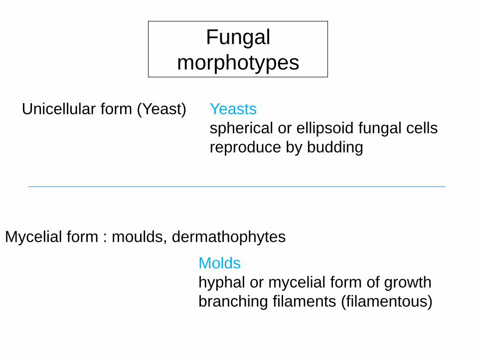
Mycelial form : moulds, dermathophytes
Unicellular form (Yeast)
Fungal
morphotypes
Yeasts
spherical or ellipsoid fungal cells
reproduce by budding
Molds
hyphal or mycelial form of growth
branching filaments (filamentous)

.
Fungal morphotypes
Unicellular form (Yeast)
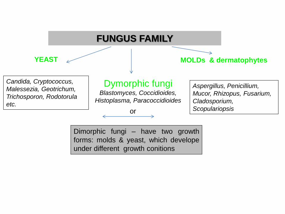
FUNGUS FAMILY
Dymorphic fungi Blastomyces, Coccidioides,
Histoplasma, Paracoccidioides
YEAST MOLDs & dermatophytes
or
Candida, Cryptococcus,
Malessezia, Geotrichum,
Trichosporon, Rodotorula
etc.
Aspergillus, Penicillium,
Mucor, Rhizopus, Fusarium,
Cladosporium,
Scopulariopsis
Dimorphic fungi – have two growth
forms: molds & yeast, which develope
under different growth conitions

SPORULATION
sexual by
sexual spores:
- ascospores
- zygospores
non common
asexual by
asexual spores
common
CONIDIOSPORES
borne externally
on
AERIAL HYPHA
SPORANGIOSPORES
borne in a sac or ascus
on
AERIAL HYPHA
ARTROSPORES
fragmentation
of
VEGETATIVE
HYPHA
size of fungal spores ranges from 2–3 m (Cladosporium , Aspergillus,Penicillium) up to
160 m (Helminthosporium)
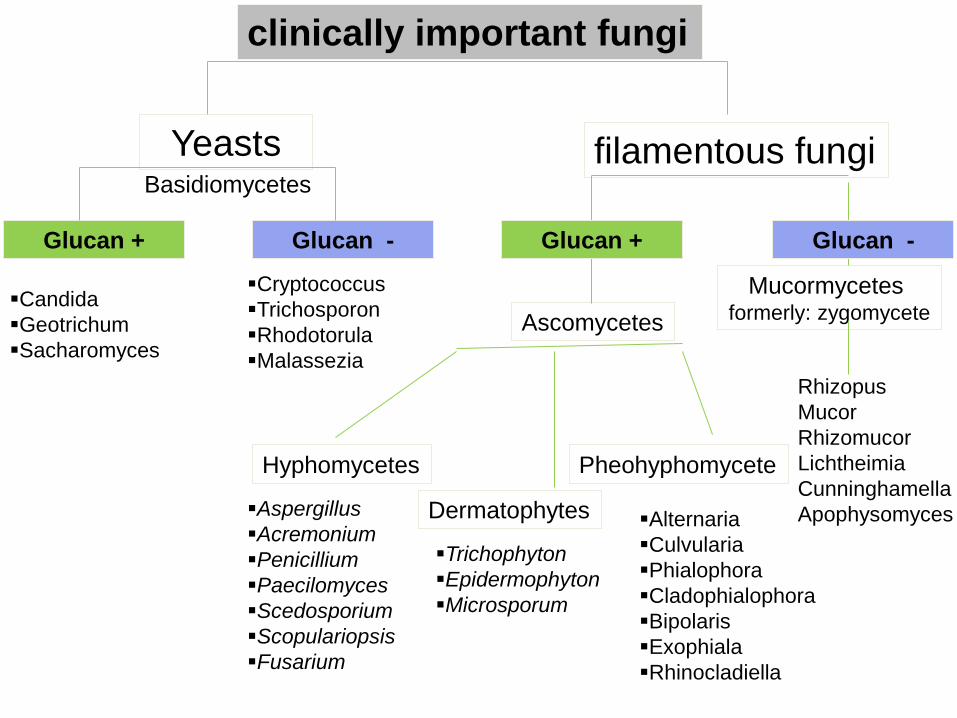
Ascomycetes
Mucormycetes formerly: zygomycete
clinically important fungi
filamentous fungi Yeasts
Glucan + Glucan -
Candida
Geotrichum
Sacharomyces
Cryptococcus
Trichosporon
Rhodotorula
Malassezia
Hyphomycetes Pheohyphomycete
Dermatophytes
Glucan + Glucan -
Aspergillus
Acremonium
Penicillium
Paecilomyces
Scedosporium
Scopulariopsis
Fusarium
Trichophyton
Epidermophyton
Microsporum
Alternaria
Culvularia
Phialophora
Cladophialophora
Bipolaris
Exophiala
Rhinocladiella
Rhizopus
Mucor
Rhizomucor
Lichtheimia
Cunninghamella
Apophysomyces
Basidiomycetes
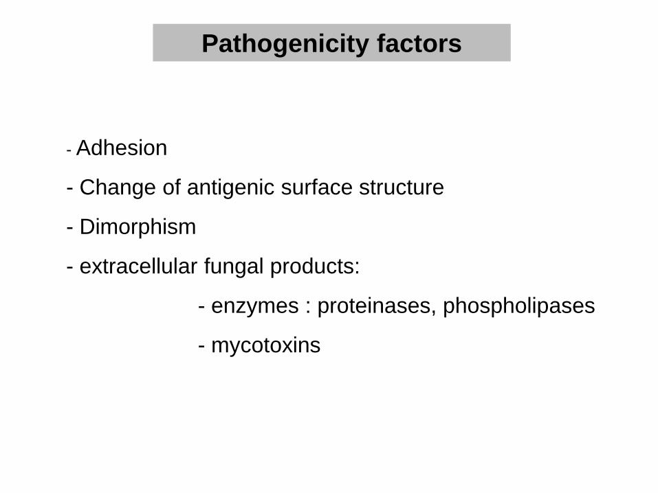
Pathogenicity factors
- Adhesion
- Change of antigenic surface structure
- Dimorphism
- extracellular fungal products:
- enzymes : proteinases, phospholipases
- mycotoxins

Mycoses
1. superficial affect outermost layers
2. cutaneous affect deeper layers (dermatophytes)
3. subcutaneous subcutaneous tissue,
connective tissue, muscle, fascia
4. systemic A) systemic primary – dimorphi fungi
B) systemic opportunistic (exogenous/endogenous NF)
5. allergic mycoses affects lungs or sinuses
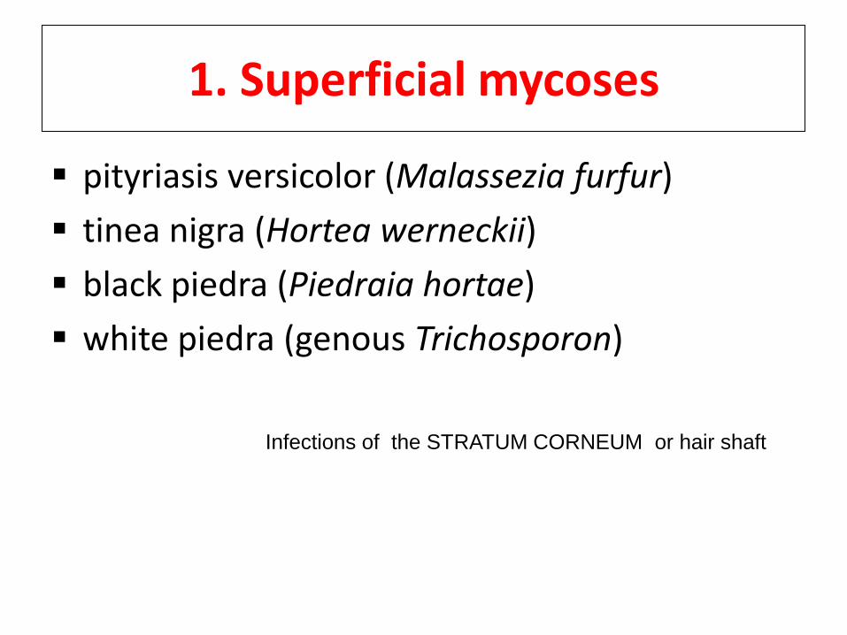
1. Superficial mycoses
pityriasis versicolor (Malassezia furfur)
tinea nigra (Hortea werneckii)
black piedra (Piedraia hortae)
white piedra (genous Trichosporon)
Infections of the STRATUM CORNEUM or hair shaft

Malassezia furfur – lipophilic yeast
Pityriasis versicolor chronic infection
occur as macular patches of discolored skin
inflamation, scaling, irritation are minimal
lesins fluoresce under Wood’s lamp
opportunistic fungemia in patients
receiving total parenteral nutrition
(contamination of the lipid emulsion)
folliculitis – rarely
contributor to dandruff and seborrheic dermatitis
Considered part of microbial flora previously
known as Pityrosporum ovale

TINEA NIGRA – Hortaea werneckii
appear as a dark discoloration often on the palm
PIEDRA - endemic in tropical countries
Black piedra -Piedra hortae: nodular infection of the hair shaft
White piedra - Trichosporon spp.: large, soft, yellowish nodules on the hair
https://mycology.adelaide.edu.au/mycoses/superficial/

Cutaneous mycoses - dermatophytoses
fungi that infect only the superficial keratinized tissue (nails, skin, hair)
unable to grow in 37C
unable to grow in the presence of serum = no systemic spread
genera:
- Trichophyton
- Epidermophyton
- Microsporum
identification, based on morphological
criteria (macroconidia and microconidia)
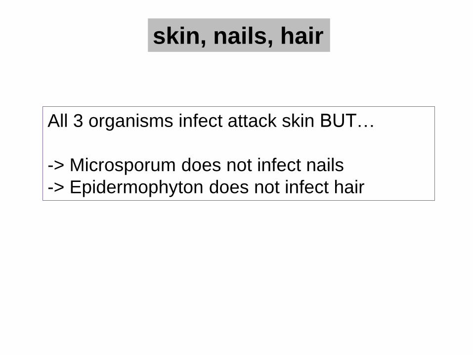
skin, nails, hair
All 3 organisms infect attack skin BUT…
-> Microsporum does not infect nails
-> Epidermophyton does not infect hair

antropophilic zoophilic or geophilic
- relatively mild and chronic
infections in human
- may be difficult to eradicate
- more acute inflammatory inf.
- tend to resolve more quickly
Dermatophytes

Onychomycosis = fungal infections of the nail
Dermatophytids - an allergic reaction to the fungus = fungus-free skin lesions
Dermatophytes >80%
Candida 10%
nondermatophytic molds 6%
Ringworm infection may cause skin lesions in a part of the body that is remote from the actual infection. Such lesions are called "dsermatophytid The lesions themselves are fungus-free, and normally disappear upon treatment of the actual infection

- weighting of the nail (jogging, tennis, badminton, climbing, marches)
- beauty treatments (pedicure, manicure)
- occupation requiring wearing footwear industry
- underlying diseases (diabetes, Cushing's syndrome, hypothyroidism, AIDS, cancer)
Dermatophyte nail mycosis - risk factors
- it occurs in adults, especially the elderly. Rare in children
- nail infections are usually secondary to athlete's foot
- shoes are often „incubator” for fungi
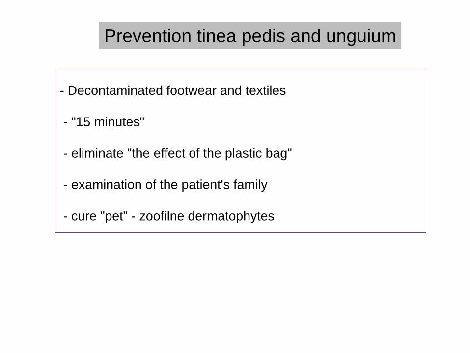
- Decontaminated footwear and textiles
- "15 minutes"
- eliminate "the effect of the plastic bag"
- examination of the patient's family
- cure "pet" - zoofilne dermatophytes
Prevention tinea pedis and unguium

acquired through traumatic lacerations or
puncture wounds to enter
usually confined to tropics and subtropics with
exception of Sporotrichosis
common among those who work with soil and
vegetation and have little protective clothing
SUBCUTANEOUS MYCOSES

http://cmapspublic3.ihmc.us/rid=1GNQT38TZ-1V40FR1-HMZ/Subcutaneous%20Mycoses.cmap

Sporothrix scheneckii
Dimprphic: mycelial in nature, yeast in tissue
Raised skin lesions with proximal spread along
lymphatic channels
Causative agent of sporotrichosis ("rose gardener's disease")
-Cutaneous sporotrichosis
-Extracutaneous sporotrichoses
-Central nervous system sporotrichosis
Risk groups: gardeners, forestry workers, miners, laboratory workers,
veterynarians
Transmission : traumatically introduced into the skin typically by a thorn
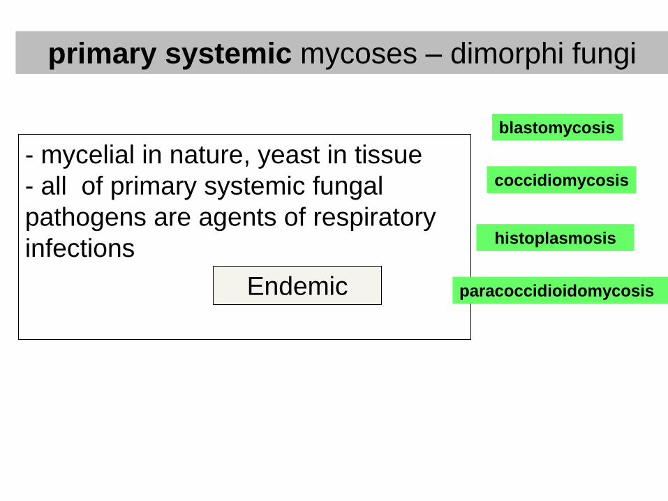
- mycelial in nature, yeast in tissue
- all of primary systemic fungal
pathogens are agents of respiratory
infections
primary systemic mycoses – dimorphi fungi
Endemic paracoccidioidomycosis
blastomycosis
coccidiomycosis
histoplasmosis

inhalation
colonisation
infection
Asymptomatic pneumonia
Chronic lung disease
healing
Acute symptomatic pneumonia
Chronic progressive lung disease
Endogenous
reactivation
Extrapulmonary dissemination
Possible clinical courses of mycosis by dimorphic fungi
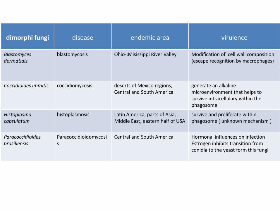
dimorphi fungi
disease
endemic area
virulence
Blastomyces dermatidis
blastomycosis Ohio-;Misissippi River Valley Modification of cell wall composition (escape recognition by macrophages)
Coccidioides immitis
coccidiomycosis deserts of Mexico regions, Central and South America
generate an alkaline microenvironment that helps to survive intracellulary within the phagosome
Histoplasma capsulatum
histoplasmosis
Latin America, parts of Asia, Middle East, eastern half of USA
survive and proliferate within phagosome ( unknown mechanism )
Paracoccidioides brasiliensis
Paracoccidioidomycosis
Central and South America Hormonal influences on infection Estrogen inhibits transition from conidia to the yeast form this fungi

Opportunisctic systemic mycoses
Candidiasis ( Candida albicans, Candida spp.)
Cryptococcosis (Cryptococcus neoformans)
Aspergillosis (Aspergillus fumigatus, Aspergillus spp.)
Mucormycosis (Rhizopus, Mucor, Absidia)
Hyalohyphomycosis (Fusarium, Scopulariopsis, Beauveria)
Pheohyphomycosis (Cladosporium, Bipolaris, Curvularia)

- broad-spectrum antibiotic therapy
- corticosteroid therapy
- pregency
- oral contraceptive use
- systemic disease ( diabetes mellitis etc.)
- neutropenia (especially >7 days)
- hematological & solid tumor malignancy
- postsurgical intensive care patients
- prolonged intravenous catheterization
- parental nutrition
- severe burns
- neonates
clinical groups and predisposing factors
for invasive candidiasis

Clinical manifestations of Candida infections:
Oral candidiasis (including thrush, glossitis, stomatitis)
Candida vulvovaginitis
Cutaneous candidiasis(including diaper candidiasis, paronychia, onychomycosis)
Candiduria
Candidemia and disseminated candidiasis
Causative agents:
Candida albicans
Candida parapsilosis
Candida glabrata
Candida tropicalis

- enzymes (proteinases,phospholipases)
- composition of the cell surface/ hydrophobicity
- ability to undergo the yeast-to-hypha transformation
(regulated by both pH and temperature)
- thigmotropism
Candida albicans virulence factors

Cryptococcus neoformans
endemic in Australia, Papua New Guinea, parts of Africa, the Mediterranean region, India, south-east Asia, Mexico, Brazil, Paraguay and Southern California
C. neoformans var neoformans
reservoir: bird droppings
host predisposition: immunocompromissed
C .neoformans var gatti
reservoir: eucalyptus trees
host predisposition: mostly healthy people

Cryptococcus neoformans
Cryptococcosis :
pulmonary infections
CNS infections (cryptococcal meningoencephalitis)
rare infect other body sides
cryptococcosis is an AIDS- defining illness in patients with HIV
virulence:
polysacharide capsule
phenoloxidase (enzyme that converts hydroxybenzoic substances to melanin;
protect against oxidative host defense)
ability to grow in 37 °C

Aspergillus
have a global distribution
small spore size
thermo-tolerance allowing growth at human body temperature
resistance to oxidative killing
produce metabolites and enzymes with proteolytic and
immunosuppressive activity
A.fumigatus
A. flavus
A. niger
A. terreus
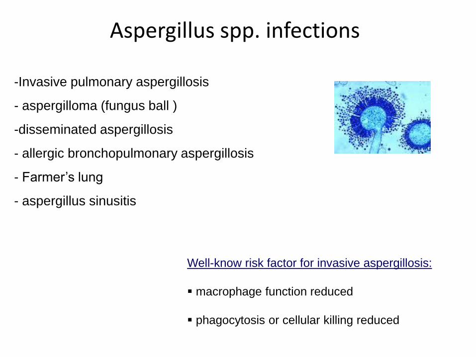
Aspergillus spp. infections
-Invasive pulmonary aspergillosis
- aspergilloma (fungus ball )
-disseminated aspergillosis
- allergic bronchopulmonary aspergillosis
- Farmer’s lung
- aspergillus sinusitis
Well-know risk factor for invasive aspergillosis:
macrophage function reduced
phagocytosis or cellular killing reduced
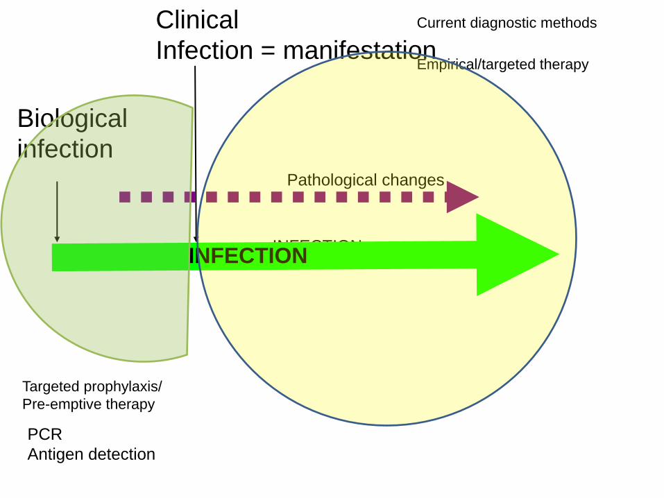
Biological
infection Pathological changes
Empirical/targeted therapy
PCR
Antigen detection
Current diagnostic methods
INFECTION
Clinical
Infection = manifestation
Targeted prophylaxis/
Pre-emptive therapy
INFECTION

Galactomannan (GM)
- polysaccharide component of the cell wall
- highly immunogenic antigen
- exo-antigen that can be detected in serum, BAL or CSF
- monitoring of GM during antifungal therapy allows progression of
treatment to be measured
Diagnosis of invasive Aspergillosis
(1→3)-β-D-glucan
- exo-antigen
-present in molds, yeast, bacteria, plants
- may also be used in diagnosis of candidiasis or fusariosis
- absent in Cryptococcus species, zygomycetes and humans
-- may be used as a complementary test to GM

Based on detection of antigen:
> β-glucan
> Mannan
Mannan
-polysaccharide component of the cell wall of Candida spp.
-highly immunogenic antigen
-immunologically more active then β-glucan
-positive results may be obtained 2-15 days before positive blood cultures
-negative results of the tests do not exclude infection
Invasive Candidiasis
Only based on detection of capsular polysaccharide
(glucuronoxylomannan) antigen
detection in serum, BAL or CSF
Invasive Cryptococcosis

biomarker Best detection method
speciment disease
(1→3)-β-D-glucan
Enzimetic fungitell Blood serum Invasive Candidiasis Fusariosis Aspergillosis
Mannan/ anty-
mannan EIA Blood serum
Specific for Candidiasis 2x - = no IFI
galactomannan EIA Blood serum BAL CSF
Invasive Aspergillosis
! + in 50% fusariosis ! Cross reaction with Geotrichum
glucuronoxylomannan EIA Latex aglutination
Blood serum CSF urine
Invasive Cryptococcosis
Invasive fungal infections

– secondary metabolites produced by fungi
– impair the immune system
- neurotoxic, mutagenic, carcinogenic and teratogenic effects.
- toxic effects depends on the type of mycotoxin, the duration and
dose of exposure and the age, health and nutritional status
of the individual affected
aflatoxin (Aspergillus flavus and A. parasiticus)
ergot alkaloids (Claviceps spp., A. fumigatus and Penicillium chermesinum)
ochratoxins (A. ochraceus , A. alliaceus , A.terreus , P. niger and P. viridicatum)
Mycotoxins & mycotoxicoses
Chronic exposure to mycotoxins causes
immunosuppression of varying extent.
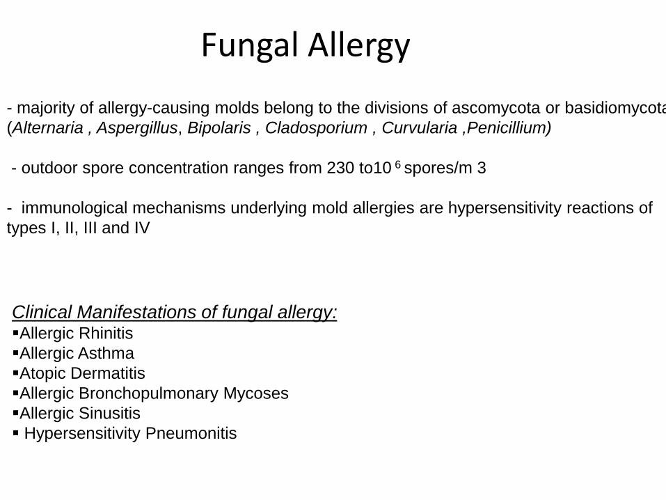
Fungal Allergy
- majority of allergy-causing molds belong to the divisions of ascomycota or basidiomycota
(Alternaria , Aspergillus, Bipolaris , Cladosporium , Curvularia ,Penicillium)
- outdoor spore concentration ranges from 230 to10 6 spores/m 3
- immunological mechanisms underlying mold allergies are hypersensitivity reactions of
types I, II, III and IV
Clinical Manifestations of fungal allergy: Allergic Rhinitis
Allergic Asthma
Atopic Dermatitis
Allergic Bronchopulmonary Mycoses
Allergic Sinusitis
Hypersensitivity Pneumonitis

Pneumocystis jiroveci ( formally P. carinii)
1. Pneumocystis pneumonia (PCP)
- AIDS defining illnes
- probably transmission from person to person
- CD4 level = predicting risk factor for develope PCP
CD4 count of <200 cells/mm3 (90% AIDS patient develope PCP)
SYMPTOMS:
- shortness of breath (especially with exeration)
- nonproductive cough
- fever
2. Extrapulmonar infections are rare (may infect any area of body)
occur in <3%of patients
P. jiroveci does not contain ergosterol and has not been cultured

polyenes (amphotericinB, nystatines, pimarcin)
yests + molds (also Mucormycetes) + dimorphic fungi
yeasts: hialopyphomycetes:
Trichosporon spp - A. tereus
- Fusarium spp.
- Scedosporium apiosperum
resistance
resistanceRare ;
if-> may be present in Candida spp.
most efective drug for severe
cidial
insoluble in water
nephrotoxicity (new formulations with liposomes)
widely distributed in tissues, poor in body fluids
half-life >15 days
Spectrum
of activity
Formation of complexes with ergosterol in fungal cell
membranes, resulting in membrane demage and leakage
Mechanism of
action
other
yests + molds (also Mucormycetes) + dimorphic fungi
yeasts: hialopyphomycetes:
Trichosporon spp - A. tereus
- Fusarium spp.
- Scedosporium apiosperum
Rare ;
if-> may be present in Candida spp.
most efective drug for severe
cidial
insoluble in water
nephrotoxicity (new formulations with liposomes)
widely distributed in tissues, poor in body fluids
half-life >15 days
Formation of complexes with ergosterol in fungal cell
membranes, resulting in membrane demage and leakage
drug of choice

azoles (ketokonazole, itraconazole, fluconazole, vericonazole, posaconazole)
resistance
resistance
Spectrum
of activity
Mechanism of
action
other
interfer with the synthesis of ergosterol
AmB + azoles
Antifungal spectrum different for each agent
Aspergillus spp. - resistant to fluconazole
C. krusei - resistant to fluconazole
50% C. glabrata intermediate for fluconazole
overproduction of enzyme (demethylasis of lanosterol)
efflux pomps
less permeability for antifungal agent
static : Candida spp.
cidial : II generation of triasole for Aspergillus
interfer with the synthesis of ergosterol
AmB + azoles
Antifungal spectrum different for each agent
Aspergillus spp. - resistant to fluconazole
C. krusei - resistant to fluconazole
50% C. glabrata intermediate for fluconazole
overproduction of enzyme (demethylasis of lanosterol)
efflux pomps
less permeability for antifungal agent
static : Candida spp.
cidial : II generation of triazoles for Aspergillus
Supplanted AmB in less severe mycoses because are less toxic & can
be administered orally

azoles
IMIDAZOLES
Ketoconazole
Miconazole
Clotrimazole
TRIAZOLES
I generation:
Fluconazole
Itraconazole
II generation:
Voriconazole
Pozaconasol
izawuconazol
usually localized fungal infections
(topical agents)
exc. ketoconazole-> oral
administration for systemic inf.

echinocandins ( caspofungin, micafungin, anidulafungin)
resistance
resistance
Spectrum
of activity
Mechanism of
action
other
Perturb the sinthesis of -glucan
Candida spp., Aspergillus, spp. dimorphic fungi
Mucormycetes, Cryptococcus spp., Trichosporon spp.
C. parapsilosis (high MIC)
Fusarium (!)
C. glabrata - MDR
cidial – Candida
static – Aspergillus -> MEC (minimal effective concentration)
Echinocandins not for UTI
poor penetration for CNS

antimetabolites (5- fluorocytosine)
resistance
resistance
Spectrum
of activity
Mechanism of
action
other
Interferes with DNA synthesis
- Candida spp.
- Cryptococcus spp.
- some of pheohyphomycetes
because resistance develops quickly flucytosine in never used
alone
penetrate well into all tissues, including CSF
dose-related bone marrow suppression and hepatotoxicity, hair loss
synergistic effect:
Candida : AmB + 5’FC
Cryptococcus: Fluconasol + 5’FC