muscular involvement during the bench …libweb.calu.edu/thesis/nonemaker_cup_6020m_10004.pdf ·...
TRANSCRIPT

MUSCULAR INVOLVEMENT DURING THE BENCH PRESS USING THE ISOBAR® LITE AND STANDARD OLYMPIC BAR
A THESIS
Submitted to the Faculty of the School of Graduate Studies and
Research of
California University of Pennsylvania in partial fulfillment
of the requirements for the degree of
Master of Science
by Ashley Nonemaker
Research Advisor, Dr. Marc Federico
California, Pennsylvania 2009
ii

iii
ACKNOWLEDGEMENTS I would like to take this opportunity to thank all the people that have helped me reach this point. First, I would like to thank the staff and professors at California University of Pennsylvania. And I would especially like to thank my thesis committee: Jeff Hatton, Dr. Thomas F. West and Dr. Marc Federico. The amount of work that took place this year could not have happened without your encouragement and persistence. The enthusiasm that this staff has for the progression of research is intoxicating and has made the process of writing this thesis enjoyable.
I would like to thank my undergraduate professors and staff athletic trainers at West Virginia University. Your dedication to the development of quality athletic trainers and quality people has prepared me for the challenges that I face daily. Without your commitment and compassion, I would not be the athletic trainer I would be today.
I would also like to thank Mike and Mark Lesako; you
are amazing mentors, excellent athletic trainers and spectacular people. Your dedication to the athletes and your families is truly inspiring. Thank you for all the opportunities and for the confidence you had in me. I am truly grateful for the opportunity to work with you this year. I would do this year over again just for the opportunity to work with you guys another time.
I would also like to thank the staff and athletes of Washington and Jefferson College for always being able to make me laugh no matter how stressed I was with class work. You are truly a unique group of people and it was an honor meeting and working with you.
I would like to thank all my friends, old and new.
Whenever I needed a laugh to loosen up a little bit you were there. I wish everyone great luck in the future with every endeavor you undertake.
I would like to thank my family; through everything you have been here to support me. Thank you for always lending me an ear to listen, your mind for an inquisitive thought and a shoulder to cry upon. Thank you for

iv
providing for me so that I could have every opportunity that was possible. I will always be just a phone call away to help in any way possible and to come to bat for you. I hope that I can help you reach your dreams as you have supported me in attaining mine. I will be forever grateful for everything you have done for me and all the sacrifices made. Thank you for teaching me never to settle and to take the risks that may not be the safest option but will lead me to what I truly want. Thank you to my entire family for helping with everything in and out of school work. I hope that I can always make you proud, and “heal” you in your old age.
Finally, I would like to thank Thomas. Without you I
would not have made it through this year. Thank you for your support and reassurance to reach for my dreams, without you I would have never continued to pursue everything I truly wanted. I am so proud of everything you do and I hope that I make you proud as I continue on. I am here whenever you need someone.

v
TABLE OF CONTENTS
Page
SIGNATURE PAGE . . . . . . . . . . . . . . . ii
ACKNOWLEDGEMENTS . . . . . . . . . . . . . . . i ii
TABLE OF CONTENTS . . . . . . . . . . . . . . v
LIST OF TABLES . . . . . . . . . . . . . . . viii
LIST OF FIGURES . . . . . . . . . . . . . . . ix
INTRODUCTION . . . . . . . . . . . . . . . . 1
METHODS . . . . . . . . . . . . . . . . . . 6
Research Design . . . . . . . . . . . . . . . 6
Subjects . . . . . . . . . . . . . . . . . 7
Preliminary Research . . . . . . . . . . . . . 8
Instruments . . . . . . . . . . . . . . . . 8
Procedures . . . . . . . . . . . . . . . . . 10
Hypotheses. . . . . . . . . . . . . . . . . 17
Data Analysis . . . . . . . . . . . . . . . 17
RESULTS . . . . . . . . . . . . . . . . . . 18
Demographic Data . . . . . . . . . . . . . . 18
Hypothesis Testing . . . . . . . . . . . . . 20
Additional Findings . . . . . . . . . . . . . 26
DISCUSSION . . . . . . . . . . . . . . . . . 31
Discussion Of Results . . . . . . . . . . . . 31
Conclusions . . . . . . . . . . . . . . . . 39
Recommendations . . . . . . . . . . . . . . . 40

vi
REFERENCES. . . . . . . . . . . . . . . . . . 44
APPENDICES . . . . . . . . . . . . . . . . . 47
APPENDIX A: Review of Literature . . . . . . . . 48
Introduction . . . . . . . . . . . . . . . . 49
Shoulder Anatomy. . . . . . . . . . . . . . . 50
Muscle Activation . . . . . . . . . . . . . . 53
Motion Analysis of the Upper Extremity . . . 56
Electromyography. . . . . . . . . . . . . . 61
Techniques and Mechanics of the Bench Press . 64
Bar Manipulations . . . . . . . . . . . . . 67
Summary . . . . . . . . . . . . . . . . . 70
APPENDIX B: The Problem . . . . . . . . . . . . 71
Definition of Terms . . . . . . . . . . . . . 72
Basic Assumptions . . . . . . . . . . . . . . 73
Limitations of the Study . . . . . . . . . . . 74
Delimitations of the Study . . . . . . . . . . 75
Significance of the Study . . . . . . . . . . . 75
APPENDIX C: Additional Methods . . . . . . . . . 77
IRB: California University of Pennsylvania (C1) . . 78
Informed Consent Form (C2) . . . . . . . . . . 85
Demographic Information Sheet(C3). . . . . . . . 94
Individual Data Collection Sheet (C4) . . . . . . 97
Isobar® Example Lift (C5) . . . . . . . . . . 99
Isobar® Lite Hand Position Example (C6). . . . . 101

vii
REFERENCES . . . . . . . . . . . . . . . . . 103
ABSTRACT . . . . . . . . . . . . . . . . . 106

viii
LIST OF TABLES
Table Title Page 1 Subject Physical Activity Participation . . . . . . . . . . . . 19
2 Mean Peak Muscle Activation Scores of Sample . . . . . . . . . . . 21 3 Peak Muscle Activation Within- Subject Effects . . . . . . . . 21 4 Peak Muscle Activation Pairwise Comparisons . . . . . . . . . 22 5 Mean Average Muscle Activation Scores of Sample . . . . . . . . . . . 23 6 Average Muscle Activation With-in Subject Effects . . . . . . . . 24 7 Average Muscle Activation Pairwise Comparisons . . . . . . . . . 25 8 Overall Peak Muscle Activation Mean
Scores of Sample . . . . . . . . . . . 27
9 Overall Average Muscle Activation Mean Scores of Sample . . . . . . . . . . . 27
10 Overall Peak Test of Within-Subjects Effects . . . . . . . . . . . . . . 28 11 Overall Peak Muscle Activation Pairwise Comparison . . . . . . . . . 28 12 Overall Average Test of Within-Subjects Effects . . . . . . . . . . . . . . 29 13 Overall Average Muscle Activation Pairwise Comparison . . . . . . . . . 30

ix
LIST OF FIGURES
Figure Title Page 1 Distribution of Subject Age . . . . . . 19 2 Effect of Contraction Type on Muscles for Average Muscle Activation . . . . . 26 3 Individual Data Collection Sheet . . . . 98 4 Exaggerated Motion with an Isobar® During the Incline Bench Press . . . . . 100 5 Isobar® Lite with Handles Positioned at the Furthest Points . . . . . . . . 102 6 Isobar® Lite with Handles Positioned at the Test Lift Starting Position . . . 102

1
INTRODUCTION
Strength training is a crucial element in athletics
today. The bench press is one component of the essential
weight lifting program, which increases strength in the
upper body. Although the bench press is part of many
strength and conditioning programs, there is no evidence it
is the most effective way to activate the musculature a
person is trying to strengthen. There have been many
studies on different ways to manipulate repetitions, load,
volume and body positioning to identify the most effective
method for increasing strength. Furthermore, recent
research has investigated manipulating the bar type by
changing the grip width or type of hand positioning.
The inclusion of a multiplanar or decreased stability
component to the bar has not been investigated. Truform’s
Isobar® Lite [Santa Barbara, CA] was developed with mobile
hand grips to allow multiplanar movement during the bench
press. 1 In this study the researcher will be testing the
efficacy of the Isobar® Lite in activating specific
musculature of the upper body during the bench press as
compared to the Olympic bar.

2
The shoulder is known as the most mobile joint in the
body, but with the increased amount of mobility comes a
decreased amount of stability. The muscles in the upper
extremity work together to provide a range of movements
that complete functional tasks. The muscles can be grouped
into categories according to their origins and insertions,
in the shoulder these groups are the scapulohumeral and
scapulothoracic. 2 The scapulohumeral group is responsible
for motions at the glenohumeral joint that include internal
and external rotation, extension, adduction, abduction and
flexion. In the scapulothoracic group the muscles
originate on the trunk and attach to the upper extremity. 3
Scapular motions completed by this group are elevation,
depression, protraction, retraction, upward and downward
rotation.
The shoulder complex is the most multifaceted
articulation in the body using three different joints to
produce multiplanar motion. The wide variety of muscular
attachments allows complex motion to occur, especially at
the scapulothoracic and glenohumeral joints. The motions
that occur at the scapulothoracic joint are adduction,
abduction, upward rotation, downward rotation, anterior
tilt, elevation and depression. 4 Upward and downward
rotation of the scapula are important to allow greater

3
range of motion of the shoulder complex. Other benefits of
scapular rotation include the movement of the glenoid fossa
thus providing the humeral head a firm but mobile
articulation. This biomechanical property is known as a
force couple. A force couple is created at the scapula
when several muscles contract simultaneously and pull the
scapula in opposite directions. Without the force couple,
scapular rotation would not be possible and would result in
limited shoulder range of motion. The uniqueness of these
joints allows for many different types of motions,
consequently needing an extensive strengthening program in
an attempt to minimize the likelihood of injury. Different
exercises are completed in multiple planes to strengthen
the muscles involved in the motions at the shoulder. If
exercises are only completed in one plane not all muscles
will be strengthened optimally because they function
normally in a different plane.
While strengthening the upper body there are different
factors which may affect the efficiency of the exercise.
If these factors interfere, the muscular activation will be
altered and the exercise will fail to produce the desired
results. One of these factors is fatigue. The function of
the upper extremity can be affected by fatigue of the
muscles involved in the exercise. During the horizontal

4
bench press the upward velocity is greatest early in the
set. As the lift progresses to the final repetition the
amount of time to lift the bar will double in duration. 5
Another way fatigue will affect upper extremity function is
changing the amount of time to complete the bench press.
With each repetition the lift duration becomes more similar
to the one repetition maximum duration. As with the other
factors, the bar path becomes more similar to the path of
the one repetition maximum when a set is done to fatigue. 5
This study detailed above by Duffey, et al illustrates that
when proper mechanics of the bench press are not followed
the maximum effects will not be achieved.
There are variations that will affect the amount of
muscles and the type of contraction. During the bench
press the bar can have a different width or grip; this will
affect type of strengthening which will occur. The width of
the bar will affect different muscles. 6 In multiple studies
a wide grip or narrow grip was found to increase
neuromuscular activation and maximal voluntary contraction
(MVC) of specific muscles. 6,7,8,9 When doing the bench press
changing the positioning of the hands can also be more
effective for certain muscles. Another variation was to
supinate or pronate the hands while performing the bench
press. Standard hand positioning during the bench press is
5
usually pronation which evidence shows is more effective in
strengthening particular muscles. 6 Sometimes changing hand
or grip positioning did not affect muscle activation or
MVC.6,9,10 Mobile parts on the bar may also have an
influence on which muscles are activated effectively, but
more research is necessary in this area. In a strength
training program it will be necessary to utilize variations
to activate multiple muscle groups.
For years the bench press has been used in all types
of strength training programs. There have been many
studies which have manipulated the body positioning, grip
or type of work out. When using the bench press some
muscles could be missed in the complex network that is the
shoulder. These studies have provided evidence that
improvements can be made to the standard bench press
exercise. This has lead companies to new developments and
variations to the Olympic bar. The Isobar® Lite is one of
these variations that have claimed to be superior to the
standard Olympic bar. The purpose of this study was to
examine the effects of the Isobar® Lite on muscle
activation as compared to the Olympic bar in active college
students aged 18-27.

6
METHODS
The purpose of this study was to investigate the
difference in muscle activation during the bench press when
using a standard Olympic bar and the Isobar® Lite. EMG
activity was measured to evaluate the activation of
specific muscles during the exercise. This section will
include research design, subjects, instruments, procedures,
hypothesis and data analysis.
Research Design
This research was a quasi-experimental, within
subjects, repeated measures design. The independent
variables were bar type, contraction type and muscles used.
The different bars used were the Isobar® Lite and a
standard Olympic bar. Results were measured during
concentric and eccentric muscle contraction to allow
comparison of muscle activity during these motions. The
muscles tested in this study were the pectoralis major,
infraspinatus, biceps brachii and triceps brachii. The
dependent variables were peak muscle activation and average
muscle activation as measured by surface EMG.

7
Subjects
The subjects used for this study were 26 male and
female volunteers from California University of
Pennsylvania. The ages of the subjects ranged from 18-27.
All the subjects were active individuals and possessed the
basic knowledge of weight lifting, including the bench
press exercise. This active individual is defined as
someone who engages in some sort of heart rate raising
physical activity at least three times a week. A person
with a basic knowledge of weight lifting is defined as
someone who has participated in a formalized weight
training program in the past. The subject must currently
lift weights or previously lifted weights and not reported
injury to the upper extremity or chest within the past six
months that resulted in medical attention or have any
current condition that may affect their performance.
It was required that each subject participate in a
preliminary meeting where a one-repetition maximum (1 RM)
on the Olympic bar was obtained. Subjects then returned to
participate in one 1-hour testing session one week later.
Each participant’s identity remained confidential and will
not be included in the study. The study was be approved by

8
the Institutional Review Board (Appendix C1) at California
University of PA. All subjects in the study signed an
Informed Consent Form (Appendix C2) and completed a
Demographic Information Sheet (Appendix C3) prior to
participation in the study.
Preliminary Research
A pilot study was completed prior to completing this
research project. Subjects who met the selection criterion
were used to test the protocol. The researcher looked for
the ability of the subjects to follow instructions,
complete the activity and warm up, the amount of time it
would take to complete each task, and if the protocol was
accurate. The data was collected and placed in the data
collection sheet (Appendix C4).
Instruments
The researcher used a demographic sheet (Appendix C3)
to accept or eliminate individuals. The study used the
following equipment: bench, bar rack, two different bars,
Biopac MP150[Goleta, CA], metronome and two one-pound cuff
weights. The first bar was the standard 45-pound Olympic

9
bar and the second bar was the Isobar® Lite which weighed
23 pounds. The Isobar® Lite had freely mobile hand grips
which slide along the bar linked together so that balance,
symmetry and control were maintained (Appendix C5). 11 The
cuff weights were added to the ends of the Isobar® Lite so
it would have the ability to equal the same total weight as
the Olympic bar during the experiment. The metronome was
used while performing the bench press so to complete the
exercise in a controlled and uniform manner.
In collecting the EMG data, the researcher used six
channels from a Biopac MP150® electromyography machine.
Four channels were designated for the muscles tested and
the other two channels were connected to an electronic
biaxial goniometer. The Biopac MP150 was connected to a
Microsoft Windows based personal computer with the Biopac’s
AcqKnowledge® program [Goleta, CA] to collect analyze the
data. The study utilized pre-gelled disposable Ag-AgCl
surface electrodes with a diameter of one centimeter. 12,13
The electrodes were placed on the subject’s dominate arm
over the motor points of each muscle belly with a center-
to-center spacing of 2.5 centimeters. 13 This goniometer was
applied to the subject’s arm at the elbow to measure the
angle of the arm when a peak muscular contraction occurred.
The raw EMG signal was band pass filtered at 10 and 1000

10
Hertz (Hz). 6,14,15 The researcher utilized a sampling rate of
2000 Hz using the AcqKnowledge software. 16,17 The signals
were rectified and normalized before the data analysis was
completed.
Procedures
Once informed consent and a demographic sheet were
obtained from all potential subjects, there was an
explanatory session to inform the participants of the
process. The Institutional Review Board at California
University of Pennsylvania approved all testing protocol
prior to experimentation. Participants were chosen by
searching the campus of California University of
Pennsylvania to acquire volunteers. To collect the
volunteers the researcher visited various classes on the
California University of Pennsylvania campus by introducing
and explaining the study. Volunteers were disqualified
from the study if there was a self-reported recent
significant injury to the upper extremity or chest, any
other condition that may affect performance, or if they did
not meet the demographic standards.
The subjects participated in a pre-experimental lift
where the participant completed a one-repetition maximum (1

11
RM) with the standard Olympic bar. The volunteers were
asked to estimate what their 1RM was based upon their prior
experience and this value was considered their perceived
maximum. Prior to the maximum lift, the subjects peddled
the Upper Body Ergometer (UBE) for five minutes at a
moderate workload speed of 60 revolutions per minute (rpm).
The subjects peddled forward for two minutes and backwards
for three minutes on the UBE to warm up the muscles used in
the 1 RM. The warm up continued after a one-minute rest
with a set of five bench press repetitions at 50% of the
perceived maximum. During the period of rest, the subjects
were permitted to perform light self-stretching of their
choosing to the upper extremity.
To determine the volunteers 1 RM using the Olympic bar
their perceived maximum weight was placed on the bar for
the first lift. The subjects were then asked to lift the
bar. If the subject was only able to lift the bar for one
repetition, then ten pounds was added to the bar and they
were asked to lift the bar again. If they were unable lift
the bar then the earlier weight was determined to be their
1 RM. This procedure was repeated until the 1 RM was
determined. It was expected that several attempts of the
bench press exercise would be needed to be performed to
determine 1 RM. The goal was to find the volunteer’s 1RM

12
within 3-5 tries with a ten pound increment of weight added
after each successful lift until a lift attempt fails. 18
During every 1RM attempt there were two spotters closely
observing to assist the lifter with bar replacement. The
spotters were positioned at either end of the bar and
followed the bar’s path with their hands keeping the bar
within reach. The 1 RM for each subject was recorded on a
sheet with their corresponding subject number. While
waiting to complete their 1 RM the subjects had an
opportunity to practice a lift with the Isobar® Lite.
After a minimum of seven days following the 1 RM
testing, the subjects returned at a time designated by the
researcher. Prior to beginning activity, the subjects
completed a Visual Analogue Scale (VAS) to identify the
level of soreness they were experiencing as a result of the
1 RM test. There were numbers listed from 0-10 where zero
equaled no pain and ten was the most pain they have ever
experienced. If the subjects stated their discomfort was a
value over four the athlete was unable to begin the second
day until it subsided. After filling out the VAS the
subjects completed a warm-up session utilizing the UBE and
one warm up set on each of the two bars. 5 The UBE portion of
the warm up was a five minutes session as performed in the
1 RM testing. The bench press warm up exercises consisted

13
of lifting either 50% of their 1 RM or 45 pounds (the
weight of the bar alone), whichever was greater. The
subjects completed two sets of ten repetitions with one set
using each bar.
The subjects were randomly assigned to two groups, one
completed the Olympic bar lift first and the second
completed the Isobar® Lite lift first. The sites for
electrode placement were shaved, cleaned and prepared to
decrease impedance with a high grit sand paper before
electrode placement occurred. 13,15,19,20 The electrodes were
placed over the motor points in each muscle belly. 12 The
muscles tested in this study were the pectoralis major,
biceps brachii, triceps brachii and infraspinatus. After
the electrodes were in place, the goniometer was applied at
the elbow with one attachment distal to the deltoid
insertion and the other under the wrist extensor muscle
group.
The BIOPAC MP150 was then turned on and connected to
the laptop computer to begin the activity. For each muscle
tested, the participants completed three maximal voluntary
isometric contractions (MVIC). These three isometric
contractions lasted six seconds each with a three second
rest period between contractions. 20 The greatest value from
the three attempts was recorded as the value for the MVIC. 20

14
The MVIC is the value which was used to normalize the EMG
data. The subjects were also measured going through the
bench press motion with a light wooden rod in order to
obtain the zeros of the goniometer.
To complete the MVIC testing the arm needed to be
placed in specific positions optimal for initiating
contraction with the tested muscles. Each muscle completes
at least one major motion and may contribute to others.
For this study, the major action of each muscle was tested
and used for the MVIC. The subject was positioned on the
bench with the non-dominant arm placed on the bar for
stabilization during the pectoralis major, biceps brachii
and triceps brachii MVIC tests. For the infraspinatus MVIC
test the non-dominant arm was placed on the post of the bar
rack. For the pectoralis major’s MVIC the dominant arm was
placed at 90 degrees of flexion and then resisted as the
subject moves into horizontal adduction while lying on the
bench. The subject sat with their dominant arm in terminal
external rotation and was resisted while they continued to
push into external rotation for the MVIC of the
infraspinatus. The beginning positioning for the biceps
brachii and the triceps brachii was identical. The subject
laid on the bench with their shoulder in a neutral
position, elbow completely extended and their hand in full

15
supination. The biceps brachii MVIC was completed by the
researcher resisting as the subject contracts into elbow
flexion from the beginning position. For the triceps
brachii MVIC the subject extended the elbow from the
beginning position while the researcher attempted to push
the elbow into flexion.
The procedures were repeated identically for both
bars. The subjects lie on a horizontal bench ensuring they
were not rubbing the infraspinatus electrodes on the bench.
The subjects grabbed the Olympic bar outside the knurl and
the Isobar® Lite against the inner bumper of the handle
with the handle at least two inches from the collar of the
bar. The subjects then lifted the bar off the rack and
held the bar for 1 second with their elbows extended. 5 The
bar was lowered until it gently touched the subject’s
chest, paused for one second, and then lifted back up to
the beginning position. The bench motion was completed in
a slow and controlled manner, which took three seconds on
the descent and two seconds to ascend. 13,21 To keep the
movements uniform, there was a metronome to keep a beat.
There were two spotters to maintain the lifter’s safety
positioned in the same location as they were during the 1
RM testing.
16
The participants completed three repetitions at 65% of
their maximal contraction as determined by their 1 RM. The
65% of the subjects’ 1 RM were rounded down to the nearest
five pounds to increase the ease of adding plates to the
bar. The hand placement on the mobile parts of the bar was
the only difference between the experiments with the two
bars. The natural movement was used with the Isobar® Lite,
this allowed the hands to follow the natural path they
would normally take through the range of motion during the
bench press. 1 The subjects were instructed not to purposely
move their hands along the length of the Isobar® Lite, but
to keep their hands in a comfortable distance apart like
they would using the Olympic bar (Appendix C6). There was a
minimum of a three-minute rest between the tests. 8, 22
As the participant was lifting, EMG data was recorded
as waves on the computer through the Biopac’s Acqknowlege®
software system. After the data was collected the data for
each subject it was rectified and smoothed. The data was
then selected starting with the first flexion of the elbows
through the final (third) extension. The maximum (peak)
and the mean (average) were calculated by the software and
then recorded in Microsoft Excel.

17
Hypotheses
The following null hypotheses were based previous
research and the researcher’s intuition based on a review
of the literature.
1. There will not be a significant difference in peak
muscle activation for each muscle during eccentric or
concentric contractions with the different bar types.
2. There will not be a significant difference in average
muscle activation during eccentric or concentric
contractions with the different bar types.
Data Analysis
The research hypotheses were analyzed using a
multivariate repeated measures 2x2x4 analysis of variance.
All data was analyzed by SPSS version 16.0 for Windows at
an alpha level of 0.05. All EMG scores were reported as
percentage of maximal voluntary contraction. 13

18
RESULTS
The purpose of this study was to investigate the
difference in muscle activation during the bench press when
using a standard Olympic bar and the Isobar® Lite. The
following section contains the data collected through this
study and is divided into the following three subsections:
Demographic Information, Hypotheses Testing, and Additional
Findings.
Demographic Information
There were 26 physically active, healthy subjects who
participated in this study. The age range was 18-27 years
and the mean age was 21.4 years and is demonstrated in
Figure 1. Eleven (42.3%) of the subjects were male,
leaving the remaining fifteen (57.7%) female. Sixty-one
and one half percent of the population participates in
physical activity 3-4 times a week where 38.5% participate
in some type of physical activity 5-7 times a week.

Figure 1. Distribution of
All 26 subjects participated in a variety of differ ent
activities as demonstrated in
participate in more
Table 1. Subject Physical
Type of Activity
Cardiovascular
Weight Lifting
Aerobics
Sports
Other
0
1
2
3
4
5
6
7
8
18 19
Distribution of Subject Age
All 26 subjects participated in a variety of differ ent
activities as demonstrated in Table 1; many subjects
more than one.
Subject Physical Activity Participation
Frequency Percent
21 42.9
20 40.8
1
4
3
20 21 22 23
Age
19
All 26 subjects participated in a variety of differ ent
subjects
Percent
42.9
40.8
5.0
8.2
6.1
27

20
Hypothesis Testing
The following hypotheses were tested during this
study. All of the hypotheses were tested with a level of
significance set at α ≤ 0.05. A multivariate, repeated
measures 2x2x4 analysis of variance was calculated to find
the effect of the bar differences on the tested muscles.
Null Hypothesis 1: There will not be a significant
difference in peak muscle activation for each muscle during
eccentric or concentric contractions with the different bar
types.
Conclusion: Mean scores for each muscle’s peak
activation were calculated during eccentric and concentric
contractions. The mean scores for each bar during
eccentric and concentric contraction are listed in Table 2.
For the peak muscle activation, there was no significant
difference found between different bars. The individual
significances are listed in Table 3. There was also no
significant difference found between any combinations of
the three variables together. The null hypothesis is
therefore accepted.

21
Table 2. Mean Peak Muscle Activation Scores of Sample
Bar Pectoralis Major(% MVIC) Infraspinatus (% MVIC)
Con. Ecc. Con. Ecc.
Olympic 173 (±220) 160 (±211) 277 (±230) 265 (±173)
Isobar 187 (±253) 164 (±216) 251 (±185) 281 (±200)
Bar Triceps (% MVIC) Biceps (% MVIC)
Con. Ecc. Con. Ecc.
Olympic 80 (±41) 66 (±31) 91 (±108) 901 (±105)
Isobar 94 (±49) 76 (±35) 102 (±103) 100 (±106)
Table 3. Peak Muscle Activation Within- Subject Effects
Source df F Sig.
Bar 25 0.548 0.466
Muscle 25 17.453 <0.001
Contraction 25 0.997 0.328
bar*muscle 25 0.456 0.714
bar*contraction 25 0.817 0.375
Muscle*contraction 25 2.271 0.087
bar*muscle* contraction 25 2.39 0.075
In the peak testing, the only significant difference
( α ≤ 0.05) was the comparison between the individual

22
muscles, see Table 3. The significance for the muscle
variable was <0.001. The values for each individual muscle
as compared to each muscle can be seen in Table 4. Not
every muscle was significantly different from one another.
The only muscles that were not significantly different were
the triceps brachii and biceps brachii.
Table 4. Peak Muscle Activation Pairwise Comparisons
Muscle (I) Muscle (J) Mean Diff. (I-J)
Stand. Error
Sig.
Pectoralis Infraspinatus* -97.6 28.2 0.002 Triceps* 91.9 39.3 0.028 Biceps* 74.7 26.9 0.01
Infraspinatus Pectoralis * 97.6 28.2 0.002 Triceps* 189.4 33.8 <0.001 Biceps* 172.3 25.6 <0.001
Triceps Pectoralis* -91.9 39.3 0.028
Infraspinatus* -189.4 33.8 <0.001 Biceps -17.1 16.3 0.304
Biceps Pectoralis* -74.7 26.9 0.01 Infraspinatus* -172.3 25.6 <0.001
Triceps 17.1 16.3 0.304
*The mean difference is significant at the .05 level
Null Hypothesis 2: There will not be a significant
difference in average muscle activation during eccentric or
concentric contractions with the different bar types.

23
Conclusion: The comparison of the mean scores for the
average muscle activation resulted in the findings that the
difference between bars was not statistically different.
The means for the average muscle activation with for each
muscle and contraction type can be found in Table 5.
Table 5. Mean Average Muscle Activation Scores of Sample
Bar Pectoralis Major (% MVIC) Infraspinatus(% MVIC)
Con. Ecc. Con. Ecc.
Olympic 75 (±62) 64 (±80) 86 (±54) 989 (±58)
Isobar 98 (±125) 74 (±89) 89 (±61) 112 (±76)
Bar Triceps (% MVIC) Biceps (% MVIC)
Con. Ecc. Con. Ecc.
Olympic 37(±17) 32 (±18) 67 (±110) 67 (±108)
Isobar 43 (±18) 31 (±14) 70 (±107) 69 (±108)
In the average muscle activation comparison between
the different bars there was not a significant difference
found, this data is found in Table 6. This table shows
that the bar significance was 0.134 and was greater than
the specified significance level. The null hypothesis is
therefore accepted.

24
Table 6. Average Muscle Activation Within-Subject Effects
The average muscle activation had similar result for
the bar as did the peak muscle activation. There were two
variables that had significant difference in the average
muscle activation statistics; see Table 6. The muscles
compared to one another had a significance of 0.002. This
variable had two muscle comparisons that were significantly
different from one another. The triceps brachii average
muscle activation was significantly different than both the
pectoralis major and infraspinatus muscles as seen in Table
7.
Source df F Sig.
Bar 25 2.401 0.134
Muscle 25 5.295 0.002
Contraction 25 1.503 0.232
bar*muscle 25 0.745 0.529
bar*contraction 25 0.473 0.498
Muscle*contraction 25 14.093 <0.001
bar*muscle* contraction
25 1.024 0.387

25
Table 7. Average Muscle Activation Pairwise Comparisons
Muscle (I) Muscle (J) Mean Diff. (I-J)
Stand. Error
Sig.
Pectoralis Infraspinatus -18.8 14.6 0.209 Triceps* 41.9 15.5 0.012 Biceps 9.3 12.9 0.476
Infraspinatus Pectoralis 18.8 14.6 0.209
Triceps* 60.7 10.6 <0.001 Biceps 28.1 17.7 0.125
Triceps Pectoralis* -41.2 15.5 0.012 Infraspinatus* -60.7 10.6 <0.001
Biceps -32.6 20.3 0.122 Biceps Pectoralis -9.3 12.9 0.476
Infraspinatus -28.1 17.7 0.125 Triceps 32.6 20.3 0.122
*The mean difference is significant at the .05 level
The other average muscle activation that was
significantly different was the difference between the
contractions of each muscle. As shown in Figure 2, the
concentric and eccentric muscle contractions of the
pectoralis major and infraspinatus were significantly
different. The triceps brachii did have a difference
between contraction types, but it was not significant.
There was not a significant difference between the
contraction types of the biceps brachii.
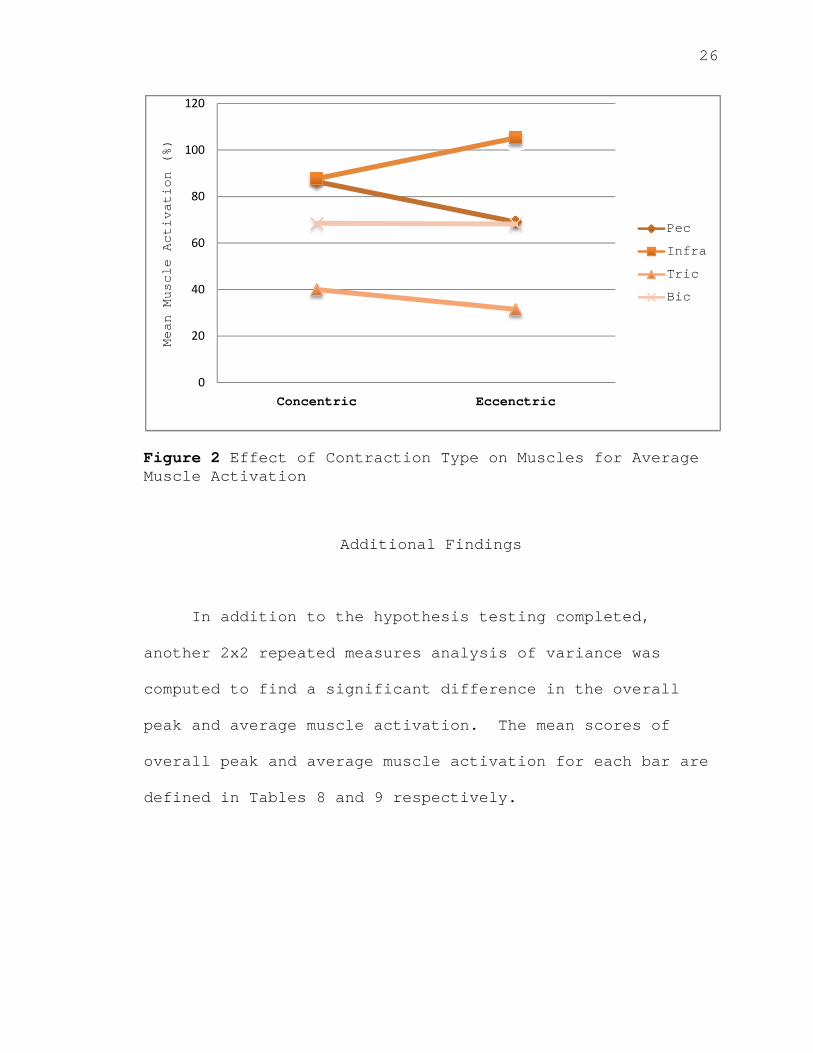
Figure 2 Effect of Muscle Activation
In addition to the hypothesis testing comp
another 2x2 repeated measures analysis of variance was
computed to find a significant difference in the overall
peak and average muscle activation.
overall peak and average
defined in Tables 8 and 9
0
20
40
60
80
100
120
Concentric
Me
an
Mu
scle
Act
iva
tion
(%
)
of Contraction Type on Muscles for Average Muscle Activation
Additional Findings
In addition to the hypothesis testing comp leted
another 2x2 repeated measures analysis of variance was
to find a significant difference in the overall
and average muscle activation. The mean scores of
overall peak and average muscle activation for each bar
defined in Tables 8 and 9 respectively.
Concentric Eccenctric
26
for Average
leted ,
another 2x2 repeated measures analysis of variance was
to find a significant difference in the overall
The mean scores of
for each bar are
Pec
Infra
Tric
Bic

27
Table 8. Overall Peak Muscle Activation Mean Scores of Sample
Bar Muscle (% MVIC)
Pectoralis Major I nfraspinatus Triceps Biceps
Olympic 187 (±227) 298 (±223) 82 (±36) 96 (±105)
Isobar 208 (±290) 304 (±210) 97 (±46) 114 (±101)
Table 9. Overall Average Muscle Activation Mean Scores of Sample
Bar Muscle (% MVIC)
Pectoralis Major Infraspinatus Triceps Biceps
Olympic 81 (±115) 92 (±54) 32 (±13) 67 (±110)
Isobar 86 (±125) 101 (±70) 37 (±15) 72 (±107)
There was not a significant difference found between
bars discovered through the results of the overall peak
muscle activation testing. These results can be seen in
Table 10. The significance level for the muscle
interactions was <0.001. There was a significant
difference found between all the muscles except between the
biceps brachii and triceps brachii muscles and is
demonstrated in Table 11.

28
Table 10.Overall Peak Test of Within-Subject Effects
Source F Sig
bar 1.816 0.19
muscle 17.101 <0.001
bar*muscle 0.136 0.938
Table 11. Overall Peak Muscle Activation Pairwise Comparisons
Muscle (I) Muscle (J) Mean Diff. (I-J)
Stand. Error
Sig.
Pectoralis Infraspinatus* -103.5 31.6 0.003 Triceps* 107.8 45.4 0.026 Biceps* 92.6 35 0.014
Infraspinatus Pectoralis* 103.5 31.6 0.003 Triceps* 211.3 36.3 <0.001 Biceps* 196.1 28.9 <0.001
Triceps Pectoralis* -107.8 45.4 0.026
Infraspinatus* -211.3 36.3 <0.001 Biceps -15.2 15.4 0.333
Biceps Pectoralis* -92.6 35 0.014 Infraspinatus* -196.2 28.9 <0.001
Triceps 15.2 15.4 0.333
*The mean difference is significant at the .05 level
The results of the overall average muscle activation
found that there was not a significant difference between

29
the Olympic bar and the Isobar® Lite. In Table 12 it is
demonstrated that there was a significant difference found
between the individual muscles. The significant difference
was found between the triceps brachii and both the
infraspinatus and pectoralis muscle. These significant
differences are demonstrated in Table 13.
Table 12. Overall Average Test of Within-Subjects Effects
Source F Sig
bar 2.609 0.119
muscle 4.53 0.006
bar*muscle 0.189 0.904

30
Table 13. Overall Average Muscle Activation Pairwise Comparisons
Muscle (I) Muscle (J) Mean Diff. (I-J)
Stand. Error
Sig.
Pectoralis Infraspinatus -13.3 20.2 0.518 Triceps* 49.1 22.7 0.04 Biceps 13.8 11.2 0.228
Infraspinatus Pectoralis 13.2 20.2 0.518
Triceps* 62.4 10.9 <0.001 Biceps 27.1 18.1 0.147
Triceps Pectoralis* -49.1 22.7 0.04 Infraspinatus* -62.4 10.9 <0.001
Biceps -35.3 20.4 0.095 Biceps Pectoralis -13.8 11.2 0.228
Infraspinatus -27.1 18.1 0.147 Triceps 35.3 20.4 0.095
* The mean difference is significant at the .05 level

31
DISCUSSION
The purpose of this research was to investigate the
claims of companies that variations of the standard Olympic
bar are better for training. The Isobar® Lite is a
variation of the standard bar and includes mobile hand
grips. The purpose of this study was to see if a
significant difference exists with muscular activation
between this bar and the Olympic bar. The following
section is divided into three subsections: Discussion of
Results, Conclusions, and Recommendations.
Discussion of Results
Upon completion of this study, it was found that the
Isobar® Lite did not produce a significantly different
amount of peak or average muscle activation as compared to
the Olympic bar. A significant difference of both peak and
average muscle activation was found between muscles. There
was no difference found between the contraction types for
the peak muscle activation but there was a significant
difference found with average muscle activation. The
results supported the null hypotheses that stated there is

32
not a significant difference between the Olympic bar and
the Isobar® Lite.
Recent literature has focused on variations that can
be applied to weight training. There have been multiple
studies which focused on the bench press specifically
altering hand positioning, grip width and body
positioning. 8, 16, 23 After an extensive literature review of
the variations of weight lifting techniques, no prior
studies had investigated the effects of mobile parts on the
bar.
Many of the prior studies found mixed results where
the increase or decrease of muscle activation was dependent
upon which muscle was tested. The researchers found that
there were specific muscles that were affected differently
dependent upon the different type of variation applied to
the exercise. In two studies that tested the effect of
grip width found that the pectoralis major, biceps brachii
and latissimus dorsi had increased muscle activation with a
wide grip but the anterior deltoid and triceps brachii
muscles were activated more efficiently with a narrow
grip. 16,23 In another study completed by Grant, et al found
that a smaller bar diameter had the lowest overall
neuromuscular activation. 8 Switching the positioning of the
hand from pronation to supination can also effect the

33
activation of the muscles. The muscles that had a greater
activation with supination are the biceps brachii and the
clavicular portion of the pectoralis major. 16
The results of this study demonstrated that there was
not a significant difference between the Olympic bar and
the Isobar® Lite for peak or average muscle activation.
The common thought would be that introducing mobile parts
would increase muscle activation, however, this study
proved otherwise under the test circumstances.
During the testing the general comment from the
subjects was, that although the amount of weight was the
same on each bar, using the Isobar® Lite was more difficult
to complete the lift. This subjective information would
imply that the subjects placed more effort into the lift
with the Isobar® Lite. Many of the subjects did not like
the Isobar® Lite due to the increased perceived difficulty.
The researcher observed that many of the subjects had
trouble keeping the mobile hand grips steady. Towards the
end of the lift was when many of the subjects had the most
instability and movement along the Isobar® Lite occurred.
The increased movement was more prominent on the
Isobar® Lite due to the mobile parts and could be a result
of fatigue in the upper extremity. Even though the
subjects felt it was more difficult, the subjectivity was

34
not reflected in the data. This could be due to the fact
that a larger percent of the subjects did not begin to
reach fatigue. If the protocol had included more
repetitions, fatigue could have been more prominent and a
significant difference might have been found between bars.
The analyzed statistics of peak muscle activation
found that there was a significant difference between the
activation levels of the different muscles. All the
muscles were significantly different from one another
except for the triceps brachii and the biceps brachii.
With the exception of the triceps brachii and the biceps
brachii, when comparing the muscles to one another, each
muscles function is significantly different from one
another. This difference between muscle function could be
the reason for the significant difference in peak muscle
activation. Because these muscles have different functions
in the upper extremity during the range of motion of the
bench press the muscles initiate the change of motion
causing peak activation for each muscle.
During the bench press, the biceps brachii and the
triceps brachii act as reciprocal inhibitors to one
another. This could be the reason for these muscles being
the only muscles that were not significantly different than
one another. The function of these muscles is opposite

35
from one another and with them exerting force against the
same amount of weight their average means should not be
significantly different.
The average muscle activation results also showed a
significant difference between muscle contractions. The
muscles that were significantly different from one another
were the triceps brachii and pectoralis major. These
muscles are the main muscles that are strengthened during
the bench press, which could be a reason for the results in
this study. The triceps brachii was also significantly
different from the infraspinatus muscle which could be due
to their different actions.
Another significant difference that was found in the
average muscle activation results was the contraction types
in each of the muscles. The concentric and eccentric
contractions were significantly different for the
pectoralis major and the infraspinatus. The pectoralis
major had a higher concentric than eccentric muscle
activation. This is to be expected in the bench press
exercise because the pectoralis major muscle is the main
muscle recruited initially to raise the bar off the chest.
This will cause the average concentric muscle activation to
be much greater than the average eccentric. During the bar
lowering process, the pectoralis major is basically

36
stabilizing and controlling the bar which recruits less
muscle than concentrically.
The contractions of the infraspinatus had the opposite
effect than the pectoralis major with the eccentric
contraction being significantly greater than the concentric
contraction. The infraspinatus is one muscle in the group
of muscles labeled the rotator cuff. The main function of
the rotator cuff muscle is to provide stability to the
shoulder complex. The function of the infraspinatus
coincides with the study’s result because during the
eccentric phase the muscle was mainly providing stability
to the upper extremity. The opposing movement caused the
concentric muscle activity where the muscle was contracting
to cause the motion of the bar.
There was a difference between the muscle contraction
for the triceps brachii but it was not as significant as
the prior two muscles. The concentric contraction was
greater than eccentric contraction. The triceps brachii
had a similar result to the pectoralis major. The triceps
brachii concentric contraction was the contraction that
lifted the bar off the chest therefore recruiting more
muscle fibers over the range than during the eccentric
contraction when it was just stabilizing the bar during
descent. It was not as significant as the pectoralis major

37
because the triceps brachii is not the prime mover the
bench press exercise.
The biceps brachii had very little change between the
concentric and eccentric contraction. This muscle was
similar in the fact that the eccentric contraction had
slightly higher average eccentric muscle activation than
concentric muscle activation. This muscle had the least
difference between the contraction types because the biceps
brachii is the muscle that is least involved in the bench
press functionally. During the biceps brachii concentric
contraction, which is elbow flexion, the bar is descending
to the chest not requiring much muscle activation. This
low level of average muscle activation during the muscle’s
concentric phase was due to the bench press’ specific range
of motion.
The overall peak muscle activation did not have a
significant difference between the Olympic bar and the
Isobar® Lite. It also only had a significance between the
different muscle types. The muscles that were not
significantly different were the triceps brachii and the
biceps brachii. This similarity of results to the peak
contractions is because there was not a significant
difference found between bars in relation to the separate
contraction types.

38
The overall average muscle activation also had similar
results to the average muscle activation. Both did not
find a significant difference between bar type but did find
a difference between the separate muscles. The triceps was
significantly different than both the pectoralis major and
infraspinatus in the overall average muscle activation
statistics. This similarity is also due to the fact that
there was not a significant difference found between muscle
contractions or bars during the average muscle activation
analysis.
Discovering the optimal techniques for strengthening
the upper extremity will improve the quality of current and
future athletes. In prior research, specific variations
have proven more efficient for targeting specific muscles.
This was the intent of this study, to determine if this
Isobar® Lite was more efficient in activating the muscles
tested. The findings implicate that the Olympic bar was
not different from the Isobar® Lite in peak and average
muscle activation. According to the results, the Isobar®
Lite is a tool that can be utilized in the weight room to
include variation to a work out, but it will not increase
the effectiveness of muscle activation during the bench
press.

39
These results are only valid for physically active
college aged students that have no recent history of injury
to the upper extremity. These subjects did not have much
experience with the Isobar® Lite, which could have had a
small effect on the study. For a wider population with
more experience the Isobar® Lite may have a different
effect on muscle activation. These results are not the
determining factor on the effects of mobile parts on the
bar during the bench press, but a block on the base of
knowledge being formed about the effect of the variations
on the bench press. To the knowledge of the researcher,
this is the only study investigating the effects of mobile
parts on the bar during the bench press.
Conclusions
This study resulted in no difference found between the
Isobar® Lite and the Olympic bar in muscle activation in
active college aged adults. The area of bar manipulations
research is one that will advance the training process for
athletes and recreational weight lifters. Determining
specific variations that target muscles more efficiently
than the standard bench press can lead to improved
rehabilitation and general strength training. As more

40
products and techniques are developed for weight training
their efficiency should be validated through research.
These variations in training are necessary for all active
individuals for advancement towards their optimal
performance.
Recommendations
The researcher’s recommendation for future research is
to test different muscles involved in the bench press, test
different types of lifts or manipulate the variables
(sets/repetitions) that were used in this experiment
concentrating on the Isobar® Light. Other research that
could be investigated is other variables as compared to the
standard Olympic bar during the bench press.
Even thought the Isobar® Lite did not have a
significant difference in this study, it could have a
different effect on different muscles not tested in this
study and there could be a significant difference between
bars. The muscles tested in this study were chosen by the
researcher based on the most effected muscles during the
bench press. Testing the other rotator cuff muscles or
even the abdominal muscles to discover their activity

41
during the bench press would be relevant to current
research in this area.
The Isobar® Lite could be tested in different types of
lifts beside just the bench press. This bar could be more
effective in activating greater percentages of muscle
during different types of lifts. The Isobar® Owner’s
manual suggests the military press, rows, biceps curls,
triceps extensions, pull-overs and pushups as exercises
that can be done more effectively with the Isobar®. 1 There
are different types of movements that can be used with the
mobile parts of the Isobar®. In this study the “natural”
movement was tested, but there are exaggerated, novel,
varying, and mid-exercise grip adjustment options that can
be tested in the future. 1
As with any strength training program, one group of
variables that can be altered to differentiate the effects
of the training are sets, repetitions and timing. These
variables chosen to be used in this research were optimal
for strength training. 21 The Isobar® Light may be more
effective in muscle activation using different variables
that are found to be optimal for different types of
training. Examples can include hypertrophy, endurance,
stabilization strength, or power. 21 Specifically, the sets
can be varied for different effects including supersets,

42
pyramid system, and multiple set systems. 21 The visual
analogue scale was used to measure pain in this study, but
in future studies it could be used to subjectively measure
the perceived difficulty of the subjects while using two
separate bars.
The percentage of the 1 RM used in this study was
chosen based upon the ability of the pilot subjects to lift
the weight with the Isobar® Lite. The original value
intended to be tested was greater than the percentage used
in the study. During the pilot testing the subjects were
unable to complete the entire lift with the higher
percentage of the 1 RM which caused the researcher to
decrease the percentage to 65%. It would be interesting to
research further the effect of a higher percentage of the 1
RM to see if fatigue has a greater effect on the difference
between the Isobar® Lite and the Olympic bar. Another
variable to manipulate would be to have the subjects lift
until they reach a fatigued state. Using the two bars, a
future study could measure the amount of repetitions it
would take the subjects to reach muscle failure and then
comparing the potential difference. This variation could
also measure the difference in muscle activation.
Another possible area to test is a long term protocol
using the Isobar® Lite. This study focused on the

43
immediate differences between the Isobar® Lite and the
Olympic bar. The Isobar® Lite may have a greater affect on
the body if it is used as a part of a weekly strength
training program. Future researchers could develop a
protocol based on standard guidelines for weight lifting
and compare subjects who used the Isobar® Lite and the
Olympic bar over the entire study.

44
REFERENCES
1. Isobar® Owner’s Manual . Truform 2006:1-17.
2. Levy O, Rath E. Traumatic Soft Tissue Injuries of the Shoulder Girdle. Trauma . 202;4:223-235.
3. Moore KL, Dalley AF. Clinically Oriented Anatomy; fifth edition . Baltimore: Lippincott Williams & Wilkins; 2006.
4. Kendall FP, McCreary EK, Provance PG, Rodgers MM,
Romani WA. Muscles Testing and Function with Posture and Pain; fifth edition. Baltimore: Lippincott Williams & Wilkins; 2005.
5. Duffey MJ, Challis JH. Fatigue Effects on Bar
Kinematics During the Bench Press. J Strength Cond Res. 2007; 21(2): 556-560.
6. Lehman GJ. The Influence of Grip Width and Forearm
Pronation/Supination on Upper-Body Myoelectric Activity During the Flat Bench Press. J Strength Cond Res. 2005;19(3):587-592.6.Barnett C.
7. Barnett C. Effects of Variations of the Bench Press
Exercise on the EMG Activity of Five Shoulder Muscles . J Strength Cond Res. 1995;9:222.
8. Grant KA, Habes DJ, Steward LL. An Analysis of Handle
Designs for Reducing Manual Effort: the Influence of Grip Diameter. Int J Indust Ergon .10: 1999-206, 1992.
9. Fioranelli D, Lee CM. The Influence of Bar Diameter
on Neuromuscular Strength and Activation: Inferences from an Isometric Unilateral Bench Press. J Strength Cond Res. 2008;22(3):661-666.
10. Ratamess NA, Faigenbaum AD, Mangine GT, Hoffman JR,
Kang J. Acute Muscular Strength Assessment Using Free Weight Bars of Different Thickness. J Strength Cond Res. 2007; 21(1):240-244.
11. Isobar® Brochure . Truform. 2006:1-4.
12. Stegeman DF, Hermens HJ. Standards for Surface
Electromyography: the European Project “Surface EMG

45
for Non-Invasive Assessment of Muscles (SENIAM)”. D. Stegeman, H.J. Hermens Research and Development . 1999; 108-112.
13. Kibler WB, Sciascia AD, Uhl TL, Tambay N, Cunningham
T. Electromyographic Analysis of Specific Exercises for Scapular Control in Early Phases of Shoulder Rehabilitation. Am J Sports Med. 2008;36: 1789.
14. Lehman GJ, Buchan DD, Lundy A, Myers N, Nalborczyk A.
Variations in Muscle Activation Levels during Traditional Latissimus Dorsi Weight Training Exercises: An experimental study. Dyn Med . 2004;3:1-5.
15. Minning S, Eliot CA, Uhl TL, Malone TR. EMG Analysis
of Shoulder Muscle Fatigue During Resisted Isometric Shoulder Elevation. J Electromyogr Kinesiol . 2007;17(2):153-159.
16. Kawcsynski A, Nie H, Jaskolska A, Jaskolski A,
Arendt-Nielsen L, Madeleine P. Mechanomyography and Electromyography During and After Shoulder Eccentric Contractions in Males and Females. Scand J Med Sci Sports . 2007;17:172-179.
17. Dark A, Ginn KA, Halaki M. Shoulder Muscle
Recruitment Patterns During Commonly Used Rotator Cuff Exercises: An Electromyographic Study. Phy Ther. 2007;87(8):1039-1046.
18. Balady GJ, et al . ACSM’s Guidelines for Exercise
Testing and Prescription; sixth edition . Baltimore: Lippincott Williams & Wilkins; 2000.
19. De Oliveira AS, de Morais Carvalho M, de Brum DP.
Activation of the Shoulder and Arm Muscles During Axial Load Exercises on a Stable Base of Support and on a Medicine Ball. J Electromyogr Kinesiol. 2008;18(3):472-479.
20. Martins J, Tucci HT, Andrade R, Araujo RC, Bevilaqua-
Grossi D, Oliveira AS. Electromyographic Amplitude Ratio of Serratus Anterior and Upper Trapezius Muscles During Modified Push-ups and Bench Press Exercises. J Strength Cond Res. 2008;22(2):477-484.

46
21. Clark M, Russell A. Integrated Resistance Training. In: NASM OPT- Optimum Performance Training for the Performance Enhancement Specialist . Calabassas, CA: NASM; 2007.
22. Marques MC, Van den Tillaar R, Vescovi JD, Gonzalez-
Badillo JJ. Relationship Between Throwing Velocity, Muscle Power, and Bar Velocity During Bench Press in Elite Handball Players. International Journal of Sports Physiology and Performance . 2007; 2:414-422.
23. Kandel ER, Schwartz JH, Jessell TM. Principles of
Neural Science; Third Edition. New York:Elsevier:1991.

47
APPENDICES

48
APPENDIX A
Review of Literature

49
REVIEW OF LITERATURE
Athletes are always striving for ways to achieve the
greatest physical advantage over their opponent. Many
athletic programs use weight training to strengthen their
athletes and reach optimum performance. Weight lifting is
useful in strengthening large muscle groups 1, but what is
not known is the connection between the strength training
and the muscles used in the more skilled areas of a sport. 2
The bench press is often used to strengthen and measure an
athlete’s ability to generate power in the upper extremity.
Further investigation is needed to determine if the
Olympic bar bench press is an optimal strengthening
practice for upper extremity athletes who need more
strength to excel in their sport. The Isobar® Lite, a
multiplanar weight lifting bar, has been introduced into
the marketplace and will be tested in this study to
determine if it is more effective than the standard Olympic
bar in activating chief muscles in the upper extremity
during the bench press. 3 This literature review will
explore 1) anatomy of the shoulder, 2) muscles activated
during a bench press exercise, 3) motion analysis of the
upper extremity, 4) the electromyography (EMG) process, 5)

50
the proper training and mechanics of a bench press, 6) the
effects of the different bar types and grip positions.
Shoulder Anatomy
The shoulder is known as the most mobile joint in the
body, but with the increased amount of mobility comes a
decreased amount of stability. The bones involved in the
upper extremity include the superior 8 ribs, sternum,
clavicle, scapula and humerus. 4 The upper limb is connected
to the trunk via the clavicle where the only direct
attachment is at the sternoclavicular joint. 4,5 On the
lateral end of the clavicle the attachment to the scapula
is at the coracoclavicular and acromioclavicular joints.
The final joints of the shoulder involve the scapula
articulating with the ribs and the humerus to form the
scapulothoracic and glenohumeral joints respectively. The
scapulothoracic joint is not a true joint due to the fact
that the scapula only articulates with the thorax and there
is no bone on bone contact. The scapula is approximately
located between the second and seventh ribs and the medial
border is 2.5 inches from the spine. 6
The muscles in the upper extremity work together to
provide a entire range of movements to complete functional

51
motion. The muscles of this region can be grouped into
categories according to their origins and insertions, in
the shoulder these groups are the scapulohumeral and
scapulothoracic. 4,5 In the scapulohumeral group the muscles
include the deltoid, teres major, supraspinatus,
infraspinatus, teres minor and the subscapularis. 5 The
deltoid is divided into the anterior, middle and posterior
parts and each has separate motions for which they are
responsible. The anterior and posterior portions are
responsible for opposite motions. The anterior initiates
flexion and internal rotation; conversely glenohumeral
extension and external rotation is achieved by the
posterior deltoid activity. The middle deltoid works in
conjunction with the supraspinatus to abduct the humerus.
The infraspinatus and teres minor are responsible for
externally rotating the upper extremity where the
subscapularis internally rotates the arm. The teres major
is responsible for adduction and internally rotating the
upper arm.
In the scapulothoracic group the muscles originate on
the trunk and attach to the upper extremity. 5 The muscles in
this group include serratus anterior, trapezius (upper,
middle, and lower), pectoralis major, latissimus dorsi and
the rhomboid major and minor. Elevation of the scapula

52
occurs when the superior trapezius and levator scapulae
contract. The combination of the pectoralis major,
latissimus dorsi, inferior trapezius and serratus anterior
depress the scapula. The pectoralis major and serratus
anterior perform scapular protraction. Retraction occurs
when the middle trapezius and rhomboids contract together.
The upper and lower trapezius and inferior part of the
serratus anterior work together to upwardly rotate the
scapula. Downward rotation is a motion which occurs when
the rhomboids, latissimus dorsi and pectoralis major
contract. The scapula is primarily stabilized by the
serratus anterior and secondarily the trapezius. 7 The
humerus is extended by the latissimus dorsi, long head of
the triceps brachii and posterior deltoid where it is
flexed by the pectoralis major, long head of the biceps
brachii and anterior deltoid.
The muscles surrounding the shoulder provide dynamic
stability, but there are other non-contractile structures
which provide static stabilization. The shallow glenoid
fossa is deepened by the glenoid labrum. Additionally there
is the joint capsule that loosely surrounds the
glenohumeral joint and the anterior portion of the capsule
thickens and attaches to the glenoid cavity and the
anatomical neck of the humerus thus providing increased

53
stability anteriorly. Also providing stability are the
three ligaments of the glenohumeral joint which are the
coracohumeral, transverse humeral and the coracoacromial. 5
The dynamic and static structures of the shoulder are
its greatest achievement and downfall. Its uniqueness
provides the most range of motion of any joint in the body
and at the same time placing it at the most risk of injury.
In order to get a movement to occur at any joint, the
muscle must receive a signal from the primary motor cortex.
When movement is initiated a single muscle or a series of
muscles must be activated in specific coordinated
combinations to complete the action.
Muscle Activation
A muscle is activated through the efferent motor
pathway. Somatic muscle fibers transmit a signal away from
the brain to the skeletal muscle controlling it to contract
either voluntarily or reflexively. 5 A signal for muscle
contraction begins in the primary motor cortex and travels
along the descending or pyramidal tract which terminates at
the ventral horn of the spinal cord. The ventral horn will
communicate with the efferent neurons which will transmit
the signal to the muscle. 8 Once the signal is at the

54
neuromuscular junction in the muscle, acetylcholine is
released depolarizing the muscle. The acetylcholine will
travel to the t-tubules in the muscle, which will activate
the sarcoplasmic reticulum to release calcium. 1 Traveling to
the muscle fiber, calcium binds to troponin causing
tropomyosin to pull away from actin. Myosin is now able to
bind with the exposed sites on the actin. Adenosine
diphosphate (ADP) and an inorganic phosphate bind causing
the actin to stroke and move along the myosin causing the
muscle contraction. 1
When feedback needs to be sent to the brain it travels
along the ascending track starting at the axons of afferent
fibers. 9 When the signal reaches the spinal cord, it
travels up its dorsal column into the thalamus and cerebral
cortex. 8 The dorsal column axons travel to the causal
medulla to synapse with the dorsal column nuclei cells. 9
Inside the thalamus, the dorsal column pathway will run
ipsilaterally but will cross to the contralateral side. 8,9
The location where the dorsal track passes to the opposite
side is labeled the medial lemniscus. 9 This crossing is
responsible for tactile sensation and limb proprioceptive
input from the right side of the body being transmitted to
the left side of the brain. 8,9 The dorsal column-medial
lemniscus is responsible for sensation and proprioception

55
to the arm, but the dorsal part of the lateral column
accepts information transmitted from the lower extremity.
The structure that transmits pain and temperature change is
the anterolateral system. These signals ascend along the
anterolateral portion of the lateral column after being
sent to the contralateral side of the body. These signals
travel to one of three parts of the brain, which include
the reticular formation of the pons and medulla, the
midbrain and the thalamus. Another function of the
anterolateral column is to relay a small amount of tactile
information, for this reason if there is a lesion on the
dorsal column a person still retains crude tactile
sensation. 9
Muscle activation can be altered by altering different
variables. Changing the positioning of the trunk is one way
to alter the amount of muscle activation in the bench
press. While performing the bench press there are four
different positions for the trunk including incline,
decline, horizontal and the military press position. The
incline bench press is where the head is above the rest of
the body. The muscles that had a higher activation when
the body was at the incline are the clavicular portion of
the pectoralis major and the triceps brachii. The decline
positioning is when the head is lower than the rest of the

56
body and the latissimus dorsi had increased activation in
this positioning. The standard positioning for the bench
press is horizontal; this is when the head and body are at
on the same level. The sternocostal portion of the
pectoralis major has increased activation in the horizontal
position. 10 The military bench press is when the person is
standing and presses the bar overhead. While in this
position, the muscle activation increased for the anterior
deltoid. 10
More than 20 muscles are responsible for motion in the
shoulder and must work in synchronization for movement to
be optimal. 4 For normal biomechanics and scapulothoracic
motion these muscles must have synergistic effects and
maintain an appropriate length-tension relationship. 7
Different muscles are activated in different motions and it
is the complex response as a result of neural signals which
allows these muscles to maintain normal biomechanics.
Motion Analysis of the Upper Extremity
The shoulder complex is the most multifaceted joint in
the body using three different joints, plus the
scapulothoracic, to produce a wide range of motion. The
wide variety of muscular attachments allow for complex

57
motion to occur, especially at the scapulothoracic and
glenohumeral joints. The motions that occur at the
scapulothoracic joint are adduction, abduction, upward
rotation, downward rotation, anterior tilt, elevation and
depression 11 and are a result of the collaboration between
the sternoclavicular and acromioclavicular joints. 6 Upward
and downward rotation at the scapula are important for
increasing range of motion at the glenohumeral joint. Other
benefits of scapular rotation include the movement of the
glenoid fossa, which gives the humeral head a firm base,
preventing inferior dislocation and impingement during full
elevation. In order to obtain the rotation motion, a force
couple, or muscles which pull the scapula in opposite
directions to create rotation must occur. The upper
trapezius, levator scapulae and rhomboids will contract to
pull the superior portion of the scapula medially and into
elevation while the lower trapezius and lower serratus
anterior contract to pull the inferior scapula into
scapular depression and laterally. The result of this
force coupling motion is upward rotation of the scapula.
The glenohumeral joint has a high range of motion due
to its positioning and its three degrees of freedom. 6 The
motions that occur at this joint are humeral flexion,
extension, internal and external rotation, abduction,

58
adduction and horizontal abduction and adduction. When the
upper extremity is at rest the scapula’s positioning
against the thorax is one that has the glenoid fossa at
approximately 35 degrees anterior to the frontal plane,
also known as the scapular plane. When abduction occurs in
this plane, as opposed to in the pure frontal plane, it
will be greater because the apex of the greater tubercle
fits into the coracoacromial arch. In order to reach this
full range of abduction the convex head of the humerus and
the concave glenoid fossa form a ball-and-socket joint
which rolls and slides. 6
In the shoulder, there must be proper scapulohumeral
rhythm for total, pain-free motion to occur. If there were
only one joint involved in the shoulder the range of motion
would be greatly compromised and the amount of accompanied
stability would increase. The normal ratio of abduction
scapulohumeral rhythm is 2:1. This ratio is the combination
of glenohumeral and scapulothoracic joint movements. There
will be two degrees of glenohumeral movement and one degree
of scapulothoracic movement for every three degrees of
shoulder abduction. 6 To maintain this motion equilibrium at
the shoulder the forces of the prime movers, gravity,
compression, friction and joint reactive forces must be
equivalent. The rotator cuff and deltoid muscles are two of

59
the prime movers and they must work together for
appropriate motion to occur in the upper extremity. The
deltoid muscle acts to elevate the humerus which
counteracts the force of gravity. The infraspinatus,
supraspinatus, teres minor and subscapularis work together
to pull the humeral head to center in the glenoid fossa and
allow for pivoting during glenohumeral flexion and/or
abduction. When the coupling effect of the deltoid and
rotator cuff muscles occur, the humeral head will be
depressed and stabilized, allowing abduction without
superior or inferior subluxation. These muscles will also
provide dynamic stability to the glenohumeral joint when
the head of the humerus is compressed in the glenoid
fossa. 6,12
A problem could occur at the scapulothoracic joint if
the movement is uncoordinated or if one of the muscles
involved in the force couple are weak or underactive. The
muscles that cause elevation and medial rotation,
especially the upper trapezius, must move excessively to
compensate for the weakness in the muscles that depress and
laterally rotate the scapula. 7 There are also issues that
can occur at the glenohumeral joint to cause dyskinesia. If
the deltoid were to contract without the muscles of the
rotator cuff the humerus would translate superiorly and

60
potentially result in shoulder impingement. The overactive
deltoid causes the sliding of the larger humeral head on
the smaller fossa, which results in the impingement of the
suprapinatus muscle, tendon and bursa. This occurs after
only 22 degrees of abduction if the rotator cuff muscles do
not activate. 6 If the opposite happens and rotator cuff
muscles contracted without the deltoid, then the humeral
head would sublux inferiorly.
The function of the upper extremity can be affected by
fatigue of the muscles involved in the exercise. During
the horizontal bench press, the upward velocity is greatest
early in the set and as it progresses to the last
repetition the amount of time to lift the bar doubled. In
addition, as a lift approaches the last repetition, each
repetition lift time becomes more similar to the one
repetition maximum. 13 A sticking region is the time where
failure to complete a lift is most likely to occur. This
region can explain the attempts to increase lift velocity
by the athlete trying to complete the lift by pushing
through the difficult region faster. 13 The path of the bar
will also change due to fatigue as a lift reaches the last
repetition. As the lift progressed to the end, the bar had
a tendency to move more over the shoulders than at the
beginning. 13 In the study conducted by Duffey the bar path

61
varied more from a straight line towards the end of the
lift. As with the lift velocity, the bar path becomes more
similar to the path of the one repetition maximum when a
set is done to fatigue. 13
With each motion that occurs in the body, there are
muscles that work together and against each other to
achieve a desired movement and stability. If these muscles’
strength is disproportional to one another it will place
the shoulder in a position for greater susceptibility to
injury. There must be a way to measure the amount of
movement in the body. The EMG machine is used to assist in
identifying the muscles involved in the motion.
Electromyography
There are two ways to objectify and quantify the
muscles activated during movement. 14 Electromyography (EMG)
uses a needle electrode which is placed into the muscle
belly to detect the size of contraction. The second option
is the surface EMG (sEMG), which is used to noninvasively
measure muscle activity and the muscular demand during
exercise. 7,15 The sEMG machine uses electrode pads which are
placed near excitable membranes on the belly of the muscle
being tested where the machine can distinguish isometric,

62
concentric and eccentric muscle actions. 15 The raw data is
normalized into useful, comparable data that measures
muscle activation. 16,17 Change in muscle contraction speed,
the muscle length at the start and the type of contraction
can influence or alter the output signal.
There are recommendations that should be followed when
preparing the subject to be tested by the sEMG. The site
of electrode attachment should be shaved, abraded and
cleaned with alcohol. 7,18,19,20 To abrade the skin it is
suggested to use the lowest grit available so the skin is
not broken. 18,19 These steps will reduces impedance from the
skin and to guarantee proper fixation. 7,20 Another way to
decrease the amount of impedance and obtain stable
recordings and low electrode noise level is to use a gel on
the electrode pad. There are electrodes that already have
gel on the pad, which makes it easier to apply and remove.
Electrode size can vary between 1 mm 2 and a few cm 2, but it
has been tested that the electrodes should not exceed 10 mm 2
to receive the best signals. Extra precautions to take
include taping the wires and electrodes to avoid pulling
artifact if the experiment requires fast dynamic
contractions. 21
The positioning of the electrodes is critical when
using the sEMG because many muscles overlap one another.

63
Stegeman, et al recommends placing the electrodes halfway
between the most distal motor endplate and distal tendon.
There are conflicting reports on the orientation of
electrodes and the muscle fibers. Different studies report
aligning the electrodes parallel or perpendicular to the
muscle fibers, so further research is needed in this area.
To confirm the proper position of the electrodes on the
muscle palpation 18 and manual muscle testing 7 must be
completed. Examples of proper positioning include
placement on the upper trapezius, lower trapezius, serratus
anterior and middle deltoid. Placement of the electrodes
on the upper trapezius muscle is on a line midway between
the acromion and the seventh cervical vertebrae and for the
lower trapezius is on a line between the intersection of
the spine and the vertebral border of the scapula. Under
the axilla region, between the pectoralis major muscle and
latissimus dorsi muscle is the appropriate location for
electrodes on the serratus anterior. For the middle deltoid
muscle, the electrodes should be placed halfway between the
deltoid tuberosity and the acromion process. 7, 20
By placing the EMG on the muscles used in a movement
one can test the amount of electricity generated. Thus,
indirectly, the EMG machine will be effective in testing
the muscles involved in the bench press. It will also be
64
helpful in showing if there are any muscular deficiencies
and if another muscle is compensating.
Techniques and Mechanics of the Bench Press
The one rep max bench press test is the gold standard
in the weight room for dynamic strengthening the upper
extremity. 22 There is evidence that large muscle group
exercises are more effective in strengthening than small
muscle group exercises. Short-term gains in strength, from
2-6 weeks after the start of a program, are caused by
neural drive. Neural drive to a muscle is the muscle fiber
recruitment and the rate of firing and is affected by
multiple factors. These factors include increased motor
unit synchronization, increased agonist activation,
decreased antagonist activation, protective mechanism
inhibition, reciprocal inhibition, motor unit coordination
and the muscles involved in the movement. If one continues
a strength-training program for longer than 10 weeks the
cause of increased strength will switch from neural drive
effects to muscle hypertrophy. Muscle hypertrophy is the
result of a combination of increased protein synthesis and
protein degradation decrease. 1 Muscle hyperplasia, or the

65
increase in number of muscle cells, has little effect on
strength increasing.
The primary goal in strength training is to find the
optimal program and condition to enhance the individual
athletes’ performance. 23 Load, volume, rest interval and
proper mechanics are main principles of the bench press.
In weight lifting the load is the resistance or amount of
weight lifted with one exercise, like one repetition of the
bench press. 1 Every load is a percentage of the individual’s
one repetition maximum; the most commonly used for strength
training is exercising above 50 percent of the one rep
maximum. The load an individual is able to lift is
dependent upon exercise order, muscle action and the length
of the rest interval. The type of load will determine the
amount of muscle fibers, which will be recruited within the
muscle group. 1 A higher amount of weight placed on the bar
the more muscle fibers the body will need to recruit to
assist in lifting it. Another effect of a greater load is
the greater the amount of strength gain or hypertrophy of
the muscle cells. If a lighter load is used at a lower
intensity local muscular endurance will increase. 1 Load and
repetitions have an inverse relationship with one another.
As the load increases, the amount of repetitions the
individual will be able to complete will decrease. Muscle

66
shape can also be affected it by varying the type of
strength training programs. 24 Altering a work out can cause
many different physiological changes to the muscle.
Two other variables which can be manipulated are the
volume of the lift and the rest period between sets. The
volume in which a person can lift can be represented by the
sets multiplied by the repetitions and the resistance. It
can be altered by changing the number of exercises, sets or
repetitions. 1 The amount of rest between sets is under
constant scrutiny due to the effects it can have upon the
body. The study completed by Kraemer, et al stated that
short rest intervals increased the amount of lactate and
growth hormone concentrations. Also stated in the study by
Kraemer was the fact that a long rest period had little to
no change.
Proper mechanics in the bench press are necessary to
obtain an optimal result. After lifting the bar off the
rack the individual must begin the bench press with their
elbows extended and hold that position for 2-3 seconds. The
bar should then be lowered until it gently touches the
chest, pause in this position and then raise the bar back
to the starting position. 13 To get the best muscle
activation the bench press should be moved in a slow and
controlled manner directly over the shoulders. 11 For the

67
lifter’s safety two spotters can stand on either end of the
bar lightly holding it so the bar can be caught if the load
is unable to be lifted. 13
The bench press will be an effective way of
strengthening the upper extremity if correct mechanics are
used. The bench press is not just one single motion in
which only load, exercises or repetitions. There can also
be different variations of the bar which affects muscles
differently during the bench press.
Bar Manipulations
During the bench press the bar can have a different
width or grip; this will affect type of strengthening which
will occur. The width of the bar will affect different
muscles. 16 A wide grip will increase activation of both
portions of the pectoralis major, the biceps brachii and
latissimus dorsi. The anterior deltoid, triceps brachii and
the clavicular portion of the pectoralis major are more
strengthened more effectively with a bar with a narrow
diameter. 10,16 When doing the bench press changing the
positioning of the hands can also be more effective for
certain muscles. In the study completed by Grant, et al.
it was found that a smaller bar diameter resulted in the

68
greatest maximal voluntary contraction (MVC) but had the
lowest neuromuscular activation. 25 In contrast to Grant’s
findings, the study completed by Fioranelli and Lee
discovered that bar diameter had no significant effect on
isometric, unilateral MVC. 26 Also during a one repetition
maximum study which tested to discover a change in load
found no difference between the bar widths when using a
standard Olympic, two inch and three inch bars. 27 While MVC
was unaffected, the Fioranelli study was able to determine
that neuromuscular activation was greater during an
isometric contraction when the bar was smaller. 26
There can be other variations on the bench press which
can activate muscles more than the standard positioning.
Another modification of the bench press can be changing
hand position. Typical hand positioning during the bench
press is pronation, which is when the palms are facing down
or away from the head. The triceps brachii muscles had
increased activation when the hands were pronated. 16
Supination is when the palms are facing up or towards the
head. The muscles whose activation increased when the
hands were supinated were the biceps brachii and the
clavicular portion of the pectoralis major. One muscle that
had no increase in activation with the change hand
positions was the sternocostal portion of the pectoralis

69
major. 16 Mobile parts on the bar may also have an influence
on which muscles are activated effectively, but more
research is necessary in this area.

70
Summary
The literature demonstrates that the musculature of
the upper extremity works together in a unique way to
produce a wide range of motion. In order for muscles to
contract there must be a signal sent efferently from the
brain, and initiating the upper extremity to complete a
fluid motion. The standard Olympic bar bench press is most
frequently used to strengthen the upper body, but the
controversy is that there are different variations that may
be more efficient than the Olympic bar. Evidence is needed
to show the different variations of bench press compared to
one another to discover which is more effective. As
athletics continue to progress there will be a greater need
to strengthen muscles effectively. For this reason
individuals are developing many new variations on strength
training programs.

71
APPENDIX B
The Problem

72
THE PROBLEM
The purpose of this study was to test the claims of
the Isobar® Lite manufacturer that their bar is superior to
the standard Olympic bar in terms of muscle activation.
This study investigated if the claims are in fact true;
therefore, changing the public’s perception of the Olympic
bar bench press. Additionally, the researcher investigated
the effectiveness of the bench press in muscle activation.
Definition of Terms
The following operational terms were defined for this
study:
1) Delayed Onset Muscle Soreness (DOMS) – Muscle soreness
that peaks 24-96 hours after activity and can last up
to 7-10 days. 27 It can cause increased muscle swelling,
stiffness, tension and resistance to stretching. 28 DOMS
can be a result from small tears in the tissue or from
disruption of the connective tissue during eccentric
exercise. 27,28
2) Maximum Voluntary Isometric Contraction (MVIC) –
Normalized the data collected by the EMG machine. 7,29
Served as the reference value to compare the peak

73
muscle activity levels which occurred during the two
bar exercises. 7
3) Muscle Activation – The level of recruitment of muscle
as sent via the afferent nerve pathway from the brain
measured by EMG. 6
4) Strength – In this study, strength was the subject’s
ability to lift 65% of their one repetition maximum
for three repetitions. Generally, strength was
defined as the ability of the neuromuscular system to
produce inner tension and exert resistance against an
external force. 30
5) Weight Lifting- The bench press is the only lift
completed in this study. When this term is used, it
refers to the proper technique of the bench press.
Basic Assumptions
The following were basic assumptions of this study:
1) There was no evidence that the volunteers would
respond differently than random subjects.
2) The subjects answered truthfully on the demographic
sheet.
3) The equipment was working correctly and properly
calibrated.

74
4) The subjects were not suffering from DOMS due to the 1
RM test completed for baseline purposes.
5) The subjects were physically active with no prior
history of upper body injury, and performed to the
best of their ability during the experiment.
Limitations of the Study
The following were possible limitations of the study:
1) The equipment used in the experiment may not have been
state of the art. The equipment that was available
for this study may not have been the newest version
available. This will not affect the study because the
equipment that was used was still reliable and valid.
2) The participants did not have a large amount of
experience using the Isobar® Lite.
3) Participants may have been suffering from DOMS during
the experiment.
4) The bars were of different weights (Isobar® Lite - 23
pounds Olympic Bar- 45 pounds) but the total weights
were the same. In order to equalize weight on each
bar a one pound cuff weight was added to each end of
the Isobar® Lite.
5) Two different facilities were used in testing
subjects, which may have effected subject

75
concentration. Although there were two testing
facilities, the same facility was used for one person
during all of the testing completed on the second day.
6) During the study, the electrodes over the
infraspinatus muscle may have come in contact with the
bench causing interference. Even though this happened
the data was still reliable due to the removal of data
that occurred during the interference.
Delimitations of the Study
The following were the delimitations of the study:
1) The subjects were college students aged 18-24, from
California University of Pennsylvania.
2) Pertained to an injury free populace.
3) Active individuals.
4) Experience with weight lifting.
Significance of the Study
The bench press is normally used for strengthening the
upper body but with new developments in strength training
it may not be the best option available. This study
investigated the Isobar® Lite, one of these newer
developments in strength training, to see if it was more
effective in activating the muscles of the upper body. If

76
it found that the Isobar® Lite was superior to the standard
bar than it could transform the bench press exercise making
it activate the muscles better. The Isobar® Lite could
activate the more musculature quicker than the Olympic bar
making the exercise time efficient.

77
APPENDIX C
Additional Methods

78
APPENDIX C1
Institutional Review Board –
California University of Pennsylvania

79
Please attach a typed, detailed summary of your project AND complete items 2 through 6.
1. Provide an overview of your project-proposal describing what you plan to do and how you will go about doing it. Include any hypothesis(ses)or research questions that might be involved and explain how the information you gather will be analyzed. For a complete list of what should be included in your summary, please refer to Appendix B of the IRB Policies and Procedures Manual
The purpose of this study is to investigate the difference in muscle activation during the bench press when using a standard Olympic bar and an Isobar® Lite. The electromyographical (EMG) activity will be measured to evaluate the activation of specific muscles during this exercise.
Hypotheses:
The following hypotheses were based previous research and the researcher’s intuition based on a review of the literature.
1. There is no significant difference in peak muscle activation during eccentric or concentric contractions with the different bar types.
2. There is no significant difference in average muscle activation during eccentric or concentric contractions with the different bar types.
Procedure:
Once approval by the Institutional Review Board at California University of Pennsylvania is received and informed consent and a demographic sheet are obtained, there will be an explanatory session to inform the volunteers of the process. Volunteers will be chosen by searching the campus of California University of Pennsylvania to find volunteers. To minimize the risk of injury these volunteers will disqualified from the study to if they had a recent injury to the upper extremity in which they received medical attention, no prior experience with the bench press exercise, if they do not meet the demographic standards or if the volunteer has any other condition that may affect performance. Some of these conditions can include rotator cuff tear/surgery, shoulder dislocation, total shoulder arthroscopy, anterior capsular shift and biceps brachii rupture. This could also include any disease or systemic conditions that may affect performance or worsen with participation. Before completing the study there will be a pilot study preformed.
The volunteers will next be asked to estimate what their 1 repetition maximum (1RM) would be and this will be called the perceived maximum. The perceived maximum is the estimated value of the 1RM based upon the volunteers’ prior experience performing the bench press exercise. Volunteers will peddle the Upper Body Ergometer (UBE) for five minutes at a moderate workload speed of 60 revolutions per minute (rpm). The warm up will continue after a one minute rest with a set of five bench press repetitions at 50% of the Please attach a typed, detailed summary of your project AND complete items 2 through 6.

80
1. Provide an overview of your project-proposal describing what you plan to do and how you will go about doing it. Include any hypothesis(ses)or research questions that might be involved and explain how the information you gather will be analyzed. For a complete list of what should be included in your summary, please refer to Appendix B of the IRB Policies and Procedures Manual
The purpose of this study is to investigate the difference in muscle activation during the
bench press when using a standard Olympic bar and an Isobar® Lite. The electromyographical (EMG) activity will be measured to evaluate the activation of specific muscles during this exercise.
Hypotheses:
The following hypotheses were based previous research and the researcher’s intuition based on a review of the literature.
1. There is no significant difference in peak muscle activation during eccentric or concentric contractions with the different bar types.
2. There is no significant difference in average muscle activation during eccentric or concentric contractions with the different bar types.
Procedure:
Once approval by the Institutional Review Board at California University of Pennsylvania is received and informed consent and a demographic sheet are obtained, there will be an explanatory session to inform the volunteers of the process. Volunteers will be chosen by searching the campus of California University of Pennsylvania to find volunteers. To minimize the risk of injury these volunteers will disqualified from the study to if they had a recent injury to the upper extremity in which they received medical attention, no prior experience with the bench press exercise, if they do not meet the demographic standards or if the volunteer has any other condition that may affect performance. Some of these conditions can include rotator cuff tear/surgery, shoulder dislocation, total shoulder arthroscopy, anterior capsular shift and biceps brachii rupture. This could also include any disease or systemic conditions that may affect performance or worsen with participation. Before completing the study there will be a pilot study preformed.
The volunteers will next be asked to estimate what their 1 repetition maximum (1RM) would be and this will be called the perceived maximum. The perceived maximum is the estimated value of the 1RM based upon the volunteers’ prior experience performing the bench press exercise. Volunteers will peddle the Upper Body Ergometer (UBE) for five minutes at a moderate workload speed of 60 revolutions per minute (rpm). The warm up will continue after a one minute rest with a set of five bench press repetitions at 50% of the perceived maximum. During the period of rest, the volunteer will be allowed to perform light stretching of the upper extremity of my choosing.
The researcher will now determine the volunteers 1 RM using the Olympic bar. Their perceived maximum weight will be placed on the bar for the first lift. The subjects will be asked to lift the bar. If the subject can only lift the bar for one repetition then ten pounds will be added to the bar and the volunteer will be asked to lift the bar again. If they cannot lift the bar then the earlier weight is determined to be their 1 RM. This procedure is repeated until the 1 RM is determined. It is expected that several attempts of the bench press exercise may need to be performed to determine 1 RM. The goal will be to find the volunteer’s 1RM within 3-5 tries with a ten-pound increment of weight added after each successful lift until a

81
lift attempt fails. During every 1RM attempt there will be two spotters closely observing to assist the lifter with bar replacement. The spotters will be positioned at either end of the bar and will follow the bars’ path with their hands keeping the bar within reach. To ensure proper spotting technique I will provide a brief instruction so that expected spotting technique will be achieved. The positioning of the spotters will allow them to utilize their entire body to support the weight in the case of the volunteer dropping the bar. The spotters would be able to support the weight of the bar so the volunteer could move from under the bar.
The 1RM for each volunteer will be recorded on a sheet with their corresponding subject number. While waiting to complete their 1RM the volunteers will have an opportunity to practice using the Isobar® Lite [Santa Barbara, CA]. Seven days following the 1RM completion, the volunteers will return at a time designated by the researcher where they will complete a warm-up session utilizing the UBE and one warm up set on each of the two bars. The UBE portion of the warm up will be a five minutes session as performed in the 1 RM testing. The bench press warm up exercises will lift either 50% of their 1 RM or 45 pounds (the weight of the bar alone), whichever is greater. The volunteers will complete two sets of ten repetitions (one set with each bar) with a metronome. The metronome will be used during the warm up as practice performing the bench press in a controlled and uniform manner by giving the volunteer an auditory command as to when to begin each phase of the lift. The metronome will beep indicating to the volunteer to begin the concentric (up) phase of the lift, and then beep a second time to indicate when to begin the eccentric (down) phase. The volunteers will be randomly assigned to two groups, one will complete the Olympic bar lift first and the second will complete the Isobar® Light lift first. The sites for the electrodes will be prepared in a standard fashion to decrease impedance and then the electrodes will be placed over the motor points in each muscle belly being tested. An electronic biaxial goniometer will also be applied to the subject’s arm to measure the angle of the arm when a peak muscular contraction occurs.
Once electrodes are in place, the BIOPAC MP150® [Goleta, CA] is turned on and connected to the laptop computer with the Acqknowledge® software [Goleta, CA] the participant will start the activity. The volunteers will complete an isometric contraction so that an EMG value for a maximal contraction can be recorded. Each volunteer will do an isometric contraction three times for each muscle. The highest value for each muscle will be recorded as the maximal voluntary isometric contraction (MVIC). The procedures will be repeated identically for both bars. They will lie on a horizontal bench and grab the Olympic bar outside the knurl. The knurl is the location on the bar where the smooth portion of the bar ends and a textured area begins. The volunteer will then lift the bar off the rack and hold the bar for 2-3 seconds with their elbows extended. The bar will be lowered until it gently touches the volunteer’s chest, pause for one second, and then lifted back up to the beginning position. The bench motion should be completed in a slow and controlled manner, taking four seconds on the descent and three seconds when extending elbows up. To keep the movements uniform, there will be a metronome to keep a beat. There will be a spotter assisting the lift to maintain the volunteer’s safety. The volunteers will complete three repetitions at 65% of their maximal contraction. As the volunteer is lifting, the EMG machine was recording the activation of four muscles. The hand placement on the mobile parts of the bar was the only difference between the experiments with the two bars.
Data Analysis: The research hypotheses will be analyzed using a multivariate repeated measures 2x2x4 analysis of variance. All data will be analyzed by Statistical Package for Social Sciences (SPSS) version 16.0 for Windows at an alpha level of 0.05. All EMG scores will be reported as percentage of maximal voluntary contraction.

82
2. Section 46.11 of the Federal Regulations state that research proposals involving human
subjects must satisfy certain requirements before the IRB can grant approval. You should describe in detail how the following requirements will be satisfied. Be sure to address each area separately.
a. How will you insure that any risks to subjects are minimized? If there are
potential risks, describe what will be done to minimize these risks. If there are risks, describe why the risks to participants are reasonable in relation to the anticipated benefits.
There will be minimal risks which will be monitored by taking every precaution possible. This study will use non-injured, physically active participants so to decrease the likelihood of injury. Volunteers will be disqualified from this study if they have no prior experience doing the bench press. During the activity there will a spotter to assist the participant if they are unable complete the lift. If at any time the subject begins to experience pain or discomfort they can discontinue the lift immediately. In the event of an injury there will a Certified Athletic Trainer present to evaluate and provide treatment to the subject. The risk to the participants is reasonable with respect to the benefits because the risk is very low. Also, this study can change the way the bench press is viewed and change how people strength train. b. How will you insure that the selection of subjects is equitable? Take into account
your purpose(s). Be sure you address research problems involving vulnerable populations such as children, prisoners, pregnant women, mentally disabled persons, and economically or educationally disadvantaged persons. If this is an in-class project describe how you will minimize the possibility that students will feel coerced.
The subjects will be volunteers from the student population enrolled in Health Sciences and Sports Studies classes of the California University of Pennsylvania campus. The study will be announced in multiple health related classes and emailed to students in these classes to obtain an even sample of the test population. The potential subject will in no way be coerced to participate in this study. I will not have any research problems pertaining to vulnerable populations because my subjects will be college aged students that will not include prisoners, pregnant, mentally disabled, educationally or economically disadvantaged people.
c. How will you obtain informed consent from each participant or the subject’s
legally authorized representative and ensure that all consent forms are appropriately documented? Be sure to attach a copy of your consent form to the project summary.
The informed consent will be distributed prior to participation during an informational meeting. This paper will inform the participant about the procedure and purpose of the study and their role. d. Show that the research plan makes provisions to monitor the data collected to
insure the safety of all subjects. This includes the privacy of subjects’ responses and provisions for maintaining the security and confidentiality of the data.

83
To monitor the data collection only the researcher and the research advisor will have access to it. The subjects’ names will never appear on the data and they will also be assigned a number to keep the subjects’ results anonymous. The data will remain in a secure location where only the researcher has access to it.
3. Check the appropriate box(es) that describe the subjects you plan to use.
Adult volunteers
CAL University Students
Other Students
Prisoners
Pregnant Women
Physically Handicapped People
Mentally Disabled People
Economically Disadvantaged People
Educationally Disadvantaged People
Fetuses or fetal material
Children Under 18
Neonates
4. Is remuneration involved in your project? Yes or No. If yes, Explain here.
5. Is this project part of a grant? Yes or No If yes, provide the following information:
Title of the Grant Proposal ____________________________________
Name of the Funding Agency
Dates of the Project Period
6. Does your project involve the debriefing of those who participated? Yes or No
If Yes, explain the debriefing process here. If your project involves a questionnaire interview, ensure that it meets the requirements of Appendix __ in the Policies and Procedures Manual.

84

85
APPENDIX C2
Informed Consent Form

86
Informed Consent
Ashley Nonemaker, ATC a graduate student at California
University of Pennsylvania has requested my participation
in a research study. The title of the research is
“Muscular Involvement During the Bench Press using a
Isobar® Lite and Standard Olympic Bar.”
I have been informed that the purpose of this study is
to compare the relative muscle activation of select upper
extremity muscles of active college students during the
bench press comparing the Olympic bar to the Isobar® Light.
Muscle activation will be determined by measuring the
muscle activity of select muscles via surface electrodes.
I know that I fit the following requirements of all
potential subjects. The subjects will include California
University of Pennsylvania students aged 18-24 years old.
Subjects also must be injury-free to the upper extremity
within the past six months, physically active, and have
basic knowledge of weight lifting. To be considered
physically active the volunteer must engage in some sort of
physical activity that raises the heart rate at least three

87
times a week. The volunteer who has the basic knowledge of
weight lifting is someone who has participated in a
formalized weight-training program in the past. The
subject must also currently lift weights or previously
lifted weights. I acknowledge that all my participation
will be on a voluntary basis and I may choose to
discontinue participation at any time.
During this study my participation will involve a one
repetition maximum (1 RM) bench press test, a maximum
voluntary isometric contraction (MVIC), a bench press test
with weight 80% of my 1 RM with the Olympic bar and the
Isobar® Lite. An isometric contraction is a muscular
contraction that creates tension in a muscle without
lengthening or shortening the muscle. I will next be asked
to estimate what my 1 RM is and this will be called the
perceived maximum. The perceived maximum is the estimated
value of the 1 RM based upon prior experience performing
the bench press exercise.
As part of my warm up I will peddle the Upper Body
Ergometer(UBE)for five minutes at a moderate workload speed
of 60 revolutions per minute (rpm). The warm up will
continue after a one-minute rest with a set of five bench

88
press repetitions at 50% of the perceived maximum. During
the period of rest, I will be allowed to perform light
stretching of the upper extremity of my choosing.
The researcher will now determine my 1 RM using the
Olympic bar. My perceived maximum weight will be placed
on the bar for the first lift. Then I will be asked to
lift the bar. If I can only lift the bar for one
repetition then ten pounds will be added to the bar and I
will be asked to lift the bar again. If I cannot lift the
bar then the earlier weight is determined to be my 1 RM.
This procedure is repeated until the 1 RM is determined.
It is expected that several attempts of the bench press
exercise may need to be performed to determine 1 RM. The
goal will be to find the my 1 RM within 3-5 tries with a
ten pound increment of weight added after each successful
lift until a lift attempt fails. During every 1 RM
attempt there will be two spotters closely observing to
assist me with bar replacement. The spotters will be
positioned at either end of the bar and will follow the
bars’ path with their hands keeping the bar within reach.
To ensure proper spotting technique the researcher will
provide a brief instruction so that expected spotting
technique will be achieved. The positioning of the
spotters will allow them to utilize their entire body to

89
support the weight in the case I dropping the bar. The
spotters would be able to support the weight of the bar so
I can move from under the bar.
The data collection will include 2 meetings each one
week apart. The first meeting I will attend will have an
informational session, where my 1 RM will be recorded and I
will practice using the Isobar® Lite. This session will
last approximately 20-30 minutes. The duration of the
second meeting will be approximately 45 minutes and this is
when I will complete the exercises with the Olympic bar and
the Isobar® Lite.
On the second day, I will be expected to complete
three maximal isometric contractions, lasting six seconds
each, for each muscle being tested where the researcher
will use the best value for each muscle. Prior to the
testing I will complete a warm-up session utilizing the UBE
and one warm up set on each of the two bars. The UBE
portion of the warm up will be a five minutes session as
performed in the 1 RM testing. The bench press warm up
exercises will lift either 50% of their 1 RM or 45 pounds
(the weight of the bar alone), whichever is greater. The
Procedure for testing with the Olympic bar and Isobar® Lite

90
will be as follows; I will lift the bar off the rack and
hold the bar for 2-3 seconds with my elbows extended. The
bar will be lowered until it gently touched my chest, pause
for one second, and then lift back up to the beginning
position. I will complete three repetitions at 80% of my
maximal contraction as determined by my 1 RM with each bar.
I understand that there are possible discomforts or
risks while participating in this study; however, every
precaution will be taken to maintain my safety. Spotters
will be present during every lift and if any discomfort is
felt during the testing I will be able to stop immediately.
I recognize that I may experience some mild muscle soreness
after the 1 RM testing. If this occurs I can expect to
receive treatment at the University’s Student Health
Services or from Ashley Nonemaker, ATC in California
University of Pennsylvania’s Athletic Training Room in
Hamer Hall. I also understand that the researcher will
properly and carefully attach the electrodes in a standard
fashion to decrease impedance on the areas over the muscles
being tested and that this may cause minimal discomfort.
The electrodes will be placed on both the front and back of
my upper arm, my shoulder blade and upper chest over the
muscles which are being tested.

91
I understand that there are possible benefits of my
participation in this study, which can contribute to
advancing knowledge about muscular activation during the
bench press. The fields of athletic training and strength
and conditioning will benefit from this research because it
will demonstrate a variation on the bench press, which will
activate the specific muscles superiorly.
I understand that the results of this study may be
published but my name and identity will remain confidential
and never be revealed. All documents will remain in the
possession of Ashley Nonemaker, the primary researcher, in
a safe location at all times. The electronic files will be
kept on the primary researcher’s personal computer to which
only she has access and any necessary paper copies will be
kept in a secure filing cabinet which only the primary
researcher will have access. The filing cabinet is located
at the researcher’s personal residence. The only people
that will have access to the information will be the
primary researcher and her research advisor, Dr. Marc
Federico. I know that I will be assigned a subject number
and all of my data will be associated with this number.
Only the primary researcher will be able to link the

92
subject number with my name, which will keep my identity
secure. The documents will be kept until the thesis is
successfully defended. The hard copies will be shredded
and disposed of and the electronic copies will be
completely deleted from the researcher’s personal computer.
I have been notified that I will not be receiving any
compensation for my participation in this study. If I have
any further questions, they can be directed to and answered
by:
Ashley Nonemaker Dr. Marc Federico 947 Cross Street, Apt #6 Department of Health Science California, PA 15419 and Sport Studies [email protected] [email protected] (240)-409-8878 (724)-938-4356
I have read and understood the above information. The
procedures and risks of the study were explained to me. I
knowingly assume the risks involved and understand that I
am a volunteer and may withdraw my consent discontinuing my
participation at any time without penalty and loss of
benefit to myself. In signing this consent form, I am not
waiving any legal claims or rights. A copy of this consent
form will be given to me upon my request.

93
________________________________________ ______________
Subject’s Signature Date
I certify that I have explained to the above
individual the purpose, potential benefits and possible
risks associated with their participation in this research,
I have answered any questions and have witnessed the above
signature.
________________________________________ ______________
Researcher’s Signature Date
Institutional Review Board (IRB) approved of
this research and is effective from 01/30/2009 and expires
on 01/29/2010.

94
Appendix C3
Demographic Information Sheet

95
Demographic Information
Subject Number: ____________
1. Gender:
Male Female
2. What is your current age?
18 19 20 21
22 23 24 Other ______
3. Do you currently participate in physical activity?
Yes No
4. What type of activities do you currently participate in?
Cardiovascular Sports (Team or Individual)
Exercise
Weight Lifting Aerobics
Other _________________
5. How many times a week do you currently engage in at least
30 minutes of physical activity?
0-2 3-4 5-7

96
6. Do you have any medical conditions that prevent you from
participating in strength training activities?
Yes No
If you answered Yes, please explain:
____________________________________________________
____________________________________________________
____________________________________________________
7. Have you had any type of injury to the upper extremity or
chest in the past six months that resulted in medical
services?
Yes No
If you answered Yes, please explain:
____________________________________________________
____________________________________________________
____________________________________________________

97
Appendix C4
Individual Data Collection Sheet

98
1
Rep
Max
65%
of
1
RM
Pec
MVC
Inf
MVC
Bic
MVC
Tri
MVC
0 1 2 3 4 5 6 7 8 9 10
No Pain
P I B T P I B T
Dominant Arm:Subject Number:
Visual Analogue Scale
Worst Pain Possible
RM Date: Test Date: 1st Bar:
Olympic Bar Isobar
Peak
Individual Data Collection Sheet
con.
ecc.
Avg.
Peak
con.
ecc.
peak
avg.
Comments:
Avg.
over
all
Figure 3. Individual Data Collection Sheet

99
Appendix C5
Isobar® Example Lift

100
Figure 4. Exaggerated Motion with an Isobar® During the Incline Bench Press 31

101
Appendix C6
Isobar® Lite Hand Position Example

102
Figure 5. Isobar® Lite with Handles Positioned at the Furthest Points
Figure 6. Isobar® Lite with Handles Positioned at the Test Lift Starting Position

103
REFERENCES
1. Kraemer WJ, Spiering BA. Skeletal Muscle Physiology:
Plasticity and Responses to Exercise. Horm Res . 2006;66:2-16.
2. Marques MC, Van den Tillaar R, Vescovi JD, Gonzalez-Badillo JJ. Relationship Between Throwing Velocity, Muscle Power, and Bar Velocity During Bench Press in Elite Handball Players. International Journal of Sports Physiology and Performance . 2007; 2:414-422.
3. Isobar® User’s Manual. Truform. 2006:1-17.
4. Levy O, Rath E. Traumatic Soft Tissue Injuries of the Shoulder Girdle. Trauma . 202;4:223-235.
5. Moore KL, Dalley AF. Clinically Oriented Anatomy;
fifth edition . Baltimore: Lippincott Williams & Wilkins; 2006.
6. Neumann DA. Kinesiology of the Musculoskeletal
System: Foundations for Physical Rehabilitation . Missouri: Mosby, Inc; 2002.
7. Martins J, Tucci HT, Andrade R, Araujo RC, Bevilaqua-
Grossi D, Oliveira AS. Electromyographic Amplitude Ratio of Serratus Anterior and Upper Trapezius Muscles During Modified Push-ups and Bench Press Exercises. J Strength Cond Res. 2008;22(2):477-484.
8. Germann WJ, Stanfield CL. Principles of Human
Physiology; second edition. New York: Pearson; 2005. 9. Kandel ER, Schwartz JH, Jessell TM. Principles of
Neural Science; Third Edition. New York:Elsevier:1991.
10. Barnett C. Effects of Variations of the Bench Press
Exercise on the EMG Activity of Five Shoulder Muscles . J Strength Cond Res. 1995;9:222.
11. Kendall FP, McCreary EK, Provance PG, Rodgers MM,
Romani WA. Muscles Testing and Function with Posture and Pain; fifth edition. Baltimore: Lippincott Williams & Wilkins; 2005.

104
12. Dark A, Ginn KA, Halaki M. Shoulder Muscle
Recruitment Patterns During Commonly Used Rotator Cuff Exercises: An Electromyographic Study. Phy Ther. 2007;87(8):1039-1046.
13. Duffey MJ, Challis JH. Fatigue effects on Bar
Kinematics During the Bench Press. J Strength Cond Res. 2007; 21(2): 556-560.
14. Kasman G. Using Surface Electromyography. Rehab
Manag. 2002;(1). 15. Kawczynski A, Nie H, Jaskolska A, Jaskolski A,
Arendt-Nielsen L, Madeleine P. Mechanomyography and Electromyography During and After Shoulder Eccentric Contractions in Males and Females. Scand J Med Sci Sports . 2007;17:172-179.
16. Lehman GJ. The influence of Grip Width and Forearm
Pronation/Supination on Upper-Body Myoelectric Activity During the Flat Bench Press. J Strength Cond Res. 2005;19(3):587-592.6.
17. Lehman GJ, Buchan DD, Lundy A, Myers N, Nalborczyk A.
Variations in Muscle Activation Levels During Traditional Latissimus Dorsi Weight Training Exercises: An Experimental Study. Dyn Med . 2004;3:1-5.
18. De Oliveira AS, de Morais Carvalho M, de Brum DP.
Activation of the Shoulder and Arm Muscles During Axial Load Exercises on a Stable Base of Support and on a Medicine Ball. J Electromyogr Kinesiol. 2008;18(3):472-479.
19. Kibler WB, Sciascia AD, Uhl TL, Tambay N, Cunningham
T. Electromyographic Analysis of Specific Exercises for Scapular Control in Early Phases of Shoulder Rehabilitation. Am J Sports Med. 2008;36: 1789.
20. Minning S, Eliot CA, Uhl TL, Malone TR. EMG Analysis
of Shoulder Muscle Fatigue During Resisted Isometric Shoulder Elevation. J Electromyogr Kinesiol . 2007;17(2):153-159.

105
21. Stegeman DF, Hermens HJ. Standards for Surface Electromyography: the European project “Surface EMG for Non-Invasive Assessment of Muscles (SENIAM)”. D. Stegeman, H.J. Hermens Research and Development . 1999; 108-112.
22. Balady GJ, et al . ACSM’s Guidelines for Exercise
Testing and Prescription; sixth edition . Baltimore: Lippincott Williams & Wilkins; 2000.
23. Rambaud O, Rahmani A, Moyen B, Bourdin M. Importance
of Upper-Limb Inertia in Calculating Concentric Bench Press Force. J Strength Cond Res. 2008; 22(2):383-389.
24. Grant KA, Habes DJ, Steward LL. An Analysis of Handle
Designs for Reducing Manual Effort: the Influence of Grip Diameter. Int J Indust Ergon .10: 1999-206, 1992.
25. Fioranelli D, Lee CM. The Influence of Bar Diameter
on Neuromuscular Strength and Activation: Inferences from an Isometric Unilateral Bench Press. J Strength Cond Res. 2008;22(3):661-666
26. Ratamess NA, Faigenbaum AD, Mangine GT, Hoffman JR,
Kang J. Acute Muscular Strength Assessment Using Free Weight Bars of Different Thickness. J Strength Cond Res. 2007; 21(1):240-244.
27. Levangie PK, Norkin CC. Joint Structure and Function:
A Comprehensive Analysis; fourth edition. Philadelphia: F.A. Davis Company; 2005.
28. Prentice WE. Arnheim’s Principles of Athletic
Training: A Competency-Based Approach; eleventh edition . New York: McGraw-Hill; 2003.
29. Santana JC, Vera-Garcia FJ, McGill SM. A Kinetic and
Electromyographic Comparison of the Standing Cable Press and Bench Press. J Strength Cond Res. 2007; 21(4):1271-1279.
30. Clark M, Russell A. Optimum Performance Training for
the Performance Enhancement Specialist; second edition . National Academy of Sports Medicine :2004.
31. Isobar® Brochure . Truform. 2006:1-4.

106
ABSTRACT
Title: MUSCULAR INVOLVEMENT DURING THE BENCH PRESS USING THE ISOBAR® LITE AND STANDARD OLYMPIC BAR
Researcher: Ashley L. Nonemaker
Advisor: Dr. Marc Federico
Date: May 2009
Research Type: Master’s Thesis
Purpose: To investigate the effect of mobile parts on muscle activation of the upper extremity during the bench press.
Problem: There are claims that the variations are
more effective than the standard bench pressing technique. There has not been any research in the area of mobile parts on the bar.
Method: A descriptive study investigated physically
active, injury-free individuals. Testing took two days a minimum of seven days apart. The first day a one repetition maximum test and introduction to the Isobar Lite was completed. During the second day, data was collected using pre-gelled Ag-AgCl surface electrodes placed over the subject’s dominant arm pectoralis major, infraspinatus, biceps brachii and triceps brachii’s motor points. The subjects completed one set of three repetitions with 65% of their 1 RM with each bar. These electrodes were connected to the Biopac MP150 electromyography machine and the data was managed using Acqknowledge Software. A peak and average activation measurement was taken for each muscle during eccentric and concentric contractions for three repetitions. This was repeated for both bars in a random order. For each muscle the

107
data’s absolute value was taken and smoothed.
Findings: The data was analyzed by using a
multivariate, repeated measures 2x2x4 ANOVA. There was no significant difference found with the peak muscle activation comparing both bars ( α=0.466). There was a significant difference found between all muscles except between the triceps and biceps brachii in the peak results. There was also no significant difference between bars found with the average muscle activation ( α=0.134). Significant differences were found between triceps brachii and both infraspinatus and pectoralis for average muscle activation. A significant difference was found between eccentric and concentric contraction in the pectoralis major and infraspinatus average muscle activation. All significances were tested at a ( α≤0.05). There was also no significance found in the overall peak ( α=0.19) and average ( α=0.119) muscle activation between bars.
Conclusions: Mobile parts on the bar does not increase
peak or average muscle activation as compared to the standard bar. Future testing could include investigating different lifts, variable manipulation with the Isobar or investigation of other bench press variations.