muscle strength measurements of the hand · cover design: kees schreuders () hand drawing from...
TRANSCRIPT

Muscle Strength Measurements
of the Hand
Ton A. R. Schreuders

Cover design: Kees Schreuders (www.ods.nl)
Hand drawing from Drawing Dynamic Hands by Burne Hogarth,
Watson-Guptill Publications, New York,1988.
ISBN 90-77595-94-5
©2004; Ton A.R. Schreuders, Reeuwijk, the Netherlands
Muscle Strength Measurements
of the Hand
Spierkracht metingen van de hand
Proefschrift
ter verkrijging van de graad van doctor aan de
Erasmus Universiteit Rotterdam
op gezag van de
Rector Magnificus
Prof.dr. S.W.J. Lamberts
en volgens besluit van het College voor Promoties.
De openbare verdediging zal plaatsvinden op
woensdag 24 november 2004 om 13.45 uur
door
Antheunis Ronald Schreuders
geboren te Johannesburg (ZA)
Promotiecommissie
Promotoren: Prof.dr.H.J. Stam
Prof.dr.S.E.R. Hovius
Overige leden: Prof.dr.P.A. van Doorn
Prof.dr.F. Nollet
Prof.dr.F.C.T. van der Helm
Copromotor: Dr. M.E.Roebroeck
Paranimfen: Jan Kwakernaak
Giel van Leeuwen
Financial support for the publication of this thesis was kindly provided by:
The Leprosy Mission International - LEPRAzending Nederland
De Leprastichting
Nederlands Gezelschap voor Hand Therapie
Nederlandse Vereniging voor Hand Chirurgie
Reumafonds

Contents page
1 General introduction 7
2 The intrinsic muscles of the hand: function, assessment 17
and therapy principles
3 Strength of the intrinsic muscles of the hand measured with a 45
hand-held dynamometer: reliability in patients with ulnar and
median nerve paralysis
4 Strength measurements of the lumbrical muscles 57
5 Measurement error in grip and pinch force measurements in patients 65
with hand injuries
6 The Rotterdam Intrinsic Hand Myometer (RIHM) A technical note 83
7 Analysis of the application of force measurements of the hand: 89
a comparison of two hand-held dynamometers
8 Measuring the strength of the intrinsic muscles of the hand in 101
patients with ulnar and median nerve injuries; reliability of the
Rotterdam Intrinsic Hand Myometer (RIHM)
9 Long term outcome of muscle strength in patients with ulnar and 115
median nerve injury: Comparing manual muscle strength testing,
grip and pinch strength dynamometers and a new intrinsic muscle
strength dynamometer.
10 Discussion 131
Summary 143
Nederlandse samenvatting 148
Publications 153
Curriculum Vitae 156
Dankwoord 157

CHAPTER 1
General Introduction
“Is it nothing to have the mind awakened to the perception of the numerous proofs
of design which present themselves in the study of the Hand - to be brought to the
conviction that everything in its structure is orderly and systematic, and that the
most perfect mechanism, the most minute and curious apparatus, and sensibilities
the most delicate and appropriate, are all combined in operation
that we may move the hand?”
Sir Charles Bell
The Hand - Its Mechanism and Vital Endowments as Evincing Design (1833)
Chapter 1
Introduction
Together with the brain, the hand is the most important organ for
accomplishing tasks of adaptation, exploration, prehension,
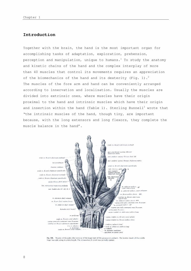
perception and manipulation, unique to humans.1 To study the anatomy
and kinetic chains of the hand and the complex interplay of more
than 40 muscles that control its movements requires an appreciation
of the biomechanics of the hand and its dexterity (Fig. 1).2
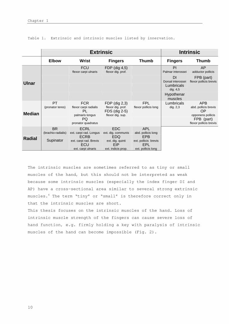
The muscles of the fore arm and hand can be conveniently arranged
according to innervation and localisation. Usually the muscles are
divided into extrinsic ones, where muscles have their origin
proximal to the hand and intrinsic muscles which have their origin
and insertion within the hand (Table 1). Sterling Bunnell3 wrote that
“the intrinsic muscles of the hand, though tiny, are important
because, with the long extensors and long flexors, they complete the
muscle balance in the hand”.
8

General introduction
Figure 1; from: Sobotta atlas van de menselijke anatomie, 2e druk. Houten, Bohn Stafleu Van Loghum, 2000. R. Putz en R. Pabst (red.), (with permission)
9
Chapter 1
Table 1. Extrinsic and intrinsic muscles listed by innervation.
Extrinsic Intrinsic
Elbow Wrist Fingers Thumb Fingers Thumb
FCUflexor carpi ulnaris
FDP (dig 4,5)flexor dig. prof.
PIPalmar interossei
APadductor pollicis
DIDorsal interossei
FPB (part) flexor pollicis brevis
Lumbricalsdig. 4,5
Ulnar
Hypothenarmuscles
PT(pronator teres)
FCRflexor carpi radialis
FDP (dig 2,3)flexor dig. prof.
FPLflexor pollicis long
Lumbricalsdig. 2,3
APBabd. pollicis brevis
PLpalmaris longus
FDS (dig 2-5)flexor dig. sup.
OPopponens pollicisMedian
PQpronator quadratus
FPB (part) flexor pollicis brevis
BR(brachio-radialis)
ECRLext. carpi rad. Longus
EDCext. dig. communis
APLabd. pollicis long
SupinatorECRB
ext. carpi rad. BrevisEDQ
ext. dig. quinti EPB
ext. pollicis brevisRadialECU
ext. carpi ulnarisEIP
ext. indicis prop. EPL
ext. pollicis long
The intrinsic muscles are sometimes referred to as tiny or small
muscles of the hand, but this should not be interpreted as weak
because some intrinsic muscles (especially the index finger DI and
AP) have a cross-sectional area similar to several strong extrinsic
muscles.4 The term “tiny” or “small” is therefore correct only in
that the intrinsic muscles are short.
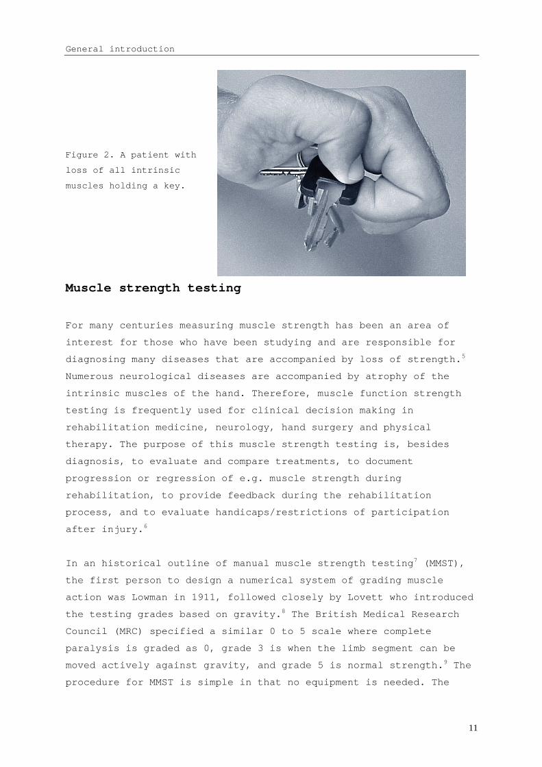
This thesis focuses on the intrinsic muscles of the hand. Loss of
intrinsic muscle strength of the fingers can cause severe loss of
hand function, e.g. firmly holding a key with paralysis of intrinsic
muscles of the hand can become impossible (Fig. 2).
10
General introduction
Figure 2. A patient with
loss of all intrinsic
muscles holding a key.
Muscle strength testing
For many centuries measuring muscle strength has been an area of
interest for those who have been studying and are responsible for
diagnosing many diseases that are accompanied by loss of strength.5
Numerous neurological diseases are accompanied by atrophy of the
intrinsic muscles of the hand. Therefore, muscle function strength
testing is frequently used for clinical decision making in
rehabilitation medicine, neurology, hand surgery and physical
therapy. The purpose of this muscle strength testing is, besides
diagnosis, to evaluate and compare treatments, to document
progression or regression of e.g. muscle strength during
rehabilitation, to provide feedback during the rehabilitation
process, and to evaluate handicaps/restrictions of participation
after injury.6
In an historical outline of manual muscle strength testing7 (MMST),
the first person to design a numerical system of grading muscle
action was Lowman in 1911, followed closely by Lovett who introduced
the testing grades based on gravity.8 The British Medical Research
Council (MRC) specified a similar 0 to 5 scale where complete
paralysis is graded as 0, grade 3 is when the limb segment can be
moved actively against gravity, and grade 5 is normal strength.9 The
procedure for MMST is simple in that no equipment is needed. The
11
Chapter 1
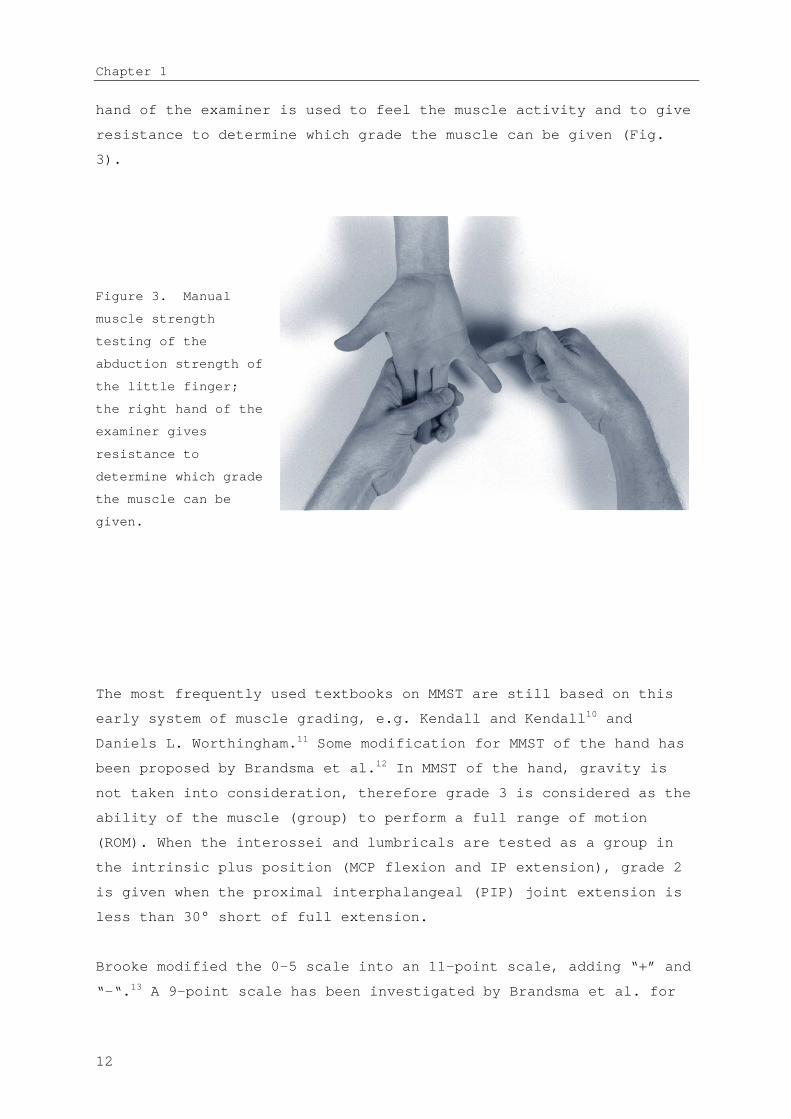
hand of the examiner is used to feel the muscle activity and to give
resistance to determine which grade the muscle can be given (Fig.
3).
Figure 3. Manual
muscle strength
testing of the
abduction strength of
the little finger;
the right hand of the
examiner gives
resistance to
determine which grade
the muscle can be
given.
The most frequently used textbooks on MMST are still based on this
early system of muscle grading, e.g. Kendall and Kendall10 and
Daniels L. Worthingham.11 Some modification for MMST of the hand has
been proposed by Brandsma et al.12 In MMST of the hand, gravity is
not taken into consideration, therefore grade 3 is considered as the
ability of the muscle (group) to perform a full range of motion
(ROM). When the interossei and lumbricals are tested as a group in
the intrinsic plus position (MCP flexion and IP extension), grade 2
is given when the proximal interphalangeal (PIP) joint extension is
less than 30° short of full extension.
Brooke modified the 0-5 scale into an 11-point scale, adding “+” and
“-“.13 A 9-point scale has been investigated by Brandsma et al. for
12

General introduction
reliability in patients with neuritis due to leprosy.14 Strength was
graded on a modified MRC scale with 9 grades: 5, 4+, 4, 3+, 3, 2+,
2, 1 and 0. Overall agreement appeared to be good or very good
(Kappa; 0.61-1.00). However, when data for hands with normal
strength (grade 5) or complete paralysis (grade 0) were excluded
from the analysis, the reliability of the remaining mid-range scale
was not acceptable.
Limitations of MMST
a) Although the textbooks usually present the muscle tests as if
muscles can be tested in isolation, clinicians should be aware
that usually a muscle group is tested rather than just one
muscle. Some have suggested labelling the movement rather than
the muscle, e.g. grading the palmar abduction movement of the
thumb instead of abductor pollicis brevis (APB), because several
muscles are active when testing the palmar abduction of the
thumb. Only a few muscles can be graded in isolation, e.g. flexor
pollicis longus, flexor digitorum profundus and first dorsal
interosseous (1DI).
b) The MRC uses a 6-point numeric scale (grades 0-5) and seems to
indicate a constant distance between points. However, it is an
ordinal scale with disproportional distances between grades; e.g.
grade 4 is not twice as strong as grade 2. It might have been
more appropriate to use terms such as normal, good, fair, trace
and paralysed.
c) Another important comment concerning MMST was made in the
American Society of Handtherapists (ASHT) recommendations,7 that
its most appropriate use is in cases of extreme muscle
deterioration. MMST is not appropriate for higher-level muscle
function due to lack of sensitivity and precision, and should be
used in conjunction with other evaluation tools. We contend that
MMST is most useful for weak muscles with grades of 1, 2 and 3,
but not for the higher grades.
13

Chapter 1
d) MMST is dependent on the examiner’s ability to assess the
pressure as a parameter for strength. Experience of the examiner
is important for reliable measurements.
History of dynamometers for the hand
One of the first dynamometers for measuring hand strength was the
Graham-Desaguliers dynamometer, which was developed in London in
1763. The Regnier dynamometer was invented in Paris in 1798 to
measure the traction properties of artillery horses, but was
designed as an all-purpose instrument to measure specific human
muscle groups as well.8
In the past decades many different dynamometers have been
introduced, e.g. cable tensiometers, sphygmomanometers,
vigorimeters,15 isokinetic dynamometers and strain gauge
dynamometers.
In response to the confusion generated by the many commercial and
experimental grip strength instruments, the California Medical
Association in 1956 evaluated the most commonly used instruments.16
They found the Jamar, first introduced by Bechtol in 1954,17 to be
the most accurate. In 1978 the American Society for Surgery of the
Hand recommended that the second position of the Jamar should be
used and in 1981 the ASHT made additional recommendations, e.g.
concerning posture and verbal instructions during measurements.7 Grip
strength measurements with a dynamometer have become popular and
have been studied extensively. Less studies have been conducted to
investigate pinch strength measurements.
Van der Ploeg et al.18 noticed the shortcoming of the MMST method by
giving an example of strength measurements of the biceps muscle as
an elbow flexor. The biceps needed 5 N of its normal strength (250
N) to overcome gravity; thus grade 3 corresponds with only 2% of the
full strength of the biceps muscle.
14

General introduction
Dynamometers for intrinsic muscle strength
measurements
In his thesis van der Ploeg noted that most dynamometers have a
scale far too crude for measuring forces in very small muscles like
the abductor digiti quinti. However, assessing the strength of these
muscles is of great importance in clinical neurology in the
evaluation of mono- and poly-neuropathies. He noted that there is a
need for an accurate device for these muscles.19
One of the first to develop a dynamometer for the intrinsic muscle
strength was Mannerfelt, who later manufactured a new device called
the Intrins-o-meter.20 In 1997 he reported a study in 48 patients
with ulnar nerve compression.21 Rosen et al. noted that assessing
muscle function using the Intrinsi-o-meter was difficult due to the
extremely small forces, and the instrument was difficult to handle
and read. They suggested using MMST and grip strength measurements
to evaluate nerve function. Interestingly they found a poor recovery
of the intrinsic muscle strength with the Mannerfelt instrument and
good grip strength recovery.22 23
Several others have developed instruments mainly to assess the
abduction of the thumb.24-26 Some needed a specially constructed jig,
e.g. to measure wrist, finger (metacarpo-phalangeal joints) and
thumb extension strength.27
Rotterdam Intrinsic Hand Myometer (RIHM)
In 1995 inventories were made at our department to establish which
clinical evaluation instruments were available to assess the outcome
after peripheral nerve surgery. Three methods were often used to
assess the recovery of muscle strength: MMST and grip and pinch
strength dynamometers.
15

Chapter 1
Having encountered several patients with good grip strength but poor
recovery of the intrinsic muscles strength, we questioned whether
grip strength measurements were appropriate. We acknowledged the
need for a dynamometer to measure the intrinsic muscles in
isolation. Such a dynamometer should be easy to handle, e.g.
portable and with an ergonomical design. It should also have the
possibility to measure the opposition force of the thumb.
Reliability should be good with acceptable measurement error making
it possible to detect reasonably small changes in muscle strength.
In this thesis we present the development of a new dynamometer for
intrinsic muscles, the validation in patients, and its application
in patients with nerve injury.
16
General introduction
Outline of this thesis
Chapter 2 Describes the functional anatomy of the intrinsic muscles
of the hand and the pathology. Possibilities to assess muscle
strength (manual and instrumental) and the various therapeutic
options (prevention of complications, exercises for strengthening)
are discussed.28
Chapter 3 The first dynamometer which was utilised was a generic
strain gauge instrument, the AIKOH. Several intrinsic muscle actions
were measured for reliability.29 The calculated Standard Error of
Measurements (SEM) and the Smallest Detectable Differences (SDD) for
intraobserver and interobserver values indicated that only
relatively large changes in strength could be confidently detected
with this technique.
Chapter 4 Presents the strength measurements of the lumbrical
muscles of the index and middle finger in a group of patients with
ulnar nerve injuries.30 The contribution of the interosseous muscles
in maintaining the intrinsic position compared to the lumbrical
muscles was measured. The MRC scale (0-5) was also compared to the
dynamometry measurements.
Chapter 5 To gain better insight into the measurement error in
strength measurements of the hand in general, we studied the
measurement error for intraexaminer and interexaminer measurements
of the grip and pinch measurements in a consecutive sample of 33
patients with hand injuries. The measurement error was compared
between measurements of the injured and noninjured hands, and
between experienced and inexperienced examiners.31 Literature was
studied concerning other reliability studies of grip and pinch force
measurements. A method to judge and compare SDDs was explored.
Chapter 6 A dynamometer was designed and fabricated with specific
requirements: i.e. improved reliability to measure the muscle force
of the intrinsic muscles, hand-held and portable, possibility to
17
Chapter 1
measure the opposition force of the thumb (Opponens Pollicis
muscle), ergonomically designed handgrip, appropriate visual
feedback of line of pull, and minimal errors from off-axis loading
thus allowing measurement of axial forces only.32 This chapter
describes the technical aspects of the dynamometer design, which was
named the Rotterdam Intrinsic Hand Myometer (RIHM).
Chapter 7 To compare the old and new dynamometers a study was
undertaken to assess the accuracy of the previously used industrial
dynamometer (AIKOH) and the new RIHM in applying the dynamometer
perpendicularly in both planes at the digit. The angle and place of
application was determined with a three-dimensional videorecording
technique. Differences between the two instruments, as well as
measurement error and variance between two sessions were calculated
and compared. The consistency of positioning the instruments at the
same point of the finger was also analysed.33
Chapter 8 The reliability of the RIHM was determined in 27 patients
with ulnar and/or median nerve injury.34 For the two ulnar and two
median nerve innervated movements the ICCs, SEMs and SDDs were
calculated and compared to the AIKOH dynamometer.
Chapter 9 Outcome of muscle strength is presented in 34 patients
with ulnar and median nerve injury. The outcome of muscle strength
with four methods (MMST, grip and pinch strength measurements and
the RIHM ) was compared.35 Correlations between the four measurement
methods were calculated.
Chapter 10 Discussion: some issues related to measurements of the
muscle strength of the hand are discussed in a more comprehensive
perspective, together with some limitations of the work presented
here.
References
18

General introduction
1. Chao EY, An K-N, Conney WP, Linscheid RL. Biomechanics of the Hand: A Basic
Research Study. Singapore: World Scientific Pub Co, 1989.
2. Linscheid RL. Historical perspective of finger joint motion: the hand-me-downs
of our predecessors. The Richard J. Smith Memorial Lecture. J Hand Surg [Am]
2002;27(1):1-25.
3. Bunnell S. Surgery of the Hand. 2nd ed. Philadelphia: J.B. Lippincott Company,
1948.
4. Brand PW. Clinical Mechanics of the Hand. St. Louis: CV Mosby, 1999.
5. Op de Coul AAW. De atrofie van de kleine handspieren. (Thesis). Amsterdam, 1970.
6. Rosen B. The sensational hand. Clinical assessment after nerve repair. (Thesis).
Lund University, 2000.
7. ASHT, editor. Clinical Assessment Recommendations. Garner, NC: American Society
of Hand Therapists, 1981.
8. Lovett RW, Martin EG. Certain aspects of infantile paralysis with a description
of a method of muscle testing. JAMA 1916;66:729-733.
9. Medical Research Council. Special Report series no 282. London: Her Majesty's
Stationary Office, 1954.
10. Kendall FP, McCreary EK. Muscles: Testing and Function. 3rd edition ed.
Baltimore: Williams & Wilkins, 1983.
11. Daniels C, Worhtingham L. Muscle Testing: Techniques of Manual Examination. 4th
ed. Philadelphia: WB Saunders, 1980.
12. Brandsma JW, Schreuders TA, Birke JA, Piefer A, Oostendorp R. Manual muscle
strength testing: intraobserver and interobserver reliabilities for the
intrinsic muscles of the hand. J Hand Ther 1995;8(3):185-90.
13. Brooke MH, Griggs RC, Mendell JR, Fenichel GM, Shumate JB, Pellegrino RJ.
Clinical trial in Duchenne dystrophy. I. The design of the protocol. Muscle
Nerve 1981;4:186-197.
14. Brandsma JW, Van Brakel WH, Anderson AM, Kortendijk AJ, Gurung KS, Sunwar SK.
Intertester reliability of manual muscle strength testing in leprosy
patients. Lepr Rev 1998;69(3):257-66.
15. Merkies IS, Schmitz PI, Samijn JP, Meche FG, Toyka KV, van Doorn PA. Assessing
grip strength in healthy individuals and patients with immune-mediated
polyneuropathies. Muscle Nerve 2000;23(9):1393-1401.
16. Kirkpatrick JE. Evaluation of grip loss; a factor of permanent partial
disability in California. Ind Med Surg 1957;26(6):285-289.
17. Bechtol C. Grips test: the use of a dynamometer with adjustable handle spacing.
J Bone Joint Surg 1954;36:820.
18. van der Ploeg RJ, Oosterhuis HJ, Reuvekamp J. Measuring muscle strength. J
Neurol 1984;231(4):200-3.
19. van der Ploeg RJO. Hand-held dynamometry. (Thesis), 1992.
20. Mannerfelt L. Studies on the hand in ulnar nerve paralysis. A clinical-
experimental investigation in normal and anomalous innervation. Acta Orthop
Scand 1966(87):61-86.
21. Mannerfelt LG. Studies on ulnar nerve compression neuropathies with a new
computerised instrument--the intrins-o-meter. Scand J Plast Reconstr Surg
Hand Surg 1997;31(3):251-60.
19

Chapter 1
22. Rosen B, Lundborg G. A model instrument for the documentation of outcome after
nerve repair. J Hand Surg [Am] 2000;25(3):535-43.
23. Rosen B, Dahlin LB, Lundborg G. Assessment of functional outcome after nerve
repair in a longitudinal cohort. Scand J Plast Reconstr Surg Hand Surg
2000;34(1):71-8.
24. Trumble TE, Kahn U, Vanderhooft E, Bach AW. A technique to quantitate motor
recovery following nerve grafting. J Hand Surg [Am] 1995;20(3):367-72.
25. Boatright JR, Kiebzak GM, O'Neil DM, Peindl RD. Measurement of thumb abduction
strength: normative data and a comparison with grip and pinch strength. J
Hand Surg [Am] 1997;22(5):843-8.
26. Liu F, Carlson L, Watson HK. Quantitative abductor pollicis brevis strength
testing: reliability and normative values. J Hand Surg [Am] 2000;25(4):752-9.
27. Richards RR, Gordon R, Beaton D. Measurement of wrist, metacarpophalangeal
joint, and thumb extension strength in a normal population. J Hand Surg [Am]
1993;18(2):253-61.
28. Schreuders TAR, Brandsma JW, Stam HJ. The intrinsic muscles of the hand;
function, assessment and therapy. In press.
29. Schreuders TAR, Roebroeck ME, van de Kar HJ, Soeters JNM, Hovius SER, Stam HJ.
Strength of the intrinsic muscles of the hand measured with a hand-held
dynamometer: reliability in patients with ulnar and median nerve paralysis. J
Hand Surg [Br] 2000;25(6):560-5.
30. Schreuders TAR, Stam HJ. Strength measurements of the lumbrical muscles. J Hand
Ther 1996;9(4):303-5.
31. Schreuders TAR, Roebroeck ME, Goumans J, van Nieuwenhuijzen JF, Stijnen TH,
Stam HJ. Measurement error in grip and pinch force measurements in patients
with handinjuries. Phys Ther 2003;83(9):806-815.
32. Schreuders TAR, Eijskoot F, den Ouden AH, Stam HJ. The Rotterdam Intrinsic Hand
Myometer (RIHM); A technical note. in press.
33. Schreuders TAR, Hoek van Dijke GA, Guler-Uysal F, Roebroeck ME, Stam HJ.
Analysis of the application of force measurements of the hand: comparison of
two hand-held dynamometers. in press.
34. Schreuders TAR, Roebroeck ME, Jaquet J-B, Hovius SER, Stam HJ. Measuring the
strength of the intrinsic muscles of the hand in patients with ulnar and
median nerve injury; reliability of the Rotterdam Intrinsic Hand Myometer
(RIHM). J Hand Surg (Am) 2004;29A(2):318-324.
35. Schreuders TAR, Roebroeck ME, Jaquet J-B, Hovius SER, Stam HJ. Longterm outcome
of muscle strength in patients with ulnar and median nerve injury: Comparing
manual muscle strength testing, grip and pinch strength dynamometers and a
new intrinsic muscle strength dynamometer. J Rehabil Med 2004;36.
20

CHAPTER 2
The Intrinsic Muscles of the Hand:
Function, Assessment and Therapy principles
The hand is an emblem of strength, skill, sexuality, and sensibility.
When it is damaged, it becomes a symbol of vulnerability of the whole person
Paul W. Brand (1914-2003)
The Intrinsic Muscles of the Hand
Introduction
There have been many valuable studies concerning the anatomy1-3,
mechanics,4-6 and architectural design,7 of the intrinsic muscles of
the hand. Understanding the mechanics of human dexterity requires an
appreciation of the kinetic chains that comprise the hand, and the
intricate interplay of muscles and ligaments that control its
movements.6 In these chains, the intrinsic muscles of the hand (Fig.
1) are of paramount importance for efficient hand function.7
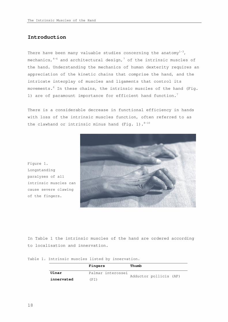
There is a considerable decrease in functional efficiency in hands
with loss of the intrinsic muscles function, often referred to as
the clawhand or intrinsic minus hand (Fig. 1).8-10
Figure 1.
Longstanding
paralyses of all
intrinsic muscles can
cause severe clawing
of the fingers.
In Table 1 the intrinsic muscles of the hand are ordered according
to localisation and innervation.
Table 1. Intrinsic muscles listed by innervation.
Fingers Thumb
Ulnar
innervated
Palmar interossei
(PI)Adductor pollicis (AP)
18

Chapter 2
Dorsal interossei
(DI)
Flexor pollicis brevis
(FPB) part
Lumbricals dig
4,5
Hypothenar
Median
innervated
Lumbricals dig
2,3
Abductor pollicis brevis
(APB)
Opponens pollicis (OP)
Flexor pollicis brevis
(FPB) part
A comprehensive analysis of hand function should include assessment
of the strength and length of the intrinsic muscles. This will
provide important information and assist the assessor in e.g.
determining nerve function, deciding which muscles need to be
strengthened, what splint is needed, what surgery needs to be
considered (tendon transfer), etc.
Although assessment of muscle strength and length are important
elements of hand function other functions, e.g. mobility,
sensibility and central properties of the brain, are equally or more
important for hand function. The latter controls e.g. tonus, co-
ordination and speed of hand movements.
Hand tests to assess the ability of the patient to perform certain
tasks have been developed by e.g. Moberg,11 Bendz,12 Sollerman et
al,13 and Light et al.14 Most of such tests record how long it takes
to finish a particular task. Clinicians often see that patients with
impairments of the hand have quickly learned compensatory mechanisms
to compensate for the lost functions. Therefore, tests at this
activity or skills level may only assess the ability of the patient
to compensate for lost function.
While there is little consensus about classifications of prehensile
patterns of hand function15-18, the general characteristic remains
largely consistent with the following categories: three pinch grips
(tip pinch, lateral or key pinch, and tripod or chuck pinch) and
three modes of gripping: (power grip, spherical or flexion grip, and
19

The Intrinsic Muscles of the Hand
extension grip in intrinsic plus position).14 It is estimated that
for a full range of natural common grips, a spherical grip is
required for 10%, a tripod grip for 10%, a power grip for 25%, a
lateral grip for 20%, a tip grip for 20%, and an extension grip for
10% of tasks of activities of daily living. Without intrinsic
muscles a power grip is somewhat weaker but still possible, but for
all other grips the intrinsic muscles play an important role.
Long-term loss of intrinsic muscle function may result in
irreversible joint contractures. An appropriate therapy plan (e.g.
which exercises are needed, what splint may enhance hand function
and prevent contractures) is needed.
The aim of this paper is fourfold:
1. to review the functional anatomy of the intrinsic muscles of
the hand.
2. to discuss the pathokinesiology of the hand with intrinsic
muscle paralysis, and its consequences for Activities for Daily
Living (ADL)/dexterity and muscle shortening.
3. to present possibilities for the assessment of muscle strength
(manual and instrumental).
4. to discuss therapy principles (prevention of complications,
exercises for strengthening).
All intrinsic muscles will be discussed separately, except for the
hypothenar muscles which are discussed as a group.
1. Intrinsic muscles of the fingers
20

Chapter 2
In general, every finger has six muscles controlling all the
movements of the fingers: three extrinsic tendons (two long flexors
and one long extensor) and three intrinsic muscles (dorsal and
palmar interosseous and lumbrical muscles). (Fig 2)
Figure 2 from R. Putz en R. Pabst (red.), Sobotta atlas van de menselijke anatomie, 2e druk. Houten, Bohn Stafleu Van Loghum, 2000 (with permission)
1.1. Dorsal and palmar interossei
1.1.1 Functional anatomy
Literature usually describes four dorsal (DI) and three palmar (PI)
interosseous muscles. Stack et al.19 suggested that it might be more
correct to divide the interossei into the proximal and distal, as it
is their insertion rather than origin, which dictates their action.
Most dorsal interossei muscles have a more proximal attachment while
21
The Intrinsic Muscles of the Hand
the palmar interossei have a more distal attachment similar to the
lumbricals.19 According to Zancolli20 there are three types of
insertions of the interossei:
Type I most proximal, attached to:
a. tubercle of proximal phalanx,
b. transverse and oblique fibers of extensor
apparatus
c. the volar plate.
Type II is like type 1 except that there is no attachment to the
bone (a.)
And part of the insertion is into:
d. the lateral band
Type III has all four attachments a, b, c and d.
The first dorsal interosseous (1 DI) insertion is of type I, all
other DI are of type III. All PI have the type II insertion, but
variations are possible.1 3 Because of this insertion the strongest
activity of the 1 DI is in key pinch when the thumb is pressed
against the mid-phalanx of the index finger. The 1 DI is also active
in tip pinch, when the tip of the thumb is pressed against the tip
of the index finger, but then the main action is as a flexor at the
metacarpo-phalangeal (MCP) joint. The first palmar interosseous (1
PI) muscle is more active in tip pinch activities.
In Type II, the insertion into the lateral band of the extensor
apparatus (d.) is responsible for an important extension force at
the proximal interphalangeal (PIP) joints. The first palmar
interosseous also produces some supination of the index finger to
get good approximation of the pulps. In this respect we might
consider the 1 PI as an “opponens indices” muscle in tip pinch
activities.
Without interossei the finger is unstable and will collapse into the
“claw” (i.e. intrinsic minus) position of flexed IP joints and
(hyper-) extension of the MCP joint: therefore the interosseous are
sometimes referred to as the “anti-claw” muscles.9 The primary
function of the interossei is MCP flexion/stabilisation with
22
Chapter 2
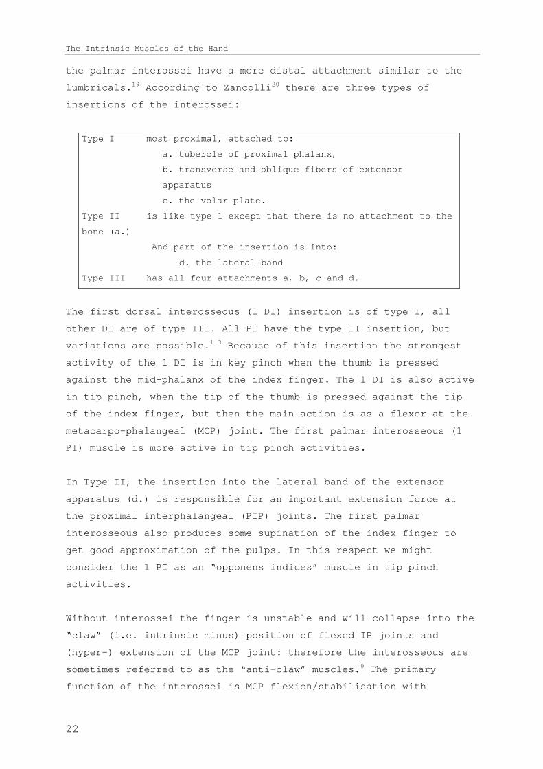
extension of the interphalangeal (IP) joints. This is especially
evident during pinch in which the collapse of the index PIP joint is
apparent in, often > 90°, flexion. This is a sign of interosseous
muscle weakness and sometimes referred to as the Mannerfelt sign.21
(Fig. 3).
Fgure 3.
Mannerfelt
and Froment
sign on left
hand of
patient with
ulnar nerve
paralysis.
Recording the moments of the intrinsic muscles that were generated
after electrical stimulation Lauer et al.22 found that the dorsal
interossei muscles were strong abductors of the fingers and
generated a significant moment in MCP joint flexion and IP joint
extension. Similarly, Ketchum et al.8 found that the interossei
muscles of the index finger contribute 73% to the overall moment for
flexion of the MCP joint.8 Li et al.23 investigated the role of the
intrinsic finger flexor muscles during finger flexion tasks. When an
external force is applied proximally to the PIP joint, the extensor
mechanism (intrinsic muscle group) is the largest component (70%) on
force production of all flexors. Thus the interossei muscles are
important flexors of the MCP joint together with the long flexors:
flexor digitorum profundus (FDP) and flexor digitorum superficiales
(FDS). However, at the PIP joint level the long flexors, primarily
the FDS, and the interossei are antagonists.
23

The Intrinsic Muscles of the Hand
When the PIP joint is flexed, some reduction of the extension moment
of the interossei takes place, due to the volar displacement of the
lateral bands (of about 4-mm) at the PIP joint level. At full
flexion of both the PIP and distal interphalangeal (DIP) joints, the
lateral bands approach the flexion-extension axis of the PIP joint,
thereby minimising the extensor moment.6
The action of the interossei muscles can be studied separately from
the extensor digitorum communis (EDC) muscles in patients with a
radial nerve paralysis. If the patient is asked to extend the
fingers, no extension of the MCP will occur, but the finger IP
joints will extend because of the ulnar and median nerve innervated
interosseous and lumbrical muscles. However, in some patients the
fingers, especially the index, can be extended in the MCP joint to
some degree, especially with the wrist flexed; this is probably
because the angle of attachment of the dorsal interossei muscles is
only 0°-5°. When the tension on the extensor tendon is increased due
to the flexed wrist, this angle will go beyond the 0° and thus the
interosseous muscle becomes an extensor of the MCP joint. The angle
of approach to the extensor mechanism for the palmar interossei
muscles is 20°-25° and for the lumbrical muscles 35°.1
The index finger is deprived of all long flexors and the lumbrical
muscles in patients with a high median nerve paralysis. The only
active muscles are the long extensors and the two interossei. In an
attempt to flex the fingers the index finger will remain extended,
and is accurately described as the “pointing finger” where the IP
joints are fully extended and the MCP joint flexed. In high median
nerve paralysis the long finger is also deprived of the long flexor,
but will usually flex in grip. This is due to the attachments
between the FDP tendons of the long finger and the ring finger and
is called the Quadriga phenomena.24 25 This term suggests a
symmetrical organisation of the four tendons; however, the
connections between the FDP of the index and middle finger are
usually absent, and a strong connection between the flexor pollicis
longus (FPL) and the FDP of the index finger can exist.
24

Chapter 2
1.1.2 Pathokinesiology (paralyses, consequences for prehension/ADL,
shortening)
The deficiency mentioned in textbooks due to loss of interossei is
usually that of controlled abduction and adduction of the fingers.
The loss of this action is important for those who play musical
instruments or operate keyboards, but in most patients this is not
recognised as a severe deficit. The loss of the interossei function
of MCP flexion and PIP extension is a much more significant loss. In
acute paralysis of the interossei muscles it will sometimes only be
visible as a mild hyperextension at the MCP joints and slight flexed
position of the PIP joints when the patient is asked to extend the
fingers. However, this deformity usually progresses depending on the
natural joint laxity of the hands, i.e. long, thin and hypermobile
fingers will develop a so-called claw hand much quicker than thick,
stiff fingers. In the mobile hands the volar ligamentous structures
stretch more easily, causing increased hyperextension and
subsequently less PIP extension. Extension of the PIP joints is only
possible by contraction of the EDC when the MCPs are 'blocked',
either actively (internally) through muscle contraction or passively
(externally) through the examiners hand or a splint, preventing
hyperextension of the MCP joints. In the latter situation this is
called assisted extension, which is also used as a test to assess
the integrity of the extensor apparatus.
Other factors that may contribute to the development of a claw hand
are: hand dominance, continued use of the hand (or the lack of),
compliance of the patient to perform the routine exercises to
prevent joint stiffness, and use of (night) splints.
Without interosseous muscles the hand can still make a full fist,
but the pattern of movement is changed (the MCP flexion takes place
later than usual). The contribution of the interossei muscles in
grip strength measurements will be discussed later.
In patients with a “high” ulnar lesion, the FDP muscles of the 4th
and 5th fingers are paralysed and less clawing is often visible
25

The Intrinsic Muscles of the Hand
because the flexion moment at the PIP joints is decreased compared
to a “low” ulnar palsy. We might conclude from this observation that
clawing is also the result of visco-elastic tension of FDP. When the
ulnar nerve recovers, the 4th and 5th finger will show an increasing
clawing. Maintaining the length of the FDPs, i.e. preventing flexor
tightness, therefore also helps to prevent clawing.
Another sign of interossei muscle weakness was first described by
Andre-Thomas in 1917 and is called the Thomas sign.26 (p. 518) (Fig. 4).
This is the tendency (compensation) of a patient with weak
interossei muscles of the fingers to automatically flex the wrist in
an attempt to gain a better opening of the hand, i.e. MCP extension,
by means of increasing the pull on the EDC. This in fact increases
the hyperextension of the MCP joints and adds to the progress of the
development of the claw hand. This “trick” movement is adopted very
quickly and even when the interossei have regained their strength,
will often take a long time to “un-learn”.
Figure 3. Thomas sign:
compensation movement when
interossei muscles are weak;
flexion of the wrist in an
attempt to gain a better
opening of the hand, i.e. by
means of increasing the pull
on the EDC.
In longstanding paralyses of the interossei, e.g. when the ulnar
nerve could not be repaired or did not recover, the PIP joints are
continuously in a flexed position. This will cause a gradual
stretching of the dorsal expansion (sometimes called the tri-angular
ligament) over the PIP joint, which secures the lateral bands of the
26
Chapter 2
extensor tendon in their dorsal position. In the normal finger, the
lateral bands shift dorsally and towards the central position of the
finger when the PIP joint is extended. Whereas when flexing the PIP
joint the dorsal expansion needs to allow the lateral bands to move
volarly towards the flexion-extension axis of movement. When this
dorsal expansion is elongated, the lateral bands are too much
volarly, resulting in a loss of PIP joint extension. Consequently,
the oblique retinacular ligament (ORL) or Landsmeers ligament is
slack most of the time and will adjust to this new situation by
shortening and this may result in hyperextension of the DIP joint.
This is a similar progression of changes as in an extensor tendon
central slip injury, causing a Boutonnière deformity.27
Another impairment in longstanding paralyses of the interossei
muscles is related to hyperextension of the MCP joint. This causes
an upward pull of the EDC tendons (bow stringing), stretching the
sagittal bands. The laxity of the sagittal bands will result in an
inability to maintain the EDC tendon on top of the MCP joint. The
drop of the luxating tendon into the groove between the MCPs is
especially observable when flexing the MCP joint. This is sometimes
called “guttering” because the tendon drops into the “gutter”
between the MCP joints.
The longstanding flexed position of the IP joints will result in a
physiological shortening of the long flexors. This flexor tightness
will increase the PIP flexion position and can cause a deterioration
of the PIP flexion contractures. This is an additional argument to
maintain the length of the extrinsic flexors in patients with
intrinsic muscle weakness.
Shortening of the interossei muscles is called intrinsic tightness
(IT), which can be caused by a trauma of the hand which can
precipitate a cascade of events. The interossei are situated in
rather tight compartments, therefore oedema/swelling will cause an
increase of pressure in these compartments. As a result blood
circulation will be hampered causing anoxia and muscle fiber death,
which results in fibrosis of the muscle and shortening. This is
27

The Intrinsic Muscles of the Hand
identical to the process which causes Volkmann's ischemic
contracture in the forearm.28
The IT test consists of two parts. First, passive PIP flexion is
tested with the MCP joint extended and, secondly, passive PIP
flexion is tested again but now with the MCP joint flexed. If there
is a large difference in PIP flexion between the two MCP positions,
intrinsic tightness is present. The long-term complications of IT
can result in decreased MCP extension and a swan neck finger, i.e.
hyperextension of the PIP joint. A longstanding swan neck deformity
might result in a painful snapping of the lateral bands at PIP level
when the finger is flexed.
In rheumatoid arthritis a different process can also lead to IT. The
role of the intrinsic muscles in producing MCP subluxation in the
rheumatoid hand has been documented.29 30
Another intricacy sometimes observed is what we call interosseous
plus, which is a paradoxal extension: the harder the patient tries
to bend the finger, the more the finger will extend in the PIP
joint. This phenomena is sometimes seen in patients in which the
interossei have been the only flexor of the finger for some time
e.g. in high median nerve palsy, or in case of adhered flexor
tendons. Although the flexors are active and can bend the finger,
when a stronger grip is required the finger will extend.
The explanation is that in a non-resistant grip of a normal
functioning hand there is only minor activity of the interossei
muscles. However, in a strong grip, the stronger the flexors are
pulling, the more the intrinsic muscles become active to stabilise
the PIP joints and prevent luxation. If the long flexor is weak or
poorly activated, the interossei will overpower the flexor causing
PIP extension. This paradoxal extension appears to be similar to the
lumbrical plus phenomena (see lumbricals) and might be called
interosseous plus. The patient has to be taught to, gently, contract
the long flexors without the action of the interossei muscles.
28

Chapter 2
1.1.3 Assessment possibilities (manual and instrumental)
Although the interosseous muscles have short fiber length, some are
strong and have physiological cross-sectional areas comparable to
the FDS muscles.27 In standard textbooks on muscle testing, the tests
suggested are usually: abduction for dorsal and adduction for palmar
interossei muscles.31 32 These tests are useful for isolated
(specific) testing of the interossei. For example, patients with an
ulnar nerve paralysis can not move their middle finger sideways,
which has been called the Egawa sign.26 Functionally, it is much more
meaningful to test the interossei muscles in the intrinsic plus
position, by giving resistance to flexion of the MCP joints and
extension of the PIP joint.33 34 (Fig. 5).
Figure 5. Testing the strength of the
intrinsic muscles in the fingers.
Some have suggested that dynamometry of the interossei muscles is
possible, indirectly, by measuring the grip strength of the hand
with e.g. a Jamar dynamometer. Janda et al. advocated to use the
smallest handle position of the Jamar because the intrinsic muscles
were most active in that position.35 Kozin et al.10 tested 21 healthy
persons who underwent median and ulnar nerve blocks at the wrist
29
The Intrinsic Muscles of the Hand
level; the average decrease in grip strength was 38% after ulnar
nerve block.
Pinch data in the study by Kozin et al. revealed a significant
decrease in key pinch of 77% after ulnar block and 60% after median
block.10 For evaluating and monitoring the motor function of the
ulnar nerve, pinch strength measurements seem more meaningful than
grip strength measurements.
Specific measurements of the first dorsal interosseous muscle can be
done with dynamometers such as the RIHM,36 the Intrins-o-meter of
Mannerfelt,37 or the Preston pinch gauge device.38
1.1.4 Therapy principles (prevention complications, strengthening,
ADL)
Prevention of contractures is directed at the PIP joints. A (night)
splint with the MCP joints in flexion and IPs in extension is
advisable. These splints can help to prevent PIP flexion
contractures and are especially important in longstanding problems.
Increased hyperextension of the MCP joints, due to stretching of the
volar plate, is also prevented in this position.
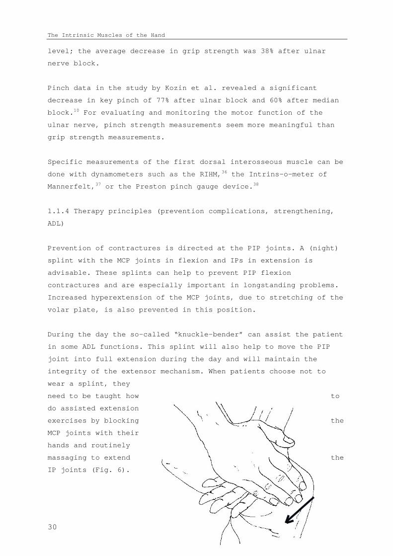
During the day the so-called “knuckle-bender” can assist the patient
in some ADL functions. This splint will also help to move the PIP
joint into full extension during the day and will maintain the
integrity of the extensor mechanism. When patients choose not to
wear a splint, they
need to be taught how to
do assisted extension
exercises by blocking the
MCP joints with their
hands and routinely
massaging to extend the
IP joints (Fig. 6).
30

Chapter 2
Figure 6. Prevention of IP flexion contractures by massaging the fingers.
Exercises to strengthen the interossei muscles are all aimed at
movements which flex the MCP and extend the IP joints. Therefore,
exercises for the interossei muscles are all activities in intrinsic
plus position: e.g. grasping a book, plate or a cylindrical object
like a large bottle. Specific training of the first dorsal and
palmar interossei are activities for which key and tip pinch
activities are required.
To correct the long flexor tightness, the patient is taught to
stretch the flexors by holding the hand flat on e.g. a table and by
moving the forearm towards an angle perpendicular to the table. In a
similar fashion, the hand can be placed on the seat of the chair
while the patient sits on the hand and pulls the forearm towards the
body. A night splint with the fingers and hand in extension might be
necessary in severe cases.
1.2 Lumbricals
1.2.1 Functional anatomy
The lumbrical muscles are unique muscles in several aspects. They
connect two extrinsic antagonistic muscles. Proximally the
31

The Intrinsic Muscles of the Hand
lumbricals are attached to the FDP and distally they are inserted
into the lateral band of the extensor tendon. The third and fourth
lumbricals also connect, by their bi-penal origin, two adjacent FDP
tendons.
The function of the lumbrical muscles is much debated and some even
considered these muscles to be redundant. Brand suggested that the
lumbrical muscles are not relevant for MCP flexion. He explained
this with an illustration of a father carrying a child; it does not
matter what the child (i.e. lumbrical) is carrying, the father (i.e.
FDP) has to carry it anyway.27 Therefore, the lumbrical muscles have
a unique ability to contract without adding flexion torque at the
MCP joint, in contrast with the interosseous muscles which, when
extending the IPs, need a stronger contraction of the EDC to
counteract the flexion moment at the MCP joint level. The lumbricals
provide a more efficient source for IP extension than the
interossei. Any contraction of the lumbrical muscle for IP extension
simultaneously, reduces the visco-elastic force of the FDP tending
to flex the IP joints. Accordingly the lumbrical can be regarded as
a deflexor of the PIP joint.39 Its direct contribution to MCP flexion
is small and in the flexed finger may be non-existent, but its
indirect contribution to IP joint extension by decreasing the
flexion torque is quite substantial.40
With the smallest physiological cross-sectional area, it is
certainly not a strong muscle. The lumbricals have a very long fiber
length (40-48 mm) which indicates that they are designed for long
excursions. If the lumbrical fiber length was short, FDP excursion
could stretch the lumbrical sarcomeres to a point that they were
unable to generate active force.7
The lumbrical muscles are richly endowed with muscle spindles, their
passive stretch by contraction of the FDP might both inhibit finger
extensors and facilitate wrist extensors.40-43 For this reason the
lumbrical muscles have been called “tensiometers” between long
flexors and extensors.44 Leijnse and Kalker25 concluded that the
32

Chapter 2
lumbricals are in an optimal position for proprioceptic feedback
concerning the PIP-DIP joint mechanism.
These unique properties of the lumbricals indicate that they are
probably important in fast, alternating movements, e.g. in typing
and playing musical instruments.45
1.2.2 Pathokinesiology (paralyses, consequences for prehension/ADL,
shortening)
In low median nerve injuries the lumbrical muscles of the index and
middle finger are paralysed. In these hands it is difficult to
discover any problems in the motion of these fingers.46 A mildly
diminished extension of the DIP joint has been noticed in a few
patients, which might be explained by the decreased extension force
on the extensor apparatus.
The “lumbrical plus” is a situation in which there is a FDP tendon
rupture distal of the lumbrical origin, or in the situation where a
too long graft has been used. The FDP now pulls through the
lumbrical muscle rather than through its tendon, causing PIP
extension.47
1.2.3 Assessment possibilities (manual and instrumental)
Manual muscle strength testing (MMST) of the lumbricals is
practically impossible because of the synergistic action of the
interossei muscles. Strength testing is most likely less relevant
than evaluating the co-ordination and dexterity in e.g. a tapping
test when the lumbrical is paralysed and normal function. However,
no studies have been found in which this has been utilised.
The strength can be measured in isolation of the interossei muscles
in patients with an ulnar nerve lesion where the index and long
fingers have only the lumbrical to maintain the intrinsic plus
position. One study measured a mean MCP joint flexion strength in
33

The Intrinsic Muscles of the Hand
the index and long finger of 0.8 kg (range 0.3-1.5) compared with
6.4 kg (range 4.6-7.9) in the non-involved hand. Thus, the affected
fingers have only about 12% of the strength of those of the non-
involved hand.46
1.2.4. Therapy principles (prevention complications, strengthening,
ADL)
No specific training/splinting program for lumbrical paralysis has
been advocated. Strength training is similar to interossei muscle
training, with perhaps more focus on speed and co-ordination.
34

Chapter 2
2. Thenar muscles
The median nerve innervates the intrinsic thumb muscles that make
the hand a “human” hand. These muscles oppose the thumb to the
fingers: abductor pollicis brevis (APB), opponens pollicis (OP) and
the flexor pollicis brevis (FPB). All these muscles originate from
the flexor retinaculum and the trapezium carpal bone. Located in the
thumb web the adductor pollicis (AP) is sometimes also considered a
thenar muscle and will be discussed separately.
2.1 Abductor Pollicis Brevis (APB)
2.1.1 Functional anatomy
The insertion of the APB is at the radial side of the proximal
phalanx of the thumb. Even though the APB is the smallest intrinsic
thenar muscle, atrophy is quickly noticed as it is the most
superficial muscle. The main function of the APB is moving the thumb
away from the palm, in a perpendicular direction to the palm of the
hand, when grasping objects. This is usually called palmar abduction
of thumb, but in more recent terminology anteposition of the thumb.48
The muscle is relatively weak as the action of abduction of the
thumb is not one that is usually done against resistance; the APB
“only” positions the thumb for action. This movement in the carpo-
metacarpophalangeal (CMC) joint of the thumb is a synergistic action
with the OP and, especially, the FPB.49
An extrinsic synergist of the APB is the abductor pollicis longus
(APL). The pull of the APL on the CMC joint of the thumb causes
palmar abduction, especially when the wrist is in flexion. Flexion
of the wrist increases the moment arm of the APL due to bow
stringing.27
Since the APB has a dual insertion into the base of the proximal
phalanx and into the extensor tendon expansion, it also has an
35

The Intrinsic Muscles of the Hand
extension moment on the IP joint. After loss of the extensor
pollicis longus (EPL) the APB together with the oblique head of the
AP may provide complete extension of the IP joint of the thumb. This
is clinically important when evaluating extension of the thumb and
the EPL function.50
36

Chapter 2
2.1.2. Pathokinesiology (paralyses, consequences for
prehension/ADL, shortening)
Loss of the APB sometimes has little effect because the ulnar nerve
innervated part of the FPB, the superficial head, can often move the
thumb in palmar abduction by itself.51 Frykman et al.52 estimated that
50% of patients with a median nerve lesion would have satisfactory
thumb opposition if no median nerve re-innervation occurred.
When the thumb can not be palmar abducted (anteposition) there will
be problems with manipulating small objects and tip-tip pinch
activities, but also in positioning the thumb to grasp larger
objects. Rosen53 found in 15 median nerve injured patients that of
the 20 tasks of the Sollerman test, only a few tasks were
particularly difficult: picking up coins from a purse, picking up
nuts, putting on bolts, and fastening of buttons.
2.1.3 Assessment possibilities (manual and instrumental)
Because it is one of the few intrinsic muscles innervated by the
median nerve, it is an important muscle in clinical practice e.g. to
evaluate motor function impairment in median nerve compression in
carpal tunnel syndrome. Unfortunately manual muscle strength testing
in isolation is often hindered by the many synergists. Especially in
the thumb, most movements are the product of multiple synergists.11 30
54
When testing the palmar
abduction of the thumb, i.e. APB
muscle strength, APL
substitution can be
diminished to a certain level by
holding the wrist in
extension. For strength testing,
pressure is applied
perpendicular to the palm of the
37

The Intrinsic Muscles of the Hand
hand at the MCP joint of the thumb33 (Fig. 7).
Figure 7. Strength test of the
abductor pollicis brevis (APB).
Several dynamometers that specifically measure the abduction
strength of the thumb have been developed.55-58 If these are not
available the strength of the APB can be evaluated indirectly with a
pinch dynamometer. We found a strong correlation between the pinch
strength and the strength of the APB.59 Weakness of the APB can cause
a diminished pinch strength, mainly because the strong thumb muscles
cannot be put into action to exert their full strength when the APB
is not able to position the thumb.36
2.1.4 Therapy principles (prevention of complications, exercises for
strengthening)
If the abduction of the thumb is weak (MRC < grade 3), there is a
danger for thumb web (adduction) contractures, especially when ADL
activities and/or work do not require opposition/abduction of the
thumb. A night splint with thumb in maximum abduction is usually
advised together with instructions how to maintain the mobility of
the thumb web by
pushing the thumb
metacarpal away from
the index, taking care
not to push distal of
the MCP joint (Fig. 8).
38

Chapter 2
Figure 8. Prevention of thumb web contractures by pulling on the thumb
proximal to the MCP joint of the thumb.
To assist in ADL several aids have been suggested: a weight to pull
the thumb away from the index when the arm is in supination has been
suggested by Wynn Parry.60 An elastic sling, pulling the thumb into
palmar abduction and a static or semi-rigid (neoprene) splint, which
maintains the thumb web in a wide position can be tried out. These
splints are usually not well accepted by the patient, due to the
restrictions they cause and the cosmetic aspects.
Strengthening exercises for weak APB (< MRC 3) are to move the thumb
towards the tip of index finger, including grasping large objects.
Trick movements, e.g. of wrist flexion to activate the APL, are
often difficult to un-learn. When MRC 3 is reached, all kinds of
pinch exercises are useful besides handling large and small objects.
2.2 Opponens Pollicis (OP)
2.2.1 Functional anatomy
The OP originates from the trapezium and flexor retinaculum and,
spiralling around the thumb, inserts into the radial border of the
metacarpal bone of the thumb. The main function is to rotate the
thumb and position the thumb towards the fingers, which is sometimes
referred to as pronation. Moving the thumb towards the index or
middle finger in an effort to pick up a coin from a smooth table,
using the nails of both the thumb and finger and the need for the
rotation of the thumb can be observed.
39

The Intrinsic Muscles of the Hand
Another important function of the OP is its ability to stabilise the
CMC joint with regard to the torsion moment in pinch movements.27
The moment arm of the ABP for abduction is increased by the OP
because the OP pushes the APB up during its contraction.27
2.2.2 Pathokinesiology (paralyses, consequences for ADL /
prehension)
OP muscle loss is often combined with other intrinsic thumb muscles,
loss in isolation is very rare. Consequences for ADL are similar to
APB loss.
2.2.3 Assessment possibilities (manual and instrumental)
In MMST the pressure is applied parallel to the palm of the hand at
the CMC joint of the thumb, while no IP flexion of the thumb is
allowed.33
Not a strength testing method but useful to evaluate the ability to
position the thumb, the Kapandji 0-10 opposition thumb test is one
in which the thumb is moved from the proximal phalanx of the index
(“1”), to the tip of the fingers (“3, 4, 5 and 6”) and flexed along
the little finger towards the distal palmar crease (“10”).61 In a
similar approach the opposition can be tested by having the patient
touch the tip of the thumb to the tip of the little finger. At the
end of the opposition, the thumbnails of the little finger and
thumb, i.e. the distal phalanges, should be in one line.
Assessment of the OP strength with pinch meters is only indirectly
possible. The RIHM is the only dynamometer which can test the OP
more or less in isolation.36
2.2.4 Therapy principles (prevention of complications, exercises for
strengthening)
40

Chapter 2
The OP can be exercised by moving from the tip of the index finger
towards the little finger, focusing on the pronation action of the
thumb. Manipulating round objects, e.g. rolling a marble from the
tip of the index finger towards the middle finger, can also be a
helpful exercise. Rotation/supination of the thumb can also be
trained with, e.g. for the right hand, unscrewing a nut and bolt.
2.3 Flexor Pollicis Brevis (FPB)
2.3.1 Functional anatomy
Similar to the AP, the FPB has two heads, but both have different
innervation: the superficial part is usually median innervated and
the deep head is ulnar innervated. The origin for the FPB is the
flexor retinaculum and the trapezium, respectively. Comparable to
the APB, the FPB inserts into the extensor tendon and the lateral
sesamoid bone, and assists in extension of the IP joint of the
thumb.
The proximal fibers of the FPB are continuous with the OP,
therefore, both act on the CMC joint of the thumb and flex the
metacarpal. The major effect of the FPB is in sequence with the AP,
in that both flex the MCP of the thumb, although the FPB pronates
and the AP supinates the thumb.27
2.3.2 Pathokinesiology (paralyses, consequences for ADL/ prehension)
Isolated weakness of the FPB is difficult to assess, not only
because of the variations in innervation, but mainly because of all
the synergists in flexion of the MCP joint and sometimes the lack of
mobility in the MCP joint of the thumb. Isolated loss of the FPB
might go unnoticed, but loss in combination with loss of the AP,
e.g. in ulnar nerve palsy, will cause significant loss of pinch
strength (see AP).
41
The Intrinsic Muscles of the Hand
Positioning of the thumb in pinch activities is a median nerve
muscle function, but the strength of the pinch grip is derived from
the ulnar nerve innervated muscles.
2.3.3. Assessment possibilities (manual and instrumental)
In MMST the strength of the FPB is evaluated by the assessment of
flexion at the MCP joint of the thumb without flexion of the IP
joint, which is the FPL action. A strength test aiming to diminish
the FPL action has been studied but without convincing results.62
Measurements of the strength of the FPB with pinch dynamometers in
isolation is not possible. In the dynamometry of the pinch grip, the
FPB together with the AP contribute significantly to pinch strength.
2.3.4. Therapy principles (prevention of complications, exercises
for strengthening)
All pinch activities can be exercised, in which the tendency to
(hyper-) extend the MCP joint is a sign of improper activation of
the FPB and AP and needs to be corrected. A pinch whereby the MCP
and IP joint is slightly flexed is also advantageous regarding the
optimum (mid) position of the sarcomers of the intrinsic muscles of
the thumb and for least tension on the soft tissues (ligaments,
volar plate) of the thumb joints.
2.4 Adductor Pollicis (AP)
2.4.1 Functional anatomy
The ulnar innervated AP is a fanshaped muscle with two heads: an
oblique part with its origin at the 2nd and 3rd metacarpals and the
transverse part with its origin at the anterior surface of the 3rd
metacarpal. The insertion of both heads is into proximal phalanx of
the thumb and the sesamoid bone.
42

Chapter 2
It is the most volar muscle in the thumb web, making atrophy visible
in the palm of the hand. Of all the intrinsic muscles working on the
thumb, the AP, working together with the FPB, has the largest
flexion moment arm at the CMC joint. Therefore, the AP, together
with the 1 DI, are the most important pinching muscle of the thumb,
while other thumb muscles are just positioners and synergists.27 p 229
The synergists for adduction of the thumb are EPL, FPL and the first
dorsal interosseus.63
2.4.2 Pathokinesiology (paralyses, consequences for prehension/ADL,
shortening)
Direct lesions of the AP sometimes occur after injuries into the
thumb web, e.g. knife wounds. Paralyses of the AP usually occur
after ulnar nerve lesion and therefore there is also weakness of the
other important muscle for pinch; the 1 DI.
In ulnar palsy there may be enough median nerve innervated FPB to
position the thumb for pinch, but when power is needed, the
diminished AP strength usually results in hyperflexion of the IP
joint of the thumb (Froment’s sign) and sometimes in hyperextension
of the MCP joint (Jeanne’s sign).27 p 54
A patient who is bedridden for a prolonged time with little activity
of the hand (coma etc.) can develop a thumb web contracture due to
shortening of the AP. After a trauma of the hand, similar to the IT,
shortening of the AP can take place. The AP rests in a compartment
and therefore muscle tightness can cause severe thumb web
contractures. Spasticity, causing a strong pull of the thumb into
the palm can be seen in patients with cerebral palsy and who have
suffered a stroke. The CMC joint is adducted, making it difficult to
grasp or hold but also to release larger objects. In a surgical
43

The Intrinsic Muscles of the Hand
procedure releasing a considerable part of the AP is often
necessary.
2.4.3 Assessment possibilities (manual and instrumental)
Tests are often described in which plain adduction of the thumb is
tested. Due to the many synergists, this usually does not provide
any useful information. Weakness or paralyses will result in loss of
pinch strength and show the so-called Froment’s sign. (Fig. 2). This
was first described by Jules Froment, who was watching a train
commuter reading his newspaper with one thumb flexed and the other
straight.64
In examining the pinch strength of both hands, e.g. pulling on a
piece of paper, the IP joint angle is observed: if more flexion
occurs at the involved hand, the Froment sign is positive. When the
MCP joint of the thumb has some laxity (hypermobile) into extension,
hyperextension of the MCP joint of the thumb will take place in
pinch, which is called Jeanne’s sign.21 Both are signs of a reduced
flexion moment at the MCP joint of the thumb, i.e. weak or paralysed
AP. Grading is not possible, besides a classification of a positive
or negative sign.
Instrumental assessment of the strength of the AP can be done,
indirectly, with pinch dynamometers.
2.4.4 Therapy principles (prevention of complications, exercises for
strengthening)
Muscle strengthening exercises are all movements/activities where a
pinch is needed, especially the key pinch or lateral pinch. If
possible, pinch strength is trained with the thumb IP and MCP joints
in slight flexion.
3. Hypothenar muscles
44

Chapter 2
The muscles of the hypothenar are from ulnar to radial: abductor
digiti minimi (ADM), flexor digiti minimi (brevis) (FDM), opponens
digiti minimi (ODM). The palmaris brevis (PB) is the most
superficial muscle overlying these muscles transversely and
originates from the aponeurosis palmaris. All the hypothenar muscles
are ulnar innervated. Isolated paralysis is rare and because there
are functionally only minor differences between these muscles, they
are discussed here as a group.
3.1 Functional anatomy
The most ulnar situated muscles (especially ADM) have the strongest
ulnar abduction action of the little finger. The ODM, attached to
the 5th metacarpal bone, has an important role in the opposition of
4th and 5th rays. All the hypothenar muscles are active in the
intrinsic plus position of the fingers, except for the ODM and the
PB. In this position, similarly to the interossei of the fingers,
they flex the MCP joint of the little finger and extend the IP
joints.
3.2 Pathokinesiology (paralyses, consequences for prehension/ADL,
shortening)
The 4th and 5th metacarpals are much more mobile in the CMC joints as
compared to the 2nd and 3rd. This makes it possible to adjust the hand
around a round object, but also e.g. the handle of a hammer.
Flattening of the palmar arch of the hand (MCP joints) is another
sign of weakness of the interossei and hypothenar muscles. The
flexion of the 4th and 5th metacarpal bones at the CMC joints is
diminished. This results in a flattening of the arch of the hand and
in a weaker and less secure grip of the hand. (Fig. 9). The loss of
cupping function of the hand can go unnoticed in many patients but,
e.g. for people accustomed to eating with their hands, can cause
some trouble.
45

The Intrinsic Muscles of the Hand
Figure 9. Loss
of the
metacarpal arch
in a patient
three months
after ulnar
nerve injury in
right hand.
In patients in whom the ulnar nerve is paralysed a Wartenberg sign
can be seen, which is usually described as a sign of activity of
long extensor tendons of the little finger running radially of the
MCP joint. The extensors produce an abduction force on the little
finger which is not opposed by the paralysed intrinsic muscles.65 In
hands in which the ulnar nerve innervated muscles are recovering,
the abducted position of the little finger often increases. In this
situation we think it is also a sign of poor recovery of the third
palmar interosseous muscle, in which the disbalance between the
intrinsic abductors and adductors may cause an abducted little
finger.
3.3 Assessment possibilities (manual and instrumental)
Due to the superficial location, the hypothenar muscles can be well
observed and palpated during MMST.34 This test is very useful to
assess the recovery of the ulnar nerve function.34 When testing the
abduction of the little finger the ADM and FDM are tested. In the
intrinsic plus position, pressing against the volar side of the PIP
of the 5th finger, the same muscles can be tested as a group. ODM is
46
Chapter 2
tested in the cupping of the hand, when flexion of the CMC of the 5th
ray is tested.
Instrumented measurements of the abduction strength of the 5th finger
are possible with dynamometers such as the RIHM and Mannerfelt’s
Intrins-o-meter.
3.4 Therapy principles (prevention complications, strengthening)
Similar training activities as described for the interossei can be
recommended. Grasping round objects emphasising the cupping of the
hand is specifically trained, and specific attention is given to the
shaping of the arch of the hand in grasping large objects and
handles.
References
1. Eyler DL, Markee JE. The anatomy and function of the intrinsic musculature of
the fingers. J Bone Joint Surg 1954;36-A:1-10.
2. Eladoumikdachi F, Valkov PL, Thomas J, Netscher DT. Anatomy of the intrinsic
hand muscles revisited: part II. Lumbricals. Plast Reconstr Surg
2002;110(5):1225-31.
3. Eladoumikdachi F, Valkov PL, Thomas J, Netscher DT. Anatomy of the intrinsic
hand muscles revisited: part I. Interossei. Plast Reconstr Surg
2002;110(5):1211-24.
4. Landsmeer JM, Long C. The mechanism of finger control, based on electromyograms
and location analysis. Acta Anat 1965;60(3):330-47.
5. Srinivasan H. Patterns of movement of totally intrinsic-minus fingers based on a
study of one hundred and forty-one fingers. J Bone Joint Surg Am
1976;58(6):777-85.
6. Linscheid RL. Historical perspective of finger joint motion: the hand-me-downs
of our predecessors. The Richard J. Smith Memorial Lecture. J Hand Surg [Am]
2002;27(1):1-25.
7. Jacobson MD, Raab R, Fazeli BM, Abrams RA, Botte MJ, Lieber RL. Architectural
design of the human intrinsic hand muscles. J Hand Surg [Am] 1992;17(5):804-
9.
47
The Intrinsic Muscles of the Hand
8. Ketchum LD, Thompson D, Pocock G, Wallingford D. A clinical study of forces
generated by the intrinsic muscles of the index finger and the extrinsic
flexor and extensor muscles of the hand. J Hand Surg [Am] 1978;3(6):571-8.
9. Brandsma JW. Intrinsic minus hand: (patho) kinesiology, rehabilitation and
reconstruction. (Thesis) [Thesis]. University of Utrecht, 1993.
10. Kozin SH, Porter S, Clark P, Thoder JJ. The contribution of the intrinsic
muscles to grip and pinch strength. J Hand Surg [Am] 1999;24(1):64-72.
11. ASHT, editor. Clinical Assessment Recommendations. 2nd ed. Garner, NC: American
Society of Hand Therapists, 1992.
12. Bendz P. Systematization of the grip of the hand in relation to finger motor
systems. A kinesiologic study using a new method for recording finger joint
motions. Scand J Rehabil Med 1974;6(4):158-65.
13. Sollerman C, Sperling L. Evaluation of ADL-function--especially hand function.
Scand J Rehabil Med Suppl 1978;6:139-43.
14. Light CM, Chappell PH, Kyberd PJ. Establishing a standardized clinical
assessment tool of pathologic and prosthetic hand function: normative data,
reliability, and validity. Arch Phys Med Rehabil 2002;83(6):776-83.
15. Kapandji IA. The physiology of the joints:upper limb. Edinburgh: Churchill
Livingstone, 1982.
16. Landsmeer JMF. Power and precision handling. Ann Rheumat Dis 1962;21:164-70.
17. Napier JR. The prehensile movements of the human hand. J Bone Joint Surg Br
1956;38-B(4):902-13.
18. Flatt AE. The care of congenital hand anomolies. St. Louis, 1994.
19. Stack HG. Muscle function in the fingers. J Bone and J Surg 1962;44B:899-909.
20. Zancolli EA. Structural and Dynamic Bases of Hand Surgery. 2nd ed.
Philadelphia: Lippincott, 1979.
21. Mannerfelt L. Studies on the hand in ulnar nerve paralysis. A clinical-
experimental investigation in normal and anomalous innervation. Acta Orthop
Scand 1966(87):61-86.
22. Lauer RT, Kilgore KL, Peckham PH, Bhadra N, Keith MW. The function of the
finger intrinsic muscles in response to electrical stimulation. IEEE Trans
Rehabil Eng 1999;7(1):19-26.
23. Li ZM, Zatsiorsky VM, Latash ML. Contribution of the extrinsic and intrinsic
hand muscles to the moments in finger joints. Clin Biomech (Bristol, Avon)
2000;15(3):203-11.
24. Neu BR, Murray JF, MacKenzie JK. Profundus tendon blockage: quadriga in finger
amputations. J Hand Surg [Am] 1985;10:878-83.
25. Leijnse JN, Kalker JJ. A two-dimensional kinematic model of the lumbrical in
the human finger. J Biomech 1995;28(3):237-49.
26. Tubiana R. The Hand Vol II. Philadelphia: WB Saunders, 1985:518.
27. Brand PW. Clinical Mechanics of the Hand. St. Louis: CV Mosby, 1999.
28. Del Pinal F, Herrero F, Jado E, Garcia-Bernal FJ, Cerezal L. Acute hand
compartment syndromes after closed crush: a reappraisal. Plast Reconstr Surg
2002;110(5):1232-9.
48
Chapter 2
29. Hueston JT, Wilson WF. The role of the intrinsic muscles in the production of
metacarpophalangeal subluxation in the rheumatoid hand. Plast Reconstr Surg
1973;52(4):342-5.
30. Smith RJ. Non-ischemic contractures of the intrinsic muscles of the hand. J
Bone Joint Surg Am 1971;53(7):1313-31.
31. Daniels L, Worthingham C. Muscle testing Techniques of Manual Examination.
Philadelphia: W.B. Saunders Co, 1986.
32. Kendall FP, McCreary EK. Muscles: Testing and Function. 3rd edition ed.
Baltimore: Williams & Wilkins, 1983.
33. Brandsma JW, Schreuders TAR, Birke JA, Piefer A, Oostendorp R. Manual muscle
strength testing: intraobserver and interobserver reliabilities for the
intrinsic muscles of the hand. J Hand Ther 1995;8(3):185-90.
34. Brandsma JW, Schreuders TA. Sensible manual muscle strength testing to evaluate
and monitor strength of the intrinsic muscles of the hand: a commentary. J
Hand Ther 2001;14(4):273-8.
35. Janda DH, Geiringer SR, Hankin FM, Barry DT. Objective evaluation of grip
strength. J Occup Med 1987;29(7):569-71.
36. Schreuders TAR, Roebroeck ME, Goumans J, van Nieuwenhuijzen JF, Stijnen TH,
Stam HJ. Measurement error in grip and pinch force measurements in patients
with hand injuries. Phys Ther 2003;83(9):806-15.
37. Mannerfelt LG. Studies on ulnar nerve compression neuropathies with a new
computerised instrument--the intrins-o-meter. Scand J Plast Reconstr Surg
Hand Surg 1997;31(3):251-60.
38. Fischer T, Nagy L, Buechler U. Restoration of pinch grip in ulnar nerve
paralysis: extensor carpi radialis longus to adductor pollicis and abductor
pollicis longus to first dorsal interosseus tendon transfers. J Hand Surg
[Br] 2003;28(1):28-32.
39. Thomas DH, Long C, Landsmeer JMF. Biomechanical Considerations of Lumbricalis
Behavior in the Human Finger. J Biomech 1968:107-115.
40. Ranney D, Wells R. Lumbrical muscle function as revealed by a new and
physiological approach. Anat Rec 1988;222(1):110-4.
41. Ranney DA, Wells RP, Dowling J. Lumbrical function: interaction of lumbrical
contraction with the elasticity of the extrinsic finger muscles and its
effect on metacarpophalangeal equilibrium. j Hand Surg (Am) 1987;12(4):110-
114.
42. Backhouse KM, Catton WT. An experimental study of the function of the lumbrical
muscles in the human hand. J Anat 1954;88(5):133-141.
43. Devanandan MS, Ghosh S, John KLT. A quantitative study of the muscle spindles
and tendon organs in some intrinsic muscles of the hand. Anat Rec
1983;207:263-266.
44. Rabischong P. [Basic problems in the restoration of prehension]. Ann Chir
1971;25(19):927-33.
45. Leijnse JN. Why the lumbrical muscle should not be bigger--a force model of the
lumbrical in the unloaded human finger. J Biomech 1997;30(11-12):1107-14.
46. Schreuders TAR, Stam HJ. Strength measurements of the lumbrical muscles. J Hand
Ther 1996;9(4):303-5.
49

The Intrinsic Muscles of the Hand
47. Parkes A. The 'lumbrical plus' finger. J Bone Joint Surg Br 1971;53(2):236-9.
48. (IFSSH) IFoSfSotH. Terminology for Hand Surgery: Harcourth Health Sciences,
2001.
49. Basmajian JV, DeLuca CJ. Muscles Alive: Their function Revealed by
Electromyography. 5th ed. Baltimore: Williams and Wilkins, 1985.
50. Lemmen MH, Schreuders TA, Stam HJ, Hovius SE. Evaluation of restoration of
extensor pollicis function by transfer of the extensor indicis. J Hand Surg
[Br] 1999;24(1):46-9.
51. Vanderhooft E. Functional outcomes of nerve grafts for the upper and lower
extremities. Hand Clin 2000;16(1):93-104, ix.
52. Strickland JW, Idler, R.S., DelSignore, J.L. Ulnar Nerve Repair. In: Gelberman
RH, editor. Operative Nerve Repair and Reconstruction. Philadelphia: J.B.
Lippincott Comp, 1991:425-436.
53. Rosen B. Recovery of sensory and motor function after nerve repair. A rationale
for evaluation. J Hand Ther 1996;9(4):315-27.
54. Sunderland S. The actions of the extensor digitorum communis, interosseus and
lumbrical muscles. J Anat 1945;77:189-209.
55. Boatright JR, Kiebzak GM, O'Neil DM, Peindl RD. Measurement of thumb abduction
strength: normative data and a comparison with grip and pinch strength. J
Hand Surg [Am] 1997;22(5):843-8.
56. An KN, Chao EY, Askew LJ. Hand strength measurement instruments. Arch Phys Med
Rehabil 1980;61(8):366-8.
57. Trumble TE, Kahn U, Vanderhooft E, Bach AW. A technique to quantitate motor
recovery following nerve grafting. J Hand Surg [Am] 1995;20(3):367-72.
58. Liu F, Carlson L, Watson HK. Quantitative abductor pollicis brevis strength
testing: reliability and normative values. J Hand Surg [Am] 2000;25(4):752-9.
59. Schreuders TAR, Roebroeck ME, Jaquet J-B, Hovius SER, Stam HJ. Longterm outcome
of muscle strength in patients with ulnar and median nerve injury: Comparing
manual muscle strength testing, grip and pinch strength dynamometers and a
new intrinsic muscle strength dynamometer. J Rehabil Med 2004;36.
60. Wynn Parr CB. Rehabilitation of the hand. 4th ed. London: Butterworth, 1981.
61. Kapandji AI. Clinical evaluation of the thumb's opposition. J Hand Ther
1992;5(2):102-6.
62. Howell JW, Rothstein JM, Lamb RL, Merritt WH. An experimental investigation for
the extensor pollicis longus and flexor pollicis brevis muscles. J Hand Ther
1989;2(2):20-28.
63. Kaufman KR, An KN, Litchy WJ, Cooney WP, 3rd, Chao EY. In-vivo function of the
thumb muscles. Clin Biomech (Bristol, Avon) 1999;14(2):141-50.
64. Froment, J. La prehension dans les paralysies du nerf cubital et le signe du
pouce. La presse medicale, Paris. 1915;23:409.
65. Blacker GJ, Lister GD, Kleinert MD. The abducted little finger in low ulnar
nerve palsy. J Hand Surg [Am] 1976;1(3):190-6.
50

Chapter 2
51
CHAPTER 3
Strength of the intrinsic muscles
of the hand measured with a hand-
held dynamometer: reliability in
patients with ulnar and median
nerve paralysis
The whole arrangement in the finger shows perfect synchronism, each muscle and
tendon doing its part, conserving its limited amplitude of motion and so relaying
its action that by coordination with each other the complete motion is carried out.
Sterling Bunnell (1882-1957)
In: Surgery of the Hand (1948)
Reprinted from Journal of Hand Surgery [British & European] volume
25 (6): Schreuders TAR, Roebroeck ME, van de Kar ThHJ, Soeters JNM,

Reliability AIKOH
Hovius SER, Stam HJ. Strength of the intrinsic muscles of the hand
measured with a hand-held dynamometer: reliability in patients with
ulnar and median nerve paralysis. pp 560-565, (2000) with permission
from the British Society for Surgery of the Hand.
46
Chapter 3
Summary
The aim of this study was to assess the reliability of a technique
to measure the strength of the intrinsic hand muscles.
Intraclass Correlation Coefficients showed an excellent level of
reliability for the comparison of muscle strength between groups of
patients. However, for the results of individual patients, the
calculated Standard Error of Measurements (10–16%) and the Smallest
Detectable Differences for intraobserver (31-36%) and interobserver
values (37-52%), indicate that only relatively large changes in
strength can be confidently detected with this technique.
The results of the present study were compared with those of four
previous grip strength studies.
Introduction
Many functions of the hand are derived from the intrinsic muscles of
the hand. According to Ketchum et al.1 loss of the intrinsic muscles
causes an “awesome decrease” in functional efficiency of the hand.
When the ulnar and/or median nerve is affected by injury,
compression or by other neurological pathology, the intrinsic
muscles of the hand are weakened or paralysed. Testing the strength
of the hand muscles provides information for diagnosis, and for the
assessment of the outcome of surgery and therapy. In view of the
many recently developed techniques for nerve reconstruction (e.g.
tubes, laser, glue, etc.)2 it is important to have a precise,
reproducible and standardised method of evaluating the muscle
strength recovery.
Manual Muscle Strength Testing (MMST) according to the Oxford
Medical Research Council (MRC) 0-5 Scale3 is a method routinely used
by physicians and therapists.4 MMST is a reliable5 and quick method
but has two weak points. First, MMST produces an outcome that is
47

Reliability AIKOH
difficult to quantify (ordinal scale). Second, the part of the MRC
scale that corresponds with the MRC 3-5 range takes up almost the
entire scale, and the sensitivity to change in this part of the
scale is low.6
Quantitative measurements of muscle strength with grip and pinch
meters may provide more accurate measurements of strength and may be
more sensitive to change. Dynamometry is a popular means of express-
ing strength in a quantitative manner.7-9 For example, grip and pinch
strength measurements are used to determine the efficacy of hand
therapy and surgical treatment.10 Other instruments used are the
MicroFET,11 the sphygmomanometer12, and the Citec hand-held
dynamometer.13 However, these instruments mainly measure the strength
of the extrinsic muscles in the upper extremity and not the intrin-
sic muscles of the hand in isolation.14 Mannerfelt15 16 has developed
an instrument which can measure the muscle strength of some
intrinsic hand muscles, but Rosen17 has reported that this instrument
is difficult to handle and read.
Test-retest reliability is concerned with the extent to which an
instrument yields reproducible results with repeated measurements
under the same conditions.
Reliability can be expressed for the same observer (intraobserver
reliability) and for different examiners (interobserver
reliability). Reliability of measurements is a prerequisite for
proper application in clinical practice.
The aims of this study were to evaluate a clinically applicable,
objective method of measuring the intrinsic muscle strength in a
quantitative way, and to determine intra- and interobserver
reliability of these measurements in the hand.
Material and methods
48

Chapter 3
The testing device was a lightweight (0.5 kg), battery operated,
strain gauge dynamometer (Aikoh Model 9520A.B. Aikoh Engineering
Co., Ltd. Tokyo, Japan) which is generally used for industrial
precision measurements. The measuring range of the dynamometer,
which records peak forces, is 0 kg to 20 kg, with an accuracy of
0.2%. A gutter shaped, 1.5 cm wide pad was custom made to match the
shape of the finger and was fitted to the dynamometer (Fig.1).
Figure 1. Photograph of the
modified dynamometer
We tested both hands of 24 patients with peripheral nerve lesions:
there were 20 males, 4 females with age range 16 to 71 (mean, 39)
years. All patients had undergone ulnar and/or median nerve repairs
more than two years before the measurements. Of the 24 patients,
nine had an isolated ulnar nerve injury, 12 a median nerve injury
and three had both ulnar and median nerve injuries. The dominant
hand was involved in 19 patients.
Manual Muscle Strength Testing (MMST) was done according to the
Medical Research Council, 0-5 Scale.3 The dynamometer measurements
were made in a similar way to the MMST with respect to posture and
the anatomical reference point where pressure was applied. The
patient was seated at a table opposite the observer and given
standard verbal instructions. The careful break test as described by
Ketchum et al.1 was used, in which the patient had to exert maximum
force against the pad on top of the dynamometer. The examiner then
exerts a little more force until movement (breaking) occurs. Within
one session the strength of three movements were tested: for the
ulnar nerve 1) the abduction of the little finger and the 2) index
finger; for the median nerve 3) the palmar abduction of the thumb.
The finger or thumb was placed in maximum abduction and the index
49

Reliability AIKOH
and little finger MP joints were fully extended. The patient was
then told to keep the finger/thumb in place. Pressure was applied at
the proximal interphalangeal joint of the little and index finger
and the metacarpo-phalangeal joint of the thumb. The order of a
testing session was always the same: abduction of the little finger,
abduction of the index finger and finally the abduction of the thumb
of the injured, and then the uninjured, hands. For each test the
mean of three peak values was calculated.
The tests in each patient were all done on the same day and did not
take longer than 45 minutes. The test sessions were first done by
one observer (TARS = Ta1) and directly after this the second
observer (JNMS=Tb) did the same tests without knowledge of the
results of the first session; finally the first observer repeated
the dynamometer measurements once again (Ta2).
Data Analysis
Statistical analyses were performed to determine intraobserver
(between Ta1 and Ta2) and interobserver (between Ta1 and Tb)
reliability. For each of the three movements separate analyses were
performed, using data of both the injured and non-injured hands
(n=48). This analysis provides one reliability index for each
movement, referring to a broad range of muscle force; from paralysed
to normal force levels. The use of one reliability index is
preferable for the application of measurements in clinical practice.
Because patients will be measured over time with force increasing
from low to almost normal. Secondly, ratios of the muscle force of
the injured hand versus those of the non-injured hand of a patient
are frequently used in interpreting muscle strength.
Figure 2. scatterplot of repeated (intraobserver) measurements
50

Chapter 3
The scatterplot (Fig. 2) of repeated (intraobserver) measurements
showed a fan-shaped appearance, implying that the amount of error
variance between the first and second measurement was associated
with the magnitude of muscle force. Therefore, a logarithmic
transformation of the data was performed prior to further analysis.
On the transformed data Analysis of Variance (ANOVA) was performed
with a SPSS/PC+ program to determine the multiple sources of
measurement error, which were calculated as the percentage of the
total variance. The variance attributed to differences between
patients and tester, also including some residual error from non-
specified error sources was designated Var (P). Var (T) is the
variance attributed to differences between sessions by the same
tester (Ta1 and Ta2); and different testers (Ta1 and Tb); and Var
(PxT) is the variance due to interaction between patient and tester.
Intraclass Correlation Coefficients (ICC) were computed as the ratio
between the variance between patients, i.e. Var (P), and the total
variance, i.e. the sum of Var (P), Var (T) and Var (PxT). The last
two components, i.e. Var (T) and Var (PxT) constitute the error
variance. From the error variance the Standard Error of Measurement
(SEM) is computed as its square root. Based on the SEM the Smallest
Detectable Difference (SDD) is calculated as 1.96x 2xSEM. By back-
transforming the results, antilogs of the SEM and SDD provide
51
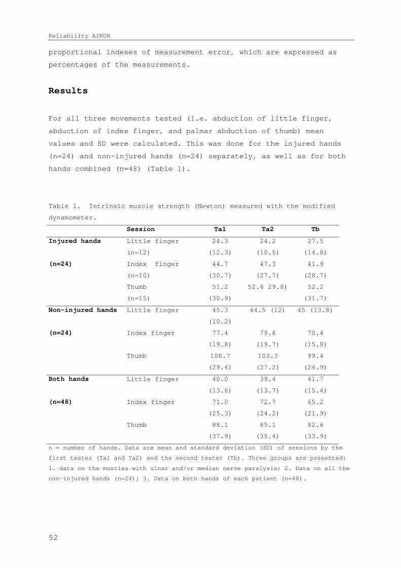
Reliability AIKOH
proportional indexes of measurement error, which are expressed as
percentages of the measurements.
Results
For all three movements tested (i.e. abduction of little finger,
abduction of index finger, and palmar abduction of thumb) mean
values and SD were calculated. This was done for the injured hands
(n=24) and non-injured hands (n=24) separately, as well as for both
hands combined (n=48) (Table 1).
Table 1. Intrinsic muscle strength (Newton) measured with the modified
dynamometer.
Session Ta1 Ta2 Tb
Injured hands Little finger
(n=12)
24.3
(12.3)
24.2
(10.5)
27.5
(14.8)
(n=24) Index finger
(n=10)
44.7
(30.7)
47.3
(27.7)
41.9
(28.7)
Thumb
(n=15)
51.2
(30.9)
52.6 29.8) 52.2
(31.7)
Non-injured hands Little finger 45.3
(10.2)
44.5 (12) 45 (13.8)
(n=24) Index finger 77.4
(19.8)
79.6
(19.7)
70.4
(15.8)
Thumb 108.7
(29.4)
103.3
(27.2)
99.4
(24.9)
Both hands Little finger 40.0
(13.6)
39.4
(13.7)
41.7
(15.4)
(n=48) Index finger 71.0
(25.3)
72.7
(24.2)
65.2
(21.9)
Thumb 88.1
(37.9)
85.1
(35.4)
82.6
(33.9)
n = number of hands. Data are mean and standard deviation (SD) of sessions by the
first tester (Ta1 and Ta2) and the second tester (Tb). Three groups are presented:
1. data on the muscles with ulnar and/or median nerve paralysis; 2. Data on all the
non-injured hands (n=24); 3. Data on both hands of each patient (n=48).
52

Chapter 3
For the hands with an ulnar nerve injury (n=12), measurements with
the dynamometer showed that the mean strength of the little finger
abduction was 25.3 N (range 3.9-57.8) and of index finger abduction
was 44.6 N (range 10.8-108.8). For the hands with a median nerve
injury (n=15) the mean strength of the thumb abduction was 52 N
(range 12.9-123.9). The recovery of muscle strength was also
calculated as a percentage of the non-injured hand. The mean value
for recovery of the little finger abduction was 57%, index finger
abduction 58% and for thumb abduction 50%. The mean muscle strength
according to the MRC scale for the little finger abduction was 4.2,
index finger abduction 3.5, and thumb abduction 3.6.
Analysis of variance (ANOVA) showed that the sources of measurement,
i.e. the variance between test sessions (Var T) and the variance due
to interaction between patient and test session (Var PxT)
contributed 4.2-9.1% to the total variance (Table 2). The largest
part of the error variance was attributed to the interaction between
patient and test session. This implies that some patients produced
the highest forces in the first test session or with the first
tester, whereas others performed better in the second test session
or with the second therapist.
Table 2. Intrinsic muscle strength measurements (mean) of both hands of 24
patients with ulnar and/or median nerve paralyses, and variance (%)
indicating reliability.
Little finger
abduction
Index finger
abduction
Thumb abduction
Intra-
observer
Inter-
observer
Intra-
observer
Inter-
observer
Intra-
observer
Inter-
observer
Mean(N) 39.7 40.9 71.9 68.1 86.6 85.3
Var(P) 95.7% 90.9% 94.9% 90.9% 95.8% 95.9%
Var(T) 0% 0% 0.1% 1.1% 0% 0.5%
Var(PxT) 4.3% 9.1% 5% 8% 4.2% 3.7%
Mean is given in Newton (N), all other data after logarithmic transformation.
Analysis of Variance: where Var(P) = variance to be attributed to differences
between patients. Var (T) = variance attributed to differences between tests by
53
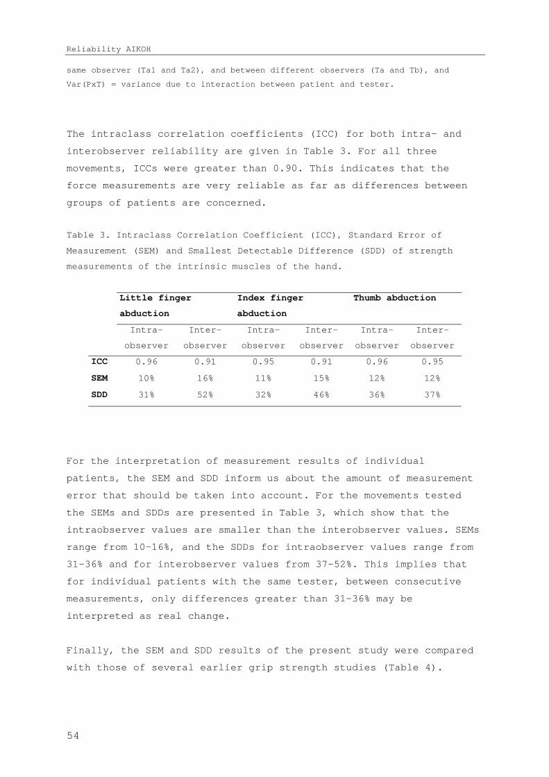
Reliability AIKOH
same observer (Ta1 and Ta2), and between different observers (Ta and Tb), and
Var(PxT) = variance due to interaction between patient and tester.
The intraclass correlation coefficients (ICC) for both intra- and
interobserver reliability are given in Table 3. For all three
movements, ICCs were greater than 0.90. This indicates that the
force measurements are very reliable as far as differences between
groups of patients are concerned.
Table 3. Intraclass Correlation Coefficient (ICC), Standard Error of
Measurement (SEM) and Smallest Detectable Difference (SDD) of strength
measurements of the intrinsic muscles of the hand.
Little finger
abduction
Index finger
abduction
Thumb abduction
Intra-
observer
Inter-
observer
Intra-
observer
Inter-
observer
Intra-
observer
Inter-
observer
ICC 0.96 0.91 0.95 0.91 0.96 0.95
SEM 10% 16% 11% 15% 12% 12%
SDD 31% 52% 32% 46% 36% 37%
For the interpretation of measurement results of individual
patients, the SEM and SDD inform us about the amount of measurement
error that should be taken into account. For the movements tested
the SEMs and SDDs are presented in Table 3, which show that the
intraobserver values are smaller than the interobserver values. SEMs
range from 10-16%, and the SDDs for intraobserver values range from
31-36% and for interobserver values from 37-52%. This implies that
for individual patients with the same tester, between consecutive
measurements, only differences greater than 31-36% may be
interpreted as real change.
Finally, the SEM and SDD results of the present study were compared
with those of several earlier grip strength studies (Table 4).
54

Chapter 3
Table 4. Data on grip strength measurements from earlier reliability (intra
observer) studies compared with data from the current study.
StudyHandheld
dynamometerUnit
subject
sN Mean SD ICC SEM SDD
% of
mean
BoissyLafayettegrip
N Healthy 10381.7
87.3 0.86 33 91.5 24%
Stroke 15130.3
83.7 0.91 25 69.3 53%
Geertzen Citec N RSDunaffectedhand
29 123 66 0.97 24 66 54%
affected 29 84 51 0.94 25 71 85%
Nitschke Jamar kgHealthywomen
32 32.5 6.9 0.93 1.8 5.7 18%
WomenwithNSRP
10 17.4 6.2 0.95 1.4 5.9 34%
Spijkerman
Grip gauge Nm Healthy 16 43.3 9.9 0.98 1.4 3.9 9%
Injuredhands
8 23.1 17.7 0.98 2.5 6.9 30%
Schreuders
abductiondig 5
40 13.6 0.96 10 31%
abductiondig 2
71 25.3 0.95 11 32%AIKOHintrinsicdynamometer
NPeripheral nerve injured
abductionthumb
48
88.1 37.9 0.96 12 36%
N = Newton; Nm = Newton meters; n= number of subjects; NSRP = Non- specific regional pain syndrome; ICC = Intraclass correlation coefficient; SEM = standard error of measurement; SDD = smallest detectable difference; % of mean = SDD percentage of the mean.
Discussion
In practically all studies evaluating peripheral nerve function,
grip and pinch strength are measured. However, grip strength
measurements do not necessarily provide useful information about the
motor function of the ulnar and median nerve in the lower arm.
Patients with a complete ulnar nerve lesion can have a considerable
grip strength, evidently because the extrinsic muscles are not
paralysed. In our opinion, grip and pinch strength measurements
provide information on the combined function of all the muscles of
the hand, including the wrist extensors innervated by the radial
nerve. In addition, generally the peripheral nerves are injured in
combination with one or more injuries of the finger and wrist
tendons. In such cases it is even more difficult to determine the
contribution of the intrinsic muscles to the total amount of grip
strength.
55
Reliability AIKOH
An attempt to analyse the contribution of the intrinsic muscles to
grip and pinch strength was made by Kozin et al.18 They measured a
38% decrease in grip strength after ulnar nerve block and a 32%
decrease after median nerve block. The only median innervated
intrinsic muscles contributing to grip are the two lumbrical muscles
to the index and middle finger. It is questionable whether two small
lumbricals can contribute as much as 32% to grip strength.
Apparently, grip strength measurements show changes after nerve
paralysis but it is difficult to determine whether this is due to
loss of extrinsic or intrinsic muscle strength, or perhaps due to
loss of sensory function.
In the present study we evaluated a device that we modified to
measure the strength of the intrinsic muscles in isolation. The
first step in assessing this new method is to establish the
reliability of these measurements.7
The intraobserver and interobserver reliability calculated as ICCs
was high. This indicates good reliability of the measurements when
used to compare groups of patients. For the purpose of measuring
individual patients to assess changes in muscle strength over time,
however, the standard errors of measurement (SEMs) and the smallest
detectable differences (SDDs) are appropriate indexes of measurement
error. These provide information about the width of the error band
around a measured value and in the amount of measurement error that
should be taken into account when comparing two consecutive
measurements.19 20 The relatively large values that we found for these
indices in the present study implies that the reliability of the
force measurements for assessing changes in individual patients is
less satisfactory than for groups of patients. Only changes of more
than 30% to 50% in muscle force in a patient can be interpreted as
real changes. It is questionable whether this amount of measurement
error is satisfactory when assessing clinically relevant change in
patients with hand and nerve injuries.
56

Chapter 3
As we are unaware of any study concerning the reliability of
intrinsic muscle strength measurements, we compared the SEM and SDD
results of the present study with those of earlier grip strength
studies.13 21-23
Because we used a log transformation the outcome was a SDD
percentage. The data from the other studies did not use the log
transformation, and thus the SDDs are in the original unit of
measurement. To allow comparison of the SDDs from these latter
studies with our results, we assessed the SDD as a percentage of the
mean (Table 4). It was found that in intra-tester reliability
studies with normal subjects the percentage of the SDD of the mean,
(in Table 4 “% of mean”), was 9-24% (mean 15%). The SDD as
percentages of the mean in the different patient groups were between
30-53% (mean 39%). Geertzen et al.13 also studied the intra-tester
reliability of grip strength in Reflex Sympathetic Dystrophy (RSD)
patients and found large SDDs of 66 N in the unaffected hand (54% of
mean) and as high as 84.5 N (85% of mean) in the affected hands.
Geertzen and colleagues concluded that their reliability study of
muscle strength measurements in RSD patients gives values which
should be “treated with scepticism”. Except for Geertzen’s group,
all other studies concluded that the intra-tester reliability was
satisfactory.
From our clinical experience we assume that an SDD of 30% or larger
is not sufficient to assess clinically relevant changes in patients
with hand and nerve injuries. The SDD for inter-tester reliability
of 37-52% leads us to conclude that we would not recommend this
dynamometer to be used by different testers for measurements in the
same patient.
To further reduce the amount of measurement error we suggest that
the shape of the dynamometer be modified because it was not designed
for hand-held measurements and is cumbersome to use. Secondly,
because a small change in the angle of pushing between the device
and the finger causes large differences in the recorded strength,
measurement made by pulling on a string, rather than pushing the
device against the device, might improve the reliability.
57

Reliability AIKOH
Adaptations to the device, and the elucidation of the relationship
between grip strength, MMST and the dynamometry of the intrinsic
muscles of the hand, will be the focus of future studies.
Conclusions
The hand-held dynamometer used in this study to measure the strength
of the intrinsic muscles of the hand after peripheral nerve lesion
has excellent reliability for comparing muscle strength between
groups of patients. However, the Smallest Detectable Difference
(SDD) of the measurements of the thumb, index and little finger
indicate that only with relatively large changes in measurements can
a clinician be confident that real changes in muscle strength have
occurred.
58

Chapter 3
Acknowledgements
The authors thank Dr. P. Hoogvliet and Mr. A.H. den Ouden for their
insight and support.
References
1. Ketchum LD, Thompson D, Pocock G, Wallingford D. A clinical study of forces
generated by the intrinsic muscles of the index finger and the extrinsic
flexor and extensor muscles of the hand. J Hand Surg [Am] 1978;3(6):571-8.
2. Frykman GK. The quest for better recovery from peripheral nerve injury: current
status of nerve regeneration research. J Hand Ther 1993;6(2):83-8.
3. Medical Research Council. Special Report series no 282. London: Her Majesty's
Stationary Office, 1954.
4. Brandsma JW, Schreuders TAR, Birke JA, Piefer A, Oostendorp R. Manual muscle
strength testing: intraobserver and interobserver reliabilities for the
intrinsic muscles of the hand. J Hand Ther 1995;8(3):185-90.
5. Brandsma JW, Van Brakel WH, Anderson AM, Kortendijk AJ, Gurung KS, Sunwar SK.
Intertester reliability of manual muscle strength testing in leprosy
patients. Lepr Rev 1998;69(3):257-66.
6. van der Ploeg RJ, Oosterhuis HJ, Reuvekamp J. Measuring muscle strength. J
Neurol 1984;231(4):200-3.
7. Fess EE. The need for reliability and validity in hand assessment instruments. J
Hand Surg [Am] 1986;11(5):621-3.
8. ASHT, editor. Clinical Assessment Recommendations. 2nd ed. Garner, NC: American
Society of Hand Therapists, 1992.
9. Mathiowetz V, Weber K, Volland G, Kashman N. Reliability and validity of grip
and pinch strength evaluations. J Hand Surg [Am] 1984;9(2):222-6.
10. Swanson GA, Swanson GG, Goran-Hagert C. Evaluation of impairment of hand
function. In: Hunter JM, Mackin EJ, Calahan AD, editors. Rehabilitation of
the hand: surgery and therapy. St. Louis: C.V. Mosby, 1995.
11. Langeveld vSHB, Aufdemkampe G, F.W.A. Av. Reliability of force measurements
with a hand-held dynamometer in healthy subjects and force measurements in
patients with poliomyelitis anterior acuta. J Rehabil Sciences 1996;9:2-10.
12. Lusardi MM, Bohannon RW. Hand grip strength: comparability of measurements
obtained with a Jamar dynamometer and a modified sphygomanometer. J Hand Ther
1991;2:117-122.
13. Geertzen JH, Dijkstra PU, Stewart RE, Groothoff JW, ten Duis HJ, Eisma WH.
Variation in measurements of grip strength. A study in reflex sympathetic
dystrophy patients. Acta Orthop Scand Suppl 1998;279:4-11.
14. Bohannon RW. Measurement, nature, and implications of skeletal muscle strength
in patients with neurological disorders. Clin Biomech 1995;10(6):283-292.
59
Reliability AIKOH
15. Mannerfelt L. Studies on the hand in ulnar nerve paralysis. A clinical-
experimental investigation in normal and anomalous innervation. Acta Orthop
Scand 1966(87):61-86.
16. Mannerfelt LG. Studies on ulnar nerve compression neuropathies with a new
computerised instrument--the intrins-o-meter. Scand J Plast Reconstr Surg
Hand Surg 1997;31(3):251-60.
17. Rosen B. Recovery of sensory and motor function after nerve repair. A rationale
for evaluation. J Hand Ther 1996;9(4):315-27.
18. Kozin SH, Porter S, Clark P, Thoder JJ. The contribution of the intrinsic
muscles to grip and pinch strength. J Hand Surg [Am] 1999;24(1):64-72.
19. Roebroeck ME, Harlaar J, Lankhorst GJ. Reliability assessment of isometric knee
extension measurements with a computer-assisted hand-held dynamometer. Arch
Phys Med Rehabil 1998;79(4):442-8.
20. Streiner DL, Norman GR. Health measurement scales: a practical guide to their
development and use. 2nd ed. Oxford: Oxford University Press,, 1995.
21. Boissy P, Bourbonnais D, Carlotti MM, Gravel D, Arsenault BA. Maximal grip
force in chronic stroke subjects and its relationship to global upper
extremity function. Clin Rehabil 1999;13(4):354-62.
22. Nitschke JE, McMeeken JM, Burry HC, Matyas TA. When is a change a genuine
change? A clinically meaningful interpretation of grip strength measurements
in healthy and disabled women. J Hand Ther 1999;12(1):25-30.
23. Spijkerman DC, Snijders CJ, Stijnen T, Lankhorst GJ. Standardization of grip
strength measurements. Effects on repeatability and peak force. Scand J
Rehabil Med 1991;23(4):203-6.
60
CHAPTER 4
Strength Measurements of the
Lumbrical Muscles
In the palm of the hand, and between the metacarpal bones, are numerous small
muscles, (lumbricales and interossei) which perform the finer movements, -
expanding the fingers, and moving them in every direction with quickness and
delicacy. They are the organs which give the hand the power of spinning, weaving,
engraving etc; and as they produce the quick motions of the musician’s fingers,
they are called by anatomists fidicinales. (= of or relating to a stringed
instrument)
Charles Bell (1774-1842)
The Hand - Its Mechanism and Vital Endowments as Evincing Design
(1833)

Lumbrical strength
Reprinted from Journal of Hand Therapy, Vol. 9, Schreuders TAR, Stam
HJ. Strength measurements of the lumbrical muscles. P 303-305,
Copyright 1996, with permission from Elsevier
Abstract
This study was designed to measure the strength of the lumbrical
muscles in the index and long fingers in patients with ulnar nerve
paralysis. A hand-held dynamometer was used.
The results show that in ulnar nerve damage the index and long
fingers have a mean metacarpo-phalangeal (MCP) joint flexion
strength of 0.8 kg (range 0.3-1.5), compared with 6.4 kg (range 4.6-
7.9) in the non-involved hand. Thus, the damaged fingers have only
about 12% of the strength of those of the non-involved hand. In the
hand with ulnar paralysis, the loss of intrinsic strength (dorsal
and palmar interosseous muscles) is considerable (almost 90%).
The contribution of the interosseous muscles in maintaining the
intrinsic position is considerably greater than that of the
lumbricals. Comparing the Medical Research Council (MRC) scale (0-5)
with the dynamometry measurements shows that MRC grade 3 correlates
with about 0.8 kg, while grade 5 correlates with about 6.5 kg of MCP
joint flexion strength.
Introduction
The lumbrical muscles are the only muscles whose origin and
insertion are tendons: the flexor digitorum profundus (FDP) and the
extensor expansion of the extensor digitorum communis (EDC),
respectively.
There is no consensus about the role of the lumbrical muscles.1
Several studies2-13 have documented the strength of the lumbricals in
normal subjects, but only a few studies have investigated the
58
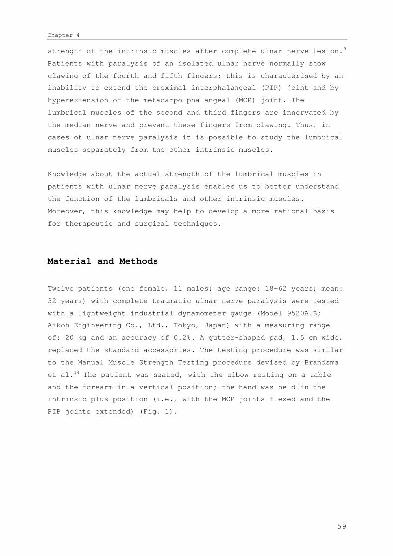
Chapter 4
strength of the intrinsic muscles after complete ulnar nerve lesion.9
Patients with paralysis of an isolated ulnar nerve normally show
clawing of the fourth and fifth fingers; this is characterised by an
inability to extend the proximal interphalangeal (PIP) joint and by
hyperextension of the metacarpo-phalangeal (MCP) joint. The
lumbrical muscles of the second and third fingers are innervated by
the median nerve and prevent these fingers from clawing. Thus, in
cases of ulnar nerve paralysis it is possible to study the lumbrical
muscles separately from the other intrinsic muscles.
Knowledge about the actual strength of the lumbrical muscles in
patients with ulnar nerve paralysis enables us to better understand
the function of the lumbricals and other intrinsic muscles.
Moreover, this knowledge may help to develop a more rational basis
for therapeutic and surgical techniques.
Material and Methods
Twelve patients (one female, 11 males; age range: 18-62 years; mean:
32 years) with complete traumatic ulnar nerve paralysis were tested
with a lightweight industrial dynamometer gauge (Model 9520A.B;
Aikoh Engineering Co., Ltd., Tokyo, Japan) with a measuring range
of: 20 kg and an accuracy of 0.2%. A gutter-shaped pad, 1.5 cm wide,
replaced the standard accessories. The testing procedure was similar
to the Manual Muscle Strength Testing procedure devised by Brandsma
et al.14 The patient was seated, with the elbow resting on a table
and the forearm in a vertical position; the hand was held in the
intrinsic-plus position (i.e., with the MCP joints flexed and the
PIP joints extended) (Fig. 1).
59
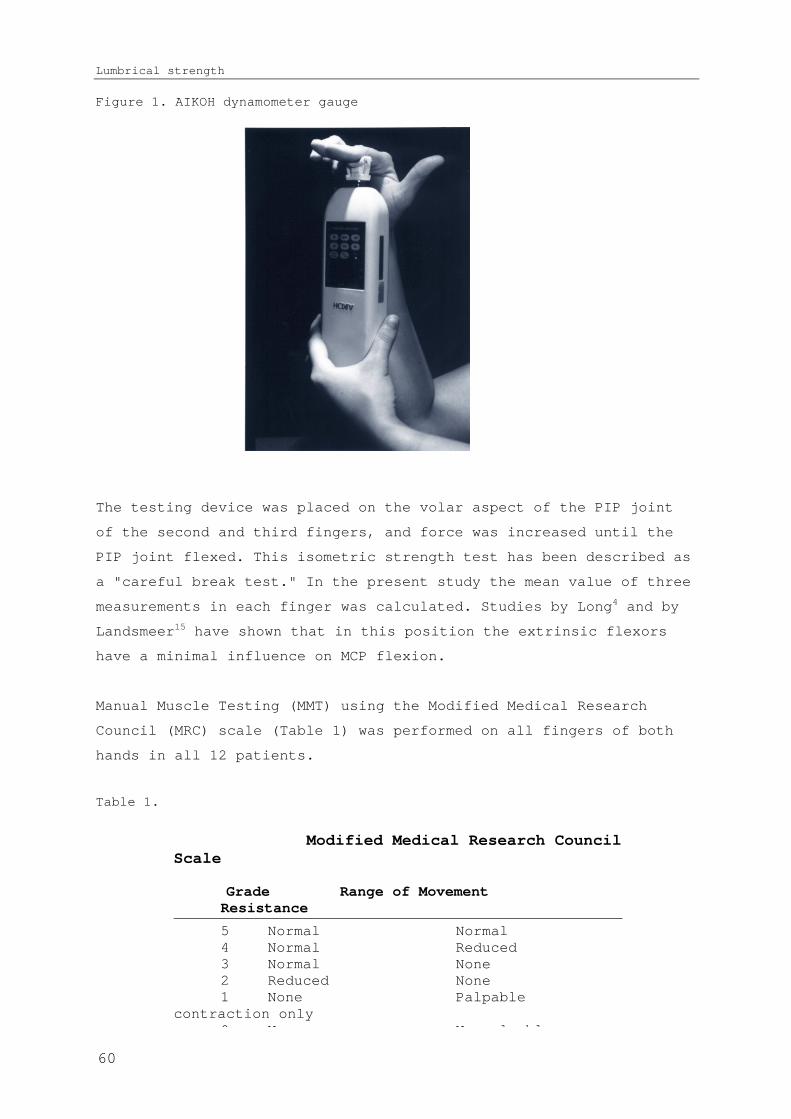
Lumbrical strength
Figure 1. AIKOH dynamometer gauge
The testing device was placed on the volar aspect of the PIP joint
of the second and third fingers, and force was increased until the
PIP joint flexed. This isometric strength test has been described as
a "careful break test." In the present study the mean value of three
measurements in each finger was calculated. Studies by Long4 and by
Landsmeer15 have shown that in this position the extrinsic flexors
have a minimal influence on MCP flexion.
Manual Muscle Testing (MMT) using the Modified Medical Research
Council (MRC) scale (Table 1) was performed on all fingers of both
hands in all 12 patients.
Table 1.
Modified Medical Research Council Scale
Grade Range of Movement Resistance
5 Normal Normal4 Normal Reduced3 Normal None2 Reduced None1 None Palpable
contraction only 0 N N l bl
60

Chapter 4
Results
Table 2 presents the results of the strength measurement tests in
the individual patients. It is clear that patients with ulnar nerve
paralysis had barely sufficient strength to maintain the intrinsic-
plus position.
Table 2. Strength (kg) of the intrinsic muscles in the second and third
fingers of the ulnar paralysed hand and in normal digits patients age
injured hand noninjured hand (n = 12)
Injured hands Noninjured
hands
Patient
(n=12)
Age
(years)digit 2 digit 3 digit 2 digit 3
DeJ 32 1 0.9 7.2 7.9
G 36 0.9 1.1 5 5.5
D 62 0.6 0.7 6.2 6.5
R 25 0.7 0.5 5.2 5.5
VdH 45 0.7 0.7 5.8 4.6
F 31 0.3 0.4 7.1 5.7
VdL 30 0.6 0.8 7 5.7
K 23 0.9 0.3 5.6 6.5
S * 18 0.4 0.6 6.9 7.3
Z 43 1 1.2 7.2 7.8
P 46 1.3 1.5 6.9 7
VH 34 1 0.8 7 6.3
Mean 32 0.8 0.8 6.4 6.4
* The only female patient in the group
61

Lumbrical strength
The mean value of the strength of the two digits of the injured hand
was the same, as was the case in the noninjured hand: 0.8 kg (range
0.3-1.5) and 6.4 kg (range 4.6-7.9), respectively.
Based on the MRC scale, all injured digits were grade 3, whereas the
fingers of the noninjured hand were grade 5.
Discussion
The intrinsic muscles are of paramount importance for efficient hand
function. The lumbrical muscles differ from other muscles in that
they both originate and insert at tendons. There are several
different theories about the function of the lumbrical muscles but
no consensus.1
According to Jacobson et al.5 the lumbricals have an extremely small
physiologic cross-sectional area (PCSA), but they have long muscle
fibres. Jacobson et al. concluded that lumbricals are designed for
high excursions and velocity production. Backhouse et al.19546 and
Long7 performed electro-myographic studies of the lumbricals and
concluded that their principal action is extension of the
interphalangeal (lP) joints and weak flexion of the MCP joints.
Ranney and coworkers1 came to the same conclusion based on
biomechanical studies of the lumbricals in human cadavers. Ketchum
et al.9 calculated the mean force of the lumbricals and estimated it
to be approximately 14% of the total force flexing the MCP joints;
they estimated that the contribution of the lumbricals to resisted
movement is 2.03 0.9 kg.
Brand10 reported that at the lP level the lumbricals act as
extensors, as do the interosseous muscles. Furthermore, the
lumbricals diminish the flexion moment of the profundus tendons at
the same joint by diverting FDP force to the extensor expansion.
During flexion, the lumbricals may dominate the sequence of closure
and ensure that the MCP joints flex ahead of the lP joints, allowing
the hand to surround a large object. The lumbricals also co-ordinate
62
Chapter 4
finger movements. Devanandan et al.11 found that the lumbricals
contain an unusually high number of muscle spindles, which are
important for proprioception of the fingers and hands.
Leijnse and Kalker12 concluded that the lumbricals are in an optimal
position for proprioceptic feedback concerning the position of the
PIP-distal interphalangeal (DIP) joint mechanism due to the large
systematic moment arm at the lP joint level and the very small
systematic moment arm at the MCP joint. They also formulated the
role of the lumbricals in the so-calIed lumbrical loop, in which the
lumbricals control the PIP and DIP joints, and the EDC and FDP
control the MCP joint. This movement may be the basis of fast
movements in, for example, a musician's hand.
Many different methods exist for surgically reconstructing a claw
hand after ulnar nerve paralysis.16 These methods include intrinsic
replacement, lumbrical replacement, or MCP flexor replacement.
Currently, it is recommended that all four fingers be corrected,17
but some clinicians suggest that correction of only the fourth and
fifth digits is sufficient.13
Objective measurement of intrinsic muscle force is necessary for
three reasons. First, it is important to establish the relative
contribution of the interosseous muscles and the lumbricals to the
balance of forces in the hand. Second, measurements are required to
monitor the recovery of strength in patients with paralysis
following ulnar nerve lesions Third, if reconstructive surgery is
considered, it is important to know the required strength of the
muscles involved.
Specific testing of the intrinsic muscles can be done manually.14
Manual Muscle Testing according to the Oxford Medical Research
Council's 0-5 Scale (MRC scale)18 is routinely used by therapists.
Dynamometry is a popular means of expressing strength in a
quantitative manner.19-21 For example, grip and pinch strength
measurements are used to determine the efficacy of hand therapy and
surgical treatment.22 However, most devices have been designed to
63

Lumbrical strength
measure extrinsic muscle strength of the long flexors. The intrinsic
muscles are not necessarily tested by grip and pinch strength
measurements.
In the present study, the strength of index and long fingers with an
ulnar nerve lesion correlated with grade 3 on the MRC scale in all
12 patients. In MRC terms, this means that the strength is
considered "reasonable," or 50%, on a 0%-100% scale.23 24 The actual
strength, however, as measured by the dynamometer was only 12% of
the strength of the noninjured hand. Therefore, we concluded that an
MRC grade 3 for the intrinsic muscles is comparable with 12% (and
not 50%) of normal strength.
The loss of 90% of intrinsic muscle strength suggests that the
primary role of the lumbricals is not to produce power but rather to
divert strength from the FDP and to contribute to the sensory
feedback for co-ordination. Evidently, the interosseous muscles are
much stronger than the lumbricals. The strength of the lumbricals is
not sufficient to withstand clawing, and latent clawing of the index
and long fingers in the hand with ulnar paralysis must be kept in
mind.10
Applying extension force to the MCP joints of these digits will
cause them to collapse into a typical claw deformity. Splinting only
the fourth and fifth fingers will in many cases be inadequate
because clawing may develop in all four fingers. When training and
testing intrinsic muscles, it is important for hand therapists to be
aware of the biomechanics of the lumbrical and interosseous muscles
and their respective contributions to the prevention of clawing in
the hand with ulnar nerve paralysis.
Conclusions
The mean strength of the lumbricals after an ulnar nerve lesion is
0.8 kg, which is about 12% of the intrinsic muscle strength of the
normal hand. There was no difference between the mean values of the
64

Chapter 4
strength of the index and long fingers after ulnar nerve paralysis.
When reconstructive surgery is considered to correct a claw hand,
the loss of strength (almost 90%) in the index and long fingers
should be taken into account.
References
1. Ranney D, Wells R. Lumbrical muscle function as revealed by a new and
physiological approach. Anat Rec 1988;222(1):110-4.
2. Brandsma JW. Intrinsic minus hand: (patho) kinesiology, rehabilitation and
reconstruction. [Thesis]. University of Utrecht, 1993.
3. Wiles CM, Karni Y. The measurement of muscle strength in patients with
peripheral neuromuscular disorders. J Neurol Neurosurg Psych 1983;46:1006-
1013.
4. Long C, Brown ME, Weiss G. An electromyographic study of the extrinsic-intrinsic
kinesiology of the hand. Arch Phys Med Rehabil. 1960;41:175-181.
5. Jacobson MD, Raab R, Fazeli BM, Abrams RA, Botte MJ, Lieber RL. Architectural
design of the human intrinsic hand muscles. J Hand Surg [Am] 1992;17(5):804-
9.
6. Backhouse KM, Catton WT. An experimental study of the function of the lumbrical
muscles in the human hand. J Anat 1954;88(5):133-141.
7. Long CH. Intrinsic-extrinsic muscle control of the fingers. J Bone Joint Surg
(Am) 1968;50(5):973-984.
8. Ranney DA, Wells RP, Dowling J. Lumbrical function: interaction of lumbrical
contraction with the elasticity of the extrinsic finger muscles and its
effect on metacarpophalangeal equilibrium. j Hand Surg (Am) 1987;12(4):110-
114.
9. Ketchum LD, Thompson D, Pocock G, Wallingford D. A clinical study of forces
generated by the intrinsic muscles of the index finger and the extrinsic
flexor and extensor muscles of the hand. J Hand Surg [Am] 1978;3(6):571-8.
10. Brand PW. Clinical Mechanics of the Hand. St. Louis: CV Mosby, 1999.
11. Devanandan MS, Ghosh S, John KLT. A quantitative study of the muscle spindles
and tendon organs in some intrinsic muscles of the hand. Anat Rec
1983;207:263-266.
12. Leijnse JN, Kalker JJ. A two-dimensional kinematic model of the lumbrical in
the human finger. J Biomech 1995;28(3):237-49.
13. Burkhalter WE, Strait JL. Metacarpophalangeal flexor replacement for intrinsic
mucsle paralysis. J Bone Joint Surg (Am) 1973;55:1667-1676.
14. Brandsma JW, Schreuders TAR, Birke JA, Piefer A, Oostendorp R. Manual muscle
strength testing: intraobserver and interobserver reliabilities for the
intrinsic muscles of the hand. J Hand Ther 1995;8(3):185-90.
15. Landsmeer JM, Long C. The mechanism of finger control, based on electromyograms
and location analysis. Acta Anat 1965;60(3):330-47.
65
Lumbrical strength
16. Omer G. Ulnar nerve palsy. In: Green D, editor. Operative Hand Surgery. 3 ed.
New York: Churchill Livingstone, 1993:1449-1466.
17. Andersen JG, Brandsma JW. Management of paralytic conditions in leprosy. Addis
Ababa, 1984.
18. Medical Research Council. Special Report series no 282. London: Her Majesty's
Stationary Office, 1954.
19. Cook JD, Glass DS. Strength evaluation in neuromuscular disease. Neurol Clin
1987;5(1):101-23.
20. Fess EE. The need for reliability and validity in hand assessment instruments.
J Hand Surg [Am] 1986;11(5):621-3.
21. van der Ploeg RJ, Oosterhuis HJ, Reuvekamp J. Measuring muscle strength. J
Neurol 1984;231(4):200-3.
22. Hunter J, Schneider LH, Mackin EJ. Rehabilitation of the Hand and Upper
Extremity: Mosby co., 2002.
23. ASHT, editor. Clinical Assessment Recommendations. 2nd ed. Garner, NC: American
Society of Hand Therapists, 1992.
24. Kendall FP, McCreary EK. Muscles: Testing and Function. 3rd edition ed.
Baltimore: Williams & Wilkins, 1983.
66

CHAPTER 5
Measurement Error in Grip and
Pinch Force Measurements in
Patients with Hand Injuries
From error to error, one discovers the
entire truth
Sigmund Freud (1856-1939)
Reprinted from Schreuders TAR, Roebroeck ME, Goumans J, van
Nieuwenhuijzen JF, Stijnen TH, Stam HJ. Measurement error in grip

Measurement error in grip - and pinch force measurements
and pinch force measurements in patients with handinjuries. Physical
Therapy 2003; 83(9): 806-815, with permission of the American
Physical Therapy Association. This material is copyrighted, and any
further reproduction or distribution is prohibited.
Abstract
There is limited documentation of measurement error of grip and
pinch force evaluation methods. The purposes of this study were (1)
to determine indexes of measurement error for intraexaminer and
interexaminer measurements of grip and pinch force in patients with
hand injuries and (2) to investigate whether the measurement error
differs between measurements of the injured and noninjured hands and
between experienced and inexperienced examiners.
The subjects were a consecutive sample of 33 patients with hand
injuries who were seen in the Department of Rehabilitation Medicine
of Erasmus MC–University Medical Center Rotterdam in the
Netherlands. Repeated measurements of grip force in 2 handle
positions. The time between two measurements and change of examiners
was 2-3 minutes. For the grip force the distance between handles
were 4.6 and 7.2 cm, respectively and of tip pinch, with the index
finger on top and the other fingers flexed with the thumb below and
key pinch force, with the thumb on top and the radial side of the
index finger below were obtained on both hands of the subjects by an
experienced examiner and an inexperienced examiner. Intraclass
correlation coefficients (ICCs), standard errors of measurement
(SEMs), and associated smallest detectable differences (SDDs) were
calculated and compared with data from previous studies.
The reliability of the measurements was expressed by ICCs between
.82 and .97. For grip force measurements (in the second handle
position) by the experienced examiner, an SDD of 61 N was found.
For tip pinch and key pinch, these values were 12 N and 11 N,
respectively. For grip force measurements by the inexperienced
examiner these values were 56 N for grip force measurements and for
tip pinch and key pinch, 13 N and 18 N, respectively
66
Chapter 5
Based on the SEMs and SDDs, in individual patients only relatively
large differences in grip and pinch force measurements can be
adequately detected between consecutive measurements. Measurement
error did not differ between injured and noninjured hands or between
experienced and inexperienced examiners. Criteria for judging
whether the measurement error does allow application of the
measurements in individual patients are discussed.
67

Measurement error in grip - and pinch force measurements
Introduction
Only data that have acceptable reliability and validity are valuable
in the clinical decision-making process for determination of
impaired function and comparison of surgical repair techniques to
document progress during rehabilitation and to evaluate disability
after injury. Such data will decrease the need for use of
unsubstantiated opinions and will increase the physical therapist’s
ability to obtain reproducible findings and meaningful results.1
Better reliability implies better precision of single measurements,
which is a prerequisite for better tracking of changes in
measurements in research and clinical practice.2
Generally accepted instruments in the evaluation of hand function
measure grip and pinch force. Grip and pinch force measurements have
been promoted as an important measure of outcome in, for example,
the evaluation of peripheral nerve function.3-5For a correct
interpretation of these force measurements, we contend information
on the reliability (measurement error) of these measurements is
required. The reliability of grip force measurements has been
studied in patients with hand injuries6 7 and in people without
impairments of the hand.8 9 It also has been studied in several other
categories of patients, including women with non-specific regional
pain (NSRP)10 and patients with reflex sympathetic dystrophy (RSD)11
or epicondylitis,12 13 and in people after a stroke.14 The reliability
of pinch force measurements has been studied in patients with RSD11
and repetitive strain injury (RSI).15
The studies of Spijkerman et al6 and Brown et al7 on the reliability
of force measurements in patients with hand injuries have important
drawbacks. The study by Spijkerman et al included only 8 patients,
and Brown et al, in their study of grip and pinch force measurements
in patients with hand injuries, did not specify the measurement
error. As in many studies, the intraclass correlation coefficient
(ICC) was used as a measure of reliability. For clinical use,
however, we believe the most relevant information is the magnitude
68

Chapter 5
of change between tests that is required to detect a real change,
preferably quantified in the same units as the force measurement.10 16
This magnitude of change is specified in the smallest detectable
difference (SDD), which is calculated from the standard error of
measurement (SEM), the absolute error component of the
measurements.17 18 The smaller the measurement error, the better the
measure.19
Various factors relating to the patient’s condition may influence
the measurement error, such as the amount of pain and loss of normal
function of the fingers or thumb after injury. The experience of the
examiner also might influence the measurement error. However,
studies on these aspects of grip and pinch force measurements for
patients with hand injuries are scarce.
One aim of our study was to assess the SEM and related indexes of
reliability for intraexaminer and interexaminer applications of grip
(2 handle positions, with distances between the handles of 4.6 and
7.2 cm) and pinch (tip pinch and key pinch) force measurements for
patients with hand injuries obtained with Lode handgrip and pinch-
grip dynamometers (Figure 1).
69

Measurement error in grip - and pinch force measurements
Figure 1. Measurements were performed with the participant seated at a
table on which the dynamometers were positioned.
A second aim was to investigate whether the measurement error of
grip and pinch force measurements differs between specific
applications, such as measurements of injured and noninjured hands,
measurements obtained by experienced and inexperienced examiners,
and measurements between 2 handle positions for grip force and 2
different types of pinch force (i.e., tip and key pinch force). A
third aim was to judge whether the measurement error is small enough
to justify use of these measurements to discern real changes in grip
and pinch force in individual patients. Because no clear criteria to
judge the SDD are available for grip and pinch force measurements,
we compared our findings with the results of studies in which grip
and pinch force measurements were examined in different patient
categories, i.e. patients with hand injuries, women with pain
(NSRP), RSD, epicondylitis and after a stroke.
Method
Subjects
A consecutive sample of 33 patients (20 male, 13 female) who were
seen in the Department of Rehabilitation Medicine of Erasmus MC-
University Medical Center Rotterdam in the Netherlands participated
in the study. They had a mean age of 36 years (SD=13.7, range 17-
67).
All patients had an injury on only one hand (6 with tendon
lacerations, 9 with nerve lesions, 4 with fractures, and 14 with a
combination of lesions or surgeries e.g., crush injury, finger
amputation, arthrodesis). During a period of 5 months, patients who
were capable of being evaluated for grip and pinch force were asked
to participate in this study. Patients who were not permitted to
grip or pinch with maximum force because of injured tissue and
patients who complained of pain were excluded. We presumed that
pain would inhibit the patients from exerting full maximum force and
70

Chapter 5
that force measurements may reflect the amount of pain rather than
the force-generating capacity of the patients. We acknowledge that
excluding those patients limited our study to only those patients
who were pain-free. The participants were informed about the purpose
of the study and gave informed consent.
The participants were in different phases of their rehabilitation
process. Study entry was, on average, 22 months after injury or
surgery when including one patient who had a 16-year period between
the date of injury and time of examination. Excluding this patient,
the average period was 9 months (SD=7.0, range 1-28) since injury or
surgery. Force measurements of the hand are routinely obtained in
our department during different phases of the rehabilitation
process. Such measurements can be used to determine progression of
muscle force recovery, the ability to perform in a vocation, and
indications for continuing therapy and to assess long-term outcome
after hand injuries.
Instrumentation
We used Lode handgrip and pinch-grip dynamometers* (Fig. 1). The
distances between the handles of the Lode handgrip dynamometer were
adapted to create spaces between the handles comparable to those of
the Jamar hand dynamometer.†,20 For our study, the grip handle
positions 2 (distance between handles=4.6 cm) and 4 (distance
between handles=7.2 cm), which we designated “grip 2” and “grip 4,”
respectively, were used with the handgrip dynamometer.
With the Lode pinch-grip dynamometer, which is similar in design to
the Preston† pinch dynamometer,21 2 types of pinch were measured: tip
pinch and key pinch. Tip pinch force was measured with only the
index finger on top and the other fingers flexed with the thumb
below, and key pinch force was measured with the thumb on top and
the radial side of the index finger below.
* Lode Medical Technology, Zernikepark 16, 9747 AN Groningen, the Netherlands.Distributed in the United States
by ElectraMed Corp, G-5332 Hill-23 Dr, Flint, MI 48507.
† Sammons Preston/Rolyan, 4 Sammons Ct, Bolingbrook, IL 60440.
71
Measurement error in grip - and pinch force measurements
The grip handle is connected to an amplifier from which the values
give a digital readout on a display. According to the manufacturer,
the measuring range is 0 to 1,000 N, with an accuracy of 1%
deviation for 2 to 500 N and 2% for 500 to 900 N. Calibration was
done with suspended weights according to the method described by
Mathiowetz et al.8 We believe the use of strain gauge technology is
preferable to dynamometers which are spring-based or use a hydraulic
pressure system, because such dynamometers might produce erroneous
data due to the wear and tear of metal, slow leaks and hysteresis.22
Procedure
Measurements were taken according to recommendations of the American
Society of Hand Therapists (ASHT).22 Participants were seated at a
table on which the dynamometers were positioned (Fig. 1). The
subjects were told to keep their elbow flexed without resting their
arm or the grip device of the dynamometer on the table. The digital
display was not visible to the subjects. Corresponding to the ASHT
recommendations for each measurement, the mean of 3 measurements was
recorded. Measurements were obtained of the left and right hands
alternately, which is the method of testing used at our clinic.
Testing both hands, we contend, enables comparisons of the injured
and noninjured hands, as proposed by Gaul.23
The side (injured or noninjured) at which measurements were started
and the order of tests (grip 2, grip 4, tip pinch, or key pinch)
were randomly selected. Both examiners obtained all the measurements
twice for each subject; the order of examiners was randomly
selected. During each measurement session, the examiners obtained 12
measurements (2 grip and 2 pinch measurements 3 repetitions from
one hand). Then the examiners were changed while the subject took a
short break (2-3 minutes) and moved the arm and hand out of the
testing position. In this way, we attempted to reflect practice with
examiners independently testing subjects, except there was a short
time interval between examiners. Both examiners participated in 2
sessions per patient, which resulted in 48 measurements per hand.
72
Chapter 5
The total time required for testing, including the breaks, was 35 to
40 minutes per patient.
Examiners
Because we were interested in the effect of experience of the
examiner on measurement error, we selected 2 examiners with widely
differing experience. The first examiner was a 54-year-old male
physical therapist (JFvN) with 20 years of experience in testing
hand function. The second examiner (JG) was a 22-year-old female
medical student with no previous experience with these tests. During
a 2-week period, the inexperienced examiner had 5 hours of training
by an experienced physical therapist to become familiarised with the
equipment and testing protocol. During the training measurements
were practised on several subjects without injuries and some
patients with hand injuries. We believe that the results obtained by
this student are reasonably representative of those that could be
obtained by an inexperienced examiner, regardless of whether this is
a starting physician or physical therapist, because at this stage
they have comparable (i.e., limited) knowledge, attitudes, and
skills concerning force measurements
Data Analysis
Statistical analyses were done to determine intraexaminer
reliability of data obtained in the 2 sessions performed by the same
examiner both for the experienced examiner (Texp1 and Texp2) and the
inexperienced examiner (Tinx 1 and Tinx 2) separately. Interexaminer
reliability was calculated between the first sessions of the
experienced examiner (Texp1) and the inexperienced examiner (Tinx1).
Analyses of the measurements of the injured hands (n=33) and
noninjured hands (n=33) were conducted separately.
An analysis of variance (ANOVA) was conducted with an SPSS/PC+
program‡ to determine the multiple sources of measurement error. The
variance attributed to differences among participants, was
‡ SPSS Inc, 233 S Wacker Dr, Chicago, IL 60606.
73

Measurement error in grip - and pinch force measurements
designated “Var (P)”. The variance attributed to differences
between sessions by the same examiner (Texp1 and Texp2, Tinx1 and
Tinx2) was designated “Var (S)”. The variance ascribed to the
different examiners (Texp1 and Tinx1) was designated “Var (T)”.
Variance due to interaction among participants for intraexaminer
sessions was designated “Var (P S)”, and variance due to interaction
among participants for interexaminer sessions was designated “Var
(P T)”. The interaction components (P S) and (P T) were confounded
by the residual error. In the intraexaminer analyses, Var (S) and
Var (P S) (i.e., the variance within participants) constituted the
error variance. Correspondingly, for the interexaminer analyses, the
variance components Var (T) and Var (P T) constituted the error
variance.
From the error variance, the SEM was computed as its square root.18
Based on the SEM, the SDD with 95% confidence was calculated as 1.96
2 SEM.24 This SDD can be applied in such a way that only
differences between 2 consecutive measurements greater than the SDD
can be interpreted with 95% certainty as real change in grip or
pinch force. Intraclass correlation coefficients were computed as
the ratio of variances among participants (i.e., Var[P]) and the
total variance.16
Comparing SEMs
In order to test whether differences existed in error variances of
different applications of grip and pinch force measurements a test
of equality of variances for paired samples was applied. In this
analysis, scores between sessions were compared by calculating the
correlation coefficient (Pearson r) between the sum and difference
of the difference scores and testing whether this correlation
differed from zero.17 25 (pp171-172) In this way, we tested whether
differences existed in error variance, and thus SEM and SDD, between
the experienced and inexperienced examiners, between measurements of
the injured and noninjured hands, and between the 2 handle positions
of the grip force measurements and between the 2 pinch force
measurements.
74

Chapter 5
Commonly accepted criteria to judge whether the SEM and SDD of
measurements are adequate for application of the measurements to
individual patients do not exist. Therefore, we compared the SEMs
found in our study with those from other studies in which grip and
pinch force measurements were investigated. We selected studies from
the literature in which values for SEM or SDD were reported and
studies from which SEM and SDD can be estimated by using the
formulas: SEM = SD (1 - ICC)16 24(p119) or SEM = SDdifference/ 2,16(p120)
where SD=standard deviation. These studies included similar groups
of patients as in our study (i.e., patients with hand injuries or
patients from different groups such as patients with pain, RSD,
epicondylitis, and stroke).
Results
Descriptive values (mean and standard deviations) were calculated
for all measurements (2 grip and 2 pinch measurements) for the
injured and noninjured hands and for the 4 sessions conducted by the
experienced and inexperienced examiners (Tab. 1). Evidently
noninjured hands have stronger grip and pinch forces. The mean grip
force in position 4 was less than in position 2. Key pinch has a
considerably greater mean force than tip pinch.
Table 1. Grip and pinch force measurements (in Newtons) obtained by
experienced and Inexperienced examiners for both hands of participants with
hand Injury of one hand (N=33)
Experienced Examiner Inexperienced
iSession 1 Session 2 Session 1 Session 2 Hand Testa X SD X SD X SD X SDInjured Grip 2 250 123 253 125 241 122 245 117
Grip 4 231 110 231 111 231 113 223 109Tip pinch 39 17 39 16 37 16 38 16Key pinch 63 22 64 22 63 25 62 24
Noninjure Grip 2 370 116 364 115 345 115 359 118Grip 4 326 110 323 108 325 112 317 109
Tip pinch 55 13 53 12 54 12 54 12
75

Measurement error in grip - and pinch force measurements
Key pinch 87 18 86 19 86 20 84 20a Grip 2 =measurements of grip force in the second handle position,
grip 4 = measurements of grip force in the fourth handle position.
Two scatterplots of measurements obtained for both injured and
noninjured hands (n=66) are presented in Figures 2 and 3. Figure 2
shows the measurements of grip force with the second handle position
for the 2 sessions conducted by the experienced examiner (Texp1 and
Texp2). Figure 3 shows the measurements of the tip pinch force
obtained by the 2
different examiners (Texp1
and Tinx1). Both
scatterplots demonstrate
that there was a high
level of correspondence
between the 2 sessions and
the 2 examiners.
Figure 2. Scatterplot of
intraexaminer grip force
measurements with the second
handle position performed in
two sessions by an
experienced examiner (Texp1 and Texp2). represents measurements of the
injured hands (n=33); represents measurements of the noninjured hands
(n=33) which are the higher values.
Figure 3; Scatterplot of interexaminer tip pinch force measurements (P1)
by an experienced (Texp1) and an inexperienced examiner (Tinx1).
represents measurements of the injured hands (n=33); represents
measurements of the noninjured hands (n=33).
76

Chapter 5
Table 2 presents the results of the ANOVA: the sum of squares, mean
of squares, and estimated variance components attributed to
different sources. Reliability indexes (i.e., SEM, SDD, and ICC) for
the intraexaminer grip and pinch measurements also are presented,
ordered by examiner (experienced and inexperienced) and by hand
(injured and noninjured). The SEMs ranged from 4 to 29 N, and the
corresponding SDDs ranged from 11 to 80 N. The lowest ICC was .82
and the highest ICCs were .97 which, according to the scale
suggested by Shrout and Fleiss,26 should be classified as excellent.
77
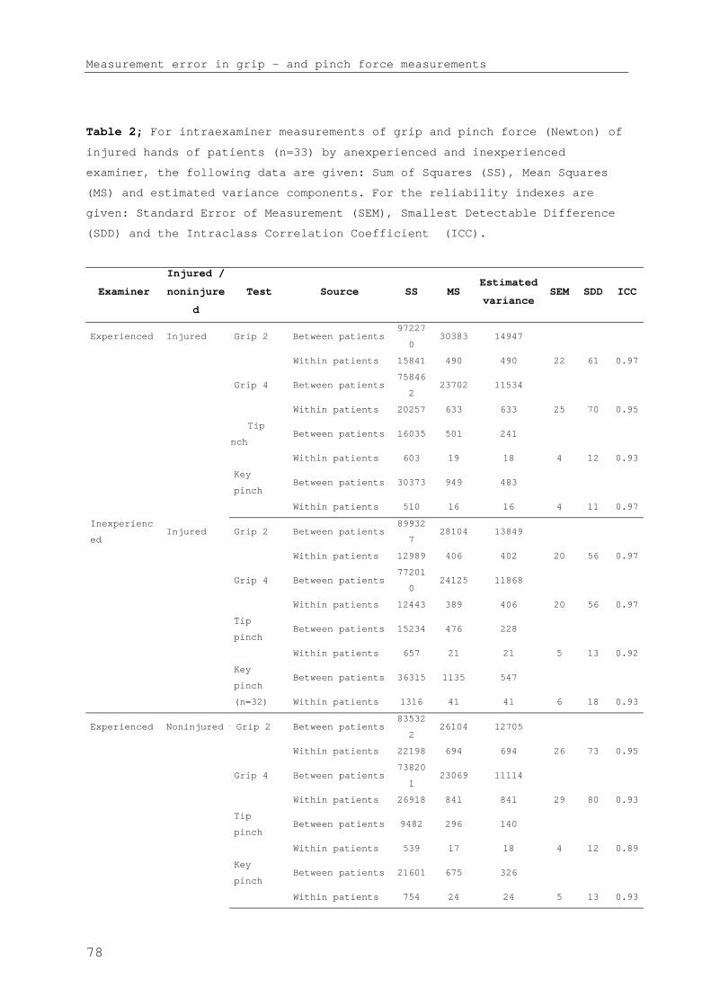
Measurement error in grip - and pinch force measurements
Table 2; For intraexaminer measurements of grip and pinch force (Newton) of
injured hands of patients (n=33) by anexperienced and inexperienced
examiner, the following data are given: Sum of Squares (SS), Mean Squares
(MS) and estimated variance components. For the reliability indexes are
given: Standard Error of Measurement (SEM), Smallest Detectable Difference
(SDD) and the Intraclass Correlation Coefficient (ICC).
Examiner
Injured /
noninjure
d
Test Source SS MSEstimated
varianceSEM SDD ICC
Experienced Injured Grip 2 Between patients97227
030383 14947
Within patients 15841 490 490 22 61 0.97
Grip 4 Between patients75846
223702 11534
Within patients 20257 633 633 25 70 0.95
i Tip
inchBetween patients 16035 501 241
Within patients 603 19 18 4 12 0.93
Key
pinchBetween patients 30373 949 483
Within patients 510 16 16 4 11 0.97
Inexperienc
edInjured Grip 2 Between patients
89932
728104 13849
Within patients 12989 406 402 20 56 0.97
Grip 4 Between patients77201
024125 11868
Within patients 12443 389 406 20 56 0.97
Tip
pinchBetween patients 15234 476 228
Within patients 657 21 21 5 13 0.92
Key
pinchBetween patients 36315 1135 547
(n=32) Within patients 1316 41 41 6 18 0.93
Experienced Noninjuredr Grip 2 Between patients83532
226104 12705
Within patients 22198 694 694 26 73 0.95
Grip 4 Between patients73820
123069 11114
Within patients 26918 841 841 29 80 0.93
Tip
pinchBetween patients 9482 296 140
Within patients 539 17 18 4 12 0.89
Key
pinchBetween patients 21601 675 326
Within patients 754 24 24 5 13 0.93
78

Chapter 5
Inexperienc
edNoninjured Grip 2 Between patients
82279
226542 12922
(n=32) Within patients 21624 698 698 28 77 0.94
Grip 4 Between patients76914
524036 11823
Within patients 13478 408 408 20 56 0.97
Tip
pinchBetween patients 8858 277 124
Within patients 892 28 28 5 15 0.82
Key
pinchBetween patients 23848 745 351
Within patients 1383 43 43 7 18 0.89
Grip 2 are measurements of grip force in the 2nd handle position, ** Grip 4 are measurements
in the 4th handle position
The interexaminer data are presented in Table 3 and show comparable
values.
Table 3. Interexaminer application of grip and pinch force measurements
(Newton) of injured and noninjured hands of patients (n=33). Sum of Squares
(SS), mean Squares (MS) and estimated variance are presented, as well as
the reliability indexes: Standard Error of Measurements (SEM), Smallest
Detectable Difference (SDD) and the Intraclass Correlation Coefficient
(ICC).
Source SS MSEstimated
varianceSEM SDD ICC
Between patients 948096 29628 14584Injured
handsGrip 2
Within patients 16279 493 493 22 62 0.97
Between patients 763813 23869 11508Grip 4
Within patients 27279 852 852 29 81 0.93
Between patients 15805 494 234Tip pinch
Within patients 855 28 29 5 15 0.89
Between patients 33541 1048 509Key pinch
(n=32) Within patients 955 30 30 5 15 0.94
Between patients 835379 26106 12899Noninjured
hands
Grip 2
Within patients 35320 1104 1111 33 92 0.92
Between patients 774343 24198 11857Grip 4
Within patients 15495 484 484 22 61 0.96
Between patients 9579 299 136Tip pinch
Within patients 857 27 27 5 14 0.84
Between patients 21771 680 315Key pinch
Within patients 1594 49 50 7 20 0.86
79

Measurement error in grip - and pinch force measurements
No differences were found in the error variances of grip and pinch
force measurements between injured and noninjured hands or performed
by experienced and inexperienced examiners. Similarly, no
differences were found in the error variances of the measurements
between the 2 handle positions of grip force or between the 2 pinch
techniques.
Tables 4 and 5 present comparisons among studies investigating grip
and pinch force measurements, including the type of dynamometer
used, characteristics of the subjects, means and standard deviations
of force measurements, and reliability indexes (i.e., SEM, SDD, and
ICC). The measurement errors of the SEMs of grip force measurements
are comparable in most of the studies, except for those of Nitschke
et al10 and Smidt et al,12 13 in which relatively low values were found
(Tab. 4). For pinch force measurements, the SEMs were 4 or 5 N, with
the exception of the higher values of 11 N found by Geertzen et al11
(Tab. 5).
80
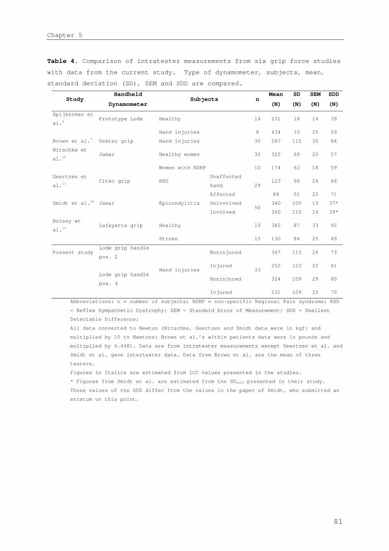
Chapter 5
Table 4. Comparison of intratester measurements from six grip force studies
with data from the current study. Type of dynamometer, subjects, mean,
standard deviation (SD), SEM and SDD are compared.
StudyHandheld
DynamometerSubjects n
Mean
(N)
SD
(N)
SEM
(N)
SDD
(N)
Spijkerman et
al.6Prototype Lode Healthy 16 231 18 14 39
Hand injuries 8 434 10 25 59
Brown et al.7 Dexter grip Hand injuries 30 267 115 30 84
Nitschke et
al.10Jamar Healthy women 32 325 69 20 57
Women with NSRP 10 174 62 18 59
Geertzen et
al.11Citec grip RSD
Unaffected
hand123 66 24 66
Affected
29
84 51 25 71
Smidt et al.12 Jamar Epicondylitis Uninvolved 340 100 13 37*
Involved50
300 110 14 39*
Boissy et
al.13Lafayette grip Healthy 10 382 87 33 92
Stroke 15 130 84 25 69
Present study Lode grip handle
pos. 2 Noninjured 367 115 26 73
Injured 252 123 22 61
Lode grip handle
pos. 4 Noninjured 324 109 29 80
Hand injuries
Injured
33
231 109 25 70
Abbreviations: n = number of subjects; NSRP = non-specific Regional Pain syndrome; RSD
= Reflex Sympathetic Dystrophy; SEM = Standard Error of Measurement; SDD = Smallest
Detectable Difference;
All data converted to Newton (Nitschke, Geertzen and Smidt data were in kgf: and
multiplied by 10 to Newtons; Brown et al.’s within patients data were in pounds and
multiplied by 4.448). Data are from intratester measurements except Geertzen et al. and
Smidt et al. gave intertester data. Data from Brown et al. are the mean of three
testers.
Figures in Italics are estimated from ICC values presented in the studies.
* Figures from Smidt et al. are estimated from the SDdiff presented in their study.
These values of the SDD differ from the values in the paper of Smidt, who submitted an
erratum on this point.
81
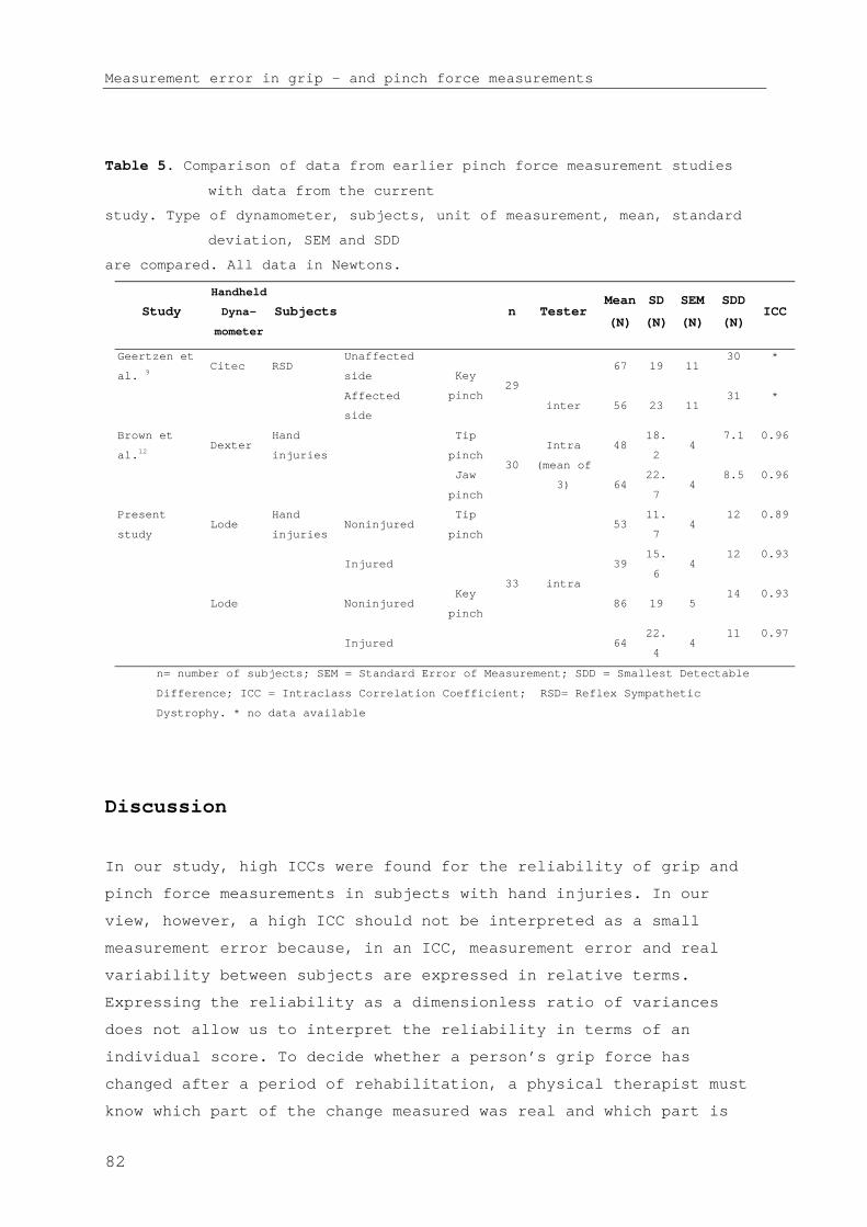
Measurement error in grip - and pinch force measurements
Table 5. Comparison of data from earlier pinch force measurement studies
with data from the current
study. Type of dynamometer, subjects, unit of measurement, mean, standard
deviation, SEM and SDD
are compared. All data in Newtons.
Study
Handheld
Dyna-
mometer
Subjects n TesterMean
(N)
SD
(N)
SEM
(N)
SDD
(N)ICC
Geertzen et
al. 9Citec RSD
Unaffected
side67 19 11
30 *
Affected
side
Key
pinch29
inter 56 23 1131 *
Brown et
al.12Dexter
Tip
pinch48
18.
24
7.1 0.96Hand
injuries
Jaw
pinch
30
Intra
(mean of
3) 6422.
74
8.5 0.96
Present
studyLode Noninjured
Tip
pinch53
11.
74
12 0.89
Injured 3915.
64
12 0.93
Lode NoninjuredKey
pinch86 19 5
14 0.93
Hand
injuries
Injured
33 intra
6422.
44
11 0.97
n= number of subjects; SEM = Standard Error of Measurement; SDD = Smallest Detectable
Difference; ICC = Intraclass Correlation Coefficient; RSD= Reflex Sympathetic
Dystrophy. * no data available
Discussion
In our study, high ICCs were found for the reliability of grip and
pinch force measurements in subjects with hand injuries. In our
view, however, a high ICC should not be interpreted as a small
measurement error because, in an ICC, measurement error and real
variability between subjects are expressed in relative terms.
Expressing the reliability as a dimensionless ratio of variances
does not allow us to interpret the reliability in terms of an
individual score. To decide whether a person’s grip force has
changed after a period of rehabilitation, a physical therapist must
know which part of the change measured was real and which part is
82

Chapter 5
due to measurement error. To accomplish this, we calculated the SEM
and SDD.
In our study, the SDD of the grip force measurements (second handle
position of the injured hands was 61 N for measurements obtained by
the experienced examiner (Tab. 2). This finding means that for grip
force measurements taken by the experienced examiner (Texp), a
change in force of at least 61 N is needed between 2 sessions to be
95% confident that a real change has occurred. The 2 different types
of pinch force measurements (tip pinch and key pinch) showed a much
smaller SDD; for the experienced examiner, the SDDs for the injured
hands were 11 to 12 N (Tab. 2).
In the case of our 2 examiners with widely differing experience,
comparison of the SEMs showed no differences between them. Thus, for
grip and pinch force measurements, it appears that a brief training
period was sufficient for an inexperienced examiner to reach a level
of reliability comparable to that of an experienced examiner. Given
the small number of examiners in our study, however, this cannot be
stated with certainty.
More examiners, however, would entail a longer measurement time,
which might fatigue the patient or decrease his or her
concentration, both of which may influence the measurement error.
We attempted to compensate for the small number of examiners by
selecting 2 examiners with what we considered maximally different
experience. Given that no differences in measurement error between
the 2 examiners were found, we contend that having more examiners
(e.g., with intermediate levels of experience) obtain the
measurements would not have affected our conclusions. Although we do
not have data to support this view, we believe it is a reasonable
interpretation.
Other factors also may influence the measurement error. For
example, we also examined the difference in SEMs between grip and
pinch force measurements of the injured and noninjured hands. The
measurement error, however, did not differ between the injured and
83

Measurement error in grip - and pinch force measurements
noninjured hands. No differences were found between the SEMs of grip
force measurements in the 2 handle positions or between both types
of pinch force measurements. Therefore, for applications in clinical
practice, we contend one SEM value can be applied for grip force
measurements for both handle positions and one SEM value can be
applied for pinch force measurements, irrespective of injury and
experience of the examiner.
Accepted criteria to judge the SDD of grip and pinch force
measurements do not exist. Therefore, we compared our findings with
those of 6 studies in which grip and pinch force measurements were
investigated in similar and different patient categories (i.e.,
patients with hand injuries,6 women with NSRP,10 patients with RSD,11
patients with epicondylitis,12 and patients after a stroke14) (Tab.
4). The SEMs of grip force measurements in these different studies
are comparable. However, the differences in means and ranges of the
measured force levels in these studies are remarkable (e.g., the
mean grip forces in the affected hands ranged from 84 to 300 N). To
determine the value of a particular measurement in a population
during clinical practice, we believe it is essential to know in
which range the changes of muscle force take place. For example, in
people with hand injury, the grip force will be lower shortly after
the trauma. During later phases of rehabilitation, the increased
muscle force generally reaches a plateau after many months. If the
difference in the grip force between the start and end of the
rehabilitation is, for example, 100 N, an SDD of 80 N will mean the
measurement is inadequate to detect changes. If the range in which
the forces change is as wide as 300 N, an SDD of 30 N is certainly
sufficient to detect changes during the rehabilitation process. In
this latter example, a virtual scale of 10 steps is achieved.
To determine whether the calculated measurement error is small
enough to make a test valuable in clinical practice, different
methods have been applied in the studies we cite. Geertzen et al 11
concluded that the SDD is too large in relation to the mean force
measured to be useful in patients with RSD. Smidt et al12 suggested
that an SDD less than 10% of the total range of the measurements
84

Chapter 5
would be acceptable.13 The range, however, is determined by the
extreme values of the measurement, whereas the standard deviation is
less influenced by the extreme values. In our study, therefore, we
re-examined the SDD in relation to the standard deviation of the
measurements by calculating the SDD/standard deviation ratios. The
SDD/standard deviation ratios were 0.5 to 0.6 for grip force
measurements using the second handle position and 0.6 to 0.7 when
using the fourth handle position. These findings mean that 6 to 7
steps of changes in force level can be detected within the 95%
distribution (“virtual scale”) of impaired to normal levels of grip
force. In our opinion, this SDD/standard deviation ratio is useful
in order to gain better insight as to whether measurements can be
used to detect clinical meaningful changes. A more definitive
conclusion concerning the usefulness of the measurements can be made
by relating the SDD relative to the magnitude of changes in force
measurements in a prospective study of patients throughout the
entire rehabilitation process.
Conclusions
According to the ICC values obtained in our study, the reliability
of grip and pinch force measurements is excellent. However,
assessment of the measurement error and detectable change in muscle
force between 2 consecutive measurements demonstrated that, in grip
and pinch force measurements in individual patients, only relatively
large changes can be adequately detected.
Measurement error did not differ between the experienced and
inexperienced examiners or the injured and noninjured hands.
Similarly, no differences were found in the grip force measurements
for the 2 handle positions or in the tip pinch and key pinch force
measurements. Further study of the SDD/standard deviation ratio is
needed to develop clear criteria to judge whether measurement error
is acceptable to detect changes in individual patients. The limited
number of examiners and the use of subjects who were pain-free
should be considered when applying our results. Further study of the
85
Measurement error in grip - and pinch force measurements
SDD/standard deviation ratio is needed to develop clear criteria to
judge whether measurement error is acceptable to detect changes in
individual patients.
References
1. Bear-Lehman J, Abreu BC. Evaluating the hand: issues in reliability and
validity. Phys Ther 1989;69(12):1025-33.
2. Beckerman H, Roebroeck ME, Lankhorst GJ, Becher JG, Bezemer PD, Verbeek AL.
Smallest real difference, a link between reproducibility and responsiveness.
Qual Life Res 2001;10(7):571-8.
3. Dellon AL. A numerical grading scale for peripheral nerve function. J Hand Ther
1993;6(2):152-60.
4. Rosen B, Dahlin LB, Lundborg G. Assessment of functional outcome after nerve
repair in a longitudinal cohort. Scand J Plast Reconstr Surg Hand Surg
2000;34(1):71-8.
5. Strickland JW, Idler, R.S., DelSignore, J.L. Ulnar Nerve Repair. In: Gelberman
RH, editor. Operative Nerve Repair and Reconstruction. Philadelphia: J.B.
Lippincott Comp, 1991:425-436.
6. Spijkerman DC, Snijders CJ, Stijnen T, Lankhorst GJ. Standardization of grip
strength measurements. Effects on repeatability and peak force. Scand J
Rehabil Med 1991;23(4):203-6.
7. Brown A, Cramer LD, Eckhaus D, Schmidt J, Ware L, MacKenzie E. Validity and
reliability of the dexter hand evaluation and therapy system in hand-injured
patients. J Hand Ther 2000;13(1):37-45.
8. Mathiowetz V, Weber K, Volland G, Kashman N. Reliability and validity of grip
and pinch strength evaluations. J Hand Surg [Am] 1984;9(2):222-6.
9. Hamilton A, Balnave R, Adams R. Grip strength testing reliability. J Hand Ther
1994;7(3):163-70.
10. Nitschke JE, McMeeken JM, Burry HC, Matyas TA. When is a change a genuine
change? A clinically meaningful interpretation of grip strength measurements
in healthy and disabled women. J Hand Ther 1999;12(1):25-30.
11. Geertzen JH, Dijkstra PU, Stewart RE, Groothoff JW, ten Duis HJ, Eisma WH.
Variation in measurements of grip strength. A study in reflex sympathetic
dystrophy patients. Acta Orthop Scand Suppl 1998;279:4-11.
12. Smidt N, van der Windt DA, Assendelft WJ, Mourits AJ, Deville WL, de Winter AF,
et al. Interobserver reproducibility of the assessment of severity of
complaints, grip strength, and pressure pain threshold in patients with
lateral epicondylitis. Arch Phys Med Rehabil 2002;83(8):1145-50.
13. de Vet HC, Roebroeck ME, Smidt N, Knol DL, van der Windt DA, Bouter LM.
Erratum. Arch Phys Med Rehabil 2003;84(6):938.
86

Chapter 5
14. Boissy P, Bourbonnais D, Carlotti MM, Gravel D, Arsenault BA. Maximal grip
force in chronic stroke subjects and its relationship to global upper
extremity function. Clin Rehabil 1999;13(4):354-62.
15. MacDermid JC, Kramer JF, Woodbury MG, McFarlane RM, Roth JH. Interrater
reliability of pinch and grip strength measurements in patients with
cumulative trauma disorders. J Hand Ther 1994;7(1):10-4.
16. Streiner DL, Norman GR. Health measurement scales: a practical guide to their
development and use. 2nd ed. Oxford: Oxford University Press,, 1995.
17. Stratford PW, Goldsmith CH. Use of the standard error as a reliability index of
interest: an applied example using elbow flexor strength data. Phys Ther
1997;77(7):745-50.
18. Roebroeck ME, Harlaar J, Lankhorst GJ. The application of generalizability
theory to reliability assessment: an illustration using isometric force
measurements. Phys Ther 1993;73(6):386-95; discussion 396-401.
19. Hopkins WG. Measures of reliability in sports medicine and science. Sports Med
2000;30(1):1-15.
20. Young VL, Pin P, Kraemer BA, Gould RB, Nemergut L, Pellowski M. Fluctuation in
grip and pinch strength among normal subjects. J Hand Surg [Am]
1989;14(1):125-9.
21. Pronk CNA. Technical note; measuring hand-grip force, using a new application
of strain gauges. Med & Biol. Eng & Comput. 1981;19:127-128.
22. ASHT, editor. Clinical Assessment Recommendations. 2nd ed. Garner, NC: American
Society of Hand Therapists, 1992.
23. Gaul JS, Jr. Intrinsic motor recovery--a long-term study of ulnar nerve repair.
J Hand Surg [Am] 1982;7(5):502-8.
24. Roebroeck ME, Harlaar J, Lankhorst GJ. Reliability assessment of isometric knee
extension measurements with a computer-assisted hand-held dynamometer. Arch
Phys Med Rehabil 1998;79(4):442-8.
25. Armitage P, Berry G. Statistical Methods in Medical Research. 3rd ed. Boston:
Mass: Blackwell Sciebtific Publications, 1994.
26. Shrout P, Fleiss J. Intraclass correlation: uses in assessing rater
reliability. Psychol Bull 1979;86:420-428.
87

Measurement error in grip - and pinch force measurements
88

CHAPTER 6
The Rotterdam Intrinsic Hand
Myometer (RIHM) A technical note
Thoughts are mightier than strength of hand
Sophocles (496-406 BC)
T.A.R. Schreuders1, F. Eijskoot2, A.H. den Ouden 3, H.J. Stam 1
1 Department of Rehabilitation Medicine, Erasmus MC - University
Medical Center Rotterdam, P.O. Box 2040, 3000 CA Rotterdam, The
Netherlands. 2 Department of Experimental Medical Instruments (EMI),
Erasmus MC, P.O. Box 1738, 3000 DR Rotterdam, The Netherlands. 3

RIHM; techincal note
Senior Advisor, Medical Technology, Den Hamel 10, St Annaland, The
Netherlands
84

Chapter 6
Abstract
The Rotterdam Intrinsic Hand Myometer (RIHM) is designed to measure
the forces of the intrinsic hand muscles for research and clinical
purposes. Earlier attempts to measure these muscle forces were with
devices designed to measure the abduction force of the thumb, some
were hand-held, or mounted on a jig or standard and could not
measure the opposition of the thumb. All such instruments made
measurements by pushing on the digit, increasing the risk to produce
forces not perpendicular to the digits. Our device is a hand-held
dynamometer that uses a pulling method and has novel technical
features which improved reliability.
Introduction
In the assessment of the muscle forces of the intrinsic muscles of
the hand there is a need for specific measurements of these muscles
in isolation. Dynamometers are available that measure grip strength
and pinch strength of the hand 1-4, but few dynamometers have the
possibility to measure the intrinsic muscles of the hand
separately.5-8 Knowledge on the specific force of the intrinsic
muscles will provide important information for developing, for
example, new methods to repair peripheral nerves of the hand, new
therapies aimed at strengthening the intrinsic muscles of the hand,
and bio-mechanical analyses of muscles forces of the lumbricals and
interossei muscles in the clinical situation.
A previous study tested the reliability of measurements made with a
generic industrially designed hand-held dynamometer (AIKOH) to
assess the force of several of the intrinsic muscles of the hand. It
was found that only relatively large changes in intrinsic muscle
force could be detected 9. Another disadvantage was that, for the
median nerve innervated muscles of the thumb, only one muscle could
be measured i.e. the Abductor Pollicis Brevis (APB), while several
ulnar nerve innervated muscles could be measured. Measuring the APB
with sufficient isolation from the other thenar muscles providing
85
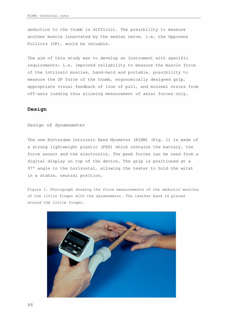
RIHM; techincal note
abduction to the thumb is difficult. The possibility to measure
another muscle innervated by the median nerve, i.e. the Opponens
Pollicis (OP), would be valuable.
The aim of this study was to develop an instrument with specific
requirements: i.e. improved reliability to measure the muscle force
of the intrinsic muscles, hand-held and portable, possibility to
measure the OP force of the thumb, ergonomically designed grip,
appropriate visual feedback of line of pull, and minimal errors from
off-axis loading thus allowing measurement of axial forces only.
Design
Design of dynamometer
The new Rotterdam Intrinsic Hand Myometer (RIHM) (Fig. 1) is made of
a strong lightweight plastic (PED) which contains the battery, the
force sensor and the electronics. The peak forces can be read from a
digital display on top of the device. The grip is positioned at a
97° angle to the horizontal, allowing the tester to hold the wrist
in a stable, neutral position.
Figure 1. Photograph showing the force measurements of the abductor muscles
of the little finger with the dynamometer. The leather band is placed
around the little finger.
86

Chapter 6
An important difference compared with other instruments is the
pulling technique of the RIHM, whereby the forces are measured by
pulling on a leather band placed on the digit. Placing a band around
the thumb allows measuring the forces of the OP muscle.
From the tester’s viewpoint pulling towards their own body with
their upper arm supported against the side of the thorax enables
better control than pushing. For practical reasons (e.g. hand size)
a 15-cm long leather band is used; this length allows the angle of
deviation angle to be easier observed.
To further decrease any erroneous forces introduced by the tester, a
small cylindrical part is connected to the handgrip of the
instrument by means of a ball joint, and contains the button load
cell (type BC301 from DS Europe). This construction, together with a
rotating handgrip, ensures loading perpendicular to the load cell
(Fig. 2). This prevents the examiner from introducing torques in the
horizontal and vertical planes, as well as rotation around the line
of work and rotation of the band around the finger.
Electronic circuit
The handgrip of the device houses the rechargeable battery, the
curved part houses the printed circuit, and the display unit with
control buttons is on top of the device. A programmable LCD digital
panel meter (LASCAR type SM1) displays the maximal reading. This
microcontroller-based module is designed around an alphanumeric LCD.
The LED back light display shows the 4.5 digit voltmeter readings as
well as a comprehensive operator menu. The user can select the
desired range, decimal place, as well as calibration settings.
Because all set-ups, including calibration, are performed via
software, there are no user adjustable potentiometers. The module
stores all settings when the power is switched off. The unit is
powered by the internal 9-volt rechargeable NiMHy battery or an
external plug in a power adapter; when connected to this adapter the
87

RIHM; techincal note
battery will be charged automatically. To save power, the device
automatically switches off every few minutes.
Figure 2 Drawing of the Rotterdam Intrinsic Hand Myometer (RIHM)
A diagram of the electronic circuit (Fig. 3) shows the strain gauge
bridge of the load cell (left-hand side): the bridge signal is
amplified by the differential amplifier 1, and amplifier 2 performs
lowpass filtering and level shifting. The force curve signal is
available on the force output connector connected to this amplifier.
The force curve signal is connected to a peak detector with a fast
peak response of < 10 msec.
Figure 3 Diagram of the electronic circuit of the RIHM
88
Chapter 6
Clinical experience
Reliability of the RIHM has been studied in patients with peripheral
nerve injuries and was found to have smaller measurement error
compared with the AIKOH measurements.10 In clinical use the
measurements have provided important data showing that the
conventionally used grip and pinch strength dynamometers do not
adequately reflect recovery of the intrinsic muscles.11
We conclude that the RIHM is an easy to use (hand-held) instrument
providing reliable measurements of the intrinsic muscles of the
hand.
89

RIHM; techincal note
Acknowledgement
The authors would like to thank Mr. R. den Ouden, Ms. B. Bartels,
Mr. M. Duchenne and Mr. R-J. Visser (students from the School for
Human Technology, The Hague) who made important contributions to
this project.
References
1. Bechtol C. Grips test: the use of a dynamometer with adjustable handle spacing.
J Bone Joint Surg 1954;36:820.
2. An KN, Chao EY, Askew LJ. Hand strength measurement instruments. Arch Phys Med
Rehabil 1980;61(8):366-8.
3. Mathiowetz V, Kashman N, Volland G, Weber K, Dowe M, Rogers S. Grip and pinch
strength: normative data for adults. Arch Phys Med Rehabil 1985;66(2):69-74.
4. Rosen B, Lundborg G. A model instrument for the documentation of outcome after
nerve repair. J Hand Surg [Am] 2000;25(3):535-43.
5. Mannerfelt L. Studies on the hand in ulnar nerve paralysis. A clinical-
experimental investigation in normal and anomalous innervation. Acta Orthop
Scand 1966(87):61-86.
6. Trumble TE, Kahn U, Vanderhooft E, Bach AW. A technique to quantitate motor
recovery following nerve grafting. J Hand Surg [Am] 1995;20(3):367-72.
7. Boatright JR, Kiebzak GM. The effects of low median nerve block on thumb
abduction strength. J Hand Surg [Am] 1997;22(5):849-52.
8. Liu F, Carlson L, Watson HK. Quantitative abductor pollicis brevis strength
testing: reliability and normative values. J Hand Surg [Am] 2000;25(4):752-9.
9. Schreuders TAR, Roebroeck ME, van de Kar HJ, Soeters JNM, Hovius SER, Stam HJ.
Strength of the intrinsic muscles of the hand measured with a hand-held
dynamometer: reliability in patients with ulnar and median nerve paralysis. J
Hand Surg [Br] 2000;25(6):560-5.
10. Schreuders TAR, Roebroeck ME, Jaquet J-B, Hovius SER, Stam HJ. Measuring the
strength of the intrinsic muscles of the hand in patients with ulnar and
median nerve injury; reliability of the Rotterdam Intrinsic Hand Myometer
(RIHM). J Hand Surg (Am) 2004;29A(2):318-324.
11. Schreuders TAR, Roebroeck ME, Jaquet J-B, Hovius SER, Stam HJ. Longterm outcome
of muscle strength in patients with ulnar and median nerve injury: Comparing
manual muscle strength testing, grip and pinch strength dynamometers and a
new intrinsic muscle strength dynamometer. J Rehabil Med 2004;36.
90
Emanuel B Kaplan.
T.A.R. Schreuders,a G.A. Hoek van Dijke,b F. Guler Uysal,c Th. H Stijnen,d M.E.
Roebroeck,a H.J. Stam.a
a Department of Rehabilitation Medicine, Erasmus MC, University Medical Center, Rotterdam,
The Netherlands b Department of Biomedical Physics and Technology, Erasmus MC,
University Medical Center, Rotterdam, The Netherlands c Cukurova University, School of

Analysis of the application of force
Medicine, Department of Physical Medicine and Rehabilitation, Adana, Turkey d Department of
Epidemiology and Biostatistics, Erasmus MC, University Medical Center, Rotterdam, The
Netherlands
. To assess the accuracy of measurement performance of two hand held
dynamometers in the application of force measurements of the intrinsic muscles of
the hand.
. Repeated measurements with a generic dynamometer (AIKOH) using a pushing
technique and with a newly designed instrument, the Rotterdam Intrinsic Hand
Myometer (RIHM) using a pulling technique.
. Three force measurements were studied of the right hand of ten healthy
persons: little finger abduction, index finger abduction and thumb palmar
abduction. The position and angle of application was recorded with a three
dimensional videorecording technique. Three repeated measurements within a session
were averaged to determine the systematic differences and variances of the
measurements of the two dynamometers
. The variance was smaller for all three measurements of the RIHM regarding
the positioning of application and the angle of application of the index finger
abduction in the frontal plane. Systematic differences between the dynamometers
were found only for the angle of application in the frontal plane of index finger
and thumb measurements in favour of the RIHM.
. Performance of force measurements of the intrinsic muscles of the hand
with the RIHM dynamometer had less variances than the AIKOH dynamometer in the
positioning of the instruments at the same anatomical reference point of the
finger. The difference in angle of application has much smaller influence on the
variance of measurements between the two instruments.
Instruments for force measurements of the hand are important in
evaluating the hand function. A new force measuring instrument, the Rotterdam
90

Chapter 7
Intrinsic Hand Myometer (RIHM), has been developed with a pulling technique to
improve reliability. Because small inaccuracies in applying a dynamometer may have
considerable consequences for measurement error, determining the cause of
measurement error will provide information on how to improve measurement
instruments.
91

Analysis of the application of force
Weakness in muscle force resulting from neurological or musculoskeletal lesions can
greatly reduce the patient's hand function and often cause major impairments and
disabilities. Measurements of the muscle force of the hand through objective and
reliable methods are important in order to follow the recovery rate and
effectiveness of treatments after upper extremity nerve injuries 1 3, or changes of
muscle force in several hereditary motor and sensory neuropathies.4 5 Different
methods for monitoring hand function have been investigated by many researchers who
work in the area of hand rehabilitation.6 11
Measurement of muscle force is possible by non instrumental techniques such as
manual muscle strength testing (MMST), or instrumental techniques such as grip and
pinch force dynamometers12 and electro physiological methods. Mannerfelt was one of
the first to measure the intrinsic muscle force of the hand with a dynamometer.13
Later, others have designed devices for e.g. force measurements of the index finger
14 16 and intrinsic thenar muscles of the thumb.17 19
In a previous study we assessed the reliability of force measurements of the
intrinsic muscles of the hand with a generic industrially designed hand held
dynamometer using the pushing technique (AIKOH) in 24 patients with ulnar and/or
median nerve injury.20 The Standard Error of Measurements (SEM) for the little
finger, index finger and thumb abduction were 10, 11 and 12 N, respectively.20 We
concluded that only relatively large changes in intrinsic muscle force could be
confidently detected. With a specifically designed dynamometer (RIHM), using a
pulling technique (Fig. 1), similar muscle strength measurements in 27 patients
with ulnar and/or median nerve injuries showed much lower SEMs of 2.2, 2.3, 5.8 N,
respectively.21
We hypothesised that this reduction of measurement error was mainly due to better
standardisation of these measurements due to an improved consistency of
application of the dynamometer at the right position and the right angle during
testing. The dynamometer is usually placed at a joint crease because it is easy to
identify. Positioning of the dynamometer needs to be done consistently at
precisely the same position (Fig. 2); e.g. when the muscles around the meta
92
Chapter 7
carpophalangeal (MCP) joint can deliver a net moment of 25 Nm, the force that is
measured at a position (arm) of 5 cm to the joint ( in Figure 1), will be 5 N
(force = moment/arm). However, when the dynamometer is positioned 0.05 m ( ) more
proximally from that point, making the arm 0.045 m from the MCP joint, the force
will be 2.5/0.045 = 5.5 N, i.e. about 10% higher than in case of accurate
placement.
Regarding the angle of application, an accurate measurement is performed when the
dynamometer is positioned perpendicular to the finger and thumb in two directions;
in the frontal plane of the hand (dividing the hand into an anterior and posterior
portion) and the transverse plane (dividing the hand into a distal and proximal
halve). A change in the angle between the device and the finger may cause large
differences in outcome force. For example, applying the dynamometer at an angle of
25° will cause an underestimation of the measured force of almost 10% (cosinus 25°
= 0.91).
Thus, relatively small changes in the position and angle when applying the
dynamometer may have considerable consequences for measurement error. The aim of
this study was to compare the accuracy of the older industrial dynamometer (AIKOH)
with a newly designed instrument, the Rotterdam Intrinsic Hand Myometer (RIHM)22 ,
in positioning the instruments at exactly the same point of the finger and
applying it at the correct angle to the digit in the two planes.
Both the angle and position of application were determined with a three
dimensional (3D) videorecording technique.23 25 Measurements with the two
instruments were compared with respect to the consistency and variance of
positioning and angle of force application during testing.
93

Analysis of the application of force
Figure 1. Measuring the
abduction force of the
little finger. A = measuring
the force at the PIP joint,
with the right distance X
(arm) from the MCP joint,
and perpendicular to the
finger in both planes (=
90°); B= when the
dynamometer is erroneously
held at a 25° angle; C= when
the dynamometer is
erroneously held at a
distance Z from the correct
point at the PIP joint.
The right hand of 10 healthy persons was tested. These were 5 males and 5 females
with a mean age of 31.5 (SD 7,1) years. Three force measurements were analysed:
little finger abduction, index finger abduction and thumb palmar abduction (Fig.
2).
94

Chapter 7
Figure 2. Force measurement of the abduction of the thumb with the RIHM dynamometer .
The first two movements are produced by ulnar nerve innervated muscles and the
latter is a movement produced by the thenar muscles of the thumb, which is mainly
a median nerve
innervated function. For little finger abduction the resistance force was applied
at the ulnar side of the proximal interphalangeal (PIP) joint crease of the little
finger. For the index finger abduction, the anatomical reference point was chosen
at the radial side of the PIP joint and for the thumb movement this was the radial
side of the MCP joint crease. Hand position, fixation and anatomical reference
point for the application of resistance force were comparable to the MMST
technique.26
The volunteer and examiner were seated at a table opposite each other.
Participants were instructed how to hold the finger/thumb in the maximally
abducted position and were told to maintain the finger in that position, while the
examiner pushed or pulled the finger in the opposite direction with the
dynamometer. An experienced tester familiar with both instruments (TARS) completed
95
Analysis of the application of force
all the measurements.
This study employed two hand held instruments: the generic AIKOH dynamometer
(Aikoh Model 9520A.B. Aikoh Engineering Co., Ltd. Tokyo, Japan) using a push
technique, and the specifically designed RIHM dynamometer (Experimental Medical
Instruments department of the Erasmus MC, Rotterdam, the Netherlands) with a pull
technique. Three measurements, comprising one session, were performed, then the
dynamometer was removed and the person asked to move the hand out of the testing
position. The dynamometer was positioned again at the same anatomical reference
point and another session of three measurements was carried out. This was repeated
one more time, providing three sessions of three measurements per movement. Both
the order of the measurements (i.e. testing the little finger, index finger and
thumb) and the order of the testing with the two instruments were randomised.
In order to obtain 3D position measurements, two videocameras were used to record
the signals from reflective markers on the hand and dynamometers. These markers
reflected the infrared light sent from a light source attached to the camera.
Three pairs of infrared light reflecting markers were used (Fig. 3). For the AIKOH
one pair (no.1 and 2) was on the dynamometer and for the RIHM the markers were on
the pull string. A small bar with two markers (no. 3 and 4) was taped on the skin
above the metacarpal bone such that these two markers were parallel to the plane
of the hand. The third pair (no. 5 and 6) was located on a small splint which was
attached with two Velcro straps to the distal phalanx of the finger.
96

Chapter 7
Figure 3. Force measurement of the abduction of the little finger with the AIKOH
dynamometer. Reflective markers no. 1 and 2 on the dynamometer, no. 3 and 4 on a small bar
which was taped on the skin above the metacarpal bone, no. 5 and 6 were fixed on a small
splint, attached with two Velcro straps to the distal phalanx of the finger.
The markers were illuminated by an infrared light source mounted on the cameras.
The image co ordinates from the two cameras were combined to 3D spatial co
ordinates using Direct Linear Transformation.23 The video signal was digitised
using a Vision Dynamics1 VCS 512 videoprocessing board to calculate the marker co
ordinates. From previous tests, the resolution of the system proved to be about
0.1 mm.24
To record the point of application of the dynamometer, the position was determined
by measuring the distance between one marker on the small splint at the fingertip
(no. 5) and a virtual line through the two markers on the dynamometer (no. 1 and
2), which provided the position in millimetres (mm). Systematic difference was not
1Hemel Hempstead, UK
97
Analysis of the application of force
relevant because the position of markers 3 and 4 was chosen at a point which gave
the least movement; proximal of the MCP joint. The exact moment arm between the
MCP joint and the position of application at the IP joint could therefore not be
established. Regarding the angle of application of the dynamometer, a perfect
measurement would provide two 90° angles, recorded as a zero degree difference in
angle in both the frontal and transverse plane of the hand. Systematic differences
could be calculated for angle measurements.
For the nine measurements, mean and SD were calculated. Mean differences in
position (mm) and angles (degrees) of application between both instruments were
calculated. To test whether differences exist between both instruments regarding
the variance of position and angle of force applications between sessions, a
repeated measurements analysis was carried out using Proc Mixed of the Statistical
Analysis System (SAS version 8.2 of the SAS Institute Inc., Cary, NC, USA). Three
repeated measurements within a session were averaged, and the analysis was done on
the three session averages. In the statistical models, ‘person‘ was a random
factor and ‘instrument’ was a fixed factor. For each outcome, two models were
fitted. The first model assumed that the between sessions variance was equal for
both instruments, the second model allowed that the variances were different. The
likelihood ratio test comparing these models was used to test the null hypothesis
that the between sessions variance of the two instruments was identical.
Table 1 presents data (mean, SD) on the position (mm) at which the two
dynamometers (AIKOH and RIHM) were placed for measurement of the little finger,
index finger and thumb abduction. There was a significant difference between the
two instruments for the thumb abduction and little finger abduction measurements.
The variance between three sessions of positioning the dynamometer was smaller (p
< 0.0001) for all three finger measurements with the RIHM as compared to the AIKOH
measurements.
Table 1 also presents data (mean, SD) on the angles, i.e. deviations from 0°,
recorded in two planes (frontal and transverse) with both dynamometers. In the
frontal plane the mean of the angle measurements of the RIHM dynamometer was
smaller (closer to 0°) for the index finger and thumb measurements as compared to
98
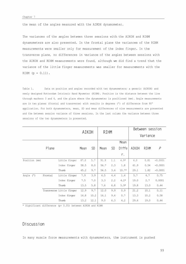
Chapter 7
the mean of the angles measured with the AIKOH dynamometer.
The variances of the angles between three sessions with the AIKOH and RIHM
dynamometers are also presented. In the frontal plane the variances of the RIHM
measurements were smaller only for measurement of the index finger. In the
transverse plane, no differences in variance of the angles between sessions with
the AIKOH and RIHM measurements were found, although we did find a trend that the
variance of the little finger measurements was smaller for measurements with the
RIHM (p = 0.11).
Table 1. Data on position and angles recorded with two dynamometers; a generic (AIKOH) and
newly designed Rotterdam Intrinsic Hand Myometer (RIHM). Position is the distance between the line
through markers 3 and 4, and the place where the dynamometer is positioned (mm). Angle measurements
are in two planes (frontal and transverse) with results in degrees (°) of difference from 90°
application. For both dynamometers, mean, SD and mean differences of nine measurements are presented
and the between session variance of three sessions. In the last column the variance between three
sessions of the two dynamometers is presented.
Position (mm) Little finger 87.0 5.7 91.9 1.1 4.9* 6.0 0.81 <0.0001
Index finger 58.5 8.0 56.7 2.3 1.8 61.9 0.54 <0.0001
Thumb 65.2 9.7 54.5 3.6 10.7* 29.1 1.82 <0.0001
Angle (°) Frontal Little finger 7.9 3.9 6.5 4.4 1.4 5.7 4.7 0.75
Index finger 7.5 7.0 3.3 2.2 4.2* 19.0 2.7 0.0001
Thumb 13.5 5.8 7.6 6.8 5.9* 19.8 13.0 0.44
Transverse Little finger 12.9 9.7 12.0 9.9 0.9 21.2 10.1 0.11
Index finger 16.8 10.2 16.1 9.6 0.7 13.3 10.2 0.58
Thumb 13.2 12.1 9.0 6.3 4.2 29.6 19.0 0.44
* Significant difference (p> 0.05) between AIKOH and RIHM
In many muscle force measurements with dynamometers, the instrument is pushed
99
Analysis of the application of force
against the body while the patient tries to resist the movement, e.g. the microFET
27, Citec 28, Nicholas CANDHY 29 and Kin Com.30 In our previous study on force
measurements of the intrinsic muscles of the hand with the AIKOH, a similar
pushing method was used. In an attempt to decrease the measurement error, we
questioned whether the reliability could be improved by using a pulling technique,
which may allow for a better visual standardisation of measurement performance.
In our previous studies on the intraobserver reliability of the forces measured
with the AIKOH and the RIHM dynamometers, in a comparable group of patients with
injuries to the ulnar and median nerve the Standard Error of Measurements (SEM)
were much lower for the RIHM measurements.21 We hypothesised that this reduction in
measurement error might be due to better standardisation of these measurements
with the RIHM dynamometer due to the improved consistency of the application of
the dynamometer at the right position and the right angle during testing.
The present study analysed the control of the point and angle of application of
the older (pushing technique) and the new dynamometer with the pulling technique.
Differences between the two instruments were found, especially in the position of
application. We found that the pulling technique with the RIHM showed systematic
differences for two measurements, while the variances between three sessions was
smaller for all three measurements of the RIHM dynamometer. Apparently the point
at which the dynamometer is placed is an important source of measurements error.
Standardisation of this aspect might be even further improved by e.g. drawing of a
small line on the finger/thumb.
In the angle of application the main differences were found between the AIKOH and
RIHM in the frontal plane measurements. This might be due to the fact that the
finger will move in this plane during testing. This knowledge is useful in
training examiners in a standardised performance of the measurements and thus
might improve the reliability and standardisation of measurement protocols.
Besides the method of application of resistance force by pulling (RIHM) or by
pushing (AIKOH), there are other factors which could affect the measurement error.
For example, differences in the size and shape of the grip between both
dynamometers and the position of the arm of tester might also influence the
measurement error. Since the RIHM could be held closer to the body of the tester,
100

Chapter 7
less strength was needed to control the dynamometer.
The described video technique provided a method to accurately quantify how the
application of the two dynamometers compared while applying a force to the hand.
Application of the splint with the two markers was straightforward, but placement
of the small bar with the markers 3 and 4 was more difficult and some minor
movements where inevitable due to movements caused by the muscles and tendons just
beneath the skin were the marker was placed. However, the same marker placement
was used for measurements of both instruments, therefore any error would have had
the same systematic inaccuracy for both dynamometers.
One limitation of this study is that the same experienced tester performed all
measurements. The difference in variances between the two instruments might have
been less obvious when performed by a person without experience. The results of
this study may help an inexperienced tester to quicker learn how to perform
adequate and reliable tests.
In conclusion, we found less variation between sessions in positioning of the
dynamometer in measuring muscle force and a smaller systematic error in the
frontal plane. The performance of several force measurements of the intrinsic
muscles of the hand with the newly designed RIHM dynamometer was more accurate and
less variable than with the AIKOH dynamometer, especially in positioning the
instrument at the same reference point of the finger. The difference in angle of
application has a much smaller influence on the variance of measurements between
the two instruments.
1. Brandsma JW. Intrinsic minus hand: (patho) kinesiology, rehabilitation and reconstruction.
[Thesis]. University of Utrecht, 1993.
2. Lundborg G. A 25 year perspective of peripheral nerve surgery: evolving neuroscientific concepts
and clinical significance. 2000;25(3):391 414.
3. Rosen B, Dahlin LB, Lundborg G. Assessment of functional outcome after nerve repair in a
longitudinal cohort. 2000;34(1):71 8.
4. Florence JM, Pandya S, King WM, Robison JD, Baty J, Miller JP, et al. Intrarater reliability of
manual muscle test (Medical Research Council scale) grades in Duchenne's muscular dystrophy.
101
Analysis of the application of force
1992;72(2):115 22; discussion 122 6.
5. Vinci P, Esposito C, Perelli SL, Antenor JAV, Thomas FP. Overwork weakness in Charcot Marie Tooth
disease. 2003;84:825 827.
6. ASHT, editor. . 2nd ed. Garner, NC: American Society of Hand
Therapists, 1992.
7. Bear Lehman J, Abreu BC. Evaluating the hand: issues in reliability and validity.
1989;69(12):1025 33.
8. Bruyns CN, Jaquet JB, Schreuders TA, Kalmijn S, Kuypers PD, Hovius SE. Predictors for return to
work in patients with median and ulnar nerve injuries. 2003;28(1):28 34.
9. Hunter J, Schneider LH, Mackin EJ. : Mosby co.,
2002.
10. Sollerman C, Sperling L. Evaluation of ADL function especially hand function.
1978;6:139 43.
11. Light CM, Chappell PH, Kyberd PJ. Establishing a standardized clinical assessment tool of
pathologic and prosthetic hand function: normative data, reliability, and validity.
2002;83(6):776 83.
12. Bohannon RW. Measurement, nature, and implications of skeletal muscle strength in patients with
neurological disorders. 1995;10(6):283 292.
13. Mannerfelt LG. Studies on ulnar nerve compression neuropathies with a new computerised
instrument the intrins o meter. 1997;31(3):251 60.
14. Zijdewind I, Kernell D. Index finger position and force of the human first dorsal interosseus
and its ulnar nerve antagonist. 1994;77(2):987 97.
15. Li ZM, Zatsiorsky VM, Latash ML. Contribution of the extrinsic and intrinsic hand muscles to the
moments in finger joints. 2000;15(3):203 11.
16. Li ZM, Pfaeffle HJ, Sotereanos DG, Goitz RJ, Woo SL. Multi directional strength and force
envelope of the index finger. 2003;18(10):908 15.
17. Liu F, Carlson L, Watson HK. Quantitative abductor pollicis brevis strength testing: reliability
and normative values. 2000;25(4):752 9.
18. Boatright JR, Kiebzak GM, O'Neil DM, Peindl RD. Measurement of thumb abduction strength:
normative data and a comparison with grip and pinch strength.
1997;22(5):843 8.
19. Trumble TE, Kahn U, Vanderhooft E, Bach AW. A technique to quantitate motor recovery following
nerve grafting. 1995;20(3):367 72.
20. Schreuders TAR, Roebroeck ME, van de Kar HJ, Soeters JNM, Hovius SER, Stam HJ. Strength of the
intrinsic muscles of the hand measured with a hand held dynamometer: reliability in patients
with ulnar and median nerve paralysis. 2000;25(6):560 5.
21. Schreuders TAR, Roebroeck ME, Jaquet J B, Hovius SER, Stam HJ. Measuring the strength of the
intrinsic muscles of the hand in patients with ulnar and median nerve injury; reliability of
the Rotterdam Intrinsic Hand Myometer (RIHM). 2004;29A(2):318 324.
22. Schreuders TAR, Eijskoot F, den Ouden AH, Stam HJ. The Rotterdam Intrinsic Hand Myometer (RIHM);
A technical note. .
23. Spoor CW. Balancing a force on the fingertip of a two dimensional finger model without intrinsic
muscles. 1983;16(7):497 504.
24. van den Bosch EW, van Zwienen CM, Hoek van Dijke GA, Snijders CJ, van Vugt AB. Sacroiliac screw
102

Chapter 7
fixation for tile B fractures. 2003;55(5):962 5.
25. Coert JH, van Dijke HG, Hovius SE, Snijders CJ, Meek MF. Quantifying thumb rotation during
circumduction utilizing a video technique. 2003;21(6):1151 5.
26. Brandsma JW, Schreuders TA, Birke JA, Piefer A, Oostendorp R. Manual muscle strength testing:
intraobserver and interobserver reliabilities for the intrinsic muscles of the hand.
1995;8(3):185 90.
27. van Meeteren J, Roebroek ME, Stam HJ. Test retest reliability in isokinetic muscle strength
measurements of the shoulder. 2002;34(2):91 95.
28. Geertzen JH, Dijkstra PU, Stewart RE, Groothoff JW, ten Duis HJ, Eisma WH. Variation in
measurements of grip strength. A study in reflex sympathetic dystrophy patients.
1998;279:4 11.
29. Roebroeck ME, Harlaar J, Lankhorst GJ. Reliability assessment of isometric knee extension
measurements with a computer assisted hand held dynamometer.
1998;79(4):442 8.
30. Stratford PW, Balsor BE. A comparison of make and break tests using a hand held dynamometer and
the Kin Com. 1994;19(1):28 32.
103
CHAPTER 8
Measuring the Strength of the
Intrinsic Muscles of the Hand in
Patients with Ulnar and Median
Nerve Injuries; Reliability of the
Rotterdam Intrinsic Hand Myometer
(RIHM)
The human hand is so beautifully formed, it has so fine a sensibility, that
sensibility governs its motions so correctly, every effort of the will is answered
so instantly, as if the hand itself were the seat of the will; its action are so
powerful, so free, and yet so delicate, as if it possessed quality of instinct in
itself, that there is no thought of its complexity as an instrument, or of the
relations which make it subservient to the mind.
Charles Bell (1774-1842)
The Hand - Its Mechanism and Vital Endowments as Evincing Design
(1833)
Reliability RIHM
Reprinted from Journal of Hand Surgery (Am), Vol 29A. Schreuders
TAR, Roebroeck ME, Jaquet J-B, Hovius SER, Stam HJ. Measuring the
strength of the intrinsic muscles of the hand in patients with ulnar
and median nerve injury; reliability of the Rotterdam Intrinsic Hand
Myometer (RIHM). p 318-324, 2004. With permission from American
Society for Surgery of the Hand.
Abstract
The aim of this study was to determine the reliability and
measurement error of measurements of the intrinsic muscle strength
of a new hand-held dynamometer (the Rotterdam Intrinsic Hand
Myometer; RIHM).
With the RIHM we obtained repeated measurements of the intrinsic
muscle strength of the hand in 27 patients with peripheral nerve
injury of the ulnar and/or median nerve in different stages of
their rehabilitation. The average time period after injury was 4.4
years (range 99 days - 11 years).
Results show that the Intraclass Correlation Coefficients (ICC’s)
were higher than 0.93. For the two ulnar nerve innervated movements
the Standard Error of Measurements (SEMs) were: 2.2 and 2.3 N, and
the Smallest Detectable Differences (SDDs) were 6.1 and 6.3 N,
respectively. For the two median nerve innervated movements of the
thumb SEMs were 5.8 and 5.5 N, and SDDs 16 N and 15.3 N,
respectively.
In patients with nerve injuries the muscle strength is usually
assessed with manual muscle strength testing (MMST) and grip and
pinch strength dynamometers. Preferably the intrinsic muscle
strength should be measured in isolation and quantitatively. The
RIHM is a new dynamometer, which allows for measurements of the
intrinsic muscle strength in isolation, with reliability comparable
to grip and pinch measurements.
102

Chapter 8
Introduction
Ulnar and/or median nerve pathology, e.g. injury, compression or
infection (leprosy) causes a strength deficit of the intrinsic
muscles which may result in severe loss of hand function. In
addition, several neuromuscular diseases (e.g. Charcot-Mary-Tooth,
Guillain-Barré) can cause loss of intrinsic hand muscle function.
Muscle strength measurements are frequently made in patients with
hand injuries and other hand problems, being an integral part of the
physical examination and providing information for diagnosis and for
the outcome of surgery and therapy.1-4 The most commonly used
classification scale for manual muscle strength testing (MMST) is
the Medical Research Council scale (MRC scale).5 The MRC uses a 6-
point numeric scale (grades 0-5) and suggests a constant distance
between points. However, because it is in fact an ordinal scale with
disproportional distances between grades, it might have been more
appropriate to use terms such as normal, good, fair, trace and
paralysed.
With the use of this 6 point ordinal scale for MMST it would be too
difficult to identify those factors that may have small but additive
beneficial effects and those that may have negative effects on nerve
regeneration.6 In contrast with the MMST, measurements with a
dynamometer are more sensitive to change and render outcome on a
continuous scale, e.g. kilogram force or Newton. An additional
advantage of such measurement methods is that the strength of the
uninjured hand can serve as the reference value for "normal" for
that person.7
In the evaluation of peripheral nerve function, grip and pinch
strength measurements with dynamometers have often been recommended
as a measure of outcome.8-10 An important comment of caution in the
usage of these dynamometers was made by Strickland et al. who argued
that grip and pinch strength data may only be used as an indirect
103

Reliability RIHM
measurement of nerve recovery.9 It is commonly overlooked that grip
strength measurements provide information on the combined function
of all the intrinsic and extrinsic muscles of the hand.
We have frequently observed that patients with weak intrinsic
muscles of the hand after ulnar and/or median nerve lesion do have a
considerable strong grip. The strength of the intrinsic muscles of
the hand affect the grip strength only to a certain level.
Improvement in grip strength can merely be a reflection of
compensatory strengthening of the, non-involved, extrinsic
musculature. Therefore, grip strength measurements do not provide
decisive information about the motor function of the ulnar and
median nerve in the lower arm. However, the hand-surgery literature
is replete with grip strength data used to measure the outcome after
nerve repair.
If we want to accurately evaluate the outcome of motor function
after nerve repair we need to distinguish between extrinsic and
intrinsic muscle strength. Clinically this can only be done with a
precision instrument measuring the strength of the intrinsic muscles
in isolation. Several methods to measure the muscle strength of the
hand have been developed.6 11-15 A number of these instruments can
measure the strength of the ulnar-innervated muscles that produce
abduction of the index and little finger. Some instruments can
measure the strength of the abduction of the thumb. However, in
measuring the abduction strength of the thumb the wrist position
needs to be controlled to minimise the action of the radial
innervated abductor pollicis longus muscle.3
A new hand-held dynamometer, the Rotterdam Intrinsic Hand Myometer
(RIHM), that meets the specific needs of measuring the intrinsic
muscle strength of the hand in isolation has been designed and
produced by Experimental Medical Instrumentation (EMI, P.O. Box
1738, 3000 DR Rotterdam, The Netherlands). The RIHM allows measuring
the abduction of the little finger and index finger. In addition the
opposition and palmar abduction of the thumb can be measured, making
104

Chapter 8
two measurements available for both median and ulnar nerve
innervated intrinsic muscles.
The aim of the present study is to assess the reliability of
measurements with the RIHM in patients with ulnar and/or median
nerve injury. Several indexes of measurement error were assessed for
intraexaminer applications of strength measurements with this new
device and compared with those of reliability studies on grip and
pinch strength measurements.
Material and Methods
Subjects
All patients had a laceration of the ulnar and/or median nerve in
the forearm, between the elbow and wrist. They were operated by
surgeons of the Plastic and Reconstructive Surgery Department and
received their hand therapy at the Rehabilitation Department of the
same hospital. The therapy protocol consisted of 3-4 weeks of
protection for the sutured nerve with a thermo-plastic splint, which
in case of combined flexor tendon injuries of the fingers, was a
splint with the wrist in 30° flexion and the meta-carpal phalangeal
(MP) joints in 60° flexion.
All patients followed a rehabilitation program consisting of
exercises to maintain the ROM, strengthening exercises and
instruction on how to prevent injuries due to loss of protective
sensation. At regular time intervals assessments of muscle strength
were done.
The patients included in this study were in different phases of
their rehabilitation process. A total of 27 patients (22 male/5
female; mean age 35.4 years, range 16-70 years) with peripheral
nerve lesions that were able to exert maximum strength against
resistance were included in this study. There were 11 ulnar nerve
injuries, 9 median nerve injuries and 7 patients had a combined
105

Reliability RIHM
ulnar and median nerve injury. Two patients had an isolated nerve
injury; all others had a combined nerve-tendon injury. The average
time period after injury was 4.4 years (range 99 days - 11 years).
The majority of injuries were due to sharp objects (e.g. glass,
knife) causing injury to the forearm between the elbow and wrist.
The measurement protocol was explained to the patients and all gave
written informed consent. The ethical board of the Erasmus MC –
University Medical Center Rotterdam approved this study.
Instrument
Measurements were performed by means of the Rotterdam Intrinsic Hand
Myometer (RIHM). Compared to a generic industrial dynamometer, the
RIHM is a dynamometer with the advantage of an easy-to-hold,
ergonomic handgrip and a different method of giving resistance i.e.
by means of pulling, in which the angle of the applied strength is
easy to control. Another advantage of the RIHM is that opposition of
the thumb can be tested. This provides us with two tests for both
the ulnar and median nerve innervated muscles, which is valuable
when comparing the muscle strength recovery of both nerves.
The RIHM was developed because in a previous study we found that
with a generic industrial dynamometer, only relatively large changes
in muscle strength measurement of the thumb, index and little finger
could be adequately detected.16
Testing protocol
The measurements were done in the clinical setting of a
Rehabilitation department. The examiner and patient were seated at
opposite sides of a table. The patient was shown how to hold the
finger or thumb and instructed that he/she should try to keep the
finger or thumb in that position with maximum strength. The strength
on the finger or thumb was slowly increased while the examiner
verbally encouraged the patient to hold the finger or thumb in
place, which is known as a break test.17 The places at which the
106

Chapter 8
strength was applied were similar to the anatomical reference points
of the manual muscle testing as described by Brandsma et al.3 18
For the ulnar nerve innervated muscles, two measurements were done:
- Ulnar abduction of the little finger. The patient’s hand was in
supination while the second, third and fourth fingers were held by
the examiner’s hand. The patient’s little finger was placed in
maximum abducted position with the MP joint in slight flexion. The
patient was told to keep the finger in that position while the sling
of the dynamometer was applied at the PIP joint of the little
finger. The pull was always perpendicular to the little finger in a
straight line with the palm of the hand.
- Radial abduction of the index finger (mainly the first dorsal
interosseous muscle). The patient's hand was in pronation and the
third, fourth and fifth fingers were held by the examiner. The point
at which the sling was applied was at the radial side of the PIP
joint of the index finger. The pull was always perpendicular to the
finger; parallel to the palm of the hand
For the median nerve innervated muscles, two measurements were done:
- Palmar abduction of the thumb. (or anteposition19) The lower arm
was in supination with the elbow resting on the table with the wrist
manually supported by the examiner in dorsiflexion. The sling was
applied at the MP joint level of the thumb. The patient was asked to
move the thumb away from the palm of the hand. The strength was in
one line with the flexion-extension axis of the MP joint of the
thumb.
- Opposition of the thumb. The lower arm was supinated and all
fingers of the hand were fixed flat on the table by the examiner.
The pull was at the MP joint in a horizontal plane in line with the
palm of the hand. The patient was instructed not to flex the
interphalangeal joint of the thumb.
107
Reliability RIHM
All movements were tested three times within one session; after a
short break in which the patient left the testing position, the same
examiner performed a second session. All measurements were completed
within 30 minutes.
Statistics
The reliability indexes intraclass correlation coefficient (ICC) and
the standard error of measurement (SEM) were calculated. Although
both of these measures are related they define different properties.
The magnitude of the ICC defines a measure's ability to discriminate
among subjects, and the SEM quantifies measurement error in the same
unit as the original measurement (Newton).20-26
An analysis of variance (ANOVA) was performed with a SPSS/PC+ 10.1
version program to determine the multiple sources of measurement
error. We calculated the variances attributed to differences between
patients differences, between sessions, and the interaction between
these factors, also including some residual error from non-specified
error sources. The total variance minus the variance between
patients constitute the error variance, from which the SEM is
computed as its square root.
The magnitude of change between tests which is required to detect a
real change in a subject's performance is indicated as the smallest
detectable difference (SDD). This SDD represents the magnitude of
the measured difference, of which the examiner can be (95%)
confident that a genuine change of strength has taken place rather
than a change due to measurement error. The SDD can be calculated by
multiplying the SEM with 1.96 * 2. The smaller the measurement
error, the better the measure.23
Since no clear criteria to judge the SDD are available for such
measurements we compared our findings with a previous study
investigating grip and pinch strength measurements in a group of
patients with hand injuries. For this comparison we expressed the
108
Chapter 8
SDD in relation to the SD of the measurements, which was labelled as
the SDD/SD ratio.27
Results
Table 1 gives the mean value and standard deviation (SD) of all
measurements (two ulnar and two median nerve measurements). The
strength of the thumb measurements was considerably larger than that
of the little finger and index finger. Results of the two
measurement sessions of the index finger, little finger and thumb
abduction and opposition of the thumb are shown in four scatter plot
diagrams (Figures 1, 2, 3 and 4). These figures show a high level of
correspondence between the two sessions.
Table 1. Two sessions of muscle strength measurements of four movements of
the hand with a hand-held dynamometer in patients (n=27) with an ulnar and
median nerve injury. (Newton)
Session
1
Session
2
Mean(N) SD range Mean(N) SD range
Abduction little
finger22.8
14.
5
5.6 -
68.222.6
13.
1
5.9 -
56.8
Abduction index
finger27.3 18
4.7 -
60.628.5
17.
9
7.1 -
61.7
Abduction thumb 5524.
2
13.5 -
10155.3 23
13.9 -
101.7
Opposition thumb 86.925.
6
50.8 -
144.285.9
24.
8
41.7 -
142.5
109

Reliability RIHM
Figure 1. and 2 Scatter plot of two sessions of measurements of the force of the
abduction of the little and index finger.
Figure 3 and 4 Scatter plot of two sessions of measurements of the force of the
abduction and opposition of the thumb.
Table 2 gives data on ICC, SEMs and SDDs for the intraexaminer
measurements of all four tests. The ICCs of all measurements were
greater than or equal to 0.94. For the ulnar innervated muscles the
SEM for abduction of the little finger and index finger were 2.2 and
2.3 N, while the SDDs were 6.1 and 6.3 N respectively. The SEM for
the median nerve innervated muscles of abduction and opposition of
the thumb were 5.8 and 5.5 N, respectively, while the SDDs were 16 N
and 15.3 N.
110

Chapter 8
Table 2. Reliability indexes (ICC, SEM, and SDD) for measurements with a
hand-held dynamometer in 27 patients with ulnar and median nerve paralyses.
Nerve Movement tested ICC SEM (N) SDD (N) SDD/SD
ratio*
Abduction little
finger
0.97 2.2 6.1 0.4
UlnarAbduction index
finger
0.98 2.3 6.3 0.4
Abduction thumb 0.94 5.8 16 0.7Media
n Opposition thumb 0.95 5.5 15.3 0.6
* SDD/SD ratio is presented for comparison of SDD with grip and pinch
strength measurements
111

Reliability RIHM
To summarise, only differences between two measurements greater than
6.3 N can be interpreted as a real change in assessing the strength
of the abduction of the little and index finger and for the median
innervated muscles of the thumb this value is 16 N.
To allow comparison with the results of the present study, Table 3
gives data on grip and pinch strength measurements from a study on
33 patients with hand injuries.27 It can be seen that the RIHM has
similar reliability with regards to ICC and error measurement
compared with the grip and pinch strength measurements. The ICCs for
the RIHM measurements were between 0.94-0.98 compared to 0.93 – 0.97
for the grip- and pinch strength measurements, which according to
the scale suggested by Shrout et al.28 should be classified as
excellent. The SDD/SD ratios for the RIHM were between 0.4 and 0.7,
for the grip and pinch, measurements they were comparable; between
0.5 and 0.7, respectively.
Table 3. Reliability study of 33 patients with hand injuries. (intra-examiner,
experienced examiner)
mean
(N)
SD(N
)ICC
SEM
(N)
SDD
(N)
SDD/SD
ratio
Grip strength (2nd handle
position)250 123
0.9
722 61 0.5
Grip strength (4th handle
position)231 110
0.9
525 70 0.6
Tip pinch strength 39 17
0.9
34 12 0.7
Key pinch strength 64 22
0.9
74 11 0.5
Discussion
The intrinsic muscles of the hand have their origin and insertion
within the hand. There are four dorsal interosseous muscles, three
112
Chapter 8
palmar interosseous, two ulnar lumbricals and the adductor pollicis
muscle innervated by the ulnar nerve. The intrinsic muscles
innervated by the median nerve are the abductor pollicis brevis
muscle, opponens pollicis muscle and two radial lumbricals. Both
ulnar and median nerves usually innervate the flexor pollicis brevis
muscle. Many valuable studies on the function of the intrinsic
muscles of the hand have been performed.17 29-34
The widely used manual muscle strength testing, grip strength
measurements and pinch strength dynamometers provide important
information on the recovery of muscle strength after peripheral
nerve injury, but also have considerable limitations. Grip strength
is a vital function of the hand in general since it is essential in
activities of daily living. However, in our opinion it is a less
adequate parameter to evaluate the muscle strength of the intrinsic
muscles of the hand. Grip strength is not only a reflection of both
the long and short flexor musculature of forearm and hand35 but is
also dependent on the strength of the extensor muscles.36 When
measuring grip and pinch strength, the question will remain what the
contribution of the intrinsic muscles to grip and pinch strength is.
Consequently, an instrument that is capable of measuring the
intrinsic muscle strength in isolation is preferable in evaluating
the recovery of muscle strength in the ulnar and median nerve
injuries.16
In this study we tested the reliability of measurements with the
RIHM for the intrinsic muscle strength measurements in 27 patients
with ulnar and/or median nerve injury. The results show that the
reliability of the RIHM, as determined by the ICC, was excellent. We
have only examined the intra examiner reliability, since this is the
most occurring situation with these measurements in clinical
practice.
The SDD for the ulnar and median nerve innervated muscles were 6.3 N
and 16 N respectively. The amount of measurement error considered to
be acceptable is a matter of clinical judgement. One useful approach
is to judge the SDD in relation to the SD of the measurements. We
113
Reliability RIHM
have applied the same method and compared these with data from our
reliability study of grip and pinch strength measurements (Table 3).
To determine the clinical value of a particular measurement in a
specific population it is essential to know in which range the
changes of muscle strength take place. For example, in patients with
hand injury, the grip strength will have weakened shortly after the
trauma. In later rehabilitation phases the increase of muscle
strength will generally reach a plateau after many months. If the
difference in the grip strength between the start and end of the
rehabilitation is for example 100 N, an SDD of e.g. 80 N will reveal
the measurements to be inadequate to detect changes. If the range in
which the strengths change is as wide as 300 N, an SDD of 30 N is
certainly sufficient to detect changes during the rehabilitation
process. In this latter example a virtual scale of 10 steps is
achieved.
It can be seen that both ICC values and SDD/SD ratios are very
similar for intrinsic muscle strength measurements and grip and
pinch strength measurements.
Until now, manual muscle strength testing and pinch and grip
strength measurements with dynamometers are the most widely-used
tools to assess the muscle strength of the hand after nerve injuries
though both have important limitations in determining the strength
of the intrinsic muscles. The RIHM is a new dynamometer, which
allows for measurements of the intrinsic muscle strength in
isolation. The reliability of measurements with the RIHM is
comparable to grip and pinch strength measurements and is acceptable
to study the recovery and function of the intrinsic muscles of the
hand in isolation. The measurements with the RIHM are a valuable
addition to the existing instruments in the evaluation of nerve
function of the hand.
References
114

Chapter 8
1. Wadsworth CT, Krishnan R, Sear M, Harrold J, Nielsen DH. Intrarater reliability
of manual muscle testing and hand-held dynametric muscle testing. Phys Ther
1987;67(9):1342-7.
2. Florence JM, Pandya S, King WM, Robison JD, Baty J, Miller JP, et al. Intrarater
reliability of manual muscle test (Medical Research Council scale) grades in
Duchenne's muscular dystrophy. Phys Ther 1992;72(2):115-22; discussion 122-6.
3. Brandsma JW, Schreuders TA, Birke JA, Piefer A, Oostendorp R. Manual muscle
strength testing: intraobserver and interobserver reliabilities for the
intrinsic muscles of the hand. J Hand Ther 1995;8(3):185-90.
4. Rosen B, Dahlin LB, Lundborg G. Assessment of functional outcome after nerve
repair in a longitudinal cohort. Scand J Plast Reconstr Surg Hand Surg
2000;34(1):71-8.
5. Medical Research Council. Special Report series no 282. London: Her Majesty's
Stationary Office, 1954.
6. Trumble TE, Kahn U, Vanderhooft E, Bach AW. A technique to quantitate motor
recovery following nerve grafting. J Hand Surg [Am] 1995;20(3):367-72.
7. Gaul JS, Jr. Intrinsic motor recovery--a long-term study of ulnar nerve repair.
J Hand Surg [Am] 1982;7(5):502-8.
8. Rosen B, Lundborg G. A model instrument for the documentation of outcome after
nerve repair. J Hand Surg [Am] 2000;25(3):535-43.
9. Strickland JW, Idler, R.S., DelSignore, J.L. Ulnar Nerve Repair. In: Gelberman
RH, editor. Operative Nerve Repair and Reconstruction. Philadelphia: J.B.
Lippincott Comp, 1991:425-436.
10. Dellon AL. A numerical grading scale for peripheral nerve function. J Hand Ther
1993;6(2):152-60.
11. Mannerfelt L. Studies on the hand in ulnar nerve paralysis. A clinical-
experimental investigation in normal and anomalous innervation. Acta Orthop
Scand 1966(87):61-86.
12. Mannerfelt LG. Studies on ulnar nerve compression neuropathies with a new
computerised instrument--the intrins-o-meter. Scand J Plast Reconstr Surg
Hand Surg 1997;31(3):251-60.
13. An KN, Chao EY, Askew LJ. Hand strength measurement instruments. Arch Phys Med
Rehabil 1980;61(8):366-8.
14. Boatright JR, Kiebzak GM, O'Neil DM, Peindl RD. Measurement of thumb abduction
strength: normative data and a comparison with grip and pinch strength. J
Hand Surg [Am] 1997;22(5):843-8.
15. Liu F, Carlson L, Watson HK. Quantitative abductor pollicis brevis strength
testing: reliability and normative values. J Hand Surg [Am] 2000;25(4):752-9.
16. Schreuders TAR, Roebroeck ME, van de Kar HJ, Soeters JNM, Hovius SER, Stam HJ.
Strength of the intrinsic muscles of the hand measured with a hand-held
dynamometer: reliability in patients with ulnar and median nerve paralysis. J
Hand Surg [Br] 2000;25(6):560-5.
17. Ketchum LD, Thompson D, Pocock G, Wallingford D. A clinical study of forces
generated by the intrinsic muscles of the index finger and the extrinsic
flexor and extensor muscles of the hand. J Hand Surg [Am] 1978;3(6):571-8.
115

Reliability RIHM
18. Brandsma JW, Schreuders TA. Sensible manual muscle strength testing to evaluate
and monitor strength of the intrinsic muscles of the hand: a commentary. J
Hand Ther 2001;14(4):273-8.
19. (IFSSH) Terminology for Hand Surgery: Harcourth Health Sciences, 2001.
20. Roebroeck ME, Harlaar J, Lankhorst GJ. The application of generalizability
theory to reliability assessment: an illustration using isometric force
measurements. Phys Ther 1993;73(6):386-95; discussion 396-401.
21. Roebroeck ME, Harlaar J, Lankhorst GJ. Reliability assessment of isometric knee
extension measurements with a computer-assisted hand-held dynamometer. Arch
Phys Med Rehabil 1998;79(4):442-8.
22. Nitschke JE, McMeeken JM, Burry HC, Matyas TA. When is a change a genuine
change? A clinically meaningful interpretation of grip strength measurements
in healthy and disabled women. J Hand Ther 1999;12(1):25-30.
23. Hopkins WG. Measures of reliability in sports medicine and science. Sports Med
2000;30(1):1-15.
24. Stratford PW, Norman GR, McIntosh JM. Generalizability of grip strength
measurements in patients with tennis elbow. Phys Ther 1989;69(4):276-81.
25. Stratford PW, Goldsmith CH. Use of the standard error as a reliability index of
interest: an applied example using elbow flexor strength data. Phys Ther
1997;77(7):745-50.
26. Streiner DL, Norman GR. Health measurement scales: a practical guide to their
development and use. 2nd ed. Oxford: Oxford University Press,, 1995.
27. Schreuders TAR, Roebroeck ME, Goumans J, van Nieuwenhuijzen JF, Stijnen TH,
Stam HJ. Measurement error in grip and pinch force measurements in patients
with handinjuries. Phys Ther 2003;83(9):806-815.
28. Shrout P, Fleiss J. Intraclass correlation: uses in assessing rater
reliability. Psychol Bull 1979;86:420-428.
29. Landsmeer JM, Long C. The mechanism of finger control, based on electromyograms
and location analysis. Acta Anat 1965;60(3):330-47.
30. Long C, 2nd, Conrad PW, Hall EA, Furler SL. Intrinsic-extrinsic muscle control
of the hand in power grip and precision handling. An electromyographic study.
J Bone Joint Surg Am 1970;52(5):853-67.
31. Li ZM, Zatsiorsky VM, Latash ML. Contribution of the extrinsic and intrinsic
hand muscles to the moments in finger joints. Clin Biomech (Bristol, Avon)
2000;15(3):203-11.
32. Linscheid RL. Historical perspective of finger joint motion: the hand-me-downs
of our predecessors. The Richard J. Smith Memorial Lecture. J Hand Surg [Am]
2002;27(1):1-25.
33. Eladoumikdachi F, Valkov PL, Thomas J, Netscher DT. Anatomy of the intrinsic
hand muscles revisited: part II. Lumbricals. Plast Reconstr Surg
2002;110(5):1225-31.
34. Eladoumikdachi F, Valkov PL, Thomas J, Netscher DT. Anatomy of the intrinsic
hand muscles revisited: part I. Interossei. Plast Reconstr Surg
2002;110(5):1211-24.
35. Kozin SH, Porter S, Clark P, Thoder JJ. The contribution of the intrinsic
muscles to grip and pinch strength. J Hand Surg [Am] 1999;24(1):64-72.
116

Chapter 8
36. Richards RR, Gordon R, Beaton D. Measurement of wrist, metacarpophalangeal
joint, and thumb extension strength in a normal population. J Hand Surg [Am]
1993;18(2):253-61.
117

CHAPTER 9
Long term outcome of muscle
strength in patients with ulnar
and median nerve injury: Comparing
manual muscle strength testing, grip and
pinch strength dynamometers and a new
intrinsic muscle strength dynamometer
Normally, the movements of the interphalangeal joints are beautifully coordinated
and the metacarpophalangeal joint can be stabilized or maintained in an infinite
number of mutually varying positions.
J.M.F. Landsmeer (1919-1999)
The coordination of finger-joint motions
J Bone Joint Surg (Am) 1963; 45 A(8)
Accepted Journal of Rehabiliation Medicine
Ton A.R. Schreuders, Marij E. Roebroeck, Jean-Bart Jaquet, Steven
E.R. Hovius and Henk J. Stam. Long term outcome of muscle strength

Comparing outcome of muscle strength measurements
in patients with ulnar and median nerve injury: Comparing manual
muscle strength testing, grip and pinch strength dynamometers and a
new intrinsic muscle strength dynamometer. J Rehabil Med 2004; 36
(reprinted with permission)
116
Chapter 9
Abstract
Objective: To compare the outcome of the muscle strength with
manual muscle strength testing grip and pinch strength measurements
and a dynamometer which allows for measurements of the intrinsic
muscles of the hand in isolation (the Rotterdam Intrinsic Hand
Myometer, RIHM).
Method: Thirty-four patients more than 2 years after ulnar and/or
median nerve injury. Muscle strength was evaluated using manual
muscle strength testing (MMST), grip, pinch and intrinsic muscle
strength measurements.
Results: Manual muscle strength testing showed that most muscles
recover to grade 3 or 4. Average grip strength recovery, as
percentage of the uninjured hand, was 83%. Pinch strength recovery
was 75%, 58% and 39% in patients with ulnar, median and combined
nerves injuries, respectively. The RIHM measurements revealed a poor
recovery of especially the ulnar nerve innervated muscles (26-37%).
No significant correlation (Pearson) was found between the
measurements of the RIHM and grip strength. Pinch strength was
significantly correlated with strength of the abduction of thumb and
opposition of the thumb strength (r 0.55 and 0.72, p = .026, .002)
as measured with the RIHM.
Conclusions: While manual muscle strength testing and grip strength
measurements show a reasonable to good recovery, measurements of the
intrinsic muscles by means of the RIHM showed poor recovery of the
intrinsic muscle strength after peripheral nerve injury. No
correlation was found between the recovery of the intrinsic muscle
strength and the grip strength measurements.
Introduction
117

Comparing outcome of muscle strength measurements
Peripheral nerve lesions constitute a major reason for severe and
longstanding impairment in hand function1. Outcome is often
unpredictable and disappointing with poor recovery of sensibility,
loss of motor function, cold intolerance and pain, leading to loss
of function, limitations in activities and social participation2 and
can cause considerable psychological stress.3 The most frequently
used method to evaluate the outcome of motor function is the manual
muscle strength testing (MMST) introduced by the British Medical
Research Council (MRC) 0-5 scale.4 In addition to MMST, grip and
pinch strength measurements with dynamometers are increasingly
becoming a standard for evaluation of the outcome of peripheral
nerve function 5-11. There are, however, reasons to question the
appropriateness of the present methods in evaluating the recovery
of muscle strength after peripheral nerve injuries. Firstly,
measuring the recovery with the MMST using the ordinal MRC scale
provides too little differentiation in the 4-5 segment of the
scale.12 Secondly, grip and pinch strength measurements provide
information on the strength of the combined action of all the
muscles of the forearm and hand, and not exclusively of the median
or ulnar nerve innervated intrinsic muscles. Thirdly, traumas of the
forearm usually involve injuries of associated tissues, e.g. flexor
tendons, which will have a negative effect on the grip strength.13-15
To avoid the aforementioned difficulties, quantitative measurements
which can specifically measure the intrinsic muscle strength are
required. Such instruments have been developed, e.g. to measure the
abduction strength of the thumb16-19 or the Mannerfelt intrins-o-meter
to measure the abduction strength of the little and the index
finger.20 The Rotterdam Intrinsic Hand Myometer (RIHM) is a new
dynamometer which enables to measure two intrinsic muscle movements
for the ulnar nerve innervated movements; i.e. abduction of index
and little finger, and two for the median nerve innervated intrinsic
muscles of the thumb; i.e. palmar abduction and opposition of the
thumb. Reliability and measurement error in patients with peripheral
nerve injuries are reported to be acceptable.21
118

Chapter 9
The aim of this study was to compare the long term outcome of muscle
strength in patients with ulnar and median nerve injuries assessed
by means of MMST, grip and pinch strength measurements and by means
of dynamometric measurements of the intrinsic muscles with the the
newly developed RIHM.
Material and method
We requested patients who had suffered a unilateral traumatic
median, ulnar, or combined median ulnar nerve injury, which were
primarily repaired by surgeons specialised in hand surgery, to
participate in this study. Thirty-four patients (28 men/6 women)
with a mean age of 36 (range 16-70, SD 12.7) years, who had suffered
an injury more than two years previously, were included. The average
time between the assessment and the injury was 7.3 (range 2.1 – 32.2
years; SD 6.4) years. Of the 34 patients 27 patients (79%) sustained
an injury to one or more tendons and major blood vessels, 6 patients
(18%) to one or more muscle bellies and two patients had a fracture.
Only 4 out of the 34 patients had an isolated nerve injury. In 42%
the left hand, and in 58% the right hand was injured, respectively.
In 65% the dominant hand was injured including the two left hand
dominant patients.
Patients were divided into three groups; ulnar nerve injury (14
patients), median nerve (12 patients) and combined ulnar and median
nerve injury (8 patients). All injuries were in the forearm located
between the elbow and the wrist.
Patients had attended the rehabilitation department and were treated
according to a standard hand therapy protocol.22 The sutured nerve
was protected for 3-4 weeks with a splint to prevent tension on the
nerve. The 4-6 weeks following this first phase patients visited the
rehabilitation department 1-2 times a week for exercises to regain
full mobility. Patients were taught exercises to prevent
contractures and wounds to the insensate parts of their hand.
Following this period patients were usually attending the
119

Comparing outcome of muscle strength measurements
Rehabilitation department once a month when routine nerve
assessments were done.
Four methods of measuring the muscle strength of the hand were
studied. Firstly, intrinsic muscle strength was assessed by use of
MMST according to Brandsma et al.23 Four movements were measured; the
abduction of little and index finger for the ulnar nerve, and
abduction and opposition of the thumb for the median nerve. In
addition, we examined whether patients were able to adduct the
little finger and move the middle finger sideways. An inability to
adduct the little finger is called the Wartenberg sign24 and the
inability to ab- and adduct the middle finger is the Egawa sign25,
both signs of ulnar nerve muscle weakness.
Secondly, grip and pinch strength measurements were performed with
the Lode “hand grip” and “pinch grip” dynamometers, (produced and
distributed by Lode Medical Technology, Zernikepark 16, 9747 AN
Groningen, The Netherlands) which are similar to the Jamar and
Preston dynamometers. For the grip strength measurements the second
handle position was used (distance between the handles of 4.6 cm)
and for the pinch strength measurements the “tip-to-tip” pinch was
done between the tip of the index finger and thumb, with the other
fingers flexed. According to the recommendations of the American
Society of Hand Therapists (ASHT)26 participants were comfortably
seated at a table on which the dynamometers were positioned. The
subjects were told to keep their shoulders adducted and their elbow
flexed without resting their arm or the grip handle of the
dynamometer on the table. After an explanation of the tasks, the
examiner told each subject, “Squeeze the dynamometer as hard as you
can”, and “Go”. No feedback regarding the performance was given
during the measurement. Measurements were obtained of the left and
right hands alternately, and for each measurement, the mean of 3
repetitions was recorded.
Finally, the RIHM was used to measure the strength of the intrinsic
muscles of the hand. As with MMST, the same four movements were
tested and were performed with a comparable procedure concerning
120

Chapter 9
e.g. the hand position, position of applying pressure, and
instructions:
The patient was seated with their elbow resting on the table. The
examiner showed how to hold the finger or thumb and instructed the
patient to keep it in that position with maximum strength. A
breaktest was performed in which the examiner slowly increased the
force on the patient’s finger or thumb, while encouraging the
patient to hold the finger or thumb in place. All the measurements
were performed within half an hour on the same day by one person,
who was an experienced examiner (TARS).
For the ulnar nerve innervated muscles, two measurements were done:
- Ulnar abduction of the little finger. The patient’s hand was in
supination while the second, third and fourth fingers were held
by the examiner’s hand. The patient’s little finger was placed
in maximum abducted position with the metacarpal phalangeal
(MP) joint in slight flexion. The patient was told to keep the
finger in that position while the sling of the dynamometer was
applied at the proximal interphalangeal (PIP) joint of the
little finger. The pull was always perpendicular to the little
finger in a straight line with the palm of the hand.
- Radial abduction of the index finger (mainly the first dorsal
interosseus muscle). The patient's hand was in pronation and
the third, fourth and fifth fingers were held by the examiner.
The point at which the sling was applied was at the radial side
of the PIP joint of the index finger. The pull was always
perpendicular to the finger; parallel to the palm of the hand.
For the median nerve innervated muscles, two measurements were done:
- Palmar abduction of the thumb. The lower arm was in supination
with the elbow resting on the table with the wrist manually
supported by the examiner in dorsiflexion. The sling was
applied at the MP joint level of the thumb. The patient was
121

Comparing outcome of muscle strength measurements
asked to move the thumb away from the palm of the hand. The
strength was in one line with the flexion-extension axis of the
MP joint of the thumb.
- Opposition of the thumb. The lower arm was supinated and all
fingers of the hand were fixed flat on the table by the
examiner. The pull was at the MP joint in a horizontal plane in
line with the palm of the hand. The patient was instructed not
to flex the interphalangeal joint of the thumb.
Measurement results were expressed as the “percentage recovery” of
the uninjured hands. Evaluating motor function recovery as a
quantitative percentage of the uninjured side provides a useful
method of normalising the results to compensate for the variability
of patient strength.18 Of the measurements with the dynamometers the
mean and SD were calculated. In 10 measurements of 6 patients muscle
strength was less than grade 3 (Table 1). In these cases RIHM
dynamometry was not possible because no resistance could be given in
these cases a “0” score was recorded.
For abduction of the little and index finger, the relation between
the four different methods of muscle strength testing was assessed
in patients with a single ulnar nerve injury and in those with
combined ulnar and median nerve injuries (n=22). In the same way,
for both thumb movements, one group was formed of patients with
median nerve and combined injuries (n=20).
The relation between the MMST and RIHM dynamometry muscle strength
testing was assessed by calculating the Spearman correlation
coefficient. The Pearson correlation coefficient was calculated
between the percentage recovery as measured with the three
dynamometers for grip strength, pinch strength and intrinsic muscle
strength (RIHM).
Results
122
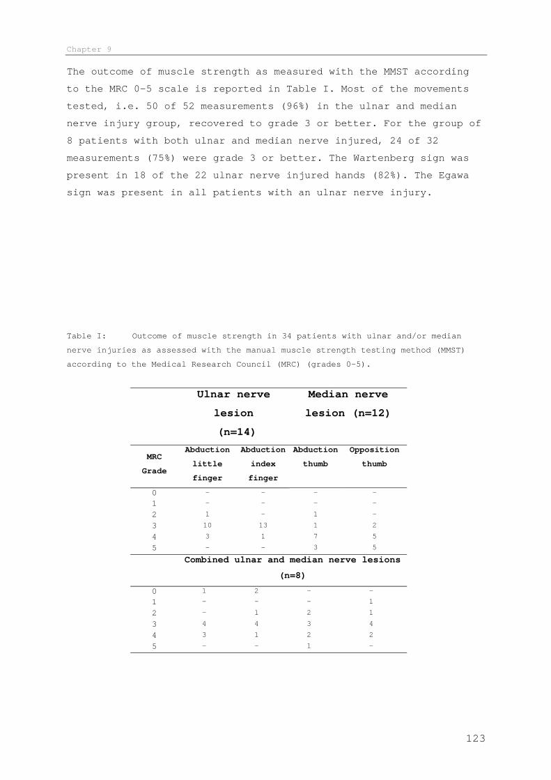
Chapter 9
The outcome of muscle strength as measured with the MMST according
to the MRC 0-5 scale is reported in Table I. Most of the movements
tested, i.e. 50 of 52 measurements (96%) in the ulnar and median
nerve injury group, recovered to grade 3 or better. For the group of
8 patients with both ulnar and median nerve injured, 24 of 32
measurements (75%) were grade 3 or better. The Wartenberg sign was
present in 18 of the 22 ulnar nerve injured hands (82%). The Egawa
sign was present in all patients with an ulnar nerve injury.
Table I: Outcome of muscle strength in 34 patients with ulnar and/or median
nerve injuries as assessed with the manual muscle strength testing method (MMST)
according to the Medical Research Council (MRC) (grades 0-5).
Ulnar nerve
lesion
(n=14)
Median nerve
lesion (n=12)
MRC
Grade
Abduction
little
finger
Abduction
index
finger
Abduction
thumb
Opposition
thumb
0 - - - -
1 - - - -
2 1 - 1 -
3 10 13 1 2
4 3 1 7 5
5 - - 3 5
Combined ulnar and median nerve lesions
(n=8)
0 1 2 - -
1 - - - 1
2 - 1 2 1
3 4 4 3 4
4 3 1 2 2
5 - - 1 -
123

Comparing outcome of muscle strength measurements
Figures 1 and 2 present the relation between the muscle strength
grading of MRC grades 3, 4 and 5 as compared to the RIHM
measurements in Newton (N). The grade 5 measurements refer to the
uninvolved hands of the 34 patients. Of the median nerve injured
hands the four measurements with MRC grade 5 were included.
Figure 1: Strength of the abduction of the little finger: assessed by means of MRC
grade 3, 4 and 5 and measured with the RIHM dynamometer (N). The box represents the
interquartile range, which contains the 50% of values. The whiskers are lines that
extend from the box to the highest and lowest values. The line across the box
indicates
the median. The
MRC grade 5
values are of the
uninvolved
hand.
124
Figure 2: Strength of the abduction of the thumb: assessed by means MRC grade 3, 4
and 5 and measured with the RIHM dynamometer (N). The box represents the

Chapter 9
interquartile range, which contains the 50% of values. The whiskers are lines that
extend from the box to the highest and lowest values. The line across the box
indicates the median. The MRC grade 5 values are of the uninvolved hand with the 4
grade 5 grades of the uninvolved hands.
Table 2 presents the outcome of the muscle strength, expressed as
“percentage recovery” of the uninjured hand, of grip and pinch
strength measurements and the RIHM measurements of the four
movements. The average outcome of grip strength in the three groups
of patients was comparable; i.e. 77 to 88%. Loss of pinch strength
is greatest in patients with the combined nerve injuries, and is
greater in patients with a median nerve injury than in patients with
an ulnar nerve injury. The abduction of the index finger strength as
measured with the RIHM in 14 ulnar nerve injured hands is remarkably
low; only 26% of the uninjured side. The abduction of the thumb
strength in patients with single median nerve and combined nerve
injuries was 59% and 27% of the uninjured hand, respectively.
125

Comparing outcome of muscle strength measurements
Table 2: Outcome of muscle strength as percentage (mean and SD) of the uninjured
hands measured with grip, pinch and intrinsic muscle strength (RIHM) dynamometers.
RIHM
Grip Pinch Abduction
little
finger
Abduction
index
finger
Abductio
n thumb
Oppositi
on thumb
Ulnar nerve (n=14) 84 (15) 75 (14) 37 (22) 26(13)
Median nerve (n=12) 88 (21) 58 (22) 59 (31) 82 (21)
Combined ulnar and
median nerve (n=8) 77 (18) 34 (30) 45 (35) 25 (22) 27 (25) 36 (28)
RIHM = Rotterdam Intrinsic Hand Myometer
Figure 3: Average percentage recovery of grip , pinch and intrinsic
muscle strength of three groups of patients; ulnar nerve, median nerve and
0
10
20
30
40
50
60
70
80
90
100
ulnar nerve(n=14)
median nerve(n=12)
combined ulnarand median (n=8)
126

Chapter 9
combined nerve injury.
Figure 3 presents the average percentage recovery of the intrinsic
muscles compared to grip and pinch strength measurements for the
three groups of patients.
There was a significant relation found between MMST and the RIHM
dynamometry measurements of all four movements (Table 3); Spearman
correlation coefficients were 0.65, 0.63, 0.85 and 0.56 for little
finger, index and thumb abduction and opposition of thumb,
respectively (p-values from 0.001 to 0.014). No significant
correlation was found between the four measurements of the RIHM and
the grip strength measurements. Pinch strength was significantly
correlated only with strength of the abduction of thumb and
opposition of the thumb strength (Pearson r; 0.55 and 0.72, p =
0.026, 0.002) as measured with the RIHM. Pinch strength was not
correlated with grip strength measurements.
Table 3; Correlation between different muscle strength testing methods.
RIHM measurements
Abduction
little
finger
(n=21)
Abduction
index
finger
(n=21)
Abduction
thumb
(n=20)
Oppositi
on thumb
(n=20)
Manual muscle strength 0.65* 0.63* 0.85* 0.56*
Grip strength b ns ns ns ns
Pinch strength b ns ns 0.55* 0.72*
RIHM = Rotterdam Intrinsic Hand Myometer. a = Spearman correlation
coefficient, b = Pearson correlation coefficient, * (p<0.05), ns = not
significant
Discussion
The evaluation of muscle strength is, in combination with the
assessment of sensibility, an important clinical method to determine
ulnar and median nerve function. This information is valuable in
127
Comparing outcome of muscle strength measurements
decision-making concerning surgery (e.g. tendon transfers), therapy
(e.g. splints), advice in work-related issues (e.g. safety to work
with machines) and research issues (e.g. nerve repair technique).
Concerning this latter topic, Trumble et al.18 noted that without
extremely sensitive methods for monitoring the functional outcome of
nerve regeneration, it will be difficult to identify those factors
that may have small but additive beneficial effects and those that
may have negative effects on nerve regeneration.
MMST according to the MRC method (grades 0-5) is the most widely
used clinical method to measure muscle strength. MMST is the only
instrument which is useful in the MRC 0 - 2 grades i.e.in the early
phases of nerve recovery. Grade 3 is an important cut-off point; the
muscles are just strong enough to provide a full range of motion on
all the joints that the muscle crosses. When the muscles have
reached this level of recovery usually there is no risk of joint
contractures. Grade 3 can be easily determined, in contrast to grade
4 which is defined as “complete range of motion with some
resistance”, while grade 5 is defined as the ability to hold against
“maximum pressure”. Objective assessment of “some resistance” and
“maximum pressure” will depend on the experience and subjective
judgement of the examiner.
Besides the problem with the grading, there is disagreement as to
what constitutes a functional level of muscle strength recovery
after peripheral nerve injury using grades 0-5. In literature,
useful or functional motor recovery has been defined anywhere from
2+ level of recovery to a 4-5 level.24 Seddon27 defined a “good” motor
outcome as grade 3 or better and reported that 47.6% of his patients
with nerve injuries obtained this level. Frykman 28 reached a higher
percentage of grade 3 or better; 81% and 64% after median and ulnar
nerve injuries, respectively. Strickland et al.24 selected grade 4
for determining a good result, which was obtained in 9 of 17
patients with ulnar nerve injuries. In the patients in the present
study, very few measurements of the intrinsic muscles (9/84) reached
grade 5. Of all the 84 MMST measurements 75% were grade 3 and 4.
128
Chapter 9
Although it is imperative to come to an agreement as to what should
be considered as a good motor outcome on the MRC scale, the more
important question is whether this method is sensitive enough to
sufficiently differentiate above the grade 3 level. We consider MMST
to be an important and useful method in the 0-3 grades of the MRC
scale, but for strength measurements above grade 3 a more accurate
evaluation method is required. Grip and pinch strength dynamometry
is a more sensitive method to determine muscle strength and to
render outcome on a continuous scale, e.g. kilogram force or Newton.
These quantitative outcomes, which will also facilitate statistical
analyses, have been proposed as a standardised method to evaluate
motor recovery in patients with peripheral nerve injuries.5 7 8
Reliable application of these measurements requires little training
and is not influenced by the experience of the examiner.29 Another
benefit is that evaluating motor function recovery as a percentage
of the uninjured side is possible using the uninvolved hands as the
normal level of strength for that patient. Therefore the data can be
normalised and allows to compensate for the large variability in
patient strength.
In our study the mean recovery of grip strength, compared with the
uninjured hand, was 84% in the 14 patients with an ulnar nerve
injury. Rosén 7 and Strickland et al.24 found comparable grip
strength recoveries of 88 and 89.9%, respectively. Commenting on
these grip strength results, Strickland et al.24 made a noteworthy
remark that grip and pinch strength may only be used as an indirect
measurement of ulnar nerve outcome. Although grip strength
measurements do fulfil the requirements for quantitative outcome of
the strength of the hand, they cannot differentiate between the
intrinsic and extrinsic muscles. Strong grip strength does not
necessarily coincide with strong intrinsic muscles. The results of
the present study give more specific insight in the recovery of the
intrinsic muscle strength.
It remains debatable what the precise contribution of the intrinsic
muscles are in grip and pinch strength measurements.30 31 It has been
suggested that the most narrow handle position (position 1) of the
129
Comparing outcome of muscle strength measurements
Jamar could be used to test the strength of the intrinsic muscles,
whereas the wider handle (position 4 and 5) could be used to test
the extrinsic finger flexors.32 In another study EMG recordings of
hand muscles in sustained grasp in the different handle positions
seems to confirm this, but again it was concluded that the strength
of the intrinsic muscles could not be isolated from that of the
extrinsic muscles.33
In our study results there was a remarkable discrepancy between the
poor outcomes of the intrinsic muscles strengths as measured with
the RIHM and the high levels of outcome of grip strength in patients
with ulnar nerve injuries. Rosén7 obtained similar values using the
Mannerfelt “intrins-o-meter” and found a recovery percentage for the
ulnar nerve innervated muscles of 31 and 36% for abduction of index
and little finger, respectively, while the average grip strength
recovery was 88%.
In our study no correlation was found between the recovery of the
intrinsic muscle strength and the grip strength measurements. This
seems to affirm our assumption that recovery of grip strength does
not reflect the recovery of the intrinsic muscles of the hand after
peripheral nerve injury. Thus, outcome of the intrinsic muscles
strength with the RIHM dynamometer is a valuable addition to grip
and pinch strength measurements in patients with peripheral nerve
injuries. The RIHM measurements might have further use in evaluation
of the intrinsic hand muscle strength in e.g. leprosy, neuropathies
like hereditary motor and sensory neuropathy and Guillain-Barré
syndrome.
Examining the pinch strength outcome, the loss of pinch strength was
noticeably greater in the median nerve injury group of patients than
in the ulnar nerve injury group. Pinch strength measurements were
not significantly correlated with the strength measurements of the
abduction of the little and index finger, but were correlated with
the intrinsic muscle strength measurements of the thumb. It appears
from these results that positioning of the thumb by the abductor
130

Chapter 9
pollicis brevis and opponens pollicis muscles is an important
requirement for a strong pinch.
Another notable finding in this study was the good recovery of the
thumb intrinsic muscle strength in patients with median nerve
injury, which is in contrast with the poor recovery of the abduction
of little and index finger strength in patients with ulnar nerve
injury. An explanation might be that the test for the thumb muscle
strength is more affected by synergistic muscle activity e.g. of the
ulnar nerve innervated part of the flexor pollicis brevis muscle or
due to crossover of ulnar innervation.34 Frykman et al.24 estimated
that 50% of the evaluated patients would have satisfactory thumb
opposition even if no median nerve re-innervation occurred. Brand35
mentioned another synergistic movement of the thumb palmar abduction
while testing the abductor pollicis brevis due to bowstringing of
the radial nerve innervated abductor pollicis longus muscle with the
wrist in flexion. For this reason examiners should test the abductor
pollicis brevis of the thumb with the wrist in extension. This
position is not always possible with the dynamometer measurements
shown in other studies but can be accomplished with the RIHM
dynamometer.
A limitation in our study is that there were only 8 patients in the
third group, i.e. those with both ulnar and median nerve injury. Two
patients in this group had a remarkably good recovery (i.e. grip
strength recovery of 98 and 91%). In one patient, with an ulnar and
median nerve injury at the wrist level, the abduction strength of
the little finger recovered to 93%. Because the results of this
patient had a strong impact on the average strength of the group, we
probably overestimated the average abduction strength of the little
finger and the grip strength for patients with combined ulnar and
median nerve injuries.
Conclusion
131

Comparing outcome of muscle strength measurements
In conclusion the measurement methods used to determine the
recovery of the motor function of ulnar and median nerve injured
hands need further consideration. In patients who sustained an ulnar
nerve injury the mean recovery of the abduction force of the index
finger was only 26% of the uninjured side. The abduction of the
thumb strength in patients with single median nerve and combined
nerve injuries was 59% and 27% of the uninjured hand, respectively.
We regard these average percentages recovery as poor, especially in
contrast with the grip strength percentages of 77 to 88% recovery.
A dynamometer like the RIHM provides a more accurate clinical
assessment of the outcome for two reasons. Firstly, with the RIHM
intrinsic muscle strength is measured in isolation, in contrast to
grip and pinch strength measurements in which many uninvolved
muscles are tested besides the intrinsic muscles. Secondly, because
of its quantitative results the RIHM measurements provide a more
accurate clinical assessment as compared with the MMST especially in
MRC grades 4 and 5.
References
1. Lundborg G. A 25-year perspective of peripheral nerve surgery: evolving
neuroscientific concepts and clinical significance. J Hand Surg [Am]
2000;25(3):391-414.
2. Bruyns CN, Jaquet JB, Schreuders TA, Kalmijn S, Kuypers PD, Hovius SE.
Predictors for return to work in patients with median and ulnar nerve
injuries. J Hand Surg [Am] 2003;28(1):28-34.
3. Jaquet JB, Kalmijn S, Kuypers PD, Hofman A, Passchier J, Hovius SE. Early
psychological stress after forearm nerve injuries: a predictor for long-term
functional outcome and return to productivity. Ann Plast Surg 2002;49(1):82-
90.
4. Medical Research Council. Special Report series no 282. London: Her Majesty's
Stationary Office, 1954.
5. Dellon AL. A numerical grading scale for peripheral nerve function. J Hand Ther
1993;6(2):152-60.
6. Gaul JS, Jr. Intrinsic motor recovery--a long-term study of ulnar nerve repair.
J Hand Surg [Am] 1982;7(5):502-8.
132
Chapter 9
7. Rosen B. Recovery of sensory and motor function after nerve repair. A rationale
for evaluation. J Hand Ther 1996;9(4):315-27.
8. Rosen B, Dahlin LB, Lundborg G. Assessment of functional outcome after nerve
repair in a longitudinal cohort. Scand J Plast Reconstr Surg Hand Surg
2000;34(1):71-8.
9. Rosen B, Lundborg G. A model instrument for the documentation of outcome after
nerve repair. J Hand Surg [Am] 2000;25(3):535-43.
10. Lagerstrom C, Nordgren B. Methods for measuring maximal isometric grip strength
during short and sustained contractions, including intra-rater reliability.
Ups J Med Sci 1996;101(3):273-85.
11. Lagerstrom C, Nordgren B, Olerud C. Evaluation of grip strength measurements
after Colles' fracture: a methodological study. Scand J Rehabil Med
1999;31(1):49-54.
12. van der Ploeg RJ, Oosterhuis HJ, Reuvekamp J. Measuring muscle strength. J
Neurol 1984;231(4):200-3.
13. Baktir A, Turk CY, Kabak S, Sahin V, Kardas Y. Flexor tendon repair in zone 2
followed by early active mobilization. J Hand Surg [Br] 1996;21(5):624-8.
14. Gault DT. Reduction of grip strength, finger flexion pressure, finger pinch
pressure and key pinch following flexor tendon repair. J Hand Surg [Br]
1987;12(2):182-4.
15. Riaz M, Hill C, Khan K, Small JO. Long term outcome of early active
mobilization following flexor tendon repair in zone 2. J Hand Surg [Br]
1999;24(2):157-60.
16. Boatright JR, Kiebzak GM, O'Neil DM, Peindl RD. Measurement of thumb abduction
strength: normative data and a comparison with grip and pinch strength. J
Hand Surg [Am] 1997;22(5):843-8.
17. An KN, Chao EY, Askew LJ. Hand strength measurement instruments. Arch Phys Med
Rehabil 1980;61(8):366-8.
18. Trumble TE, Kahn U, Vanderhooft E, Bach AW. A technique to quantitate motor
recovery following nerve grafting. J Hand Surg [Am] 1995;20(3):367-72.
19. Liu F, Carlson L, Watson HK. Quantitative abductor pollicis brevis strength
testing: reliability and normative values. J Hand Surg [Am] 2000;25(4):752-9.
20. Mannerfelt LG. Studies on ulnar nerve compression neuropathies with a new
computerised instrument--the intrins-o-meter. Scand J Plast Reconstr Surg
Hand Surg 1997;31(3):251-60.
21. Schreuders TAR, Roebroeck ME, Jaquet J-B, Hovius SER, Stam HJ. Measuring the
strength of the intrinsic muscles of the hand in patients with ulnar and
median nerve injury; reliability of the Rotterdam Intrinsic Hand Myometer
(RIHM). J Hand Surg (Am) 2004;29A(2):318-324.
22. Shaw Wilgis EF, Brushart TM. Nerve Repair and Grafting. In: Green DP, editor.
Operative Hand Surgery. 3rd ed. New York: Churchill Livingstone, 1993:1335-
1336.
23. Brandsma JW, Schreuders TA, Birke JA, Piefer A, Oostendorp R. Manual muscle
strength testing: intraobserver and interobserver reliabilities for the
intrinsic muscles of the hand. J Hand Ther 1995;8(3):185-90.
133

Comparing outcome of muscle strength measurements
24. Strickland JW, Idler, R.S., DelSignore, J.L. Ulnar Nerve Repair. In: Gelberman
RH, editor. Operative Nerve Repair and Reconstruction. Philadelphia: J.B.
Lippincott Comp, 1991:425-436.
25. Tubiana R. The Hand Vol II. Philadelphia: WB Saunders, 1985:518.
26. ASHT, editor. Clinical Assessment Recommendations. 2nd ed. Garner, NC: American
Society of Hand Therapists, 1992.
27. Seddon HG. Surgical disorders of the peripheral nerves. 2 ed. New York:
Churchill-Livingstone, 1975.
28. Frykman GK. The quest for better recovery from peripheral nerve injury: current
status of nerve regeneration research. J Hand Ther 1993;6(2):83-8.
29. Schreuders TAR, Roebroeck ME, Goumans J, van Nieuwenhuijzen JF, Stijnen TH,
Stam HJ. Measurement error in grip and pinch force measurements in patients
with handinjuries. Phys Ther 2003;83(9):806-815.
30. Kozin SH, Porter S, Clark P, Thoder JJ. The contribution of the intrinsic
muscles to grip and pinch strength. J Hand Surg [Am] 1999;24(1):64-72.
31. Mahaffey P. The contribution of the intrinsic muscles to grip and pinch
strength. J Hand Surg [Am] 1999;24(4):870-1.
32. Stokes HM. The seriously uninjured hand--weakness of grip. J Occup Med
1983;25(9):683-4.
33. Janda DH, Geiringer SR, Hankin FM, Barry DT. Objective evaluation of grip
strength. J Occup Med 1987;29(7):569-71.
34. Vanderhooft E. Functional outcomes of nerve grafts for the upper and lower
extremities. Hand Clin 2000;16(1):93-104, ix.
35. Brand PW. Clinical Mechanics of the Hand. St. Louis: CV Mosby, 1999.
.
134

Chapter 9
135

“
”
Discussion
Chao EY, An K N, Conney WP, Linscheid RL.
132
Chapter 10
The main purpose of this thesis is to study different methods of measuring muscle
strength of the hand and to develop a reliable instrument to quantitatively
measure the strength of the intrinsic muscles of the hand. Such an instrument
would allow the function of the intrinsic muscles to be studied in clinical
practice. This can be valuable in monitoring the recovery or deterioration of the
intrinsic muscle strength of the hand in patients who have suffered from nerve
injuries in their arm. Intrinsic muscle strength could also be examined in other
neuromuscular diseases which affect the intrinsic muscles, such as Charcot Marie
Tooth disease, Amyotrophic Lateral Sclerosis and Guillan Barré syndrome. A
reliable instrument which can accurately measure the intrinsic muscle strength may
assist in developing more effective therapies.
This final chapter addresses some issues related to measurements of the muscle
strength of the hand in a larger perspective and discusses some limitations of
this study.
Nerve function can be determined by several methods. All of the clinical methods
evaluate nerve function indirectly by testing e.g. muscle strength, sensation or
sweating. According to Bunnell “
”.1 p.319 The motor and sensation
function is usually reported separately, but some, e.g. Dellon2 and Rosen et al.3,
present figures on sensation and motor function to provide, conveniently, just one
figure as the function or outcome of a particular nerve. In this way the loss of
motor function and sensory loss is in accordance with Bunnell’s remark, weighed as
equally important.
We want to challenge the statement that motion and sensation are of equal
importance. It seems to be a reasonable assumption that the consequence of motor
and/or sensory loss is dependent on e.g. profession and the patient’s ability to
adapt to a particular loss of function. One can imagine that a violin player with
normal muscle function but loss of sensation would have major difficulties playing
the violin, while a farmer with normal sensation but loss of muscle function would
be unable to do his work.
133

Discussion
As long as it is not precisely known how strength and sensation relate to
particular tasks of the hand, it seems more logical to present these results
separately when testing the hand.
Brink and Mackel4 developed a functional and simple test for evaluation of
peripheral nerve function. The authors stated that patients suffering from severe
sensory neuropathies can perform a wide range of movements, but are unable to
sustain constant levels of muscle contraction or perform fine motor adjustments
when visual guidance is prevented. They concluded that their results underline the
need for sensation in the successful execution of a sequence of co ordinated,
skilful movements.
Studies investigating the ability of the muscles to control the forces of the
fingers in gripping objects have shown that decreased sensation is often
associated with increased grip strength levels. Grip strength control requires
sensory feedback to signal the effectiveness of the motor commands. Sensory
feedback uses object properties and mechanical actions, e.g. at the skin object
interface, to modify motor commands; consequently, intact sensory feedback is
essential for controlling the grip strength.5 6
Complete loss of sensation causes severe problems in hand function. Paul Brand has
shown that the loss of protective sensation causes a destructive increase of grip
strength.7 8 He searched for a method to give leprosy patients an artificial (pain)
feedback system to protect their hands against injury caused by gripping their
tools too powerfully. Experimenting with a prototype he observed a patient who
switched the system off when he required a stronger grip. Brand realised that a
protective system, which could be switched off, was ineffective.
Apparently, the influence of sensation and pain needs to be considered when
interpreting grip strength measurements for the evaluation of nerve function. The
134

Chapter 10
above mentioned studies support the proposition that sensation does play an
important role in the control of the muscle strength; however, to our knowledge no
study has determined the role of sensation in maximum strength measurements of the
hand in a quantitative way.
In the above mentioned book by Sterling Bunnell1 (1948) one chapter has been
dedicated to the intrinsic muscles of the hand. In the introduction Bunnell wrote
(p. 467): “
”. He also stated
that “
”. It is open to discussion how at the same time
the role of the intrinsic muscles can be important and controversial, and to what
extent the strength of the grip is dependent on the intrinsic muscles.
The contribution of the intrinsic muscles in grip has been studied by e.g. Kozin
et al.9 Twenty one healthy subjects were given either an initial median nerve
block at the wrist followed by an ulnar nerve block, or an ulnar nerve block
followed by a subsequent median nerve block. The anaesthetic solution was chosen
because of its effectiveness in producing complete motor and sensory block. The
average decrease in grip strength was roughly 38% after ulnar and 32% after median
nerve block. The total grip loss after both nerves were injected averaged 49%
compared with the pre injection strength.
These results were commented on by Mahaffey10 who found it surprising that Kozin
did not take into account the effect of sensory loss on the results of the grip
strength measurements. Both Kozin and Mahaffey agreed that, ideally, motor
recovery is best studied with intact sensory function. However, in clinical
practice this is rarely seen and results of strength measurements will therefore
be influenced by loss of sensation, (see 9.2) especially in these experiments
where there is a sudden and complete loss of sensation.
135
Discussion
In relation to Bunnel’s quotation that strength of grip is dependent on the
intrinsic muscle strength, we have observed several patients with poor intrinsic
muscle recovery and strong grip strength. In accordance with this observation, in
our long term outcome study (Chapter 9) we found no significant correlation
between the grip strength and the strength of the intrinsic muscles as measured
with the Rotterdam Intrinsic Hand Myometer (RIHM).11
The intrinsic muscle strength may influence the grip strength, but grip strength
measurements will not provide sufficient information to assess the precise
strength of the intrinsic muscles. This is because in grip strength measurements
all the muscles in the lower arm and hand are active making it practically
impossible to determine the precise component of the intrinsic muscles. Secondly,
loss of sensation will probably also influence the grip strength, making it even
more complicated to determine the contribution of the intrinsic muscles.
In clinical practice there is a third reason why grip strength is difficult to
interpret when evaluating the intrinsic muscle strength. Generally the peripheral
nerves are injured in combination with one or more injuries of the finger and
wrist tendons, and it is known that tendon injuries alone cause a decrease of grip
strength.12 13
Loss of nerve function has an important effect on e.g. hand function and the
patients’ abilities to perform certain tasks. In a group of 81 ulnar and/or median
nerve injured patients the mean time off work was more than 31 weeks. Within one
year after combined nerve injuries only 24% versus 80% after isolated median, and
59% after ulnar nerve injuries, returned to their work.14
Measuring hand function more specifically using the Sollerman test, Rosen found
that patients with peripheral nerve injuries had difficulty in picking up coins,
putting nuts on bolts and doing up four buttons.15 Handling these small objects was
especially difficult for patients with median nerve loss possibly because of the
136

Chapter 10
loss of sensibility of the index and thumb, or due to impaired muscle strength of
the thumb. These three selected tasks provided information about the integrated
sensory/motor function of the hand and were included in the sensory domain of
Rosen’s test battery design after nerve repair.16
An Indian study of 62 leprosy patients investigated the relation between grip and
pinch strength and (using an Indian designed questionnaire) the basic activities
of daily living (BADL).17 The study showed a significant relationship between grip
and pinch strengths (in particular the tripod pinch) and the BADL questionnaire.
In a long term study of patients with peripheral nerve injuries, the relation
between the DASH questionnaire and grip strength and sensibility measurements
(Semmes Weinstein filaments) was analysed. A strong association was shown between
the outcome as measured with the DASH questionnaire for both the sensory and motor
recovery.18 19 Evidently, both functions of the nerve, i.e. sensation and muscle
strength, can have an important influence on ADL.
However, interpreting the outcome of tests which assess the patient’s ability to
perform a certain task by measuring the time taken (Sollerman test) or by asking
if the patient has encountered problems (BADL, DASH questionnaires) has some
important drawbacks, e.g. they might only assess the patient’s ability to adapt or
the patient’s creativity in finding coping strategies, e.g. in using the non
injured hand or certain trick movements.
Therefore, we propose to present hand function in the domain of function, skills,
activities and participation as was suggested by the 2001 World Health
Organization report as the International classification of functioning,
disability, and health (ICF).20
No measurement is perfect: whether it is used to make a diagnosis or to determine
a change in strength, measurements will have some error associated with them.
Hence, we have to decide how to live with these errors. The primary step is to
decide which method to choose to determine the error of measurements.21
137
Discussion
Studies concerning measuring instruments and methods generally provide Intraclass
Correlation Coefficient (ICC) values which are interpreted as showing either good,
moderate or poor reliability of the instrument. However, reliability relates to
how the measurement will be used, i.e. is the measurement error so large that it
would be unlikely to provide valuable information? Estimates of reliability are
arranged along a scale and clinicians need to know at what point along this scale
they lie and if the change in e.g. muscle strength as measured with the instrument
is a clinically relevant deterioration or improvement, or is due to measurement
errors only.
Insight in the sources of measurement error can be obtained by performing an
analysis of variance. From the error variance, the standard error of measurement
(SEM) can be computed as its square root, which is advantageously expressed in the
same value as the measurements performed.22
Based on the SEM, the smallest detectable difference (SDD) can be calculated as
1.96 2 SEM.23 This SDD can be applied in such a way that only differences
between two consecutive measurements greater than the SDD can be interpreted with
95% certainty as real change in strength. If the change between measurements is
less than the SDD, the change is interpreted as a result of variation and not as a
real change.
Although the calculation of SEM and SDD is becoming an accepted method in
reliability studies21 24 26 to determine the clinical value of a particular
measurement in a specific population, it is essential to know in which range the
changes of e.g. muscle strength take place. For example, in patients with hand
injury, the grip strength will weaken shortly after the trauma. In later
rehabilitation phases the increase of muscle strength will generally reach a
plateau after many months. If the difference in the grip strength between the
start and end of the rehabilitation is, for example, 100 N, an SDD of e.g. 80 N
will reveal the measurements to be inadequate to detect changes during the
rehabilitation process. If the range in which the strengths change is as wide as
300 N, an SDD of 30 N is certainly sufficient to detect changes during the
rehabilitation process. In this latter example a virtual scale of 10 steps is
achieved.27 Whether the SDD is sufficiently small to be acceptable, is a matter of
clinical judgement.28
138

Chapter 10
In Chapter 3 we considered the SDD of the AIKOH measurements and other instruments
proportional to the mean of the measurements; this approach was also suggested by
Geertzen et al.29 In a later phase of this study we reported that the mean may not
be the best criterion for this purpose because the mean gives no insight into the
range that will be covered from poor to good results.
In a study investigating the interobserver reproducibility of grip strength in
patients with lateral epicondylitis, Smidt et al.25 initially stated that an
interobserver difference of 10% of the total range of measurements would be
acceptable. Later they found that the correct SDD values of grip strength
measurements were larger than the predefined acceptable difference of 10% of the
total range of the scale, while for maximum grip strength the SDD approximately
equalled the predefined acceptable difference.30 They concluded that in their study
population these outcome measures were insufficiently reproducible to detect
changes in an individual patient. In studies comparing larger groups of patients
these outcome measures perform adequately because the influence of measurement
error decreases with increasing sample size.
In Chapter 5 we proposed to judge the SDD proportional to the standard deviation
(SD) rather than to the mean or the range of the measurements. Arbitrarily, we
accepted that an SDD/SD ratio should be larger than 0.5 for the instrument to have
acceptable SDDs for application in individual patients.31 Further research is
needed to gain insight into the usefulness of this criterion to judge the amount
of measurement error.
This SDD/SD ratio should be further studied in different patient groups. We
propose that this ratio should be reported in reliability studies of measurement
instruments as it will be especially useful for clinicians.
139

Discussion
The function of a muscle can be divided into three aspects: force or strength (N),
endurance (sec) and velocity (m/sec). In most muscle testing methods, the first
aspect, maximum strength is tested, which is sometimes referred to as the maximum
voluntary contraction (MVC). In grip and pinch strength measurements this MVC is
generally used by asking the patient to squeeze or push “ ” and
has been shown to provide reliable data. In ADL, however, these maximum muscle
contractions are rarely used, except for some specific actions, e.g. when lifting
a heavy suitcase or opening a tight jar. Usually, a much lower level of strength
is needed to perform most of our ADL, especially in white collar workers. The
second aspect, endurance or muscle fatigue, is probably particularly relevant in
patients with impaired muscle function due to e.g. multiple scleroses, amyotrophic
lateral sclerosis and hereditary motor and sensory neuropathy (HMNS). Videler et
al.32 studied muscle fatigue of grip in patients with HMSN. Many patients with HMSN
indicate that during daily activities they experience most difficulty when an
activity has to be maintained for a longer period of time.33 34 Videler and
colleagues studied the reproducibility of a fatigue test with measurements of
three sets of 15 grip and pinch contractions with a handgrip dynamometer on two
separate occasions with a 1 week interval, but found that reproducibility was
poor. With regard to instruments to measure muscle fatigue, the authors concluded
that no reproducible method is yet available for patients with neuromuscular
disorders.
Although many studies have attempted to identify a single muscle fatigue factor,
there seems little doubt that the concept of fatigue refers to a class of acute
effects that can impair motor performance. Factors known to influence muscle
fatigue include muscle blood flow, neuromuscular properties and motivation
(sincerity of effort).35 However, consensus on the definition of muscle fatigue and
a standardised method to measure muscle fatigue of the hand are still lacking.
140
Chapter 10
The third aspect of muscle function, the velocity component of muscle function,
was not specifically explored in this study. However, it is known that the ability
to rapidly move the hand is an important attribute which determines the co
ordination of the hand. The consequences of diminished co ordination can be
observed in patients with spasticity. The patient might have a very strong grip
but has major problems in ADL because releasing the grip on an object is not
possible. The velocity or co ordination as part of the muscle function can be
evaluated in certain tests that measure time to perform particular tasks.
In Chapter 4, study of lumbrical muscle strength demonstrates that these muscles
only have a small ‘strength’ effect on the metacarpo phalangeal joint flexion of
the fingers. The interossei muscles are much stronger than the lumbricals. The
importance of the lumbrical in fast movements rather than in maximum strength
circumstances, as suggested by Leijnse et al.36, is interesting and warrants
further clinical study.
In the long term outcome study presented in Chapter 9 the muscle strength of
patients’ hands was mainly measured at a certain point in time (cross sectional).
We have not yet reported the change in muscle strength of the intrinsic muscles
over time. To further explore the recovery of muscle strength longitudinally in
patients with nerve injuries data from measurements performed regularly (e.g.
every three months after nerve injury) can be used.
Even though a period of two years after nerve injury is generally considered as
the end point for recovery, recent studies have indicated that muscle strength can
improve after an even longer period of time.37
In this thesis we have studied the reliability of the RIHM only in patients with
peripheral nerve injuries and determined the reliability of the RIHM with two
experienced examiners only. Although we believe that performing these tests in
141

Discussion
different patient groups will result in few differences, this should be
investigated.
Despite the fact that the clinician’s experience in taking the measurements might
also influence the measurement error, in grip and pinch strength measurements we
found that this did not influence the reliability. This needs, however, to be
confirmed in further studies.
Another issue is the time needed for a non experienced therapist or physician to
learn to use the RIHM in a reliable way. Although RIHM measurements might be
slightly more difficult to perform than grip and pinch strength measurements, they
are very similar to the measurement protocol of manual muscle testing regarding
the point of pressure and methods of holding the hand of the patient.
In the evaluation of peripheral nerve function, interpretation of manual muscle
strength testing and grip strength measurements needs reconsideration. Although
grip strength measurements do fulfil the requirements for quantitative measurement
of the strength of the hand, they cannot differentiate between the intrinsic and
extrinsic muscles. Strong grip strength does not necessarily imply strong
intrinsic muscles. The results of the present study give more specific insight
into the recovery of intrinsic muscle strength.
When choices are made about which technique provides the best results with regard
to motor recovery of the muscles, an instrument such as the RIHM should be used.
Concerning the reliability of measurement instruments, we suggest to calculate the
SDD/SD ratio. This ratio should probably need to be larger than 0.5 for an
instrument to have acceptable SDDs, but this too needs further study.
1. Bunnell S. . 2nd ed. Philadelphia: J.B. Lippincott Company, 1948.
142
Chapter 10
2. Dellon AL. A numerical grading scale for peripheral nerve function. 1993;6(2):152 60.
3. Rosen B, Lundborg G. A model instrument for the documentation of outcome after nerve repair.
2000;25(3):535 43.
4. Brink EE, Mackel R. Sensorimotor performance of the hand during peripheral nerve regeneration.
1987;77(2 3):249 66.
5. Nowak DA, Glasauer S, Hermsdorfer J. How predictive is grip force control in the complete absence
of somatosensory feedback? 2004;127(1):182 192.
6. Monzee JL, Lamarre Y, Smith AM. The effects of digital anesthesia on force control using a
precision grip. 2003(89):672 683.
7. Brand PW. Management of the insensitive limb. 1979;59(1):8 12.
8. Brand PW. . St. Louis: CV Mosby, 1999.
9. Kozin SH, Porter S, Clark P, Thoder JJ. The contribution of the intrinsic muscles to grip and
pinch strength. 1999;24(1):64 72.
10. Mahaffey P. The contribution of the intrinsic muscles to grip and pinch strength.
1999;24(4):870 1.
11. Schreuders TAR, Roebroeck ME, Jaquet J B, Hovius SER, Stam HJ. Longterm outcome of muscle
strength in patients with ulnar and median nerve injury: Comparing manual muscle strength
testing, grip and pinch strength dynamometers and a new intrinsic muscle strength
dynamometer. 2004;36.
12. Gault DT. Reduction of grip strength, finger flexion pressure, finger pinch pressure and key
pinch following flexor tendon repair. 1987;12(2):182 4.
13. Ejeskar A. Finger flexion force and hand grip strength after tendon repair.
1982;7(1):61 5.
14. Bruyns CN, Jaquet JB, Schreuders TA, Kalmijn S, Kuypers PD, Hovius SE. Predictors for return to
work in patients with median and ulnar nerve injuries. 2003;28(1):28 34.
15. Rosen B. Recovery of sensory and motor function after nerve repair. A rationale for evaluation.
1996;9(4):315 27.
16. Rosen B. The sensational hand. Clinical assessment after nerve repair. (Thesis). Lund
University, 2000.
17. Rajkumar P, Premkumar R, Richard J. Grip and pinch strength in relation to function in
denervated hands. 2002;74(4):319 28.
18. Jaquet JB, Luijsterburg AJ, Kalmijn S, Kuypers PD, Hofman A, Hovius SE. Median, ulnar, and
combined median ulnar nerve injuries: functional outcome and return to productivity.
2001;51(4):687 92.
19. Jaquet J, Schreuders TAR, Stam HJ, Hovius SER. Relationship between DASH and functional recovery
after nerve injury. (in press).
20. WHO. Geneva: World
Health Organization, 2001.
21. Rothstein JM. Living with error. 2003;83(5):422 423.
22. Roebroeck ME, Harlaar J, Lankhorst GJ. The application of generalizability theory to reliability
assessment: an illustration using isometric force measurements. 1993;73(6):386 95;
discussion 396 401.
143
Discussion
23. Roebroeck ME, Harlaar J, Lankhorst GJ. Reliability assessment of isometric knee extension
measurements with a computer assisted hand held dynamometer.
1998;79(4):442 8.
24. de Vet HCW. Klinimetrie: De maat van de geneeskunde (inaugurale rede). Amsterdam: VU Amsterdam,
2002.
25. Smidt N, van der Windt DA, Assendelft WJ, Mourits AJ, Deville WL, de Winter AF, et al.
Interobserver reproducibility of the assessment of severity of complaints, grip strength,
and pressure pain threshold in patients with lateral epicondylitis.
2002;83(8):1145 50.
26. Hopkins WG. Measures of reliability in sports medicine and science. 2000;30(1):1 15.
27. Schreuders TAR, Roebroeck ME, Jaquet J B, Hovius SER, Stam HJ. Measuring the strength of the
intrinsic muscles of the hand in patients with ulnar and median nerve injury; reliability of
the Rotterdam Intrinsic Hand Myometer (RIHM). 2004;29A(2):318 324.
28. Rothstein JM. Sick and tired of reliability? 2001;81(2):774 5.
29. Geertzen JH, Dijkstra PU, Stewart RE, Groothoff JW, ten Duis HJ, Eisma WH. Variation in
measurements of grip strength. A study in reflex sympathetic dystrophy patients.
1998;279:4 11.
30. de Vet HC, Roebroeck ME, Smidt N, Knol DL, van der Windt DA, Bouter LM. Erratum.
2003;84(6):938.
31. Schreuders TAR, Roebroeck ME, Goumans J, van Nieuwenhuijzen JF, Stijnen TH, Stam HJ. Measurement
error in grip and pinch force measurements in patients with handinjuries.
2003;83(9):806 815.
32. Videler AJ, Beelen A, Aufdemkampe G, de Groot IJ, Van Leemputte M. Hand strength and fatigue in
patients with hereditary motor and sensory neuropathy (types I and II).
2002;83(9):1274 1278.
33. Brooke MH, Miller R. Fatigue testing. 1990;13 Suppl:35 37.
34. Lindeman E, Leffers P, Reulen J, Spaans F, Drukker J. Reduction of knee torques and leg related
functional abilities in hereditary motor and sensory neuropathy.
1994;75(11):1201 5.
35. Enoka RM, Stuart DG. Neurobiology of muscle fatigue. 1992;72(5):1631 48.
36. Leijnse JN. Why the lumbrical muscle should not be bigger a force model of the lumbrical in the
unloaded human finger. 1997;30(11 12):1107 14.
37. Ghandi A, Jaquet J B, Kalmijn S, Hovius SER. Motor recovery following median and ulnar nerve
injuries. 2003;28 Suppl(1):16.
144

Summary

The hand has been called an extension of the brain, and the sensory
and motor performance of the hand is based on adequate function of
components in both the peripheral nerves as well as the central
nervous systems. Damage to the nerves (e.g., injury, compression,
infection [e.g. leprosy]) causes a loss of sensation and strength
that may result in serious loss of hand function. According to
Lundborg (2000) peripheral nerve lesions constitute one major reason
for severe and longstanding impairment of hand function. He called
the situation of nerve repair after injury frustrating because,
although a nerve suture may be technically perfect and the
rehabilitation carried out meticulously, the outcome is
unpredictable and often disappointing.
The evaluation of muscle strength is, in combination with the
assessment of sensibility, an important clinical method to determine
ulnar and median nerve function. This information is valuable in
decision-making concerning surgery (e.g. tendon transfers), therapy
(e.g. splints), advice in work-related issues (e.g. safety to work
with machines) and research issues (e.g. nerve repair technique).
Concerning this latter topic, Trumble et al. (1995) noted that
without extremely sensitive methods for monitoring the functional
outcome of nerve regeneration, it will be difficult to identify
those factors that may have small but additive beneficial effects
and those that may have negative effects on nerve regeneration.
The main objective of the work presented in this thesis was to
investigate the methods currently used to evaluate the motor domain
of nerve function, i.e. manual muscle strength testing, and grip and
pinch strength measurements. We questioned whether these methods
give appropriate and sufficient information about the intrinsic
muscle strength of the hand. Our hypothesis was that a dynamometer
that provides quantitative data on the intrinsic muscle strength
would provide more valuable information when monitoring the muscle
strength of patients who have suffered nerve injuries of their arm.
Chapter 1 presents a general introduction to Manual Muscle Strength
Testing (MMST), which was the first numerical system for the grading
144

of muscle action. In 1954 the British Medical Research Council (MRC)
specified a similar 0-5 scale in which grade 0 indicates total
paralysis, grade 3 indicates that the limb can be moved against
gravity, and grade 5 is normal. Several limitations of MMST are
discussed: a) usually a muscle group is tested rather than just one
muscle, b) the MRC scale is an ordinal one with disproportional
distances between the grades, c) it is not appropriate for use in
higher level muscle function (grades 4 and 5), and d) MMST is
dependent on the examiner’s experience to acquire reliable
measurements.
Then we give a brief history of the development of dynamometers for
the hand. In the past decades many different dynamometers have been
introduced; for example, grip strength measurements using a
dynamometer became popular and have been studied extensively.
Dynamometers for the measurement of intrinsic muscle strength of the
hand are very important in clinical neurology for the evaluation of
mononeuropathology and poly-neuropathology. One of the first to
develop a dynamometer to measure intrinsic muscle strength was
Mannerfelt. Later he manufactured a new device that was called the
intrins-o-meter. Since then, several others have developed
instruments mainly to assess the abduction of the thumb.
Chapter 2 describes the functional anatomy of the intrinsic muscles
of the hand and its pathology, and discusses ways to assess muscle
strength (both manual and instrumental) and the therapeutic options
available.
Chapter 3 focuses on the first dynamometer to be utilised, which
was a generic strain gauge instrument called the AIKOH. In a group
of 24 patient with ulnar and/or median nerve injuries, several
intrinsic muscle actions were measured to test reliability. There
was an excellent level of reliability when comparing muscle strength
between groups of patients. However, for individual patients, the
calculated Standard Error of Measurements (SEM) and the Smallest
Detectable Differences (SDD) for intra-observer and inter-observer
145
values indicated that only relatively large changes in muscle
strength could be confidently detected with this technique.
Chapter 4 Using the AIKOH, strength measurements were made of the
lumbrical muscles of the index and middle finger in 12 patients with
ulnar nerve injuries. The contribution of the interosseous muscles
in maintaining the intrinsic position compared to the lumbrical
muscles was measured and showed that the contribution of the
lumbricals is only 12% compared to the non-involved hands.
Chapter 5 presents a study on grip and pinch measurements performed
to gain more insight into the measurement error in strength
measurements of the hand in general. The SEM and SDD of grip and
pinch measurements were calculated in a consecutive sample of 33
patients with hand injuries. The aim was to determine indexes of
measurement error for intra-examiner and inter-examiner measurements
of grip and pinch force in patients, and to establish whether
measurement error differs between measurements of injured and
noninjured hands and between experienced and inexperienced
examiners. Based on the SEMs and SDDs, in individual patients it was
concluded that only relatively large differences in grip and pinch
strength measurements could be adequately detected between
consecutive measurements. Measurement error did not differ between
injured and noninjured hands, or between experienced and
inexperienced examiners. We proposed to judge the SDD proportional
to the standard deviation, i.e. the SDD/SD ratio.
Chapter 6 describes the technical aspects of a dynamometer designed
by the Instrument department of Erasmus University following our
specific requirements: i.e. improved reliability to measure the
muscle force of the intrinsic muscles, hand-held and portable,
possibility to measure the opposition force of the thumb (opponens
pollicis muscle), ergonomically designed grip, appropriate visual
feedback of line of pull, and minimal errors from off-axis loading
thus allowing measurement of axial forces only. This device is
called the Rotterdam Intrinsic Hand Myometer (RIHM).
146
Chapter 7 A study was performed to assess the accuracy of the
previously used industrial dynamometer (the AIKOH) and the new RIHM
in applying the dynamometer perpendicularly in the frontal and
transverse plane on the digit. The angle and place of application
was determined using a three-dimensional videorecording technique.
Differences between the two instruments, as well as measurement
error and variance between two sessions, were calculated and
compared. The ability to consistently position the instruments at
the same reference point of the finger was also analysed. Less
variation between the two sessions was found in positioning the RIHM
dynamometer to measure muscle strength, and there was a smaller
systematic error in the frontal plane.
Chapter 8 The reliability of the RIHM was investigated in 27
patients with ulnar and/or median nerve injury. For the two ulnar
and two median nerve innervated movements the ICCs, SEMs and SDDs
were calculated. The results showed that the ICC values and the
SDD/SD ratios were very similar for intrinsic muscle strength
measurements, and for grip and pinch strength measurements. The
reliability of measurements with the RIHM is comparable to grip and
pinch strength measurements and is acceptable to study the recovery
and function of the intrinsic muscles of the hand in isolation.
Chapter 9 presents a study investigating muscle strength in 34
patients more than 2 years after ulnar and/or median nerve injury.
The outcome of the muscle strength measured with four methods
(manual muscle strength testing, grip and pinch strength
measurements and the RIHM) were compared. Manual muscle strength
testing and grip strength measurements showed a reasonable to good
recovery, but measurements of the intrinsic muscles by means of the
RIHM showed poor recovery of the intrinsic muscle strength. No
correlation was found between the recovery of the intrinsic muscle
strength and the grip strength measurements.
The RIHM dynamometer provides a more accurate clinical assessment of
the muscle strength of the intrinsic muscles for two reasons.
Firstly, with the RIHM intrinsic muscle strength is measured in
147
isolation, in contrast to grip and pinch strength measurements in
which many (uninvolved) muscles are tested besides the intrinsic
muscles. Secondly, because it provides quantitative results the RIHM
measurements provide a more accurate clinical assessment compared
with the manual muscle strength testing, especially in Medical
Research Council grades 4 and 5.
Chapter 10 Finally, in this last chapter we address some issues
related to the measurement of the muscle strength of the hand in a
larger perspective: e.g. role of sensibility in strength
measurements, relation between grip strength and intrinsic muscle
strength, relation between intrinsic muscle strength and ADL, and
reliability issues. Several of these questions need to be answered
in order to find the best measurement method to determine the
peripheral nerve function and, especially, the intrinsic muscle
strength of the hand.
148

Nederlandse samenvatting
149

De hand wordt wel eens als het verlengstuk van de hersenen gezien,
waarbij de sensorische (gevoel) en motorische (beweging) functies
van de hand afhankelijk zijn van een adequaat functioneren van zowel
de hersenen als de verbinding tussen de organen (hand) en de
hersenen, n.l. de perifere zenuwen. Beschadiging van de zenuwen,
bijvoorbeeld door een ongeval, compressie (bv carpaal tunnel
syndroom) of infectie ( bijvoorbeeld lepra) veroorzaakt een verlies
van gevoel en spierkracht waardoor de handfunctie ernstig bedreigd
kan worden. Volgens Lundborg (2000) zijn perifere
zenuwbeschadigingen een belangrijke oorzaak van ernstig en langdurig
verlies van de handfunctie. Hij noemt de situatie van zenuwherstel
na een ongeval frustrerend omdat, ondanks het feit dat een zenuw
chirurgisch technisch perfect gehecht kan zijn en er een optimale
revalidatie heeft plaatsgevonden, de uitkomst toch onvoorspelbaar en
vaak teleurstellend is.
Het evalueren van de spierkracht van de hand is, samen met het
onderzoeken van de sensibiliteit, een belangrijke klinische methode
om het functioneren van de ulnaris en medianus zenuwen te bepalen.
Deze informatie is waardevol wanneer keuzes moeten worden gemaakt
over operaties (bv pees transposities), therapie (spalk), advies
over werksituaties (bv veiligheid om met machines te werken) en
wetenschappelijk onderzoek (bv evaluatie van chirurgische
hechttechnieken van zenuwen). Wat dit laatste onderwerp betreft
schreef Trumble (1995) dat, zonder gevoelige methodes om de
zenuwfunctie te bepalen, het moeilijk is om de factoren te
identificeren die kleine maar waardevolle effecten kunnen hebben en
factoren die negatieve effecten hebben op het herstel van de
zenuwfunctie.
Het hoofddoel van dit proefschrift is om de bestaande meetmethodes
die gebruikt worden om de zenuwfunctie te bepalen aan de hand van de
spierkrachtmetingen, nader te onderzoeken. Beschikbare meetmethodes
zijn het manueel spierkracht meten en knijp- en pincetkracht
metingen. Wij betwijfelden of deze methodes wel de juiste zijn en
voldoende informatie geven over de kracht van de intrinsieke spieren
van de hand en veronderstelden dat een dynamometer die kwantitatieve
150

data kan leveren over de intrinsieke spierkracht waardevollere
informatie oplevert om de zenuwfunctie te bepalen
Hoofdstuk 1 Het eerste hoofdstuk van dit proefschrift is een
algemene inleiding over het manueel spierkracht testen (MMST) welke
een van de eerste systemen was, bedacht in 1954 door de Britisch
Medical Research Council (MRC), om de kracht per spier te meten door
middel van een 0-5 schaal. In deze schaal is graad 0 complete
uitval, graad 3 is wanneer de vinger of duim een complete beweging
kan maken zover als het gewricht dat toelaat en graad 5 is normale
kracht. Deze MRC schaal heeft enkele nadelen: a) meestal wordt een
spiergroep gemeten en is het geïsoleerd testen van een spier meestal
niet mogelijk, b) de MRC schaal is een ordinale schaal met een
ongelijke verdeling (graad 4 is niet 2x zo sterk als graad 2), c) in
het gedeelte van de schaal rond graad 4 en 5 geeft het onvoldoende
differentiatie, d) de betrouwbaarheid van het MMST is afhankelijk
van de ervaring van de onderzoeker.
Hierna wordt een korte geschiedenis gegeven van de ontwikkeling van
de dynamometers met betrekking tot spierkracht meting van de hand.
In de laatste decennia zijn er verschillende dynamometers
geïntroduceerd bijvoorbeeld de knijpkracht dynamometers, welke veel
worden gebruikt.
Dynamometers om de intrinsieke spierkracht te meten kunnen
belangrijk zijn in bijvoorbeeld de klinische neurologie om mono- en
poli-neuropathologien te evalueren (van der Ploeg 1992). Een van de
eerste die een dynamometer heeft ontwikkeld voor de intrinsieke hand
spieren is Mannerfelt (1966) geweest. Later ontwikkelde hij een
elektronische dynamometer die de intrins-o-meter werd genoemd. In de
laatste decennia zijn er meerdere dynamometers ontwikkeld met name
om de duimkracht te meten.
Hoofdstuk 2 In dit hoofdstuk beschrijven we de functionele anatomie
van de intrinsieke spieren samen met de patho-kinesiologie ten
gevolge van ulnaris en/of medianus uitval. Mogelijkheden van
spierkracht meten (manueel en instrumenteel) en de verschillende
151

therapie opties (preventie van complicaties, oefeningen) worden ook
besproken.
Hoofdstuk 3 De eerste dynamometer die werd onderzocht om de
intrinsieke spierkracht te meten was een generieke dynamometer; de
AIKOH. Het onderzoek naar de betrouwbaarheid van de AIKOH metingen
werd gedaan bij een groep van 24 patiënten met ulnaris en/of
medianus zenuwletsels. De betrouwbaarheid indices zoals de
Intraclass Correlatie Coëfficiënt (ICC), Standaard Error of
Measurements (SEM) en de Smallest Detectable Differences (SDD) voor
intraobserver en interobserver betrouwbaarheid toonden aan dat bij
individuele patiënten alleen relatief grote veranderingen in
spierkracht met zekerheid konden worden vastgesteld.
Hoofdstuk 4 Krachtmetingen van de lumbricales spieren van de wijs-
en middel vinger werden uitgevoerd met de AIKOH bij12 patiënten met
een ulnaris zenuw beschadiging. Het aandeel van de interossei
spieren en de lumbricales om de vingers in de zgn. intrinsieke plus
positie te houden werd vergeleken. Het aandeel van de lumbricales
was slechts 12% vergeleken met de niet-aangedane hand.
Hoofdstuk 5 Om een beter inzicht te krijgen in de meetfout van
krachtmetingen in de hand werd in een groep van 33 patiënten met een
handletsel een onderzoek gedaan naar de intra- en inter-observer
betrouwbaarheid van de knijp- en pincetkrachtmetingen van de hand.
De meetfouten werden vergeleken tussen de aangedane en niet-
aangedane hand en ook tussen de metingen van een ervaren en een niet
ervaren tester. Gebaseerd op de Standaard Error of Measurements
(SEM) en Smallest Detectable Differences (SDD) die we vonden werd
geconcludeerd dat alleen relatief grote verschillen tussen twee
opeenvolgde metingen van een individuele patiënt als werkelijke
verandering konden worden beschouwd. Er werd geen verschil gevonden
in de meetfout van de aangedane en niet-aangedane hand en ook niet
tussen de ervaren en niet-ervaren tester. In dit hoofdstuk bespreken
we ook welke criteria kunnen worden gebruikt om te beoordelen of de
meetfout klein genoeg is. We stellen voor om de SDD te beoordelen in
152

vergelijking met de Standaard Deviatie (SD) tussen personen met de
zogenaamde SDD/SD ratio.
Hoofdstuk 6 gaat over het ontwerpen van de Rotterdam Intrinsic Hand
Myometer (RIHM) voor het kwantitatief meten van de kracht van de
intrinsieke spieren. In de literatuur zijn enkele dynamometers
beschreven die de abductiekracht van de duim (antepostie) kunnen
meten. Sommige van deze instrumenten zijn draagbaar, andere moeten
op een standaard worden gemonteerd. Alle apparaten werken volgens de
methode dat de dynamometer tegen de vinger en/of duim aan wordt
geduwd. Geen van deze instrumenten kan de oppositie kracht van de
duim meten.
Een instrument dat de spierkracht van de intrinsieke spieren meet
heeft de volgende eisen: goede betrouwbaarheid en gevoeligheid voor
verandering, draagbaar, ergonomische greep, goed zicht op de lijn
waarin de kracht wordt geleverd waardoor deze zo min mogelijke
afwijkt van de lijn loodrecht op de vinger. De Rotterdam Intrinsic
Hand Myometer (RIHM) is ontworpen met deze eisen en meet door middel
van een trekkracht via een lus die om de vinger/duim wordt gelegd.
De RIHM heeft ook de mogelijkheid om de oppositiekracht van de duim
te meten.
Hoofdstuk 7 Een onderzoek werd verricht naar de nauwkeurigheid van
de loodrechte applicatie van de industriële dynamometer (AIKOH) en
de nieuwe RIHM in het frontale en transversale vlak. De hoek
waaronder met de dynamometer werd geduwd (AIKOH) en getrokken (RIHM)
werd gemeten met een drie dimensionale video-recording techniek. Per
meetinstrument werd de variatie in correcte (loodrechte) applicatie
tussen meetsessies berekend en we beoordeelden of deze verschilde
tussen de twee instrumenten. Ook de consistente plaatsing van de
dynamometers op hetzelfde punt van de vinger en duim werd
geanalyseerd. Met de RIHM dynamometer werd minder variatie gevonden
tussen sessies in het correct plaatsen van de dynamometer en een
kleinere systematische fout in het frontale vlak.
153

Hoofdstuk 8 De betrouwbaarheid van de metingen met de RIHM werd
getoetst bij 27 patiënten met een ulnaris en/of medianus zenuw
letsel. Per zenuw werden twee bewegingen getest en ICC, SEM, SDD en
SDD/SD ratio's werden berekend. De resultaten lieten zien dat de
betrouwbaarheid van metingen met de RIHM vergelijkbaar is met die
van de knijp- en pincet-krachtmetingen De betrouwbaarheid van de
RIHM is voldoende om het herstel van de intrinsieke spieren te
kunnen meten.
Hoofdstuk 9 Resultaten van herstel van de spierkracht op de lange
termijn werden onderzocht bij 34 patiënten met een ulnaris en/of
medianus zenuw letsel. Resultaten van vier verschillende
meetmethodes werden vergeleken: manuele spierkrachttesten (MMST),
knijp- en pincet-kracht dynamometrie en de metingen met de RIHM
dynamometer. Het herstel van de spierkracht van de intrinsieke
handspieren, gemeten met de RIHM, was opvallend matig terwijl de
knijpkracht op lange termijn goed herstelt. Metingen met de RIHM
dynamometer leveren een nauwkeuriger bepaling van de spierkracht van
de intrinsieke spieren om twee redenen. Ten eerste worden met
metingen van de RIHM de intrinsieke spieren meer geïsoleerd gemeten
in tegenstelling tot de knijp- en pincetkrachtmetingen waarbij
meerdere spieren bv van de onderarm ook worden getest, (en ook de
niet aangedane spieren). Ten tweede leveren de metingen met de RIHM
kwantitatieve waarden op en zijn daarom nauwkeuriger dan de manuele
spierkrachttesten met name op krachtniveaus van MRC schaal graad 4
(verminderde weerstand) en 5 (normale kracht).
Hoofdstuk 10 In het laatste hoofdstuk worden nog enkele zaken
besproken aangaande het meten van de spierkracht van de hand zoals
de invloed van de sensibiliteit, de relatie knijpkracht en de
spierkracht van de intrinsieke spieren, de relatie tussen
intrinsieke spierkracht en ADL, en betrouwbaarheid die van belang
zijn voor het beantwoorden van de vraag welke meetmethodes het meest
geschikt zijn om de perifere zenuwfuncties te bepalen, met name de
spierkracht van de intrinsieke spieren in de hand.
154

Publications
Schreuders TAR, Stam HJ. Strength measurements of the lumbrical muscles.
Journal of Hand Therapy 1996;9(4):303-5.
Schreuders TAR, Stam HJ, Hovius SER. Training of muscle excursion after
tendon transfer. Journal of Hand Therapy 1996;9(3):243-5.
Schreuders TAR, Brandsma JW. Het onderzoek van de spierkracht van de hand.
Fysiopraxis 1996(8):14-20.
Schreuders TAR, Roebroeck ME, van de Kar THJ, Soeters JNM, Hovius SER, Stam
HJ. Strength of the intrinsic muscles of the hand measured with a
hand-held dynamometer: reliability in patients with ulnar and median
nerve paralysis. Journal of Hand Surgery [Br] 2000;25(6):560-5.
Schreuders TAR, Roebroeck ME, Goumans J, van Nieuwenhuijzen JF, Stijnen TH,
Stam HJ. Measurement error in grip and pinch strength measurements in
patients with handinjuries. Physical Therapy 2003;83(9):806-15.
Schreuders TAR, Roebroeck ME, Jaquet J-B, Hovius SER, Stam HJ. Measuring
the Strength of the Intrinsic Muscles of the Hand in Patients with
Ulnar and Median Nerve Injury; Reliability of the Rotterdam Intrinsic
Hand Myometer (RIHM). Journal of Hand Surgery (Am) 2004;29A(2):318-24
Schreuders TAR, Roebroeck ME, Jaquet J-B, Hovius SER, Stam HJ. Longterm
outcome of muscle strength in patients with ulnar and median nerve
injury: Comparing manual muscle strength testing, grip and pinch
strength dynamometers and a new intrinsic muscle strength
dynamometer. (accepted Journal of Rehabilitation Medicine March
2004).
Schreuders TAR, Brandsma JW, Stam HJ. The intrinsic muscles of the hand;
function, assessment and therapy. (in press).
Schreuders TAR, Hoek van Dijke GA, Guler-Uysal F, Roebroeck ME, Stijnen TH,
Stam HJ. Analysis of the application of force measurements of the
hand: comparison of two hand-held dynamometers. (in press)
Schreuders TAR, Eijskoot F, den Ouden AH, Stam HJ. The Rotterdam Intrinsic
Hand Myometer(RIHM); A technical note. (in press)
Co -author
Kuipers M, Schreuders TAR. The predictive value of sensation testing in the
development of neuropathic ulceration on the hands of leprosy
patients. Leprosy Review 1994;65(3):253-61.
155

Brandsma JW, Schreuders TAR, Birke JA, Piefer A, Oostendorp R. Manual
muscle strength testing: intraobserver and interobserver
reliabilities for the intrinsic muscles of the hand. Journal of Hand
Therapy 1995;8(3):185-90.
Brandsma JW, Schreuders TAR, Theuvenet W. De verlamde hand: revalidatie
aspecten en chirurgische mogelijkheden. Jaarboek Fysiotherapie: Bohn
Stafeleu van Loghum, 1995: 68-82.
Brandsma JW, Schreuders TAR, Leijnse JNAL. Het onderzoek van de kracht en
functie van de extrinsieke spieren van de hand. Fysiopraxis
1997(14):14-19.
Lemmen MH, Schreuders TAR, Stam HJ, Hovius SER. Evaluation of restoration
of extensor pollicis function by transfer of the extensor indicis. J
Hand Surg [Br] 1999;24(1):46-9.
Tanis PJ, Poelhekke LM, Schreuders TAR, Hovius SER. Tendon transfers to
restore hand function following peripheral nerve injury in the arm.
Nederlands Tijdschrift voor Geneeskunde 2000;144(18):825-30.
Birke JA, Brandsma JW, Schreuders TAR, Piefer A. Sensory testing with
monofilaments in Hansen's disease and normal control subjects.
International.Journal of Leprosy Other Mycobact Diseases
2000;68(3):291-8.
Brandsma JW, Schreuders TAR. Sensible manual muscle strength testing to
evaluate and monitor strength of the intrinsic muscles of the hand: a
commentary. Journal of Hand Therapy 2001;14(4):273-8..
Bruyns CN, Jaquet JB, Schreuders TAR, Kalmijn S, Kuypers PD, Hovius SER.
Predictors for return to work in patients with median and ulnar nerve
injuries. Journal of Hand Surgery [Am] 2003;28(1):28-34.
Jaquet JB, van der Jagt I, Schreuders TAR, Kalmijn S, Kuypers PDL, Hovius
SER. Spaghetti wrist trauma: Functional recovery, return to work and
psychologicl impact. Accepted Plastic and Reconstructive Surgery.
Jaquet JB, Schreuders TAR, Ghandi A, Kalmijn S, Stam HJ, Hovius SER.
Content validaty of the DASH and a long-term functional outcome of
median and ulnar nerve injuries. Accepted Journal of Hand Surgery
[Am}.
Congresses 1-5
156
Schreuders TAR, Stam HJ. Dynamometry of the intrinsic muscles after nerve
damage. 3rd International Congres of the International Federation of
Societies of Hand Therapists 1995, Helsinki, Finland.
Schreuders TAR, Roebroeck ME, Hovius SER, Stam HJ. Instrumented
measurements of muscle strength of the hand after peripheral nerve
lesions. A comparison between grip and intrinsic muscle strength
measurements. 5th Congress of the International Federation of
Societies for Hand Therapy 2001, Istanbul, Turkey.
Schreuders TAR, Roebroeck ME, Stam HJ. Measuring intrinsic muscle strength
of the hand. 1st World Congress of the International Society of
Physical and Rehabilitation Medicine 2001, Amsterdam.
Schreuders TAR. Key note address: A review of function and pathology of the
Intrinsic muscles of the hand. 7th Congress of the European
Federation of Societies for Hand Therapy (EFSHT) 2002, Amsterdam.
Schreuders TAR, Roebroeck ME, Hovius SER, Stam HJ. Measuring recovery of
intrinsic muscle strength in patients with peripheral nerve lesions:
a comparison of different measurment methods. EFSHT 7th Congress
2002, Amsterdam.
Schreuders TAR, Mink van der Molen AB, Ardon MS, Stam HJ. Validity of
goniometry of the PIP joint. EFSHT 7th congress 2002, Amsterdam
Ardon MS, Stam HJ, Schreuders TAR, Roebroeck ME. Reliability and clinical
applicability of a new goniometer for joint angle measurements of the
hand. EFSHT 7th congress 2002, Amsterdam.
Mink van der Molen AB, Schreuders TAR, Ardon MS, Stam HJ, Hovius SER. Loss
of distal interphalanegal joint motion and quadriga phenomena after
PIP joint arthrodesis. FESSH 8th congress 2002, Amsterdam.
Janssen WGM, Schreuders TAR, Hovius SER, Stam HJ. Tendon surgery on
Charcot-Marie-Tooth disease - What are the criteria? FESSH 8th
congress 2002, Amsterdam.
157
Curriculum Vitae
The author was born in Johannesburg (South Africa) and at the age of
5 moved with his family to the Netherlands. After primary school in
Reeuwijk (Johannes Calvijn MAVO) and secondary schooling in Gouda
(Christelijk Lyceum), a vocational advisor suggested he should study
physiotherapy. These studies were started in 1977 at the School for
Physiotherapy (SUPA) in Utrecht.
His first job as a physiotherapist was at private physiotherapy
clinics (owned by Mr. Huib Pieneman) in Gouda and Stolwijk where he
worked from 1982 to1984. In 1985 he and his wife, Hilde, joined The
Leprosy Mission International (TLMi). After one year at the London
Bible College, Summer Institute of Linguistics, three months of
leprosy training in India, and seven months attending a language
school in Bangkok, they moved to Chiangmai (the second largest city
in Thailand) to work at the McKean Rehabilitation Center. He was
head of the Physiotherapy department there from 1986 to 1992. During
this period he also lectured at the school for Physio- and
Occupational Therapy in Chiangmai.
After returning to the Netherlands in 1992 he worked on a part-time
basis at the Nederlands Paramedisch Instituut (NPI) and at the
Rehabilitation department of the Medisch Centrum Alkmaar. He also
worked as a consultant for TLMi and the Netherlands Leprosy Relief
organisation.
From 1994 onwards he was employed as a Hand Therapist at the
department of Rehabilitation Medicine of the Erasmus MC Rotterdam.
After receiving a grant from the local Revolving Fund, a research
project was started in 1999 that has culminated in the work
presented in this thesis.
Since 2000 the author has also has been co-ordinating a hand therapy
course (Praktijk Opleiding Hand Therapie) at the Erasmus University
Hospital Rotterdam.
158

159

Dankwoord
Dit proefschrift was een hele berg waar ik overheen moest, maar het
uitzicht was mooi en terugkijkend, een mooie ervaring en
belangrijker: niet alleen, maar omringd door een groot aantal mensen
die stuk voor stuk hebben bijgedragen bij het ontstaan van dit
proefschrift.
In dit dankwoord wil ik een aantal van deze mensen noemen.
In de eerste plaats mijn beide promotoren; Prof.dr. H.J. Stam en
Prof.dr. S.E.R. Hovius. Beste Henk, je scherpe analyses waren altijd
weer verrassend (en) raak en het brainstormen over nieuwe ideeën was
stimulerend. Ik heb het als bijzonder positief ervaren dat je me
veel ruimte hebt gegeven om me te ontwikkelen en ideeën uit te
werken, bedankt. Beste Steven, ik werkte nog in Thailand toen ik je
proefschrift onder ogen kreeg en was geboeid door de stelling: bij
moderne handchirugie hoort gespecialiseerde handtherapie. Toen dacht
ik; het lijkt me geweldig om met een handchirurg met zo’n visie
samen te werken. Na meer dan 10 jaar zeg ik; ik had gelijk. Je
enthousiasme over het vak van de handchirurgie en altijd weer het
benadrukken van het belang van handtherapie zijn en blijven
bijzonder plezierig.
Het grootste aandeel in dit proefschrift is zonder twijfel van mijn
copromotor: dr. M. Roebroeck. Beste Marij, je komst naar Rotterdam
had niet beter getimed kunnen worden: precies toen de Revolving Fund
aanvraag geschreven moest worden. Dankzij jou haalden we de subsidie
binnen waardoor Hans Soeters en ik 2 jaar lang halftime onderzoek
konden doen. Het werd een privé opleiding in onderzoek en wetenschap
met een redelijk geduldige en buitengewoon goede docent, bedankt
Marij.
Alle overige leden van de promotie commissie wil ik bedanken;
Prof.dr. van Doorn, Prof.dr. Koes, Prof.dr. van der Helm, Prof.dr.
Nollet, Prof.dr. McGrouther en Dr. Brandsma.
160

Wim Brandsma heeft als medeauteur, commissielid en goede vriend een
bijzondere plaats. Wim, onze eerste ontmoeting op de compound van
het lepra ziekenhuis in Karigiri India, herinner ik me nog erg goed.
Hilde en ik waren daar in opleiding om lepra werk in Thailand te
gaan doen en jij was in Madras voor een congres, o.a. Met Prof.
Landsmeer. Toen wist je me te overdonderen met al je kennis over de
spieren van de hand en de biomechanica van de intrinsieke van de
hand. Het heeft even geduurd voordat ik al die patronen zag die jij
zo duidelijk zag, maar toen was mijn interesse ook serieus gewekt.
En het blijft fascineren wat je allemaal kan ontdekken in de hand.
Ik wil je ook hier nog eens bedanken voor al je hulp met name bij
mijn herintreden in Nederland.
Alle nog niet genoemde medeauteurs wil ik bedanken die allemaal
hebben bijgedragen aan het ontstaan van de artikelen dus ook van dit
proefschrift zijn o.a. Theo van de Kar, Janine Goumans, Han van
Nieuwenhuijzen, Jean-Bart Jaquet, Gilbert Hoek van Dijke, Frist
Eijskoot, Arie den Ouden, Theo Stijnen, Fusun Uysal en de studenten
van de Hogeschool Den Haag (Bewegings technologie) (m.n. Rob den
Ouden) die een belangrijke aandeel hebben gehad in het ontwerpen van
de RIHM dynamometer.
Mijn collega’s handtherapeuten van de afdeling Revalidatie Anneke,
Ellen, Monique, Vera, Willie, Hans en Joop wil ik bedanken voor
jullie medewerking en getoonde interesse. Laraine Visser heeft me
enorm geholpen met de engelse correctie; the red pen is patient
stond er op een van mijn artikelen. Voor assistentie met alle
administratieve rompslomp van deze promotie wil ik ook Betty
Koehorst bedanken. Jean-Bart Jaquet en Erik Walbeehm wil ik bedanken
voor de bijzonder prettige samenwerking en hopelijk een mooie dag op
24 november met z’n drieën.
Mijn secondanten en paranimfen tijdens deze dag waren; Giel en Jan.
Na veel inspanning is ontspanning onontbeerlijk en dat lukte elk
jaar toch weer door samen hier en daar een gites d’étappe of berghut
onveilig te maken en te genieten van moeder natuur. Een flinke dosis
Waarheid, filosofie, humor en een scheutje wijn, maakt alles weer
161

goed, soms relatief en daarna toch weer waardevol. In Vent zeiden ze
het zo: Berge erleben damit Körper, Geist und Seele in Bewegung
bleibt. Hinauf, die Wolken greifen, neue Kraft schöpfen und Geist
und Seele fliegen lassen. Jan en Giel, bedankt en op naar de
volgende top.
Veel mensen hebben me gestimuleerd met dit project en me het gevoel
gegeven dat toch wel een beetje bijzonder was, van de familie waren
dat natuurlijk Ma, Kees en Wilmy, zwagers en schoonzussen Kees, jij
nog bedankt voor de mooie cover en hulp bij lay-out van dit boekje.
Op een andere manier, maar ook familie: Fred & Pia Verstoep; bedankt
voor jullie vriendschap en hartverwarmende enthousiasme en meeleven.
Dit deel van het proefschrift wordt veel gelezen omdat, analoog aan
stelling VI en X, er niet meer alleen objectief geschreven wordt,
maar juist aan het eind steeds subjectiever en diepgravender omdat
het gevoel nu mag gaan spreken. En alhoewel ik bepaald geen
voorstander ben van geëtaleerde, naar exhibitionisme riekende
ongeremde emoties, komt helemaal aan het eind van dit proefschrift
natuurlijk het allervoornaamste. Hilde, jij wist dat ik in mid-life
een nieuwe uitdaging nodig had en stimuleerde me steeds enorm,
thanks.
Hilde, Vanya, Talitha en David, als de waarde van dit proefschrift
gewogen zou kunnen worden, dan was het zijn prijs in oud papier nog
niet waard, vergeleken bij wat jullie waard zijn, ook voor mij.
162