multivalent histone engagement by the linked tandem tudor...
TRANSCRIPT

Multivalent histone engagement by thelinked tandem Tudor and PHD domainsof UHRF1 is required for the epigeneticinheritance of DNA methylation
Scott B. Rothbart,1,2 Bradley M. Dickson,3,8 Michelle S. Ong,4,5,6,7,8 Krzysztof Krajewski,1,8
Scott Houliston,4,5,6,7 Dmitri B. Kireev,3 Cheryl H. Arrowsmith,4,5,6,7 and Brian D. Strahl1,2,9
1Department of Biochemistry and Biophysics, University of North Carolina at Chapel Hill, Chapel Hill, North Carolina 27599,USA; 2Lineberger Comprehensive Cancer Center, University of North Carolina at Chapel Hill, Chapel Hill, North Carolina27599, USA; 3Center for Integrative Chemical Biology and Drug Discovery, Division of Chemical Biology and MedicinalChemistry, University of North Carolina Eshelman School of Pharmacy, University of North Carolina at Chapel Hill, ChapelHill, North Carolina 27599, USA; 4Structural Genomics Consortium, University of Toronto, Toronto, Ontario M5G 1L7,Canada; 5Department of Medical Biophysics, University of Toronto, Toronto, Ontario M5S 1A8, Canada; 6Ontario CancerInstitute, Toronto, Ontario M5G 2M9, Canada; 7Campbell Family Cancer Research Institute, Toronto, Ontario M5G 2M9,Canada
Histone post-translational modifications regulate chromatin structure and function largely through interactionswith effector proteins that often contain multiple histone-binding domains. While significant progress has beenmade characterizing individual effector domains, the role of paired domains and how they function ina combinatorial fashion within chromatin are poorly defined. Here we show that the linked tandem Tudor andplant homeodomain (PHD) of UHRF1 (ubiquitin-like PHD and RING finger domain-containing protein 1) operatesas a functional unit in cells, providing a defined combinatorial readout of a heterochromatin signature withina single histone H3 tail that is essential for UHRF1-directed epigenetic inheritance of DNA methylation. Thesefindings provide critical support for the ‘‘histone code’’ hypothesis, demonstrating that multivalent histoneengagement plays a key role in driving a fundamental downstream biological event in chromatin.
[Keywords: histone code; UHRF1; DNMT1; multivalency; epigenetic inheritance; DNA methylation]
Supplemental material is available for this article.
Received April 23, 2013; revised version accepted May 10, 2013.
Histone post-translational modifications (PTMs) andDNA methylation are two key epigenetic regulators ofDNA information in chromatin. The N-terminal andC-terminal tails of histones are rich in PTMs, includingmethylation, acetylation, phosphorylation, and ubiquiti-nation (Kouzarides 2007; Tan et al. 2011). Histone PTMshave been shown to alter the physical structure ofchromatin (Shogren-Knaak et al. 2006) and may functionin the form of a ‘‘histone code’’ to facilitate the binding ofeffector proteins that elicit selective effects on chroma-tin-templated biological processes like gene expressionand DNA repair (Strahl and Allis 2000; Jenuwein andAllis 2001; Taverna et al. 2007).
Effector proteins associate with (i.e., ‘‘read’’) specificPTMs and surrounding residues on histone tails through
specialized recognition domains (Taverna et al. 2007;Musselman et al. 2012a). To date, a wide number ofreader domains have been characterized and includemodules that bind acetyllysine (e.g., bromodomains),methyllysine (e.g., chromo, Tudor, plant homeodomain[PHD], PWWP, and MBT domains), methylarginine (e.g.,Tudor domains), phosphoserine (e.g., 14-3-3 and BRCTdomains), and even unmodified histone residues (e.g.,ADD and PHD domains) (Taverna et al. 2007; Musselmanet al. 2012a). Although many chromatin-associated pro-teins encode only a single reader module, a significantnumber of these proteins in fact contain multiple chro-matin recognition domains or are partnered with othereffector proteins containing one or more reader modulesthrough membership in macromolecular complexes. Thus,multivalent chromatin engagement is likely to be animportant aspect of how chromatin-associated proteinsfunction—a concept suggested by the ‘‘histone code’’hypothesis (Strahl and Allis 2000; Jenuwein and Allis2001; Ruthenburg et al. 2007). Indeed, several examples of
8These authors contributed equally to this work.9Corresponding authorE-mail [email protected] is online at http://www.genesdev.org/cgi/doi/10.1101/gad.220467.113.
1288 GENES & DEVELOPMENT 27:1288–1298 � 2013 by Cold Spring Harbor Laboratory Press ISSN 0890-9369/13; www.genesdev.org
Cold Spring Harbor Laboratory Press on May 23, 2018 - Published by genesdev.cshlp.orgDownloaded from
multivalent histone engagement by effector proteinshave recently been described (Tsai et al. 2010; Eustermannet al. 2011; Ruthenburg et al. 2011; Xi et al. 2011; Ali et al.2012; Musselman et al. 2012b; Oliver et al. 2012; Qiu et al.2012). However, the contribution of these combinatorialinteractions to the biological functions associated withthese proteins remains poorly defined.
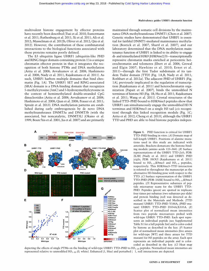
The E3 ubiquitin ligase UHRF1 (ubiquitin-like PHDand RING finger domain-containing protein 1) is a uniquechromatin effector protein in that it integrates the rec-ognition of both histone PTMs and DNA methylation(Arita et al. 2008; Avvakumov et al. 2008; Hashimotoet al. 2008; Nady et al. 2011; Rajakumara et al. 2011). Assuch, UHRF1 harbors multiple domains that bind chro-matin (Fig. 1A). The UHRF1 SET and RING-associated(SRA) domain is a DNA-binding domain that recognizes5-methylcytosine (5mC) and 5-hydroxymethylcytosine inthe context of hemimethylated double-stranded CpGdinucleotides (Arita et al. 2008; Avvakumov et al. 2008;Hashimoto et al. 2008; Qian et al. 2008; Frauer et al. 2011;Spruijt et al. 2013). DNA methylation patterns are estab-lished during early embryogenesis by de novo DNAmethyltransferases DNMT3a and DNMT3b (with theassociated, but noncatalytic, DNMT3L) (Okano et al.1999; Bourc’his et al. 2001; Jia et al. 2007) and are primarily
maintained through somatic cell divisions by the mainte-nance DNA methyltransferase DNMT1 (Chen et al. 2007).Genetic studies have demonstrated that UHRF1 is essen-tial for faithful DNMT1-mediated maintenance methyla-tion (Bostick et al. 2007; Sharif et al. 2007), and ourlaboratory determined that the DNA methylation main-tenance function of UHRF1 is linked to its ability to engagedi- and trimethylated H3K9 (H3K9me2/3)—transcriptionallyrepressive chromatin marks enriched at pericentric het-erochromatin and telomeres (Ebert et al. 2006; Grewaland Elgin 2007; Hawkins et al. 2010; Ernst et al.2011)—through the first Tudor subdomain of its tan-dem Tudor domain (TTD) (Fig. 1A,B; Nady et al. 2011;Rothbart et al. 2012a). The adjacent PHD of UHRF1 (Fig.1A), previously implicated in the regulation of transcrip-tion (Rajakumara et al. 2011) and heterochromatin orga-nization (Papait et al. 2007), binds the unmodified Nterminus of histone H3 (Fig. 1B; Hu et al. 2011; Rajakumaraet al. 2011; Wang et al. 2011). Crystal structures of thelinked TTD–PHD bound to H3K9me3 peptides show thatUHRF1 can simultaneously engage the unmodified H3 Nterminus and H3K9me3 on a single H3 tail (cis recogni-tion) through this linked recognition module (Fig. 1C;Arita et al. 2012; Cheng et al. 2013), although the UHRF1TTD and PHD are able to bind histone peptides indepen-
Figure 1. PHD function is critical for UHRF1TTD–PHD binding in vitro. (A) Domain map offull-length UHRF1. Positions of alanine muta-tions used in this study are indicated withasterisks. Brackets demarcate the histone-bind-ing module (amino acids 123–366). (B) Surfacerepresentations of the UHRF1 TTD (left; PDB:2L3R) (Nady et al. 2011) and UHRF1 PHD(right; PDB: 3SOU) (Rajakumara et al. 2011)bound to H31–11K9me3 and H31–8 peptides,respectively. This H3K9me3–TTD interactionis referred to throughout the manuscript as thealternative H3-binding pose with respect to theTTD. (C) Surface representation of the UHRF1TTD–PHD (PDB: 3ASK) bound to H31–11K9me3peptides. (D) Representative subarrays of pep-tide microarray scans for the UHRF1 TTD–PHD. Peptides (green) are spotted in triplicatefour times per subarray (two subarrays per slide)and protein binding (red) was detected as de-scribed in the Materials and Methods. (TTDmutant) UHRF1 TTD–PHD Y188A; (PHD mu-tant) UHRF1 TTD–PHD D334A/E335A. (E)Scatter plot of normalized mean intensitiesfrom two peptide microarrays probed withwild-type UHRF1 TTD–PHD. Each spot repre-sents an individual peptide (see SupplementalTable S1 for a full peptide list) and is color-codedby histone as described in the key. (F) Scatterplot of normalized mean intensities (five arraysfor wild-type [WT] and three arrays for TTDmutant) for H3 peptides on the array. Each spotrepresents an individual peptide and is color-coded as described in the key. (G) Heat map
depicting the effects of single PTMs on the binding of wild-type UHRF1 TTD–PHD to H31–20 peptides. Normalized mean intensities arerepresented relative to unmodified H31–20 (0, white). Enhanced (1, blue) and perturbed (�1, red) interactions are depicted.
Multivalency guides UHRF1 chromatin function
GENES & DEVELOPMENT 1289
Cold Spring Harbor Laboratory Press on May 23, 2018 - Published by genesdev.cshlp.orgDownloaded from

dently of one another in vitro (Fig. 1B; Hu et al. 2011;Nady et al. 2011; Rajakumara et al. 2011; Wang et al. 2011).However, it remains to be determined whether multiva-lent engagement of H3 by the linked reader modules ofUHRF1 is biologically significant.
Here, we comprehensively characterize the specificity,selectivity, and molecular dynamics of the TTD–PHDdual-effector module of UHRF1. Notably, we demonstratethat this structural unit functions as a single histonerecognition module in cells, providing a combinatorialreadout of a histone H3 tail in cis that is required for theepigenetic inheritance of DNA methylation. Our findingsare among the first to demonstrate that multivalenthistone engagement in a defined manner plays a funda-mental role in driving a downstream biological event inchromatin, providing critical support for the combinato-rial nature of the ‘‘histone code’’ hypothesis.
Results
The UHRF1 PHD dominates multivalent histoneH3 engagement in vitro
To decipher the contribution of the ‘‘histone code’’ oneffector protein recognition, we recently developeda high-density (>4000 features) peptide microarray plat-form harboring a comprehensive library of known singleand combinational PTMs on each of the core histoneproteins (H3, H4, H2A, and H2B) (Supplemental Table S1;Fuchs et al. 2011; Rothbart et al. 2012b). This technologywas recently used to define the isolated UHRF1 TTD asa bona fide H3K9 di- and trimethyl reader module (Fig. 1B)and also uncovered a novel and biologically relevantproperty of the UHRF1 TTD as an H3K9 effector domainthat is insensitive to neighboring H3S10 phosphorylation(H3S10p) (Rothbart et al. 2012a). In addition to the TTD,UHRF1 contains an adjacent PHD finger that interactswith the unmodified N terminus of H3 (Fig. 1B; Hu et al.2011; Rajakumara et al. 2011; Wang et al. 2011). Recentstructural studies indicated that the linked TTD andPHD domains of UHRF1 simultaneously bind H3K9me3peptides on a single histone tail in vitro (Fig. 1C; Arita et al.2012; Cheng et al. 2013). We therefore sought to character-ize this multivalent cis interaction further in the context ofthe ‘‘histone code’’ and asked whether such binding isbiologically important for UHRF1 function in the cell.
Peptide microarrays probed with the wild-type UHRF1TTD–PHD revealed a strong preference for histone H3peptides encompassing the N-terminal tail (Fig. 1D,E;Supplemental Fig. S1A). Notably, an aromatic cage mu-tation of the UHRF1 TTD (Y188A; TTD mutant), pre-viously shown to abolish the interaction with H3K9methylation (Nady et al. 2011), showed interactions sim-ilar to those found with the wild-type dual domain, withthe exception that a number of H3K9 methylated pep-tides that were ‘‘strong’’ hits in the wild-type case(Supplemental Fig. S1B) were shifted slightly to a lowerrange of detection (Fig. 1D,E). In striking contrast, alaninemutations at two acidic residues in the UHRF1 PHD(D334A/E335A; PHD mutant) (Rajakumara et al. 2011),
previously shown to strongly inhibit the isolated PHDinteraction with N-terminal H3 tail peptides (Rajakumaraet al. 2011), completely disrupted array binding, even inthe presence of a functional TTD (Fig. 1D). Consistent withan important role for the N terminus of H3 in TTD–PHDbinding, PTMs at the extreme N terminus of H3, includingasymmetric Arg 2 methylation or citrullination (H3R2me2aand Cit2, respectively), Thr 3 phosphorylation (H3T3p), andLys 4 acetylation (H3K4ac), strongly perturbed binding ofthe wild-type UHRF1 TTD–PHD (Fig. 1F,G). Interestingly,while array analysis showed a slight inhibitory effect in thepresence H3K4me3, in-solution peptide pull-down andisothermal titration calorimetry (ITC) analysis indicatedthat this change was not significant (data not shown). Col-lectively, these data suggest that the UHRF1 PHD providesa dominant affinity for the unmodified N terminus of H3,while the TTD facilitates the localization of UHRF1 toH3K9me2/3-marked chromatin.
Quantitative analysis of UHRF1 TTD–PHD bindingto H3K9me3 reveals individual domain contributionsto multivalency
While peptide microarray analysis is a powerful tool fordiscriminating between strong and weak binding events,the technology is limited to semiquantitative measures.To further define the contributions of the UHRF1 TTDand PHD to multivalent engagement, we sought toquantify binding events using fluorescence polarization.The UHRF1 TTD–PHD bound tightly to H31–20K9me2(Kd ;0.17 mM) and H31–20K9me3 (Kd ;0.35 mM) peptidesfluorescently labeled at the C terminus (Fig. 2A; Supple-mental Fig. S1C), consistent with Kd measurements forH3K9me3 peptides by ITC (Arita et al. 2012). Perturbingthe TTD interaction with H3K9 methylation (Y188A;TTD mutant) diminished the affinity for these peptides10-fold to 15-fold (Kd ;2.5 and 4.5 mM, respectively) (Fig.2A; Supplemental Fig. S1C). Consistent with microarrayresults, perturbing the PHD interaction with the N ter-minus of H3 (D334A/E335A; PHD mutant) resulted inundetectable binding to H31–20K9me2 and H31–20K9me3peptides fluorescently labeled at the C terminus (Fig. 2A;Supplemental Fig. S1C).
Many effector modules that recognize the extremeN terminus of H3 (residues 1–4)—including the UHRF1PHD—specifically interact with the N-terminal aminogroup of Ala 1. Therefore, we predicted that placement ofa fluorescent probe on the N terminus of H31–20K9me3peptides would block PHD interaction with the free Nterminus. Indeed, N-terminal probe placement resultedin no detectable binding to wild-type UHRF1 TTD–PHDor to the individual domain mutants of this dual bindingmodule (Fig. 2A). These results agree with those of Aritaet al. (2012), in which no measurable binding of wild-typeUHRF1 TTD–PHD was detected by ITC to H3K9me3 pep-tides acetylated at their N-terminal amino group. Further-more, binding of the UHRF1 TTD–PHD to H31–20K9me3peptides fluorescently labeled at the C terminus was out-competed by unlabeled H31–5 peptides containing eitherL-alanine or L-valine at position 1 but not with unlabeled
Rothbart et al.
1290 GENES & DEVELOPMENT
Cold Spring Harbor Laboratory Press on May 23, 2018 - Published by genesdev.cshlp.orgDownloaded from

H31–5 peptides containing either D-alanine or D-valine,demonstrating that stereochemistry at the first positionis critical for PHD binding and that the functional PHDis likely able to accommodate more bulky side chains(Supplemental Fig. S1D).
Collectively, these results demonstrate that the high-affinity interaction of the UHRF1 TTD–PHD for H3K9me2
and H3K9me3 peptides requires multivalent engagementof H3K9 methylation by the UHRF1 TTD and the H3 Nterminus by the UHRF1 PHD. Furthermore, the UHRF1TTD–PHD cannot maintain a measurable interactionwith H3K9me2/3 in the absence of a functional PHDthat interacts with the H3 N terminus, while TTD–PHDaffinity for H3K9me2/3 peptides is reduced ;10-fold inthe absence of a functional methyllysine binding pocketwithin the TTD (Fig. 2A).
The UHRF1 PHD–linker fragment physicallyoccludes alternative H3 peptide-binding poseswith the UHRF1 TTD
Strong experimental evidence shows that the UHRF1TTD can function as a H3K9me2/3 reader domain inisolation (Supplemental Fig. S1E; Nady et al. 2011; Aritaet al. 2012; Rothbart et al. 2012a; Cheng et al. 2013), butour data demonstrate that in the context of the dual do-main, the TTD has no measurable affinity for H3K9me2/3peptides in the absence of PHD function. This led us toquestion why the physical linkage of the TTD to the PHDfinger renders this H3K9 reader domain incapable ofengaging H3K9 methylated histone tails in the absenceof PHD coordination of the H3 N terminus. In the crystalstructure of the UHRF1 TTD–PHD bound to H3K9me3(Protein Data Bank [PDB]: 3ASK) (Arita et al. 2012), the15-amino-acid region (linker) connecting the TTD andPHD domains (amino acids 286–300) resides in the samebinding groove occupied by the H3 peptide in the solutionstructure of the isolated UHRF1 TTD (PDB: 2L3R) (Fig.1B,C; Nady et al. 2011). Here, we refer to this H3–TTDinteraction (PBD: 2L3R) (Nady et al. 2011) as an alterna-tive H3-binding pose with respect to the TTD. Nuclearmagnetic resonance (NMR) titration of the 15-residueTTD–PHD linker peptide confirms its interaction withthe alternate H3 peptide-binding groove of the free TTD(Fig. 3F,G; Supplemental Fig. S4A), and ITC indicates thatthis intrinsic affinity of the free linker peptide for theTTD is weak (>100 mM) in the absence of covalent inter-action in the intact protein (Supplemental Fig. S4B).
Mutations of R295 and R296 or phosphorylation ofS298, both of which disrupt the linker–TTD interaction,were previously suggested to uncouple the cooperativebinding potential of the UHRF1 TTD from the PHD inthe context of the dual domain (Arita et al. 2012). Con-sistent with these findings, a mutant of the linker regionwithin the TTD–PHD (R295A/R296A) was able to bindH31–20K9me3 peptides fluorescently labeled at both theN and C termini (Fig. 2B). This supports a model in whichdissociation of the linker from the TTD allows the al-ternate mode of H3 peptide binding with the TTD, aninteraction that does not involve the N terminus of H3and is therefore not affected by the N-terminal fluores-cent probe. This model also predicts that linker occu-pancy of the TTD peptide-binding groove would preventthe TTD from binding H3K9me2/3 peptides in the alter-native pose (Fig. 1B). To test this predicted occlusionfunction of the linker region, we compared the bindingaffinity of N-terminally labeled H3K9me3 peptides (which
Figure 2. High-affinity interaction of UHRF1 with H3K9me3peptides requires multivalent TTD and PHD engagement in cis.(A,B) Fluorescence polarization binding assays of H31–20K9me3peptides labeled with 5-carboxyfluorescein (5-FAM) at the Nterminus (red) or C terminus (blue) with the indicated wild-type(WT) and mutant UHRF1 TTD–PHD proteins. Error bars representSD for three independent experiments. All Kd values were deter-mined using a one-site binding model as described in the Materialsand Methods. Asterisk in B indicates that Kd is likely an averagecontribution of two binding sites as suggested by published ITC-binding stoichiometry calculations (n-value) (Arita et al. 2012). (n.m)Not measurable; (TTD mutant) UHRF1 TTD–PHD Y188A; (PHDmutant) UHRF1 TTD–PHD D334A/E335A; (linker mutant) UHRF1TTD–PHD R295A/R296A. Cartoons depict predicted peptide-binding modes for wild-type (A, top panel) and Linker mutant(B) UHRF1 TTD–PHD based on experimental observations.
Multivalency guides UHRF1 chromatin function
GENES & DEVELOPMENT 1291
Cold Spring Harbor Laboratory Press on May 23, 2018 - Published by genesdev.cshlp.orgDownloaded from

can only bind the TTD in the alternative pose) with theTTD–PHD dual domain and with a protein fragmentcontaining just the TTD and linker sequence (TTD–linker)and compared the latter with the disruptive R295A/R296A
mutation in the linker region (TTD–linkermut) (Fig. 3A).Surprisingly, unlike the TTD–PHD dual domain, theTTD–linker was able to bind N-terminally taggedH3K9me3, and this interaction was enhanced by the
Figure 3. The UHRF1 linker–PHD physically occludes TTD coordination of H3K9 methylated peptides in alternative binding poses. (A)Fluorescence polarization binding assays of N-terminal 5-FAM-H31–20K9me3 peptides with wild-type (WT; black) and linker mutant (blue)UHRF1 TTD–linker (amino acids 123–301) or wild-type UHRF1 TTD–PHD (red). Error bars represent SD for three independent experiments.Kd values were determined using a one-site binding model as described in the Materials and Methods. (TTD–linkermut) UHRF1 TTD–linkerR295A/R296A. (B) Estimate of the free-energy landscape in the TTD–PHD molecular dynamics simulation with several representativeconformations. An H31–11K9me3 peptide (yellow) is modeled into the structure in the isolated TTD peptide-binding pose as a point ofreference. The linker region (amino acids 286–301) is depicted as a white ribbon. Contours on the free energies are drawn every 4kbT, wherekb is the Boltzmann constant and T is the temperature. (C) Estimate of the free-energy landscape in the TTD–linker (amino acids 123–301)molecular dynamics simulation. Contours on the free energies are drawn every 4kbT, where kb is the Boltzmann constant and T is thetemperature. (D) An overlay of linker (left) and R295 (right) conformations for the TTD–PHD (gray) and TTD–linker (red) moleculardynamics simulations. An H31–11K9me3 peptide (yellow) is modeled into the structure in the isolated TTD peptide-binding pose as a point ofreference. (E) Fluorescence polarization competition of N-terminal 5-FAM-H31–20K9me3 peptides bound to UHRF1 TTD–linker (red) orUHRF1 TTD (black; amino acids 126–280) in the presence of the indicated concentrations of UHRF1 PHD (amino acids 301–366). Error barsrepresent SD for three independent experiments. (F) Histogram profiles showing NMR chemical shift changes of the UHRF1 TTD (aminoacids 126–285) upon addition of the linker (orange; amino acids 286–300), PHD (black; amino acids 310–366), or linker–PHD (blue; aminoacids 286–387). (G) Surface representation of the UHRF1 TTD–linker (PDB: 3ASK). The linker region (amino acids 286–300) is depicted aswhite sticks. Significant NMR chemical shifts observed in titrations of the linker (orange), linker–PHD (blue), and both (red) are depicted.
Rothbart et al.
1292 GENES & DEVELOPMENT
Cold Spring Harbor Laboratory Press on May 23, 2018 - Published by genesdev.cshlp.orgDownloaded from

R295A/R296A linker mutation. Collectively, these re-sults demonstrate that the linker region alone cannotfully occlude the alternative H3-binding mode of theTTD in the absence of the PHD, and they suggest thatthe linker functions with the PHD in some way topromote multivalent histone engagement by the dualTTD–PHD module.
To gain further insight into how the PHD and linkermodulate the interaction of the TTD with the H3 tail, wefirst performed a series of microsecond-scale nonequilib-rium molecular dynamics simulations for the UHRF1TTD–PHD and UHRF1 TTD–linker (Fig. 3B–D; Supple-mental Movie S1; Supplemental Fig. S3). Because we wereinterested in observing rare events (e.g., the dynamicrearrangement of the PHD or linker with respect to theTTD), we made use of an adaptive biasing potential(Dickson 2011) to accelerate conformational samplingand assess the free-energy landscapes. The TTD–PHDfree-energy projection was designed to correlate directlywith PHD positioning (see Materials and Methods fordetails), and the position of the PHD in the crystalstructure (PDB: 3ASK) (Arita et al. 2012) is located atthe origin of the free-energy map (Fig. 3B). The projectedhistograms from 10 400-nsec simulations of TTD–PHDare shown in Supplemental Figure S3C, and a collectiveaverage of the free-energy landscape is depicted in Figure3B. Simulations of the TTD–PHD indicated a rugged free-energy landscape created by nonspecific interactionsbetween the PHD and the TTD–linker (Fig. 3B; Supple-mental Fig. S3C). This simulation suggests that the PHDis ‘‘scanning’’ the TTD surface rather than residing ina single conformation relative to the TTD. The numerousnonspecific interactions of the PHD with the TTD resultin a compact coordination of the linker and PHD near theTTD surface. Several metastable conformations of thePHD in this simulation are depicted in Figure 3B, in-cluding several poses that may physically occlude theTTD aromatic cage that coordinates methylated H3K9(Nady et al. 2011). Notably, closer examination of thelinker positions in the TTD–PHD simulations suggestthat these weak interactions between TTD and PHDpromote occupancy of the linker within the TTD peptide-binding groove, as depicted in Figure 3D.
In stark contrast, the TTD–linker simulation displays amuch less complex free-energy landscape. The free-energyestimates from five independent simulations are shown inSupplemental Fig. S3D, and a collective average of the free-energy landscape is depicted in Figure 3C. Here, the originof the coordinates corresponds to the location of the Ca
atom of S301 in the crystal structure (PDB: 3ASK) (Aritaet al. 2012). Consistently, in all five free-energy estimates,this reference position is rarely visited. Instead, the linkerlargely resides away from the TTD peptide-binding groove(Fig. 3D). Collectively, these simulations suggest a role forthe PHD in maintaining the linker in or near the TTD-binding groove through transient interactions with theTTD. Hence, both the linker and PHD appear to block theTTD from binding H3 in the alternative binding pose.
Consistent with the model described above, the freeUHRF1 PHD was able to perturb TTD–linker coordina-
tion of H3K9me3 peptides fluorescently labeled at the Nterminus (Fig. 3E). This observed competition seems to bespecific to the UHRF1 PHD, as the BPTF PHD finger hadno inhibitory effect on TTD–linker binding (Supplemen-tal Fig. S2). However, the UHRF1 PHD was unable toperturb the isolated UHRF1 TTD from binding thispeptide (Fig. 3E), demonstrating that the PHD inhibitionseen in Figure 3E is mediated through interaction withthe linker. Furthermore, analysis of the HSQC spectra ofUHRF1 TTD titrated with linker peptide, PHD, andlinker–PHD demonstrated that the majority of significantchemical shift changes observed are a result of interac-tion between the linker and the TTD (Fig. 3F; Supple-mental Fig. S4). Both molecular dynamics and NMRtitrations suggest that the PHD does not engage inspecific interactions with the TTD (Fig. 3F; SupplementalFig. S4). Additionally, discreet differences in chemicalshift changes observed for the TTD in the linker andlinker–PHD titrations suggest a role for the PHD in fine-tuning the orientation of the linker in the TTD groove.For example, several significant chemical shift changesfor residues outside of the TTD groove (e.g., K136, N228,K233, and E278) in the linker titration were absent in thelinker–PHD titration (Fig. 3F,G; Supplemental Fig. S4).Furthermore, D190, shown to play key roles in theinteraction of the isolated TTD with the H3 tail (Nadyet al. 2011) and the TTD–PHD with the linker (Arita et al.2012), displayed a significant chemical shift change in thelinker–PHD titration only (Fig. 3F,G; Supplemental Fig.S4). Collectively, these results suggest a key role for thePHD in preventing TTD binding to H3 peptides in thealternative pose and reveal the importance of the co-ordinated ensemble of TTD–linker–PHD conformationsin UHRF1 multivalent histone engagement, in which theTTD–linker–PHD module reads the PTM status of the H3tail from Ala 1 through Lys 9.
UHRF1 TTD–PHD multivalent histone engagementin cis is required to facilitate DNA methylationmaintenance
We next sought to determine whether UHRF1 TTD–PHDmultivalent histone engagement is important for theinteraction of UHRF1 with bulk chromatin in cells.Wild-type and TTD (Y188A) and PHD (D334A/E335A)mutants of full-length UHRF1 were transiently expressedin HeLa cells for 72 h, followed by biochemical separationinto chromatin-enriched and soluble fractions (Fig. 4A).Consistent with our previous findings (Rothbart et al.2012a), the TTD mutant was unable to bind bulk chro-matin, and protein was found primarily in the solublefraction (Fig. 4A,C). Strikingly, similar results were obtainedwith both the PHD mutant and linker mutations (R295A/R296A) that uncouple H3 N-terminal binding or thehistone-binding properties of TTD from the PHD, re-spectively (Fig. 4A,B). Consistently, confocal microscopyof TTD, PHD, and linker mutants of full-length GFP-tagged UHRF1 in HEK293 cells showed that these mu-tants remain in the nucleus but no longer associate withDAPI-dense pericentric heterochromatin (Fig. 3C).
Multivalency guides UHRF1 chromatin function
GENES & DEVELOPMENT 1293
Cold Spring Harbor Laboratory Press on May 23, 2018 - Published by genesdev.cshlp.orgDownloaded from

We further hypothesized that the ability of UHRF1 tofacilitate DNA methylation maintenance may requirecoordinated multivalent histone engagement. We showedpreviously that UHRF1 knockdown with shRNAs inHeLa cells resulted in ;60% reduction in global 5mCcontent (as analyzed by immunofluorescence), and rein-troduction of wild-type full-length UHRF1 (but not aTTD mutant [Y188A]) was able to restore DNA methyl-ation in these cells (Fig. 4D; Rothbart et al. 2012a). Usingthis system, we queried whether PHD histone-bindingmutants (D334A/E335A) or linker mutants (R295A/R296A)could genetically complement for UHRF1 knockdown.Consistent with the notion that UHRF1 DNA methyla-tion maintenance function requires the physical associa-tion of UHRF1 with chromatin, neither UHRF1 PHD norlinker mutants were able to restore global DNA methyl-ation levels in UHRF1 knockdown HeLa cells (Fig. 4D).To further examine the role of UHRF1 histone multi-valency in DNA methylation maintenance, we examinedthe methylation status of a CpG island from the 59 end ofthe intergenic spacer of ribosomal DNA (IGS-rDNA)using methylation-specific PCR (Herman et al. 1996).We and others have shown that DNA methylation at this
locus is regulated by UHRF1 (Bostick et al. 2007; Rothbartet al. 2012a). Consistent with our previous findings,knockdown of UHRF1 in HeLa cells diminished DNAmethylation at this locus, and transient expression ofwild-type (but not TTD mutant) UHRF1 partially re-stored DNA methylation (Fig. 4E). Furthermore, neitherlinker nor PHD mutants of UHRF1 were able to restoreDNA methylation at this locus (Fig. 4E). Collectively,these results provide definitive evidence that linked histone-binding domains of an effector protein must act coordinatelyin cis to functionally engage chromatin in cells and that thisbinding is critically important to mediate a fundamentaldownstream epigenetic event in chromatin–DNA methyla-tion maintenance.
Discussion
Our studies comprehensively define the contributions ofthe linked TTD and PHD domains of UHRF1 to multi-valent histone engagement. This dynamic ensemble islikely sampling multiple conformations within the chro-matin microenvironment until an appropriately markedH3 tail is engaged. Notably, the SRA domain of UHRF1
Figure 4. The UHRF1 TTD–PHD functions as a single unit in cells to maintain global DNA methylation. (A,B) Western blot analysisof wild-type (WT) and the indicated mutant forms of full-length UHRF1 in HeLa cell lysates biochemically separated into chromatinand soluble fractions. (b-Tubulin and H3) Fractionation controls; (Mock) no DNA transfection. (C) Representative confocal microscopyimages of GFP-tagged wild-type and mutant forms of full-length UHRF1 in HEK293 cells. (GFP only) Transfection of empty GFP vector.Bars, 10 mm. (D) Representative immunofluorescence staining for 5mC in control and UHRF1 knockdown HeLa cells after geneticcomplementation with wild-type and mutant forms of full-length UHRF1. (Ctrl. KD) Nontargeted shRNA; (Mock) transfection in theabsence of DNA. Bars, 10 mm. Error is represented as 6SD. (E) Methylation-specific PCR for IGS-rDNA in control and UHRF1knockdown HeLa cells after genetic complementation with wild-type and mutant forms of full-length UHRF1. Representativeunmethylated (U) and methylated (M) gene-specific PCR products are shown. (Ctrl. KD) Nontargeted shRNA; (Mock) transfection inthe absence of DNA. Error is represented as 6SD.
Rothbart et al.
1294 GENES & DEVELOPMENT
Cold Spring Harbor Laboratory Press on May 23, 2018 - Published by genesdev.cshlp.orgDownloaded from

harbors an additional chromatin-binding domain capableof recognizing hemimethylated DNA (i.e., DNA in needof maintenance methylation). As such, UHRF1 is one ofa few proteins known to recognize both histone and DNAregulatory marks on chromatin. Unpublished findingsfrom our laboratory and recent work by Liu et al. (2013)suggest an important role for SRA binding in cells, andcareful examination of this apparent multivalent interac-tion with histones and DNA will be an important area offuture biochemical study.
The original posit of the ‘‘histone code’’ hypothesisstated that there is a combinatorial code embedded inhistone tails that is read by effector proteins to mediatea downstream biological event in chromatin (Strahl andAllis 2000). Much research over the past decade hasprovided support for this hypothesis, primarily throughthe cataloging of individual reader domains recognizingindividual histone marks. Our results demonstrate thathistone-binding proteins themselves have evolved to in-terpret complex chromatin signatures through linked and‘‘collaborative’’ reader modules. Furthermore, the low-affinity, but functionally independent, nature of the UHRF1TTD, linker, and PHD domains suggests that multivalentchromatin-binding modules may be more susceptible tofunctional modulation by cellular events/factors such asPTMs (compared with a single high-affinity interaction).
Beyond UHRF1, these studies underscore the complex-ity of chromatin recognition likely to be harbored ineffector proteins with closely spaced reader modulesand within multiprotein complexes with multiple readermodules. While it remains important to define the spec-ificity of reader domains in isolation, multivalent chroma-tin engagement by linked or coordinated reader domains islikely to be a more physiologically relevant mode of inter-preting the complex language of chromatin modifications.
Materials and methods
Materials
Histone peptides were synthesized, purified, and analyzed asdescribed (Rothbart et al. 2012b). Linker peptide (amino acids286–300) used for NMR titrations was purchased from Genscript.Antibodies used in this study were as follows: anti-Flag (1:5000;Sigma, F1804), anti-b-tubulin (1:1000; Cell Signaling, 2146), anti-H3(1:20,000; EpiCypher, 13-0001), and anti-5mC (1:100; Diagenode,Mab-081). Human cDNAs encoding the UHRF1 TTD–PHD andTTD–linker were cloned into pGEX-4T-1 (GE Lifesciences) asN-terminal GST fusions for peptide microarray and fluorescencepolarization studies, expressed in Escherichia coli BL21(DE3)using standard procedures, and purified with GST-Bind Resin(Novagen) according to the manufacturer’s protocol. The humanUHRF1 PHD for fluorescence polarization was a gift from Y. Shi(Harvard University). This N-terminal 6x-His fusion was expressedin E. coli BL21(DE3) using standard procedures and was purifiedwith Talon Resin (Clontech) according to the manufacturer’sprotocol. An N-terminal GST fusion of the BPTF PHD was a giftfrom A. Ruthenburg (University of Chicago) and was expressedand purified as described above. Full-length human UHRF1 wascloned into pCMV-Tag 2 (Agilent) as an N-terminal Flag fusionand into pEGCP-C1 (gift from M. Bedford, MD Anderson CancerCenter) as an N-terminal GFP fusion for mammalian expression.
Point mutations were generated by QuickChange site-directedmutagenesis (Stratagene).
Histone peptide microarrays
Peptide synthesis and validation, microarray fabrication, effec-tor protein hybridization and detection, and data analysis wereperformed essentially as described (Rothbart et al. 2012b) withthe following modification. Each peptide listed in SupplementalTable S1 was spotted in triplicate eight times per array. Triplicatespots were averaged and treated as a single value for subsequentstatistical analysis.
Fluorescence polarization binding assays
Peptides for fluorescence polarization were synthesized essen-tially as described (Rothbart et al. 2012b) with an additional 6-hcoupling with three equivalents of 5-carboxyfluorescein (5-FAM)and diidopropylcarbodiimide and nine equivalents of diisopro-pylethylamine in 1:1 volume equivalents of DMF/DMSO.C-terminally modified peptides were synthesized with additionalorthogonally protected lysine at the C terminus after selectivedeprotection of the e-amino group that lysine 5-FAM was coupledto. Binding assays were performed in 50 mL in black flat-bottom384-well plates (Costar) as described (Rothbart et al. 2012a).
Molecular dynamics
Adaptive biasing potential (Dickson 2011) was implemented inGROMACS 4.5.5 (Hess et al. 2008). For the UHRF1 TTD–PHD,the collective variables were defined as the three Cartesiancoordinates of the center of geometry of the PHD. The positionof the PHD in the crystal structure (PDB: 3ASK) (Arita et al.2012) is located at the origin of the free-energy map (Fig. 3B). Forthe TTD–linker, the collective variables were defined as thethree Cartesian coordinates of the Ca atom of residue Ser 301,the last residue of the linker. The position of the Ser 301 Ca inthe TTD–PHD crystal structure is located at the origin of thecoordinates in Figure 3C. A grid in collective variable space isshown for the UHRF1 TTD–PHD in Supplemental Figure S3A.For better visibility, the visitation histograms were projectedonto a two-dimensional surface, as illustrated in SupplementalFigure S3B. During the simulations, the UHRF1 TTD backbone(up to residue Glu 285) was loosely restrained by a harmonicpotential to eliminate rotations and translations of the TTD,allowing the motion of the PHD or linker to be tracked directlyas movement in the collective variable grid. To further facilitatevisualization and enable the comparison of free-energy land-scapes, the crystal structure (PDB: 3ASK) (Arita et al. 2012) wastaken as a reference. The TTD backbones of all simulationsnapshots were aligned to the TTD backbone of the presimula-tion structure using visual molecular dynamics (Humphrey et al.1996) before simulation. The motion of the PHD or linker in thesimulations can thus be directly compared with the referencestructure.
The Cartesian coordinate system used here was selected sothat the origin was at the center of geometry of the TTD and theZ-axis pointed from the origin to the center of geometry of thePHD in the 3ASK structure (or in the case of the TTD–linker, tothe Ca atom of residue S301). The position of the PHD (Ca atomof residue S301) was tracked as rotations around the X-axis andY-axis of this coordinate system. The projected histograms wereobtained by integrating over the radial component of the data ata fixed rotation around the X-axis and Y-axis. Rotations aroundthe X-axis were denoted with u1, and rotations around the Y-axis
Multivalency guides UHRF1 chromatin function
GENES & DEVELOPMENT 1295
Cold Spring Harbor Laboratory Press on May 23, 2018 - Published by genesdev.cshlp.orgDownloaded from

were denoted with u2. The Euler-Rodrigues formula was used toexpress the rotations around an axis.
The bias parameters were b = 0.9, c = 0.1/dt = 50 ps�1, where dt
is the molecular dynamics time step. The Gaussian width wasa = 0.3 nm. All simulations were fully solvated in TIP3P water atphysiological salt conditions. For the UHRF1 TTD–PHD, thesimulation box was a dodecahedron with a minimum distance of17 A from the protein to the wall to allow for potential linkerextension. This resulted in a total of 70,147 atoms after addingsalt and water. For the TTD–linker, the box was a dodecahedronwith a minimum distance of 1.3 A from the protein to the boxwall, resulting in a total of 40,830 atoms after adding salt andwater. The proteins were described by the OPLS-AA force field(Kaminski et al. 2001). For the TTD–PHD, bond and anglerestraints were added to maintain the geometry of coordinationwith zinc as described (Peters et al. 2010). System constructionincluded 5 nsec of isothermal–isobaric equilibration at 1 bar.Production simulations were carried out in the canonical en-semble with stochastic velocity rescaling (Bussi et al. 2007). Vander Walls and direct electrostatic interactions used a 10 A cutoff.Long-range electrostatics were treated with the particle meshEwald approach with a grid spacing of 1.6 A. The neighbor listwas updated every five steps. Ten trajectories, each 400 nsecin duration, were generated for the TTD–linker–PHD. Fivetrajectories, each 200 nsec long, were generated for the TTD–linker.
NMR spectroscopy and data analysis
The UHRF1 TTD (amino acids 126–285) and linker–PHD (aminoacids 288–387) were cloned into pET28- MHL and pNICCHvectors, respectively. The UHRF1 PHD (amino acids 310–366)was cloned into a pET15b-MHL vector using the In-Fusionmethod (BD Biosciences). UHRF1 TTD was expressed and purifiedas previously described (Nady et al. 2011). UHRF1 linker–PHDand PHD were expressed in E. coli cultured in Terrific Brothmedium following overnight induction at 15°C with 1 mMIPTG, when OD600 reached 1.5. Cell pellets were resuspendedin 20 mM Tris-HCl (pH 8.0), 0.5 M NaCl, 5% glycerol, 2 mMImidazole, 2 mM BME, and 1 mM PMSF. After sonication andcentrifugation, Ni-NTA agarose beads were added to the clarifiedlysate, and binding to the beads was allowed to proceed for 1 h.Bound protein was washed with 20 mM Tris-HCl (pH 8.0), 0.5 MNaCl, 5% glycerol, and 20 mM Imidazole and eluted in similarbuffer containing 250 mM imidazole. The eluate was passedthrough a size exclusion S200 16/60 column and then dialyzedinto 20 mM NaPO4 (pH 7.0), 0.25 M NaCl, and 1 mM DTT at4°C. Chemical shift changes of residues on the UHRF1 TTDupon titration of unlabeled linker peptide (amino acids 286–300;Genscript), PHD, and linker–PHD,were mapped using prioravailable chemical shift assignments deposited in the BiologicalMagnetic Resonance Bank (BMRB) (Nady et al. 2011). 1H–15NHSQC spectra were collected at 25°C with a Bruker Avance 500-MHz spectrometer. Composite chemical shift changes (Dppm) werecalculated using the equation [(DppmN/6.5)2 N + (DppmHN
2)]0.5.
ITC
Proteins and peptides were extensively dialyzed into buffercontaining 20 mM Tris-HCl (pH 8.0), 150 mM NaCl, and 5%glycerol at 4°C. ITC measurements were performed at 20°Cwith a MicroCal VP-ITC (GE Healthcare). Linker andH3K9me2 peptides at 500 uM were titrated (25 10-mL in-jections at 10-sec intervals) with 30 uM UHRF1 TTD. Datapoints were fit to a one-site binding model using MicroCalOrigin 7.0 software.
Cell culture and manipulation
HeLa and HEK293 (American Type Culture Collection) werecultured in MEM and DMEM (Gibco), respectively, supple-mented with 10% FBS (PAA Laboratories), maintained in a37°C incubator with 5% CO2 and passaged every 2–3 d. Tran-sient transfections were performed using TurboFect (Fermentas)according to the manufacturer’s protocol. shRNA knockdown ofUHRF1 in HeLa cells was described previously (Rothbart et al.2012a). For GFP-UHRF1 analysis, transfected cultures of HEK293cells growing in eight-well chamber slides (Lab-Tek) were fixedwith 4% paraformaldehyde, washed with 13 PBS, and mountedusing SlowFade Anti-fade (Life Technologies) containing DAPI.Images were acquired using an FV1000 multiphoton invertedconfocal microscope (Olympus) at 643 magnification followingexcitation with 405-nm and 488-nm single-photon lasers.
Chromatin fractionation
Chromatin and associated proteins were isolated from asynchro-nously growing HeLa cells as described (Kuo et al. 2012; Rothbartet al. 2012a).
DNA methylation analysis
Cells were labeled with anti-5mC (Diagenode) essentially asdescribed (Rothbart et al. 2012a). Images were acquired using anFV1000 multiphoton inverted confocal microscope (Olympus)at 603 magnification following excitation with 405-nm and633-nm single-photon lasers. Methylation percentage was de-rived from quantification of 5mC fluorescence normalized toDNA fluorescence from at least four optical fields. For methyl-ation-specific PCR analysis, genomic DNA (gDNA) was isolatedfrom transfected cells using a DNeasy blood and tissue kit(Qiagen) according to the manufacturer’s protocol. Five-hundrednanograms of gDNA was bisulfite-converted and desulphonatedusing an EZ DNA Methylation-Gold kit (Zymo Research) ac-cording to the manufacturer’s protocol. One microliter of bi-sulfite-converted gDNA was used to amplify IGS-rDNA usingprimers specific for methylated (sn primer, 59-ACGAGAGTGAGAAGCGCGTGTTCGG-39; asn primer, 59-TCGACCTCCCGAAATCGTACACCGAA-39) and unmethylated (sn primer, 59-ATGAGAGTGAGAAGTGTGTGTTTGG-39; asn primer, 59-TCAACCTCCCAAAATCATACACCAAA-39) sequences (Esteve et al.2006). Thirty cycles of PCR were performed using EpiMarkPolymerase (New England Biolabs) according to the manufac-turer’s protocol using annealing temperatures of 66.6°C (meth-ylated primer pair) and 59°C (unmethylated primer pair) beforeresolving amplified DNA fragments by 12% native-PAGE.
Acknowledgments
We thank Shili Duan for cloning of constructs used for NMRtitrations. This work was supported in part by grants from theNational Institutes of Health (NIH) to B.D.S. (GM068088) andD.B.K. (GM090732 and GM100919); the Carolina Partnershipand the University Cancer Research Fund, University of NorthCarolina at Chapel Hill to D.B.K.; and the Natural Sciences andEngineering Research Council of Canada to C.H.A. (372475-10).S.B.R. was supported by the University of North Carolina Line-berger Comprehensive Cancer Center Basic Sciences TrainingProgram (T32CA09156) and a Post-doctoral Fellowship from theAmerican Cancer Society (PF-13-085-01-DMC). The StructuralGenomics Consortium is a registered charity (no. 1097737) thatreceives funds from AbbVie, Boehringer Ingelheim, the CanadaFoundation for Innovation, the Canadian Institutes of Health
Rothbart et al.
1296 GENES & DEVELOPMENT
Cold Spring Harbor Laboratory Press on May 23, 2018 - Published by genesdev.cshlp.orgDownloaded from

Research, Genome Canada through the Ontario Genomics In-stitute (OGI-055), GlaxoSmithKline, Janssen, Lilly Canada, theNovartis Research Foundation, the Ontario Ministry of Eco-nomic Development and Innovation, Pfizer, Takeda, and theWellcome Trust (092809/Z/10/Z). B.D.S. is a cofounder ofEpiCypher, Inc. S.B.R. designed, performed, and analyzed peptidemicroarray, polarization, and cellular studies. B.M.D. designed,performed, and analyzed molecular dynamics simulations. M.S.O.and S.H. designed, performed, and analyzed NMR studies. M.S.O.also prepared protein for NMR and ITC and carried out andanalyzed ITC studies. K.K. designed, synthesized, and analyzedpeptides and contributed to the design of peptide microarray andpolarization experiments. D.B.K. contributed to analysis ofmolecular dynamics. C.H.A. contributed to the design and analysisof NMR and ITC experiments. B.D.S. contributed to the analysisof peptide microarray, polarization, and cellular studies. Allauthors made key intellectual contributions. S.B.R, B.M.D.,C.H.A., and B.D.S. wrote the manuscript.
References
Ali M, Yan K, Lalonde ME, Degerny C, Rothbart SB, Strahl BD,Cote J, Yang XJ, Kutateladze TG. 2012. Tandem PHD fingersof MORF/MOZ acetyltransferases display selectivity foracetylated histone H3 and are required for the associationwith chromatin. J Mol Biol 424: 328–338.
Arita K, Ariyoshi M, Tochio H, Nakamura Y, Shirakawa M.2008. Recognition of hemi-methylated DNA by the SRAprotein UHRF1 by a base-flipping mechanism. Nature 455:818–821.
Arita K, Isogai S, Oda T, Unoki M, Sugita K, Sekiyama N,Kuwata K, Hamamoto R, Tochio H, Sato M, et al. 2012.Recognition of modification status on a histone H3 tail bylinked histone reader modules of the epigenetic regulatorUHRF1. Proc Natl Acad Sci 109: 12950–12955.
Avvakumov GV, Walker JR, Xue S, Li Y, Duan S, Bronner C,Arrowsmith CH, Dhe-Paganon S. 2008. Structural basis forrecognition of hemi-methylated DNA by the SRA domain ofhuman UHRF1. Nature 455: 822–825.
Bostick M, Kim JK, Esteve PO, Clark A, Pradhan S, Jacobsen SE.2007. UHRF1 plays a role in maintaining DNA methylationin mammalian cells. Science 317: 1760–1764.
Bourc’his D, Xu GL, Lin CS, Bollman B, Bestor TH. 2001.Dnmt3L and the establishment of maternal genomic im-prints. Science 294: 2536–2539.
Bussi G, Donadio D, Parrinello M. 2007. Canonical samplingthrough velocity rescaling. J Chem Phys 126: 014101.
Chen T, Hevi S, Gay F, Tsujimoto N, He T, Zhang B, Ueda Y,Li E. 2007. Complete inactivation of DNMT1 leads tomitotic catastrophe in human cancer cells. Nat Genet 39:391–396.
Cheng J, Yang Y, Fang J, Xiao J, Zhu T, Chen F, Wang P, Li Z,Yang H, Xu Y. 2013. Structural insight into coordinatedrecognition of trimethylated histone H3 lysine 9 (H3K9me3)by the plant homeodomain (PHD) and tandem tudor domain(TTD) of UHRF1 (ubiquitin-like, containing PHD and RINGfinger domains, 1) protein. J Biol Chem 288: 1329–1339.
Dickson BM. 2011. Approaching a parameter-free metadynam-ics. Phys Rev E Stat Nonlin Soft Matter Phys 84: 037701.
Ebert A, Lein S, Schotta G, Reuter G. 2006. Histone modifica-tion and the control of heterochromatic gene silencing inDrosophila. Chromosome Res 14: 377–392.
Ernst J, Kheradpour P, Mikkelsen TS, Shoresh N, Ward LD,Epstein CB, Zhang X, Wang L, Issner R, Coyne M, et al. 2011.Mapping and analysis of chromatin state dynamics in ninehuman cell types. Nature 473: 43–49.
Esteve PO, Chin HG, Smallwood A, Feehery GR, Gangisetty O,Karpf AR, Carey MF, Pradhan S. 2006. Direct interactionbetween DNMT1 and G9a coordinates DNA and histonemethylation during replication. Genes Dev 20: 3089–3103.
Eustermann S, Yang JC, Law MJ, Amos R, Chapman LM,Jelinska C, Garrick D, Clynes D, Gibbons RJ, Rhodes D,et al. 2011. Combinatorial readout of histone H3 modifica-tions specifies localization of ATRX to heterochromatin. Nat
Struct Mol Biol 18: 777–782.Frauer C, Hoffmann T, Bultmann S, Casa V, Cardoso MC, Antes
I, Leonhardt H. 2011. Recognition of 5-hydroxymethylcyto-sine by the Uhrf1 SRA domain. PLoS ONE 6: e21306.
Fuchs SM, Krajewski K, Baker RW, Miller VL, Strahl BD. 2011.Influence of combinatorial histone modifications on anti-body and effector protein recognition. Curr Biol 21: 53–58.
Grewal SI, Elgin SC. 2007. Transcription and RNA interferencein the formation of heterochromatin. Nature 447: 399–406.
Hashimoto H, Horton JR, Zhang X, Bostick M, Jacobsen SE,Cheng X. 2008. The SRA domain of UHRF1 flips 5-methyl-cytosine out of the DNA helix. Nature 455: 826–829.
Hawkins RD, Hon GC, Lee LK, Ngo Q, Lister R, Pelizzola M,Edsall LE, Kuan S, Luu Y, Klugman S, et al. 2010. Distinctepigenomic landscapes of pluripotent and lineage-committedhuman cells. Cell Stem Cell 6: 479–491.
Herman JG, Graff JR, Myohanen S, Nelkin BD, Baylin SB. 1996.Methylation-specific PCR: A novel PCR assay for methyla-tion status of CpG islands. Proc Natl Acad Sci 93: 9821–9826.
Hess B, Kutzner C, van der Spoel D, Lindahl E. 2008.GROMACS 4: Algorithms for highly efficient, load-balanced,and scalable molecular simulation. J Chem Theory Comput 4:435–447.
Hu L, Li Z, Wang P, Lin Y, Xu Y. 2011. Crystal structure of PHDdomain of UHRF1 and insights into recognition of unmod-ified histone H3 arginine residue 2. Cell Res 21: 1374–1378.
Humphrey W, Dalke A, Schulten K. 1996. VMD: Visual molec-ular dynamics. J Mol Graph 14: 33–38.
Jenuwein T, Allis CD. 2001. Translating the histone code.Science 293: 1074–1080.
Jia D, Jurkowska RZ, Zhang X, Jeltsch A, Cheng X. 2007.Structure of Dnmt3a bound to Dnmt3L suggests a modelfor de novo DNA methylation. Nature 449: 248–251.
Kaminski GA, Friesner RA, Tirado-Rives J, Jorgensen WL. 2001.Evaluation and reparametrization of the OPLS-AA force fieldfor proteins via comparison with accurate quantum chemicalcalculations on peptides. J Phys Chem 105: 6474–6487.
Kouzarides T. 2007. Chromatin modifications and their func-tion. Cell 128: 693–705.
Kuo AJ, Song J, Cheung P, Ishibe-Murakami S, Yamazoe S, ChenJK, Patel DJ, Gozani O. 2012. The BAH domain of ORC1links H4K20me2 to DNA replication licensing and Meier-Gorlin syndrome. Nature 484: 115–119.
Liu X, Gao Q, Li P, Zhao Q, Zhang J, Li J, Koseki H, Wong J.2013. UHRF1 targets DNMT1 for DNA methylation throughcooperative binding of hemi-methylated DNA and methyl-ated H3K9. Nat Commun 4: 1563.
Musselman CA, Lalonde ME, Cote J, Kutateladze TG. 2012a.Perceiving the epigenetic landscape through histone readers.Nat Struct Mol Biol 19: 1218–1227.
Musselman CA, Ramirez J, Sims JK, Mansfield RE, Oliver SS,Denu JM, Mackay JP, Wade PA, Hagman J, Kutateladze TG.2012b. Bivalent recognition of nucleosomes by the tandemPHD fingers of the CHD4 ATPase is required for CHD4-mediated repression. Proc Natl Acad Sci 109: 787–792.
Nady N, Lemak A, Walker JR, Avvakumov GV, Kareta MS,Achour M, Xue S, Duan S, Allali-Hassani A, Zuo X, et al.
Multivalency guides UHRF1 chromatin function
GENES & DEVELOPMENT 1297
Cold Spring Harbor Laboratory Press on May 23, 2018 - Published by genesdev.cshlp.orgDownloaded from

2011. Recognition of multivalent histone states associatedwith heterochromatin by UHRF1 protein. J Biol Chem 286:24300–24311.
Okano M, Bell DW, Haber DA, Li E. 1999. DNA methyltrans-ferases Dnmt3a and Dnmt3b are essential for de novomethylation and mammalian development. Cell 99: 247–257.
Oliver SS, Musselman CA, Srinivasan R, Svaren JP, KutateladzeTG, Denu JM. 2012. Multivalent recognition of histone tailsby the PHD fingers of CHD5. Biochemistry 51: 6534–6544.
Papait R, Pistore C, Negri D, Pecoraro D, Cantarini L, BonapaceIM. 2007. Np95 is implicated in pericentromeric heterochro-matin replication and in major satellite silencing. Mol BiolCell 18: 1098–1106.
Peters MB, Yang Y, Wang B, Fusti-Molnar L, Weaver MN, MerzKM. 2010. Structural survey of zinc-containing proteins anddevelopment of the zinc AMBER force field (ZAFF). J Chem
Theory Comput 6: 2935–2947.Qian C, Li S, Jakoncic J, Zeng L, Walsh MJ, Zhou MM. 2008.
Structure and hemimethylated CpG binding of the SRAdomain from human UHRF1. J Biol Chem 283: 34490–34494.
Qiu Y, Liu L, Zhao C, Han C, Li F, Zhang J, Wang Y, Li G, Mei Y,Wu M, et al. 2012. Combinatorial readout of unmodifiedH3R2 and acetylated H3K14 by the tandem PHD finger ofMOZ reveals a regulatory mechanism for HOXA9 transcrip-tion. Genes Dev 26: 1376–1391.
Rajakumara E, Wang Z, Ma H, Hu L, Chen H, Lin Y, Guo R, WuF, Li H, Lan F, et al. 2011. PHD finger recognition ofunmodified histone H3R2 links UHRF1 to regulation ofeuchromatic gene expression. Mol Cell 43: 275–284.
Rothbart SB, Krajewski K, Nady N, Tempel W, Xue S, BadeauxAI, Barsyte-Lovejoy D, Martinez JY, Bedford MT, Fuchs SM,et al. 2012a. Association of UHRF1 with methylated H3K9directs the maintenance of DNA methylation. Nat Struct
Mol Biol 19: 1155–1160.Rothbart SB, Krajewski K, Strahl BD, Fuchs SM. 2012b. Peptide
microarrays to interrogate the ‘histone code.’ Methods
Enzymol 512: 107–135.Ruthenburg AJ, Li H, Patel DJ, Allis CD. 2007. Multivalent
engagement of chromatin modifications by linked bindingmodules. Nat Rev Mol Cell Biol 8: 983–994.
Ruthenburg AJ, Li H, Milne TA, Dewell S, McGinty RK, YuenM, Ueberheide B, Dou Y, Muir TW, Patel DJ, et al. 2011.Recognition of a mononucleosomal histone modificationpattern by BPTF via multivalent interactions. Cell 145:692–706.
Sharif J, Muto M, Takebayashi S, Suetake I, Iwamatsu A, EndoTA, Shinga J, Mizutani-Koseki Y, Toyoda T, Okamura K,et al. 2007. The SRA protein Np95 mediates epigeneticinheritance by recruiting Dnmt1 to methylated DNA. Na-ture 450: 908–912.
Shogren-Knaak M, Ishii H, Sun JM, Pazin MJ, Davie JR, PetersonCL. 2006. Histone H4-K16 acetylation controls chromatinstructure and protein interactions. Science 311: 844–847.
Spruijt CG, Gnerlich F, Smits AH, Pfaffeneder T, Jansen PW,Bauer C, Munzel M, Wagner M, Muller M, Khan F, et al.2013. Dynamic readers for 5-(hydroxy)methylcytosine andits oxidized derivatives. Cell 152: 1146–1159.
Strahl BD, Allis CD. 2000. The language of covalent histonemodifications. Nature 403: 41–45.
Tan M, Luo H, Lee S, Jin F, Yang JS, Montellier E, Buchou T,Cheng Z, Rousseaux S, Rajagopal N, et al. 2011. Identifica-tion of 67 histone marks and histone lysine crotonylationas a new type of histone modification. Cell 146: 1016–1028.
Taverna SD, Li H, Ruthenburg AJ, Allis CD, Patel DJ. 2007. Howchromatin-binding modules interpret histone modifications:
Lessons from professional pocket pickers. Nat Struct Mol
Biol 14: 1025–1040.Tsai WW, Wang Z, Yiu TT, Akdemir KC, Xia W, Winter S, Tsai
CY, Shi X, Schwarzer D, Plunkett W, et al. 2010. TRIM24links a non-canonical histone signature to breast cancer.Nature 468: 927–932.
Wang C, Shen J, Yang Z, Chen P, Zhao B, Hu W, Lan W, Tong X,Wu H, Li G, et al. 2011. Structural basis for site-specificreading of unmodified R2 of histone H3 tail by UHRF1 PHDfinger. Cell Res 21: 1379–1382.
Xi Q, Wang Z, Zaromytidou AI, Zhang XH, Chow-Tsang LF, LiuJX, Kim H, Barlas A, Manova-Todorova K, Kaartinen V, et al.2011. A poised chromatin platform for TGF-b access tomaster regulators. Cell 147: 1511–1524.
Rothbart et al.
1298 GENES & DEVELOPMENT
Cold Spring Harbor Laboratory Press on May 23, 2018 - Published by genesdev.cshlp.orgDownloaded from

10.1101/gad.220467.113Access the most recent version at doi: 27:2013, Genes Dev.
Scott B. Rothbart, Bradley M. Dickson, Michelle S. Ong, et al. methylationdomains of UHRF1 is required for the epigenetic inheritance of DNA Multivalent histone engagement by the linked tandem Tudor and PHD
Material
Supplemental
http://genesdev.cshlp.org/content/suppl/2013/06/10/27.11.1288.DC1
References
http://genesdev.cshlp.org/content/27/11/1288.full.html#ref-list-1
This article cites 52 articles, 13 of which can be accessed free at:
License
ServiceEmail Alerting
click here.right corner of the article or
Receive free email alerts when new articles cite this article - sign up in the box at the top
Copyright © 2013 by Cold Spring Harbor Laboratory Press
Cold Spring Harbor Laboratory Press on May 23, 2018 - Published by genesdev.cshlp.orgDownloaded from