multitox-fluor multiplex cytotoxicity assay technical bulletin tb348
TRANSCRIPT

T e c h n i c a l B u l l e t i n
MultiTox-Fluor MultiplexCytotoxicity AssayINSTRUCTIONS FOR USE OF PRODUCTS G9200, G9201 AND G9202.
PRINTED IN USA.Revised 12/12 Part# TB348
Promega Corporation · 2800 Woods Hollow Road · Madison, WI 53711-5399 USA Toll Free in USA 800-356-9526 · Phone 608-274-4330 · Fax 608-277-2516 · www.promega.comPrinted in USA. Part# TB348Revised 12/12 Page 1
1. Description ..........................................................................................................1
2. Product Components and Storage Conditions ............................................6
3. Reagent Preparation and Storage ...................................................................7
4. Protocols ..............................................................................................................7A. Example Cytotoxicity and Viability Assay Protocol.......................................8B. Example Multiplex Protocol (with a luminescent caspase assay) ................8C. Recommended Controls ......................................................................................9D. Determining Assay Sensitivity, Method 1......................................................10E. Determining Assay Sensitivity, Method 2......................................................11
5. General Considerations ..................................................................................13
6. Troubleshooting...............................................................................................15
7. References .........................................................................................................19
8. Related Products ..............................................................................................20
1. Description
The MultiTox-Fluor Multiplex Cytotoxicity Assay(a) is a single-reagent-additionfluorescent assay that simultaneously measures the relative number of live anddead cells in cell populations. The MultiTox-Fluor Multiplex Cytotoxicity Assaygives ratiometric, inversely correlated measures of cell viability and cytotoxicity.The ratio of viable cells to dead cells is independent of cell number and, there-fore, can be used to normalize data. Having complementary cell viability andcytotoxicity measures reduces errors associated with pipetting and cell clumping.Assays often are subject to chemical interference by test compounds andmedium components and can give false-positive or false-negative results.Independent cell viability and cytotoxicity assay chemistries serve as internalcontrols and allow identification of errors resulting from chemical interferencefrom test compounds or medium components.
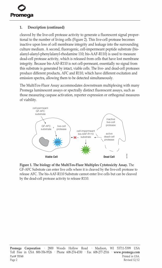
The MultiTox-Fluor Assay simultaneously measures two protease activities: oneis a marker of cell viability, and the other is a marker of cytotoxicity (Figure 1).The live-cell protease activity is restricted to intact viable cells and is measuredusing a fluorogenic, cell-permeant peptide substrate (glycyl-phenylalanyl-amino fluorocoumarin; GF-AFC). The substrate enters intact cells where it is
MultiTox-Fluor MultiplexCytotoxicity Assay
All technical literature is available on the Internet at: www.promega.com/protocols/ Please visit the web site to verify that you are using the most current version of this
Technical Bulletin. Please contact Promega Technical Services if you have questions on useof this system. E-mail: [email protected]
1. Description (continued)
cleaved by the live-cell protease activity to generate a fluorescent signal propor-tional to the number of living cells (Figure 2). This live-cell protease becomesinactive upon loss of cell membrane integrity and leakage into the surroundingculture medium. A second, fluorogenic, cell-impermeant peptide substrate (bis-alanyl-alanyl-phenylalanyl-rhodamine 110; bis-AAF-R110) is used to measuredead-cell protease activity, which is released from cells that have lost membraneintegrity. Because bis-AAF-R110 is not cell-permeant, essentially no signal fromthis substrate is generated by intact, viable cells. The live- and dead-cell proteasesproduce different products, AFC and R110, which have different excitation andemission spectra, allowing them to be detected simultaneously.
The MultiTox-Fluor Assay accommodates downstream multiplexing with manyPromega luminescent assays or spectrally distinct fluorescent assays, such asthose measuring caspase activation, reporter expression or orthogonal measuresof viability.
Promega Corporation · 2800 Woods Hollow Road · Madison, WI 53711-5399 USA Toll Free in USA 800-356-9526 · Phone 608-274-4330 · Fax 608-277-2516 · www.promega.comPart# TB348 Printed in USA.Page 2 Revised 12/12
5847
MA
cell-permeantGF-AFC
substrate
GF-AFCsubstrate
cell-impermeantbis-AAF-R110
substrate
live-cellprotease
AFC
inactive live-cell
protease
active dead-cell protease
R110
Viable Cell Dead Cell
Figure 1. The biology of the MultiTox-Fluor Multiplex Cytotoxicity Assay. The GF-AFC Substrate can enter live cells where it is cleaved by the live-cell protease torelease AFC. The bis-AAF-R110 Substrate cannot enter live cells but can be cleavedby the dead-cell protease activity to release R110.

Promega Corporation · 2800 Woods Hollow Road · Madison, WI 53711-5399 USA Toll Free in USA 800-356-9526 · Phone 608-274-4330 · Fax 608-277-2516 · www.promega.comPrinted in USA. Part# TB348Revised 12/12 Page 3
5820
MB
A.
LDH
Rele
ase
Fluo
resc
ence
(RFU
)
0
50,000
100,000
150,000
200,000
250,000
bis-AAF-R110 Fluorescence (RFU)
r2 = 0.9782
0 10,000 20,000 30,000 40,000 50,000
B.
C.
Reso
rufin
Flu
ores
cenc
e (R
FU)
2,500
2,600
2,700
2,800
2,900
3,000
3,100
GF-AFC Fluorescence (RFU)
r2 = 0.963
500 600 700 800 900 1,000 1,100
D.
Excl
udab
le D
ye F
luor
esce
nce
(RFU
)
0
2,000
4,000
6,000
8,000
10,000
12,000
bis-AAF-R110 Fluorescence (RFU)
r2 = 0.9982
0 10,000 20,000 30,000 40,000 50,000
ATP
Lum
ines
cenc
e (R
LU)
250 750 1,250 1,750 2,250 2,750
40,000
80,000
120,000
160,000
0
GF-AFC Fluorescence (RFU)
r2 = 0.9923
Figure 3. Live- and dead-cell protease activities detected using the MultiTox-FluorAssay show strong correlation with established methods for measuring viabilityand cytotoxicity. Panel A. bis-AAF-R110 signal plotted against CellTiter-Blue® Assayfluorescence. Panel B. GF-AFC signal plotted against CellTiter-Glo® Assay lumines-cence. Panel C. GF-AFC signal plotted against CytoTox-ONE™ Assay fluorescence.Panel D. bis-AAF-R110 signal plotted against ethidium homodimer fluorescence.
A.
5821
MB
Cells or Cell Equivalents/Well
R110
Flu
ores
cenc
e (R
FU)
Treated
Viable0
10,000
20,000
30,000
40,000
50,000
60,000
0 2,000 4,000 6,000 8,000 10,000 12,000
B.
500
1,500
2,500
3,500
4,500
5,500
6,500
7,500
Cells or Cell Equivalents/Well
AFC
Fluo
resc
ence
(RFU
) Viable
Treated
0 2,000 4,000 6,000 8,000 10,000 12,000
Figure 2. The MultiTox-Fluor Assay measures distinct and differential proteaseactivities. Live-cell and dead-cell protease activities were measured in populationsof viable cells (untreated) or cells lysed by sonication (treated) using the GF-AFCSubstrate (live-cell protease substrate) and bis-AAF-R110 Substrate (dead-cell proteasesubstrate). Panel A. GF-AFC fluorescence. Panel B. bis-AAF-R110 fluorescence. Notethat the Y-axis scales in Panels A and B are different. Results are provided in relativefluorescence units (RFU), and the scales reflect the difference in signal output ofAFC and R110.

1. Description (continued)
Assay Advantages
• Simultaneously Measure the Number of Live Cells and Number of DeadCells in Culture: Single-reagent-addition, “add-mix-measure” protocol.
• Normalize Data with a Built-In Internal Control: The ratio of number of livecells to number of dead cells is independent of cell number and normalizesdata. This normalization makes results more comparable well-to-well, plate-to-plate and day-to-day.
• Identify More False Positives and False Negatives Immediately: Independentcell viability and cytotoxicity chemistries serve as controls for each other. Iftest compounds interfere with one assay chemistry, the other serves as aninternal control.
• Get More Data from Every Well: Multiplex the MultiTox-Fluor Assay withmany Promega bioluminescent assays (apoptosis determination, reporter geneand protease activity assays, etc.).
An overview of the protocol is provided in Figure 5.
Promega Corporation · 2800 Woods Hollow Road · Madison, WI 53711-5399 USA Toll Free in USA 800-356-9526 · Phone 608-274-4330 · Fax 608-277-2516 · www.promega.comPart# TB348 Printed in USA.Page 4 Revised 12/12
5822
MA
0
25
50
75
100
% Viable Cells/Well
% o
f Max
imal
Ass
ay R
espo
nse
Live Cell Response(GF-AFC)
r2 = 0.9998 r2 = 0.9998
Dead Cell Response(bis-AAF-R110)
0 25 50 75 100
Figure 4. Viability and cytotoxicity measurements are inversely correlated andratiometric. When viability is high, the live-cell signal is highest and the dead-cellsignal is lowest. When viability is low, the live-cell signal is lowest and the dead-cellsignal is highest. Solid line, live-cell signal; dotted line, dead-cell signal.

Promega Corporation · 2800 Woods Hollow Road · Madison, WI 53711-5399 USA Toll Free in USA 800-356-9526 · Phone 608-274-4330 · Fax 608-277-2516 · www.promega.comPrinted in USA. Part# TB348Revised 12/12 Page 5
5814
MA
Assay Buffer
MultiTox-Fluor Multiplex Cytotoxicity Assay Reagent
GF-AFC (live-cell substrate)
bis-AAF-R110 (dead-cell substrate)
Add GF-AFC and bis-AAF-R110 Substrates to Assay Buffer to create the MultiTox-Fluor Multiplex Cytotoxicity Assay Reagent.
Add reagent to plate in proportional volumes, mix and incubate.
Record 400Ex/505Em
485Ex/520Em
Figure 5. Schematic diagram of the MultiTox-Fluor Multiplex CytotoxicityAssay. Live-cell fluorescence is measured at 400Ex/505Em; dead-cell fluorescenceis measured at 485Ex/520Em.
2. Product Components and Storage Conditions
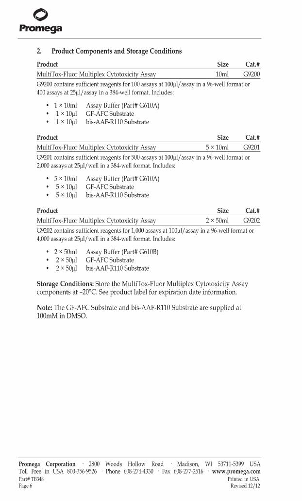
Product Size Cat.#MultiTox-Fluor Multiplex Cytotoxicity Assay 10ml G9200G9200 contains sufficient reagents for 100 assays at 100µl/assay in a 96-well format or 400 assays at 25µl/assay in a 384-well format. Includes:
• 1 × 10ml Assay Buffer (Part# G610A)• 1 × 10µl GF-AFC Substrate• 1 × 10µl bis-AAF-R110 Substrate
Product Size Cat.#MultiTox-Fluor Multiplex Cytotoxicity Assay 5 × 10ml G9201G9201 contains sufficient reagents for 500 assays at 100µl/assay in a 96-well format or2,000 assays at 25µl/well in a 384-well format. Includes:
• 5 × 10ml Assay Buffer (Part# G610A)• 5 × 10µl GF-AFC Substrate• 5 × 10µl bis-AAF-R110 Substrate
Product Size Cat.#MultiTox-Fluor Multiplex Cytotoxicity Assay 2 × 50ml G9202G9202 contains sufficient reagents for 1,000 assays at 100µl/assay in a 96-well format or4,000 assays at 25µl/well in a 384-well format. Includes:
• 2 × 50ml Assay Buffer (Part# G610B)• 2 × 50µl GF-AFC Substrate• 2 × 50µl bis-AAF-R110 Substrate
Storage Conditions: Store the MultiTox-Fluor Multiplex Cytotoxicity Assaycomponents at –20°C. See product label for expiration date information.
Note: The GF-AFC Substrate and bis-AAF-R110 Substrate are supplied at100mM in DMSO.
Promega Corporation · 2800 Woods Hollow Road · Madison, WI 53711-5399 USA Toll Free in USA 800-356-9526 · Phone 608-274-4330 · Fax 608-277-2516 · www.promega.comPart# TB348 Printed in USA.Page 6 Revised 12/12

3. Reagent Preparation and Storage
The MultiTox-Fluor Reagent can be prepared and used at two different concen-trations, depending on the intended application. The 2X concentration is preferred for standard viability and cytotoxicity determinations. The 5X concen-tration is required to accommodate volume restraints when multiplexing withother compatible downstream assay chemistries.
1. Thaw the MultiTox-Fluor Multiplex Cytotoxicity Assay components in a 37°Cwater bath. The substrate vials may require a brief centrifugation to recoverthe entire volume.
2a. To make the 2X reagent, transfer the contents of the GF-AFC and bis-AAF-R110Substrates (10µl each for Cat.# G9200 and G9201; 50µl for G9202) to the AssayBuffer (10ml for Cat.# G9200 and G9201; 50ml for G9202). Mix by vortexingthe contents until the substrates are thoroughly dissolved. The 2X reagentshould be used in equal-volume additions (i.e., 100µl for 96-well plates; 25µlfor 384-well plates).
OR
2b. To make the 5X reagent, transfer the contents of the GF-AFC and bis-AAF-R110Substrates to 2.0ml of Assay Buffer (96-well plates) or 2.5ml of Assay Buffer(384-well plates) for Cat.# G9200 and G9201. [Transfer the substrates to 10mlof Assay Buffer for Cat.# G9202.] Mix by vortexing until the substrates arethoroughly dissolved. The 5X reagent should be used in 1/5 volume additions,(i.e., 20µl for 96-well plates and 5µl for 384-well plates).
Note: Once prepared, the MultiTox-Fluor Multiplex Cytotoxicity Reagent containing both substrates should be used within 24 hours if stored at roomtemperature. Unused MultiTox-Fluor Multiplex Cytotoxicity Reagent can bestored at 4°C for up to 7 days with no appreciable loss of activity.
4. Protocols
Materials to Be Supplied by the User• 96- or 384-well opaque-walled tissue culture plates compatible with fluorometer
(clear or solid bottom)• multichannel pipettor• reagent reservoirs• fluorescence plate reader equipped with filter sets of excitation ~400nm and
emission ~505nm, and excitation ~485nm and emission ~520nm• orbital plate shaker• positive control cytotoxic reagent or lytic detergent (e.g., digitonin at 20mg/ml in
DMSO)
If you have not performed this assay on your cell line previously, we stronglyrecommend determining assay sensitivity using your cells and one of the twomethods described in Section 4.D or 4.E. A description of recommended controlscan be found in Section 4.C.
Promega Corporation · 2800 Woods Hollow Road · Madison, WI 53711-5399 USA Toll Free in USA 800-356-9526 · Phone 608-274-4330 · Fax 608-277-2516 · www.promega.comPrinted in USA. Part# TB348Revised 12/12 Page 7
!

4.A. Example Cytotoxicity and Viability Assay Protocol
1. Set up 96-well assay plates containing cells in culture medium at thedesired density.
2. Add test compounds and vehicle controls to the appropriate wells so thatthe final volume is 100µl in each well (25µl for a 384-well plate).
3. Culture cells for the desired test exposure period.
4. Add the 2X MultiTox-Fluor Multiplex Cytotoxicity Assay Reagent in anequal volume (100µl per well) to all wells. Mix briefly on an orbital shaker,then incubate for at least 30 minutes at 37°C.Note: Longer incubations may improve assay sensitivity and dynamicrange. Do not incubate longer than 3 hours.
5. Measure fluorescence:• Viability: Excitation ~400nm; Emission ~505nm• Cytotoxicity: Excitation ~485nm; Emission ~520nmNotes:Adjusting the instrument gain setting (applied photomultiplier tubeenergy) may be necessary.Data are collected as relative fluorescence units (RFU), and the scales forAFC and R110 fluorescence will differ due to the difference in signal outputof the two molecules (Figure 2).
4.B. Example Multiplex Protocol (with a luminescent caspase assay)
Representative data are shown in Figure 6.
1. Set up 96-well assay plates containing cells in medium at the desired density.
2. Add test compounds and vehicle controls to the appropriate wells so thatthe final volume in each well is 100µl (25µl for a 384-well plate).
3. Culture cells for the desired test exposure period.
4. Add 20µl of 5X MultiTox-Fluor Reagent (prepared as 10µl of each substratein 2ml of Assay Buffer) to each well, and mix briefly by orbital shaking.Incubate for at least 30 minutes at 37°C.Note: Longer incubations may improve assay sensitivity and dynamicrange. Do not incubate longer than 3 hours.
5. Measure fluorescence using a fluorometer (live-cell fluorescence at400Ex/505Em; dead-cell fluorescence at 485Ex/520Em).
6. Add an equal volume of Caspase-Glo® 3/7 Reagent to each well (100–120µlper well), incubate for 30 minutes, then measure luminescence using aluminometer.
Promega Corporation · 2800 Woods Hollow Road · Madison, WI 53711-5399 USA Toll Free in USA 800-356-9526 · Phone 608-274-4330 · Fax 608-277-2516 · www.promega.comPart# TB348 Printed in USA.Page 8 Revised 12/12

4.C. Recommended Controls
No-Cell Control: Set up triplicate wells without cells to serve as the negativecontrol to determine background fluorescence.
Untreated-Cells Control: Set up triplicate wells with untreated cells to serve asa vehicle control. Add the same solvent used to deliver the test compounds tothe vehicle control wells.
Test Compound Control (optional): Set up triplicate wells without cells, andadd the vehicle and test compound to test for possible fluorescence interference.
Positive Control for Cytotoxicity: Set up triplicate wells containing cellstreated with a compound known to be toxic to the cells used in your modelsystem (e.g., treat cells with digitonin at a final concentration of 30μg/ml for15 minutes).
Promega Corporation · 2800 Woods Hollow Road · Madison, WI 53711-5399 USA Toll Free in USA 800-356-9526 · Phone 608-274-4330 · Fax 608-277-2516 · www.promega.comPrinted in USA. Part# TB348Revised 12/12 Page 9
5824
MA
0.00
25
50
75
100
125 Dead-CellFluorescence (R110)Live-CellFluorescence (AFC)Caspase-3/7Luminescence
1,000 10,000 100,000
Staurosporine (nM)
% o
f Max
imal
Ass
ay R
espo
nse
Figure 6. The MultiTox-Fluor Assay can be multiplexed with other assays. LN-18cells were plated at a density of 10,000 cells per well in 50µl volumes of MEM + 10%fetal bovine serum and allowed to attach overnight. Staurosporine was seriallydiluted twofold and added to wells in 50µl volumes. The plate was incubated at 37°Cin 5% CO2 for 6 hours. MultiTox-Fluor Reagent was prepared by combining 10µl ofeach substrate with 1ml of Assay Buffer, and 10µl was used per well. The plate wasmixed and incubated for 30 minutes at 37°C. Fluorescence was measured at400Ex/505Em and 485Ex/520Em using a BMG PolarStar plate reader. Caspase-Glo® 3/7Reagent then was added in an additional 100µl volume, and luminescence was measured after a 10-minute incubation. The resulting signals were normalized to apercentage of maximal response and plotted using GraphPad Prism® software.

4.D. Determining Assay Sensitivity, Method 1
1. Harvest adherent cells (by trypsinization, etc.), wash with fresh medium (to remove residual trypsin) and resuspend in fresh medium.Note: For cells growing in suspension, proceed directly to Step 2.
2. Determine the number of viable cells by trypan blue exclusion using ahemacytometer, then adjust the concentration by dilution to 100,000 viablecells/ml in at least 3.0ml of fresh medium.Note: Concentration by centrifugation may be necessary if the cell suspen-sion is less dense than 100,000 cells/ml.
3. Add 100µl of the 100,000 cell/ml dilution (10,000 cells/well) into each wellof rows A and B of a 96-well plate. See Table 1.
4. Add 100µl of fresh medium to each well in rows B–H.
5. Using a multichannel pipettor, mix the cell suspensions in row B by pipet-ting. Be careful not to create foam or bubbles. Transfer a 100µl volume fromrow B to row C. Repeat mixing, and transfer 100µl from row C to row D.Continue this process to row G. After mixing the cell suspension in row G,remove 100µl from the wells and discard it. This procedure dilutes the cellsfrom 10,000 cells/well in row A to 156 cells/well in row G. Wells in row Hserve as no-cell background controls.
6. Dilute digitonin to 300µg/ml in water. Using a multichannel pipette, care-fully add 10µl of the digitonin solution to all wells in columns 7–12 to lysecells (treated cells). Add 10µl of water to all wells in columns 1–6 to normalize the volume in all wells (untreated cells).
Promega Corporation · 2800 Woods Hollow Road · Madison, WI 53711-5399 USA Toll Free in USA 800-356-9526 · Phone 608-274-4330 · Fax 608-277-2516 · www.promega.comPart# TB348 Printed in USA.Page 10 Revised 12/12
Table 1. Schematic Diagram of 96-Well Plate Layout.1 2 3 4 5 6 7 8 9 10 11 12
ABCDEFGH
untreated treated
10,000 Cells/Well5,000 Cells/Well
1,250 Cells/Well
2,500 Cells/Well
625 Cells/Well
313 Cells/Well
156 Cells/Well
0 Cells/Well

7. Add 100µl of the MultiTox-Fluor Multiplex Cytotoxicity Assay Reagent toeach well, mix briefly by orbital shaking to ensure homogeneity and incubateat 37°C for at least 30 minutes. Protect plates from light.Note: Longer incubations may improve assay sensitivity and dynamicrange. Do not incubate longer than 3 hours.
8. Measure fluorescence:• Viability (live-cell fluorescence): Excitation ~400nm; Emission ~505nm• Cytotoxicity (dead-cell fluorescence): Excitation ~485nm; Emission ~520nmNote: Adjusting the instrument gain setting (applied photomultiplier tubeenergy) may be necessary.
9. Calculate the signal-to-noise ratios (S:N) to determine practical sensitivity foryour cell type for each dilution of cells (10,000 cells/well, 5,000 cells/well,2,500 cells/well, etc.)
Viability S:N = (Average Untreated, RFU – Average Treated, RFU) S.D. of H-1 through H-12
Cytotoxicity S:N = (Average Treated, RFU – Average Untreated, RFU)S.D. of H-1 through H-12
Note: The practical level of assay sensitivity for either assay is a signal-to-noise ratio of greater than 3 standard deviations (derived from reference 1).
4.E. Determining Assay Sensitivity, Method 2
1. Harvest adherent cells (by trypsinization, etc.), wash with fresh medium (toremove residual trypsin) and resuspend in fresh medium.Note: For cells growing in suspension, proceed directly to Step 2.
2. Determine the number of viable cells by trypan blue exclusion using ahemacytometer, then adjust the concentration by dilution to 100,000 viablecells/ml in at least 20ml of fresh medium.Note: Concentration by centrifugation may be necessary if the cell suspen-sion is less dense than 100,000 cells/ml.
3. Divide the volume of cells into two separate tubes. Subject one tube to“moderate” sonication (empirically determined by post-sonication morpho-logical examination) to disrupt cell membrane integrity and simulate a100% cytotoxic population. The second tube of untreated cells will serve asthe maximum viable population.
Promega Corporation · 2800 Woods Hollow Road · Madison, WI 53711-5399 USA Toll Free in USA 800-356-9526 · Phone 608-274-4330 · Fax 608-277-2516 · www.promega.comPrinted in USA. Part# TB348Revised 12/12 Page 11

4.E. Determining Assay Sensitivity, Method 2 (continued)
4. Create a spectrum of viability by blending sonicated and untreated cellpopulations in 1.5ml microcentrifuge tubes as described in Table 2.
5. After mixing each blend by gently vortexing, pipet 100µl of each blend into eight replicate wells of a 96-well plate. Add the 100% viable sample tocolumn 1, 95% viable to column 2, etc. Add cell culture medium only tocolumn 10 to serve as the no-cell control.
6. Add MultiTox-Fluor Multiplex Cytotoxicity Assay Reagent in an equal volume (100µl per well) to all wells, mix briefly by orbital shaking andincubate for at least 30 minutes at 37°C Note: Longer incubations may improve assay sensitivity and dynamicrange. Do not incubate longer than 3 hours.
7. Measure fluorescence:• Viability (live-cell fluorescence): Excitation ~400nm; Emission ~505nm• Cytotoxicity (dead-cell fluorescence): Excitation ~485nm; Emission ~520nmNote: Adjusting the instrument gain setting (applied photomultiplier tubeenergy) may be necessary.
8. Determine the practical sensitivity for your cell type by calculating the signal-to-noise ratio (S:N) for each blend of cell viability (X = 95, 90% etc.)
Viability S:N = (Average X%, RFU – Average 0%, RFU)S.D. of 0%
Cytotoxicity S:N = (Average X%, RFU – Average 100%, RFU)S.D. of 100%
Note: The practical level of assay sensitivity for either assay is a signal-to-noise ratio of greater than 3 standard deviations (derived from reference 1).
Promega Corporation · 2800 Woods Hollow Road · Madison, WI 53711-5399 USA Toll Free in USA 800-356-9526 · Phone 608-274-4330 · Fax 608-277-2516 · www.promega.comPart# TB348 Printed in USA.Page 12 Revised 12/12
Table 2. Spectrum of Viability Generated by Blending Sonicated andUntreated Cells.
Percent ViabilityVolume of Sonicated
Cells (µl)Volume of Untreated
Cells (µl)100 0 1,00095 50 95090 100 90075 250 75050 500 50025 750 25010 900 1005 950 500 1,000 0

5. General Considerations
Background Fluorescence and Inherent Serum Activity
Tissue culture medium that is supplemented with animal serum may containdetectable levels of the protease marker used for dead-cell measurements. Thelevel of this protease activity may vary among different lots of serum. To correctfor this variability, background fluorescence should be determined using samplescontaining medium plus serum without cells.
Temperature
Generation of the fluorescent product is proportional to the protease activity ofthe markers associated with cell viability and cytotoxicity. The activities ofthese proteases are influenced by temperature. For best results, we recommendincubating assays at a constant controlled temperature to ensure uniformityacross the plate. After reagent addition and a brief mixing, we suggest one oftwo methods:
1. Incubate at 37°C in a water-jacketed incubation module (Me’Cour, etc.).Note: Incubation at 37°C in a CO2 culture cabinet may lead to edge effects dueto thermal gradients.
2. Incubate at room temperature with or without orbital shaking.Note: Assays performed at room temperature may require more than 30 minutesof incubation. Do not incubate longer than 3 hours.
Assay Controls
In addition to a no-cell control to establish background fluorescence, we recom-mend including an untreated-cell (maximum viability) and positive (maximumcytotoxicity) control in the experimental design. The maximum viability controlis established by adding vehicle only. Vehicle is used to deliver the test com-pound to test wells and, in most cases, consists of a buffer system or mediumand the equivalent amount of solvent added with the test compound. Maximumcytotoxicity can be determined using a compound that causes cytotoxicity or alytic reagent added to compromise viability (e.g., nonionic detergents such asdigitonin or zwitterionic detergents). See Section 4.C.
Triton® X-100, NP-40 and SDS interfere with the assay.
Cytotoxicity Marker Half-Life
The activity of the protease marker released from dead cells has a half-life ofapproximately 9–10 hours (2). In situations where cytotoxicity occurs veryrapidly (necrosis) and the treatment time is greater than 24 hours, the degree ofcytotoxicity may be underestimated. Addition of a lytic detergent may be usefulto determine the total cytotoxicity marker activity remaining (from remaininglive cells) in these extended treatments.
Promega Corporation · 2800 Woods Hollow Road · Madison, WI 53711-5399 USA Toll Free in USA 800-356-9526 · Phone 608-274-4330 · Fax 608-277-2516 · www.promega.comPrinted in USA. Part# TB348Revised 12/12 Page 13
!

5. General Considerations (continued)
Light Sensitivity
The MultiTox-Fluor Multiplex Cytotoxicity Assay uses two fluorogenic peptidesubstrates. Although the substrates demonstrate good general photostability,the fluors liberated after contact with the proteases can degrade with prolongedexposure to ambient light sources. We recommend shielding the plates fromambient light at all times.
Cell Culture Medium
The GF-AFC and bis-AAF-R110 Substrates are introduced into the test wellusing an optimized buffer system that mitigates differences in pH due to thetreatment. In addition, the buffer system supports protease activity in a host ofdifferent culture media with varying osmolarity. With the exception of mediumformulations with very high serum content or phenol red indicator levels, nosubstantial performance differences will be observed among media.
Instrument Settings and Measurement Parameters
To acquire both cell viability and cytotoxicity readings from one Multitox-Fluorreaction, the instrument must have the recommended filter sets. An incorrectfilter set will result in inadequate signal separation or high background readings.If the recommended filter sets are not readily available, contact PromegaTechnical Services for alternatives.
Many fluorescent microplate readers have a detection gain setting. Setting thedetection gain too low can result in low signal and output with high variation(i.e., large error bars). Setting the detection gain too high may result in signalplateau (i.e., signal saturation) and inadequate signal separation. See Section 6.
Promega Corporation · 2800 Woods Hollow Road · Madison, WI 53711-5399 USA Toll Free in USA 800-356-9526 · Phone 608-274-4330 · Fax 608-277-2516 · www.promega.comPart# TB348 Printed in USA.Page 14 Revised 12/12

6. Troubleshooting
For questions not addressed here, please contact your local Promega Branch Office or Distributor.Contact information available at: www.promega.com. E-mail: [email protected]
Symptoms Causes and Comments
Little difference in fluorescence Improper filter set. Measure live-cell between treated and untreated fluorescence at 400Ex/505Em and dead-cell cell populations (Figure 7) fluorescence at 485Ex/520Em.
Treatment did not induce cytotoxicity. Check the positive control for cytotoxicity; there shouldbe a significant difference in signal between the treated and untreated cell populations.
Promega Corporation · 2800 Woods Hollow Road · Madison, WI 53711-5399 USA Toll Free in USA 800-356-9526 · Phone 608-274-4330 · Fax 608-277-2516 · www.promega.comPrinted in USA. Part# TB348Revised 12/12 Page 15
9904
TA
Fluo
resc
ence
(RFU
)
Cell Number
TreatedUntreated
r2 = 0.9996
r2 = 0.9874
Figure 7. An example showing little difference in GF-AFC fluorescence betweentreated (dead) and untreated (viable) cell populations. Fluorescence increases withincreasing cell number, but there is little difference in fluorescence between treatedand untreated cell populations.

6. Troubleshooting (continued)
Symptoms Causes and Comments
Little change in fluorescence with Improper filter set. Measure live-cellincreasing cell number for both fluorescence at 400Ex/505Em and dead-cell treated and untreated cell populations fluorescence at 485Ex/520Em.(Figure 8)
Promega Corporation · 2800 Woods Hollow Road · Madison, WI 53711-5399 USA Toll Free in USA 800-356-9526 · Phone 608-274-4330 · Fax 608-277-2516 · www.promega.comPart# TB348 Printed in USA.Page 16 Revised 12/12
9905
TA
Fluo
resc
ence
(RFU
)
Cell Number
TreatedUntreated
Figure 8. An example showing little change in GF-AFC fluorescence with increas-ing cell number for both treated and untreated cell populations. Fluorescence isrelatively high for both treated and untreated cells at low cell numbers and does notchange dramatically with increasing cell number.

Symptoms Causes and Comments
High variation (i.e., large error bars) The instrument gain setting was too low. and low fluorescence despite Increase the gain setting.increasing cell number (Figure 9)
Mixing of reagent and cells was inadequate. Mixreagent and cells gently on an orbital platform.
Promega Corporation · 2800 Woods Hollow Road · Madison, WI 53711-5399 USA Toll Free in USA 800-356-9526 · Phone 608-274-4330 · Fax 608-277-2516 · www.promega.comPrinted in USA. Part# TB348Revised 12/12 Page 17
9947
TA
Fluo
resc
ence
(RFU
)
Cell Number
TreatedUntreated
r2 = 0.597
r2 = 0.9839
25
20
15
5
10
0
Figure 9. An example showing high variation and low bis-AAF-R110 fluorescencedespite increasing cell number.

6. Troubleshooting (continued)
Symptoms Causes and Comments
Fluorescence reached plateau for The instrument gain setting was too high, and one or both cell populations the signal reached saturation. Reduce the gain (Figure 10) setting.
Fluorescence was too high, and signal reached saturation. Be sure fluorescence levels are within the linear range of the detection instrument.
Narrow signal window (i.e., small Incubation time was inadequate. Incubate difference in signal between treated reactions for at least 30 minutes at 37°C. An and untreated cell populations) additional incubation time of up to 3 hours (Figure 11) often improves results. Do not incubate longer
than 3 hours.
Improper filter set. Measure live-cellfluorescence at 400Ex/505Em and dead-cell fluorescence at 485Ex/520Em.
Improper instrument gain setting.
Live-cell signal decreased, but Improper assay time. The dead cell protease hasdead-cell signal also decreased a half life of approximately 9–10 hours. With
prolonged treatment times, the dead-cell protease signal can decay before fluorescence is measured. Optimize the assay time. Measure live-cell and dead-cell protease activities at earlier time points.
Live signal decreased, but Cell cycle arrest without secondary necrosis. dead signal remained the same Extend the treatment to promote cell death and
loss of membrane integrity and increase dead-cell protease activity.
High variation (i.e., large error bars) Mixing of reagent and cells was inadequate. Mixin signal between replicate reactions reagent and cells gently on an orbital platform
prior to incubation at 37°C.
There was a bubble in the well. Pop any bubbles with a sharp object before measuring fluorescence.
EC50 values determined using this The susceptibility of cells to a drug is affected byassay differ from EC50 values many factors such as cell type, treatment time, in the literature seeding density and cell passage number. These
factors can cause EC50 values to be different.
Promega Corporation · 2800 Woods Hollow Road · Madison, WI 53711-5399 USA Toll Free in USA 800-356-9526 · Phone 608-274-4330 · Fax 608-277-2516 · www.promega.comPart# TB348 Printed in USA.Page 18 Revised 12/12

7. References
1. Zhang, J.H. et al. (1999) A simple statistical parameter for use in evaluation and vali-dation of high-throughput screening assays. J. Biomol. Screen. 4, 67–73.
2. Niles, A.L. et al. (2007) A homogeneous assay to measure live and dead cells in thesame sample by detecting different protease markers. Anal. Biochem. 366, 197–206.
Promega Corporation · 2800 Woods Hollow Road · Madison, WI 53711-5399 USA Toll Free in USA 800-356-9526 · Phone 608-274-4330 · Fax 608-277-2516 · www.promega.comPrinted in USA. Part# TB348Revised 12/12 Page 19
9949
TA
Fluo
resc
ence
(RFU
)
Cell Number
TreatedUntreated r2 = 0.9905
r2 = 0.8315
Figure 11. An example showing a narrow signal window between treated anduntreated cell populations. bis-AAF-R110 fluorescence levels are similar for bothtreated and untreated cell populations.
9948
TA
Fluo
resc
ence
(RFU
)
Cell Number
TreatedUntreated
r2 = 0.9997
r2 = 0.7581
Figure 10. An example showing signal plateau for treated cells.

8. Related Products
Cell Viability Assays
Product Size Cat.#ApoLive-Glo™ Multiplex Assay 10ml G6410ApoTox-Glo™ Triplex Assay 10ml G6320CellTiter-Fluor™ Cell Viability Assay 10ml G6080
5 × 10ml G60812 × 50ml G6082
CytoTox-Fluor™ Cytotoxicity Assay 10ml G9260CellTiter-Glo® Luminescent Cell Viability Assay 10ml G7570CytoTox-ONE™ Homogeneous Membrane Integrity Assay 1,000–4,000 assays G7891CellTiter-Blue® Cell Viability Assay 20ml G8080Additional Sizes Available.
Apoptosis Assays
Product Size Cat.#Caspase-Glo® 3/7 Assay 100ml G8092
10 × 10ml G8093Caspase-Glo® 8 Assay 100ml G8202Caspase-Glo® 9 Assay 100ml G8212Additional Sizes Available.
Reporter Gene Assays
Product Size Cat.#ONE-Glo™ Luciferase Assay System 10ml E6110Bright-Glo™ Luciferase Assay System 10ml E2610Steady-Glo® Luciferase Assay System 10ml E2510Additional Sizes Available.
Protease Assays
Product Size Cat.#Calpain-Glo™ Protease Assay 10ml G8501DPPIV-Glo™ Protease Assay 10ml G8350Proteasome-Glo™ 3-Substrate Cell-Based Assay System 10ml G1180Additional Sizes Available.
Fluorometers
Product Size Cat.#GloMax®-Multi Base Instrument 1 each E7031GloMax®-Multi Fluorescence Module 1 each E7051GloMax®-Multi Optical Kit Blue 1 each E8921
Promega Corporation · 2800 Woods Hollow Road · Madison, WI 53711-5399 USA Toll Free in USA 800-356-9526 · Phone 608-274-4330 · Fax 608-277-2516 · www.promega.comPart# TB348 Printed in USA.Page 20 Revised 12/12

Promega Corporation · 2800 Woods Hollow Road · Madison, WI 53711-5399 USA Toll Free in USA 800-356-9526 · Phone 608-274-4330 · Fax 608-277-2516 · www.promega.comPrinted in USA. Part# TB348Revised 12/12 Page 21
(a)Patent Pending.© 2006, 2009–2012 Promega Corporation. All Rights Reserved.Caspase-Glo, CellTiter-Blue, CellTiter-Glo, GloMax and Steady-Glo are registered trademarks of Promega Corporation.ApoLive-Glo, ApoTox-Glo, Bright-Glo, Calpain-Glo, CellTiter-Fluor, CytoTox-Fluor, CytoTox-ONE, DPPIV-Glo, ONE-Gloand Proteasome-Glo are trademarks of Promega Corporation.GraphPad Prism is a registered trademark of GraphPad Software, Inc. Triton is a registered trademark of Union CarbideChemicals and Plastics Technology Corporation.Products may be covered by pending or issued patents or may have certain limitations. Please visit our Web site for moreinformation. All prices and specifications are subject to change without prior notice.Product claims are subject to change. Please contact Promega Technical Services or access the Promega online catalog for themost up-to-date information on Promega products.