mucosa-adherent lactobacilli: commensal and pathogenic
TRANSCRIPT
Mucosa-Adherent Lactobacilli: Commensal and Pathogenic Characteristics
Sanna Edelman
General Microbiology Faculty of Biosciences
and Viikki Graduate School in Biosciences
University of Helsinki
Academic Dissertation in General Microbiology
To be presented, with the permission of the Faculty of Biosciences, Department of Biological and Environmental Sciences,
University of Helsinki, for public criticism in the Auditorium 1041, Viikinkaari 5
on the 1st of April, at 12 o´clock noon.
Helsinki 2005

II
Supervisor Professor Timo K. Korhonen, PhD General Microbiology
Department of Biological and Environmental Sciences University of Helsinki Reviewers Docent Pentti Kuusela, MD, PhD Department of Bacteriology and Immunology Haartman Institute University of Helsinki and Division of Clinical Microbiology
Helsinki University Central Hospital, Helsinki, Finland Professor Airi Palva, PhD Division of Microbiology and Epidemiology Department of Basic Veterinary Sciences University of Helsinki Cover figure: Immuno-EM image of the subcellular localization of glyceraldehyde-3-phosphate dehydrogenase (GAPDH) and enolase on Lactobacillus crispatus. ISSN 1239-9469 ISBN 952-10-2345-7 (paperpack) ISBN 952-10-2346-5 (pdf) http://ethesis.helsinki.fi Yliopistopaino, Helsinki 2005

III
TO HARRY AND MARTIN

IV
V
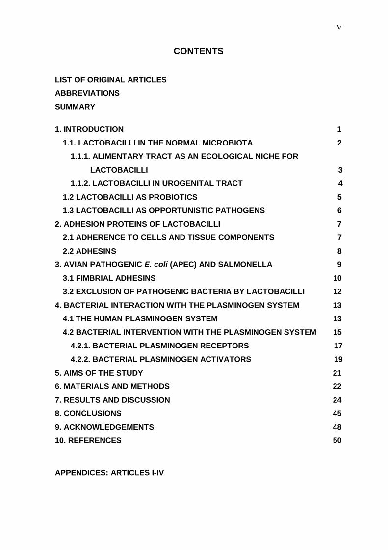
CONTENTS
LIST OF ORIGINAL ARTICLES
ABBREVIATIONS
SUMMARY
1. INTRODUCTION 1
1.1. LACTOBACILLI IN THE NORMAL MICROBIOTA 2
1.1.1. ALIMENTARY TRACT AS AN ECOLOGICAL NICHE FOR
LACTOBACILLI 3
1.1.2. LACTOBACILLI IN UROGENITAL TRACT 4
1.2 LACTOBACILLI AS PROBIOTICS 5
1.3 LACTOBACILLI AS OPPORTUNISTIC PATHOGENS 6
2. ADHESION PROTEINS OF LACTOBACILLI 7
2.1 ADHERENCE TO CELLS AND TISSUE COMPONENTS 7
2.2 ADHESINS 8
3. AVIAN PATHOGENIC E. coli (APEC) AND SALMONELLA 9
3.1 FIMBRIAL ADHESINS 10
3.2 EXCLUSION OF PATHOGENIC BACTERIA BY LACTOBACILLI 12
4. BACTERIAL INTERACTION WITH THE PLASMINOGEN SYSTEM 13
4.1 THE HUMAN PLASMINOGEN SYSTEM 13
4.2 BACTERIAL INTERVENTION WITH THE PLASMINOGEN SYSTEM 15
4.2.1. BACTERIAL PLASMINOGEN RECEPTORS 17
4.2.2. BACTERIAL PLASMINOGEN ACTIVATORS 19
5. AIMS OF THE STUDY 21
6. MATERIALS AND METHODS 22
7. RESULTS AND DISCUSSION 24
8. CONCLUSIONS 45
9. ACKNOWLEDGEMENTS 48
10. REFERENCES 50
APPENDICES: ARTICLES I-IV
VI
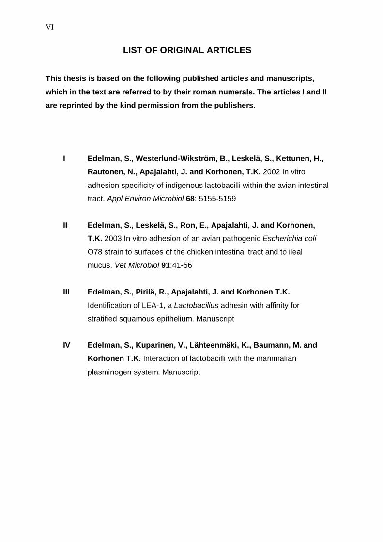
LIST OF ORIGINAL ARTICLES
This thesis is based on the following published articles and manuscripts,
which in the text are referred to by their roman numerals. The articles I and II
are reprinted by the kind permission from the publishers.
I Edelman, S., Westerlund-Wikström, B., Leskelä, S., Kettunen, H.,
Rautonen, N., Apajalahti, J. and Korhonen, T.K. 2002 In vitro
adhesion specificity of indigenous lactobacilli within the avian intestinal
tract. Appl Environ Microbiol 68: 5155-5159
II Edelman, S., Leskelä, S., Ron, E., Apajalahti, J. and Korhonen,
T.K. 2003 In vitro adhesion of an avian pathogenic Escherichia coli
O78 strain to surfaces of the chicken intestinal tract and to ileal
mucus. Vet Microbiol 91:41-56
III Edelman, S., Pirilä, R., Apajalahti, J. and Korhonen T.K.
Identification of LEA-1, a Lactobacillus adhesin with affinity for
stratified squamous epithelium. Manuscript
IV Edelman, S., Kuparinen, V., Lähteenmäki, K., Baumann, M. and
Korhonen T.K. Interaction of lactobacilli with the mammalian
plasminogen system. Manuscript
VII
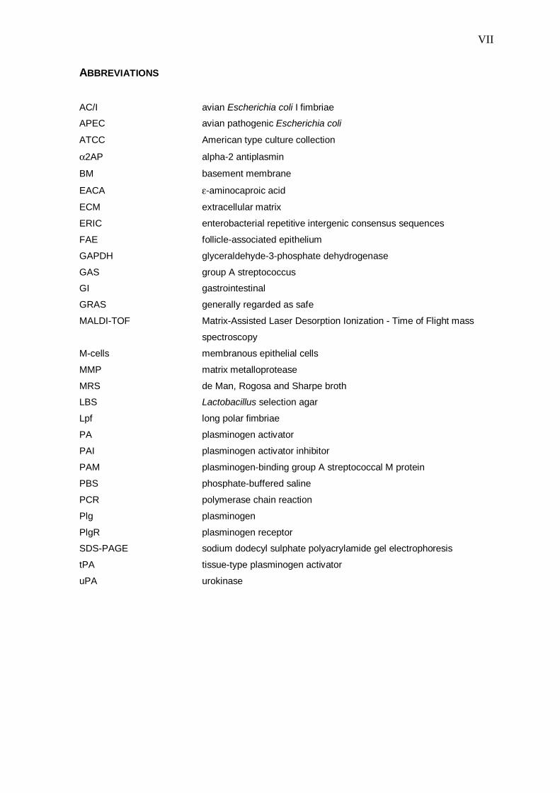
ABBREVIATIONS
AC/I avian Escherichia coli I fimbriae
APEC avian pathogenic Escherichia coli
ATCC American type culture collection
α2AP alpha-2 antiplasmin
BM basement membrane
EACA ε-aminocaproic acid
ECM extracellular matrix
ERIC enterobacterial repetitive intergenic consensus sequences
FAE follicle-associated epithelium
GAPDH glyceraldehyde-3-phosphate dehydrogenase
GAS group A streptococcus
GI gastrointestinal
GRAS generally regarded as safe
MALDI-TOF Matrix-Assisted Laser Desorption Ionization - Time of Flight mass
spectroscopy
M-cells membranous epithelial cells
MMP matrix metalloprotease
MRS de Man, Rogosa and Sharpe broth
LBS Lactobacillus selection agar
Lpf long polar fimbriae
PA plasminogen activator
PAI plasminogen activator inhibitor
PAM plasminogen-binding group A streptococcal M protein
PBS phosphate-buffered saline
PCR polymerase chain reaction
Plg plasminogen
PlgR plasminogen receptor
SDS-PAGE sodium dodecyl sulphate polyacrylamide gel electrophoresis
tPA tissue-type plasminogen activator
uPA urokinase
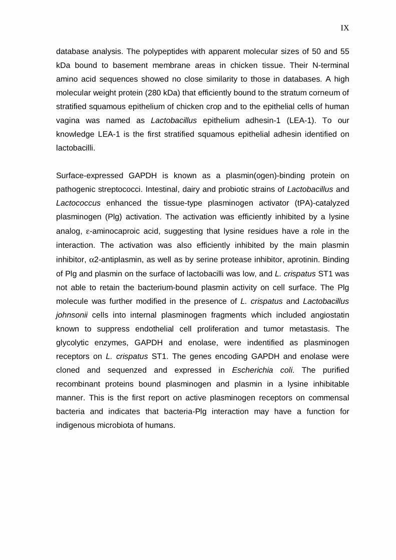
VIII
SUMMARY
Lactobacilli belong to the commensal gastrointestinal and urogenital microbiota of
man and animals and are proposed to endow the host with several beneficial
health effects. Adhesion is considered important for bacterial colonization at the
host mucosal surfaces as well as for bacteria-host interactions both in health and
disease. In this thesis work, the in vitro tissue tropism in adhesiveness of
lactobacilli to the mucosal structures of the alimentary tract of the host and the
underlying molecular bases of the adhesion were studied. To characterize the
potential health effects of the adhesion of lactobacilli, the ability of lactobacilli to
prevent in vitro adhesion of bacterial pathogens was tested. In addition, the ability
of lactobacilli to interact with the mammalian plasminogen system, known to be
harnessed by several invasive bacterial pathogens, was demonstrated.
Lactobacillus ssp. isolated from the avian alimentary tract exhibited strain-specific
cell and tissue tropism in their in vitro adhesion to chicken intestinal tissue
structures. In general, adhesion to crop epithelium was a common trait in well-
colonizing strains. Lactobacillus crispatus strain ST1, characterized as a potential
colonizer, showed a strong adhesion to the epithelial areas of crop as well as small
and large intestine. Notably, ST1 also adhered efficiently to the immunologically
important follicle-associated epithelium in ileum.
The avian pathogenic E. coli (APEC) and Salmonella strains adhered via their
type-1 fimbriae essentially to the same tissue areas in the chicken intestinal
mucosa as did the adhesive lactobacilli. The strongly adhesive L. crispatus ST1
efficiently inhibited the adhesion of APEC at the crop and the follicle-associated
epithelia, whereas the poorly adhesive L. crispatus 134mi inhibited the adhesion of
pathogens only partially.
Adhesive surface proteins from L. crispatus ST1 were isolated by mutanolysin
treatment. A 39-kDa peptide bound to singular intraepithelial and lamina propria
cells of chicken intestine and was identified as glyceraldehyde-3-phosphate
dehydrogenase (GAPDH) by N-terminal amino acid sequenzing and protein
IX
database analysis. The polypeptides with apparent molecular sizes of 50 and 55
kDa bound to basement membrane areas in chicken tissue. Their N-terminal
amino acid sequences showed no close similarity to those in databases. A high
molecular weight protein (280 kDa) that efficiently bound to the stratum corneum of
stratified squamous epithelium of chicken crop and to the epithelial cells of human
vagina was named as Lactobacillus epithelium adhesin-1 (LEA-1). To our
knowledge LEA-1 is the first stratified squamous epithelial adhesin identified on
lactobacilli.
Surface-expressed GAPDH is known as a plasmin(ogen)-binding protein on
pathogenic streptococci. Intestinal, dairy and probiotic strains of Lactobacillus and
Lactococcus enhanced the tissue-type plasminogen activator (tPA)-catalyzed
plasminogen (Plg) activation. The activation was efficiently inhibited by a lysine
analog, ε-aminocaproic acid, suggesting that lysine residues have a role in the
interaction. The activation was also efficiently inhibited by the main plasmin
inhibitor, α2-antiplasmin, as well as by serine protease inhibitor, aprotinin. Binding
of Plg and plasmin on the surface of lactobacilli was low, and L. crispatus ST1 was
not able to retain the bacterium-bound plasmin activity on cell surface. The Plg
molecule was further modified in the presence of L. crispatus and Lactobacillus
johnsonii cells into internal plasminogen fragments which included angiostatin
known to suppress endothelial cell proliferation and tumor metastasis. The
glycolytic enzymes, GAPDH and enolase, were indentified as plasminogen
receptors on L. crispatus ST1. The genes encoding GAPDH and enolase were
cloned and sequenzed and expressed in Escherichia coli. The purified
recombinant proteins bound plasminogen and plasmin in a lysine inhibitable
manner. This is the first report on active plasminogen receptors on commensal
bacteria and indicates that bacteria-Plg interaction may have a function for
indigenous microbiota of humans.

X
1
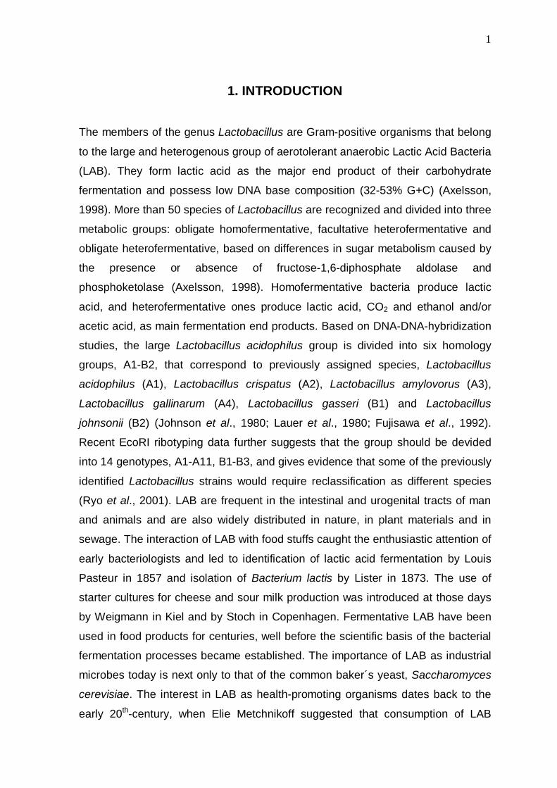
1. INTRODUCTION
The members of the genus Lactobacillus are Gram-positive organisms that belong
to the large and heterogenous group of aerotolerant anaerobic Lactic Acid Bacteria
(LAB). They form lactic acid as the major end product of their carbohydrate
fermentation and possess low DNA base composition (32-53% G+C) (Axelsson,
1998). More than 50 species of Lactobacillus are recognized and divided into three
metabolic groups: obligate homofermentative, facultative heterofermentative and
obligate heterofermentative, based on differences in sugar metabolism caused by
the presence or absence of fructose-1,6-diphosphate aldolase and
phosphoketolase (Axelsson, 1998). Homofermentative bacteria produce lactic
acid, and heterofermentative ones produce lactic acid, CO2 and ethanol and/or
acetic acid, as main fermentation end products. Based on DNA-DNA-hybridization
studies, the large Lactobacillus acidophilus group is divided into six homology
groups, A1-B2, that correspond to previously assigned species, Lactobacillus
acidophilus (A1), Lactobacillus crispatus (A2), Lactobacillus amylovorus (A3),
Lactobacillus gallinarum (A4), Lactobacillus gasseri (B1) and Lactobacillus
johnsonii (B2) (Johnson et al., 1980; Lauer et al., 1980; Fujisawa et al., 1992).
Recent EcoRI ribotyping data further suggests that the group should be devided
into 14 genotypes, A1-A11, B1-B3, and gives evidence that some of the previously
identified Lactobacillus strains would require reclassification as different species
(Ryo et al., 2001). LAB are frequent in the intestinal and urogenital tracts of man
and animals and are also widely distributed in nature, in plant materials and in
sewage. The interaction of LAB with food stuffs caught the enthusiastic attention of
early bacteriologists and led to identification of lactic acid fermentation by Louis
Pasteur in 1857 and isolation of Bacterium lactis by Lister in 1873. The use of
starter cultures for cheese and sour milk production was introduced at those days
by Weigmann in Kiel and by Stoch in Copenhagen. Fermentative LAB have been
used in food products for centuries, well before the scientific basis of the bacterial
fermentation processes became established. The importance of LAB as industrial
microbes today is next only to that of the common baker´s yeast, Saccharomyces
cerevisiae. The interest in LAB as health-promoting organisms dates back to the
early 20th-century, when Elie Metchnikoff suggested that consumption of LAB
2
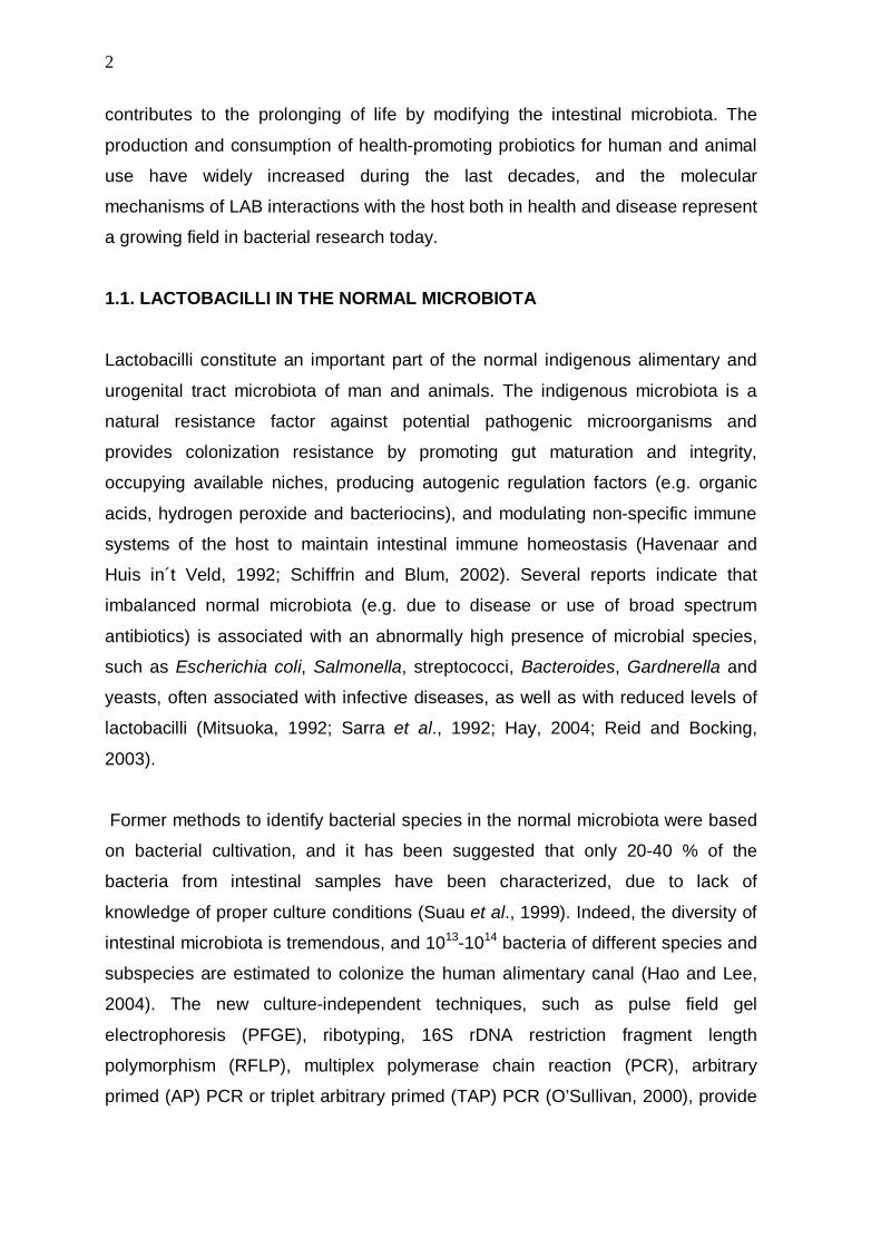
contributes to the prolonging of life by modifying the intestinal microbiota. The
production and consumption of health-promoting probiotics for human and animal
use have widely increased during the last decades, and the molecular
mechanisms of LAB interactions with the host both in health and disease represent
a growing field in bacterial research today.
1.1. LACTOBACILLI IN THE NORMAL MICROBIOTA
Lactobacilli constitute an important part of the normal indigenous alimentary and
urogenital tract microbiota of man and animals. The indigenous microbiota is a
natural resistance factor against potential pathogenic microorganisms and
provides colonization resistance by promoting gut maturation and integrity,
occupying available niches, producing autogenic regulation factors (e.g. organic
acids, hydrogen peroxide and bacteriocins), and modulating non-specific immune
systems of the host to maintain intestinal immune homeostasis (Havenaar and
Huis in´t Veld, 1992; Schiffrin and Blum, 2002). Several reports indicate that
imbalanced normal microbiota (e.g. due to disease or use of broad spectrum
antibiotics) is associated with an abnormally high presence of microbial species,
such as Escherichia coli, Salmonella, streptococci, Bacteroides, Gardnerella and
yeasts, often associated with infective diseases, as well as with reduced levels of
lactobacilli (Mitsuoka, 1992; Sarra et al., 1992; Hay, 2004; Reid and Bocking,
2003).
Former methods to identify bacterial species in the normal microbiota were based
on bacterial cultivation, and it has been suggested that only 20-40 % of the
bacteria from intestinal samples have been characterized, due to lack of
knowledge of proper culture conditions (Suau et al., 1999). Indeed, the diversity of
intestinal microbiota is tremendous, and 1013-1014 bacteria of different species and
subspecies are estimated to colonize the human alimentary canal (Hao and Lee,
2004). The new culture-independent techniques, such as pulse field gel
electrophoresis (PFGE), ribotyping, 16S rDNA restriction fragment length
polymorphism (RFLP), multiplex polymerase chain reaction (PCR), arbitrary
primed (AP) PCR or triplet arbitrary primed (TAP) PCR (O’Sullivan, 2000), provide
3
more specific methods for detailed investigations at the species and the strain
levels and are valuable tools for ecological Lactobacillus studies as well.
1.1.1. ALIMENTARY TRACT AS AN ECOLOGICAL NICHE FOR
LACTOBACILLI
Lactobacilli belong to the normal microbiota of human oral cavity and human
gastrointestinal (GI) tract (Mitsuoka, 1992; Lidbeck and Nord, 1993; Vaughan et
al., 2002). The most frequent Lactobacillus species colonizing the human
alimentary canal are L. acidophilus, L. crispatus, L. brevis, L. casei, L. delbrueckii,
L. fermentum, L. gasseri, L. johnsonii, L. paracasei, L. plantarum, L. rhamnosus, L.
reuteri, L. ruminis, L. sakei, L salivarius, L. vaginalis, L. curvatus and L.
fructovorans (Mitsuoka, 1999; Vaughan et al., 2002). Several factors, such as pH,
peristalsis, redox potential, bacterial adhesion, bacteria-bacteria interactions,
mucus and bile secretion, immunoglobulins, intestinal enzymes, exfoliated
epithelial cells, nutrient availability, diet, as well as bacterial antagonism, affect the
prevalence of bacteria in different parts of the GI tract (Holzapfer et al., 1998;
Tannock, 1999; Hao and Lee, 2004). In general, the alimentary bacterial numbers
are highly dependent on intestinal pH and peristalsis. In the neutral pH of oral
cavity, bacteria reach high numbers (up to 109 per ml of fluids). In the stomach and
the upper two-thirds of the small intestine (e.g. duodenum and jejunum) only 103-
105 bacteria per g of contents are present, probably because of the acidic
conditions of stomach and the fast peristaltic movement in the upper parts of the
alimentary canal. In the distal small intestine, ileum and the large intestine, the pH
rises and the peristaltic movement decreases, and the bacterial numbers are
higher again. The colon is the largest bacterial reservoir in the body and contains
up to 1012 bacteria per g of contents (Mitsuoka, 1992; Hao and Lee, 2004). The
acid-tolerant lactobacilli are one of the dominating species in the oral cavity,
stomach and duodenum and jejunum, with 103-107 bacteria per g of contents
(Mitsuoka, 1992; Lidbeck and Nord, 1993). In the colon, lactobacilli are frequent
(107 bacteria per g of contests) but not the major bacterial species (Mitsuoka,
1992).

4
The presence and composition lactobacilli in the microbiota of the GI tract of
mammalian animals closely resemble those found in humans, although some
variations at the species level occur depending on the host (Mitsuoka, 1992;
Tannock, 1992). Also, the anatomical differences of the alimentary canals
influence the microbiota: the non-secreting stratified squamous epithelia in the
fore-stomach of pigs, mice, rats and horses are efficiently colonized by lactobacilli
(Tannock, 1992; Yuki et al., 2000). In the fowl intestinal tract, the dominating
Lactobacillus species are L. crispatus, L. gallinarum, L. johnsonii, L. salivarius and
L. reuteri (Fuller and Turvey, 1971; Mitsuoka, 1992; Sarra et al., 1992; Guan et al.,
2003; Lan et al., 2002). Lactobacilli efficiently colonize the stratified squamous
epithelium lining the crop, which functions as a food storage pouch in the middle of
esophagus. Due to their fermentation, the colonizing lactobacilli secrete acid and
lower the pH of crop to 4.5-6 (Sarra et al., 1992).
For successful colonization, intestinal bacteria, including lactobacilli, have been
suggested to resist the peristaltic movement by adhering to intestinal epithelia
and/or mucus, particularly in the upper parts of the alimentary canal (Fuller, 1989;
Tannock, 1992, Rojas and Conway, 1996). The topic has remained controversial
since there is also evidence that in vitro adhesion ability of lactobacilli to epithelia
is not a prerequisite for in vivo colonization in an animal model (Pedersen and
Tannock, 1989). Most probably, successful colonization by intestinal bacteria is
dependent on several bacterial factors, and adhesiveness can be one of them.
1.1.2. LACTOBACILLI IN UROGENITAL TRACT
Lactobacilli are the dominating bacteria (107 to 108 cells per g of fluids) in vagina of
healthy premenopausal females (Redondo-Lopez et al., 1990; Paavonen, 1983).
The most prevalent species in vagina are L. crispatus, L. gasseri, L. acidophilus, L.
jensenii, and L. iners (Song et al., 1999; Vásquez et al., 2002; Silvester and Dicks,
2003; Zhou et al., 2004). Microbial colonization is affected by glycogen content, pH
and hormone levels and/or treatments (Galask, 1988). The antagonism of
lactobacilli against urogenital infections, e.g. yeast vaginitis, bacterial vaginosis or
urinary tract infections, has been demonstrated to depend on production of
antimicrobial compounds such as H2O2 and bacteriocins, on low pH and on high
5
redox potential. Also, adherence of lactobacilli to and colonization at the stratified
squamous epithelium of vagina has a protective role against pathogens (McLean
and Rosenstein, 2000; Barbés and Boris, 1999; Pabich et al., 2003; St. Amant et
al., 2002; Marelli et al., 2004).
1.2 LACTOBACILLI AS PROBIOTICS
The findings that colonization by lactobacilli and other lactic acid bacteria, e.g.
bifidobacteria, improves infection resistance of the host, have let to the production
and consumption of probiotics. Probiotics are live microbial cultures or cultured
dairy products which beneficially influence the health and nutrition of the host
(Salminen, 1996). To date, several health-promoting effects of probiotics have
been proposed (Table 1, for reviews, see Reid and Burton, 2002; Sanders, 2003;
Mercenier et al., 2003; Vaarala, 2003; Tuohy et al., 2003). However, the
mechanisms underlying the health effects and the host-probiotic communication in
prophylactic and/or therapeutic treatments have remained poorly characterized.
The probiotic strains are expected to fulfil several health-promoting characteristics
and safety criteria (Table 2).
Table 1. Proposed beneficial effects of probiotics (Modified from Klaenhammer, 2001; Mercenier et al., 2003 and Sanders, 2003) Alleviation of lactose intolerance
Prevention of gastric, intestinal and urogenital infections
Modulation of intestinal immune system
Reduction of inflammatory or allergenic reactions
Anticarcinogenic effects
Antihypertensive effects
Reduction of serum cholesterol level
Regulation of gut motility
6
Table 2. Expected characteristics and safety criteria of probiotics (Mercenier et al., 2003)
Non toxic and non pathogenic
Accurate taxonomic identification
Normal inhabitant of the targeted species
Capability to survive, proliferate and be metabolically active in the targeted site, which implies:
resistance to gastric juice and bile
ability to persist in the GI tract
ability to adhere
ability to compete with the resident microbiota
Production of antimicrobial substances
Antagonism towards pathogenic bacteria
Ability to modulate immune responses
Ability to exert at least one clinically documented health benefit
Genetically stable
Amenability of the strain and stability of the desired characteristics during processing, storage and
delivery
Viability at high populations
Desirable organoleptic and technological properties when included in industrial prosesses
1.3 LACTOBACILLI AS OPPORTUNISTIC PATHOGENS
Lactobacilli are Generally Regarded As Safe (GRAS) and non-pathogenic (Adams
and Marteau, 1995). Occasionally, isolates of Lactobacillus are associated with
opportunistic infectious diseases in humans, such as infective endocarditis (IE),
bacteremia, urinary tract infections, dental caries, chorioamnionitis, endometritis,
meningitis, deep abscesses and empyema (Husni et al., 1997; Aguirre and Collins,
1993; Brouqui and Raoult, 2001). The Lactobacillus-associated infections have
often been polymicrobial, and no direct indication of a primary role for lactobacilli in
the infection has been found. The portal of entry for lactobacilli into circulation has
remained unresolved in most cases; it has been suggested that the bacteria from
the microbiota of the oral cavity or the GI tract can be introduced into the blood
circulation as a result of poor dental hygiene, dental manipulation, gastrointestinal
lesions or surgery (Husni et al., 1997; Aguirre and Collins, 1993). Also, sepsis has
been reported to induce translocation of indigenous intestinal bacteria from
intestine to underlying host tissues in mouse model (Naaber et al., 2000).
However, the molecular mechanisms that contribute to the opportunistic
7
pathogenicity of commensal lactobacilli have remained unknown. The wide-spread
ability of lactobacilli to aggregate platelets coupled with their ability to bind
fibrinogen, fibronectin and collagen, in particular type V collagen demonstrated at
the sites of endothelial damage (Kerényi et al., 1985), as well as their proteolytic
activities are so far the only suggested pathogenic factors of lactobacilli (Harty et
al., 1993; Harty et al., 1994; Oakey et al., 1995). These characteristics are
common in all Lactobacillus strains and their role in lactobacilli-associated
infectious diseases remains unclear. In most cases the infections associated with
lactobacilli have preceded predisposing microbial infections (Husni et al., 1997;
Aguirre and Collins, 1993; Salminen et al., 2004), and the pathogenic potential of
lactobacilli in a healthy host is considered very low, particularly in a view of the
ubiquitous presence of these bacteria in the hosts and the environment (Adams
and Marteau, 1995).
2. ADHESION PROTEINS OF LACTOBACILLI
2.1 ADHERENCE TO CELLS AND TISSUE COMPONENTS
Lactobacilli have been frequently observed to bind to epithelial cells and dissected
tissue samples of the alimentary canal from humans and animals (Conway and
Adams, 1989; Henriksson et al., 1991; Yuki et al., 2000; Sarem-Damerdji et al.,
1995; Fuller, 1973 and 1978; Jin et al., 1996; Mäyra-Mäkinen et al., 1983;
Kotarsky and Savage, 1979; Conway et al., 1987), to human vaginal epithelial
cells (Andreu et al., 1995; Osset et al., 2001; McLean and Rosenstein, 2000;
Redondo-Lopez et al., 1990), to intestinal mucus (Rojas and Conway, 1996;
Matsumura et al., 1999; Kirjavainen et al., 1998 and 1999; Tuomola et al., 1999;
Roos et al., 2000; Roos and Jonsson, 2002; Gusils et al., 2003), to cultured human
carcinomal intestinal cell lines (Adlerberth et al., 1996; Kirjavainen et al., 1999;
Granato et al., 1999; Todoroki et al., 2001) and to the components of the
extracellular matrix (ECM) (Aleljung et al., 1991; Nagy et al., 1992, Harty et al.,
1994; Toba et al., 1995; McGrady et al., 1995; Styriak et al., 2003). The reports on
the adherence of lactobacilli are numerous, but detailed knowledge of the
adhesion mechanisms is very limited. Species-specificity in the adherence of

8
lactobacilli has also been suggested (Fuller, 1973; Mäyrä-Mäkinen et al., 1983;
Yuki et al., 2000), but the topic has remained controversial, since intestinal and
environmental lactobacilli adhere to non-host tissue targets as well (Kotarsky and
Savage, 1979; Lin and Savage, 1984; Conway et al., 1987; Jacobsen et al., 1999;
Todoroki et al., 2001; Sarem-Damerdji et al., 1995).
2.2 ADHESINS
The reduced adhesiveness of lactobacilli treated with proteinases has led to the
hypothesis that proteinaceous molecules mediate the adhesion of lactobacilli in the
host intestine (Fuller, 1975; Henriksson et al., 1991; Reid et al., 1993; Greene and
Klaenhammer, 1994). The involvement of carbohydrates and lipoteichoic acids in
the adherence of lactobacilli to intestinal and genital epithelia has also been
reported (Fuller, 1975; Adlerberth et al., 1996; Henriksson et al., 1991; Coconnier
et al., 1992; Greene and Klaenhammer, 1994; Ahrné et al., 1998; Boris et al.,
1998; Granato et al., 1999; Neeser et al., 2000), but the adhesive structures have
not been identified. Overall, the various results suggest that lactobacilli adhere to
host tissues via mechanisms that vary in different species.
Few adhesins of lactobacilli have been characterized at the molecular level. These
include the collagen binding CnBP of L. reuteri (Aleljung et al., 1994; Roos et al.,
1996), the collagen and laminin-binding CbsA of L. crispatus (Sillanpää et al.,
2000; Antikainen et al., 2002), fibronectin binding SlpA of L. brevis (Hynönen et al.,
2002), and the pig and hen mucus-binding Mub of L. reuteri (Roos and Jonsson,
2002). CnBP is a 29 kDa-sized surface protein that is encoded in an ABC
transporter operon in L. reuteri. The native CnBP and recombinant CnBP
produced in E. coli bind solubilized type I collagen (Roos et al., 1996). From L.
reuteri, another collagen binding protein with 31 kDa molecular mass has been
purified. The protein showed immunocrossreactivity with CnBP, but the N-terminal
peptide sequences of these proteins were not related (Roos et al., 1996). CbsA is
a 43-kDa S-layer protein, which constitutes the major cellular protein and consists
of single subunit which forms a regular crystalline array surrounding the cell (Toba
et al., 1995). CbsA carries adhesive sequences for collagen type I and IV as well
as laminin in its N-terminal domain and binds to bacterial cell wall by its C-terminal
9
domain (Antikainen et al., 2002). SlpA is also a S-layer protein that binds via its N-
terminal domain to immobilized fibronectin and cultivated human intestinal cell
lines possible via fibronectin bridging mechanism (Hynönen et al., 2002). Mub has
a high molecular weigh (358 kDa) and contains 14 approximately 200 amino acid-
long sequences and regions typical for other cell surface proteins in Gram positive
bacteria, such as an N-terminal secretion signal peptide, a cell wall anchoring motif
(LPXTG), a putative membrane-spanning region and a cell-membrane anchor. It
was also detected by immunogical methods in the growth medium, suggesting
secretion and/or release of the protein from bacterial surface (Roos and Jonsson,
2002).
Further, a 29-kDa surface protein of L. fermentum that binds to pig small intestinal
mucus and gastric mucins has been identified. The N-terminal amino acid
sequence of this protein shows no similarity to peptide sequences in databases
(Rojas et al., 2002) and further investigations are needed to characterize the
adhesin. In preliminary studies, L. acidophilus was observed to express a 15-kDa
protein that binds to fibronectin and 45-kDa and 58-kDa proteins that bind to
collagen type I (Lorca et al., 2002).
3. AVIAN PATHOGENIC E. coli (APEC) AND SALMONELLA
Avian colisepticaemia caused by virulent strains of E. coli O1, O2 or O78
serotypes, is an important cause of morbidity and mortality in poultry (Dho-Moulin
and Fairbrother, 1999). Poultry is also a major source of food-born infections in
humans, particularly in cases of enterocolitis caused by pathogenic salmonella
(Humphries et al., 2001; Jordan Lin et al., 1997). E. coli and Salmonella ssp. are
present in the normal microbiota of the lower intestinal tract of birds with 103-107
bacteria per gram of contents (Dho-Moulin and Fairbrother, 1999; Fedorka-Cray et
al., 2001).
Adherence of pathogenic bacteria to host tissues has been considered to be a
crucial first step in the infection process (Finlay and Cossard, 1997; Finlay and
Falkow, 1997; Klemm and Schembri, 2000). The avian colisepticaemia begins by
10
bacterial adherence and invasion at the upper respiratory tract and later develops
into systemic infection (Dho-Moulin and Fairbrother, 1999). Adherence and
subsequent invasion through intestinal epithelium also play a critical role during
diseases caused by Salmonella serovars (Humphries et al., 2001). Overall, the
bacterial adherence to and persistence on intestinal mucosa offers an intestinal
reservoir for shedding and horizontal transmission of bacteria via environment.
This may influence the frequency of infections in chickens (Cerquett and Gherardi,
2000; Carlson and Whenham, 1968).
3.1 FIMBRIAL ADHESINS
E. coli and salmonella frequently express fimbriae, thin, proteinaceous, polymeric
surface appendages that mediate binding to host tissues structures (Stordeur et
al., 2002; Humphries et al., 2001). In APEC, the most frequent and important
fimbria are type-1, AC/I and P fimbriae as well as curli (Janben et al., 2001;
Stordeur et al., 2002; Yerushalmi et al., 1990). According to genome analysis,
Salmonella enterica serovars have 14 putative fimbrial operons in their genome;
only few of them have been characterized (Townsend et al., 2001). This indicates
that our understanding of fimbrial functions in salmonellosis is inadequate at the
present. Also, flagella, the bacterial motility elements, have shown to play a role in
bacterial adhesion to mucus-protected epithelia in vitro and invasion in vivo (Allen-
Vercoe and Woodward, 1999a; La Ragione et al., 2000 and 2003).
The type-1 fimbriae that binds to oligomannoside chains of glycoproteins (Neser et
al., 1986), have been observed to be more often present on APEC than on non-
pathogenic strains isolated from chickens (Dozois et al., 1992; Wooley et al., 1992;
Janben et al., 2001) In in vitro and in vivo studies, the type-1 fimbriae have been
shown to mediate E. coli adherence to avian pharyngeal and tracheal enterocytes
(Naveh et al., 1984; Dho and Lafont, 1984; Vidotto et al., 1997) and the fimbriated
strains are also better colonizers of chicken trachea as well as intestine (Dho and
Lafont, 1982; Marc et al., 1998). The adherence of E. coli to mucosal enterocytes
of human oral cavity was also shown to be mannose sensitive (Ofek et al., 1977).
In salmonella, the type-1 fimbriae have been observed to be important in adhesion
to rat, mouse and pig small intestinal enterocytes as well as to HeLa and Hep-2
11
cell lines (Lindquist et al., 1987; Thankavel et al., 1999; Hancox et al., 1998;
Althouse et al., 2003) In chickens, D-mannose abolished the adhesion of S.
enterica serovar Typhimurium to intestinal enterocytes in vitro, and the mannose-
fed chickens were significantly less colonized by S. enterica serovar Typhimurium
in vivo (Oyofo et al., 1989 a and b). These results indicate that type-1 fimbrial
receptor epitopes most likely are present in the host intestinal mucosa and
contribute to bacterial adhesion and colonization, however the role of type-1
fimbriae in the infectious diseases is still unclear (Marc et al., 1998; Dho-Moulin
and Fairbrother, 1999; La Ragione and Woodward, 2002).
AC/I fimbriae are expressed on APEC O78 serotypes with prevalence of 50 %
(Babai et al., 1997 and 2000) and belong to the S- fimbriae family, although they
do not exhibit the sialyl oligosaccharide-sensitive hemagglutination mediated by S-
fimbriae (Yerushalmi et al., 1990). AC/I have been shown to mediate E. coli
adhesion to chicken tracheal and intestinal epithelial cells (Yerushalmi et al.,
1990), but its role in the virulence is not known. The P fimbriae are expressed in
vivo in the later stages of APEC infections (Pourbakhsh et al., 1997 a and b) and
do not the mediate in vitro adhesion to primary infections sites, i.e. trachea or
pharynx (van den Bosch et al., 1993; Vidotto et al., 1997; Dozois et al., 1994).
These results suggest that P fimbriae do not have role in the early stages of APEC
pathogenicity in chickens. The specific binding of curli to ECM and serum proteins
(Olsen et al., 1989) may contribute to bacterial adherence and colonization in the
initial stages of infection, and in bacterial internalization into human epithelial cells
(Gophna et al., 2001) although, the overall role of curli in the pathogenesis of
APEC has not yet been elucidated (La Ragione and Woodward, 2002).
One of the characterized mucosal adhesins of salmonella associated with
intestinal infections is the long polar fimbria (Lpf) which mediates specific
adherence to murine intestinal membranous epithelial cells (M-cells) (Bäumler et
al., 1996). The M-cells are specialized epithelial cells that occur in the lymphoid
follicle associated epithelia (FAE) and by transepithelial transport deliver luminal
samples to the underlying lymphoid tissues aggregated in ileum (Peyer´s patches)
and scattered in other parts of small and large intestine (Sieber and Finlay, 1996;
Gebert et al., 1996). Following transport through M cells, salmonella are
12
phagocytosed by resident macrophages in which they are able to multiply and are
subsequently disseminated to secondary infection sites (Jepson and Clark, 2001;
Santos and Bäumler, 2004). The specific receptor epitope for salmonella Lpf is not
known and its role in the virulence has remained unclear since oral admistration of
Lpf deficient salmonella show only limited reduction in their virulence in mice
(Bäumler et al., 1996).
3.2 EXCLUSION OF PATHOGENIC BACTERIA BY LACTOBACILLI
One of the physiological effects of lactobacilli in the host has proposed to be the
antagonism against pathogens. Lactobacilli have been shown to affect the
prevalence of several intestinal and urogenital pathogens in vitro and in vivo (Reid
et al., 1988; Coconnier et al., 1998; Boris et al., 1998; Jin et al., 1996; Pascual et
al., 1999; Mack et al., 1999; McLean et al., 2000; Osset et al., 2001; La Ragione et
al., 2004). The detailed molecular mechanisms underlying this phenomenon
remain poorly characterized, and the reports vary in degrees of successful
inhibition depending on the strains used as competitive exclusion agents, the
pathogens as well as the methods of assessment.
The importance of adhesion in pathogen exclusion at chicken ileal epithelia has
been shown in vitro with L. acidophilus against Salmonella pullorum (Jin et al.,
1996) but L. acidophilus failed to exclude Salmonella enterica serovar Enteritidis
and Salmonella enterica serovar Typhimurium and APEC (Jin et al., 1996 and
1998). At human vaginal epithelia, adherent Lactobacillus ssp. diminished
adhesion of Candida albicans, bacterial vaginosis associated species and
uropathogens (Boris et al., 1998; McLean et al., 2000; Ossest et al., 2001). Also,
exclusion of E. coli and salmonalla by lactobacilli in human and piglet mucus have
been reported (Blomberg et al., 1993; Lee et al., 2003), but lactobacilli failed to
inhibit the adhesion of Salmonella ssp. to chicken mucus (Gusils et al., 2003). L.
crispatus and its collagen binding S-layer protein inhibit the adherence of E. coli to
the components of basement membrane (Horie et al., 2002) and a 29-kDa surface
protein from L. fermentum inhibited adhesion of uropathogenic Enterococcus
faecalis to polystyrene (Heinemann et al., 2000).
13
4. BACTERIAL INTERACTION WITH THE PLASMINOGEN SYSTEM
4.1 THE HUMAN PLASMINOGEN SYSTEM
Plasminogen (Plg) is a proenzyme of the serine protease plasmin, which is
involved in several physiological and pathological processes such as fibrinolysis,
degradation of the extracellular matrix (ECM), eukaryotic cell migration, tissue
remodelling, embryonic development, inflammation and tumor metastasis (Mignatti
and Rifkin, 1993; Lijnen and Collen, 1995; Plow et al., 1999; Boyle and Lottenberg,
1997; Berger, 2002; Oh et al., 2003, Myöhänen and Vaheri, 2004). Plg circulates
at high concentrations in human blood and is abundant in other body fluids, such
as saliva, urine and breast milk as well (Chen et al., 1980; Moody, 1982; Lijnen
and Collen, 1995; Heegaard et al., 1997). Plasmin is important for cell migration as
it directly degrades laminin, the major glycoprotein in basement membranes, and
indirectly enhances tissue damage by activating latent matrix metalloproteases
(MMPs) which are capable of degrading collagens and other constituents of ECM
(Zucker and Vacirca, 2004). The basement membranes form important tissue
barriers and also offer a milieu where components of the Plg system are present
and can be activated (Lähteenmäki et al., 2001), thus the proteolytic activity of
plasmin is efficiently targeted to these physiological barrier structures. Notably, the
metastatic tumor cells have shown to adhere to the components of ECM as well as
bind and activate Plg (Schwartz, 1996; Berger, 2002), which subsequently enables
focal tissue degradation and remodelling.
Plasmin(ogen) binds to fibrin, components of ECM and to lysine-containing
receptors that are broadly distributed on mammalian cells. Plg receptors (PlgRs)
include proteins with C-terminal lysines, e.g. α-enolase, proteins rich in internal
lysines, e.g. amphoterin, and non-protein receptors, e.g. gangliosides and
glycosaminoglycans (Plow et al., 1995). Binding of Plg to lysine-containing
receptors leads to conformational changes which render it more susceptible to
cleavage by Plg activators (PAs)(Mangel et al., 1990). Mammalians have two PAs,
the tissue-type plasminogen activator (tPA) and urokinase (uPA) (Rijken, 1995),
which cleave a peptide bond at Arg561-Val562 of the Plg molecule. The resulting
heavy (65 kDa) and light (25 kDa) chains of plasmin are held together via two
14
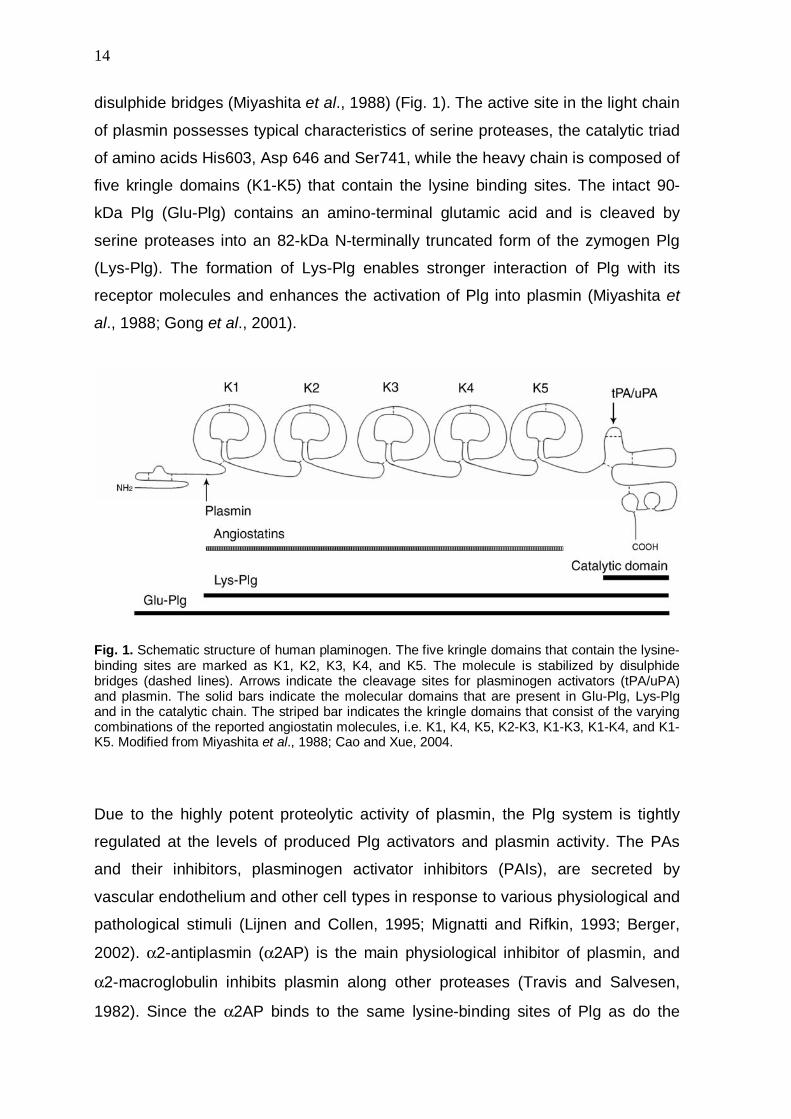
disulphide bridges (Miyashita et al., 1988) (Fig. 1). The active site in the light chain
of plasmin possesses typical characteristics of serine proteases, the catalytic triad
of amino acids His603, Asp 646 and Ser741, while the heavy chain is composed of
five kringle domains (K1-K5) that contain the lysine binding sites. The intact 90-
kDa Plg (Glu-Plg) contains an amino-terminal glutamic acid and is cleaved by
serine proteases into an 82-kDa N-terminally truncated form of the zymogen Plg
(Lys-Plg). The formation of Lys-Plg enables stronger interaction of Plg with its
receptor molecules and enhances the activation of Plg into plasmin (Miyashita et
al., 1988; Gong et al., 2001).
Fig. 1. Schematic structure of human plaminogen. The five kringle domains that contain the lysine-binding sites are marked as K1, K2, K3, K4, and K5. The molecule is stabilized by disulphide bridges (dashed lines). Arrows indicate the cleavage sites for plasminogen activators (tPA/uPA) and plasmin. The solid bars indicate the molecular domains that are present in Glu-Plg, Lys-Plg and in the catalytic chain. The striped bar indicates the kringle domains that consist of the varying combinations of the reported angiostatin molecules, i.e. K1, K4, K5, K2-K3, K1-K3, K1-K4, and K1-K5. Modified from Miyashita et al., 1988; Cao and Xue, 2004.
Due to the highly potent proteolytic activity of plasmin, the Plg system is tightly
regulated at the levels of produced Plg activators and plasmin activity. The PAs
and their inhibitors, plasminogen activator inhibitors (PAIs), are secreted by
vascular endothelium and other cell types in response to various physiological and
pathological stimuli (Lijnen and Collen, 1995; Mignatti and Rifkin, 1993; Berger,
2002). α2-antiplasmin (α2AP) is the main physiological inhibitor of plasmin, and
α2-macroglobulin inhibits plasmin along other proteases (Travis and Salvesen,
1982). Since the α2AP binds to the same lysine-binding sites of Plg as do the
15
receptors, the receptor-bound Plg is protected from the inhibitor. On the contrary,
free circulating plasmin is rapidly inactivated by the antiprotease.
The proteolytic cleavage of Plg can further lead to formation of internal Plg
fragments, collectively called as angiostatins, which lack the catalytic light chain
and vary in the number of kringle domains (Cao, 1999; Cao and Xue, 2004) (Fig.
1). The internal Plg fragments inhibit the endothelial cell proliferation and act as
natural regulators of physiological and pathological angiogenesis (O´Reilly et al.,
1994; Cao, 1999; Cao and Xue 2004). The Plg proteolysis into internal fragments
has been shown with variety of physiological proteases, such as members of the
matrix metalloprotease (MMP) family and elastase (Dong et al., 1997; Patterson
and Sang, 1997; Cao and Xue, 2004). Reductases secreted by tumor cells
(Stathakis et al., 1997 and 1999; Lay et al., 2000) generate angiostatin from
plasmin and also, tPA, uPA and the streptococcal PA streptokinase cleave Plg to
angiostatin in the presence of free sulfhydryl donors (Gately et al., 1997). Also,
autoproteolytic conversion of membrane-associated plasmin into angiostatin on
tumor cells has been reported (Wang et al., 2004). Fragmentation of the Plg into
angiostatin has been characterized with fungi-derived nonlysine triprenyl phenol
metabolites (Ohyama et al., 2004), but bacterial cells have not been reported to
modulate Plg fragmentation.
4.2 BACTERIAL INTERVENTION WITH THE PLASMINOGEN SYSTEM
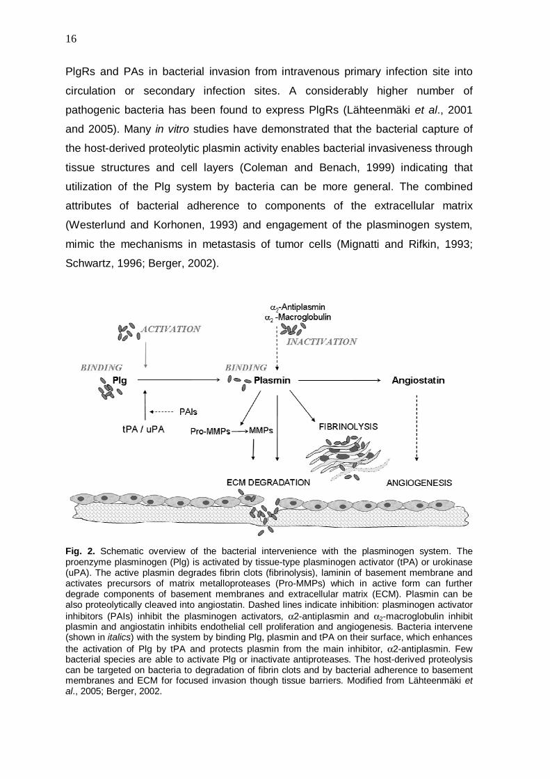
Invasive bacterial pathogens intervene with the Plg system by expressing PAs
and/or PlgRs (Fig. 2) which enables the pathogens to gain surface-bound
proteolytic activity (Lähteenmäki et al., 2005). Also, the production levels of
mammalian PAs, uPA-receptor (uPAR), PAIs and MMPs have been reported to be
affected by bacteria and/or bacteria-derived components, such as endotoxin
(Suffredini et al., 1989; Brandtzaeg et al., 1990; Behera et al., 2005). The central
role of bacteria-Plg interaction in bacterial invasiveness has been so far
documented in vivo in infections caused by Yersinia pestis, Borrelia species,
Streptococcus pneumoniae and group A streptococci (GAS) (Sodeinde et al.,
1992; Gebbia et al., 1999; Nordstrand et al., 2001; Sun et al., 2004; Bergmann et
al., 2003). These studies have highlighted the importance of synergistic action of
16
PlgRs and PAs in bacterial invasion from intravenous primary infection site into
circulation or secondary infection sites. A considerably higher number of
pathogenic bacteria has been found to express PlgRs (Lähteenmäki et al., 2001
and 2005). Many in vitro studies have demonstrated that the bacterial capture of
the host-derived proteolytic plasmin activity enables bacterial invasiveness through
tissue structures and cell layers (Coleman and Benach, 1999) indicating that
utilization of the Plg system by bacteria can be more general. The combined
attributes of bacterial adherence to components of the extracellular matrix
(Westerlund and Korhonen, 1993) and engagement of the plasminogen system,
mimic the mechanisms in metastasis of tumor cells (Mignatti and Rifkin, 1993;
Schwartz, 1996; Berger, 2002).
Fig. 2. Schematic overview of the bacterial intervenience with the plasminogen system. The proenzyme plasminogen (Plg) is activated by tissue-type plasminogen activator (tPA) or urokinase (uPA). The active plasmin degrades fibrin clots (fibrinolysis), laminin of basement membrane and activates precursors of matrix metalloproteases (Pro-MMPs) which in active form can further degrade components of basement membranes and extracellular matrix (ECM). Plasmin can be also proteolytically cleaved into angiostatin. Dashed lines indicate inhibition: plasminogen activator inhibitors (PAIs) inhibit the plasminogen activators, α2-antiplasmin and α2-macroglobulin inhibit plasmin and angiostatin inhibits endothelial cell proliferation and angiogenesis. Bacteria intervene (shown in italics) with the system by binding Plg, plasmin and tPA on their surface, which enhances the activation of Plg by tPA and protects plasmin from the main inhibitor, α2-antiplasmin. Few bacterial species are able to activate Plg or inactivate antiproteases. The host-derived proteolysis can be targeted on bacteria to degradation of fibrin clots and by bacterial adherence to basement membranes and ECM for focused invasion though tissue barriers. Modified from Lähteenmäki et al., 2005; Berger, 2002.
17
4.2.1. BACTERIAL PLASMINOGEN RECEPTORS
The best characterized PlgRs in Gram-positive bacteria are the plasminogen-
binding group A streptococcal M protein (PAM) as well as streptococcal glycolytic
enzymes, glyceraldehyde-3-phosphate dehydrogenase (GAPDH) and α-enolase
(Wistedt et al., 1995; Winram and Lottenberg, 1998; Pancholi and Fischetti, 1998).
In Gram negative bacterial species, such as E. coli and salmonella, the
filamentous surface appendages, fimbriae and flagella, act as PlgR molecules
(Parkkinen and Korhonen, 1989; Lähteenmäki et al, 1993; Kukkonen et al., 1998;
Sjöbring et al., 1994). Also, aspartase in Haemophilus influenzae, outer surface
protein A (OspA) and a 70-kDa surface protein OppA in Borrelia burgdorferi as
well as the surface proteins PgbA and PgbB in Helicobacter pylori have been
characterized as PlgRs (Sjöström et al., 1997; Fuchs et al., 1994; Hu et al., 1997;
Jönssön et al., 2004).
PAM is associated with GAS strains that cause skin infections in humans
(Svensson et al., 1999) and binds Plg via lysine residues present in two N-
terminally located repeat regions of 13 amino acids. Inactivation of the PAM-
encoding emm53 gene abolishes the Plg binding in vitro and leads to attenuated
infection in an experimental animal model of infection in vivo (Svensson et al.,
2002; Sun et al., 2004).
Streptococcal GAPDH, also called as streptococcal surface hydrogenase (SDH),
has been shown to be surface localized and to bind human Plg and plasmin
(Pancholi and Fischetti, 1992; Lottenberg et al., 1992; Winram and Lottenberg,
1998; Bergmann et al., 2004). The C-terminal lysine residue of GAPDH seems to
be important in the Plg binding, since substitution of this amino acid to leucine
abolished the binding (Winram and Lottenberg, 1998). However, finding that the
recombinant bacteria with mutated GAPDH still retained the Plg-binding ability,
emphasizes the importance of other PlgRs in streptococci (Winram and
Lottenberg, 1998; Jobin et al., 2004). The GAPDH protein has also been observed
to possess multifunctional activity, such as binding to fibronectin, lysozyme,
cytoskeletal proteins as well as displaying auto-ribosylating and phosphorylating
18
activities (Pancholi and Fischetti, 1992 and 1993), but the in vivo role of these
multiple characteristics are not known.
The surface-localized α-enolase is reported to be the main PlgR in streptococci
and binds Plg and plasmin in a lysine dependent manner (Pancholi and Fischetti,
1998; Bergmann et al., 2001). Similar to GAPDH, α-enolase binds with greater
affinity to Lys-Plg and plasmin than to Glu-Plg (Winram and Lottenberg, 1998;
Derbise et al., 2004). The C-terminal lysine residues have been shown to be
important in the PlgR function of GAS and pneumococci (Pancholi and Fischetti,
1998; Bergmann et al., 2001; Derbise et al., 2004), although recent report of the
3D structure of pneumococcal α-enolase suggests that the internal binding epitope 248FYDKERKVY256 (Bergmann et al., 2003), provides the major binding site for Plg
(Ehinger et al., 2004). Substituting of lysines at this sequence reduces Plg-binding
and attenuates the bacterium in a mouse model of intranasal infection (Bergmann
et al., 2003). The α-enolase performs also several other functions in addition to
glycolytic activity and plasmin(ogen) binding, e.g. binding to laminin, salivary mucin
and DNA. Enolase is also a heat-shock protein and a constructing component of
turtle cellular lenses and playes a role in autoimmunity disorders (Williams et al.,
1985; Wistow et al., 1988; al-Giery and Brewe, 1992; Aaronson et al., 1995;
Pancholi, 2001; Carneiro et al., 2004; Ge et al., 2004).
The mechanisms that contribute to surface localization of GAPDH and α-enolase
have remained unknown. These house-keeping molecules are traditionally
considered as cytoplasmic enzymes and they do not possess known signal
sequences, membrane-anchoring motifs or hydrophobic membrane-spanning
regions; however, they have been reported to be surface-localized or secreted in
several prokaryotic and eukaryotic organisms (Table 3).

19
Table 3. Surface localization and/or secretion of GAPDH and α-enolase by prokaryotic and eukaryotic cells.
Enzyme Bacteria Fungi Parasites Mammalian
GAPDH
Group A, B, C, E, G, H
and L streptococci1
Staphylococcus aureus2
Staphylococcus
epidermidis2
Mycobacteria3
Neisseria meningitidis4
Neisseria lactimica4
enteropathogenic E. coli5
Candida
albicans10
Saccharomyces
cerevisiae11
Kluyveromyces
marxianus12
Schistosoma
mansoni15
Trypanosoma16
Leishmania16
Fasciola hepatica17
erythroid19
α-enolase
Group A, B, C, D, E, F, G,
H and L streptococci6
Staphylococcus aureus7
Aeromonas hydrophila8
Actinobacillus
actinomycetemcomitans9
Candida
albicans13,
Pneumocystis
carinii14
Onchocerca
volvulus18
F. hepatica17
hematopoietic20
pharyngeal21
carcinomal22
endothelial23
neuronal24
muscular25
(Pancholi and Fischetti, 19921; Modun and Williams, 19992; Bermudez et al., 19963; Grifanttini et al., 20024; Kenny and Finlay, 19955; Pancholi and Fischetti, 19986; Mölkänen et al., 20027; Sha et al., 20038; Hara et al., 20009; Gil-Navarro et al., 199710; Crowe et al., 200310; Delgado et al., 200111; Fernandes et al., 199212; Angiolella et al., 199613; Fox and Smulian, 200114; Goudot-Crozel et al., 198915; Pancholi and Chhatwal, 200316; Bernal et al., 200417; Jolodar et al., 200318; Allen et al., 198719; Miles et al., 199120; Redlitz et al., 199520; Pancholi et al., 200321; López-Alemany et al., 199422; Dudani et al., 199323; Nakajima et al., 199424; Lopez-Alemany et al., 200325).
4.2.2. BACTERIAL PLASMINOGEN ACTIVATORS
Few invasive bacterial species (e.g. streptococci, staphylococci, Yersinia pestis
and salmonella) have PAs that can be functionally divided into two groups:
secreted non-enzymatic activators and surface localized protease activators.
Streptokinase (SK) and staphylokinase (SAK) belong to the former group and form
molecular complexes with Plg and plasmin which leads to changes in conformation
and specificity of Plg (Rabijns et al., 1997; Wang et al., 1998). SK and SAK share
little sequence homology but have a similar protein folding (Parry et al., 2000). The
binary SAK-Plg complex remains proteolytically inactive and requires additional
activation by PAs (Schlott et al., 1997). SK activates Plg by forming first a binary
complex with Plg and subsequently ternary Plg-SK-Plg complex in which the
catalytic triad of Plg is functional without cleavage at Arg561-Val562 (Young et al.,
1998). The SKs produced by strains of human and non-human origin differ
structurally and form ternary complexes with Plg mainly in a host species-specific
20
manner (Caballero et al., 1999). Expression of ska gene encoding SK has been
shown to be increased in GAS isolated from spleens of infected mice (Rezcallah et
al., 2004). In a mouse model of human skin impetigo inactivation of ska reduced
bacterial virulence after subcutaneous infection of conventional as well as
transgenic mice expressing human Plg (Svensson et al., 2002; Khil et al., 2003;
Sun et al., 2004); thus the synergistic action of bacterial PAs and PlgRs is
important in the virulence of GAS (Sun et al., 2004). The protection of receptor–
associated plasmin from α2AP is observed also in vitro in SAK-promoted
activation of staphylococci-immobilized Plg (Mölkänen et al., 2002). The PauA of
Streptococcus uberis also is a secreted PA (Leigh, 1994; Leigh and Lincoln, 1997)
that has a low sequence similarity but high predicted structural analogy with SK
(Rosey, et al., 1999; Ward et al., 2004) and activates bovine, ovine and equine,
but not human Plg via SK-like activation (Rosey, et al., 1999; Ward et al., 2004). A
few Streptococcus uberis strains associated with clinical bovine mastitis are also
found to secrete a novel broad host spectrum PA, PauB (Ward and Leigh, 2002
and 2004), which activates Plg by unknown mechanism.
The Pla of Y. pestis and PgtE of enteropathogenic salmonella belong to the beta-
barrel surface proteases of the omptin family (Sodeinde and Goguen, 1988). Pla
activates Plg by cleaving at the same Arg561-Val562 site as do tPA and uPA, and
thus resemble the mammalian PAs in function (Sodeinde et al., 1992). The Plg
activation by PgtE seems similar in mechanisms to Pla, but is significantly less
efficient (Kukkonen et al., 2004). Notably, both proteases are highly capable of
inactivating α2AP and thus creating uncontrolled plasmin proteolysis (Kukkonen et
al., 2001; Lähteenmäki et al, 2004). The functions of omptins are strongly
influenced by lipopolysaccharide (LPS) on bacteria: the activities of Pla and PgtE
are sterically inhibited by O-antigen repeats present in smooth LPS, whereas
rough LPS allows full activity (Kukkonen et al., 2004). LPS in Y. pestis is rough
whereas the clinical isolates of salmonella nearly constantly possess smooth LPS
(Skurnik et al., 2000; Kukkonen et al., 2004). The importance of Pla for Y. pestis
virulence is convincingly demonstrated in vivo (Sodeinde et al., 1992) and elevated
expression of PgtE and alterations of LPS O-chain in salmonella cells from
intracellular vacuoles of infected macrophages (Lähteenmäki et al., 2004) suggest
that PgtE has a role in salmonellosis.
21
5. AIMS OF THE STUDY
The bacterial adhesion to the epithelium is considered important for colonization
as well as for bacteria-host crosstalk at host mucosal surfaces. While the
molecular bases of adhesion of bacterial pathogens have been well characterized,
much less is known about the surface proteins and adhesion mechanisms of
commensal lactobacilli. These bacteria, however, form a major prokaryotic group
colonizing the human and animal mucosa, and knowledge on their adhesion
mechanisms is important both for basic bacterial ecology and for a more optimal
use of lactobacilli as probiotics. Overall, the probiotic as well as opportunistic
pathogenic mechanisms of intestinal lactobacilli have remained poorly understood.
This study was initiated to characterize the adhesive properties of a chicken
intestinal isolate of Lactobacillus crispatus within the alimentary tract of its host
and to identify the molecules mediating the adhesion. The effects of lactobacilli on
adhesiveness of avian pathogenic E. coli to chicken tissues were also studied. As
it became evident that commensal lactobacilli possess a surface-expressed
glycolytic enzyme, GAPDH, which is known to act as plasminogen receptor on
pathogenic streptococci, hence we also studied the interaction of lactobacilli with
the human plasminogen system.
22
6. MATERIALS AND METHODS
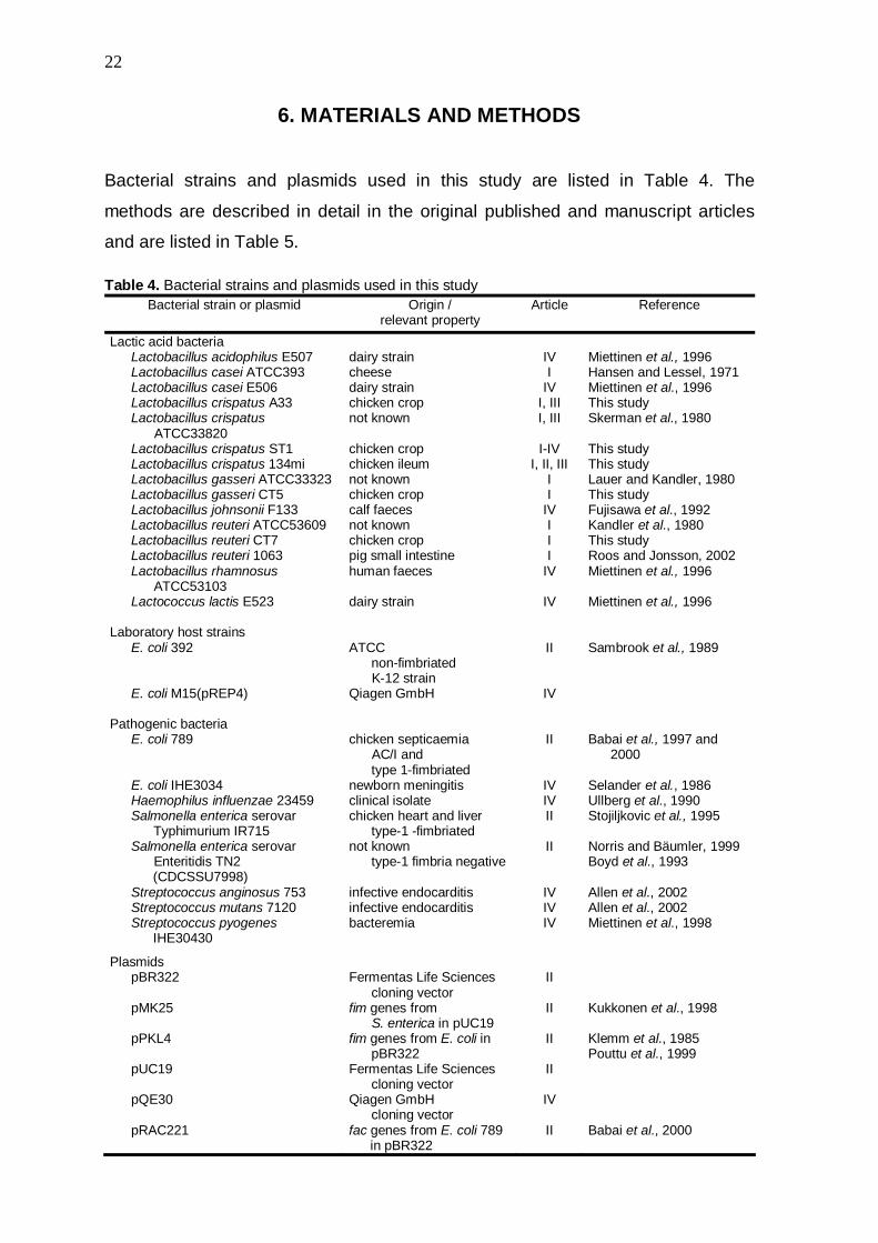
Bacterial strains and plasmids used in this study are listed in Table 4. The
methods are described in detail in the original published and manuscript articles
and are listed in Table 5.
Table 4. Bacterial strains and plasmids used in this study
Bacterial strain or plasmid Origin / relevant property
Article Reference
Lactic acid bacteria Lactobacillus acidophilus E507 Lactobacillus casei ATCC393 Lactobacillus casei E506 Lactobacillus crispatus A33 Lactobacillus crispatus
ATCC33820 Lactobacillus crispatus ST1 Lactobacillus crispatus 134mi Lactobacillus gasseri ATCC33323 Lactobacillus gasseri CT5 Lactobacillus johnsonii F133 Lactobacillus reuteri ATCC53609 Lactobacillus reuteri CT7 Lactobacillus reuteri 1063 Lactobacillus rhamnosus ATCC53103 Lactococcus lactis E523
dairy strain cheese dairy strain chicken crop not known chicken crop chicken ileum not known chicken crop calf faeces not known chicken crop pig small intestine human faeces dairy strain
IV I
IV I, III I, III
I-IV
I, II, III I I
IV I I I
IV
IV
Miettinen et al., 1996 Hansen and Lessel, 1971 Miettinen et al., 1996 This study Skerman et al., 1980 This study This study Lauer and Kandler, 1980 This study Fujisawa et al., 1992 Kandler et al., 1980 This study Roos and Jonsson, 2002 Miettinen et al., 1996 Miettinen et al., 1996
Laboratory host strains E. coli 392 E. coli M15(pREP4)
Pathogenic bacteria
E. coli 789 E. coli IHE3034 Haemophilus influenzae 23459 Salmonella enterica serovar Typhimurium IR715 Salmonella enterica serovar Enteritidis TN2
(CDCSSU7998) Streptococcus anginosus 753 Streptococcus mutans 7120 Streptococcus pyogenes
IHE30430
ATCC non-fimbriated K-12 strain Qiagen GmbH chicken septicaemia AC/I and type 1-fimbriated newborn meningitis clinical isolate chicken heart and liver type-1 -fimbriated not known type-1 fimbria negative infective endocarditis infective endocarditis bacteremia
II
IV
II
IV IV II
II
IV IV IV
Sambrook et al., 1989 Babai et al., 1997 and 2000 Selander et al., 1986 Ullberg et al., 1990 Stojiljkovic et al., 1995 Norris and Bäumler, 1999 Boyd et al., 1993 Allen et al., 2002 Allen et al., 2002 Miettinen et al., 1998
Plasmids pBR322 pMK25 pPKL4 pUC19 pQE30 pRAC221
Fermentas Life Sciences cloning vector fim genes from S. enterica in pUC19 fim genes from E. coli in pBR322 Fermentas Life Sciences cloning vector Qiagen GmbH
cloning vector fac genes from E. coli 789
in pBR322
II
II
II
II
IV
II
Kukkonen et al., 1998 Klemm et al., 1985 Pouttu et al., 1999 Babai et al., 2000

23
Table 5. Methods used in this study Method Described and used in
Adhesion studies with bacteria and purified proteins Adhesion to chicken intestinal tissue sections Adhesion to vaginal epithelial cells Adhesion to ileal mucus Exclusion of adhesion of E. coli by lactobacilli
Interaction studies of bacteria and purified proteins with the plasminogen
Kinetic plasminogen activation assay Binding of 125I-labeled Plg, plasmin and tPA to bacteria Binding of Plg and plasmin on purified Plg-receptors Bacteria-bound plasmin activity measurement
Genetic methods
Sequencing of chromosomal DNA Cloning and sequencing the Plg-receptors from lactobacilli
Protein work
Extraction of surface proteins from lactobacilli Expression and purification of the Plg-receptors of L. crispatus ST1 in E. coli N-terminal sequencing MALDI-TOF analysis
Immunological methods Production of antisera Western-blotting Indirect immunofluorescence Immuno-EM
I, II, III
III I, II II
IV IV IV IV
IV IV
III IV
III, IV IV
III, IV III, IV I, II, III
IV
24
7. RESULTS AND DISCUSSION
This study was initiated to characterize molecular mechanisms important for
adhesion and colonization of lactobacilli at host intestinal tissues. We used the
chicken as a host model since lactobacilli are frequent and adhesive in their
alimentary canal (Fuller and Turvey, 1971; Fuller, 1973; Brooker and Fuller, 1975;
Mitsuoka, 1992; Sarra et al., 1992; Guan et al., 2003; Lan et al., 2002) and as
tissue samples from the animals are easily available.
7.1. Identification of Lactobacillus crispatus strains with colonization
potential (unpublished)
Our first aim was to isolate a Lactobacillus stain with colonization and adhesion
potential, and we used a chicken colonization trial for this task. The Lactobacillus
strains A33 and 134mi were isolated from chicken crop and ileum, respectively,
and identified as L. crispatus by their 16S rRNA-gene sequences. The PCR
amplification of genomic DNA with primers designed for Enterobacterial Repetitive
Intergenic Consensus (ERIC) sequences that have been used in analysis of
various bacterial genera and species (Sharples et al., 1990; Versalovic et al.,
1991; de Bruijn, 1992), gave different patterns for both strains (data not shown)
verifying identification of separate strains. In primary adhesion tests, the strain A33
was found highly adhesive to frozen tissue sections of chicken intestinal tract,
whereas the strain 134mi was less efficient in adhesion (see 7.2.). As a
comparison, the type strain of L. crispatus from culture collection, ATCC33820,
was also tested for its adherence and found to be poorly adhesive. For
colonization trials, we inoculated newly hatched ross 208 chickens orally with
6x10⁷ bacteria per strains in two pairs of lactobacilli, A33 + ATCC33820 or 134mi +
ATCC33820, to compare colonization of adhesive and poorly-adhesive strains.
Lactobacilli at days 2, 5 and 12 postinoculum were isolated by cultivating on
Lactobacillus selection (LBS) agar from crop and ileum of inoculated chickens as
well as from chickens that received no exogenous bacteria. Within two days after
hatching, the total number of lactobacilli isolates reached similar high levels both in
crop (mean 4.1x10⁸ cells per crop) and ileum (2.6x10⁸ cells per ileum) with no
significant difference between the three test groups. The cut-off level of detection
25
was 105 bacteria per intestinal sample. Genomic DNA of 120 randomly chosen
Lactobacillus colonies from each time point were subjected to PCR amplification
with ERIC primers, and 67 isolates representing the most frequent PCR fingerprint
patterns as well as isolates yielding patterns indistinguishable from those obtained
with A33 and 134mi, were subjected to 16S rRNA-gene sequencing. Based on the
16S rRNA-gene sequences, the isolates belonged to the species L. crispatus, L.
gasseri and L. reuteri. Based on PRC profiling, both A33 and 134mi also persisted
in the chicken intestine. A bacterial colony representing the most commonly
detected PCR profile within an identified species was chosen for the adherence
tests. This selection gave the strains L. crispatus ST1, L. gasseri CT5, and L.
reuteri CT7, which were considered capable of colonizing young chickens as they
outgrow the challenge lactobacilli in the host intestine. Lactobacilli with the same
PCR profiles were not detected among bacteria isolated from chicken egg shells,
and it seems probable that ST1, CT5 and CT7 originated from the air of the
brooder room or the diet of chickens similar to observations by Fuller (1973).
7.2. Tissue tropisms of indigenous lactobacilli in the avian intestinal tract (I)
Histological analysis of bacterial colonization in the chicken intestine has revealed
that the major sites of bacterial colonization are crop, ileum and caecum (Fuller
and Turvey, 1971). The alimentary canal consists of multiple tissue structures that
vary in physiological functions. Therefore tissue sections rather than isolated
epithelial cells or tissue homogenates were used in this PhD work to reveal
possible tissue tropism in the adherence of lactobacilli. We used frozen sections
from crop, duodenum, jejunum, ileum, caecum and colon to cover the whole
alimentary canal.
The adherence of lactobacilli to the tissue domains in the avian alimentary canal is
summarized in Table 1; I. L. crispatus ST1 adhered efficiently to the non-secreting
stratified squamous epithelium of chicken crop, to the apical poles of mature
enterocytes at tip of villi, to basolateral poles of enterocytes along the villus-crypt
axis as well as to FAE and underlying lymphoid follicles. Also, efficient adherence
to lamina propria composed of connective tissue, blood vessels and lymphoid cells
was observed. (Fig. 1 and 2; I). The strain A33 showed similar and strong
26
adhesion profile as did the strain ST1. Instead, the strain 134mi adhered only
poorly to the epithelium of crop and to FAE. The adhesiveness of L. gasseri CT5
was targeted to the same tissue structures as the strains ST1 and A33, but CT5
showed lower adhesion efficiency to apical surfaces of enterocytes and,
importantly, to FAE. The L. reuteri CT7 adhered only to the epithelium of crop, and
the type culture collection strains L. crispatus ATCC33820, L. gasseri ATCC33323,
L. reuteri ATCC53609 L. casei ATCC393, showed no adhesion to chicken tissues.
These results demonstrate that fresh isolates of lactobacilli exhibit considerable
cell and tissue tropism in their adherence to the tissue domains in chicken
intestine.
None of the Lactobacillus strains adhered to mucus-producing goblet cells on
tissue sections, and only A33 and ATCC 53609 showed adherence to secreted
ileal mucus; this, however, was low when compared to that of L. reuteri strain 1063
reported to bind to hen mucus (Roos and Jonsson, 2002) (Fig. 3; I). To estimate
the role of abundant intestinal mucus layer as a colonization niche for lactobacilli,
we tested the ability of L. crispatus strains ST1, A33, 134mi and ATCC33820, L.
reuteri CT7, L. gasseri CT5 and L. casei 393 to grow in a minimal M9 salt medium
supplemented with ileal mucus as a carbon source (data not shown). For
comparison, the growth of lactobacilli in MRS medium was also tested. Bacteria
(1x10⁵ cfu) were inoculated to 1 ml of the media. After 16-hours cultivation,
lactobacilli reached average numbers of 2x10⁵ cells per ml in mucus medium and
1x10⁹ cells per ml in MRS medium.
Our results on the in vitro cell specificity of the adherence of lactobacilli are in
agreement with the findings of Fuller and Turvey (1971) and Brooker and Fuller
(1975) on bacterial colonization and in vivo adhesion. Thus there appears to be a
similar tissue distribution of the in vitro adhesion and the in vivo colonization by
lactobacilli in the chicken intestine. Furthermore, our adhesion and colonization
data support the idea that adhesion to the stratified squamous epithelium of crop is
an important factor in successful colonization of lactobacilli to chicken intestine
(Fuller, 1973). This idea is highlighted by the fact that strain CT7 adhered in vitro
only to crop epithelium and was found to be persistent in vivo in the intestine
during the colonization trial.
27
In the small and large intestine, the epithelium is simple columnar and serves the
functions of secretion and absorption, and undergoes continuous and rapid
renewal. The epithelial stem cells are located at the crypts of villi and give rise to
mature enterocytes, mucus-producing goblet cells, paneth cells and entero
endocrine cells (Huet et al., 1987; McCracken and Lorenz, 2001). Our finding that
lactobacilli preferentially adhere to the apical poles of mature enterocytes at villus
tips is in line with the observation of Fuller and Turvey (1971) that colonizing
bacteria were only rarely detected below the top third of the villi. The crypt-villus
migration of intestinal enterocytes involves changes in expression of ECM
receptors (Louvard et al., 1992) but much less is known about the distribution of
surface antigens in the polarized epithelium. The mature enterocytes possess
apical brush border with well organized microvilli and underlying terminal web,
while the immature enterocytes contain only few surface microvilli and lack well-
formed terminal web. The maturating process is known to involve changes in the
levels of luminal membrane proteins (Burgess et al., 1989; Fath et al., 1990;
Shibayama et al., 1987; Weiser et al., 1986). Also, the intestinal brush-border
associated enzymes as well as laminin expression and basement membrane (BM)
organizations are known to differ in healthy vs. colorectal conditions (Real et al.,
1992; Rémy et al., 1992). The enterocyte receptors for adhering lactobacilli remain
to be elucidated.
The frozen tissue sections fixed with paraformaldehyde have been successfully
used to determine tissue tropism of variety of bacterial adhesins (Korhonen et al.,
1990; Sillanpää et al., 2000). The intact protective mucus layer, however, may be
lost under these experimental procedures (Deplancke and Gaskins, 2001). We did
not detect bacterial adherence to mucus-producing goblet cells in the tissue
sections and hence wanted to confirm the adherence with freshly isolated
intestinal mucus. Isolates of Lactobacillus have been reported to bind to pig and
hen intestinal mucus and human faecal mucus (Rojas and Conway, 1996; Roos et
al., 2000; Roos and Jonsson, 2002; Tuomola et al., 1999; Gusils et al., 2003). No
adhesion to secreted mucus was observed with Lactobacillus strain tested in this
study, expect for the strains A33 and ATCC53609 that showed poor adhesion.
Further, in contrast to pathogenic E. coli that are able to grow in intestinal mucus
28
(Rang et al., 1999; Wadolkowski et al., 1988), the lactobacilli tested in this study
were not able to grow efficiently in a minimal medium supplemented with salts and
mucus. However, Rojas and Conway (1996) reported that Lactobacillus ssp. were
able to grow in pig ileal mucus suggesting that some lactobacilli are able to occupy
the mucus-layer niche in the intestine. The mucus is observed to prevent bacterial
adhesion onto epithelial cells in vitro (Jin et al., 1996; Todoroki et al., 2001) and
hence promote removal of bacteria from the intestine (Weiser et al., 1986).
Lactobacilli have also been observed to inhibit adhesion of enteropathogenic E.
coli to cultured intestinal cell line by inducing mucin gene expression (Mack et al.,
1999). On the other hand, commensal bacteria alter surface mucus and diminish
its deposition in caecum (McCracken and Lorenz, 2001) and enhanced adherence
to intestinal mucus in vitro has been reported by L. reuteri strains grown in the
presence of intestinal mucus (Jonsson et al., 2001). Overall, the role of mucus in
bacterial and in particular Lactobacillus colonization in the GI remains controversial
(Deplancke and Gaskins, 2001). However, our results suggest that in chicken
intestine, the adherence to stratified squamous epithelium of crop which is not
secreting mucus (Hodges, 1974) may be important for colonization by lactobacilli.
In the colonization trial, the outgrowth of L. crispatus strain ST1 over the strain
A33, which both adhered to crop epithelium, might be explained by the difference
of the strains to adhere to the mucus layer.
Lymphoid follicles are scattered throughout the intestinal mucosa and form
lymphatic aggregates i.e. Peyer=s patches in the distal small intestine (Ross and
Romrell, 1989). We found that the L. crispatus strains were adherent to FAE and
over the entire area of the lymphoid follicles, including the B-cell-rich germinal
centers and the surrounding T-cell-rich dome area. Notably, a sharp contrast was
seen in the adherence of ST1 cells onto the FAE and onto the apical enterocyte
surface of the same ileal region, where bacteria selectively bound to the FAE. FAE
contains M cells that are important for pathogen invasion into the circulation and
for presentation of antigens to the immune system (Neutra et al., 1999). Our
findings are in line with the reports of association of certain lactobacilli with Peyer=s
patches of the mouse (Ma et al., 1990; Perdigón et al., 2000). The light
microscopic techniques do not allow identification of individual M cells in the
chicken ileum (Clark et al., 1993; Kitagawa et al., 2000; Buda et al., 2005), and the
29
possible interaction of lactobacilli with M cells requires more detailed studies.
Interaction of lactic acid bacteria with M cells in vivo in the mouse intestine was
recently reported on the basis of immune reactions against orally inoculated lactic
acid bacteria (Perdigón et al., 1999). It was also proposed that lactobacilli are
selectively internalized in vivo into Peyer=s patches or lamina propria of the mouse
intestine and that these interactions may be involved in different secretory
immunoglobulin A and CD4+ T-cell responses induced by the lactic acid bacteria
(Ma et al., 1990; Perdigón et al., 1999, 2000 and 2001; Plant and Conway, 2001).
Our in vitro adhesion studies suggest that adherent lactobacilli may indeed make
contact with M cells and the underlying Peyer=s patches, and the adherence of
lactobacilli to FAE observed in this study may denote to presentation of lactobacilli
to the immune cells.
7.3. Adhesion of APEC and salmonella in avian intestinal tract (II)
The shedding and horizontal transmission of APEC and salmonella between avian
hosts and subsequently to humans causes annually considerable health problems
and economical losses both in animal production and public health (Gross, 1994;
Gomez et al., 1997; Cerquetti and Gherardi, 2000). Not much is known about the
role of fimbrial expression on the tissue tropism of APEC and salmonella in
chicken intestine. We utilized the tissue sections to screen tissue specificity in
fimbriae-mediated adhesion of APEC and salmonella along the host intestinal
mucosa. This was done as the first step in evaluating the use of adhesive
lactobacilli in exclusion of pathogens in the chicken intestine.
The wild type (wt) APEC and salmonella and the recombinant strains expressing
mannose-binding type-1 fimbriae were observed to adhere to the stratified
squamous epithelium of crop (Fig. 1; II) as well as to apical and basolateral poles
of simple columnar epithelia of small and large intestine and ileal FAE (Fig. 2 and
4; II). Also, adhesion to submucosal connective tissue areas was observed.
Notably, poor adherence to the mucus-producing goblet cells but strong
adherence to secreted ileal mucus was observed with type-1 fimbriated strains
except for S. enterica serovar Typhimurium, which did not bind to either of these
samples (Fig. 3 and 5; II). The adherence was efficiently inhibited by α-methyl-D-
30
mannoside, in accordance with the role of type-1 fimbriae in the binding (Fig. 2; II).
Adhesion to intestinal goblet cells was detected only with AC/I-fimbriated wt and
recombinant strains (Fig. 3; II).
The frequent expression of type-1 fimbriae by APEC and salmonella (Dozois et al.,
1994; Babai et al., 1997; Humphries et al., 2001) and the efficient type-1 fimbriae
mediated adhesion to chicken intestinal epithelial surfaces observed in this study,
support the previous findings (Oyofo et al., 1989 a and b; Marc et al., 1998) that
type-1 fimbriae may have a role in the colonization of enteropathogens in chicken
intestine. Prevention of this mannose-dependent adhesion of APEC and
salmonella thus may interfere with the fecal-oral transmission and subsequently
reduce the frequency of colibacillosis and salmonellosis in the avian and the
human hosts.
The type-1 fimbriae were observed to mediate the adherence of APEC and
salmonella to the immunologically important FAE area in the chicken ileum. On the
other hand, the specific adherence of salmonella to ileal FAE M-cells in the mouse
was reported to be mediated by Lpf fimbriae (Bäumler et al., 1996). In lack of
specific markers for chicken M-cells, these cells could not be identified on the
sections used in the light microscopic analysis and the role of host specificity in the
differences of results remains open. Lpf-deficient salmonella have shown only
limited reduction in their virulence in mice (Bäumler et al., 1996) and translocation
of unspecific luminal particles and microorganisms to underlying lymphoid areas
by M-cells is also reported (Jepson et al., 1996; Owen, 1999), suggesting that
close association of bacteria with M-cells might affect their translocation frequency.
Hence, the type-1 fimbriae mediated adhesion of APEC and salmonella to chicken
FAE may also contribute to their translocation to underlying tissues.
The type-1 fimbriae were also observed to mediate adhesion of APEC and
salmonella to ileal mucus but not to the goblet cells. The intestinal mucus layer is
composed of secreted mucins mainly synthesized by intestinal goblet cells to form
a protective barrier over the epithelium (Deplancke and Gaskins, 2001). The
bacterial entrapment into the mucus is regarded as an antimicrobial and defensive
function which removes luminal bacteria by peristalsis (Weiser et al., 1986).
31
Hence, it would not be beneficial for enteric bacteria to express type-1 fimbriae in
vivo in the intestinal lumen. Bacterial motility is reported to be important in vitro
and in vivo for colonization and invasion of the host intestinal mucosa by
salmonella (Dibb-Fuller et al., 1999; Allen-Vercoe and Woodward, 1999 a and b;
Allen-Vercoe et al., 1999). Penetration of mucus layer by E. coli strain with “on”
phase-locked type-1 fimbrial expression was slower than that observed with the
strain lacking type-1 fimbriae (McCormick et al., 1993), indicating that type-1
fimbria may promote bacterial entrapment into intestinal mucus in mice. Also, the
phase-locked mutant showed reduced colonization ability in mouse large intestine
when compared to the strain lacking type-1 fimbriae (McCormick et al., 1993). On
the other hand, mucus may serve as a nutrient source for the growth of enteric
pathogens, and hence contribute to their colonization and virulence in mice (Rang
et al., 1999; Wadolkowski et al., 1988). The efficient growth of both APEC and
salmonella in chicken ileal mucus supplemented with salts was also observed in
this study (Table 1; II), whereas intestinal isolates of L. crispatus, L. gasseri, and L.
reuteri did not multiply under the same experimental conditions (see 7.2). The
ability of motile enteropathogens to grow in the mucus may resist peristaltic
exhaustion and increase bacterial persistence within the host intestine. For non-
motile lactobacilli, on the other hand, the adhesion to mucus might on the contrary
be unfavourable.
Opposite to type-1 fimbriae, the AC/I fimbria preferentially bound to goblet cell-
associated mucus rather than to the secreted mucus. The difference may be due
to variation in the molecular composition of the mucins in the secreted or the cell-
associated forms (Deplancke and Gaskins, 2001), to bacterial modification of the
mucins (McCracken and Lorenz, 2001) or to different technical conditions in the
two experiments (McCracken and Lorenz, 2001). Our results disagree with
previously reported AC/I fimbriae-mediated adhesion of APEC to isolated avian
intestinal epithelia cells (Yerushalmi et al., 1990) in that we did not observe AC/I-
mediated bacterial adherence to the intestinal enterocytes of tissues sections. The
reasons for the differences remain open, however, our results do not support a
role for AC/I fimbriae in colonization of chicken intestine.
32
7.4. Exclusion of pathogens by lactobacilli (II)
The prevention of intestinal tract infections is one of the health benefits attributed
to lactobacilli. The adherence of lactobacilli to intestinal epithelium could prevent
pathogen colonization by steric hindrance or competition for epithelial receptors
(McGroarty, 1993). The colocalization of the type-1 fimbriae-mediated adhesion of
pathogenic E. coli and salmonella to the same epithelial areas that were
recognized by adhering lactobacilli, encouraged us to test the potential of
lactobacilli to exclude the pathogens at sites important for colonization and
invasion, i.e. crop and FAE. The strongly adhering L. crispatus strain ST1
inhibited the adhesion of APEC strain 789 to the epithelium of crop and FAE, while
the poorly adhesive L. crispatus strain 134mi inhibited the pathogen adhesion only
partially (Fig. 4; II). These results demonstrate that adhesion may be a factor
contributing to exclusion of pathogens from intestinal surfaces.
Adhesion is considered to be an important step for pathogens in their invasion
(Finlay and Cossard, 1997; Finlay and Falkow, 1997; Dho-Moulin and Fairbrother,
1999; Neutra et al., 1999; Klemm and Schembi, 2000), and hence the adhesion-
based exclusion of pathogens by commensal bacteria might be important in
prevention of bacterial invasion particularly at the area of FAE. In many in vivo
animal feeding experiments, the supplementation of hosts with Lactobacillus ssp.
has decreased the colonization and infection ability of enteric pathogens (Cole and
Fuller, 1984; Hudault et al., 1997; Pascual et al., 1999; Zhao et al., 1998; Perdigón
et al., 2001; Sgouras et al., 2004), although results with no significant effects have
also been reported (Adler and DaMassa, 1980; Hinton and Mead, 1991; Stavric et
al., 1992). In human trials, the most promising results have been obtained with
Lactobacillus treatments to decrease rotavirus infections and prevention of
pathogen overgrowth during antibiotic therapy (Hilton et al., 1997; Isolauri et al.,
1991 and 1994; Kaila et al., 1992; Majamaa et al., 1995; Biller et al., 1995;
Gorbach et al., 1987; Siitonen et al., 1990; Arvola et al., 1999). These studies,
however, did not address the mechanisms of exclusion of pathogens by
lactobacilli. In the chicken crop, lactobacilli reduce the luminal pH to 4,5-6 (Sarra et
al., 1992), which most probably affects the viability of enteric pathogens. This has
been seen in vitro with Lactobacillus salivarius strains against E. coli and S.
33
enterica serovar Enteritidis (Garriga et al., 1998). However, Fuller (1977) reported
that the bacteriostatic property of lactobacilli in crop is due to low pH, but the
bactericidal activity could not be accounted by pH alone. In human vagina, the
production of H2O2 rather than alteration of pH by Lactobacillus isolates has been
observed to decrease gonococcal growth (St. Amant et al., 2002), while low pH
alone acts against uropathogenic E. coli (Juárez Tomás et al., 2003).
Coaggregation of lactobacilli with type-1 fimbriated uropathogenic E. coli has also
been demonstrated, and unlike the bacterial coaggregation in oral cavity leading to
formation of dental plague, the finding was not related to onset of urogenital
disease but rather to prevention of infections (Reid et al., 1988). Since we did not
observe coaggragation of L. crispatus isolates with type-1 fimbriated APEC strain,
the mechanisms of inhibition of pathogen adhesion in crop and FAE was most
likely due to unspecific steric hindrance of adhesion to tissue receptors and to the
strong affinity of the adhesion of lactobacilli. The idea of steric hindrance is in line
with the report of mannose sensitive adhesion of L. plantarum and type-1
fimbriated E. coli to the human colonic cell line HT-29. Due to different reactivity to
periodate treatment, the prefered receptor epitopes on HT-29 cells were however,
suggested not to be identical for lactobacilli and E. coli (Adlerberth et al., 1996).
7.5. Isolation of L. crispatus adhesins (III)
L. crispatus ST1 was chosen for the isolation and characterization of adhesins due
to its strong adhesion ability to several tissue domains in chicken intestine. The
strain A33 was also included as an adhesive strain and strains 134mi and
ATCC33820 as poorly adhesive comparative strains. The removal of S-layer-
protein from the surface of ST1 cells by chaotropic treatment with guanidine
hydrochloride (GnHCl) did not affect the adherence of ST1 cells to chicken
intestinal tissues (Fig 1; III) and we could not detect adherence with the extracted
S-layer proteins released from ST1 (data not shown). By treating the bacteria with
peptidoglycan-cleaving bacterial autolysin, mutanolysin, the adhesion was
abolished, yielding detached proteins that retained their binding ability (Fig. 1; III).
Extracts from the adhesive and poorly-adhesive strains differed only with two
protein bands in SDS-PAGE (Fig. 2; III). After fractionating the released surface
proteins of STI, three different protein fractions with binding affinity to the stratum
34
corneum of stratified squamous epithelium of crop, basement membrane areas or
singular intraepithelial and lamina propria cells were isolated (Fig. 3; III).
The Lactobacillus surface protein that binds to stratum corneum layer of stratified
squamous epithelium of crop was named as Lactobacillus epithelium adhesin-1
(LEA-1). To our knowledge, this is the first stratified squamous epithelial adhesin
isolated form lactobacilli. Lactobacillus strains also show adhesion to other
epithelial surfaces, indicating a likely existence of other epithelial adhesins. LEA-1
is a high molecular weight protein with an apparent molecular size of approx. 280
kDa and may be a glycoprotein since it bound the conacavalin A lectin. The N-
terminal sequencing of LEA-1 was not successful. The finding that LEA-1 and a
related proteins could be detected on the surface of the adhesive L. crispatus
stains ST1 and A33, but not on the poorly adhering strains 134mi and ATCC33820
(Fig. 2; III), also suggest a role in adhesion of lactobacilli to crop epithelium.
Different ability of the strains ST1 and 134mi to inhibit pathogen adhesion onto the
outermost surface of crop epithelium may also involve different expression of LEA-
1 by the strains. Since lactobacilli are also frequent and dominating in human oral
cavity and vagina which are lined with stratified squamous epithelia (Mitsuoka,
1992; Redondo-Lopez et al., 1990; Paavonen, 1983), we tested whether LEA-1
could mediate adhesion to these tissue structures as well. ST1 cells were found to
be adhesive to buccal and vaginal cells freshly isolated from a healthy host,
whereas the purified LEA-1 adhered to ca. half of the isolated vaginal cells (Fig. 4;
III) but not at all to the buccal cells. The cells isolated from vagina probably contain
epithelial cells from stratum corneum as well as from underlying epithelial layers,
which might explain the selective binding of LEA-1 to only a fraction of the cell
population. However, ST1 cells were observed to bind throughout the stratified
squamous epithelium of crop in tissue sections and to all isolated vaginal and
buccal cells, suggesting that other adhesins, not identified in this study, also
participate in the epithelial adhesion.
Binding to the basement membrane was observed with the protein fraction
containing peptides with apparent molecular sizes of 50 and 55 kDa (50/55 kDa)
that could not be separated from each other by gel filtration or ion exchange
chromatography. N-terminal sequencing of these proteins gave amino acid
35
sequences that showed no close similarity to those in databases, including the
previously characterized collagen and laminin-binding proteins of lactobacilli,
CnBP or CbsA (Aleljung et al., 1994; Roos et al., 1996; Sillanpää et al., 2000).
Hence, our results give evidence that L. crispatus possessess yet a novel
basement membrane-binding adhesin(s). The 50/55 kDa adhesins or their
receptor proteins were not determined in this study and further investigations are
required to fully characterize these adhesins. Adhesion of several pathogenic
bacteria to ECM proteins is thought to contribute to bacterial invasiveness
(Westerlund and Korhonen, 1993). Lactobacilli commonly adhere to the
components of ECM and BM (Aleljung et al., 1991; Nagy et al., 1992; Harty et al.,
1994; Toba et al., 1995; McGrady et al., 1995; Styriak et al., 2003), and the activity
has been proposed to have both protective (Horie et al., 2002) and pathogenic
functions (Harty et al., 1993 and 1994), but overall, the biological functions of the
adhesiveness of lactobacilli to ECM remain open.
The protein binding to singular intraepithelial and lamina propria cells was
putatively identified by N-terminal sequencing and peptide database analysis as
GAPDH. The 20 amino acid long N-terminal sequence of the protein shows 100%
identity with L. delbrueckii and L. johnsonii GAPDH sequences (Pridmore et al.,
2004; Branny et al., 1998). The gene encoding GAPDH was cloned into an
expression vector and the recombinant GAPDH protein was expressed and
purified from E. coli (see 7.7). The extracted and the purified recombinant GAPDH
were found to bind with similar characteristics to chicken tissues. Histologically the
cells recognized by GAPDH resembled lymphoid cells that are numerous in lamina
propria and scattered in intraepithelial regions (Ross and Romrell, 1989;
McCracken and Lorenz, 2001). Double staining of tissue sections with specific
anti-chicken T-cell receptor 2 antibodies (Chen et al., 1988) showed a partial
colocalization with GAPDH-binding. However, due to lack of specific markers for
chicken lymphoid cells, the receptor cells for GAPDH could not be identified. The
surface-localization of the glycolytic enzyme GAPDH is well characterized in
several prokaryotic and eukaryotic species (Pancholi and Chhatwal, 2003) (Table
3.), and our results indicate that this is valid for lactobacilli also. The sequence of
GAPDH from L. crispatus ST1 contains no features associated with secretion of
proteins from Gram positive bacteria, and its secretion mechanisms remain open.
36
GAPDH has a variety of functions seemingly unrelated to glycolysis (Pancholi and
Fischetti, 1992 and 1993) and it has been observed to bind Plg (Pancholi and
Fischetti, 1992; Lottenberg et al., 1992). Binding of the GAPDH of lactobacilli to
intraepithelial and lamina propria cells in chicken intestine may represent a novel
function for bacterial GAPDHs.
The results described above indicate that a single strain of Lactobacillus
possesses several adhesins with affinity to distinct tissue domains in the host
intestine. However, as ST1 bacteria exhibited even broader tissue tropisms than
the adhesins identified here, more adhesion factors are likely to have a role in the
adhesiveness of ST1. The detailed knowledge of lactobacilli-host interaction at
molecular level could be important for developing efficiently targeted probiotic
products or efficient oral vaccine carriers. The results from lactobacilli as oral
tetanus toxin carriers have shown that the immunization efficiency is dependent on
the expression levels of the antigens, the Lactobacillus strain used as carrier, and
the viability and persistence of the carrier strain in the host GI tract (Grangette et
al., 2002; Seegers, 2002).
7.6. Interaction of commensal lactobacilli with the plasminogen system (IV)
Several pathogenic bacterial species intervene with the human proteolytic
plasminogen system to enhance their invasion and migration through tissue
barriers within the human host (Lähteenmäki et al., 2005). The Plg-binding has so
far been linked to bacterial pathogenesis in invasive infections (Coleman and
Benach, 1999; Lähteenmäki et al., 2001). Our results indicated that GAPDH, which
is a well characterized PlgR on pathogenic streptococci (Pancholi and Fischetti,
1992; Seifert et al., 2003; Jobin et al., 2004; Bergmann et al., 2004), is surface-
exposed also on lactobacilli. Lactobacilli are GRAS microbes (Adams and
Marteau, 1995) and have been associated only with sporadic cases of infective
diseases (Husni et al., 1997; Aguirre and Collins, 1993; Brouqui and Raoult, 2001;
Salminen et al., 2004). We begun to study the possible interaction of lactobacilli
with Plg as a possible mechanism contributing to the opportunistic pathogenesis of
lactobacilli; however, in view of the range of physiological functions of plasmin, this
37
interaction might have non-pathogenic functions as well. The methods we first
used have successfully been applied in studies of bacterial pathogens.
7.6.1. Plasminogen receptor function in lactobacilli (IV)
The ability of lactobacilli and lactococci to enhance Plg activation was tested in a
kinetic assay by incubating the bacteria with human Plg in the presence or
absence of the physiological PA, tPA, and the formed plasmin activity was
measured as the breakdown of a choromogenic substrate of plasmin. In inhibition
tests, the lysine analog ε-aminocaproic acid, EACA, the plasmin inhibitor α2-
antiplasmin (α2AP), and the serine protease inhibitor aprotinin, were included to
evaluate the role of lysine residues in bacteria-Plg interaction and the level of
bacteria-bound plasmin. The bacteria were highly efficient in enhancing the tPA-
catalyzed Plg-activation (Fig. 1; IV). The strains represented different species and
origins of isolation or use: intestinal, dairy and probiotic. Hence the interaction with
Plg-system seems to be a common characteristic for lactobacilli and lactococci.
For comparison, pathogenic bacteria isolated from systemic infections, i.e. GAS,
Streptococcus anginosus, Streptococcus mutans, E. coli and Haemophilus
influenzae, and earlier found to express PlgR activity (Pancholi and Fischetti, 1992
and 1998; Urdaneta et al., 2004; Lähteenmäki et al., 1993; Virkola et al., 1996)
were tested under the same conditions. These bacteria exhibited a significantly
less efficient enhancement of Plg activation that did the lactic acid bacteria (Fig. 1;
IV). Comparison of these results with previous reports on enhancement of Plg
activation by pathogenic bacteria e.g. E. coli (Lähteenmäki et al., 1993; Kukkonen
et al., 1998) is complicated due to different experimental conditions, such as the
ratio of the concentrations of Plg and tPA. The Plg activation on lactic acid bacteria
was strongly inhibited by EACA suggesting lysine-dependent interaction of
bacteria with the Plg. Also, α2AP efficiently inhibited the activation, indicating that
only a small proportion of the formed plasmin activity was bacterium-bound.
Aprotinin consistently inhibited the plasmin activity formed in the experiment (Fig.
1; IV). No plasmin activity could be measured in the absence of tPA, suggesting no
release of PAs from lactobacilli under these experimental conditions (data not
shown).
38
We next tested the binding of Plg, plasmin and tPA, onto the bacteria. The strains
L. crispatus ST1, L. rhamnosus GG were chosen to represent non-pathogenic
bacteria, and the strains S. anginosus 753 and S. mutans 7120 were chosen to
represent pathogenic species. Bacteria were first incubated with excess of 125I-
labeled proteins to allow binding, and after washing, the radioactivity remaining
bacteria-bound was measured (Fig. 2; IV). The binding of Plg and plasmin to
bacteria was poor (0,7-4,3%) when compared to the values reported in binding of
Plg and plasmin onto pathogenic GAS, S. aureus, Neisseria meningitidis,
Neisseria gonorrhoeae, H. influenzae, E. coli, Branhamella catarrhalis and strains
of Proteus species (Broeseker et al., 1988; Ullberg et al., 1990 and 1992; Kuusela
and Saksela, 1990). The binding was inhibited by EACA, which is in line with the
role of lysine residues in the binding. No binding of tPA to bacterial strains tested
here was observed. Previously tPA has been reported to bind only to curli and
fimbriae of E. coli and S. enterica (Sjöbring et al., 1994).
To further investigate the binding of plasmin(ogen) to lactobacilli, we analysed the
ability of these bacteria to obtain and retain plasmin activity on their surface.
Plasminogen was allowed to bind to L. crispatus ST1 cells and after washing the
excess Plg, tPA was added to catalyze the activation. After incubation for 1, 2 and
3 hours, the cells and the supernatants were separated, and the plasmin activity
was measured with the chromogenic substrate of plasmin. The plasmin activity on
the surface of L. crispatus ST1 was low and rapidly lost into the incubation
supernatant (Fig. 5; IV). The subsequent increase of plasmin activity in the
supernatant suggests cleavage of plasmin from bacterial surface or low affinity in
the binding. Release of plasmin from bacterial surface of group A, C and G
streptococci has also been suggested (Kuusela et al., 1992), but the importance of
this phenomenon in bacteria-host interaction has remained unknown.
Our results show that the bacterial interaction with the Plg is not restricted to
pathogenic bacteria but also commensal, dairy and probiotic lactobacilli and
lactococci express Plg receptors. The data from our and previous studies
demonstrate that lactobacilli are able to both adhere to components of ECM
(Aleljung et al., 1991; Nagy et al., 1992; Harty et al., 1994; Toba et al., 1995;
McGrady et al., 1995; Styriak et al., 2003) and bind Plg. These are common
39
characteristics for metastatic cancer cells and for several invasive bacterial
pathogens (Mignatti and Rifkin, 1993; Myöhänen and Vaheri, 2004; Westerlund
and Korhonen, 1993; Lähteenmäki et al., 2005) and might hence also enhance the
opportunistic pathogenic potential of lactobacilli. However, lactobacilli have been
associated with only sporadic cases of infective diseases in humans (Husni et al.,
1997; Aguirre and Collins, 1993; Brouqui and Raoult, 2001), and these cases have
usually preceded other severe predisposing diseases (Husni et al., 1997;
Salminen et al., 2004). Healthy persons get exceedingly rarely infectious diseases
associated with lactobacilli even though these bacteria are highly ubiquitous in the
indigenous microbiota of the hosts and in the environment. The risks of lactobacilli
to cause septicaemia and endocarditis have so far been evaluated by their ability
to trigger platelet aggregation, to participate in the platelet-fibrin-clot formation on
endothelial surface and to bind collagens in vitro, as well as by their proteolytic
activities (Harty et al., 1993 and 1994; Oakey et al., 1995). None of these
characteristics are restricted to strains associated with infective diseases and thus
their role in the virulence of lactobacilli remains open. Our data show that although
lactobacilli and lactococci were highly efficient in enhancing the tPA-catalyzed Plg
activation, the binding of Plg and plasmin onto the bacterial cells was poor.
Further, lactobacilli were not able to retain the cell-bound plasmin activity which
was further susceptible to physiological antiproteases, suggesting that they might
not be able to take advantage of the Plg system for proteolysis in a manner similar
to what pathogenic bacteria and tumor cells do (Lähteenmäki et al., 2005; Berger,
2002). In a view of our results the expression of PlgRs alone seems not to be
sufficient for bacterial virulence but might rather be a general property utilized for
bacteria-host interactions and in predisposing conditions for opportunistic
pathogenicity. The PlgR function of lactobacilli may also be utilized for nutritional
demands and persistence in the host e.g. oral cavity and intestine or the
environment e.g. milk.
7.6.2. Modulation of the plasminogen molecule by lactobacilli (IV)
It is known that in presence of tumor cells, Plg is cleaved into a small internal Plg
fragment, angiostatin, which is known to suppress endothelial proliferation and
tumor metastasis (O’Reilly et al., 1994; Cao and Xue, 2004). We allowed L.
40
crispatus ST1 bacteria to interact with Plg in PBS in the presence or absence of
tPA for 6, 12 and 24 hours and analysed the proteolytic cleavage of the Plg
molecule into smaller fragments by Western-blotting, N-terminal sequencing and
MALDI-TOF.
In the presence of tPA, ST1 enhanced the fragmentation of Plg to molecules
corresponding in size to plasmin and angiostatin (Fig. 6; IV). Plasmin has been
reported to possess autoproteolytic nature (Gately et al., 1997) which was also
observed here when incubating Plg in presence of tPA in plain PBS for 24 h.
However, the presence of L. crispatus ST1 significantly enhanced the Plg
fragmentation. N-terminal sequencing and MALDI-TOF analysis showed that in 24-
h incubation of ST1 with Plg and tPA, the main fragments modified from Plg were
K78-W783, corresponding to Lys-plasmin, and K78-R474 and V79-R493,
corresponding to angiostatin molecules (Fig. 6; IV)(Cao, 1999). Also, fragments
V79-W685 and K78-P670 lacking the S741 of the catalytic triad of plasmin were
generated. The fragments V79-W685 and K78-P670 had also been hydrolysed at
the position R474-A475 but the resulting molecules were still held together via the
disulphide bond C462-C541.
In the 24-h incubation in the absence of tPA, the L. crispatus ST1 and L. johnsonii
F133 were the only strains able to promote generation of internal Plg fragments.
The formed molecules resembled plasmin and angiostatin in molecular size (Fig.
6; IV). Based on N-terminal sequencing and MALDI-TOF data, the proteolytic
processing of Glu-Plg in the presence of L. crispatus ST1 yielded the internal
fragments of Plg, V79-W685 and K78-P670, as well as angiostatins, K78-R474
and V79-R493. Fragment corresponding to the Lys-plasmin was not detected from
the sample lacking tPA. We did not detect plasmin activity in the 24-h samples in
the kinetic plasminogen activation assay. The generated internal Plg fragments
were however, N-terminally cleaved at K77-K78 or K78-V79 sites, known to be
recognized by plasmin and other serine proteases as well as other
trypsin/chymotrypsin enzymes. Also, the fragmentation was efficiently inhibited
with α2AP and aprotinin, further suggesting trypsin-like cleavage of Plg. The
interaction of lactobacilli with Plg might involve bacterial cell envelope-associated
serine proteases (Kunji et al., 1996; Fira et al., 2001) that could initiate the N-
41
terminal cleavage of Plg at K77-K78 or K78-V79 and subsequently promote
fragmentation of Plg. The mechanimsm of Plg fragmentation by lactobacilli might
also involve action of surface exposed of secreted reductases similar to those in
tumor cells (Stathakis et al., 1997 and 1999; Lay et al., 2000), or autoproteolysis
catalyzed by free sulfhydryl donors or cell surface receptors (Gately et al., 1997;
Wang et al., 2004). Finally, secreted bacterial PAs such as streptokinase and
staphylokinase (Wang et al., 1998; Rabijns et al., 1997) have been shown to alter
the conformation of Plg to make it more susceptible for cleavage. We did not find
any indication of secreted PAs in the kinetic activation assays performed with the
bacteria or their supernatants containing secreted PlgRs under the experimental
conditions and the potential expression of PAs by lactobacilli need to be studied in
the future.
These results demonstrate that Lactobacillus cells promote modulation of Plg to
potential antitumorigenic angiostatin molecules both in the presence or absence of
tPA. The formation of internal Plg fragments and angiostatin could suppress tumor
growth or inhibit pathological plasmin-mediated cell invasion (Ji et al., 1998; Stack
et al., 1999; Gonzalez-Gronow et al., 2005). Commensal and probiotic lactobacilli
have been proposed to have an anti-colon cancer effect (Mercenier et al., 2003),
but the mechanisms of the tumor suppression have remained poorly known. In
acute toxoplasmosis, the infection-induced resistance to tumors has been shown
to result from suppression of tumor neovascularization rather than antitumor
cytotoxic signalling (Hunter et al., 2001). Characterization of Lactobacillus factor(s)
responsible for modulating Plg into angiostatin and their potential to suppress
intestinal tumor neovascularization or plasmin mediated tumor metastasis would
be of great importance for understanding the influence of indigenous microbiota on
the host in health and disease as well as for efficient targeting of health-promoting
probiotics.
7.7. Identification of GAPDH and enolase as plasminogen receptors in
lactobacilli (IV)
We found that GAPDH was surface-localized on lactobacilli (III). GAPDH and
another glycolytic enzyme, α-enolase, have been characterized as PlgRs on
42
pathogenic streptococci (Pancholi and Fischetti, 1992; Seifert et al., 2003; Jobin et
al., 2004; Bergmann et al., 2004; Pancholi and Fischetti, 1998; Bergmann et al.,
2001), and we tested the possible role of both of these proteins in the Plg-binding
of lactobacilli. The open reading frames of gap encoding the 338 amino acid
GAPDH and eno1 encoding the 428 amino acid enolase-1 were cloned from the L.
crispatus ST1 genome. Unlike pathogenic GAS and pneumococci which possess
only single genes coding for α-enolase in their genomes (Hoskins et al., 2001;
Ferretti et al., 2001), three enolase genes are present in L. johnsonii genome and
two in L. plantarum and L. lactis (Pridmore et al., 2004; Kleerebezem et al., 2003;
Bolotin et al., 2001). The GAPDH encoding gene exists as one copy both in
streptococci and lactobacilli and lactococci (Hoskins et al., 2001; Ferretti et al.,
2001; Pridmore et al., 2004; Kleerebezem et al., 2003; Bolotin et al., 2001).
Expression of other possible α-enolases in L. crispatus and their potential
interactions with Plg remain to be characterized in the future.
The recombinant GAPDH and enolase-1 were expressed and purified from E. coli
as N-terminal His6-tag fusion proteins under nonreducing conditions. To identify
their function as PlgRs, the proteins were tested in the kinetic plasminogen
activation assay at same molar concentrations. GAPDH was slightly more efficient
than enolase-1 in enhancing the tPA-catalyzed activation (Fig. 3; IV). The reaction
was inhibited by EACA, suggesting that lysine residues have a role in the
interaction. Also, α2AP and aprotinin inhibited the reactions. Next, the binding of
GAPDH and enolase-1 to Plg and plasmin were tested. Both GAPDH and enolase-
1 were observed to bind Plg (Fig. 3; IV) and plasmin in a lysine dependent
manner, GAPDH being slightly more efficient in binding than enolase-1. L.
crispatus ST1 GAPDH shows 57% and enolase-1 51% sequence identity to
GAPDH and enolase of pneumococci and GAS. L. crispatus ST1 GAPDH and
enolase-1 lack C-terminal lysines that are important in streptococcal Plg-binding,
but carry internal lysines in areas that are hydrophilic and predictably exposed on
protein surface. The 3D structure of the pneumococcal octameric enolase show
that the internal Plg-binding regions of the subunits are well exposed on protein
surface while the C-terminal lysine residues are only poorly displayed, suggesting
that the internal binding-regions are more important in the interaction with Plg
(Ehinger et al., 2004). The enolase-1 of L. crispatus ST1 possesses two lysines,

43
K251 and K255, at internal region which in sequence is highly similar to the
internal Plg-binding epitope of pneumococcal enolase (Bergmann et al., 2003).
The GAPDH and α-enolase are traditionally classified as cytoplasmic enzymes
and their predicted sequences possess no putative signal sequences, anchoring
motifs or membrane-spanning regions (Pancholi and Chhatwal, 2003). The
secretion mechanisms are thus undefined. Proteins with these characteristics have
been postulated to be present on the cell surface of pathogens which possess
either only an outer cell membrane such as parasites, or a membrane and a wall
such as Gram positive bacteria, but only rarely on the surface of organisms with
two cell membrane layers such as Gram negative bacteria (Pancholi and
Chhatwal, 2003). Secretion of GAPDH and α-enolase from Gram positive as well
as Gram negative bacteria (Nelson et al., 2001; Fluegge et al., 2004; Kenny and
Finlay, 1995) and human parasites (Bernal et al., 2004) has been shown. To
assess whether these proteins are surface located in L. crispatus, we used
immunoelectron microscopy (IEM) to characterize their subcellular localization. In
the postcutting IEM detection with antibodies raised against the GAPDH and
enolase-1, both proteins were detected in bacterial cell wall and cell membrane as
well as in cytoplasm (Fig. 4; IV). The porous nature of bacterial S-layers (Sleytr
and Messner, 1988) may allow the PlgRs of lactobacilli to be well exposed on
bacterial surface. We also tested possible secretion of GAPDH and enolase-1 from
L. crispatus ST1. Bacteria were incubated in culture medium or in PBS for 5 and
24 hours, cells were pelleted, and the spent culture media and the spent
incubation buffers were analysed in SDS-PAGE and by Western-blotting with anti-
GAPDH and anti-enolase-1 antibodies. Both GAPDH and enolase-1 were
observed to be secreted from lactobacilli (Fig. 4; IV), the spent incubation buffers
enhanced also the tPA-catalyzed plasmin formation. No activation of Plg was
observed in the absence of tPA, suggesting that lactobacilli did not secrete a PA
under these experimental conditions.
Plg-binding most likely is not the primary function of GAPDH and α-enolase, and
we therefore also tested the glycolytic enzyme activities in Lactobacillus cells, in
the GAPDH and the enolase-1 secreted from ST1 cells, and in the purified
recombinant proteins. No GAPDH or α-enolase activities were found in native L.
44
crispatus ST1 cells, but when the S-layer was removed, strong GAPDH activity but
no α-enolase activity could be detected. The spent incubation buffers of lactobacilli
did not contain proteins with GAPDH or α-enolase activity, which might, however,
result from low-level of secreted proteins available in the detection. The purified
recombinant GAPDH and enolase-1 on the contrary were glycolytically active.
GAPDH and enolase, either when purified as recombinants or located on
streptococcal surface, have been reported to be enzymatically active (Pancholi
and Fischetti, 1992 and 1998). Tetrameric and octameric structures are required
for the enzyme activity of GAPDH and α-enolase (Roitel et al., 1999; Brown et al.,
1998) and at the present, we do not know the oligomerization state of the GAPDH
and enolase-1 of lactobacilli.
The results described above demontrate that lactobacilli are not only capable of
expressing plasmin(ogen)-binding GAPDH and enolase, but also secreting PlgRs
to their environment which could further lead to enhanced plasmin formation and
offer proteolytic benefit of shared detached plasmin which is protected from α2AP.
The same effect could result from the lactobacilli-released plasmin if still receptor-
bound after deassociation from bacterial surface and could hence enhance
virulence of lactobacilli. However, these bacteria have not been observed to be
invasive in healthy hosts. In our experiments, the plasmin activities were efficiently
inhibited by α2AP, suggesting that the lactobacilli-promoted plasmin formation is
susceptible to its physiological inhibitor and would not be able to counteract the
host´s control mechanisms. The biological functions of the release of PlgRs and
surface-bound plasmin by commensal lactobacilli remain open. In contrast to
pathogenic bacteria, L. crispatus and L. johnsonii were also observed to promote
modification of Plg into internal fragments, including angiostatin, indicating that the
interaction of lactobacilli with Plg is not restricted to formation of highly proteolytic
plasmin but instead, may also provide heath-promoting effects for the host.

45
8. CONCLUSIONS
This work characterized adherence of lactobacilli in the avian alimentary canal and
the interaction of lactobacilli with the mammalian proteolytic plasminogen system.
The results of this study show that commensal lactobacilli and pathogenic bacterial
species possess partially similar adhesion characteristics and express similar
surface proteins. Lactobacilli, avian pathogenic E. coli and salmonella adhered to
the same important tissue areas at the chicken intestine; i.e. to epithelial surface of
crop, apical surfaces of enterocytes in small and large intestinal villi and follicle-
associated epithelium in ileum as well as to basement membranes along the
intestine. The expression of plasminogen receptors, GAPDH and enolase, which
are considered as virulence factors in pathogenic streptococci, was also described
on lactobacilli.
Our results suggest that the adherence of lactobacilli to crop epithelium may be
important for successful colonization as well as for exclusion of pathogenic
bacteria by steric hindrance. Hence, the adhesion of lactobacilli to the epithelial
surfaces may play a role in the prevention of pathogen colonization and invasion in
the host intestine. Indigenous microbiota has been proposed to provide the host
with a colonization resistance. This idea is in line with our results and previous
reports of successful suppression of intestinal and urogenital pathogens by
lactobacilli (Jin et al., 1996; Boris et al., 1998; McLean et al., 2000; Ossest et al.,
2001) and highlights the importance of efficient adhesion of lactobacilli. We
isolated the Lactobacillus epithelium adhesin (LEA-1) which was detected on the
strongly adhesive strains of L. crispatus. Characterization of this adhesin and its
receptor(s) remain to be completed; this would provide tools to better evaluate the
adhesive characteristics in lactobacilli and to utilize these factors in lactobacilli-
host interactions. The adherence of commensal and pathogenic bacteria to
mucosal surfaces may be explained by competition of an important colonization
niche that may lead to commensal persistence in the intestine or, in the case of
pathogenic bacteria, to enhanced virulence.
Bacterial adhesion to the components of basement membrane and engagement of
the proteolytic plasminogen system are important for bacterial invasiveness into
46
circulation or secondary infection sites (Westerlund and Korhonen, 1993;
Lähteenmäki et al., 2005). The results of the present and previous studies show
that the plasminogen receptor function and the adhesiveness to components of
ECM are also frequent properties among commensal Lactobacillus isolates
(Aleljung et al., 1991; Nagy et al., 1992; Harty et al., 1994; Toba et al., 1995;
Mcgrady et al., 1995 and Styriak et al., 2003). Lactobacilli are regarded as low-
virulent microorganisms and are only sporadically associated with infectious
diseases in human hosts. Their mechanisms as opportunistic pathogens have
remained unknown. Our results offer a mechanism that may contribute to virulence
of lactobacilli in opportunistic diseases such as infective endocarditis, intra-
abdominal abscesses and bacteremia. However, the lactobacilli-Plg interaction in
vitro was susceptible to physiological antiproteases and we hence consider this
finding as a low risk factor for healthy hosts. Lactobacilli are frequent in oral and
intestinal cavities of the humans (Mitsuoka, 1992). The components of the Plg
system are also present and tightly regulated in saliva and oral cavities of healthy
hosts (Moody, 1982), whereas less is known about the availability of Plg to
intestinal bacteria in the gut lumen under the normal physiological conditions. The
predisposing pathological conditions related to lactobacilli-associated infectious
diseases (Husni et al., 1997; Aguirre and Collins, 1993; Salminen et al., 2004) may
render the Plg system for intervenience with commensal lactobacilli. The
fragmentation of Plg promoted by lactobacilli also offers a potential non-pathogenic
function in inhibition of plasmin-mediated invasion of pathogenic bacteria or
metastatic tumor cells by internal Plg fragments (Stack et al., 1999; Gonzalez-
Gronow et al., 2005). Also, several endogenous inhibitors of angiogenesis and
tumor neovascularization are shown to evolve from proteolytic prosessing of host’s
large precursor proteins, such as Plg (Cao, 1999). Putative lactobacilli-derived
compounds that reduce disulphide bridges in Plg may also inhibit the proteolytic
action of plasmin as was reported with denatured β-lactoglobulin that inhibit
plasmin-mediated proteolysis of milk casein (Grufferty and Fox, 1986). Overall, the
in vivo importance of ECM adhesion as well as Plg receptor function in lactobacilli
remains to be elucidated and offers an interesting field of research to compare the
role of the plasminogen system in bacterial commensalism and pathogenicity.

47
Based on current results it seems that the more we learn about commensal
bacteria the more attributes previously linked to pathogenic bacteria are found also
in commensals. The non-pathogenic probiotic E. coli strain Nissle1917 has the
overall genetic structure homologous to pathogenic strains and possesses also
insertion (IS)-elements and transposons homologous to those of pathogenic E. coli
strains (Grozdanov et al., 2004). The sequence similarity in the lysogenic
conversion genes of prophages of commensal L. johnsonii strain NCC533 and
pathogenic streptococci and staphylococci suggests that the genes previously
linked to virulence also may be present in commensal bacteria (Ventura et al.,
2004). Sequence similarity between pneumococcal colonization and virulence
factor PspC and a putative surface protein of Lactobacillus reuteri DSM20016T
further support this idea (Wall et al., 2003). These findings together with the
adhesion of lactobacilli to ECM and interaction with the plasminogen system
reported here raise the possibility that at least part of the previously classified
pathogenic characteristics are not virulence factors as such but rather common
bacterial properties that may be utilized for competition for the available ecological
niches.
48
9. ACKNOWLEDGEMENTS
This work was carried out at the University of Helsinki, Department of Biological
and Environmental Sciences, in the Division of General Microbiology with financial
and educational support from the Viikki Graduate School in Biosciences, University
of Helsinki, Academy of Finland, and Danisco Innovation, which are gratefully
acknowledged. I warmly thank Professor Kielo Haahtela, the Head of the
Department, Professor Timo Korhonen, the Head of the Division, and Professor
Marja Makarow, the Head of the Viikki Graduate School in Biosciences for
providing excellent working and educational facilities.
To my supervisor, Professor Timo Korhonen, I want to express my gratitude for his
valuable visions, vast knowledge and criticism that guided me during this thesis
work. I have been given an opportunity to work in a research group that provides
young scientists with inspiring atmosphere and leads them into an exciting and
fascinating world of science. Docent Kaarina Lähteenmäki and Docent Benita
Westerlund-Wikström have also been my witty mentors both in my thesis work and
in my early academic career. Kaarina as a student advisor had already an
influence on me during my undergraduate studies, and at least partly, she may be
thanked for me becoming a microbiologist. Kaarina’s scientific knowledge on the
plasminogen system is also highly appreciated. Benita has given me an enormous
support not only on the scientific issues related to the early studies of the thesis
but also issues far beyond science. I truly respect her impact on the research
group.
Docent Pentti Kuusela and Professor Airi Palva are warmly acknowledged for the
efficient and thorough reviewing of the thesis and for their valuable comments as
well as encouragement.
I warmly thank all the other co-workers of the project, Juha Apajalahti, Nina
Rautonen, Hannele Kettunen, Susanna Leskelä, Eliora Ron, Riikka Pirilä, Veera
Kuparinen and Marc Baumann. This work would not have been possible to carry
out without your efficient and fruitful collaboration. My special thanks belong to you
Veera for your interest in this work and excellent participation in the late phase of
49
the project and for sharing the ups and downs of the enolase studies. I also want
to acknowledge Professor Per Saris, a member of my PhD thesis follow-up group
for his critical comments and appreciable encouragement during this work.
I want to thank all the present and the former members of Timå’s research group
for creating such a pleasant and joyful working atmosphere. The old, green, and
ugly sofa in the coffee corner did not flag conversations during coffee breaks, but
instead, created a nice and cosy place in which to share and solve the problems of
science. Ann-Mari, Hannu, Jarna, Jenni, Jouko, Katariina, Katri, Lena, Maini,
Marjo, Päivi, Raili, Riitta, Ritva, Sirkku and Ulla, I have been lucky to have an
opportunity to work with you. My warm thanks belong to Jenni for sharing the office
with me, for numerous sympathetic conversations, and especially for the
friendship. My warm thanks belong also to Marjo for her friendship. Jarna is
acknowledged for the instructions during my first summer job period at the division
as well as for the hilarious country music played in the lab and for talks that could
have been endless. Also, Maini has given a nice companionship especially during
the late working hours and weekends. Recently, Katri has served as a model for
female scientists how to cope with the tight schedule of the thesis writing and
pregnancy.
I am grateful to my parents for the support and encouragement that they have
given me during the years of my studies. Finally, I want to thank my family, Harry
and Martin for their love and care which have given a backbone for my life. Harry’s
unfathomable support and faith in me are more appreciated than can be
expressed by words.
Helsinki, March 2005
50
10. REFERENCES
Aaronson, R.M., Graven, K.K., Tucci, M., McDonald, J. and Farber, H.W. (1995) Non-neuronal
enolase is an endothelial hypoxic stress protein. J Biol Chem 270:27752-27757 Adams, M.R. and Marteau, P. (1995) On the safety of lactic acid bacteria from food. Int J Food
Microbiol 27:263-264 Adler, H.E. and DaMassa, A.J. (1980) Effect of ingested lactobacilli on Salmonella infantis and E.
coli and intestinal flora, pasted vents and chick growth. Avian Dis 24:868-878 Adlerberth, I., Ahrné, S., Johansson, M.-L., Molin, G., Hanson, L.Å. and Wold, A.E. (1996) A
mannose-specific adherence mechanism in Lactobacillus plantarum conferring binding to the human colonic cell line HT-29. Appl Environ Microbiol 62:2244-2251
Aguirre, M., and Collins, M.D. (1993) Lactic acid bacteria and human clinical infection. J Appl
Bacteriol 75:95-107 Ahrné, S., Nobaek, S., Jeppsson, B., Adlerberth, I., Word, A.E. and Molin, G. (1998) The normal
Lactobacillus flora of healthy human rectal and oral mucosa. J Appl Microbiol 85:88-94 Andreu, A., Stapleton, A.E., Fennell, C.L., Hillier, S.L. and Stamm, W.E. (1995) Hemagglutination,
adherence, and surface properties of vaginal Lactobacillus species. J Infect Dis 171:1237-1243 Aleljung, P., Paulsson, M., Emödy, L., Andersson, M., Naidu, A.S. and Wadström, T. (1991)
Collagen binding by lactobacilli. Curr Microbiol 23:33-38 Aleljung, P., Shen, W., Rozalska, B., Hellman, U., Ljungh, Å. and Wadström T. (1994) Purification
of collagen-binding proteins of Lactobacillus reuteri NCIB 11951. Curr Microbiol 28:231-236 al-Giery, A.G. and Brewer, J.M. (1992) Characterization of the interaction of yeast enolase with
polynucleotides. Biochim Biophys Acta 1159:134-140 Allen, B.L., Katz, B. and Höök, M. (2002) Streptococcus anginosus adheres to vascular
endothelium basement membrane and purified extracellular matrix proteins. Microbiol Pathogen 32:191-204
Allen, R.W., Trach, K.A. and Hock, J.A. (1987) Identification of the 37-kDa protein displaying a
variable interaction with the erythroid cell membrane as glyceraldehyde-3-phosphate dehydrogenase. J Biol Chem 262:649-653
Allen-Vercoe, E., Sayers, A.R. and Woodward, M.J. (1999) Virulence of Salmonella enterica
serotype Enteritidis aflagellate and afimbriate mutants in a day-old chick model. Epidemiol Infect 122:395-402
Allen-Vercoe, E. and Woodward, M.J. (1999a) The role of flagella, but not fimbriae, in the
adherence of Salmonella enterica serotype Enteritidis to chick gut explant. J Med Microbiol 48:771-780
Allen-Vercoe, E. and Woodward, M.J. (1999b) Colonization of the chicken caecum by afimbriate
and aflagellate derivates of Salmonella enterica serotype Enteritidis. Vet Microbiol 69:265-275 Angiolella, L., Facchin, M., Stringaro, A., Maras, B., Simonetti, N. and Cassone, A. (1996)
Identification of a glucan-associated enolase as a main cell wall protein of Candida albicans and an indirect target of lipopeptide antimycotics. J Infect Dis 173:684-690
Antikainen, J., Anton, L., Sillanpää, J. and Korhonen, T.K. (2002) Domains in the S-layer protein
CbsA of Lactobacillus crispatus involved in adherence to collagens, laminin and lipoteichoic acids and in self-assebly. Mol Microbiol 46:381-394
51
Althouse, C., Patterson, S., Fedorka-Cray, P. and Isaacson, R.E. (2003) Type-1 fimbriae of
Salmonella enterica serovar Typhimurium bind to enterocytes and contribute to colonization of swine in vivo. Infect Immun 71:6446-6452
Arvola, T., Laiho, K., Torkkeli, S., Mykkänen, H., Salminen, S., Maunula, L. and Isolauri, E. (1999)
Prophylactic Lactobacillus GG reduces antibiotic-associated diarrhea in children with respiratory infections: a randomized study. Pediatrics 104:1-4
Axelsson, L. (1998) Lactic Acid Bacteria: classification and physiology. 2nd ed. Lactic acid bacteria:
microbiology and functional aspects. (Salminen, S and von Wright, A. eds.) pp.1-72 Marcel Dekker Inc. New York, USA
Babai, R., Blum-Oehler, G., Stern, B.E., Hacker, J. and Ron, E.Z. (1997) Virulence patterns from
secticemic Escherichia coli O78 strains. FEMS Microbiol Letters 149:99-105 Babai, R., Stern, B.E., Hacker, J. and Ron, E.Z. (2000) New fimbrial gene cluster of S-fimbrial
adhesin family. Infect Immun 68:5901-5907 Barbés, C. and Boris, S. (1999) Potential role of lactobacilli as prophylactic agents against genital
pathogens. AIDS Patient Care STDS 13:747-751 Behera, A.K., Hildebrand, E., Scagliotti, J., Steere, A.C. and Hu, L.T. (2005) Induction of host
matrix metalloproteinases by Borrelia burgdorferi differs in human and murine lyme arthritis. Infect Immun 73:126-134
Berger, D.H. (2002) Plasmin/plasminogen system in colorectal cancer. World J Surg 26:767-771 Bergmann, S., Rohde, M., Chhatwal, S. and Hammerschmidt, S. (2001) α-Enolase of
Streptococcus pneumoniae is a plasmin(ogen)-binding protein displayed on the bacterial cell surface. Mol Microbiol 40:1273-1287
Bergmann, S., Rohde, M. and Hammerschmidt, S. (2004) Glyceraldehyde-3-phosphate
dehydrogenase of Streptococcus pneumoniae is a surface-displayed plasminogen-binding protein. Infect Immun 72:2416-2419
Bergmann, S., Wild, D., Diekmann, R.F., Bracht, D., Chhatwal, G.S. and Hammerschmidt, S.
(2003) Identification of a novel plasmin(ogen)-binding motif in surface displayed α-enolase of Streptococcus pneumoniae. Mol Microbiol 49:411-423
Bermudez, L.E., Petrofsky, M. and Shelton, K. (1996) Epidermal growth factor-binding protein in
Mycobacterium avium and Mycobacterium tuberculosis: a possible role in the mechanism of infection. Infect Immun 64:2917-2922
Bernal, D., de la Rubia, J.E., Carrasco-Abad, A.M., Toledo, R., Mas-Coma, S. and Marcilla, A.
(2004) Identification of enolase as a plasminogen-binding protein in excretory-secretory products of Fasciola hepatica. FEBS Lett 563:203-206
Biller, J.A., Katz, A.J., Flores, A.F., Buie, T.M. and Gorbach, S.L. (1995) Treatment of recurrent
Clostridium difficile colitis with Lactobacillus GG. J Pediatr Gastroenterol Nutr 21:224-226 Blomberg, L., Henriksson, A. and Conway, P.L. (1993) Inhibition of adhesion of Escherichia coli
K88 to piglet ileal mucus by Lactobacillus ssp. Appl Environ Microbiol 59:34-39 Bolotin, A., Wincker, P., Mauger, S., Jailloin, O., Malarme, K., Weissenbach, J., Ehrlich, S.D. and
Sorokin, A. (2001) The complete genome sequence of the lactic acid bacterium Lactococcus lactis ssp. lactis IL1403. Genome Res 11:731-753
Boris, S., Suárez, J.E., Vázquez, F. and Barbés, C. (1998) Adherence of human vaginal lactobacilli
to vaginal epithelial cells and interaction with uropathogens. Infect Immun 66:1985-1989
52
Boyd, E.F., Wang, F.-S., Beltran, P., Plock, S.A., Nelson, K. and Selander, R.K. (1993) Salmonella reference collection B (SARB): strains of 37 serovars of subspecies I. J Gen Microbiol 139:1125-1132
Boyle, M.D.P. and Lottenberg, R. (1997). Plasminogen activation by invasive human pathogens.
Thromb Haemost 77:1-10 Brandtzaeg, P., Joo, G.B., Brusletto, B. and Kierulf, P (1990) Plasminogen activator inhibitor 1 and
2, alpha-2-antiplasmin, plasminogen, and endotoxin levels in systemic meningococcal disease. Thromb Res 57:271-278
Branny, P., de la Torre, F. and Garel, J.R. (1998) An operon encoding three glycolytic enzymes in
Lactobacillus delbrueckii subsp. bulgaricus: glyceraldehyde-3-phosphate dehydrogenase, phosphoglycerate kinase and triosephosphate isomerase. Microbiol 144:905-914
Broeseker, T.A., Boyle, M.D.P. and Lottenberg, R. (1988) Characterization of the interaction of
human plasmin with its specific receptor on a group A streptococcus. Microbial Pathogenesis 5:19-27
Brooker, B.E. and Fuller, R. (1975) Adhesion of lactobacilli to the chicken crop epithelium. J
Ultrastruct Res 52:21-31 Brouqui, P. and Raoult, D. (2001) Endocarditis due to rare and fastidious bacteria. Clin Microbiol
Rev 14:177-207 Brown, C.K., Kuhlman, P.L., Mattingly, S., Slates, K., Calie, P.J. and Farrer, W.W. (1998) A model
of the quaternary structure of enolases, based on the structural and evolutionary analysis of the octameric enolase from Bacillus subtilis. J Prot Chem 17:855-866
Buda, A., Sands, C. and Jepson, M.A. (2005) Use of fluorescence imaging to investigate the
structure and function of intestinal M cells. Adv Drug Deliv Rev 57:123-134 Burgess, D.R., Jiang, W., Mamajiwalla, S. and Kinsey, W. (1989) Intestinal crypt stem cells
possess high levels of cytoskeletal-associated phosphotyrosine-containing proteins and tyrosine kinase activity relative to differentiated enterocytes. J Cell Biol 109: 2139-2144
Bäumler, A.J., Tsolis, R.M. and Heffron, F. (1996) The lpf fimbrial operon mediates adhesion of
Salmonella typhimurium to murine Peyer's patches. Proc Natl Acad Sci 93:279-283 Caballero, A.R., Lottenberg, R. and Johnston, K.H. (1999) Cloning, expression, sequence analysis,
and characterization of streptokinases secreted by porcine and equine isolates of Streptococcus equisimilis. Infect Immun 67:6478-6486
Cao, Y. (1999) Therapeutic potential of angiostatin in the treatment of cancer. Haematol 84:643-
650 Cao, Y. and Xue, L. (2004) Angiostatin. Sem Thromb Hemostasis 30:83-93 Carlson, H.C. and Whenham, G.R. (1968) Coliform bacteria in chicken broiler house dust and their
possible relationship to coli-septicemia. Avian Dis 12:297-302 Carneiro, C.R.W., Postol, E., Nomizo, R., Reis, L.F.L. and Brentani, R.R. (2004) Identification of
enolase as a laminin-binding protein on the surface of Staphylococcus aureus. Microbes Infect 6:604-608
Cerquetti, M.C. and Gherardi, M.M. (2000) Orally administered attenuated Salmonella enteritidis
reduces chicken cecal carriage of virulent Salmonella challenge organisms. Vet Microbiol 76:185-192
Chen, C.L., Cihak, J., Losch, U. and Cooper, M.D. (1988) Differential expression of two T cell
receptors, TcR1 and TcR2, on chicken lymphocytes. Eur J Immunol 18:539-543
53
Chen, H.F., Nakabayashi, M., Satoh, K. and Sakamoto, S. (1980) Studies on the purification and characterization of human urinary plasminogen and plasmin. Thromb Haemost 42:1536-1547
Clark, M.A., Jepson, M.A., Simmons, N.L., Booth, T.A. and Hirst, B.H. (1993) Differential
expression of lectin-binding sites defines mouse intestinal M-cells. J Histochem Cytochem 41:1679-1687
Coconnier, M.-H., Klaenhammer, T.R., Kernéis, S., Bernet, M.-F. and Servin, A.L. (1992) Protein-
mediated adhesion of Lactobacillus acidophilus BG2FO4 on human enterocyte and mucus-secreting cell lines in culture. Appl Environ Microbiol 58:2034-2039
Coconnier, M.-H., Lieven, V., Hemery, E. and Servin, A.L. (1998) Antagonistic activity against
Helicobacter infection in vitro and in vivo by the human Lactobacillus acidophilus strain LB. Appl Environ Microbiol 64:4573-4580
Cole, B.B. and Fuller, R. (1984) A note on the effect on host specific fermented milk on the coliform
population of the neonatal rat gut. J Appl Bacteriol 56:495-498 Coleman, J.L. and Benach, J.L. (1999) Use of the plasminogen activation system by
microorganisms. J Lab Clin Med 134:567-576 Conway, P.L. and Adams, R.F. (1989) Role of erythrosine in the inhibition of adhesion of
Lactobacillus fermentum strains 737 to mouse stomach tissue. J Gen Microbiol 135:1167-1173 Conway, P.L., Gorbach, S.L. and Golding, B.R. (1987) Survival of lactic acid bacteria in the human
stomach and adhesion to intestinal cells. J Dairy Sci 70:1-12 Crowe, J.D., Sievwright, I.S., Auld, G.C., Moore, N.R., Gow, N.A.R. and Booth, N.A. (2003)
Candida albicans bind human plasminogen: identification of eight plasminogen-binding proteins. Mol Microbiol 47:1637-1651
de Bruijn, F. (1992) Use of repetitive (repetitive extragenic palindromic and enterobactrerial
repetitive intergenic consensus) sequences and the polymerase chain reaction to fingerprint the genomes of Rhizobium meliloti isolates and other soil bacteria. Appl Environ Microbiol 58:2180-2187.
Delgado, M.L., O´Connor, J.E., Azorín, I., Reuna-Piqueras, J., Gil, M.L. and Gozalbo, D. (2001)
The glyceraldehydes-3-phosphate dehydrogenase polypeptides encoded by the Saccharomyces cerevisiae TDH1, TDH2 and TDH3 genes are also cell wall proteins. Microbiol. 147:411-417
Deplancke, B. and H.R. Gasskins. (2001) Microbial modulation of innate defense: goblet cells and
the intestinal mucus layer. Am J Clin Nutr 73:1131S-1141S Derbise, A., Song, Y.P., Parikh, S., Fischetti, V.A. and Pancholi, V. (2004) Role of the C-terminal
lysine residues of streptococcal surface enolase in Glu- and Lys-plasminogen-binding activities of group A streptococci. Infect Immun 72:94-105
Dho, M. and Lafont, J.P. (1982) Escherichia coli colonization of the trachea in poultry: comparison
of virulent strains in gnotoxenic chickens. Avian Dis 26:787-797 Dho, M. and Lafont, J.P. (1984) Adhesive properties and iron uptake ability in Escherichia coli
lethal and nonlethal for chicks. Avian Dis 28:1016-1025 Dho-Moulin, M. and Fairbrother, J.M. (1999) Avian pathogenic Escherichia coli (APEC). Vet Res
30:299-316 Dibb-Fuller, M.P., Allen-Vercoe, E., Thorns, C.J. and Woodward, M.J. (1999) Fimbriae- and
flagella-mediated association with and invasion of cultured epithelial cells by Salmonella enteritidis. Microbiol 145:1023-1031
54
Dong, Z., Kumar, R., Yang, X. and Fidler, I.J. (1997) Macrophage-derived metalloelastase is responsible for the generation of angiostatin in Lewis lung carcinoma. Cell 88:801-810
Dozois, C.M., Chanteloup, N., Dho-Moulin, M., Brée, A., Desautels, C. and Fairbrother, J.M. (1994)
Bacterial colonization and in vivo expression of F1 (type 1) fimbrial antigens in chickens experimentally infected with pathogenic Escherichia coli. Avian Dis 38:231-239
Dozois, C.M., Fairbrother, J.M., Harel, J. and Bossé, M. (1992) pap- and pil-related DNA
sequences and other virulence determinant associated with Escherichia coli isolated from secticemic chickens and turkeys. Infect Immun 60:2648-2656
Dudani, A.K., Cummings, C., Hashemi, S. and Ganz, P.R. (1993) Isolation of a novel 45 kDa
plasminogen receptor from human endothelial cells. Thromb Res 69:185-196 Ehinger, S., Schubert, W-D., Bergmann, S., Hammerschmidt, S. and Heinz, D.W. (2004)
Plasmi(ogen)-binding α-enolase from Streptococcus pneumoniae: crystal structure and evaluation of plasmin(ogen)-binding sites. J Mol Biol 343:997-1005
Fath, K.R., Obenauf, S.D. and Byrgess, D.R. (1990) Cytoskeletal protein and mRNA accumulation
during brush border formation in adult chicken enterocytes. Development 109:449-459 Fedorka-Cray, P.J., Ladely, S.R., Bailey, J.S. and Stern, N.J. (2001) Colonization of broiler chicks
by Salmonella typhimurium definite phage type 104. J Food Prot 64:1698-1704 Fernandes, P.A., Keen, J.N., Findlay, J.B.C. and Moradas-Ferreira, P. (1992) A protein
homologous to glyceraldehyde-3-phosphate dehydrogenase is induced in the cell wall of a flocculent Kluyveromyces marxianus. Biochim Biophys Acta 1159:67-73
Ferretti, J.J., McShan, W.M. Adjic, D., Savic, D., Savic, G., Lyon K., Primeaux, C., Sezate, S.,
Suvorov, A.N., Kenton, S., Lai, H.S., Lin, S.P., Qian, Y., Jia, H.G., Najar, F.Z., Ren, Q., Zhu, H., Song, L., White, J., Yuan, X., Clifton, S.W., Roe, B.A. and McLaughlin, R. (2001) Complete genome sequence of an M1 strain of Streptococcus pyogenes. Proc Natl Acad Sci 98:4658-4663
Finlay, B.B. and Cossard, P. (1997) Exploitation of mammalian host cell functions by bacterial
pathogens. Science 276:718-725 Finlay, B.B. and Falkow, S. (1997) Common themes in microbial pathogenicity revisited. Microbiol
Mol Biol Rev 61:136-169 Fira, D., Kojic, M., Banina, A., Spasojevic, I., Strahinic, I. and Topisirovic, L. (2001) Characterization
of cell envelope-associated proteinases of thermophilic lactobacilli. J Appl Microbiol 90:123-130 Fluegge, K., Schweier, O., Schiltz, E., Batsford, S. and Berner, R. (2004) Identification and
immunoreactivity of proteins released from Streptococcus agalactiae. Eur J Clin Microbiol Infect Dis 23:818-824
Fox, D. and Smulian, A.G. (2001) Plasminogen-binding activity of enolase in the opportunistic
pathogen Pneumocystis carinii. Med Mycol 39:495-507 Fuchs, H., Wallich, R., Simon, M.M. and Kramer, M.D. (1994) The outer surface protein A of the
spirochete Borrelia burgdorferi is a plasmin(ogen) receptor. Proc Natl Acad Sci 91:12594-12598 Fujisawa, S., Benno, Y., Yaeshima, T. and Mitsuoka, T. (1992) Taxonomic study of the
Lactobacillus acidophilus group, with recognition of Lactobacillus gallinarum sp. nov. and Lactobacillus johnsonii sp. nov. and synonymy of Lactobacillus acidophilus group A3 (Johnson et al., 1980) with the type strain of Lactobacillus amylovorus (Nakamura 1981) Int J Syst Bacteriol 42:487-491
Fuller, R. (1973) Ecological studies on the Lactobacillus flora associated with the crop epithelium of
the fowl. J Appl Bacteriol 36:131-139
55
Fuller, R. (1975) Nature of the determinant responsible for the adhesion of lactobacilli to chicken
crop epithelial cells. J Gen Microbiol 87:245-250 Fuller, R. (1977) The importance of lactobacilli in maintaining normal microbial balance in the crop.
Br Poult Sci 18:85-94 Fuller, R. (1978) Epithelial attachment and other factors controlling the colonization of the intestine
of the gnotobiotic chicken by lactobacilli. J Appl Bacteriol 45:389-395 Fuller, R. (1989) Probiotics in man and animals J Appl Bacteriol 66:365-378 Fuller, R. and Turvey, A. (1971) Bacteria associated with the intestinal wall of the fowl (Gallus
domesticus). J Appl Bacteriol 34:617-622 Galask, R.P. (1988) Vaginal colonization by bacteria and yeast. Am J Obstet Gynecol 158:993-995 Garriga, M., Pascual, M., Monfort, J.M. and Hugas, M. (1998) Selection of lactobacilli for chicken
probiotic adjuncts. J Appl Microbiol 84:125-132 Gately, S., Twardowski, P., Stack, M.S., Cundiff, D.L., Grella, D., Castello, F.J., Enghild, J., Kwaan,
H.C., Lee, F., Kramer, R.A., Volpert, O., Bouck, N. and Soff, G.A. (1997) The mechanism of cancer-mediated concersion of plasminogen to the angiogenesis inhibitor angiostatin. Proc Natl Acad Sci 94:10868-10872
Ge, J., Catt, D.M. and Gregory, R.L. (2004) Streptococcus mutans surface α-enolase binds salivary
mucin MG2 and human plasminogen. Infect Immun 72:6748-6752 Gebbia, J.A., Monco, J.C., Degen, J.L., Bucce, T.H. and Benach, J.L. (1999) The plasminogen
activation system enhances brain and heart invasion in murine relapsing fever borreliosis. J Clin Invest 103:81-87
Gebert, A., Rothkötter, H.J. and Pabst, R. (1996) M cells in Peyer’s patches of the intestine. Int Rev
Cytol 167:91-159 Gil-Navarro, I., Gil, M.L., Casanova, M., O’Connor, J.E., Martinez, J.P. and Gozalbo, D. (1997) The
glycolytic enzyme glyceraldehyde-3-phosphate dehydrogenase of Candida albicans in a surface antigen. J Bacteriol 179:4992-4999
Gomez, T.M., Motarjemi, Y., Miyagama, S., Kaferstein, F.K. and Stohr, K. (1997) Foodborne
salmonellosis. World Health Stat Q 50:81-89 Gong Y., Kim, S.O., Felez, J., Grella, D.K., Castellino, F.J. and Miles, L.A. (2001) Conversion of
Glu-plasminogen to Lys-plasminogen is necessary for optimal stimulation of plasminogen activation on the endothelial cell surface. J Biol Chem 276:19078-19083
Gonzalez-Gronow, M., Grenett, H.E., Gawdi, G. and Pizzo, S.V. (2005) Angiostatin directly inhibits
human prostate tumor cell invasion by blocking plasminogen binding to its cellular receptor, CD26. Exp Cell Res 303:22-31
Gophna, U., Berlev, M., Seijffers, R., Oelschlager, A., Hacker, J. and Ron, E.Z. (2001) Curli fibers
mediate inernalization of Escherichia coli by eukaryotic cells. Infect Immun 69:2659-2665 Gorbach, S., Chang, T.W. and Goldin, B. (1987) Successful treatment of relapsing Clostridium
difficile colitis with Lactobacillus GG. Lancet 2:1519 Goudot-Crozel, V., Caillol, D., Djabali, M. and Dessein, A.J. (1989) The major parasite surface
antigen associated with human resistance to schistosomiasis is a 37-kDa glyceraldehyde-3P-dehydrogenase. J Exp Med 170:2065-2080
56
Granato, D., Perotti, F., Masserey, I., Rouvet, M., Golliard, M., Servin, A. and Brassart, D. (1999) Cell surface-associated lipoteichoid acid acts as an adhesion factor for attachment of Lactobacillus johnsonii La1 to human enterocyte-like Caco-2 cells. Appl Environ Microbiol 65:1071-1077
Grangette, C., Müller-Alouf, H., Geoffroy, M.-C., Goudercourt, D., Turneer, M. and Mercenier, A.
(2002) Protection against tetanus toxin after intragastric administration of two recombinant lactic acid bacteria: impact of strain viability and in vivo persistence. Vaccine 20:3304-3309
Greene, J.D. and Klaenhammer, T.R. (1994) Factors involved in adherence of lactobacilli to human
Caco-2 cells. Appl Environ Microbiol 60:4487-4494 Grifantini, R., Bartolini, E., Muzzi, A., Draghi, M., Frigimelica, E., Berger, J., Randazzo, F. and
Grandi, G. (2002) Gene expression profile in Neisseria meningitidis and Neisseria lactamica upon host-cell contact: from basic research to vaccine development. Ann NY Acad Sci 975:202-216
Gross, W.G. (1994) Diseases due to Escherichia coli in poultry. In Escherichia coli in domestic
animals and humans. (Gyles, C.L. ed.) pp. 237-259 CAB International, Wallingford, UK Grozdanov, L., Raasch, C., Schulze, J., Sonnenborn, U., Gottschalk, G., Hacker, J. and Dobrindt,
U. (2004) Analysis of the genome structure of the non-pathogenic probiotic Escherichia coli strain Nissle 1917. J Bacteriol 186:5432-5441
Grufferty, M.B. and Fox, P.F. (1986) Potassium iodate-induced proteolysis in ultra heat treated milk
during storage: the role of β-lactoglobulin and plasmin. J Dairy Res 53:601-613 Guan, L.L., Hagen, K.E., Tannock, G.W., Korver, D.R., Fasenko, G.M. and Allison, G.E. (2003)
Detection and identification of Lactobacillus species in crops of broilers of different ages by using PCR-denaturing gradient gel electrophoresis and amplified ribosomal DNA restriction analysis. Appl Environ Microbiol 69:6750-6757
Gusils, C., Oppezzo, O., Pizarro, R. and González, S. (2003) Adhesion of probiotic lactobacilli to
chick intestinal mucus. Can J Microbiol 49:472-478 Hancox, L.S., Yeh, K.-S. and Clegg, S. (1998) Construction and characterization of type 1 non-
fimbriate and non-adhesive mutants of Salmonella typhimurium. FEMS Immun Med Microbiol 19:289-296
Hansen, P.A. and Lessel, E.F. (1971) Lactobacillus casei (Orla-Jensen) comb. nov. Int J Syst
Bacteriol 21:69-71 Hao, W.-L. and Lee, Y.-K. (2004) Microflora of the gastrointestinal tract. In Methods in molecular
biology. Vol 268. Public health microbiology: methods and protocols pp. 491-502 (Spencer, J.F.T. and Ragout de Spencer, A.L. ed.) Humana Press Inc., Totowa, NJ, USA
Hara, H., Ohta, H., Inoue, T., Ohashi, T., Takashiba, S., Murayama, Y. and Fukui, K. (2000) Cell
surface-associated enolase in Actinobacillus actinomycetemcomitans. Microbiol Immunol 44:349-356
Harty, D.W.S., Patrikakis, M. and Knox, K.W. (1993) Identification of Lactobacillus strains isolated
from patients with infective endocarditis and comparison of their surface-associated properties with those of other strains of same species. Microbial Ecol Health Disease 6:191-201
Harty, D.W.S., Oakey, H.J., Patrikakis, M., Hume, E.B.H. and Knox, K.W. (1994) Pathogenic
potential of lactobacilli. Int J Food Microbiol 24:179-189 Havernaar, R. and Huis in´t Veld, J.H.J. (1992) Probiotics: a general view. In The lactic acid
bacteria. Vol 1. The lactic acid bacteria in health and disease. (Wood, B.J.B. ed.) pp. 151-170 Elsevier Applied Science, NY, USA
57
Hay, P.E. (2004) Bacterial vaginosis and miscarriage. Curr Opin Infect Dis 17:41-44 Heegaard, C.W., Larsen, L.B., Rasmussen, L.K., Hojberg, K.E., Petersen, T.E. and Andreasen,
P.A. (1997) Plasminogen activation system in human milk. J Pediatr Gastroenterol Nutr 25:159-166
Heinemann, C., van Hylcama Vlieg, J.E.T., Janssen, D.B., Busscher, H.J., van der Mei, H.C and
Reid, G. (2000) Purification and characterization of a surface-binding protein from Lactobacillus fermentum RC-14 that inhibits adhesion of Enterococcus faecalis 1131. FEMS Microbiol Lett 190:177-180
Henriksson, A., Szewzyk, R. and Conway, P.L. (1991) Characteristics of adhesive determinants of
Lactobacillus fermentum 104. Appl Environ Microbiol 57:499-502 Hilton, E., Kolakowski, P., Singer, C. and Smith, M. (1997) Efficacy of Lactobacillus GG as a
diarrhea preventative. J Travel Med 4:41-43 Hinton, M. and Mead, G.C. (1991) Salmonella control in poultry: the need for the satisfactory
evaluation of probiotics for this purpose. Lett Appl Microbiol 13:49-50 Hodges, R.D. (1974) The histology of the fowl. Chapter 2: The digestive system. London Academic
Press, UK Holzapfel, W.H., Haberer, P., Snel, J., Schillinger, U. and Huis in’t Veld, J.H. (1998) Overview of gut
flora and probiotics. Int J Food Microbiol 41:85-101 Hoskins, J.A., Alborn, W. Jr., Arnold, J., Blaszczak, L., Burgett, S., DeHoff, B.S., Estrem, S.T., Fritz,
L., Fu, D.J., Fuller, W., Geringer, C., Gilmour, R., Glass, J.S., Khoja, H., Kraft, A.R., Lagace, R.E., LeBlanc, D.J., Lee, L.N., Lefkowitz, E.J., Lu, J., Matsushima, P., McAhren, S.M., McHenney, M., McLeaster, K., Mundy, C.W., Nicas, T.I., Norris, F.H., O'Gara, M., Peery, R.B., Robertson, G.T., Rockey, P., Sun, P.M., Winkler, M.E., Yang, Y., Young-Bellido, M., Zhao, G., Zook, C.A., Baltz, R.H., Jaskunas, S.R., Rosteck, P.R. Jr, Skatrud, P.L. and Glass, J.I. (2001) The genome of the bacterium Streptococcus pneumoniae strain R6. J Bacteriol 183:5709-5717
Horie, M., Ishiyama, A., Fujihira-Ueki, Y., Sillanpää, J., Korhonen, T.K. and Toba, T. (2002)
Inhibition of the adherence of Escherichia coli strains to basement membrane by Lactobacillus crispatus expressing an S-layer. J Appl Microbiol 92:396-403
Hu, L.T., Pratt, S.D., Perides, G., Katz, L., Rogers, R.A. and Klempner, M.S. (1997) Isolation,
cloning, and expression of a 70-kilodalton plasminogen binding protein of Borrelia burgdorferi. Infect Immun 65:4989-4995
Hudault, S., Liévin, V., Bernet-Camard, M.-F. and Servin, A.L. (1997) Antagonistic activity exerted
in vitro and in vivo by Lactobacillus casei (strain GG) against Salmonella typhimurium C5 infection. Appl Environ Microbiol 63:513-518
Huet, C., Sahuquillo-Merino, C., Coudrier, E. and Louvard, D. (1987) Absorptive and mucus-
secreting subclones isolated from a multipotent intestinal cell line (HT-29) provide new models for cell polarity and terminal differentiation. J Cell Biol 105:345-357
Humphries, A.D., Townsend, S.M., Kingsley, A., Nicholson, T.L., Tsoli, R.M. and Bäumler, A.J.
(2001) Role of fimbriae as antigens and intestinal colonization factors of Salmonella serovars. FEMS Microbiol Lett 201:121-125
Hunter, C.A., Yu, D., Gee, M., Ngo, C.V., Sevignani, C., Goldschmidt, M., Golovkina, T.V., Evans,
S., Lee, W.F. and Thomas-Tikhonenko, A. (2001) Cutting edge: systemic inhibition of angiogenesis underlies resistance to tumors during acute toxoplasmosis. J Immunol 166:5878-5881
Husni, R.N., Gordon, S.M., Washington, J.A. and Longworth, D.L. (1997) Lactobacillus bacteremia
and endocarditis: Review of 45 cases. Clin Inf Dis 25:1048-1055
58
Hynönen, U., Westerlund-Wikström, B., Palva, A. and Korhonen, T.K. (2002) Identification by flagellum display of an epithelial cell- and fibronectin-binding function in the SlpA surface protein of Lactobacillus brevis. J Bacteriol. 184:3360-3367
Isolauri, E., Juntunen, M., Rautanen, T., Sillanaukee, P. and Salminen, S. (1991) A human
Lactobacillus strain (Lactobacillus casei strain GG) promotes recovery from acute diarrhea in children. Pediatrics 88:90-97
Isolauri, E., Kaila, M., Mykkänen, H., Ling, W.H. and Salminen, S. (1994) Oral bacteriotherapy for
viral gastroenteritidis. Dig Dis Sci 39:2595-2600 Jacobsen, C.N., Nielsen, V.R., Hayford, A.E., Møller, P.L., Michaelsen, K.F., Pœrregaard, A.,
Sandtröm, B., Tvede, M. and Jacobsen, M. (1999) Screening of probiotic activities of forty-seven strains of Lactobacillus ssp. by in vitro techniques and evaluation of the colonization ability of five selected strains in humans. Appl Environ Microbiol 65:4949-4956
Janben, T., Schwarz, C., Preikschat, P., Voss, M., Philipp, H.-C. and Wieler, L.H. (2001) Virulence-
associated genes in avian pathogenic Escherichia coli (APEC) isolated from internal organs of poultry having diet from colibacillosis. Int J Med Microbiol 291:371-378
Jepson, M.A. and Clark, M.A. (2001) The role of M cells in Salmonella infection. Microbes Infect
3:1183-1190 Jepson, M.A., Clark, M.A., Foster, N., Mason, C.M., Bennett, M.K., Simmons, N.L. and Hirst, B.H.
(1996) Targeting to intestinal M cells. J Anat 189:507-516 Ji, W.-R., Castellino, F.J., Chang, Y., Deford, M.E., Gray, H., Villarreal, X., Kondri, M.E., Marti,
D.N., Llinás, M., Schaller, J., Kramer, R.A. and Trail, P.A. (1998) Characterization of kringle domains of angiostatin as antagonists of endothelial cell migration, an important process in angiogenesis. FASEB J 12:1731-1738
Jin, L.Z., Ho, Y.W., Ali, M.A., Abdullah, N. and Jalaludin, S. (1996) Effect of adherent Lactobacillus
spp. on in vitro adherence of salmonellae to the intestinal epithelial cells of chicken. J Appl Bacteriol 81:201-206
Jin, L.Z., Ho, Y.W., Abdullah, N., Ali, M.A. and Jalaludin, S. (1998) Lack of influence of adherent
Lactobacillus isolates on the attachment of Escherichia coli to the intestinal epithelial cells of chicken in vitro. J Appl Microbiol 84:1171-1174
Jobin, M.-C., Brassard, J., Quessy, S., Gottschalk, M. and Grenier, D. (2004) Acquisition of host
plasmin activity by the swine pathogen Streptococcus suis serotype 2. Infect Immun 72:606-610 Johnson, J.L., Phelps, C.F., Cummins, C.S., London, J. and Gasser, F. (1980) Taxonomy of the
Lactobacillus acidophilus group. Int J Syst Bacteriol 30:53-68 Jolodar, A., Fischer, P., Bergmann, S., Buttner, D.W., Hammerschmidt, S. and Brattig, N.W. (2003)
Molecular cloning of an alpha-enolase from the human filarial parasite Onchocerca volvulus that binds human plasminogen. Biochim Biophys Acta 1627:111-120
Jonsson, H., Ström, E. and Roos, S. (2001) Addition of mucin to the growth medium triggers
mucus-binding activity in different strains of Lactobacillus reuteri in vitro. FEMS Microbiol Lett 204:19-22
Jordan Lin, C.T., Morales R.A. and Ralston, K. (1997) Raw and under-cooked eggs: a danger of
salmonellosis. Food Rev 20:27-32 Juárez Tomás, M.S., Ocaña, V.S., Wiese, B. and Nader-Macías, M.E. (2003) Growth and lactic
acid production by vaginal Lactobacillus acidophilus CRL 1259, and inhibition of uropathogenic Escherichia coli. J Med Microbiol 52:1117-1124

59
Jönssön, K., Guo, B.P., Monstein, H.J., Mekalanos, J.J. and Kronvall, G. (2004) Molecular cloning and characterization of two Helicobacter pylori genes coding for plasminogen-binding proteins. Proc Natl Acad Sci 101:1852-1857
Kaila, M., Isolauri, E., Soppi, E., Viitanen, V., Lane, S. and Arvolommi, H. (1992) Enhancement of
circulating antibody secreting cell responce in human diarrhoea by a human Lactobacillus strain. Pediatr Res 32:141-144
Kandler, O., Stetter, K.O. and Köhl, R. (1980) Lactobacillus reuteri sp. nov., a new species of
heterofermentative lactobacilli. Zbl Bakt Hyg I Abt Orig C 1:264-269 Kenny, B. and Finlay, B.B. (1995) Protein secretion by enteropathogenic Escherichia coli is
essential for transducing signals to epithelial cells. Proc Natl Acad Sci 92:7991-7995 Kerényi, T., Voss, B., Rauterberg, J., Fromme, H.G., Jellinek, H. and Hauss, W.H. (1985)
Connective tissue proteins on the injured endothelium of the rat aorta. Exp Mol Pathol 43:151-161
Khil, J., Im, M., Heath, A., Ringdahl, U., Mundada, L., Cary Engleberg, N. and Fay, W.P. (2003)
Plasminogen enhances virulence of group A streptococci by streptokinase-dependent and streptokinase-independent mechanisms. J Infect Dis 188:497-505
Kirjavainen, P.V., Ouwehand, A.C., Isolauri, E. and Salminen, S.J. (1998) The ability of probiotic
bacteria to bind to human intestinal mucus. FEMS Microbiol Lett 167:185-189 Kirjavainen, P.V, Tuomola, E.M., Crittenden, R.G., Ouwehand, A.C., Harty, D.W.S., Morris, L.F.,
Rautelin, H., Playne, M.J., Donohue, D.C. and Salminen, S.J. (1999) In vitro adhesion and platelet aggregation properties of bacteremia-associated lactobacilli. Infect Immun 67:2653-2655
Kitagawa, H., Shiraishi, S., Imagawa, T. and Uehara, M. (2000) Ultrastructural characteristics and
lectin-binding properties of M cells in the follicle-associated epithelium of chicken caecal tonsils. J Anat 197:607-616
Klaenhammer, T.R. (2001) Probiotics and prebiotics. In Food microbiology, fundamentals and
frontiers 2nd ed. pp. 797-811 (Doyle, M.P., Beuchat, L.R. and Montville, T.J. ed.) ASM Press Washington D.C. USA
Kleerebezem, M., Boekhorst, J., van Kranenburg, R., Molenaar, D., Kuipers, O.P., Leer, R.,
Tarchini, R., Peters, S.A., Sandbrink, H.M., Fiers, M.W.E.J., Stiekema, W., Lankhorst, R.M.K., Bron, P.A., Hoffer, S.M., Groot, M.N.N., Kerkhoven, R., de Vries, M., Ursing, B., de Vos, W.M. and Sizen, R.J. (2003) Complete genome sequence of Lactobacillus plantarum WSFS1. Proc Natl Acad Sci 100:1990-1995
Klemm, P., Jörgensen, B.J., van Die, I., de Ree, H. and Bergmans, H. (1985) The fim genes
responsible for synthesis of type 1 fimbriae in Escherichia coli, cloning and genetic organization. Mol Gen Genet 199:410-414
Klemm, P. and Schembri, M.A. (2000) Bacterial adhesins: function and structure. Int J Med
Microbiol 290:27-35 Korhonen, T.K., Virkola R., Westerlund B., Holthöfer H. and Parkkinen J. (1990) Tissue tropism of
Escherichia coli adhesins in human extraintestinal infections. Curr Top Microbiol Immunol 151:115-127.
Kotarsky, S.F. and Savage, D.C. (1979) Models for study of the specificity by which indigenous
lactobacilli adhere to murine gastric epithelium. Infect Immun 26:966-975 Kukkonen, M., Lähteenmäki, K., Suomalainen, M., Kalkkinen, N., Emody, L., Lång, H. and
Korhonen, T.K. (2001) Protein regions important for plasminogen activation and inactivation of alpha2-antiplasmin in the surface protease Pla of Yersinia pestis. Mol Microbiol 40:1097-1111
60
Kukkonen, M., Saarela, S., Lähteenmäki, K., Hynönen, U., Westerlund-Wikström, B., Rhen, M., and Korhonen, T.K. (1998) Identification of two laminin-binding fimbriae, the type-1 fimbria of Salmonella enterica serovar Typhimurium and the G fimbria of Escherichia coli, as plasminogen receptors. Infect Immun 66:4965-4970
Kukkonen, M., Suomalainen, M., Kyllönen, P., Lähteenmäki, K., Lång, H., Virkola, R., Helander,
I.M., Holst, O. and Korhonen, T.K. (2004) Lack of O-antigen is essential for plasminogen activation by Yersinia pestis and Salmonella enterica. Mol Microbiol 51:215-225
Kunji, E.R.S, Mierau, I., Hagting, A., Poolman, B., and Konings, W.N. (1996) The proteolytic system
of lactic acid bacteria. Antonie van Leeuwenhoek 70:187-221 Kuusela, P., and Saksela, O. (1990) Binding and activation of plasminogen at the surface of
Staphylococcus aureus. Increase in affinity after conversion to the Lys form of the ligand. Eur J Biochem 193:759-765
Kuusela, P., Ullberg, M., Saksela, O. and Kronvall, G. (1992) Tissue-type plasminogen activator-
mediated activation of plasminogen on the surface of group A, C, and G streptococci. Infect Immun 60:196-201
Lan, P.T.N., Hayashi, H., Sakamoto, M. and Benno, Y. (2002) Phylogenetic analysis of cecal
microbiota in chicken by the use of 16S rDNA clone libraries. Microbiol Immunol 46:371-382 La Ragione, R.M., Cooley, W.A., Velge, P., Jepson, M.A. and Woodward, M.J. (2003) Membrane
ruffling and invasion of human and avian cell lines is reduced for aflagellate mutants of Salmonella enterica serotype Enteritidis. Int J Med Microbiol 293:261-272
La Ragione, R.M., Cooley, W.A. and Woodward, M.J. (2000) The role of fimbriae and flagella in the
adherence of avian strains of Escherichia coli O78:K80 to tissue culture cells and tracheal and gut explants. J Med Microbiol 49:327-338
La Ragione, R.M., Narbad, A., Gasson, M.J. and Woodward, M.J. (2004) In vivo characterization of
Lactobacillus johnsonii FI9785 for use as a defined competitive exclusion agent against bacterial pathogens in poultry. Lett Appl Microbiol 38:197-205
La Ragione, R.M. and Woodward, M.J. (2002) Virulence factors of Escherichia coli serotypes
associated with avian colisepticaemia. Res Vet Sci 73:27-35 Lauer, E., Helming, C. and Kandler, O. (1980) Heterogeneity of the specied Lactobacillus
acidophilus (Moro) Hansen and Mocquot as released by biochemical and DNA-DNA hybridization. Zenralbl Bakteriol Hyg Abt l Orig C 1:150-168
Lauer, E. and Kandler, O. (1980) Lactobacillus gasseri sp. nov., a new species of the subgenus
Thermobacterium. Zentralbl Bakteriol Hyg Abt I Orig C 1:75-78 Lay, A.J., Jiang, X.M., Kisker, O., Flynn, E., Underwood, A., Condron, R. and Hogg, P.J. (2000)
Phosphoglycerate kinase acts in tumour angiogenesis as a disulphide reductase. Nature 408:869-873
Lee, Y.-K., Puong, K.-Y., Ouwehand, A.C. and Salminen, S. (2003) Displacement of bacterial
pathogens from mucus and Caco-2 cell surface by lactobacilli. J Med Microbiol 52:925-930 Leigh, J.A. (1994) Purification of a plasminogen activator from Streptococcus uberis. FEMS
Microbiol Lett 118:153-158 Leigh, J.A. and Lincoln, R.A. (1997) Streptococcus uberis acquires plasmin activity following growth
in the presence of bovine plasminogen through the action of its specific plasminogen activator. FEMS Microbiol Lett 154:123-129
Lidbeck, A. and Nord, C.E. (1993) Lactobacilli and the normal human anaerobic microflora. Clin
Infect Dis 16:S181-187
61
Lijnen, H.R. and Collen, D. (1995) Mechanisms of physiological fibrinolysis. Baillière´s Clin Haemat 8:277-290
Lin, J.H.-C. and Savage, D.C. (1984) Host specificity of the colonization of murine gastric
epithelium by lactobacilli. FEMS Microbiol Lett 24:67-71 Lindquist, B.L., Lebenthal, E., Lee, P.-C. Stinson, M.W. and Merrick, J.M. (1987) Adherence of
Salmonella typhimurium to small-intestinal enterocytes of the rat. Infect Immun 55:3044-3050 López-Alemany, R., Correc, P., Camoin, L. and Burtin, P. (1994) Purification of the plasmin
receptor from human carcinoma cells and comparison to alpha-enolase. Thromb Res 75:371-381
López-Alemany, R., Suelves, M. and Muñoz-Cánoves, P (2003) Plasmin generation dependent on
α-enolase-type plasminogen receptor is required for myogenesis. Thromb Haemost 90:724-733 Lorca, G., Torino, M.I., de Valdez, G.F. and Ljungh, Å. (2002) Lactobacilli express cell surface
proteins which mediate binding of immobilized collagen and fibronectin. FEMS Microbiol Lett 206:31-37
Lottenberg, R., Broder, C.C., Boyle, M.D.P., Kain, S.J., Schroeder, B.L. and Curtiss, R. (1992)
Cloning, sequence analysis, and expression in Escherichia coli of a streptococcal plasmin receptor. J Bacteriol 174:5204-5210
Louvard, D., Keringer, M. and Hauri, H.P. (1992) The differentiating intestinal epithelial cell:
Establishment and maintenance of functions through interactions between cellular structures. Annu Rev Cell Biol 8:157-195
Lähteenmäki, K., Westerlund, B., Kuusela, P. and Korhonen, T.K. (1993) Immobilization of
plasminogen on Escherichia coli flagella. FEMS Micobiol Lett 106:309-314 Lähteenmäki, K., Kuusela, P. and Korhonen, T.K. (2001) Bacterial plasminogen activators and
receptors. FEMS Microbiol Rev 25:531-552 Lähteenmäki, K., Kyllönen, P., Partanen, L. and Korhonen, T.K. (2004) Antiprotease interaction by
Salmonella enterica released from infected macrophages. Cell Microbiol, in press, early online doi: 10.1111/j.1462-5822.2004.00483.x
Lähteenmäki, K., Edelman, S. and Korhonen, T.K. (2005) Bacterial metastasis: the host
plasminogen system in bacterial invasion. Trends Microbiol 13:79-85 Lähteenmäki, K., Westerlund, B., Kuusela, P. and Korhonen, T.K. (1993) Immobilization of
plasminogen on Escherichia coli flagella. FEMS Microbiol Lett 106:309-314 Ma, L., Deitch, E., Specian, R., Steffen, E. and Berg, R. (1990) Translocation of Lactobacillus
murinus from the gastrointestinal tract. Curr Microbiol 20:177-184 Mack, D.R., Michail, S., Wei, S., McDougall, L. and Hollingsworth, M.A. (1999) Probiotics inhibit
enteropathogenic E. coli adherence in vitro by inducing intestinal mucin gene expression. Am J Physiol 276:G941-G950
Majamaa, H., Isolauri, E., Saxelin, M. and Vesikari, T. (1995) Lactic acid bacteria in the treatment of
acute rotavirus gastroenteritis. J Pediatr Gastroenterol Nutr 20:333-338 Mangel, W.F., Lin, B. and Ramakrishnan, V. (1990) Characterization of an extremely large, ligand-
induced conformational change in plasminogen. Science 248:69-73 Marc, D., Arné, P., Brée, A. and Dho-Moulin, M. (1998) Colonization ability and pathogenic
properties of a fim- mutant of an avian strain of Escherichia coli. Res Microbiol 149:473-485
62
Marelli, G., Papaleo, E. and Ferrari, A. (2004) Lactobacilli for prevention of urogenital infections: a review. Eur Rev Med Pharmacol Sci 8:87-95
Matsumura, A., Saito, T., Arakuni, M., Kitazawa, H., Kawai, Y. and Itoh, T. (1999) New binding
assay and preparative trials of cell-surface lectin from Lactobacillus acidophilus group lactic acid bacteria. J Dairy Sci 82:2525-2529
McCormick, B.A., Klemm, P., Krogfelt, K.A., Burghoff, R.L. Pallesen, L., Laux, D.C. and Cohen,
P.S. (1993) Escherichia coli F-18 phase locked “on” for expression of type 1 fimbriae is a poor colonizer of the streptomycin-treated mouse large intestine. Microbial Pathogenesis 14:33-43
McCracken, V.J. and Lorenz, R.G. (2001) The gastrointestinal ecosystem: a precarious alliance
among epithelium, immunity and microbiota. Cell Microbiol 3:1-11 McCroarty, J.A. (1993) Probiotic use of lactobacilli in the human female urogenital tract. FEMS
Immunol Med Microbiol 6:251-264 McGrady, J.A., Butcher, W.G., Beigthon, D. and Switalski, L.M. (1995) Specific and charge
interactions mediate collagen binding by oral lactobacilli. J Dent Res 74:649-657 McLean, N.W. and Rosenstein, I.J. (2000) Characterization and selection of a Lactobacillus
species to re-colonize the vagina of women with recurrent bacterial vaginosis. J Med Microbiol 49:543-552
Mercenier, A., Pavan, S. and Pot, B. (2003) Probiotics as biotherapeutic agents: present
knowledge and future prospects. Curr Pharmaceut Design 8:99-110 Miettinen, M., Matikainen, S., Vuopio-Varkila, J., Pirhonen, J., Varkila, K., Kurimoto, M. and
Julkunen, I. (1998) Lactobacilli and Streptococci induce interleukin-12 (IL-12), IL-18, and gamma interferon production in human peripheral blood mononuclear cells. Infect Immun 66:6058-6062
Miettinen, M., Vuopio-Varkila, J. and Varkila, K. (1996) Production of human tumor necrosis factor
alpha interleucin-6 and interleucin-10 is induced by lactic acid bacteria. Infect Immun 64:5403-5405
Mignatti, P. and Rifkin, D.B. (1993) Biology and biochemistry of proteinases in tumor invasion.
Physiol Rev 73:161-195 Miles, L.A., Dahlberg, C.M., Plescia, J., Felez, J., Kato, K. and Plow, E.F. (1991) Role of cell-
surface lysines in plasminogen binding to cells: identification of α-enolase as a candidate plasminogen receptor. Biochem 30:1682-1691
Mitsuoka, T. (1992) The human gastrointestinal tract. In The lactic acid bacteria. Vol 1. The lactic
acid bacteria in health and disease. (Wood, B.J.B. ed.) pp. 69-114 Elsevier Applied Science, NY, USA
Miyashita, C., Wenzel, E. and Heiden, M. (1988) Plasminogen: a brief introduction into its
biochemistry and function. Haemostasis 18 suppl.:7-13 Modun, B. and Williams, P. (1999) The staphylococcal transferrin-binding protein is a cell wall
glyceraldehyde-3-phosphate dehydrogenase. Infect Immun 67:1086-1092 Moody, G.H. (1982) Plasminogen in human saliva. Int J Oral Surg 11:110-114 Myöhänen, H. and Vaheri, A. (2004) Regulation and interactions in the activation of cell-associated
plasminogen. Cell Mol Life Sci 61:2840-2858 Mäyrä-Mäkinen, A., Manninen, M. and Gyllenberg, H. (1983) The adherence of lactic acid bacteria
to the columnar epithelial cells of pigs and calves. J Appl Bacteriol 55:241-245
63
Mölkänen, T., Tyynelä, J., Helin, J., Kalkkinen, N. and Kuusela, P. (2002) Enhanced activation of bound plasminogen on Staphylococcus aureus by staphylokinase. FEBS Lett 517:72-78
Naaber, P., Smidt, I., Tamme, K., Liigant, A., Tapfer, H., Mikaelsaar, M. and Talvik, R. (2000)
Translocation of indigenous microflora in an experimental model of sepsis. J Med Microbiol 49:432-439
Nagy, E., Fröman, G. and Mårdh, P.-A. (1992) Fibronectin binding of Lactobacillus species isolated
from women with and without bacterial vaginosis. J Med Microbiol 37:38-42 Nakajima, K., Hamanoue, M., Takemoto, N., Hattori, T., Kato, K. and Kohsaka, S. (1994)
Plasminogen binds specifically to α-enolase on rat neuronal plasma membrane. J Neurochem 63:2048-2057
Naveh, M.W., Zusman, T., Skutelsky, E. and Ron E.Z. (1984) Adherence pili in avian strains of
Escherichia coli: effect on pathogenicity. Avian Dis 28:651-661 Neeser, J.-R., Granato, D., Rouvet, M., Servin, A., Teneberg, S. and Karlsson, K.-A. (2000)
Lactobacillus johnsonii La1 shares carbohydrate-binding specificities with several enteropathogenic bacteria. Glycobiol 10:1193-1199
Nelson, D., Goldstein, J.M., Boatright, K., Harty, D.W.S., Cook, S.L., Hickman, P.J., Potempa, J.,
Travis, J. and Mayo, J.A. (2001) pH-regulated secretion of a glyceraldehydes-3-phosphate dehydrogenase from Streptococcus gordonii FSS2: Purification, characterization and cloning of the gene encoding this enzyme. J Dent Res 80:371-377
Neser, J.-R., Koellreutter, P., Wuersch, P. (1986) Oligomannoside-type glycopeptides inhibiting
adhesion of Escherichia coli strains mediated by type 1 pili: preparation of potent inhibitors from plant glycoproteins. Infect Immun 52:428-436
Neutra, M.R., Mantis, N.J., Frey, A. and Giannasca, P.J. (1999) The composition and function of M
cell apical membranes: Implications for microbial pathogenesis. Immunol 11:171-181 Nordstrand, A., Shamaei-Tousi, A., Ny, A. and Bergström, S. (2001) Delayed invasion of the kidney
and brain by Borrelia crocidurae in plasminogen-deficient mice. Infect Immun 69:5832-5839 Norris, T.L. and Bäumler, A.J. (1999) Phase variation of the lpf operon is a mechanism to evade
cross-immunity between Salmonella serotypes. Proc Natl Acad Sci 96:13393-13398 Oakey, H.J., Harty, D.W. and Knox, K.W. (1995) Enzyme production by lactobacilli and the
potential link with infective endocarditis. J Appl Bacteriol 78:142-148 Ofek, I., Mirelman, D. and Sharon, N. (1977) Adherence of Escherichia coli to human mucosal cells
mediated by mannose receptors. Nature 265:623-625 Oh, C.-W., Hoover-Plow, J. and Plow, E.F. (2003) The role of plasminogen in angiogenesis in vivo.
J Thromb Haemost 1:1683-1687 Ohyama, S., Harada, T., Chikanishi, T., Miura, Y. and Hasumi, K. (2004) Nonlysine-analog
plasminogen modulators promote autoproteolytic generation of plasmin(ogen) fragments with angiostatin-like activity. Eur J Biochem 271:809-820
Olsen, A., Jonsson, A. and Normark, S. (1989) Fibronectin binding mediated by a novel class of
surface organelles on Escherichia coli. Nature 338:652-655 O´Reilly, M.S., Holmgren, L., Shing, Y., Chen, C., Rosenthal, A., Moses, M., Lane, W.S., Cao, Y.,
Sage, E.H. and Folkman, J. (1994) Angiostatin: a novel angiogenesis inhibitor that mediates the suppression of metastases by a Lewis lung carcinoma. Cell 79:315-328
64
Osset, J., Bartolomé, R.M., García, E. and Andreu, A. (2001) Assessment of the capacity of Lactobacillus to inhibit the growth of uropathogens and block their adhesion to vaginal epithelial cell. J Infect Dis 183:485-491
O’Sullivan, D.J. (2000) Methods for analysis of the intestinal flora. Curr Issues Intest Microbiol 1:39-
50 Owen, R.L (1999) Uptake and transport of intestinal macromolecules and microorganisms by M
cells in Peyer's patches -a personal and historical perspective. Semin Immunol 11:157-162 Oyofo, B.A., DeLoach, J.R., Corrier, D.E., Norman, J.O., Ziprin, R.L. and Mollenhauer, H.H.
(1989a) Prevention of Salmonella typhimurium colonization of broilers with D-mannose. Poultry Sci 68:1357-1360
Oyofo, B.A., Droleskey, R.E., Norman, J.O., Mollenhauer, H.H., Ziprin, R.L., Corrier, D.E. and
DeLoach, J.R. (1989b) Inhibition by mannose of in vitro colonization of chicken small intestine by Salmonella typhimurium. Poultry Sci 68:1351-1356
Paavonen, J. (1983) Physiology and ecology of the vagina. Scand J Infect Dis Suppl 40:31-35 Pabich, W.L., Fihn, S.D., Stamm, W.E., Scholes, D., Boyko, E. and Gupta, K. (2003) Prevalence
and determinants of vaginal flora alterations in postmenopausal women. J Infect Dis 188:1054-1058
Pancholi, V. (2001) Multifunctional α-enolase: its role in diseases. Cell Mol Life Sci 58:902-920 Pancholi, V. and Chhatwal, G.S. (2003) Housekeeping enzymes as virulence factors for pathogens.
Int J Med Microbiol 293:391-401 Pancholi, V. and Fischetti, V.A. (1992) A major surface protein on Group A Streptococci is a
glyceraldehydes-3-phosphate-dehydrogenase with multiple binding activity. J Exp Med 176:415-426
Pancholi, V. and Fischetti, V.A. (1993) Glyceraldehyde-3-phosphate dehydrogenase on the surface
of group A streptococci is also an ADP-ribosylating enzyme. Proc Natl Acad Sci 90:8154-8158 Pancholi, V. and Fischetti, V.A. (1998) α-enolase, a novel strong plasmin(ogen) binding protein on
the surface of pathogenic streptococci J Biol Chem 273:14503-14515 Pancholi, V., Fontan, P. and Jin, H. (2003) Plasminogen-mediated group A streptococcal
adherence to and pericellular invasion of human pharyngeal cells. Microb Pathogen 35:293-303 Parkkinen, J. and Korhonen, T.K. (1989) Binding of plasminogen to Escherichia coli adhesion
proteins. FEBS Lett 250:437-440 Parry, M.A.A., Zhang, X.C. and Bode, W. (2000) Molecular mechanisms of plasminogen activation:
bacterial cofactors provide clues. Trends Biochem Sci 25:53-59 Pascual, M., Hugas, M., Badiola, J.I., Monfort, J.M. and Garriga, M. (1999) Lactobacillus salivarius
CTC2197 prevents Salmonella enteritidis colonization in chickens. Appl Environ Microbiol 65:4981-4986
Patterson, B.C. and Sang, Q.A. (1997) Angiostatin-converting enzyme activities of human
matrilysin (MMP-7) and gelatinaseB/type IV collagenase (MMP-9). J Biol Chem 272:28823-28825
Plant, L. and Conway, P. (2001) Association of Lactobacillus ssp. with Peyer’s patches in mice.
Clin Diag Lab Immunol 8:320-324 Pedersen, K. and Tannock, G.W. (1989) Colonization of the porcine gastrointestinal tract by
lactobacilli. Appl Environ Microbiol 55:279-283
65
Perdigón, G., Fuller, R. and Raya, R. (2001) Lactic acid bacteria and their effect on the immune
system. Curr Issues Intest Microbiol 2:27-42 Perdigón, G. Medina, M., Vitiñi, E. and Valdez, J.C. (2000) Intestinal pathway of internalization of
lactic acid bacteria and gut mucosal immunostimulation. Int J Immunopath Pharmacol 13:141-150
Perdigón, G., Vintiñi, E., Alvaraz, S., Medina, M. and Medici, M. (1999) Study of the possible
mechanisms involved in the mucosal immune system activation by lactic acid bacteria. J Dairy Sci 82:1108-1124
Plow, E.F., Herren, T., Redlitz, A., Miles, L.A. and Hoover-Plow, J.L. (1995) The cell biology of the
plasminogen system. FASEB J 9:939-945 Plow, E.F., Ploplis, V.A., Carmeliet, P. and Colen, D. (1999) Plasminogen and cell migration in vivo.
Fibrinolys Proteolys 13:49-53 Pourbakhsh, S.A., Boulianne, M., Martineau-Doizé, B. and Fairbrother, J.M. (1997a) Virulence
mechanisms of avian fimbriated Escherichia coli in experimentally inoculated chickens. Vet Microbiol 58:195-213
Pourbakhsh, S.A., Dho-Moulin, M., Brée, A., Desautels, C., Martineau-Doizé, B. and Fairbrother,
J.M. (1997b) Localization of the in vivo expression of P and F1 fimbriae in chickens experimentally inoculated with pathogenic Escherichia coli. Microb Pathog 22:331-341
Pouttu, R., Puustinen, T., Virkola, R., Hacker, J., Klemm, P. and Korhonen, T.K. (1999) Amino acid
residue Ala-62 in the FimH fimbrial adhesin is critical for the adhesiveness of meningitis-associated Escherichia coli to collagens. Mol Microbiol 31:1747-1757
Pridmore, R.D., Berger, B., Desiere, F., Vilanova, D., Barretto, C., Pittet, A.-C., Zwahlen, M.-C.,
Rouvet, M., Altermann, E., Barrangou, R., Mollet, B., Mercenier, A., Klaenhammer, T., Arigoni, F. and Schell, M.A. (2004) The genome sequence of the probiotic intestinal bacterium Lactobacillus johnsonii NCC 533. Proc Natl Acad Sci 101:2512-2517
Rabijns, A., De Bondt, H.L. and De Ranter, C. (1997) Three-dimensional structure of
staphylokinase, a plasminogen activator with therapeutic potential. Nature Struct Biol 4:357-360 Rang, C.U., Licht, T.R., Midvedt, T., Conway, P.L., Chao, L., Krogfelt, K.A., Cohen, P.S. and Molin,
S. (1999) Estimation of growth rates of Escherichia coli BJ4 in streptomycin-treated and previously germfree mice by in situ rRNA hybridization. Clin Diagn Lab Immunol 6:434-436
Real, F.X., Xu, M., Vilá, M.A. and de Bolós, C. (1992) Intestinal brush-border-associated enzymes:
co-ordinated expression in colorectal cancer. Int J Cancer 51:173-181 Redlitz, A., Fowler, B.J., Plow, E.F. and Miles, L.A. (1995) The role of an enolase-related molecule
in plasminogen binding to cells. Eur J Biochem 227:407-415 Redondo-Lopez, V., Cook, R.L. and Sobel, J.D. (1990) Emerging role of lactobacilli in the control
and maintainance of the vaginal bacterial microflora. Rev Infect Dis 12:856-872 Reid, G. and Bocking, A. (2003) The potential for probiotics to prevent bacterial vaginosis and
preterm labor. Am J Obstet Gynecol 189:1202-1208 Reid, G., and Burton, J. (2002) Use of Lactobacillus to prevent infection by pathogenic bacteria.
Microbes Infect 4:319-324 Reid, G., McGroarty, J.A., Angotti, R. and Cook, R.L. (1988) Lactobacillus inhibitor production
against Escherichia coli and coaggregation ability with uropathogens. Can J Microbiol 34:344-351
66
Reid, G., Servin, A.L., Bruce, A.W. and Busscher, H. (1993) Adhesion of three Lactobacillus strains to human urinary and epithelial cells. Microbios 75:57-65
Rémy, L., Lissitzky. J.-C., Daemi, N., Jacquier, M.F., Bailly, M., Martin, P.M., Bignon, C. and Doré,
J.F. (1992) Laminin expression by two clones isolated from the colon carcinoma cell line LoVo that differ in metastatic potential and basement-membrane organization. Int J Cancer 51:204-212
Rezcallah, M.S., Boyle, M.D. and Sledjeski, D.D. (2004) Mouse skin passage of Streptococcus
pyogenes results in increased streptokinase expression and activity. Microbiol 150:365-371 Rijken, D.C. (1995) Plasminogen activators and plasminogen activator inhibitors: biochemical
aspects. Baillière’s Clin Haematol 8:291-312 Roitel, O., Sergienko, E. and Branland, G. (1999) Dimers generated from tetrameric
phosphorylating glyceraldehydes-3-phosphate dehydrogenase from Bacillus stearothermophilus are inactive but exhibit cooperativity in NAD binding. Biochem 38:16084-16091
Rojas, M., Ascencio, F. and Conway, P.L. (2002) Purification and characterization of a surface
protein from Lactobacillus fermentum 104R that binds to porcine small intestinal mucus and gastric mucin. Appl Environ Microbiol 68:2330-2336
Rojas, M. and Conway, P.L. (1996) Colonization by lactobacilli of piglet small intestinal mucus. J
Appl Bacteriol 81:474-480 Roos, S., Aleljung, P., Robert, N., Lee, B., Wadström, T., Lindberg, M. and Jonsson, H. (1996) A
collagen binding protein from Lactobacillus reuteri is part of an ABC transporter system? FEMS Microbiol Lett 144:33-38
Roos, S. and Jonsson, H. (2002) A high-molecular-mass cell-surface protein from Lactobacillus
reuteri 1063 adheres to mucus components. Microbiol 148:433-442 Roos, S., Karner, F., Axelsson, L. and Jonsson, H. (2000) Lactobacillus mucosae sp. nov., a new
species with in vitro mucus-binding activity isolated from pig intestine. Int J Syst Evol Microbiol 50:251-258
Rosey, E.L., Lincoln, R.A., Ward, P.N., Yancey, R.J. Jr and Leigh, J.A. (1999) PauA: a novel
plasminogen activator from Streptococcus uberis. FEMS Microbiol Lett 178:27-33 Ross, M.H. and Romrell, L.J. (1989) Histology: A text and atlas. 2nd edit. Williams&Wilkins, Baltimore, USA Ryu, C.S., Czajka, J.W., Sakamoto, M. and Benno, Y. (2001) Characterization of the Lactobacillus
casei group and the Lactobacillus acidophilus group by automated ribotyping. Microbiol Immunol 45:271-275
Salminen, S. (1996) Uniqueness of probiotic bacteria. IDF Nutrition Newsletter 145:18-19 Salminen, M.K., Rautelin, H., Tynkkynen, S., Poussa, T., Saxelin, M., Valtonen, V. and Järvinen, A.
(2004) Lactobacillus bacteremia, clinical significance and patient outcome, with special focus on probiotic L. rhamnosus GG. Clin Inf Dis 38:62-69
Sambrook, J., Fritsch, E.F., Maniatis, T. (1989) Molecular Cloning: a Laboratory Manual, 2nd ed.
Cold Spring Harbor, NY: Cold Spring Harbor Laboratory Press. Sanders, M.E. (2003) Probiotics: considerations for human health. Nutr Rev 61:91-99 Santos, R.L. and Bäumler, A.J. (2004) Cell tropism of Salmonella enterica. Int J Med Microbiol
294:225-233 Sarem-Damerdji, L., Sarem, F., Marchal, L. and Nicolas, J.-P. (1995) In vitro colonization ability of
human colon mucosa by exogenous Lactobacillus strains. FEMS Microbiol Lett 131:133-137

67
Sarra, P.G., Morelli, L. and Bottazzi, V. (1992) The lactic microflora of fowl. Vol 1. The lactic acid
bacteria in health and disease. (Wood, B.J.B. ed.) pp. 3-19 Elsevier Applied Science, NY, USA Schiffrin, E. and Blum, S. (2002) Interactions between the microbiota and the intestinal mucosa.
Eur J Clin Nutr 56:S60-S64 Schlott, B., Gührs, K.H., Hartmann, M., Röcker, A. and Collen, D. (1997) Staphylokinase requires
NH2-terminal proteolysis for plasminogen activation. J Biol Chem 28:6067-6072 Schwartz, G.L. (1996) Invasion and metastasis in gastric cancer: in vitro and in vivo models with
clinical correlations. Sem Oncol 23:316-324 Seegers, J.F.M.L. (2002) Lactobacilli as live vaccine delivery vectors: progress and prospects.
Trends Microbiol 20:508-515 Seifert, K.N., McArthur, W.P., Bleiweis, A.S. and Brady, L.J. (2003) Characterization of group B
streptococcal glyceraldehyde-3-phosphate dehydrogenase: surface localization, enzymatic activity, and protein-protein interactions. Can J Microbiol 49:350-356
Selander, R.K., Korhonen, T.K., Väisänen-Rhen, V., Williams, P.H., Pattison, P.A. and Caugant,
D.A. (1986) Genetic relationship and clonal structure of strains of Escherichia coli causing neonatal septicaemia and meningitis. Infect Immun 52:213-222
Sgouras, D., Maragkoudakis, P., Petraki, K., Martinez-Gonzalez, B., Eriotou, E., Michopoulos, S.,
Kalantzopoulos, G., Tsakalidou, E. and Mentis, A. (2004) In vitro and in vivo inhibition of Helicobacter pylori by Lactobacillus casei strain Shirota. Appl Environ Microbiol 70:518-526
Sha, J., Galindo, C.L., Pancholi, V., Popov, V.L., Zhao, Y., Houston, C.W. and Chopra, A.K. (2003)
Differential expression of the enolase gene under in vivo versus in vitro growth conditions of Aeromonas hydrophila. Microb Pathog 34:195-204
Sharples, G.J. and Lloyd, R.G. (1990) A novel repeated DNA sequence located in the intergenic
regions of bacterial chromosomes. Nuc.Acids Res 18: 6503-6508 Shibayama, T., Carboni, J.M. and Mooseker, M.S. (1987) Assembly of the intestinal brush border:
Appearance and redistribution of microvillar core proteins in developing chick enterocytes. J Cell Biol 105:335-344
Sieber, A. and Finlay, B.B. (1996) M cells and the pathogenesis of mucosal and systemic
infections. Trends Microbiol 4:22-29 Siitonen, S., Vapaatalo, H., Salminen, S., Goldin, A., Saxelin, M., Wikberg, R. and Kirkkola, A.L.
(1990) Effect of Lactobacillus GG yoghurt in prevention of antibiotic associated diarrhea. Ann Med 22:57-59
Sillanpää, J., Martínez, B., Antikainen, J., Toba, T., Kalkkinen, N., Tankka, S., Lounatmaa, K.,
Keränen, J., Hook, M., Westerlund-Wikström, B., Pouwels, P.H. and Korhonen, T.K. (2000) Characterization of the collagen-binding S-layer protein CbsA of Lactobacillus crispatus. J Bacteriol 182:6440-6450
Silvester, M.E. and Dicks, L.M.T. (2003) Identification of lactic acid bacteria isolated from human
vaginal secretions. Antonie van Leeuwenhoek 83:117-123 Sjöbring, U., Pohl, G. and Olsén, A. (1994) Plasminogen, absorbed by Escherichia coli expressing
curli or by Salmonella enteritidis expressing thin aggregative fimbriae, can be activated by simultaneously captured tissue-type plasminogen activator (tPA). Mol Microbiol 14:443-452
Sjöström, I., Gröndahl, H., Falk, G., Kronvall, G. and Ullberg, M. (1997) Purification and
characterisation of a plasminogen-binding protein from Haemophilus influenzae. Sequence determination reveals identity with aspartase. Biochem Biophys Acta 1324:182-190
68
Skerman, V. B., McGowan, D.V. and Sneath, P.A.H. (1980) Approved lists of bacterial names. Int J Syst Bacteriol 30:225-420
Skurnik, M., Peippo, A. and Ervelä, E. (2000) Characterization of the O-antigen gene cluster of
Yersinia pestis shows that the plague bacillus is most closely related to and has evolved from Y. pseudotuberculosis serotype O:1b. Mol Microbiol 37:316-330
Sleytr, U.B. and Messner, P. (1988) Crystalline surface layers in procaryoties. J Bacteriol 170:2891-
2897 Sodeinde, O.A. and Goguen, J.D. (1988) Genetic analysis of the 9.5-kilobase virulence plasmid of
Yersinia pestis. Infect Immun 56:2743-2748 Sodeinde, O.A. Subrahmanyam, Y.V.B.K., Stark, K., Quan, T., Bao, Y. and Goguen, J.D. (1992) A
surface protease and the invasive character of plague. Science 258:1004-1007 Song, Y.-L., Kato, N., Matsumiya, Y., Liu, C.-X., Kato, H. and Watanabe, K. (1999) Identification of
and hydrogen peroxide production by fecal and vaginal lactobacilli isolated from Japanese women and newborn infants. J Clin Microbiol 37:3062-3064
Stack, M.S., Gately, S., Bafetti, L.M., Enghild, J.J. and Soff, G.A. (1999) Angiostatin inhibits
endothelial and melanoma cellular invasion by blocking matrix-enhanced plasminogen activation. Biochem J 340:77-84
St. Amant, D.C., Valentin-Bon, I.E. and Jerse, A.E. (2002) Inhibition of Neisseria gonorrhoeae by
Lactobacillus species that are commonly isolated from the female genital tract Infect Immun 70:7169-7171
Stathakis, P., Fitzgerald, M., Matthias, L.J., Chesterman, C.N. and Hogg, P.J. (1997) Generation of
angiostatin by reduction and proteolysis of plasmin. J Biol Chem 272:20641-20645 Stathakis, P., Lay, A.J., Fitzgerald, M., Schlieker, C., Matthias, L.J. and Hogg, P.J. (1999)
Angiostatin formation involves disulfide bond reduction and proteolysis in kringle 5 of plasmin. J Biol Chem 274:8910-8916
Stavric, S., Gleeson, T.M., Buchanan, B. and Blanchfield, B. (1992) Experience of the use of
probiotics for Salmonella control in poultry. Lett Appl Microbiol 14:69-71 Stojiljkovic, I., Bäumler, A.J. and Heffron, F. (1995) Enthanolamine utilization in Salmonella
typhimurium: nucleotide sequence, protein expression, and mutational analysis of the cchA cchB eutE eutH gene cluster. J Bacteriol 177:1357-1366
Stordeur, P., Marlier, D., Blanco, J., Oswald, E., Biet, F., Dho-Moulin, M. and Mainil, J. (2002)
Examination of Escherichia coli from poultry for selected adhesin genes important in disease caused by mammalian pathogenic E. coli. Vet Microbiol 84:231-241
Styriak, I., Nemcová, R., Chang, Y.-H. and Ljungh, Å. (2003) Binding of extracellular matrix
molecules by probiotic bacteria. Lett Appl Microbiol 37:329-333 Suau, A., Bonnet, R., Sutren, M., Godon, J.-J., Gibson, G.R., Collins, M.D. and Dore, J. (1999)
Direct analysis of genes encoding 16S rRNA from complex communities reveals many molecular species within the human gut. Appl Environ Microbiol 65:4799-4807
Suffredini, A.F., Halper, P.C. and Parrillo, J.E. (1989) Promotion and subsequent inhibition of
plasminogen activation after admistration of intravenous endotoxin to normal subjects. New Engl J Med 320:1165-1172
Sun, H., Ringdahl, U., Homeister, J.W., Fay, W.P., Engleberg, N.C., Yang, A.Y., Rozek, L.S.,
Wang, X., Sjöbring, U. and Ginsburg, D. (2004) Plasminogen is a critical host pathogenicity factor for group A streptococcal infection. Science 305:1283-1286
69
Svensson, M.D., Sjöbring, U. and Bessen, D.E. (1999) Selective distribution of a high-affinity plasminogen-binding site among group A streptococci associated with impetigo. Infect Immun 67:3915-3920
Svensson, M.D., Sjöbring, U., Luo, F. and Bessen, D.E. (2002) Roles of the plasminogen activator
streptokinase and the plasminogen-associated M protein in an experimental model for streptococcal impetigo. Microbiol 148:3933-3945
Tannock, G.W. (1992) The lactic microflora of pigs, mice and rats. In The lactic acid bacteria. Vol 1.
The lactic acid bacteria in health and disease. (Wood, B.J.B. ed.) pp. 21-48 Elsevier Applied Science, NY, USA
Tannock, G.W. (1999) Analysis of the intestinal microflora: a renaissance. Antonie van
Leeuwenhok 76:265-278 Thankavel, K., Shah, A.H., Cohen, M.S., Ikeda, T., Lorenz, R.G., Curtiss, R. and Abraham, S.N.
(1999) Molecular basis for the enterocyte tropism exhibited by Salmonella typhimurium type 1 fimbriae. J Biol Chem 274:5797-5809
Toba, T., Virkola, R., Westerlund, B., Bjökman, Y., Sillanpää, J., Vartio, T., Kalkkinen, N. and
Korhonen, T.K. (1995) A collagen-binding S-layer protein in Lactobacillus crispatus. Appl Environ Microbiol 61:2467-2471
Todoroki, K., Mukai, T., Soto, S. and Toba, T. (2001) Inhibition of adhesion of food-borne
pathogens to Caco-2 cells by Lactobacillus strains. J Appl Microbiol 91:1-6 Townsend, S.M., Kramer, N.E., Edwards, R., Baker, S., Hamlin, N., Simmonds, M., Stevens, K.,
Maloy, S., Parkhill, J., Dougan, G. and Bäumler, A.J. (2001) Salmonella enterica serotype Typhi possesses a unique repertoire of fimbrial gene sequences. Infect Immun 69:2894-2901
Travis, J. and Salvesen, G.S. (1982) Human plasma proteinase inhibitors. Ann Rev Biochem
52:655-709 Tuohy, K.M., Probert, H.M., Smejkal, C.W. and Gibson, G.R. (2003) Using probiotics and prebiotics
to improve gut healt. Drug Disc Today 8:692-700 Tuomola, E.M., Ouwehand, A.C. and Salminen, S.J. (1999) Human ileostomy glycoproteins as a
model for small intestinal mucus to investigate adhesion of probiotics. Lett Appl Microbiol 28:159-163
Ullberg, M., Kronvall, G., Karlsson, I. and Wiman, B. (1990) Receptors for human plasminogen on
Gram-negative bacteria. Infect Immun 58:21-25 Ullberg, M., Kuusela, P., Kristiansen, B.E. and Kronvall, G. (1992) Binding of plasminogen to
Neisseria meningitidis and Neisseria gonorrhoeae and formation of surface-associated plasmin. J Infect Dis 166:1329-1334
Urdaneta, L., Vanegas, G., Premoli, G. and Avilan, L. (2004) Plasminogen interaction and
activation on Streptococcus mutans surface. Oral Microbiol Immunol 19:257-261 Vaarala, O. (2003) Immunological effects of probiotics with special reference to lactobacilli. Clin
Exp Allergy 33:1634-1640 Van den Bosch, J.F., Hendriks, J.H., Gladigau, I., Willems, H.M., Storm, P.K. and deGraaf, F.K.
(1993) Identification of F11 fimbriae on chicken Escherichia coli strains. Infect Immun 61:800-806
Vásquez, A., Jakobsson, T., Ahrné, S., Forsum, U. and Molin, G. (2002) Vaginal Lactobacillus flora
of healthy Swedish women. J Clin Microbiol 40:2746-2749 Vaughan, E.E., de Vries, M.C., Zoeteldal, E.G., Ben-Amor, K., Akkermans, A.D.L. and de Vos,
W.M. (2002) The intestinal LABs. Antonie van Leeuwenhoek 82:341-352
70
Ventura, M., Canchaya, C., Pridmore, D. and Brüssow, H. (2004) The prophage of Lactobacillus
johnsonii NCC 533: comparative genomics and transcriptional analysis. Virology 320:229-242 Versalovic, J., Koeuth, T. and Lupski, R. (1991) Distribution of repetitive DNA sequences in
eubacteria and application to fingerprint of bacterial genomes. Nuc Acid Res 19:6823-6831 Vidotto, M.C., Navarro, H.R. and Gaziri, L.C.J. (1997) Adherence pili of pathogenic strains of avian
Escherichia coli. Vet Microbiol 59:79-87 Virkola, R., Lähteenmäki, K., Eberhard, T., Kuusela, P., van Alphen, L., Ullberg, M. and Korhonen,
T.K. (1996) Interaction of Haemophilus influenzae with the mammalian extracellular matrix. J Infect Dis 173:1137-1147
Wadolkowski, E.A.., Laux, D.C. and Cohen, P.S. (1988) Colonization of the streptomycin-treated
mouse large intestine by a human fecal Escherichia coli strain: role of adhesion to mucosal receptors. Infect Immun 56:1036-1043
Wall, T., Roos, S., Jacobsson, K., Rosander, A. and Jonsson, H. (2003) Phage display revelas 52
novel extracellular and transmembrane proteins from Lactobacillus reuteri DSM20016T. Microbiol 149:3493-3505
Wang, X., Lin, X., Loy, J.A., Tang, J. and Zhang, X.C. (1998) Crystal structure of the catalytic
domain of human plasmin complexed with streptokinase. Science 281:1662-1665 Wang, H., Schultz, R., Hong, J., Cundiff, D.L., Jiang, K. and Soff, G.A. (2004) Cell surface-
dependent generation of angiostatin4.5. Cancer Res 64:162-168 Ward, P.N., Field, T.R., Rosey, E.L., Abu-Median, A.-B., Lincoln, R.A. and Leigh, J.A. (2004)
Complex interactions between bovine plasminogen and streptococcal plasminogen activator PauA. J Mol Biol 342:1101-1114
Ward, P.N. and Leigh, J.A. (2002) Characterization of PauB, a novel broad-spectrum plasminogen
activator from Streptococcus uberis. J Bacteriol 184:119-125 Ward, P.N. and Leigh, J.A. (2004) Genetic analysis of Streptococcus uberis plasminogen
activators. Indian J Med Res 119S:136-140 Williams, L.A., Ding, L., Horwitz, J. and Piatigorsky, J. (1985) tau-Crystallin from the turtle lens:
purification and partial characterization. Exp Eye Res 40:741-749 Winram, S.B. and Lottenberg, R. (1998) Site-directed mutagenesis of streptococcal plasmin
receptor protein (Plr) identifies the C-terminal Lys334 as essential for plasmin binding, but mutation of the plr gene does not reduce plasmin binding to group A streptococci. Microbiol 144:2025-2035
Wistedt, A.C., Ringdahl, U., Müller-Esterl, W. and Sjöbring, U. (1995) Identification of a
plasminogen-binding motif in PAM, a bacterial surface protein. Mol Microbiol 18:569-578 Wistow, G.J., Lietman, T., Williams, L.A., Stapel, S.O., de Jong, W.W., Horwitz, J. and Piatigorsky,
J. (1988) τ-Crystallin/α-enolase: one gene encodes both an enzyme and a lens structural protein. J Cell Biol 107:2729-2736
Weiser, M.M., Walters, J.R.F. and Wilson, J.R. (1986) Intestinal cell membranes. Int Rev Cytol
101:1-57 Westerlund, B. and Korhonen T.K. (1993) Bacterial proteins binding to the mammalian extracellular
matrix. Mol Microbiol 9:687-694
71
Wooley, R.E., Spears, K.R., Brown, J., Nolan, L.K. and Fletcher, O.J. (1992) Relationship of complement resistance and selected virulence factors in pathogenic avian Escherichia coli. Avian Dis 36:679-684
Yerushalmi, Z., Smorodinsky, N.I., Naveh, M.W. and Ron, E.Z. (1990) Adherence pili of avian
strains of Escherichia coli O78. Infect Immun 58:1129-1131 Young, K.C., Shi, G.Y., Wu, D.H., Chang, L.C., Chang, B.I., Ou, C.P. and Wu, H.L. (1998)
Plasminogen activation by streptokinase via unique mechanisms. J Biol Chem 273:3110-3116 Yuki, N., Shimazaki, T., Kushiro, A., Watanabe, K., Uchida, K., Yuyama, T. and Morotomi, M.
(2000) Colonization of the stratified squamous epithelium of the nonsecreting area of horse stomach by lactobacilli. Appl Environ Microbiol 66:5030-5034
Zhao, T., Doyle, M.P., Harmon, B.G., Brown, C.A., Mueller, P.O.E. and Parks, A.H. (1998)
Reduction of carriage of enteric Escherichia coli O157:H7 in cattle by inoculation with probiotic bacteria. J Clin Microbiol 36:641-647
Zhou, X., Bent, S.J., Schneider, M.G., Davis, C.C., Islam, M.R. and Forney, L.J. (2004)
Characterization of vaginal microbial communities in adult healthy women using cultivation-independent methods. Microbiol 150:2565-2573
Zucker, S. and Vacirca, J. (2004) Role of matrix metalloproteinases (MMPs) in colorectal cancer.
Cancer Metastasis Rev 23:101-117