lactobacilli in the normal microbiota - gupea
TRANSCRIPT
Lactobacilli in the normal microbiota and probiotic effects of Lactobacillus plantarum
Elisabet Lönnermark
Department of Infectious medicine Sahlgrenska Academy
University of Gothenburg
Sweden 2010

2
Printed by Intellecta Infolog AB Västra Frölunda, Sweden 2010 ISBN 978-91-628-8006-4 E-published at: http://hdl.handle.net/2077/21480

3
Till Giorgos och Johannes Στο Γιαννακη και Γιωργο
4
ABSTRACT Lactobacilli colonise most adult individuals and are also frequently used as probiotics, i.e. bacteria which possibly have health promoting effects when ingested. In this thesis, the intestinal Lactobacillus microbiota was studied in longitudinally followed infants. The oral and intestinal Lactobacillus microbiota of adults with and without IgA deficiency was examined to investigate the influence of secretory-IgA (S-IgA) on mucosal lactobacilli. Probiotic effects of the strain L. plantarum 299v were studied in antibiotic-treated patients and in patients with salmonellosis. In infants, colonisation by lactobacilli increased until six months of age, when 45 % were colonised, most often by L. rhamnosus or L. gasseri. Colonisation dropped and reached its lowest point by one year, to increase again by 18 months. By that time, L. paracasei and other food-related Lactobacillus species were most common. Only 30% of the infants harboured the same strain on at least two sampling occasions, indicating that stable colonisation by lactobacilli is quite uncommon in infants. Colonisation by L. rhamnosus was more common in breastfed than in weaned infants at six months, suggesting that breastfeeding favours this species. Lactobacillus colonisation was not significantly related to delivery mode, or to contact with siblings or pets. The influence of S-IgA on the oral and faecal Lactobacillus microbiota was studied by comparing IgA-deficient and healthy adult individuals. Expression of mannose-specific (MS) adhesins by lactobacilli was studied since such adhesins could possibly interact with mannose-containing polysaccharide chains of S-IgA. Lactobacilli were isolated from the oral cavity and faeces of the majority of both IgA-deficient and healthy individuals. L. paracasei and L. gasseri dominated in oral samples, and L. paracasei was the most common species in faecal samples from both groups. The only significant difference in species distribution was a lower colonisation by L. fermentum in the oral cavity of IgA-deficient individuals. Thus, the presence of S-IgA seems to have little influence on the Lactobacillus species distribution. The expression of MS adhesins was more common in oral than in faecal lactobacilli, indicating that these adhesins may be of advantage for oral colonisation. Faecal isolates from IgA-deficient individuals more often expressed MS adhesins than faecal isolates from controls. Possibly, expression of MS adhesins is less advantageous for lactobacilli in the presence of S-IgA in the gut. In two double-blind placebo-controlled studies we explored if intake of L. plantarum 299v could counteract gastrointestinal side-effects during treatment with antibiotics, and reduce time to clearance and symptoms of Salmonella in patients with non-typhoid salmonellosis, respectively. Intake of L. plantarum reduced the risk of experiencing loose stools or nausea in antibiotic-treated patients. The risk of diarrhoea, i.e. at least three loose stools a day for at least two days, was not reduced, and there was no effect on colonisation by toxin-producing C. difficile. In patients with salmonellosis, intake of L. plantarum 299v did not reduce time to clearance of Salmonella, or time to resolution of diarrhoea and other symptoms. After clearance of Salmonella, patients receiving L. plantarum less frequently had hard stools, but tended to have more loose stools than patients on placebo. The differences regarding effects of L. plantarum 299v in the two studies could relate to e.g. differences between the studies regarding doses and formulas of the probiotic strain. Gender seemed to influence the course of salmonellosis. Women tended to clear Salmonella more quickly than men, but had diarrhoea for a longer period. After Salmonella clearance, women had more loose stools, nausea and flatulence than men. Also, effects of L. plantarum after clearance of Salmonella were influenced by gender. Women receiving L. plantarum had more abdominal pain than those on placebo, whereas men in the L. plantarum group had less hard stools, but more diarrhoea than men on placebo. The gender-related differences regarding salmonellosis and probiotic effects need to be further explored. Key words: Lactobacillus, oral microbiota, gut microbiota, infants, adults, secretory IgA, IgA-deficiency, adherence, Lactobacillus plantarum, probiotics, antibiotics, salmonellosis, diarrhoea
5
ORIGINAL PAPERS The thesis is based on the following papers, which are referred to in the text by their roman number (I-IV)
I. Siv Ahrné, Elisabet Lönnermark, Agnes E. Wold, Nils Åberg, Bill Hesselmar,
Robert Saalman, Inga-Lisa Strannegård, Göran Molin, Ingegerd Adlerberth. Lactobacilli in the intestinal microbiota of Swedish infants. Microbes and Infection 2005;7:1256-1262
II. Elisabet Lönnermark, Forough Nowrouzian, Ingegerd Adlerberth, Siv Ahrné,
Agnes E. Wold, Vanda Friman. Oral and faecal lactobacilli and their expression of mannose-specific adhesins in individuals with and without IgA deficiency. In manuscript.
III. Elisabet Lönnermark, Vanda Friman, Georg Lappas, Torsten Sandberg, Anna
Berggren, Ingegerd Adlerberth. Intake of Lactobacillus plantarum reduces certain gastrointestinal symptoms during treatment with antibiotics. Journal of Clinical Gastroenterology 2009 [Epub ahead of print]
IV. Elisabet Lönnermark, Georg Lappas, Vanda Friman, Agnes E. Wold, Erik
Backhaus, Ingegerd Adlerberth. Effects of probiotic intake and gender on non-typhoid Salmonella infection. In manuscript.
6
TABLE OF CONTENTS
ABBREVIATIONS ____________________________________________________________ 7
INTRODUCTION _____________________________________________________________ 8
The alimentary tract ____________________________________________________________ 8
Establishment of the microbiota of the alimentary tract _______________________________ 15
Bacterial pathogens causing gastroenteritis _________________________________________ 16
Defences of the alimentary tract _________________________________________________ 18
Factors of importance for the composition of the microbiota of the alimentary tract _________ 25
Effects on the host of the gut microbiota ___________________________________________ 31
Disturbances of the gut microbiota _______________________________________________ 33
Probiotics ___________________________________________________________________ 35
AIMS ______________________________________________________________________ 41
MATERIALS AND METHODS USED IN THE STUDIES ___________________________ 42
Study populations and study design (I-IV) _________________________________________ 42
Permission from the Ethics Committee (I-IV)_______________________________________ 44
Sampling and culture for the isolation of lactobacilli (I, II) ____________________________ 44
Identification of lactobacilli by PCR (I, II) _________________________________________ 45
Adherence of lactobacilli to HT-29 cells (II)________________________________________ 47
Sampling for and detection of C. difficile toxin in faeces (III) __________________________ 47
Sampling and culture for the detection of faecal bacterial pathogens (IV, III) ______________ 48
Salmonella serotyping (IV) _____________________________________________________ 48
Statistical methods ____________________________________________________________ 48
RESULTS __________________________________________________________________ 50
Studies on the Lactobacillus microbiota of infants and adults (I, II)______________________ 50
Studies on probiotic effects of L. plantarum 299v (III, IV)_____________________________ 55
DISCUSSION _______________________________________________________________ 61
ACKNOWLEDGMENTS ______________________________________________________ 79
REFERENCES_______________________________________________________________ 80
7
ABBREVIATIONS AAD Antibiotic associated diarrhoea APC Antigen presenting cell CFU Colony forming units IFN Interferon IBS Irritable bowel syndrome IL Interleukin LPS Lipopolysaccharide MS Mannose-specific NO Nitric oxide NOD Nucleotide-binding oligomerisation domain PAMP Pathogen-associated molecular patterns PBS Phosphate buffered saline PCR Polymerase chain reaction PRR Pattern recognizing receptors RAPD Random Amplification of Polymorphic DNA SCFA Short-chain fatty acids S-IgA Secretory Immunoglobulin A TH cell T helper cell TLR Toll like receptor TNF Tumour necrosis factor
8
INTRODUCTION
The alimentary tract It is becoming increasingly clear that the alimentary tract is not only a tube designed for the uptake of nutrients, but an organ with many tasks. Much of the structure and function of the intestine seems to have developed to enable the host to handle the constant exposure to high loads of microorganisms and prevent their entrance into the body. Most parts of the alimentary tract harbour complex microbial communities, and we are constantly exposed to new bacteria from various sources. There are still many basic features of the gastro-intestinal tract and its commensal microbiota which are poorly understood.
The normal microbiota Only 10 % of the cells in our body are of human origin, whereas the majority are bacteria [1] and the genomic content of all microbes colonising a human being (the microbiome) is estimated to be 100-fold greater than the human genome [2].
There is great diversity within the bacterial communities inhabiting various parts of the alimentary tract, and also great variation between different habitats. Several biotopes are devoid of oxygen under normal conditions. This is true for the colon, but also for several niches in the oral cavity, e.g. the subgingival crevices and the rough surfaces of the dorsal tongue. The vast majority of the bacteria living here are strict anaerobes, i.e. they cannot utilise oxygen, and are often killed by oxygen contact, whereas facultative anaerobes, which live in smaller numbers in these habitats and dominate in aerobic niches, grow better in oxygen, but can still grow and multiply without it.
Even under optimal conditions many of the bacterial species inhabiting the alimentary tract cannot be cultivated. The recent development of non-culture based identification methods has led to the discovery of several new species, and many more remain to be detected [3]. Culture-independent studies of the entire genome of a mixed microbial community, including bacteria, viruses, fungi, archaea and sometimes parasites, are referred to as metagenomics [2]. Estimates for the total number of species compromising the collective gut microbiome have recently been extended up to several thousand [4].
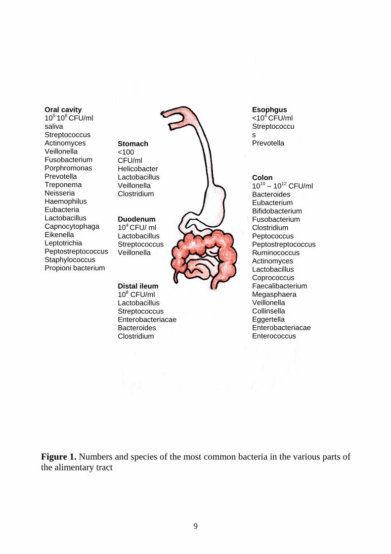
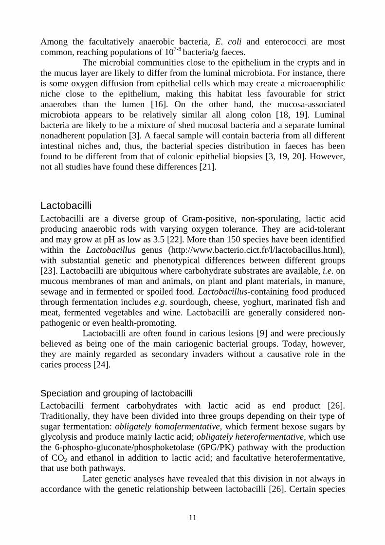
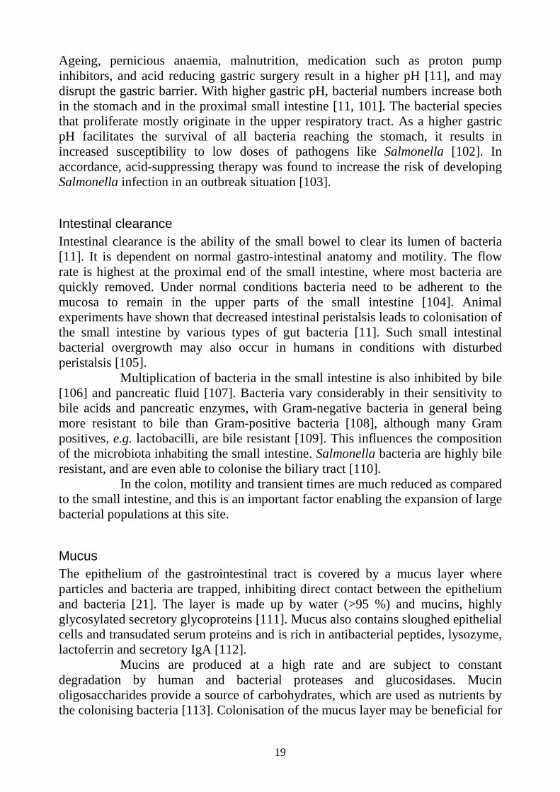
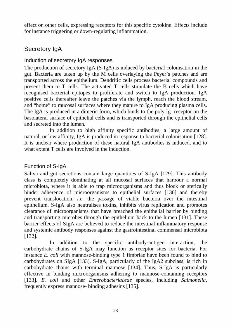
Numbers and species in the various parts of the gastro-intestinal tract The approximate numbers and important groups of bacteria inhabiting the various parts of the gastro-intestinal tract are shown in Figure 1.
9
Figure 1. Numbers and species of the most common bacteria in the various parts of the alimentary tract
Oral cavity 105-108 CFU/ml saliva Streptococcus Actinomyces Veillonella Fusobacterium Porphromonas Prevotella Treponema Neisseria Haemophilus Eubacteria Lactobacillus Capnocytophaga Eikenella Leptotrichia Peptostreptococcus Staphylococcus Propioni bacterium
Stomach <100 CFU/ml Helicobacter Lactobacillus Veillonella Clostridium
Duodenum 104 CFU/ ml Lactobacillus Streptococcus Veillonella
Distal ileum 108 CFU/ml Lactobacillus Streptococcus Enterobacteriacae Bacteroides Clostridium
Colon 1010 – 1012 CFU/ml Bacteroides Eubacterium Bifidobacterium Fusobacterium Clostridium Peptococcus Peptostreptococcus Ruminococcus Actinomyces Lactobacillus Coprococcus Faecalibacterium Megasphaera Veillonella Collinsella Eggertella Enterobacteriacae Enterococcus
Esophgus <104 CFU/ml Streptococcus Prevotella Veillonella
10
The oral cavity More than 700 different bacterial species have been identified in the oral microbiota [5], the majority being anaerobes [6]. It is also clear that a number of species still remain to be identified [7]. Each individual usually harbours 100-200 species [5], the majority of which grow at a particular site, such as the back of the tongue, the hard palate, or the dental surfaces [8]. However, some bacterial groups, e.g. various streptococci, Prevotella and Lactobacillus species grow at most sites and are found in most individuals [5, 9]. The bacterial density varies widely between different oral niches, but the counts in saliva are approximately 105 -108 / ml, and higher in dental plaques [10].
The stomach The stomach harbours only a low number of bacteria due to the harsh conditions, where the low pH kills most bacteria within minutes. More acid-resistant bacteria, e.g. lactobacilli, Veillonella spp. and some clostridia can still survive, and some bacteria may even colonise niches where the pH is higher due to secretion of bicarbonate [11, 12]. In individuals colonised by H. pylori, the bacterial community is very much dominated by this species [13], whereas the same authors found evidence of a larger number of species in individuals who did not harbour H. pylori [13].
The small intestine Moving from the ventricle towards the ileocaecal valve, the number of bacteria and the complexity of the microbiota gradually increase. The small intestine offers an aerobic environment and bacteria like lactobacilli and streptococci are common. Proximally, the bacterial numbers are low, only 102-4 /ml, increasing to 105-8/ml in the distal ileum. Here the oxygen content decreases, and the microbiota also includes Bacteroides, clostridia and other anaerobes, along with facultative anaerobes such as enterococci and E. coli [12, 14].
The colon The total number of bacteria in the colon amounts to 1014, or 1011 / g faeces, which is equivalent to 60 % of the faecal mass [15]. The most common bacterial groups are presented in Figure 1. More than 99 % of the bacteria are strict anaerobes, as they are favoured by the lack of oxygen and low redox potential in this environment [16]. Bacteroides, Bifidobacterium, Clostridium, Eubacterium, Ruminococcus, Coprococcus, Faecalibacterium, Megasphaera, Veillonella, Collinsella, Eggerthella and Fusobacterium are most common [3, 17]. Species belonging to the genus Lactobacillus, which are defined as anaerobic or microaerophilic bacteria, are present in populations up to 106-8 bacteria/g faeces.
11
Among the facultatively anaerobic bacteria, E. coli and enterococci are most common, reaching populations of 107-8 bacteria/g faeces.
The microbial communities close to the epithelium in the crypts and in the mucus layer are likely to differ from the luminal microbiota. For instance, there is some oxygen diffusion from epithelial cells which may create a microaerophilic niche close to the epithelium, making this habitat less favourable for strict anaerobes than the lumen [16]. On the other hand, the mucosa-associated microbiota appears to be relatively similar all along colon [18, 19]. Luminal bacteria are likely to be a mixture of shed mucosal bacteria and a separate luminal nonadherent population [3]. A faecal sample will contain bacteria from all different intestinal niches and, thus, the bacterial species distribution in faeces has been found to be different from that of colonic epithelial biopsies [3, 19, 20]. However, not all studies have found these differences [21].
Lactobacilli Lactobacilli are a diverse group of Gram-positive, non-sporulating, lactic acid producing anaerobic rods with varying oxygen tolerance. They are acid-tolerant and may grow at pH as low as 3.5 [22]. More than 150 species have been identified within the Lactobacillus genus (http://www.bacterio.cict.fr/l/lactobacillus.html), with substantial genetic and phenotypical differences between different groups [23]. Lactobacilli are ubiquitous where carbohydrate substrates are available, i.e. on mucous membranes of man and animals, on plant and plant materials, in manure, sewage and in fermented or spoiled food. Lactobacillus-containing food produced through fermentation includes e.g. sourdough, cheese, yoghurt, marinated fish and meat, fermented vegetables and wine. Lactobacilli are generally considered non-pathogenic or even health-promoting. Lactobacilli are often found in carious lesions [9] and were preciously believed as being one of the main cariogenic bacterial groups. Today, however, they are mainly regarded as secondary invaders without a causative role in the caries process [24].
Speciation and grouping of lactobacilli Lactobacilli ferment carbohydrates with lactic acid as end product [26]. Traditionally, they have been divided into three groups depending on their type of sugar fermentation: obligately homofermentative, which ferment hexose sugars by glycolysis and produce mainly lactic acid; obligately heterofermentative, which use the 6-phospho-gluconate/phosphoketolase (6PG/PK) pathway with the production of CO2 and ethanol in addition to lactic acid; and facultative heterofermentative, that use both pathways.
Later genetic analyses have revealed that this division in not always in accordance with the genetic relationship between lactobacilli [26]. Certain species
12
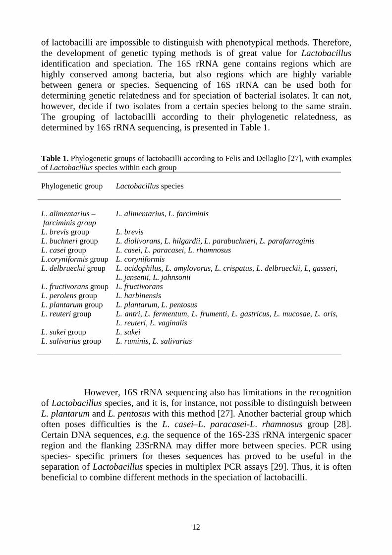
of lactobacilli are impossible to distinguish with phenotypical methods. Therefore, the development of genetic typing methods is of great value for Lactobacillus identification and speciation. The 16S rRNA gene contains regions which are highly conserved among bacteria, but also regions which are highly variable between genera or species. Sequencing of 16S rRNA can be used both for determining genetic relatedness and for speciation of bacterial isolates. It can not, however, decide if two isolates from a certain species belong to the same strain. The grouping of lactobacilli according to their phylogenetic relatedness, as determined by 16S rRNA sequencing, is presented in Table 1. Table 1. Phylogenetic groups of lactobacilli according to Felis and Dellaglio [27], with examples of Lactobacillus species within each group Phylogenetic group Lactobacillus species L. alimentarius – farciminis group
L. alimentarius, L. farciminis
L. brevis group L. brevis L. buchneri group L. diolivorans, L. hilgardii, L. parabuchneri, L. parafarraginis L. casei group L. casei, L. paracasei, L. rhamnosus L.coryniformis group L. coryniformis L. delbrueckii group L. acidophilus, L. amylovorus, L. crispatus, L. delbrueckii, L, gasseri,
L. jensenii, L. johnsonii L. fructivorans group L. fructivorans L. perolens group L. harbinensis L. plantarum group L. plantarum, L. pentosus L. reuteri group L. antri, L. fermentum, L. frumenti, L. gastricus, L. mucosae, L. oris,
L. reuteri, L. vaginalis L. sakei group L. sakei L. salivarius group L. ruminis, L. salivarius
However, 16S rRNA sequencing also has limitations in the recognition of Lactobacillus species, and it is, for instance, not possible to distinguish between L. plantarum and L. pentosus with this method [27]. Another bacterial group which often poses difficulties is the L. casei–L. paracasei-L. rhamnosus group [28]. Certain DNA sequences, e.g. the sequence of the 16S-23S rRNA intergenic spacer region and the flanking 23SrRNA may differ more between species. PCR using species- specific primers for theses sequences has proved to be useful in the separation of Lactobacillus species in multiplex PCR assays [29]. Thus, it is often beneficial to combine different methods in the speciation of lactobacilli.
13
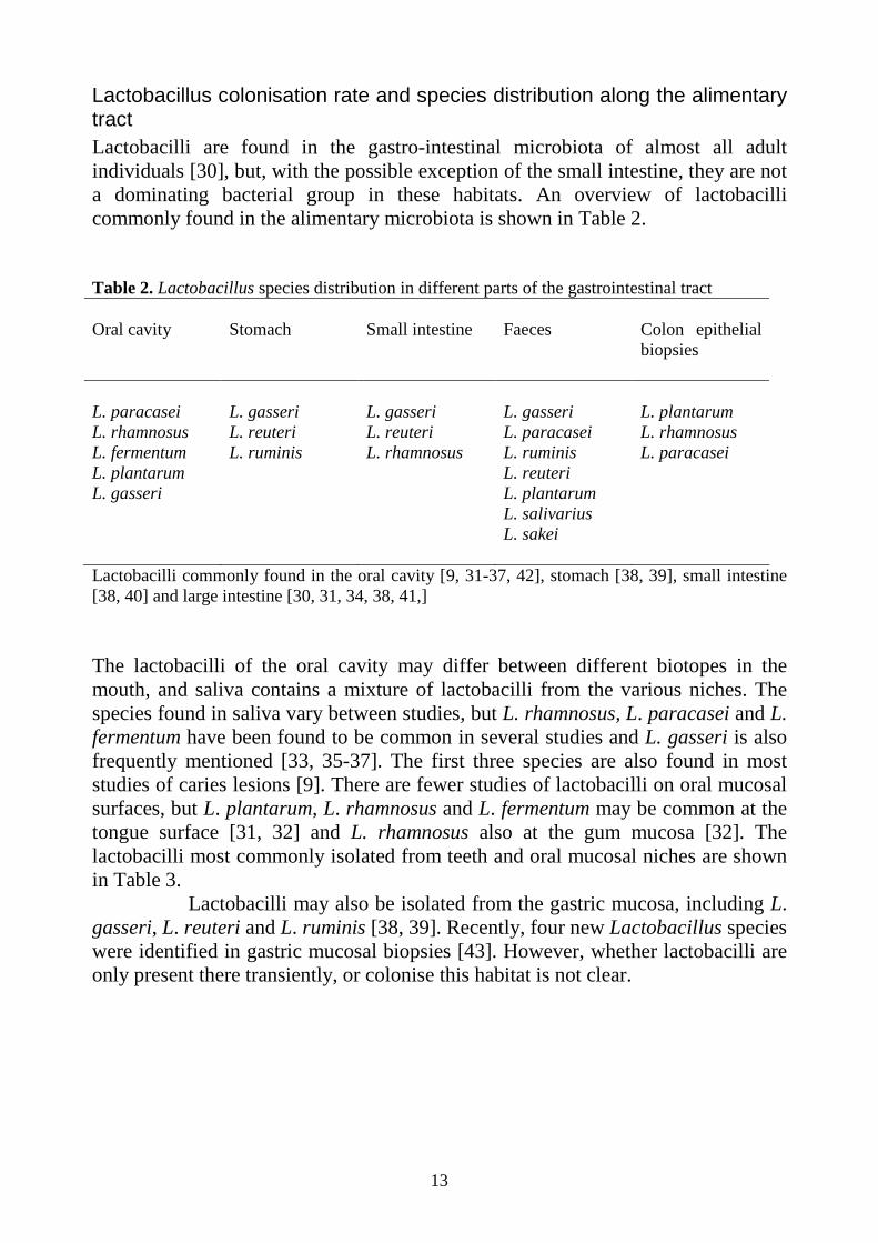
Lactobacillus colonisation rate and species distribution along the alimentary tract Lactobacilli are found in the gastro-intestinal microbiota of almost all adult individuals [30], but, with the possible exception of the small intestine, they are not a dominating bacterial group in these habitats. An overview of lactobacilli commonly found in the alimentary microbiota is shown in Table 2. Table 2. Lactobacillus species distribution in different parts of the gastrointestinal tract Oral cavity Stomach Small intestine Faeces Colon epithelial
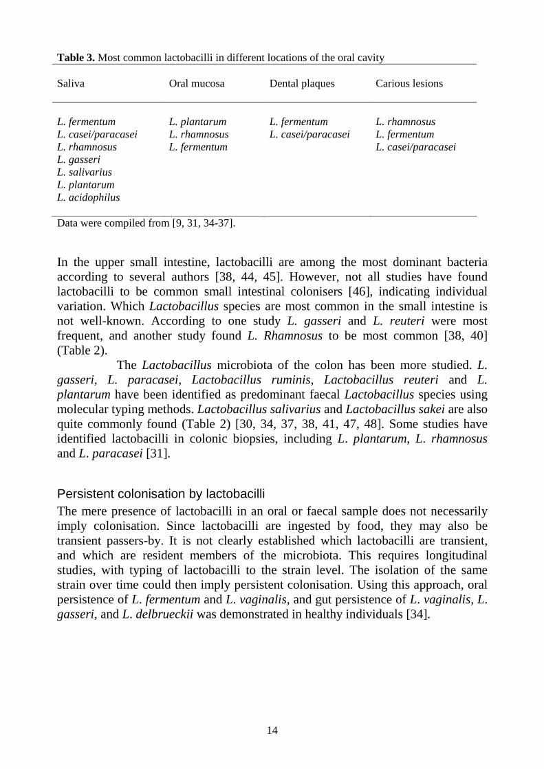
biopsies L. paracasei L. gasseri L. gasseri L. gasseri L. plantarum L. rhamnosus L. reuteri L. reuteri L. paracasei L. rhamnosus L. fermentum L. ruminis L. rhamnosus L. ruminis L. paracasei L. plantarum L. reuteri L. gasseri L. plantarum L. salivarius L. sakei Lactobacilli commonly found in the oral cavity [9, 31-37, 42], stomach [38, 39], small intestine [38, 40] and large intestine [30, 31, 34, 38, 41,] The lactobacilli of the oral cavity may differ between different biotopes in the mouth, and saliva contains a mixture of lactobacilli from the various niches. The species found in saliva vary between studies, but L. rhamnosus, L. paracasei and L. fermentum have been found to be common in several studies and L. gasseri is also frequently mentioned [33, 35-37]. The first three species are also found in most studies of caries lesions [9]. There are fewer studies of lactobacilli on oral mucosal surfaces, but L. plantarum, L. rhamnosus and L. fermentum may be common at the tongue surface [31, 32] and L. rhamnosus also at the gum mucosa [32]. The lactobacilli most commonly isolated from teeth and oral mucosal niches are shown in Table 3.
Lactobacilli may also be isolated from the gastric mucosa, including L. gasseri, L. reuteri and L. ruminis [38, 39]. Recently, four new Lactobacillus species were identified in gastric mucosal biopsies [43]. However, whether lactobacilli are only present there transiently, or colonise this habitat is not clear.
14
Table 3. Most common lactobacilli in different locations of the oral cavity Saliva Oral mucosa Dental plaques Carious lesions L. fermentum L. plantarum L. fermentum L. rhamnosus L. casei/paracasei L. rhamnosus L. casei/paracasei L. fermentum L. rhamnosus L. fermentum L. casei/paracasei L. gasseri L. salivarius L. plantarum L. acidophilus Data were compiled from [9, 31, 34-37]. In the upper small intestine, lactobacilli are among the most dominant bacteria according to several authors [38, 44, 45]. However, not all studies have found lactobacilli to be common small intestinal colonisers [46], indicating individual variation. Which Lactobacillus species are most common in the small intestine is not well-known. According to one study L. gasseri and L. reuteri were most frequent, and another study found L. Rhamnosus to be most common [38, 40] (Table 2).
The Lactobacillus microbiota of the colon has been more studied. L. gasseri, L. paracasei, Lactobacillus ruminis, Lactobacillus reuteri and L. plantarum have been identified as predominant faecal Lactobacillus species using molecular typing methods. Lactobacillus salivarius and Lactobacillus sakei are also quite commonly found (Table 2) [30, 34, 37, 38, 41, 47, 48]. Some studies have identified lactobacilli in colonic biopsies, including L. plantarum, L. rhamnosus and L. paracasei [31].
Persistent colonisation by lactobacilli The mere presence of lactobacilli in an oral or faecal sample does not necessarily imply colonisation. Since lactobacilli are ingested by food, they may also be transient passers-by. It is not clearly established which lactobacilli are transient, and which are resident members of the microbiota. This requires longitudinal studies, with typing of lactobacilli to the strain level. The isolation of the same strain over time could then imply persistent colonisation. Using this approach, oral persistence of L. fermentum and L. vaginalis, and gut persistence of L. vaginalis, L. gasseri, and L. delbrueckii was demonstrated in healthy individuals [34].
15
Lactobacilli in other human habitats Lactobacilli are not only found in the alimentary tract canal, but also in the vagina of fertile women where they dominate the microbiota. The most commonly found vaginal lactobacilli in women of reproductive ages are L. crispatus, L. iners, L. jensenii and L. gasseri [49-51]. Lactobacilli are also isolated from breast milk, e.g. the species L. gasseri, L. rhamnosus, L. plantarum and L. fermentum [51, 52], but the origin of these bacteria is not clear.
Establishment of the microbiota of the alimentary t ract Colonisation of the alimentary tract starts at birth, when the baby leaves the sterile milieu of the uterus, and proceeds over several years.
Establishment of the oral microbiota The oral microbiota is initially simple, but expands with teething, which provides new surfaces for adhering bacteria [53]. Among the earliest colonisers are streptococci, e.g. S. mitis, S. oralis, and S. salivarius, which usually appear in the infant within a few days [54]. Actinomyces species are other early colonisers [55] and also various anaerobes including Bacteroides, Veillonella, Prevotella, and Fusobacterium may be detected in the first two months of life [55, 56]. Colonisation by these and other anaerobes increases steadily over the first years of life. Colonisation by the caries pathogen Streptococcus mutans has previously been found to occur between 19 and 31 months of age [57]. However, more recent studies found S. mutans earlier [58, 59], including in 60 % of six month old predentate infants [58].
Establishment of the gut microbiota The gut is initially colonised by facultatively anaerobic and oxygen-tolerant bacteria, since it is rich in oxygen. In older studies, Enterobacteriaceae, mainly E. coli, and enterococci dominated immediately after birth [60, 61]. However, in recent studies from Western countries, coagulase negative staphylococci, which are typical skin bacteria, have become the earliest colonisers, most likely because of a limited exposure of neonates to traditional faecal bacteria [62, 63]. S. aureus has also become a frequent early coloniser [63, 64].
The facultatively anaerobic bacteria soon consume the oxygen in the gut, and the anaerobic bacterial population starts to expand. Bifidobacteria are the most common anaerobes in the first weeks of life, followed by clostridia and Bacteroides [60, 61, 65, 66]. Most studies describing this early gut colonisation
16
pattern are based on stool cultures. The few and usually small culture-independent studies mostly agree with the results of the culture-based studies [67-69].
Over time, the number of species increases in the gut microbiota, and the dominance of anaerobes becomes more and more pronounced [69]. It takes several years until a full “adult-type” microbiota has developed [70]. Thereafter, it becomes more difficult for new species to colonise the intestine [71], and the species composition of the microbiota remains quite stable over time in healthy adults [72]. An adult is believed to harbour a few hundred different bacterial species in the gut [3] and the anaerobes outnumber the facultatives by a factor of 100 to 1000 [73].
Establishment of the Lactobacillus microbiota Lactobacilli are rarely isolated from the oral cavity of infants in the first few months of life [55, 74], but are found in a majority of children aged two to five years [9, 59].
Regarding the presence of lactobacilli in the early gut microbiota, the results differ between studies. Most studies have reported low Lactobacillus colonisation rates in infants [60, 61, 75-77], and Stark and Lee, who followed infants over time, questioned that lactobacilli were able to form stable populations in the infant gut. Others claim that lactobacilli are present in substantial quantities (107-9 CFU/g faeces) in most infant’s stools [78, 79]. Variations in methodology may, at least to some degree, account for the differences between older studies, since lactobacilli are hard to identify by traditional biochemical methods.
The Lactobacillus species most commonly isolated from infant faeces include L. gasseri [77, 80], L. paracasei and L crispatus [77, 80]. However, there are substantial differences also in the Lactobacillus species distribution between different studies.
Bacterial pathogens causing gastroenteritis Certain bacterial groups colonising the intestines have the capacity to cause gastroenteritis, the most common being diarrheagenic subgroups of E. coli, Campylobacter, Salmonella, and Shigella. Clostridium difficile is another enteric pathogen which may cause disease, especially during or after treatment with antibiotics.
Diarrhoea is the main symptom of gastroenteritis. Other common symptoms include vomiting, fever, nausea and abdominal cramps. Infectious diarrhoea can be divided into inflammatory and non-inflammatory. Inflammatory diarrhoea is characterised by signs of inflammation like blood and mucus in stools and fever. Campylobacter, Salmonella, and Shigella invade the intestinal mucosa and thereby induce acute inflammation. Non-invasive bacteria like
17
enteroaggregative E. coli, enterohemorragic E. coli and C. difficile also cause inflammatory diarrhoea by producing cytotoxins that stimulate the release of inflammatory mediators and damage the mucosa. The secretory diarrhoea caused by Vibrio cholera and enterotoxigenic E. coli is induced by enterotoxins which activate adenylate cyclase and cAMP leading to massive loss of fluid, but little inflammation and little damage to the mucosa [81].
Salmonella
Non-typhoid salmonellosis Salmonella enterica is the type species of Salmonella including more than 2500 serotypes based on variation in O (LPS) and H (flagellar) antigens [82]. Salmonella Typhi and Paratyphi causes severe systemic disease, whereas other serotypes, often labelled as non-typhoid Salmonella, primarily cause intestinal disease. The most common non-typhoid serotypes worldwide include S. Enteritidis, S. Typhimurium and S. Newport [83]. Non-typhoid Salmonella is mainly acquired from contaminated foods. Attack rates are highest in infants, and lowest between 20 and 70 years of age [88]. Estimates of global incidence of non-typhoid Salmonella infection range from 200 million to 1.3 billion cases.
Diarrhoea is the cardinal symptom in non-typhoid Salmonella infection and is often accompanied by symptoms like abdominal cramps, myalgia, headaches, fever, and chills [85]. In some patients, septicaemia and focal infections occur. Most patients recover within a few weeks without treatment, but the acute symptoms may be quite severe and persistent gastro-intestinal disturbances are common [86].
Also, many individuals remain positive for Salmonella in faeces for various lengths of time after a symptomatic infection, median duration of excretion being approximately five weeks [87]. Less than one per cent continue to excrete Salmonella in faeces for more than a year [87], and they are defined as chronic or persistent carriers [88]. It is not clear if Salmonella actually colonises the gut for such prolonged periods, or if bacteria are only seeded to the intestine from other foci of colonisation, e.g. the biliary tract. Very low or very high age, biliary tract abnormalities, schistosomiasis, and diverticulitis are known risk factors for the carrier state [88]. Salmonella is cleared more rapidly in asymptomatic infection [89].
Pathogenesis in non-typhoid salmonellosis Salmonella adhere [90] to epithelial cells and colonise the distal ileum and proximal colon [91]. They use type III secretion systems, a kind of molecular syringe consisting of more than 20 proteins, to inject so called effector proteins into the cells, which enables the invasion of the epithelium, induces fluid secretion and stimulates the production of inflammatory mediators [92]. Whereas S. Typhi is
18
spread systemically within macrophages and neutrophils, non-typhoid Salmonella normally remains in the intestinal tissues, and large numbers of neutrophils are attracted to the small intestinal wall [91,93]. Necrosis may be seen in the superficial mucosa layers in areas of the terminal ileum and colon [92].
Clostridium difficile C. difficile is found in the normal gut microbiota of approximately ten per cent of adult individuals, but normally in low counts only [94, 95]. When permitted to reach high population numbers, e.g. during treatment with antibiotics, it may cause enteritis through its elaboration of toxins. Symptoms of C. difficile infection range from mild diarrhoea to life-threatening pseudomembranous colitis [96], a severe inflammation of the colon with production of fibrous membranes. C. difficile may produce two exotoxins, toxin A and B, which induce mucosal inflammation, fluid secretion, epithelial damage and in some cases necrosis of intestinal epithelial cells [97]. An aggressive Clostridium difficile clone has spread over several countries in the last decades, causing complicated and relapsing infections with a high mortality rate [98].
Defences of the alimentary tract
Barrier functions
Saliva Saliva flushes microorganisms from teeth and oral mucosa and transports bacteria to the stomach through swallowing. The bacterial content of saliva is approximately 105-8 CFU/ml and as we produce 750 to 1500 ml per day, 108-1011 bacteria from the oral cavity reach the acidic environment of the stomach daily. Saliva contains several protective factors and antimicrobial agents, e.g. secretory IgA, lactoferrin and lysozyme [99], which are described below.
The gastric acid barrier The low pH of the stomach kills bacteria very efficiently. Most bacteria can pass only when the pH is higher, e.g. during meals, and reach the lower parts of the gastro-intestinal tract to possibly establish residency [11]. In neonates, gastric pH is relatively high, which might facilitate the establishment of the gut microbiota [100]. Some bacteria, i.e. lactobacilli, Veillonella and clostridia are, however, able to survive in acidic environments [12].
With a normal fasting pH below 3, gastric aspirate contains less than 103-4 CFU/ml, whereas at a pH of 6-7.5 bacterial levels rise to 106-8 CFU/ml [11].
19
Ageing, pernicious anaemia, malnutrition, medication such as proton pump inhibitors, and acid reducing gastric surgery result in a higher pH [11], and may disrupt the gastric barrier. With higher gastric pH, bacterial numbers increase both in the stomach and in the proximal small intestine [11, 101]. The bacterial species that proliferate mostly originate in the upper respiratory tract. As a higher gastric pH facilitates the survival of all bacteria reaching the stomach, it results in increased susceptibility to low doses of pathogens like Salmonella [102]. In accordance, acid-suppressing therapy was found to increase the risk of developing Salmonella infection in an outbreak situation [103].
Intestinal clearance Intestinal clearance is the ability of the small bowel to clear its lumen of bacteria [11]. It is dependent on normal gastro-intestinal anatomy and motility. The flow rate is highest at the proximal end of the small intestine, where most bacteria are quickly removed. Under normal conditions bacteria need to be adherent to the mucosa to remain in the upper parts of the small intestine [104]. Animal experiments have shown that decreased intestinal peristalsis leads to colonisation of the small intestine by various types of gut bacteria [11]. Such small intestinal bacterial overgrowth may also occur in humans in conditions with disturbed peristalsis [105].
Multiplication of bacteria in the small intestine is also inhibited by bile [106] and pancreatic fluid [107]. Bacteria vary considerably in their sensitivity to bile acids and pancreatic enzymes, with Gram-negative bacteria in general being more resistant to bile than Gram-positive bacteria [108], although many Gram positives, e.g. lactobacilli, are bile resistant [109]. This influences the composition of the microbiota inhabiting the small intestine. Salmonella bacteria are highly bile resistant, and are even able to colonise the biliary tract [110].
In the colon, motility and transient times are much reduced as compared to the small intestine, and this is an important factor enabling the expansion of large bacterial populations at this site.
Mucus The epithelium of the gastrointestinal tract is covered by a mucus layer where particles and bacteria are trapped, inhibiting direct contact between the epithelium and bacteria [21]. The layer is made up by water (>95 %) and mucins, highly glycosylated secretory glycoproteins [111]. Mucus also contains sloughed epithelial cells and transudated serum proteins and is rich in antibacterial peptides, lysozyme, lactoferrin and secretory IgA [112]. Mucins are produced at a high rate and are subject to constant degradation by human and bacterial proteases and glucosidases. Mucin oligosaccharides provide a source of carbohydrates, which are used as nutrients by the colonising bacteria [113]. Colonisation of the mucus layer may be beneficial for
20
the bacteria, as it protects them from being swept away by peristalsis [114]. The mucus layer provides binding sites for various bacteria, which is likely to facilitate their colonisation of this habitat [115]. Lactobacilli are among the bacteria which are likely to inhabit the mucus layer. This has been studied mainly in vitro [116, 117], but also in vivo [118].
The epithelium Underneath the mucus layer, the epithelium covers the gastrointestinal canal from the mouth to the rectum. The continuous shedding of epithelial cells limits microbial colonisation of the mucosa. The area of the mucosa of the intestinal tract is 200 to 300 m2 [119]. This large area is created by the organisation of the mucosa into villi and crypts in the small intestine, and crypts in the large intestine. Villi are about one mm long projections into the lumen, which increase the absorptive surface. In the large intestine there are no villi, but the crypts are generally deeper. The intestinal epithelial barrier is made up of a single columnar epithelium where the cells are tightly linked to each other with junctional complexes, providing a physical barrier that prevents bacteria from invading the body. There are several different types of epithelial cells in the small intestine: crypt cells, absorptive enterocytes, enteroendocrine cells, goblet cells and Paneth cells. Paneth cells are located at the base of the crypts from the duodenum to the ileum. Apically they are filled with granules which contain antibacterial substances produced within the cells [120]. In the large intestine, the epithelium consists of columnar epithelial cells, goblet cells, crypt cells and endocrine cells.
Antimicrobial compounds Epithelial cells and Paneth cells are able to produce a large number of antibacterial compounds. Among these are antimicrobial peptides like defensins and cathelicidins with broad spectrum antimicrobial effect. Their mechanism of action is the formation of pores in bacterial membranes, resulting in bacteriolysis [122]. Other examples of antimicrobial compounds are the bacteriolytic enzymes lysozyme and group IIa phospholipase A2 (PLA2) [123], and lactoferrin which deprives microbes of nutrients through iron sequestration and induces cell lysis [124].
The immune system of the alimentary tract
Architecture The alimentary tract is defended by organised lymphoid tissue, and a large number of immune cells scattered in the submucosal niches.
21
Around the oral cavity organised lymphoid tissues make up a ring consisting of the palatine tonsils, the lingual tonsils and the adenoid tissue in the roof of the nasopharynx. These tissues are rich in T and B lymphocytes, macrophages and dendritic cells, and are inductive sites for immune responses in the oral cavity. In the oral cavity, there are also many plasma cells, located in the salivary glands, producing dimeric IgA that is converted to secretory IgA during passage through the duct epithelium.
Organised lymphoid follicles are present along the entire small intestine, but become more abundant in the distal ileum. They are also frequent in the colon, especially in the caecum and the rectum. In the distal ileum they are grouped in large patches, the so called Peyer’s patches, with B cells in the centre surrounded by T-lymphocytes. The Peyer’s patches also contain macrophages and dendritic cells and are the primary sites for induction of immune responses towards bacteria that colonise or pass the gut. The epithelium overlaying lymphoid follicles differs from the villus epithelium. Instead of goblet cells and enteroendocrine cells it contains epithelial M cells which actively transport antigens and whole microbes across the epithelium.
The intestinal epithelium contains a large number of intraepithelial lymphocytes. Underneath, there is a thin layer of loose cell-rich connective tissue, the lamina propria containing fibroblasts and immune cells, including large numbers of activated T helper (TH) lymphocytes, B cells, plasma cells, macrophages, and dendritic cells.
Innate defence Bacteria that cross the epithelial barriers in the gastrointestinal tract will encounter the innate immune system which consists of cells and soluble proteins e.g. factors of the complement system. The innate immune system is activated within minutes and bacteria are eliminated by phagocytosis and complement activation, a response which produces inflammation.
Cells in the innate system, including e.g. macrophages, dendritic cells, mast cells, NK (natural killer) cells, monocytes and granulocytes are able to recognize microbial structures. Epithelial cells may also recognise and take part in the clearance of invading bacteria. Both commensal bacteria and pathogens are recognised by means of conserved structures that are specific for prokaryotes, e.g. LPS, peptidoglycan, lipoteichoic acid and flagellin [125]. These structures are defined as pathogen-associated molecular patterns, (PAMPs) and are recognised by specific receptors called pattern recognizing receptors (PRRs) [126] which are present on cells of the innate immune system and to some degree on epithelial cells. Receptor activation results in intracellular signalling via different pathways, and to expression of various genes, e.g. genes for production of inflammatory mediators and chemotactic compounds. The most well-known and well-characterised PRRs are the toll-like receptors expressed on the surface of e.g. macrophages, dendritic
22
cells and epithelial cells [127]. The intracellular NOD receptors are another important group.
The acquired immune system When bacteria are taken up by M cells overlying lymphoid follicles, or cross the epithelial layer, the acquired immune system is also activated. This is a slower process, as lymphocytes produced in the bone marrow and thymus need time to mature into antibody producing plasma cells and effector T lymphocytes.
The cells of the acquired immune system are very specialised and can recognise an enormous array of structures. Each T- and B-lymphocyte has a unique receptor in its membrane, specific for one single structure, the antigen. T-lymphocytes recognise their specific antigen when presented by antigen-presenting cells (APCs), most commonly dendritic cells or macrophages. The T-lymphocytes are divided depending on which presentation-molecule, the so called MHC-molecule, they prefer. CD4+ T cells recognise their specific antigen presented on MHC II molecules, whereas the cytotoxic CD8+ T-cells recognise their antigen on MHC I molecules. CD4+ T cells are further divided into T helper (TH) cells or regulatory T cells.
The antigen-presenting cells are present in all tissues where they take up antigen or entire pathogens by endocytosis or phagocytosis. After degradation, pieces of antigens are presented on the MHC molecule to T-cells. This may occur in the Peyer’s patches, where both APCs and T cells are present, or, if the APC has encountered bacteria in the lamina propria, they may migrate to the nearest lymph nodes, to present antigens to T cells. The CD4+T cell recognises its specific antigen through the T-cell receptor and starts to divide and mature into TH1, TH2 or TH17 cells. TH1 cells activate macrophages, promote cytotoxic T lymphocyte activity, and mediate inflammation through the production of cytokines. TH2 cells are involved in the stimulation of antibody responses. TH17 cells are found mainly in the skin and intestinal epithelium and recruit neutrophils and induce a strong inflammatory response upon bacterial stimulation.
B cells recognise their antigens directly, with their surface bound antibodies. They can then proliferate and differentiate into plasma-cells, a process which usually requires activation by T-helper cells. B cells express MHC II and are able to present antigens to CD4+ T cells. First, the antigen is presented by an APC to a T cell recognising the antigen. Then the T cell proliferates and differentiates into cytokine producing TH2 cells. The TH2 cell binds to the B cell presenting the same antigen as the APC did, and with the secretion of additional cytokines, the B cell differentiates into an antibody-producing plasma cell. Most naive B cells are originally expressing antibodies of the IgD or IgM class on their surfaces. Depending on the stimuli provided by the T cell, they may switch their production to another immunoglobulin class or subclass, i.e. IgG1, 2, 3 or 4, IgA1 or 2, or IgE.
Both the innate and the acquired immune system are regulated by cytokines. A cytokine is a soluble protein or glycoprotein released by cells, with an
23
effect on other cells, expressing receptors for this specific cytokine. Effects include for instance triggering or down-regulating inflammation.
Secretory IgA
Induction of secretory IgA responses The production of secretory IgA (S-IgA) is induced by bacterial colonisation in the gut. Bacteria are taken up by the M cells overlaying the Peyer’s patches and are transported across the epithelium. Dendritic cells process bacterial compounds and present them to T cells. The activated T cells stimulate the B cells which have recognised bacterial epitopes to proliferate and switch to IgA production. IgA positive cells thereafter leave the patches via the lymph, reach the blood stream, and “home” to mucosal surfaces where they mature to IgA producing plasma cells. The IgA is produced in a dimeric form, which binds to the poly Ig- receptor on the basolateral surface of epithelial cells and is transported through the epithelial cells and secreted into the lumen.
In addition to high affinity specific antibodies, a large amount of natural, or low affinity, IgA is produced in response to bacterial colonisation [128]. It is unclear where production of these natural IgA antibodies is induced, and to what extent T cells are involved in the induction.
Function of S-IgA Saliva and gut secretions contain large quantities of S-IgA [129]. This antibody class is completely dominating at all mucosal surfaces that harbour a normal microbiota, where it is able to trap microorganisms and thus block or sterically hinder adherence of microorganisms to epithelial surfaces [130] and thereby prevent translocation, i.e. the passage of viable bacteria over the intestinal epithelium. S-IgA also neutralises toxins, inhibits virus replication and promotes clearance of microorganisms that have breached the epithelial barrier by binding and transporting microbes through the epithelium back to the lumen [131]. These barrier effects of SIgA are believed to reduce the intestinal inflammatory response and systemic antibody responses against the gastrointestinal commensal microbiota [132].
In addition to the specific antibody-antigen interaction, the carbohydrate chains of S-IgA may function as receptor sites for bacteria. For instance E. coli with mannose-binding type 1 fimbriae have been found to bind to carbohydrates on SIgA [133]. S-IgA, particularly of the IgA2 subclass, is rich in carbohydrate chains with terminal mannose [134]. Thus, S-IgA is particularly effective in binding microorganisms adhering to mannose-containing receptors [133]. E. coli and other Enterobacteriaceae species, including Salmonella, frequently express mannose- binding adhesins [135].
24
The influence of SIgA is probably most profound in the small intestine as the rapid peristalsis may remove bacteria which are not able to adhere to the mucosa. For instance, segmented filamentous bacteria, anaerobes closely related to clostridia, expand in the small intestine of mice in the absence, but not in the presence, of secretory IgA [136]. In the colon, however, S-IgA does not seem to prevent colonisation. E. coli colonise the gut regardless of the presence of specific S-IgA antibodies towards E. coli [137]. Indeed, a large share of faecal bacteria are coated with S-IgA under normal conditions [138]. However, it is likely that the presence of S-IgA may influence population numbers of certain bacteria in the large intestine. For instance, as S-IgA may prevent bacterial access to the epithelial surface, bacteria that prefer this specific niche could be disfavoured.
IgA deficiency IgA deficiency is a lack of both IgA1 and IgA2 in serum and secretions and the most common primary immune deficiency. The prevalence of IgA deficiency is approximately one in 700 in Caucasian populations [139]. The background is not fully elucidated, but involves a failure of B lymphocytes to switch to IgA production [140]. The majority of IgA deficient individuals are healthy and may be discovered at e.g. blood donor screenings. Approximately one third have recurrent respiratory tract infections. Several in vitro studies have demonstrated that B cells from IgA deficient individuals become able to produce IgA when stimulated through CD40 together with IL-10 or IL-4 + IL-10 or [141, 142], especially B cells from healthy IgA deficient individuals [143]. Thus, defects in cytokine production may be involved in the pathogenesis of IgA deficiency.
Why some IgA deficient individuals suffer from recurrent infections could relate to differences in the ability to compensate for the absence of S-IgA, for instance by S-IgM production [144], but other studies show that healthy individuals with IgA deficiency do not have higher S-IgM concentrations at mucosal sites than patients with many infections [145]. Yet another hypothesis is that they compensate by production of antibacterial peptides, however, no effect of antimicrobial peptides on the expanding bacteria was found in small intestinal expansion of anaerobes due to lack of IgA [146].
IgA deficiency is associated with increased risk of certain autoimmune disorders [147], celiac disease and perhaps inflammatory bowel disease [148]. The latter condition could possibly relate to an increased inflammatory response towards gut bacteria in the absence of IgA.
Few studies have investigated the gastrointestinal microbiota of individuals with IgA deficiency. One study found increased counts of Actinomyces spp. in the oral cavity of IgA deficient individuals [149]. The same authors found increased counts of anaerobic bacteria and enterococci in faecal stool samples from IgA deficient as compared to healthy individuals [149].
25
Salmonella and the immune system After invasion of the mucosa, non-typhoid Salmonella induces a massive inflammatory response, with production of inflammatory cytokines such as TNF, IL-1, IL-6, IL-12 and IL-18, and chemokines that recruit monocytes, macrophages, neutrophils and cells of the adaptive immune system to the site of infection [150].
Most studies of Salmonella pathogenesis and immune response are performed in vitro or in animals. The importance of S-IgA in Salmonella infections in humans is not clarified, but IgA deficient individuals do not seem to be at risk for more severe disease than individuals with normal IgA levels. Instead, individuals with deficiencies involving IFN-γ or IL-12 production have increased susceptibility to Salmonella infection [151], pointing towards the importance of cell-mediated immunity and macrophage activation.
Influence of gender on the immune system Women seem to exert stronger cellular as well as humoral immune responses than men. For example, serum immunoglobulin levels and responses to a variety of antigens are higher in women [152]. Gender related differences have been observed in several infectious conditions, including parasitic infections, trauma-related bacterial sepsis and virus infections [153, 154].
Factors of importance for the composition of the microbiota of the alimentary tract
Host factors
A number of host factors, including the various mechanical and chemical barriers towards colonisation described above are important for if and where different bacteria are able to establish. Some additional host factors which possibly influence the microbiota are described below.
Hereditary factors The predominant species of the gut microbiota are very stable over time in healthy adults and appear to be host-specific [155]. The highest levels of similarity is found in monozygotic twins [156], while the microbiota of the individuals of a married couple living together is not more similar than the microbiota of unrelated individuals [155]. Knowledge of how this genetic regulation is carried out is still scarce, but mechanisms could include e.g. genetic variations in carbohydrate-structures expressed in the mucosa, which could influence both adherence of
26
bacteria and the availability of nutrients. However, the high level of similarity between monozygotic twins could also indicate that the environmental conditions present when acquiring the microbiota in the first place have great influence on the final composition of the microbiota. Various physiological and anatomical factors which are genetically determined could play a role for the composition of the oral microbiota. For instance tooth eruption, tooth fissure shape, and interdental space and amount of saliva play a role in deciding which bacteria will be favoured by their preferred niche in the oral cavity [157]. Also variations in host derived nutrients for bacteria available in e.g. saliva and gingival crevicular fluid are likely to be important [158].
Gender Composition of the microbiota may to some extent be gender-dependent. Phylogenetic profiles of mice have been found to cluster together according to gender [159]. One group found that bacteria belonging to the Bacteroides-Prevotella group were more numerous in the male gut, without further distinctions [160] and another group that three Clostridium species, one species from the Bacteroides group and two from the phylum Proteobacteria are more common in males [161].
Environmental and lifestyle factors influencing the oral microbiota The composition of the oral microbiota is influenced by several factors which are only briefly mentioned here. Lifestyle factors influencing the microbiota include e.g. diet, smoking, oral hygiene, and the presence of foreign materials [158]. Environmental factors, like variation in the level of exposure to different bacteria are also likely to influence the microbiota. In infants, acquisition of S. mutans was associated with habits that allowed saliva transfer from parents to infants [58].
Environmental and lifestyle factors of importance for the establishment and composition of the gut microbiota The establishment of the gut microbiota early in life is influenced by several environmental and life style factors, including delivery mode, feeding pattern, and levels of bacterial exposure [162].
The origin of the bacteria colonising neonates may be the maternal faecal and vaginal microbiota, but also various environmental sources [62, 163]. Infants delivered by caesarean section show delayed acquisition of for instance E. coli and Bacteroides, and to some degree of bifidobacteria, indicating the importance of the maternal faecal microbiota as a source of these bacteria [62, 163]. Lactobacilli may sometimes be acquired from the maternal vaginal
27
microbiota during delivery, and some authors report somewhat delayed colonisation in sectio delivered infants [163, 164]. Clostridia and enterobacteria other than E. coli, e.g. Klebsiella and Enterobacter species are found equally early, or even earlier, in sectio delivered as compared to vaginally delivered infants [62] [63], indicating that these bacteria are easily acquired from the environment. By one year of age, sectio-delivered infants in Western countries have a lower ratio of anaerobic to facultatively anaerobic bacteria in the gut than vaginally delivered infants, possibly indicating a less “mature” microbiota [63]
Close contact with other individuals facilitates acquisition of bacteria. Infants with siblings have a higher ratio of anaerobic to aerobic bacteria by one year of age, which possibly indicates a more adult-like microbiota [63]. It is not clear if an early childhood with animals influence the colonisation pattern.
Breast milk may be a source of bacteria colonising infants. Staphylococcus, Streptococcus, Bifidobacterium and Lactobacillus are frequently isolated from breast milk [165]. Other foods given to neonates may also contain bacteria that colonise the infantile gut [166].
Other, less well defined, lifestyle factors also play a role. In a study of the microbiota of children aged five to 13 years from three European countries there were no differences between countries, but children attending anthroposophic schools had a more diverse dominant faecal microbiota than controls as well as farm children [167].
Influence of environmental bacterial exposure on the gut microbiota It is clear that the level of microbial exposure influences the gut colonisation pattern. In neonatal intensive care units where great efforts are taken to reduce bacterial spread, infants acquire a microbiota where coagulase-negative staphylococci, enterococci and Enterobacteriaceae dominate, while anaerobes are almost absent [168, 169] Great differences are also observed when comparing the early microbiota between infants from different parts of the world, with different levels of environmental bacterial exposure. Infants in developing countries have a more rapid turnover of bacteria and a larger number of species in their early microbiota than infants in Western countries [166]. Also, in developing countries, infants delivered by caesarean section may be as rapidly colonised by E. coli, Bacteroides, and bifidobacteria as vaginally delivered infants, indicating pronounced spread of faecal bacteria in the hospital milieu [171]. Lactobacilli have been found to be more common in Ethiopian (two to six weeks of age) and also in Estonian infants (at one year) compared to Swedish infants of similar ages [172, 173].
It is not known if the level of environmental microbial exposure also influences the composition and complexity of the more stable adult gut microbiota.
28
Influence of diet on the gut microbiota In young infants, the gut microbiota differs in several aspects between breastfed and bottlefed infants. Breastfed infants harbour less clostridia [66, 174], but tend to have more staphylococci [75]. Furthermore, Bacteroides, enterococci and Enterobacteriaceae, especially Klebsiella and Enterobacter, tend to be more common in bottle-fed infants [171, 175]. A majority of studies find no differences in Lactobacillus colonisation between breast and bottle-fed infants [79, 162, 176]. Also, Bifidobacteria are equally common in breast- and bottle-fed infants in most recent studies [177].
In adults, the gut microbiota differs between individuals in different geographical regions consuming different types of diets [178], but this may also relate to other factors differing between the populations.
Roseburia, Eubacterium and Bifidobacterium have been found to decrease as a result of a diet low in carbohydrate [179]. Vegetarians harbour less clostridia and Faecalibacterium than omnivores [180, 181].
Diet lipid content may also influence the gut microbiota, as fat stimulates bile flow, by which expansion of e.g. Bacteroides may be stimulated [182]. In a study of mice, dietary iron deprivation resulted in elevation of anaerobes including lactobacilli [183].
Bacterial factors of importance for establishment and composition of the gastrointestinal microbiota
Ability to utilise available nutrients To establish in the gastrointestinal tract, bacteria must multiplicate at a rate exceeding their rate of elimination. Bacteria therefore compete for available nutrients which include both dietary and host derived components. Apart from indigestible carbohydrates and low levels of non-absorbed protein, not much of the food ingested by the host reaches the colon. However there is a broad range of other nutrient sources in this habitat, e.g. mucus and exfoliated epithelial cells.
Some bacteria are able to utilise a variety of different substrates, whereas others are much more specialised [184]. Many gut bacteria ferment indigestible carbohydrates into short chain fatty acids, which is discussed later.
Adhesins Bacteria colonising the gastro-intestinal tract commonly express structures called adhesins mediating adherence to host cell receptors or mucus structures [135]. Most bacterial adhesins are proteinacious structures that recognise defined carbohydrate sequences in host tissues: glycoproteins, glycolipids or less often a defined protein
29
structure. Adhesins of Gram negative bacteria are often found on fimbriae, rigid protein rods reaching out from the bacteria, whereas adhesins of Gram-positive bacteria most commonly form part of the cell wall or of the cell coat [185]. The same bacteria may have more than one adhesin, and the expression of adhesins is commonly subject to phase variation and may be turned on and off by the bacteria depending on environmental stimuli, which has been shown for S layer protein adhesin in L. acidophilus and L. brevis [186].
Adherence is an important step in colonisation of mucosal sites. This has been shown most clearly for pathogenic bacteria, for instance enterotoxigenic E. coli, which colonise the small intestine of man and animals [187]. Salmonella adheres to enterocytes and M cells as a first step in the pathogenesis [90]. In the oral cavity and the small intestine, adherence is likely to be extra important for colonisation because of the salivary flow and peristalsis, respectively, which otherwise wash bacteria away. In the oral cavity, bacteria form complex biofilms, and in this process, they commonly also coaggregate, i.e. adhere to each other [158].
In the colon, the flow of gut contents and transient time are much lower, which could indicate that adherence is less important for colonisation of this habitat. However, adherence may promote colonisation of the epithelium or mucus layer, and provide advantages by increasing access to nutrients squamated from the tissues or present in mucus [188].
For E. coli colonising the large intestine, the possession of P-fimbriae and type-1-fimbriae mediating adherence to galactose- and mannose-containing receptors, respectively, on colonic epithelial cells, seem to promote long-term persistence in the gut [189, 190]. E. coli with mannose-binding type 1 fimbriae are also able to bind to mannose-containing oligosaccharides on S-IgA [133, 191]. E. coli retrieved from the commensal microbiota of IgA deficient individuals express less mannose-binding adhesins than E. coli from individuals with normal levels of IgA [191, 192]. Possibly, binding of E. coli to S-IgA, which is present in mucus, facilitates colonisation of the mucus layer, which may be the preferred niche for E. coli in the large intestine.
Adherence of lactobacilli Lactobacilli have been shown to adhere to epithelial cells, mucus, and extracellular matrices. Several structures have been identified as target substances or receptors for lactobacilli, e.g. collagen, fibronectin, laminin, lectins, and oligosaccharide-chains of glycoproteins [193-195].
Most studies of Lactobacillus adhesion to epithelial cells have been performed using cell lines, but there are also studies of biopsy samples [196, 197]. A number of surface layer proteins of lactobacilli have been reported to mediate adhesion to intestinal epithelial cells [198, 199] but few specific receptors for lactobacilli on epithelial cells or in mucus have been identified. However, several Lactobacillus species are able to express adhesins mediating adherence to
30
mannose-containing receptors on colonic epithelial cells [31, 196]. Such mannose-specific (MS) adhesins have been demonstrated in L. plantarum, L. salivarius, L. johnsonii, L. paracasei and L. fermentum, all members of the oral and/or faecal microbiota [31, 196, 200-202]. At least in L. plantarum, the expression of these adhesins is enhanced by oxygen, since the mannose-specific adherence of lactobacilli to colonic epithelial cells is reduced after culture of bacteria under anaerobic conditions [196]. In L. plantarum, the adhesin was identified as a proteinaceous structure [196], more specifically as a multi-domain cell surface protein [201].
Adhesion to mucus structures by lactobacilli expressing MS adhesins have been described [201]. Also, in addition to the proteinaceous MS adhesin of L. plantarum [201], other mucus-binding Lactobacillus proteins have been identified: the extracellular mucus-binding protein (mub) of L. reuteri [203], and the mub of L. acidophilus [204].
Oral adhesion of lactobacilli is less well studied than adhesion to intestinal structures. Epithelial cells and teeth are covered with saliva, and several studies show Lactobacillus adherence to salivary structures [205-207]. Adherence to buccal epithelial cells of L. rhamnosus has also been found [205].
Adherence of Salmonella As mentioned above, adherence to host cells is an important step in the induction of infection by mucosal pathogens, and may even be a prerequisite [90]. Several types of fimbriae have been found in Salmonella, including mannose-binding type 1 fimbriae, plasmid-encoded fimbriae, long polar fimbriae and thin aggregative fimbriae (curli) [208]. Variable expression of the many adhesin structures may enable adherence to different cell types and the colonisation of different hosts [209].
Bacteriocins and other antagonistic compounds produced by bacteria Antibacterial substances are not only produced by the host but also by the bacteria themselves, and antimicrobial activity is thought to be an important way for members of the normal microbiota to competitively exclude or inhibit newcomers.
Most bacteria can make one or more antibacterial peptides, i.e. bacteriocins, with the function to suppress competing bacteria of the same or different species [210]. The best characterised bacteriocins are those from lactic acid bacteria, including lactobacilli. They are most often active towards closely related Gram-positive bacteria, while the producer cells are immune to their own bacteriocins [211]. However, activity against Gram-negatives, e.g. Salmonella, by Lactobacillus bacteriocins or bacteriocin-like substances has also been described [212]. Like for human antimicrobial peptides, the most common mode of action is the formation of pores in the bacterial membrane, but they can also act by
31
prevention of cell-wall synthesis, inhibition of RNA synthesis and inhibition of bacterial phospholipase [213]. Other antagonistic compounds produced by bacteria are also known. Lactobacilli produce a number of other substances with non-specific antimicrobial activity, e.g. SCFA, lactic acid, formic acid, and hydrogen peroxide [211]. For instance, the SCFA could possibly lower colonic pH, which may then limit the growth of certain bacteria [214]. However, little is actually known about the possible role of these compounds in the interaction between bacteria and for the composition of the microbiota in the gastro-intestinal tract.
Colonisation resistance A full microbiota greatly hampers the implantation of new bacteria into the ecosystem, and, thus, protects its host from the colonisation by pathogens. This is called colonisation resistance [215], and is believed to be the result of competition for binding sites and nutrients, and from the production of bacteriocins and other substances harmful for the competitors. The same forces are likely to strictly regulate the population size of each bacterial strain in the microbiota.
Colonisation resistance is indeed an important defence mechanism against colonisation and proliferation of pathogenic or potentially pathogenic bacteria. For example, it has been shown that a dose of only ten Salmonella bacteria causes lethal infection in germfree animals, whereas animals with a normal gut microbiota can stand infection with up to 5 x 10⁶ bacteria before lethal infection occurs [216]. Also, the colonisation resistance provided by the microbiota keeps down the population counts of the potential pathogen C. difficile in the gut. Other potential pathogens which are kept at relatively low numbers in the microbiota include e.g. E. coli and other enterobacteria.
Effects on the host of the gut microbiota
Gut maturation Establishment of the gut microbiota contributes to the maturation of the intestines with thickening of the mucosa and deepening of crypts. The villi become broader and shorter and the mass of the small intestine increases [217]. The regulation of intestinal epithelial cell turnover and mucus biosynthesis increases as new genes are turned on [218]. Increased peristalsis reduces bacterial counts in the small intestine [219]. Germ-free animals have fewer goblet cells than conventional animals [114]. The intestinal microbiota may also contribute to the development of the capillary network in the small intestinal villi [220].
32
Maturation of the immune system Both the innate and the adaptive immune systems need stimulation from bacteria to develop and function optimally. The commensal microbiota induces mediators of the innate defence. For example, Paneth cells in small intestinal crypts are stimulated through colonisation to secrete antimicrobial compounds. Macrophage chemotaxis and phagocytic activity are reduced in germ-free as compared to conventional animals [221]. There are even greater differences between germfree and conventional animals in the acquired immune system. The gut associated lymphoid tissue is poorly developed in germ free animals. There are fewer lymphocytes in the lamina propria and in the epithelium, and intestinal lymphoid aggregates, such as the Peyer’s patches and mesenteric lymph nodes, are smaller [222]. Spleen and thymus are underdeveloped as compared to conventionally raised animals [223]. Also, concentrations of serum immunoglobulins are much lower in germfree than in conventional animals, and germ-free mice have very low levels of S-IgA at mucosal sites [223].
Energy and nutrients for the host The gut microbiota has an important function in the processing of nutrients [25]. The presence of bacteria alters the metabolic apparatus of host cells, resulting in more efficient uptake and utilisation of nutrients [223, 224]. It contributes to the regulation of host fat storage, and increases the capacity to extract energy from the diet [225]. Bacteria are also able synthesise several vitamins, e.g. vitamin K which is taken up and utilised by the host [25].
Gut bacteria enable metabolism of otherwise indigestible dietary carbohydrates to host-absorbable compounds and thus contribute to energy production [25]. The major end products of the fermentation of indigestible carbohydrates by anaerobes in the colon are the short-chain fatty acids (SCFAs) acetate, propionate and butyrate [214].
Involvement of the gut microbiota in health and disease The gut microbiota influences our wellbeing in several ways. For instance, the SCFA butyrate produced by gut bacteria, serves as energy for epithelial cells and may have a protective role against colon cancer and ulcerous colitis [226, 227], and propionate enhances colonic muscular contraction contributing to relief of constipation [228]. Stimulation of the immune system by the commensal microbiota may be beneficial for health. Such stimulation possibly contributes to the maturation of immunoregulatory mechanisms. It has been shown that so called regulatory T cells, which are important for the prevention of immune reactivity towards autoantigens

33
and harmless environmental antigens, e.g. allergens, have impaired function in germfree animals [229].
There is increasing evidence that the composition of the gastro-intestinal microbiota could have implications for the development of a number of diseases, including allergy [230], inflammatory bowel disease [231], colon cancer colitis [226], and obesity [232].
Disturbances of the gut microbiota
Disturbances induced by antibiotics Treatment with antibiotics is a common cause of disturbances of the normal gastro-intestinal microbiota. Different agents have different effects, depending on antibiotic spectrum, dose, route of administration, pharmacokinetics and pharmacodynamics. In neonates and young infants, antibiotic treatment often has profound effect on the microbiota, and most anaerobic bacteria are strongly suppressed.
Suppression or elimination of large bacterial populations in the microbiota with loss of colonisation resistance may lead to the expansion and colonisation of potential pathogens in the gut microbiota, like Clostridium difficile, S. aureus, C. perfringens, and various Enterobacteriaceae or Candida species [233, 234]. Antibiotic-induced disturbances in the microbiota treatment also increases the risk of acquiring gastrointestinal pathogens. For instance, prior antibiotic treatment increases the risk of acquiring Salmonella [235].
Antibiotic associated diarrhoea Antibiotic associated diarrhoea (AAD) is common during and after the administration of antibiotics. The incidence varies between five and 35 % in different studies [236]. Common risk factors include high age, hospitalisation and concomitant disease [236]. Clindamycin, extended-spectrum penicillins, cephalosporins and possibly fluoroquinolones are associated with a higher risk [236-238].
Toxin-producing C. difficile is reported to be the cause of 10 - 25% of AAD cases in most studies [239], but higher figures have been found [240]. Possibly, other pathogens like S. aureus, C. perfringens, various Enterobacteriaceae or Candida species may sometimes be responsible [233, 234]. The remainder of episodes of AAD may be due to several factors, including increased gastro-intestinal motility [233], osmotic diarrhoea secondary to decreased digestion and absorption of carbohydrates [233], and reduction of short chain fatty acids in faeces [241].
34
Clostridium difficile enteritis is treated with metronidazole or vancomycin, if discontinuation of antibiotics is not enough, but relapses are common. For other types of AAD there is no effective treatment available.
Disturbances in the microbiota induced by infectious gastroenteritis Bacterial gastroenteritis may involve changes in the normal gut microbiota. There are several contributing mechanisms. Increased intestinal motility and fluid secretion change the intestinal environment and increase the oxygen content. Resident or introduced aerobic or facultatively anaerobic bacteria can expand, whereas several anaerobes decrease in numbers [242, 243]. In a recent study of children in India, significantly lower levels of Bacteroides, the Prevotella-Porphyromonas group, Eubacterium rectale and Faecalibacterium prauznitzii were found during acute diarhoea [70]. There is also a decrease in bacterial fermentation products, particularly short-chain fatty acids, and an associated increase in luminal pH [243]. It is possible that this could then in turn allow further growth of bacteria that are usually inhibited by a lower pH.
Post infectious irritable bowel syndrome Symptoms of irritable bowel syndrome (IBS), i.e. abdominal pain or discomfort, and altered bowel habits, commonly arise after gastroenteritis caused by Campylobacter, Salmonella, Shigella or diarrheagenic E. coli [86] but does not seem to be common after viral infection.
Proposed risk factors for developing IBS after bacterial gastroenteritis include infection by a virulent pathogen, long duration of diarrhoea, young age, female gender, and antibiotic treatment [244, 245].
The pathogenesis of IBS is unknown, but may include low grade inflammation of the gut mucosa [246]. Overgrowth in the small intestine by bacteria normally found in the colon has been described in IBS [247] and colonic gas-production (hydrogen or methane) is greater, possibly as a result of changes in the fermentation of carbohydrates by gut bacteria [248]. Changes found in the faecal microbiota include e.g. decreased numbers of E. coli and other Enterobacteriaceae, lactobacilli, Collinsella and to a lesser extent, bifidobacteria [249]. Veillonella was increased in IBS with constipation [250].
The prognosis of post infectious IBS is similar to that of IBS without preceding infection, with 50 % of the patients recovering within six years [251]. There is no widely accepted treatment for postinfectious IBS.
35
Probiotics Probiotics are defined as “live microorganisms which, when consumed in adequate amounts as part of food, confer a health benefit on the host” [252]. Microorganisms commonly used as probiotics include the yeast Saccharomyces boulardii, and various bifidobacterial and Lactobacillus species. Lactobacillus species used as probiotics are e.g. L. rhamnosus, L. reuteri, L. acidophilus, L. paracasei, L. johnsonii, L. salivarius, and L. plantarum.
Why use probiotics? There are a number of situations where probiotics could be an attractive treatment alternative. Conditions involving a disturbed microbiota could possibly benefit from the effects of probiotic bacteria on the microbiota. This includes e.g. patients treated with antibiotics.
Effective treatment is often lacking in bacterial gastroenteritis. For instance in Salmonella, antibiotics are indicated for severe and complicated infections, but have marginal effect against uncomplicated gastroenteritis. Antibiotics have also been reported to prolong the time Salmonella is excreted in stools [88], although this has been questioned [245]. Furthermore, there is an increasing problem with antibiotic resistance in enteric pathogens, including Salmonella [253].
Influence of probiotics on the gut microbiota Intake of lactobacilli and other probiotics may alter the composition of the microbiota, but the changes are usually small and transient and there are large variations between individuals regarding the effects [47, 254]. Still, intake of probiotics may considerably alter levels of diverse bacterial metabolites, e.g. amino-acids, and SCFA [254]. Some studies have found no changes in the microbiota during or after the ingestion of a probiotic Lactobacillus strain (apart from an increase in Lactobacillus counts due to the strain administered) [255], whereas some find minor changes like increased numbers of Enterococcus [47]. A study of infants aged 12-24 months showed moderate changes in the faecal microbiota, with increased counts of several Lactobacillus species and decreased counts of clostridia with intake of the strain Lactobacillus paracasei A [256]. Clearly, the effects on the microbiota induced by probiotic bacteria are strain specific.
Other proposed effects of probiotics Probiotics have been reported to influence the immune system through increased production of IFN-γ by lymphocytes [257] enhanced phagocytosis by polymorphonuclear leucocytes [258-260] and enhanced expression of complement
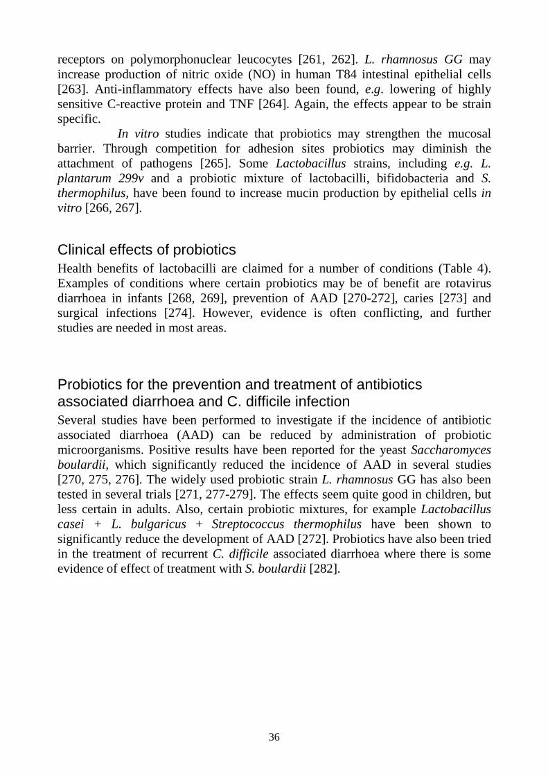
36
receptors on polymorphonuclear leucocytes [261, 262]. L. rhamnosus GG may increase production of nitric oxide (NO) in human T84 intestinal epithelial cells [263]. Anti-inflammatory effects have also been found, e.g. lowering of highly sensitive C-reactive protein and TNF [264]. Again, the effects appear to be strain specific. In vitro studies indicate that probiotics may strengthen the mucosal barrier. Through competition for adhesion sites probiotics may diminish the attachment of pathogens [265]. Some Lactobacillus strains, including e.g. L. plantarum 299v and a probiotic mixture of lactobacilli, bifidobacteria and S. thermophilus, have been found to increase mucin production by epithelial cells in vitro [266, 267].
Clinical effects of probiotics Health benefits of lactobacilli are claimed for a number of conditions (Table 4). Examples of conditions where certain probiotics may be of benefit are rotavirus diarrhoea in infants [268, 269], prevention of AAD [270-272], caries [273] and surgical infections [274]. However, evidence is often conflicting, and further studies are needed in most areas.
Probiotics for the prevention and treatment of antibiotics associated diarrhoea and C. difficile infection Several studies have been performed to investigate if the incidence of antibiotic associated diarrhoea (AAD) can be reduced by administration of probiotic microorganisms. Positive results have been reported for the yeast Saccharomyces boulardii, which significantly reduced the incidence of AAD in several studies [270, 275, 276]. The widely used probiotic strain L. rhamnosus GG has also been tested in several trials [271, 277-279]. The effects seem quite good in children, but less certain in adults. Also, certain probiotic mixtures, for example Lactobacillus casei + L. bulgaricus + Streptococcus thermophilus have been shown to significantly reduce the development of AAD [272]. Probiotics have also been tried in the treatment of recurrent C. difficile associated diarrhoea where there is some evidence of effect of treatment with S. boulardii [282].
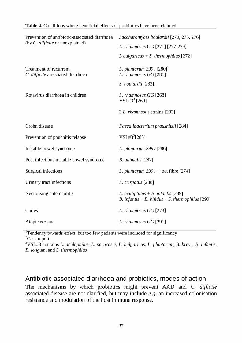
37
Table 4. Conditions where beneficial effects of probiotics have been claimed
Saccharomyces boulardii [270, 275, 276]
L. rhamnosus GG [271] [277-279]
Prevention of antibiotic-associated diarrhoea (by C. difficile or unexplained)
L bulgaricus + S. thermophilus [272]
L. plantarum 299v [280]1 Treatment of recurrent
C. difficile associated diarrhoea L. rhamnosus GG [281]2
S. boulardii [282].
L. rhamnosus GG [268] VSL#33 [269]
Rotavirus diarrhoea in children
3 L. rhamnosus strains [283]
Crohn disease Faecalibacterium prausnitzii [284] Prevention of pouchitis relapse VSL#33[285] Irritable bowel syndrome L. plantarum 299v [286] Post infectious irritable bowel syndrome B. animalis [287] Surgical infections L. plantarum 299v + oat fibre [274] Urinary tract infections L. crispatus [288]
L. acidiphilus + B. infantis [289] Necrotising enterocolitis B. infantis + B. bifidus + S. thermophilus [290]
Caries L. rhamnosus GG [273] Atopic eczema L. rhamnosus GG [291] 1Tendency towards effect, but too few patients were included for significancy 2Case report 3VSL#3 contains L. acidophilus, L. paracasei, L. bulgaricus, L. plantarum, B. breve, B. infantis, B. longum, and S. thermophilus
Antibiotic associated diarrhoea and probiotics, modes of action The mechanisms by which probiotics might prevent AAD and C. difficile associated disease are not clarified, but may include e.g. an increased colonisation resistance and modulation of the host immune response.
38
As described elsewhere, suppression of the normal microbiota by antibiotics allows opportunists like C. difficile to multiply. By competing for space and nutrients probiotics may counteract the growth of this and other pathogens. Also, spore germination of C. difficile may be inhibited by the production of hydrogen peroxide and short chain fatty acids by probiotic Lactobacillus strains [293].
In a study by Gorbach the numbers of plasma cells in the intestinal mucosa increased with administration of L. rhamnosus GG, resulting in an enhanced immune response towards C. difficile or C. difficile toxins [294]. Certain Lactobacillus strains, including L. plantarum 299v, have been shown to increase the production of SCFAs in the gut [241]. A positive correlation between SCFA production, absorption of sodium and water in the colon, and decreased diarrhoeal symptoms has been observed in several studies [295].
Probiotics for the prevention or treatment of Salmonella infection Several animal studies support an effect of probiotic lactobacilli against Salmonella. Good effects have been observed in chickens [296], pigs [297], and mice [298]. However, there are also studies showing no beneficial effects.
There are few studies involving human subjects. Alm et al. found in a non-blinded study that intake of Lactobacillus acidophilus shortened the duration of the Salmonella carrier state in patients that were asymptomatic at study start [300].
Salmonella and probiotics, modes of action There are several possible mechanisms whereby probiotics could be effective against Salmonella: competition for binding sites in the gut, production of antibacterial substances, competition for nutrients, local reduction of luminal pH, and an influence on the host immune response. A number of studies, mostly in vitro, have been performed to test these possibilities. A short review of such studies is presented below, but to what extent these findings mirror what actually happens in the human gut is unknown.
Strains of several Bifidobacterium and Lactobacillus species, for instance B. longum, L. acidophilus, and L. plantarum are able to inhibit the adhesion of Salmonella to intestinal epithelial cells in vitro [301, 302]. It is not known if adhesion is blocked by competition for a specific receptor, or by steric hindrance.
Furthermore, several Bifidobacterium and Lactobacillus species, including B. bifidum, B. animalis, L. acidophilus, L. johnsonii and L. plantarum, have been shown to possess antagonistic action against Salmonella ssp. as a result of production of bacteriocins, organic acids like lactic acid, or other substances toxic to Salmonella [212, 303]. Antagonism by competition for growth limiting
39
amino acids between S. Typhimurium and a combination of five faecal species has been shown in vitro with a reduction of growth of the Salmonella strain [304].
There are also studies indicating that probiotic strains can down-regulate the inflammatory response induced by Salmonella [302], and increase the production of S-IgA in the gut against this pathogen [305]. On the other hand, certain probiotics may stimulate the inflammatory response, for example through activation of macrophages [306]. Probiotics were also found to stimulate proliferation of lymphocytes in response to Salmonella [307].
Lactobacillus plantarum 299v Lactobacillus plantarum 299v (DSM 9843) is a probiotic strain which survives passage through the gastrointestinal tract and transiently colonises small intestinal and colonic mucosa [308, 309]. In animal models it has been found to reduce intestinal injury and inflammation after radiation, to reduce the severity of dextran sulfate sodium-induced colitis [310, 311] and to abolish E. coli-induced increase in intestinal permeability [312].
Two clinical studies have shown a reduction of abdominal pain in patients with irritable bowel disease receiving L. plantarum 299v, and trends towards less constipation and flatulence [286, 313]. However, another study found no clinical improvement in IBS using this probiotic strain [314].
Supply of L. plantarum 299v significantly reduced the incidence of post-operative infections in transplant recipients [315]. Furthermore, critically ill patients treated with this strain had decreased colonisation by C. difficile as compared to controls [316], and the same strain in combination with metronidazole tended to be more effective than metronidazole alone in the treatment of patients with recurrent C. difficile associated diarrhoea [280].
Safety of probiotics There area several reports of sepsis associated with the probiotic yeast S. boulardii [317], which is widely used for the treatment of AAD and C. difficile infections. Infections have even occurred by aerosol transmission to patients with central venous catheters neighbouring a patient treated with the yeast [318]. Most infections occurred in severely debilitated patients.
Lactobacilli are generally regarded as safe [319] and it is believed that the risk of infection with a probiotic Lactobacillus strain is similar to the risk of becoming infected with a Lactobacillus strain from the commensal microbiota [320]. As mentioned earlier, endocarditis, bacteraemia and localised infections including abscesses caused by lactobacilli have been reported, mainly in individuals with severe underlying disease [321, 322].

40
There are some reports of infections caused by the widely used probiotic strain L. rhamnosus GG including bacteraemia in children with underlying disease and one case of endocarditis and one case of liver abscess in adults [317]. In a review from 2005 only one out of 241 reported Lactobacillus infections was caused by a probiotic Lactobacillus strain [322]. Plasmids carrying antibiotic resistance genes have been found in several Lactobacillus species, including L. plantarum [323]. Transfer of such plasmids to other bacteria has been considered to be rare, but is as yet not well studied [324]. Some studies indicate a higher risk of antibiotic resistance transfer than previously believed, especially during treatment with antibiotics [325, 326].
41
AIMS The aims of this thesis were: to study the establishment of the Lactobacillus microbiota at the species and strain level in Swedish infants followed over the first 18 months of life, and to identify factors influencing the colonisation pattern to investigate how S-IgA influences the Lactobacillus microbiota, by studying the oral and faecal Lactobacillus microbiota of adults with and without IgA deficiency regarding species distribution and expression of mannose-specific adhesins to determine if intake of Lactobacillus plantarum can prevent diarrhoea and other gastro-intestinal disturbances and decrease colonisation by toxin-producing C. difficile during treatment with antibiotics to study if intake of Lactobacillus plantarum can reduce symptoms during and after Salmonella enteritis and/or reduce the time until clearance of Salmonella in faeces
42
MATERIALS AND METHODS USED IN THE STUDIES
A summary of the individuals studied and the methods used in study I-IV is presented here. Please refer to the individual papers for more detailed information.
Study populations and study design (I-IV)
Paper I The establishment of the Lactobacillus microbiota was studied in Swedish infants followed over the first 18 months of life.
One hundred and twelve healthy Swedish infants born at the Sahlgrenska University Hospital were followed. The children were recruited to a prospective birth-cohort study aiming to investigate the relation between intestinal colonisation pattern and allergy development, the ALLERGYFLORA study [62] [63]. Parents-to-be were enrolled at the maternity clinic, and background data collected on atopic heredity, pets and siblings was collected.
During the infant’s first 18 months of life, faecal samples were collected at regular intervals and analysed for lactobacilli. Parents recorded the child’s health status and feeding habits during the first year. The influence of delivery mode, feeding mode and of siblings or pets in the household on Lactobacillus colonisation pattern was studied.
Paper II The oral and faecal Lactobacillus microbiota of adult individuals with and without IgA deficiency was characterised regarding species distribution and expression of mannose-specific adhesins.
Thirty three individuals with selective IgA deficiency and thirty-four age-matched healthy individuals with normal serum immunoglobulin levels were included in the study. IgA deficiency was defined as a serum IgA level of less than 0.05 g/l in the presence of IgM, IgG and IgG1 - 4 in the normal range [327]. Ten of the individuals with IgA deficiency were healthy blood donors diagnosed at screening, while 23 at some time point had sought medical advice at the infectious diseases clinic for symptoms which could be related to their IgA deficiency. Symptoms included a history of frequent respiratory tract infections and/or of gastrointestinal complaints. Fourteen had at least one diagnosed auto-immune disease. All participants agreed not to consume any probiotic products for two weeks before sampling.
43
From each individual, one oral and one faecal sample were obtained and analysed for lactobacilli. The Lactobacillus species distribution and adhesin expression were compared between IgA deficient and control individuals, and between oral and faecal samples.
Paper III The ability of the probiotic strain L. plantarum 299v to prevent diarrhoea and other gastro-intestinal disturbances during treatment with antibiotics was tested in a double blind placebo controlled study.
Inclusion criteria were age of at least 16 years, antibiotic treatment started within 48 hours before inclusion, an expected treatment period of 7 to 14 days, and ability to actively participate in the study by keeping a diary and providing stool samples. Patients were randomized to consume 200 ml/day of a test drink containing blueberries and 5% oats gruel with or without 5x10⁷ colony forming units (CFU) of L. plantarum/ml once daily until a week after termination of antibiotics. Gastrointestinal symptoms, intake of antibiotics and intake of test drink were recorded daily by the patients. The patients were asked to avoid foods or products containing live bacteria during the study period and to continue the registration in the diary for at least one week after the last day of intake of test drink. On completion, the diary was mailed to the study centre. Faecal samples for the analysis of C. difficile toxins were obtained at study start before first intake of test drink and after discontinuation of antibiotics.
Primary outcomes were the proportion of patients in each group developing diarrhoea, defined as at least 3 loose or watery stools per day for at least 2 consecutive days, and the proportion of patients in each group positive for C. difficile toxin in faeces after treatment with antibiotics. Secondary outcomes were the risk of developing loose or watery stools, hard stools, abdominal pain, nausea, vomiting, flatulence, or blood in stools in each group.
Paper IV The effect of intake of L. plantarum 299v on symptoms during and after Salmonella enteritis and on time until clearance of Salmonella in faeces was studied.
Patients were eligible for the study if they were at least one year of age, had sought medical advice for gastrointestinal symptoms and had a stool sample positive for Salmonella. Patients were randomised to consume 1 g of a skim milk powder with or without 5 x 10¹⁰ colony forming units (CFU)/g of freeze-dried Lactobacillus plantarum 299v. They were instructed not to consume other foods or products containing live bacteria during the study period, and to register symptoms, intake of study preparation and any use of antibiotics in the study diary. Faecal samples for the analysis of Salmonella were obtained at inclusion and then weekly during the study.
44
Primary outcomes were time until clearance of Salmonella and time until resolution of diarrhoea, i.e. time to the first week without a day with diarrhoea (defined as at least three loose stools in 24 hours). Secondary outcomes were time until cessation of other acute symptoms, i.e. loose stools, blood in stools, fever, nausea, vomiting, abdominal pain, or any of these symptoms. The occurrence of symptoms while still taking the test preparation after clearance of Salmonella, was also analysed and compared between the L. plantarum and the placebo group. Time to clearance of Salmonella symptoms, and effects of treatment, were also compared between female and male patients.
Permission from the Ethics Committee (I-IV) Informed consent was obtained from included individuals or their parents, and the Ethics Committee of the Medical Faculty of Göteborg approved all four studies.
Sampling and culture for the isolation of lactobaci lli (I, II) From infants, faecal samples were obtained at 1, 2, 4 and 8 weeks, and at 6 and 12 months of age. In 65 of the 112 infants, faecal cultures were also performed at 18 months. Parents collected freshly voided faeces at home. The samples were kept refrigerated in a container without oxygen (AnaeroGen Compact, Oxoid Ltd., Basingstoke, UK) until transported to the laboratory, where they were processed within 24 h after collection.
In paper II, one oral and one faecal sample were obtained from each healthy and each IgA deficient individual. For oral lactobacilli, a cotton-tipped swab was pressed against the back of the tongue and transported in Stuart’s transport medium [328] directly to the laboratory. For intestinal lactobacilli, freshly voided faeces was collected by the participants and kept refrigerated until transported to the laboratory within 5 h.
All faecal samples were serially diluted and plated on Rogosa agar for selective outgrowth of lactobacilli [329]. The limit of detection was 330 (102.52) CFU/g faeces. For oral lactobacilli, the cotton-tipped swab was streaked directly on Rogosa agar plates. All plates were incubated anaerobically at 37 °C for 3 days using the BBL GasPak anaerobic system (Becton Dickinson Microbiology Systems, Sparks, MD). From the plates of oral samples and from appropriate dilutions of the faecal samples, one representative colony of each morphotype (differing in e.g. size, shape, colour or texture from other colonies) was separately enumerated, Gram-stained, examined in the microscope and subcultured to purity. Unbranched Gram-positive rods were regarded as tentative lactobacilli and analysed further.
45
Identification of lactobacilli by PCR (I, II)
DNA extraction (I, II) In paper I, putative Lactobacillus isolates were grown over night in Lactobacillus carrying medium (LCM) [330] and a crude cell extract was prepared by bead beating as previously described [331]. Briefly, cell extracts were prepared from overnight cultures at 28 ºC. The cells were washed twice in 1 ml sterile Milli-Q® water (Millipore, Molsheim, France), and disrupted in an Eppendorf tube with glass beads.
In paper II, bacterial DNA was extracted as described by Song with some modifications [29]. Briefly, one to two bacterial colonies, which had been subcultured to purity on Rogosa agar, were suspended in 50 µl of 10 mM Tris HCl, 1 mM EDTA (pH 8.0) and 10 mM saline. The bacterial suspension was incubated for 10 min at 95 ° and centrifuged at 18 600 x g for 3 min. The supernatants were kept at -20 º C until used.
Exclusion of bifidobacterial isolates using a Bifidobacterium-specific PCR (I, II) As Rogosa plates are not entirely selective for lactobacilli, but also permit growth of bifidobacteria, all isolates were screened in a Bifidobacterium-specific PCR. The primers PbiF1 (5’CCG GAA TAG CTC C-3’) and PbiR2 (5’-GAC CAT GCA CCA CCT GTG AA-3’) [332] were used under previously described PCR conditions [332] (paper II) and as described in paper I, respectively. The amplicon specific for bifidobacteria, with a size of 919 bp, was visualised by agarose gel electrophoresis and ethidium bromide staining. Isolates identified as bifidobacteria were not analysed further.
RAPD for the distinction of different Lactobacillus strains combined with a Bifidobacterium-specific PCR (I) In paper I, a Random Amplification of Polymorphic DNA analysis (RAPD) was performed to distinguish different Lactobacillus strains, as previously described [331]. In RAPD, primers of arbitrary sequences bind under low stringency conditions to various sites on both strands of template DNA. This results in a pattern of amplified DNA fragments which is unique for each strain [26].
The RAPD-primer 73 (5′-ACGCGCCCT-3′) was used, and the RAPD analysis was performed in the PCR in which the Bifidobacterium specific primers were included. Gel electrophoresis for the separation of PCR products was performed on agarose gels that were stained with ethidium bromide and photographed under UV-light. Putative Lactobacillus isolates from a single child
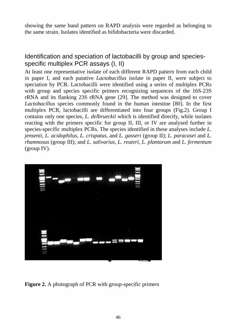
46
showing the same band pattern on RAPD analysis were regarded as belonging to the same strain. Isolates identified as bifidobacteria were discarded.
Identification and speciation of lactobacilli by group and species-specific multiplex PCR assays (I, II) At least one representative isolate of each different RAPD pattern from each child in paper I, and each putative Lactobacillus isolate in paper II, were subject to speciation by PCR. Lactobacilli were identified using a series of multiplex PCRs with group and species specific primers recognizing sequences of the 16S-23S rRNA and its flanking 23S rRNA gene [29]. The method was designed to cover Lactobacillus species commonly found in the human intestine [80]. In the first multiplex PCR, lactobacilli are differentiated into four groups (Fig.2). Group I contains only one species, L. delbrueckii which is identified directly, while isolates reacting with the primers specific for group II, III, or IV are analysed further in species-specific multiplex PCRs. The species identified in these analyses include L. jensenii, L. acidophilus, L. crispatus, and L. gasseri (group II); L. paracasei and L. rhamnosus (group III); and L. salivarius, L. reuteri, L. plantarum and L. fermentum (group IV).
Figure 2. A photograph of PCR with group-specific primers
47
Speciation of lactobacilli by sequencing of the Lactobacillus 16S rRNA gene (I, II) In paper I, one of the Lactobacillus strains that could not be typed to the species level using species-specific primers as described above was identified by partial sequencing of the Lactobacillus 16S rRNA gene. This analysis was performed at the Laboratory of Molecular Microbiology, Department of Clinical Bacteriology, Sahlgrenska University Hospital, using their standard protocol [333].
In paper II, a large number of isolates did not react in the group- or species-specific PCR assays. These isolates were speciated by partial sequencing of 16S rRNA genes, as described in the manuscript, at the Genomics Core Facility Platform at the Sahlgrenska Academy, University of Göteborg. The sequences were analysed using the Ribosomal Database Project (http://rdp.cme.msu.edu/) and FASTA (http://www.ebi.ac.uk/Tools/fasta33/nucleotide.html) databases. Only type strains were considered and a similarity of at least 98 % and a distance score of at least 0.5 % units to the next closest species required for the determination of species.
Adherence of lactobacilli to HT-29 cells (II) The expression of mannose-specific (MS) adhesins by lactobacilli from IgA deficient and control individuals was tested by assessing adherence in a mannose-sensitive manner to cells of the colonic carcinoma cell line HT-29 as previously described [196]. Bacteria and cells were incubated in Hank’s balanced salt solution with or without methyl-α-D-mannoside at 4 ° for 30 min. The cells were spun down, washed in PBS and fixed with neutral buffered formalin. The number of bacteria attached to each of 40 epithelial cells was determined using interference contrast microscopy. In each experiment, the Lactobacillus isolates from one IgA deficient and one control individual were analysed, the person examining adherence being blinded as to their identity. To calculate MS adherence, the number of bacteria adhering in the presence of methyl-α-D-mannoside were subtracted from the number of bacteria adhering in the absence of methyl-α-D-mannoside. Isolates adhering with at least three bacteria/cell and showing at least 30 % reduction of adherence in the presence of methyl-α-D-mannoside, were considered positive for MS adhesins [31].
Sampling for and detection of C. difficile toxin in faeces (III) In paper III, faecal samples for the detection of C. difficile toxin were obtained before administration of the first dose of test drink and seven to ten days after discontinuation of antibiotics. Patients were instructed to deliver an extra faecal sample in case of diarrhoeal symptoms. Freshly voided faeces was collected by the patient and sent by mail if sampling was not performed at the hospital.
48
An ELISA was used to detect C. difficile toxin in faecal samples, either toxin A only (patients 1-154) or, as the standard method was changed at the clinical laboratory, toxin A and B (patient 155 and onwards) [334, 335]. The analyses were performed at the Clinical Bacteriology Laboratory at Sahlgrenska University Hospital.
Sampling and culture for the detection of faecal bacterial pathogens (IV, III) In paper IV, rectal swab samples for culture and detection of Salmonella were performed at inclusion and once a week until four consecutive negative samples had been obtained. Thereafter, intake of test products was discontinued, but weekly samples were obtained and cultured for four additional weeks (Fig. 1, paper IV). The rectal swab samples were taken by the patients and sent in by mail in Stuart’s transport medium [328].
In paper III, the faecal samples delivered during diarrhoeal symptoms were cultured and analysed for Salmonella, Shigella, Yersinia, and Campylobacter.
Culture and analysis for the identification of Salmonella, Shigella, Yersinia and Campylobacter were performed at the Clinical Bacteriology Laboratory at Sahlgrenska University Hospital according to their standard methods [336].
Salmonella serotyping (IV) Salmonella isolates were serotyped at the Clinical Bacteriology Laboratory at Sahlgrenska University Hospital according to the Kauffman-White scheme [328].
Statistical methods
Fischer, Man-Whitney (I, II, III, IV) Comparisons of characteristics between infants (paper I), study groups (paper II, III, IV), women and men (paper IV), and bacterial groups (paper II) were performed using Fisher exact test for proportions and the Mann-Whitney test for continuous characteristics.
McNemar’s test (II) McNemar’s chi-squared test was used for comparing paired proportions, i.e. oral and faecal isolates.
49
Exact logistic regression (III) The probability of antibiotic associated diarrhoea was compared between the treatment and placebo groups using exact logistic regression.
Generalised estimating equations (III, IV) To analyze the effect of treatment on the risk of experiencing loose or watery stools or other gastrointestinal symptoms in paper III, we used generalized estimating equations [337, 338], modelling the correlation structure between the responses within an individual by the autoregressive model, where the correlation between responses gets smaller with time.
In paper IV, we used the empirical model of generalized estimating equations to compare the treatment groups, as well as women and men, regarding symptoms after clearance of Salmonella while still taking the test preparation.
Survival analysis (IV)
Kaplan Meier (IV) Kaplan Meier curves with the log rank test were used to detect the effects of probiotic treatment and gender on the primary outcomes time to clearance of Salmonella and resolution of diarrhoea, and on time to resolution of other acute symptoms. Finally, they were used to study the effect of certain other factors and patient characteristics on time to Salmonella clearance and resolution of diarrhoea, respectively.
Cox regression (IV) Cox regression was used to perform a multivariable analysis of the factors that were found to influence time to clearance of Salmonella and resolution of diarrhoea in the univariable analyses, to determine the possible independent contribution of these factors to the outcomes.
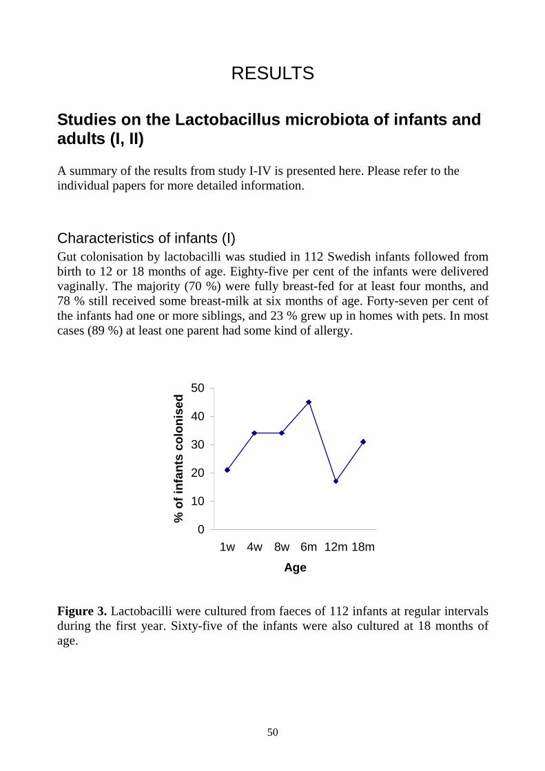
50
RESULTS
Studies on the Lactobacillus microbiota of infants and adults (I, II) A summary of the results from study I-IV is presented here. Please refer to the individual papers for more detailed information.
Characteristics of infants (I) Gut colonisation by lactobacilli was studied in 112 Swedish infants followed from birth to 12 or 18 months of age. Eighty-five per cent of the infants were delivered vaginally. The majority (70 %) were fully breast-fed for at least four months, and 78 % still received some breast-milk at six months of age. Forty-seven per cent of the infants had one or more siblings, and 23 % grew up in homes with pets. In most cases (89 %) at least one parent had some kind of allergy.
0
10
20
30
40
50
1w 4w 8w 6m 12m 18m
Age
% o
f inf
ants
col
onis
ed
Figure 3. Lactobacilli were cultured from faeces of 112 infants at regular intervals during the first year. Sixty-five of the infants were also cultured at 18 months of age.
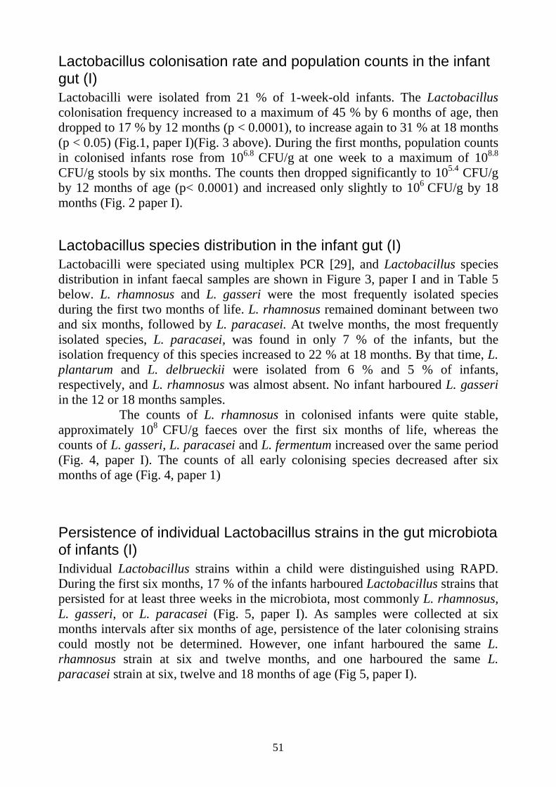
51
Lactobacillus colonisation rate and population counts in the infant gut (I) Lactobacilli were isolated from 21 % of 1-week-old infants. The Lactobacillus colonisation frequency increased to a maximum of 45 % by 6 months of age, then dropped to 17 % by 12 months (p < 0.0001), to increase again to 31 % at 18 months (p < 0.05) (Fig.1, paper I)(Fig. 3 above). During the first months, population counts in colonised infants rose from 106.8 CFU/g at one week to a maximum of 108.8 CFU/g stools by six months. The counts then dropped significantly to 105.4 CFU/g by 12 months of age (p< 0.0001) and increased only slightly to 106 CFU/g by 18 months (Fig. 2 paper I).
Lactobacillus species distribution in the infant gut (I) Lactobacilli were speciated using multiplex PCR [29], and Lactobacillus species distribution in infant faecal samples are shown in Figure 3, paper I and in Table 5 below. L. rhamnosus and L. gasseri were the most frequently isolated species during the first two months of life. L. rhamnosus remained dominant between two and six months, followed by L. paracasei. At twelve months, the most frequently isolated species, L. paracasei, was found in only 7 % of the infants, but the isolation frequency of this species increased to 22 % at 18 months. By that time, L. plantarum and L. delbrueckii were isolated from 6 % and 5 % of infants, respectively, and L. rhamnosus was almost absent. No infant harboured L. gasseri in the 12 or 18 months samples. The counts of L. rhamnosus in colonised infants were quite stable, approximately 108 CFU/g faeces over the first six months of life, whereas the counts of L. gasseri, L. paracasei and L. fermentum increased over the same period (Fig. 4, paper I). The counts of all early colonising species decreased after six months of age (Fig. 4, paper 1)
Persistence of individual Lactobacillus strains in the gut microbiota of infants (I) Individual Lactobacillus strains within a child were distinguished using RAPD. During the first six months, 17 % of the infants harboured Lactobacillus strains that persisted for at least three weeks in the microbiota, most commonly L. rhamnosus, L. gasseri, or L. paracasei (Fig. 5, paper I). As samples were collected at six months intervals after six months of age, persistence of the later colonising strains could mostly not be determined. However, one infant harboured the same L. rhamnosus strain at six and twelve months, and one harboured the same L. paracasei strain at six, twelve and 18 months of age (Fig 5, paper I).
52
Table 5. Per cent of infants colonised by various Lactobacillus species at different time points over their first 18 months of life Isolation frequency (%) 1 w 2 w 1 m 2 m 6 m 12 m 18 m L. rhamnosus 7.1 8.9 14 21 21 1.8 1.5 L. gasseri 4.5 8.9 11 8.9 3.6 - - L. fermentum 3.6 3.6 2.7 0.9 3.6 3.6 - L. paracasei 0.9 0.9 2.7 5.4 15 7.1 22 L. plantarum 1.8 - 1.8 - 4.5 2.7 6.2 L. delbrueckii - - - - 0.9 0.9 4.6 L. crispatus 1.8 - 1.8 - - - - L. reuteri - - - - 1.8 0.9 - L. salivarius - - - - - - 1.5 L. mucosae* 0.9 0.9 0.9 0.9 - - - *One strain identified by partial sequencing of 16S rDNA
The Lactobacillus microbiota in infants in relation to lifestyle factors (I) There were no significant differences in Lactobacillus colonisation rate or species distribution depending on delivery mode, nor did presence of siblings or pets in the household significantly influence colonisation. Because of the high breast-feeding rates in the first months of life, it was not possible to compare the Lactobacillus colonisation pattern between breast- and bottlefed infants during this period. At six months 22 % of the infants were weaned, and they harboured lactobacilli significantly less often (20 % vs. 52 %, p<0.01) and had a tendency towards lower population counts (106.8 vs. 108.8 CFU/g, p=0.18) than did breastfed infants of the same age. L. rhamnosus tended to be more common in vaginally than in sectio-delivered infants, but the difference was not significant. Colonisation by L. gasseri, which is a common species in the maternal vaginal microbiota [339], was not related to delivery mode. In infants still receiving breast milk at six months, L. rhamnosus was more commonly isolated (27 % vs. 4 %, p<0.05) and tended to
53
reach higher population counts (108.9 vs. 106,8 CFU/g, p=0.19) than in infants weaned by that age. Most of the infants studied (89 %) had at least one allergic parent. However, the Lactobacillus colonisation pattern did not differ between children of atopic and children of non-atopic parents.
Lactobacillus colonisation frequency and species distribution in the oral microbiota of adult individuals with and without IgA deficiency (II) To investigate the effect of secretory IgA on the Lactobacillus microbiota, we studied and compared the Lactobacillus microbiota of healthy and IgA deficient individuals.
Lactobacilli were isolated from the tongue of more than 80 % of control individuals, and the isolation frequency was only slightly lower in IgA deficient individuals. The various species isolated are shown in Table 1, paper II. L. paracasei and L. gasseri were the most common species in the oral microbiota of both IgA deficient and control individuals. The third most common species was L. vaginalis in IgA deficient persons and L. fermentum in controls. The latter species was significantly more common in the oral microbiota of controls as compared to IgA deficient individuals, while L. vaginalis tended to be more common in IgA deficiency. A large number of other species were also isolated, many of which were found in one or two individuals only (Table 1, paper II).
When comparing the Lactobacillus species distribution between symptomatic and asymptomatic IgA deficient individuals, L. salivarius was significantly more common in the oral cavity of the asymptomatic group. None of the individuals with a history of frequent respiratory infections carried this species.
Lactobacillus colonization frequency, population counts and species distribution in the gut microbiota of adult individuals with and without IgA deficiency (II) Lactobacilli were isolated from faeces of more than 90 % of both IgA deficient and control individuals. The mean faecal Lactobacillus population counts were approximately 106 CFU/g in both groups, slightly lower in IgA deficient individuals than in controls. L. paracasei dominated by far in the faecal microbiota of both study groups, followed by L. gasseri, L. plantarum and L. rhamnosus. There were no significant differences between IgA deficient and control individuals regarding Lactobacillus species distribution in faecal samples. We also compared the faecal Lactobacillus species distribution between symptomatic (n=23) and asymptomatic (n=10) IgA deficient individuals, but no significant differences were observed.
54
Similarities and differences between the oral and faecal Lactobacillus microbiota of IgA deficient and control individuals (II) A majority of individuals harboured lactobacilli in both oral and faecal samples. More than two thirds of these individuals had at least one species in common between the two locations and approximately one third had two or more species in common. L. gasseri, L. vaginalis and L. fermentum were significantly more common in the oral cavity than in faeces. No individual was positive for L. vaginalis or L. fermentum in faeces without being positive also in the oral sample. The diversity of species also seemed to be somewhat higher in oral than in faecal samples (Table 1, paper 2). When analysing IgA deficient and control individuals together, oral samples yielded on average 2.4 different species, and the corresponding figure for faecal samples was 2.0.
Expression of mannose-specific adhesins by oral and faecal lactobacilli from IgA deficient and control individuals (paper II) The carbohydrate chains of SIgA are rich in mannose, and could possibly function as receptors for MS adhesins of lactobacilli. Such interactions could then in turn influence the Lactobacillus microbiota by favouring, or disfavouring, lactobacilli expressing MS adhesins at mucosal surfaces. To explore this, we examined the expression of MS adhesins by Lactobacillus isolates from IgA deficient and control individuals by testing their ability to adhere in a mannose-sensitive manner to HT-29 colonic epithelial cells. MS adhesins were more commonly expressed by faecal isolates from IgA deficient individuals than by faecal isolates from controls, but a corresponding difference between IgA deficient and control individuals was not observed in oral isolates (Fig. 2a, paper 2). There were no significant differences between IgA deficient and control individuals in the level of MS adherence (bacteria/cell) in adhesin positive isolates.
Among both IgA deficient and control individuals, MS adhesins were more common in oral than in faecal Lactobacillus isolates (Fig. 2a, paper II) and adhesin-positive isolates also displayed a higher average adherence (bacteria/cell) when isolated from the oral cavity (Fig. 2b, paper II).
Isolates of a large number of species expressed MS adhesins, including L. fermentum, L. parabuchneri, L. vaginalis, L. plantarum, L. brevis, L. acidophilus, L. paracasei, L. salivarius, L. reuteri, L. gasseri, Lactobacillus hilgardii and Lactobacillus parafarraginis (Table 2, paper 2). In total more than 80 % of isolates from both oral and faecal samples and in both study groups belonged to species with the capacity to express MS adhesins. Among the more commonly isolated species, MS adherence was absent in L. rhamnosus and L. oris.
55
Studies on probiotic effects of L. plantarum 299v (III, IV)
Characteristics of patients receiving L. plantarum 299v or placebo during treatment with antibiotics (III) In this double blind placebo controlled trial, we investigated if intake of L. plantarum 299 could reduce the risk of diarrhoea or other gastrointestinal symptoms during treatment with antibiotics. Two hundred and thirty-nine patients, 93 men and 146 women, were initially included in the study, and randomised to daily intake of either a blueberry/oat drink with L. plantarum 299v (1010 CFU/day) or a blueberry/oat placebo drink. Seventy-three of the patients left the study without returning the study diary where symptoms and intake of test drink should be recorded, and were therefore excluded. In addition one patient was excluded because of cultivation of Campylobacter jejunii in blood, one because the diary was impossible to interpret, and one patient did not take the test drink. The excluded patients were equally distributed between the L. plantarum and placebo groups, and reasons for withdrawal did not differ between the study groups. Characteristics of patients who remained in the study and those who did not are presented in Table 1, paper III. The median age was significantly higher in patients who completed the study (p=0.0015).
Thus, 163 patients could be analysed and they were equally distributed between the L. plantarum and the placebo group (Table 2, paper III). The proportion of women tended to be higher in the L. plantarum group (p=0.081) and somewhat more patients in this group were positive for C. difficile on inclusion (p=0.20), but no clear differences between the study groups were observed regarding diagnoses, hospitalisation or antibiotics given (Table 2, paper III).
Side effects of the L. plantarum and placebo blueberry drinks (III) Three patients in the L. plantarum group and three in the placebo group reported constipation during the study, all during intake of test drink. No serious side effects were recorded.
Effects of L. plantarum 299v on the incidence of diarrhoea in antibiotic treated patients (III) The patients recorded symptoms, stool frequencies and intake of test preparation in a diary during the study. Diarrhoea, defined as at least three loose or watery stools per 24 hours for at least two consecutive days, was only diagnosed in eleven
56
patients during the entire study period, in six patients in the L. plantarum group and five in the placebo group.
When comparing the risk of diarrhoea and other symptoms between the study groups, we also divided the study period into three separate periods: the period of antibiotic treatment (A), the period of continued intake of test drink after discontinuation of antibiotics (B) and a follow-up period after intake of test drink (C) (Fig. 1, paper III). Four of 80 patients in the L. plantarum group and five of 83 in the placebo group had diarrhoea during treatment with antibiotics (period A) (OR 0.96; 95% confidence interval (CI) 0.18-4.7; p=1.0). During period B, with continued intake of test drink after antibiotic treatment, 3/71 vs. 0/69 patients had diarrhoea (OR not estimable, p=0.17), and during the follow-up period (C) 3/45 vs. 0/42 (OR not estimable, p=0.25). Patients developing and those not developing diarrhoea are compared in Table 3, paper III. Treatment with more than one antibiotic and treatment with clindamycin, cephalosporines or ampicillin was more common among patients developing diarrhoea than among other patients.
Effects of L. plantarum 299v on gastro-intestinal symptoms not defined as diarrhoea in antibiotic-treated patients (III) The risk of gastrointestinal symptoms not defined as diarrhoea was also compared between the study groups. The risk of developing loose or watery stools during the study was significantly lower in the L. plantarum group (OR 0.69; 95 % CI 0.52-0.92; p=0.012) (Table 4, paper III). The effect was significant during antibiotic treatment (period A) (OR 0.65; 95 % CI 0.45-0.94; p=0.020). There was a tendency towards an effect also after discontinuation of antibiotics, during continued intake of test drink (period B), but not thereafter (period C) (Table 4, paper III).
Patients in the L. plantarum group also had a lower risk of nausea than patients on placebo. When analysing the three study periods separately, the effect was significant during the period of antibiotic treatment, but not during the following periods (Table 5, paper III), possibly due to the lower number of patients included in the analysis of the two later periods.
Intake of L. plantarum did not alter the risk of abdominal pain, vomiting, hard stools, flatulence, or presence of blood in faeces (data not shown). Women had significantly more nausea (OR 3.2; 95 % CI 1.6-6.2; p=0.0006) and abdominal pain (OR 2.9; 95 % CI 1.5-5.5; p=0.0017) than men during treatment with antibiotics (period A). There were no gender-specific differences for other symptoms (data not shown). All statistical analyses performed to compare the L. plantarum and the placebo groups were adjusted for age and gender.
57
C. difficile toxin in faeces of antibiotic-treated patients receiving L. plantarum 299v or placebo (III) C. difficile toxin was found in four of 69 patients in the L. plantarum group and in one of 74 patient in the placebo group at baseline (p=0.20). Three of 74 patients in the L. plantarum group and three of 76 in the placebo group were positive for C. difficile toxin after antibiotic treatment. One of the eleven patients who developed diarrhoea was positive for C. difficile toxin on inclusion. The same patient was also the only one positive for C. difficile during diarrhoea. However, two of the patients developing diarrhoea did not provide samples for analysis at baseline, and only four delivered samples during diarrhoeal symptoms.
Characteristics of patients with non-typhoid salmonellosis receiving L. plantarum 299v or placebo (IV) In this double-blind, placebo controlled trial we investigated if intake of L. plantarum 299v could accelerate clearance of Salmonella in faeces, and reduce infection-related symptoms in patients with non-typhoid salmonellosis. The patients were randomised to daily intake of 5 x 10¹⁰ CFU of freeze dried L. plantarum 299v or placebo. One hundred and fifty-four patients were initially enrolled in the study, five of whom were excluded as they had not had any symptoms from their Salmonella infection. Of the remaining patients, 46 % were male, and the median age was 36 years (range 5 – 68 years). Patient characteristics are shown in Table 1, paper IV. No significant differences between the L. plantarum and placebo groups were observed regarding concomitant diseases, medication at study start or recent intake of antibiotics or probiotics. Initial symptoms are shown in Table 2, paper IV. All but one patient reported diarrhoea, and a majority reported fever and abdominal pain at their first hospital visit, which occurred on average five days before inclusion in the study, and seven days after first onset of symptoms. Vomiting was more common as reported on inclusion among patients later randomised to receive placebo (Table 2, paper IV). Eighteen per cent of the patients were initially hospitalised.
Patients were asked if they wanted to participate in the study once it was clear that they had a stool culture positive for Salmonella. At inclusion, a new stool sample was collected. Eighty-five per cent of the patients, 83 % in the placebo and 87 % in the L. plantarum group, still had Salmonella in stools when entering the study.
Occurrence of symptoms at study start was defined as occurrence of the symptom on at least one day during the first week after inclusion in the study (Table 2, paper IV). All symptoms were less common at study start than at the first hospital visit. Loose stools were more common in the L. plantarum group at study start and the same tendency was observed for fever (Table 2, paper IV). Median
58
time from onset of symptoms to study start was eleven days in the group receiving L. plantarum and ten days in the placebo group. The most common Salmonella serotype was S. Enteritidis in both study groups (Table 3, paper IV).
Differences at study start between female and male patients with non-typhoid salmonellosis (IV) We also analysed whether women and men differed in various patient characteristics and initial symptoms.
Median age was 37 years in female and 34 years in male patients. Women tended to have a history of gall bladder disease more often than men (p=0.13). Reported symptoms at the first hospital visit and symptoms at study start in female and male patients are shown in Table 4, paper III. Vomiting and abdominal pain were somewhat more common in women than in men before inclusion, but the differences were not significant. During the first week of the study, all symptoms were somewhat more common in female than in male patients, and significant differences were observed regarding nausea and abdominal pain. The differences remained when leaving out women reporting menstruation during the same time period.
Women in the L. plantarum group significantly more often had fever during the first study week than women in the placebo group. .
Side effects of the freeze-dried L. plantarum 299v and placebo preparations (IV) Urticaria was reported by one patient in the placebo group. Symptoms resolved quickly after discontinuation of intake of the placebo skim milk powder. Treatment with L. plantarum tended to increase some gastrointestinal symptoms, and these effects are presented below under treatment effects.
Effects of treatment with L. plantarum and influence of gender on clearance of Salmonella (IV) Faecal samples were cultured for Salmonella at inclusion and once a week thereafter until four weeks after discontinuation of intake of the study preparation. Among patients positive for Salmonella at inclusion (85 %), time to clearance of Salmonella was studied with survival analysis. There was no significant difference in time to clearance of Salmonella between the L. plantarum and the placebo groups as analysed using the log rank test (Fig. 2a, paper IV). Time to clearance was also compared between women and men. Women tended to clear Salmonella more quickly than men according to the survival analysis (Figure 2b, paper IV). The same tendencies regarding gender were seen when analysing separately patients receiving L. plantarum and patients on
59
placebo. Intake of L. plantarum did not affect time to clearance of Salmonella when analysing female and male patients separately (table 9, paper IV).
Effects of treatment with L. plantarum 299v and influence of gender on time to resolution of diarrhoea and other symptoms (IV) Among patients who had diarrhoea at study start, time to resolution of diarrhoea was analysed by survival analysis and compared between groups using the log rank test. There was no significant difference between patients receiving L. plantarum and patients receiving placebo in time to resolution of diarrhoea (Fig. 3a, paper IV). However, men cleared diarrhoea within significantly shorter time than women (Fig. 3b, paper IV). This difference was observed also when analysing only patients on placebo. There was no effect of L. plantarum on time to resolution of diarrhoea when analysing female and male patients separately.
Time to cessation of various acute symptoms in patients receiving L. plantarum or placebo is shown in table 5, paper IV. There were no significant differences in time from study start to cessation of loose stools, nausea or abdominal pain between patients receiving L. plantarum or placebo as compared using the log rank test. In addition, there were no differences in time from study start to resolution of blood in stools, fever, or vomiting, but patients still having these symptoms at study start were too few for meaningful comparisons (Table 2, paper IV). There were no significant differences between female and male patients in time to resolution of these symptoms (Table 6, paper IV), and no differences between the L. plantarum and the placebo group was observed when women and men were analysed separately.
Additional factors affecting time to clearance of Salmonella and resolution of diarrhoea (IV) To identify factors influencing time to clearance of Salmonella and resolution of diarrhoea we used survival analysis and log rank tests to compare patients positive or negative for a specific factor of possible importance. Higher age, history of diverticulitis, and vomiting at study start were associated with Salmonella clearance, and diarrhoea, fever, and abdominal pain were associated with prolonged carriage in univariate analyses. In the multivariate analysis (Cox regression), diarrhoea at study start, associated with longer Salmonella carriage, was the only factor which remained significant. There was a non-significant tendency for vomiting to shorten Salmonella carriage. Male sex and higher age were associated with shorter time to resolution of diarrhoea in univariate analyses. Only gender remained significant in the multivariate analysis.
60
Effects of treatment with L. plantarum and influence of gender on symptoms after clearance of Salmonella (IV) We also analysed the effect of treatment with L. plantarum 299v on symptoms occurring after clearance of Salmonella from stools, as gastrointestinal symptoms commonly persist for some time after the initial infection. In the L. plantarum group, the proportion of days with hard stools was lower than in the placebo group, but the proportion of days with fever was increased. Also, the risk of abdominal pain tended to be higher in the L. plantarum group (Table 7, paper IV). The risk of experiencing symptoms after clearance of Salmonella was also compared between women and men, revealing that women had a larger proportion of days with loose stools, nausea, abdominal pain and flatulence (Table 8, paper IV). The difference in abdominal pain was primarily observed among women receiving L. plantarum. Comparing the effect of L. plantarum in women and men separately, women in the treatment group reported more days with fever and abdominal pain than women in the placebo group. Men in the L. plantarum group had a higher proportion of days with diarrhoea, but fewer days with hard stools than men in the placebo group (Table 8, paper IV).
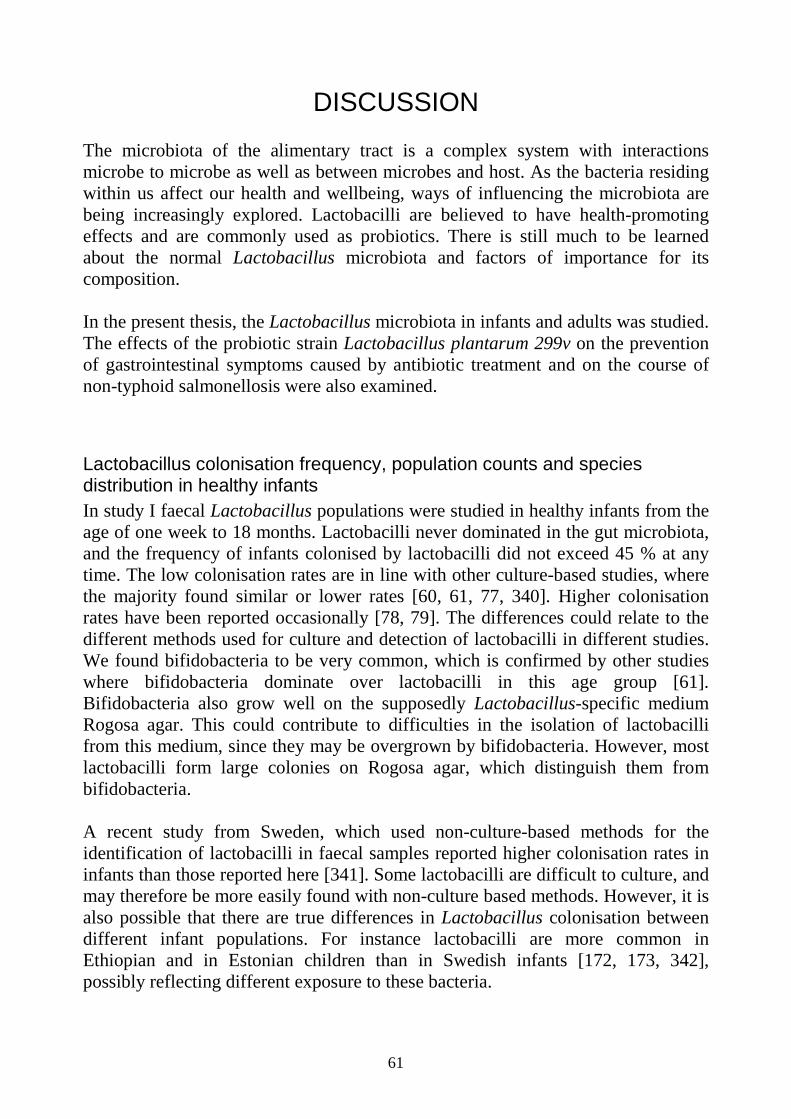
61
DISCUSSION The microbiota of the alimentary tract is a complex system with interactions microbe to microbe as well as between microbes and host. As the bacteria residing within us affect our health and wellbeing, ways of influencing the microbiota are being increasingly explored. Lactobacilli are believed to have health-promoting effects and are commonly used as probiotics. There is still much to be learned about the normal Lactobacillus microbiota and factors of importance for its composition. In the present thesis, the Lactobacillus microbiota in infants and adults was studied. The effects of the probiotic strain Lactobacillus plantarum 299v on the prevention of gastrointestinal symptoms caused by antibiotic treatment and on the course of non-typhoid salmonellosis were also examined.
Lactobacillus colonisation frequency, population counts and species distribution in healthy infants In study I faecal Lactobacillus populations were studied in healthy infants from the age of one week to 18 months. Lactobacilli never dominated in the gut microbiota, and the frequency of infants colonised by lactobacilli did not exceed 45 % at any time. The low colonisation rates are in line with other culture-based studies, where the majority found similar or lower rates [60, 61, 77, 340]. Higher colonisation rates have been reported occasionally [78, 79]. The differences could relate to the different methods used for culture and detection of lactobacilli in different studies. We found bifidobacteria to be very common, which is confirmed by other studies where bifidobacteria dominate over lactobacilli in this age group [61]. Bifidobacteria also grow well on the supposedly Lactobacillus-specific medium Rogosa agar. This could contribute to difficulties in the isolation of lactobacilli from this medium, since they may be overgrown by bifidobacteria. However, most lactobacilli form large colonies on Rogosa agar, which distinguish them from bifidobacteria. A recent study from Sweden, which used non-culture-based methods for the identification of lactobacilli in faecal samples reported higher colonisation rates in infants than those reported here [341]. Some lactobacilli are difficult to culture, and may therefore be more easily found with non-culture based methods. However, it is also possible that there are true differences in Lactobacillus colonisation between different infant populations. For instance lactobacilli are more common in Ethiopian and in Estonian children than in Swedish infants [172, 173, 342], possibly reflecting different exposure to these bacteria.
62
There were no significant differences in Lactobacillus colonisation rates between the vaginally and sectio delivered infants studied here. Several other studies have found that vaginally delivered infants are more likely to harbour lactobacilli in the early gut microbiota [78, 163, 164], whereas some find no difference [66, 343]. In most studies the difference in colonisation rate between sectio and vaginally delivered infants disappear within one or two weeks, and the studies finding equal Lactobacillus colonisation rates regardless of delivery mode have mostly studied infants at later time points only. The infants followed here were first sampled at one week of age, and it is possible that a significant difference would have been revealed, had we sampled the children a few days earlier. It seems likely that some vaginally delivered infants do pick up lactobacilli from the maternal vagina. This has been documented in Japanese infants, where approximately 20 % of vaginally delivered infants acquired maternal vaginal Lactobacillus strains [77]. However, only a few infants retained these strains until one month of age. Thus, vaginal lactobacilli are not likely to influence colonisation rates at later time points. Also, the fact that sectio delivered infants catch up so quickly regarding Lactobacillus colonisation rate indicates that other sources are more important for the acquisition of these bacteria. Differences between sectio and vaginally delivered infants in Lactobacillus colonisation could also relate to antibiotic treatment of mothers giving birth by caesarean section, or different practices in postpartum routines for sectio delivered children. For instance, in some countries all mothers receive antibiotics before sectio [163], whereas in other countries, e.g. Sweden, it is mainly used in emergency sections. Antibiotics administered to the mother during delivery may pass to the infant and influence the early colonisation pattern [344]. There may also be differences between countries and hospitals in i.e. how quickly a sectio delivered infant is handed over to the mother, which could influence the colonisation rate. Since lactobacilli are common in the oral flora of adults, they could easily be transferred to the neonates through close contact. Furthermore, differences in which other bacteria first colonise the gut may influence the establishment of lactobacilli. Close contact with other individuals, as well as contact with animals could influence the acquisition of lactobacilli. In the infants studied here, there were no significant differences in Lactobacillus colonisation pattern in relation to presence of siblings or pets, although there were tendencies towards higher colonisation frequencies in infants in households with older brothers or sisters an/or household animals. Some studies have found higher counts and/or colonisation rates of lactobacilli in the faeces of breastfed infants [345-347], but most studies report no difference or even more lactobacilli in bottle-fed infants [75, 79, 176, 348, 349]. The Swedish infants examined here were mostly breastfed exclusively until at least four months of age. At six months, when 25 infants were completely weaned, lactobacilli were
63
significantly more often isolated from infants still receiving breast milk. At the same age, population counts reached a maximum of 108.8 CFU/g faeces in breast-fed as compared to 106.8 CFU/g faeces in bottle-fed infants. These differences between breast- and bottlefed infants could indicate that infants are supplied with lactobacilli from maternal milk, as further discussed below. It could also be that lactobacilli are favoured in the gut milieu of breastfed infants. It is unclear why results regarding infants studied here differ from most other studies comparing breast- and bottlefed infants. It could relate to the fact that the Swedish infants here were compared at six months of age whereas the majority of other studies compared breast- and bottlefed infants at younger ages. Also, the influence of breast milk on the gut milieu and colonisation by lactobacilli may depend on other bacteria in the microbiota, which may differ between different infant populations studied. Regarding Lactobacillus species distribution, we found L. gasseri and L. rhamnosus to dominate in the early phase of infancy. The finding of early colonisation by L. gasseri is in agreement with several other studies [77, 80]. L. rhamnosus was not found to dominate in the majority of earlier studies of infants from other countries, e.g. the Netherlands, Japan or Scotland [77, 80, 176, 346], but was common in a recent study of Greek infants [164]. Also another recent study of Swedish infants found L. rhamnosus/L. paracasei in faeces of around 50 % of infants during the first two months of life [341]. L. casei/paracasei and L. rhamnosus are sometimes difficult to distinguish, which may lead to different results in different studies. The studies mentioned above, which found L. rhamnosus in very few infants, did find L. casei/paracasei to a somewhat higher extent [77, 80, 176, 346]. It is also possible that colonisation by L. rhamnosus has become more common in mothers in some countries in recent years, and therefore in infants, due to that L. rhamnosus has become a commonly used probiotic species in e.g. dairy products. True differences can also relate to that the early microbiota exhibits large interindividual variation [350], and the composition is likely to influence which bacteria are able to establish. In the Swedish infants studied here, infants still breastfeeding at six months were more often colonised with L. rhamnosus than weaned infants, indicating that breastfeeding favoured colonisation by this species. Thus, the differences found between countries in colonisation rates of L. rhamnosus could also be related to different feeding patterns in early infancy. L. gasseri and L. rhamnosus, as well as L. fermentum, which was also sometimes present in faeces of the infants studied here, may all be found in breastmilk [52, 351]. Thus, if breastmilk is a source of lactobacilli colonising infants, this could explain the dominance by this species in breastfed infants. Colonisation by L. gasseri was not more prevalent in breastfed than in weaned infants at six months in the present study, but strains of L gasseri with identical RAPD patterns have been
64
isolated from breast-milk and infant faeces [51, 351], which indicates that breast milk could be the source also for at least some of the L. gasseri bacteria found in infants. The origin of bacteria isolated from human milk is not clear. Some authors claim that lactobacilli are transported by macrophages from the gut of the mother to the breast tissue [352], but this has not been proven. Another possibility is that lactobacilli present in breast milk are simply transferred from the mouth of the infant. Although lactobacilli are normally not part of the early oral microbiota [74] [55], they may well be transiently present within infants’ mouths. L. gasseri is common in the vaginal flora of fertile women [49-51, 353] and, as mentioned, vaginal lactobacilli have been found to transiently colonise newborn infants [77]. However, another study found that few lactobacilli from the vagina of the mother were present in infant faeces [51] which is in line with our study where L. gasseri was no more common in vaginally delivered than in infants delivered by caesarean section. In addition, L. gasseri, L. rhamnosus and L. fermentum are common colonisers of the mouth and gastrointestinal tract of adults [30, 36, 37] and could easily be transferred from the parents through handling and kisses. The majority of the infants studied here had at least one atopic parent. Parental allergy did not significantly influence the colonisation pattern. Atopy in the parents does not seem to play an important role in infant intestinal colonisation pattern in general [62]. The Lactobacillus colonisation pattern was very different at six months as compared to at one year of age, with colonisation rates and population counts being highest at six months and reaching its lowest points at 12 months of age. It is likely that the shift is related to changes in the gut milieu occurring with weaning, making it less favourable for lactobacilli. It is also possible that less lactobacilli are consumed in the first period after weaning than during breastfeeding, if lactobacilli are ingested with the breastmilk. From 12 months, L. paracasei started to be the most prevalent Lactobacillus species, and it became even more common at 18 months, followed by L. delbrueckii, L. plantarum and L. acidophilus. These species are found in foods, e.g. cheese (L. paracasei), fermented vegetables (L. plantarum) and milk products (L. delbrueckii and L. acidophilus). L. paracasei has previously been reported to dominate in Swedish 18-month old children [342] and is a common member of the adult gut microbiota [30, 37], as is L. plantarum [30, 38].
65
The faecal Lactobacillus microbiota of healthy adults In study II, adult individuals with and without IgA deficiency were cultured for oral and faecal lactobacilli. In this section, the findings of faecal lactobacilli in healthy adult individuals with normal IgA levels are discussed. Lactobacilli were detected in more than 90 % of the individuals. It is possible that the true Lactobacillus colonisation rate is almost 100 % in the gut microbiota of adults. Indeed, a recent non-culture based study using real time quantitative PCR with a sensitivity of 102 to 104 cells / g faeces for the detection of lactobacilli, found lactobacilli in 98 % of faecal samples from healthy adults [30]. We found a mean population count in adults of around 106 CFU/g faeces. Numbers similar to ours are commonly reported in recent both culture based and non-culture based studies [30, 37]. In the faecal Lactobacillus microbiota of adults, L. paracasei was the most commonly isolated species, being present in more than 40 % of the individuals studied, and L. gasseri and L. plantarum were also quite common. These findings are in agreement with another recent culture-based study [37]. The non-culture-based study by Matsuda et al. found L. fermentum in 60 % [30], which is a considerably higher isolation frequency than we found. Since L. fermentum grows readily on Rogosa plates we do not believe that methodological differences explain our comparatively lower isolation rates, but rather that this reflects a difference between the populations studied. Japanese and Swedes differ in e.g. eating habits, which could possibly affect the Lactobacillus microbiota. Several early studies identified L. acidophilus as the predominant faecal Lactobacillus species in adults [73, 178]. Many of these bacteria probably belonged to the species L. gasseri, as these two species are not distinguished by the classical phenotypic identification methods used in early studies [354].
Comparison between infants and adults regarding faecal lactobacilli Since we used much the same methodology to isolate and identify lactobacilli in the studies involving infants and adults, the results may be directly compared. The faecal Lactobacillus microbiota differs between infants and adults in colonisation frequency, population counts and species distribution. In our studies, adults were much more frequently colonised with lactobacilli in faeces than were infants in their first 18 months of life. Thus, the acquisition of lactobacilli as a component of the gut microbiota may occur after the first 18 months in many infants.
66
In adults, a large number of species were isolated, many of which were found in one or two individuals only. Possibly, the larger species diversity in adults is related to a more varied food intake than in infants, as some of the species isolated from adults originate in food not consumed early in life, like marinated fish and meat products (L. alimentarius) [355] and wine (L. hilgardii) [356]. Furthermore, commercial baby foods are often completely devoid of bacteria, including lactobacilli (Annika Ljung, personal communication). However, minor methodological differences between our two studies probably also have a role. Seven infants had lactobacilli in their stools reacting with primers specific for Lactobacillus group III or IV, but not with any of the primers specific for these groups included in the multiplex PCR. These isolates were only sequenced in one case, and were identified as L. mucosae. The rest were not speciated. In adults, there were several isolates especially in group IV not reacting with primers in the multiplex PCR. Most of these were identified as species which were isolated from very few individuals, but L. parabuchneri, L. ruminis, and L. vaginalis were each found in the faeces of around 10% of individuals. Of these, at least L. ruminis [256] and L. vaginalis [77] have been found in infant faeces. It is possible that infant Lactobacillus isolates which were not speciated belonged to these species. It is also possible that the large number of bifidobacteria growing on Rogosa plates inoculated with infant faecal samples hid some lactobacilli. Certain Lactobacillus species, e.g. L. ruminis, and L. parabuchneri, are difficult to culture on Rogosa agar [34], and an appearance as very small colonies or a presence of very few colonies may have contributed to that they were overlooked in the infant faecal cultures. Thus, it is possible that also infants harbour a larger variety of species than revealed in our studies, especially at the older ages when starting to consume a more varied diet. The Lactobacillus species distribution differed over time in infants, and L. rhamnosus was clearly more prevalent in early infant faecal samples than in the adult faecal microbiota. As discussed above, we believe that this dominance is related to breastfeeding. The dominance of L. paracasei from twelve months of age and also in adults could possibly indicate that the Lactobacillus microbiota becomes more adult-like by one year of age. While a higher number of adults harboured lactobacilli, colonised infants had higher Lactobacillus count in their first six months of life. A probable reason for the higher counts in young infants is that they have a less complex microbiota which allows bacteria that reach the intestine to expand in a way that the more complex microbiota of adults does not. Also, the gut milieu of breast fed infants could possibly favour the proliferation of certain lactobacilli, as discussed above, and as previously described [52, 351]. The sharp decline in Lactobacillus counts in infants after weaning could support this.
67
Persistence of lactobacilli in the gut In our longitudinally followed infants, we typed lactobacilli to the strain level and could thus follow individual strains in the microbiota over time. A single Lactobacillus strain persisted for at least three weeks in 17 % of the infants. In a study of Japanese infants only 2/86 infants harboured the same strain of lactobacilli at five days and one month of age [77]. Thus, persistent gut colonisation by Lactobacillus strains appears not to be common in infancy. In our study, strains that persisted in the infant gut belonged to the species that were most common in the microbiota: L. rhamnosus, followed by L. gasseri, L. paracasei and L. fermentum. One infant harboured L. mucosae for seven weeks. These persistent strains are likely to be true colonisers of the infant gut. There are few studies of intestinal persistence of lactobacilli also in adults. In a study of three healthy adults, gut persistence of L. gasseri, L. delbrueckii and L. vaginalis was found [34]. Faecal samples in our study of adults were only obtained once, and persistence of lactobacilli could therefore not be tested. It is likely, however, that adults harbour a higher number of persistent strains, as the adult microbiota is more stable, and it seems as if several Lactobacillus species are able to persist for some time in the gut.
Are the findings representative? In our studies of the intestinal Lactobacillus microbiota of infants and adults we analysed faecal samples only. It is possible that lactobacilli isolated from biopsies, representing the mucosa-associated Lactobacillus microbiota, are the lactobacilli with the most influence on the host. A number of studies have found considerable differences between the microbiota in biopsies as compared to faeces [3, 20], whereas others have not [21]. In a study of rectal biopsies L. plantarum found in 29% of healthy adult volunteers was most common, whereas L. paracasei and L. gasseri were found much less frequently than in our study of adults [31]. This could of course also relate to differences between the populations studied. However, it is likely that all lactobacilli reaching the intestine in significant amounts are able to influence its host [241, 254] Our studies of the Lactobacillus microbiota in infants and adults, respectively, were both culture based. With initial culturing, bacteria which are difficult to culture may be missed. Many non-culture based studies are based on a very low number of individuals, which do not readily allow comparisons. The previously mentioned study by Matsuda et al. [30] reported a somewhat higher Lactobacillus isolation rate in faecal samples than what we found in healthy adult individuals, and especially higher colonisation frequencies of individual species, including e.g. L.
68
plantarum/pentosus identified in 80 % and L. fermentum in 60% of the 40 healthy adults studied [30]. However, much the same species were found as in our study.
The oral microbiota of healthy adults In our study of adult individuals, we also investigated the oral Lactobacillus microbiota.
Lactobacilli were isolated from the tongue of more than 80 % of healthy adults. There are few studies of Lactobacillus colonisation rate from this location, but a study of healthy individuals aged 9- 28 years found a Lactobacillus colonisation frequency of 42 % in tongue samples [32]. The same study found lactobacilli in the saliva of only 28 %, as compared to 100 % in a study of adults [357]. Thus oral Lactobacillus colonisation frequencies differ considerably between studies as well as between different oral niches. There are several factors associated with higher numbers of oral lactobacilli, e.g. increasing age [9], smoking [358], presence of foreign materials in the mouth [359], increased carbohydrate intake [360], intake of Lactobacillus containing food, and the presence of caries [361]. The impact of these factors was not analysed in our study.
L. paracasei and L. gasseri were most common in tongue samples of adults, present in about 40 % each. This differs from another study of lactobacilli isolated from the same location where L. plantarum was most common, found in 45 %, and L. casei/paracasei was found in only 14 %. In the same study, L. gasseri was not identified, but could have been present in the group “acidophilus-like” isolated from 7 % [31]. However, others have found L. paracasei and L. gasseri to be common in other niches of the oral cavity [36, 37]. Which lactobacilli are found in the oral microbiota may be influenced by recent food intake, since lactobacilli ingested by foods are likely to reside in the mouth for some time, even if not colonising. Like Lactobacillus colonisation frequencies and numbers, species distribution is also likely to be influenced by a number of factors as described in the previous paragraph. Since we only sampled individuals once, we do not know which lactobacilli could have been persistent oral colonisers. However, oral persistence of strains of at least L. fermentum and L. vaginalis has been reported previously [34].
Differences and similarities between the faecal and oral Lactobacillus microbiota of healthy individuals L. gasseri and L. fermentum were significantly more common in the oral cavity than in faeces of adult individuals. No individual was positive for L. fermentum or
69
L. vaginalis in faecal samples while negative in the oral sample. This could indicate that the presence of these species in the gut is dependent on their presence in the oral cavity, and in saliva, which is a constant source of lactobacilli reaching the intestine. However, persistence of a L. gasseri strain in the gut, but not in the oral cavity, was described in two of three individuals studied by Dal Bello and co-workers [34], indicating that the gut microbiota may harbour at least this species independent of its presence in the oral microbiota. There were great similarities between the oral and faecal microbiota at the species level. More than two thirds of individuals positive for lactobacilli in both the oral and faecal microbiota had at least one Lactobacillus species in common in these two locations. However, the variety of species was somewhat broader in the mouth, both collectively and individually, which could reflect a higher isolation rate of food-derived lactobacilli in the mouth. Others have also found that the same species often inhabit both the oral cavity and the gut [34, 37]. In Dal Bello’s study of three healthy adults all three harboured several Lactobacillus strains in both saliva and faeces. These strains belonged to the species L. gasseri, L. paracasei, L. rhamnosus and L. vaginalis [34], i.e. similar species as identified in both faecal and oral samples in the present study. L. fermentum was found in two of the three individuals and only in the oral cavity [34].
Influence of IgA deficiency on the Lactobacillus microbiota IgA deficiency is the most common primary immune deficiency in humans. IgA is normally abundant in the gut and in the oral cavity in the form of secretory IgA, and therefore it is possible that a lack of S-IgA could influence the composition of the Lactobacillus microbiota. However, the rate of colonisation by lactobacilli, as well as the species distribution in oral and faecal samples, was very similar in persons with and without IgA deficiency. The only significant difference was that L. fermentum was more common in the oral cavity of controls as compared to IgA deficient individuals. L. fermentum is a major species in the oral microbiota of healthy individuals, but is also common in carious lesions [9]. Lactobacilli are today mainly regarded as secondary invaders in the caries process [24], although some still consider them to be important cariogens [362]. Some Lactobacillus species have been found to be associated with a lower risk of caries. Even certain L. fermentum strains may be beneficial, as for instance a L. fermentum strain isolated from children without dental caries inhibited the formation of biofilm by the cariogenic bacterium Streptococcus mutans [363]. We found no difference in L. fermentum colonisation rates in the faecal flora between IgA deficient and control individuals, which was quite low in both groups. Lactobacillus population counts in faeces did not differ significantly between healthy and IgA deficient individuals, although a weak trend towards lower counts
70
in the latter group was observed. According to animal studies, S-IgA has a greater influence on the small intestinal than on the colonic microbiota [364]. Therefore it is possible that differences in Lactobacillus population counts between IgA deficient and control individuals would have been found had we examined small intestinal fluid from these individuals. L. salivarius was isolated less often from the oral cavity of IgA deficient individuals with a history of frequent respiratory tract infections than from oral samples of IgA deficient individuals without this history. Several studies have shown that L. salivarius has antibacterial activities against bacterial pathogens, e.g. Streptococcus pneumoniae [35].
The expression of mannose-specific adhesins by lactobacilli from IgA deficient and control individuals In addition to the classical antigen-antibody interactions between S-IgA and bacteria, the carbohydrate chains of S-IgA may also function as receptors for certain bacterial adhesins. The carbohydrate chains of S-IgA are rich in mannose, and E. coli with mannose-binding type 1 fimbriae have been found to bind to S-IgA [133, 191, 192]. Furthermore, intestinal E. coli isolates from healthy individuals express more type 1 fimbriae than isolates from IgA-deficient individuals. This suggests that the interaction between type 1 fimbriae and S-IgA may be of advantage for the E. coli in the gut [191, 192]. Many Lactobacillus species are also able to express mannose-specific (MS) adhesins [196, 200, 202]. In our study of IgA deficient and healthy individuals, we examined the ability of each Lactobacillus isolate to express MS adhesins by testing their ability to adhere to HT-29 colonic epithelial cells in a mannose-sensitive manner. Lactobacilli are known to adhere to several structures of epithelial cells, mucus, and extracellular matrices in the gut [365]. It is possible that MS adhesins of lactobacilli are able interact with mannose-containing oligosaccharides on S-IgA. Our hypothesis was that lectin-carbohydrate interactions between lactobacilli expressing MS adhesins and S-IgA could be of advantage for lactobacilli, for instance, binding to S-IgA in the mucus layer could facilitate colonisation of this habitat. However, it is also possible that binding to S-IgA could be disadvantageous for lactobacilli, since it would prevent direct contact with epithelial cells [130]. The expression of MS adhesins by intestinal lactobacilli was more common in IgA deficient persons than in controls. Thus, in the presence of IgA, expression of MS adhesins seems to be disadvantageous for lactobacilli in the gut, possibly by S-IgA trapping lactobacilli in the mucus layer and preventing their association with the epithelium, which may be most advantageous for the bacteria. However, we did not
71
find a difference in expression of MS adhesins between oral Lactobacillus isolates from IgA deficient and healthy individuals, respectively. IgA, including secretory IgA, exists in two subclasses, IgA1 and IgA2. Secretory IgA2 is somewhat more common in colonic secretions, while relatively more S-IgA1 is found in saliva [366]. The IgA2 subclass expresses much more mannose than IgA1 [134], and this could possibly influence the ability to interact with MS adhesins of lactobacilli. The lack of differences between IgA deficient and healthy individuals regarding MS adhesion of oral lactobacilli could indicate that the MS adhesins of lactobacilli interacted with S-IgA2 but not with S-IgA1. We also found that MS adhesins were significantly more common among oral than faecal Lactobacillus isolates, especially in healthy individuals. Isolates expressing MS adhesins also adhered in higher numbers when originating in the mouth, possibly supporting that this adherence specificity is beneficial for lactobacilli in the oral cavity. Adhesion of lactobacilli in this location is less well studied than intestinal adhesion, but lactobacilli have been found to bind to structures in saliva [205-207] as well as to buccal epithelial cells [205]. MS adhesins were expressed by isolates from a large number of Lactobacillus species, including L. plantarum, L. salivarius, L. johnsonii, L. paracasei and L. fermentum, in which the expression has been demonstrated previously [31, 196, 200, 202]. It was also found in several additional species in our study, and more than 80 % of all isolates from IgA deficient and healthy individuals belonged to species with the capacity to express MS adhesins. Thus, the ability to express MS adhesins is a common trait in lactobacilli.
Are lactobacilli in the gut microbiota important for health? The fact that lactobacilli are not numerous in stools does not preclude that they are an important part of the intestinal microbiota. Lactobacilli may preferentially colonise the small intestine, and adhere to the mucosa there [367]. It is clear that suppression of the commensal microbiota may lead to disease such as C. difficile enteritis, that lactobacilli are common in the gut microbiota of healthy individuals and that they are able to influence e.g. the immune system, the epithelium as well as other gut bacteria. Lactobacilli produce short chain fatty acids (SCFA), which are believed to be beneficial for the host in several ways [226, 227, 295]. Certain Lactobacillus species have been associated with a lower risk of colon cancer [178]. As previously described, lactobacilli are commonly used as probiotics, with a large number of claimed effects. In some cases there is good evidence, but in many others further studied are needed to confirm the effects.
72
L. plantarum 299v for the prevention of diarrhoea and other gastrointestinal symptoms associated with antibiotic treatment We performed a randomised placebo controlled trial where we examined the ability of the probiotic strain L. plantarum 299v to prevent diarrhoea and other gastrointestinal side effects during and after treatment with antibiotics. We found a low incidence (6.7 %) of antibiotics associated diarrhoea (AAD), defined as at least three loose or watery stools a day for at least two consecutive days. The incidence was still within the limits of previously reported figures of 5- 35 % [236] and slightly higher than in a large prospective Swedish study which found AAD in 4.9 % [240]. One explanation for the lower incidence in Sweden as compared to some other countries could be the preferred use of narrow-spectrum antibiotics in Sweden. Patients treated with clindamycin, a cephalosporin or an ampicillin derivate tended to have diarhoea more often than other patients, which is in line with previous studies [237, 368]. We also found an increased risk of AAD with treatment with more than one antibiotic, which is also known before [240, 369]. Another contributing factor to the low incidence of AAD in our study is probably that the prevalence of known risk factors such as enteral feeding or comorbidities was low among the patients studied. We did not find any protective effect of L. plantarum 299v against diarrhoea, as strictly defined. However, we did find a significantly reduced risk of loose stools during the study in patients receiving L. plantarum 299v. This effect was most obvious during antibiotic treatment and lost after the discontinuation of intake of test drink. Thus, L. plantarum seems to have some preventive effects on milder antibiotic associated intestinal disturbances. The fact that the effect on loose stools was observed only as long as the probiotic was administered indicates that continuous intake of the probiotic is required for beneficial effects. Why there was an effect against loose stools, but not against diarrhoea is not clear. It is likely that the reasons for diarrhoea, as strictly defined, differs from the aetiology of the milder symptom loose stools. A significant part of the diarrhoeal cases may have been caused by overgrowth by potential pathogens in the microbiota, whereas loose stools could result e.g. from minor disturbances in the microbiota, or other disturbances induced by the antibiotic. For instance, L. plantarum has been shown to reduce negative effects of antibiotics on colonic fermentation [241]. The risk of experiencing nausea was also reduced among patients receiving L. plantarum. The mechanisms behind this effect are not clear. A recent study showed that treatment with a mixture of L. acidophilus and B. longum reduced nausea in individuals with stress-induced gastrointestinal symptoms [370].
73
Effects of L. plantarum 299v on Clostridium difficile toxin in faeces after antibiotic treatment Few patients harboured toxin producing C. difficile after having been treated with antibiotics. Several studies have shown a much higher prevalence of toxin producing C. difficile with antibiotic treatment, including the large Swedish study by Wistrom and co-workers [239, 240]. C. difficile is a spore forming bacterium, and the spores are able to persist on surfaces like the floor for several months [371], and may spread with aerial dissemination [372]. They are easily transmitted between patients and have been isolated from the hands of hospital staff [373]. One reason for the lower isolation rate of toxin-producing C. difficile in our study may thus be that almost 50 % were outpatients. Furthermore, all the patients in our study were recruited from an infectious diseases clinic, while the patients in the study by Wistrom et al. were admitted to wards within several medical specialities. It is possible that different hygiene routines, and/or fewer patients per room at the infectious diseases clinic may have contributed to less spread of C. difficile. Furthermore, the patients in our study were younger than in the study by Wistrom and co-workers, and they had fewer concomitant diseases, and interventions like endoscopy and abdominal surgery were less common. These factors may influence colonisation by C. difficile There was no difference between patients receiving L. plantarum and those receiving placebo in the colonisation by toxin-producing C. difficile. As the total number of individuals positive for C. difficile toxin was low, it is not possible to draw any far-reaching conclusions from this. There are previous studies which found no influence on the number of clostridia after the administration of L. plantarum 299v [309, 374], and in one study there was even an increase in total Clostridium counts after the administration of this strain [375]. However, C. difficile was not studied specifically in these studies, and Clostridium is a genus containing a variety of highly differing species which are not likely to be influenced by probiotics in a similar way. Another previous study has shown reduced colonisation by C. difficile in critically ill patients receiving L. plantarum 299v [316]. In a small study of recurrent C. difficile enteritis, L. plantarum 299v combined with metronidazole tended to be more effective than only metronidazole in clearing the infection [280]. Lactobacillus strains have so far mainly been tried in very small studies or case studies for the treatment of infections caused by C. difficile [280, 281, 376]. The probiotic yeast Saccharomyces boulardii has been found to have a significant effect against recurrent, but not initial C. difficile associated diarrhoea [377] and also to prevent recurrences of C. difficile enteritis in patients with more severe disease only [282]. Whether also certain lactobacilli could have better effects against more severe C. difficile infections is not clear, but it is possible that probiotics in general
74
are more efficient in conditions where larger derangements of the gut microbiota could allow more efficient colonisation by the probiotic strain.
L. plantarum 299v for the treatment of non-typhoid salmonellosis In a second randomised placebo controlled study, we tested if intake of L. plantarum 299v could shorten the time of Salmonella carriage and ameliorate symptoms in non-typhoid Salmonella infection. There was no positive effect at all of L. plantarum 299v administered to patients diagnosed with non-typhoid Salmonella gastroenteritis neither on time to clearance of Salmonella from stools, nor on resolution of acute symptoms. The lack of effect was not due to under-powering and after Salmonella clearance, loose stools and even diarrhoea seemed to be more frequent in patients receiving L. plantarum. Patients had had symptoms of their Salmonella infection for a median of eleven days when entering the study. One possible way by which L. plantarum could counteract Salmonella infection could be through inhibition of adherence of Salmonella to epithelial cells. Both L. plantarum and Salmonella may adhere by binding to mannose-containing receptors. However, it is not clear if they can adhere to the same receptors. Adherence of Salmonella is important early in the infection [90], and thus it is possible that very early administration of L. plantarum 299v could have had some positive effects. L. acidophilus has been found in vitro to prevent attachment of Salmonella Pullorum, and L. fermentum has been shown to inhibit attachment to some extent of Salmonella Typhimurium to epithelial cells, but both lactobacilli were unable to displace already attached Salmonella bacteria [301]. Salmonella is likely to be the causative agent in a significant part of cases of travellers’ diarrhoea, but studies using probiotics to prevent this condition have not been conclusive [378]. Why L. plantarum seemed to somewhat increase the risk of loose stools and diarrhoea after clearance of Salmonella is not clear. However, L. plantarum also seemed to decrease the risk of experiencing hard stools, and the same mechanism may be responsible for these effects. L. plantarum has previously been shown to be effective in treatment of constipation [379], and a trend towards reduction of constipation in IBS patients through intake of L. plantarum 299v has also been found [286]. In a study in pigs, L. plantarum 299v decreased jejunal net fluid absorption [380]. Further, propionate may play a role. This short chain fatty acid (SCFA) which was found to increase after intake of L. plantarum 299v [308] can induce colonic muscular contraction in rats, which may counteract constipation [228]. While it seems as if some probiotics, i.e. S. boulardii and L. rhamnosus GG, may be more effective in the prevention of AAD than L. plantarum [270, 277], it is not
75
known whether other probiotics would be more effective against Salmonella carriage and symptoms, since very few studies involving humans have been performed. Lactobacillus acidophilus was used in one study and was shown to shorten the duration of the Salmonella carrier state in asymptomatic patients [300]. This lack of studies of probiotic treatment against Salmonella infection in humans could be a result of publication bias, where studies of probiotic treatment in Salmonella infections have been performed but not published due to lack of effect. It could be worth trying a Lactobacillus strain which has been shown to have effect against Salmonella in animals, but it is not at all certain that such a strain would have positive effects against salmonellosis in humans. Patients in our study were infected by a number of different Salmonella subtypes, Salmonella Enteritidis being most common. It is possible that different probiotics are to prefer against different serotypes of Salmonella. In vitro, L. acidophilus was found to prevent the attachment of S. Pullorum to ileal epithelial cells, but not the attachment of S. Typhimurium [301].
Why did L. plantarum decrease the number of loose stools during antibiotic treatment, but not during Salmonellosis? There was no effect of L. plantarum 299v against diarrhoea in either of our two studies discussed here. However, the probiotic did reduce the risk of loose stools in the study of antibiotic-treated patients, whereas there was no such effect in the study of patients with non-typhoid salmonellosis, where the number of days with loose stool and also with diarrhoea after clearance of Salmonella instead tended to be higher in patients receiving L. plantarum than in patients on placebo. The patient groups receiving the probiotic differed between the two studies – the first consisting of antibiotic treated patients with various infectious diseases, the second of patients with non-typhoid salmonellosis. Thus, it is not surprising that the effect of the probiotic differed between the studies.
The dose of the probiotic strain was lower in the study of patients on antibiotics, 1010 CFU/day compared to the study of patients with salmonellosis (5x10¹⁰ CFU/day) and also compared to other studies using L. plantarum 299v, where 2-5 x 10¹⁰ CFU/day has often been used [280, 286]. It could be that a higher dose would have contributed to a reduction not only of loose stools, but also of diarrhoea in the antibiotics-treated patients. However, it is also possible that the increased risk of loose stools observed with intake of this probiotic strain by patients with salmonellosis was a side effect of too high a dose in the Salmonella study.
76
The time when administration of the probiotic is started could be very important. In the study on antibiotic-treated patients, when L. plantarum 299v had some positive effect against loose stool and nausea, the probiotic was given prophylactically to patients who had not yet developed any gastrointestinal symptoms, whereas the patients with salmonellosis had already had gastroenteritis for more than a week in most cases. L. plantarum was administered differently in the two studies, in a fruit drink in the study of antibiotic-treated patients and as a freeze dried powder in the study of patients with salmonellosis. The viability of bacteria was checked for both preparations. The powder contained skim milk, but patients with known hypersensitivity to milk products were not included in the study. However, both blueberries and oats, which were parts of the fruit drink, are known to have an effect on the gut. Oat is rich in starch and β-glycan and may exert effects by itself in a way similar to so called prebiotics. A prebiotic is defined as ”a non-digestible food ingredient that beneficially affects the host by selectively stimulating the growth and/or activity of one or a limited number of bacteria in the colon, and thus improves health” [381]. An animal study found an increase in short chain fatty acids and bifidobacteria, and a decrease in faecal pH and coliforms when comparing an oat-based with an oat-free diet [382]. An in vitro study of oat bran fermentation by a mixed bacterial culture found an increase of bifidobacteria and lactobacilli, and a decrease in clostridia [383]. Studies on the effect of blueberries are scarce, but they have been shown in vitro to have an effect against the pathogen Giardia duodenalis [384]. Blueberries have also been found to decrease counts of caecal Enterobacteriaceae, and also to decrease disease activity, bacterial translocation and inflammation in a rat colitis model [385]. Thus, it is possible that oat and blueberries consumed together with L. plantarum 299v by patients on antibiotics, modified the mechanisms whereby L. plantarum 299v contributed to an increased risk of loose stool after clearance of Salmonella in patients with salmonellosis. The prevalence of nausea was lower in the L. plantarum group in the study of patients on antibiotics, but tended to be higher with L. plantarum treatment in the Salmonella study. This difference could relate to the factors discussed above, e.g. differences between populations studied, different doses of the probiotic and different formulas.
Gender-related findings in the studies of treatment with L. plantarum 299v The influence of gender on the outcomes measured was studied in both treatment studies and several gender-related differences were found, especially in the study of patients with salmonellosis, where the influence of gender was also more thoroughly investigated. In the study involving antibiotic-treated patients, women
77
had significantly more nausea and abdominal pain than men during treatment with antibiotics. The same symptoms were more pronounced in women as compared to men at study start in the study of patients with salmonellosis. The differences in the latter study remained when excluding women reporting menstruation at that time, and there were no significant differences between women and men before the onset of Salmonella which could explain the findings. Thus, it seems that women are more prone to suffer from abdominal pain and nausea during gastrointestinal infection and antibiotic treatment. Gastric emptying is slower in women, which may be related to differences in autonomic tone [386]. This could possibly partly explain the increased symptoms in women. Women tended to clear Salmonella more rapidly than men, but had a longer diarrhoeal phase. There are few studies of gender differences in salmonellosis, but a recent study describes increased Salmonella-related morbidity in females as compared to males in their mid-adult years [387]. A possible explanation could be that women exert a stronger inflammatory response than men, which clears the bacteria but increases symptoms. However, in contrast to a previous study [388], we found a positive correlation between fever and time to Salmonella clearance, and prolonged carriage was also associated with longer duration of gastrointestinal symptoms. Furthermore, asymptomatic carriers of Salmonella mostly clear the bacteria within a short period of time [89]. Thus, a more symptomatic response is not related to a shorter period of Salmonella carriage. Female mice clear S. Enteritidis significantly faster than male mice. The gene Slc11a1 has been found to be of importance in the early defence in salmonellosis, but also enhances persistence of these bacteria [389]. However, the gender related differences in time to clearance in mice were not dependent on the presence of this gene [390]. The IL12/IFN-γ pathway is believed to be very important for the control of Salmonella [391] and deficiencies in this pathway are associated with severe Salmonella infections. Testosterone has been found to increase IL-10 production in mice [392] and antigen presenting cells from female mice secrete IL-12, but not IL-10, during activation, whereas the opposite is true for male mice [393]. Other studies, however, found no differences [394]. During the first four weeks after Salmonella clearance in our study, when the study preparations were still administered, women had a higher risk of loose stools, nausea and flatulence than men. Women also had more abdominal pain than men, but this was mainly due to a higher risk of abdominal pain in women in the L. plantarum group. We also analysed the effect of L. plantarum in women and men separately. Women taking L. plantarum had more fever and abdominal pain than women on placebo. The increased risk of fever that was noted in women in the L. plantarum group may be related to the fact that women in this group significantly more often had fever at
78
study start. We cannot explain the increased risk of abdominal pain among women receiving L. plantarum. This strain has previously been shown to reduce abdominal pain in patients with irritable bowel syndrome [286]. Men in the L. plantarum group had more diarrhoea and tended to have more loose stools than men in the placebo group, but the most evident effect was a decreased risk of hard stools in men receiving L. plantarum. There were no significant differences between men in the L. plantarum and placebo groups regarding these symptoms at inclusion. Possible reasons for why L. plantarum could be effective in the treatment of constipation were discussed above. However, it is unclear why these effects of L. plantarum were observed in men only. Persistence of gastrointestinal symptoms after salmonellosis may in some patients represent early stages of post-infectious irritable bowel syndrome (IBS) [86]. IBS is more common in women than in men [86] and may be associated with low grade inflammation in the gut [395]. Reduced IL-10 production has been found in irritable bowel syndrome [396, 397], and gender related differences in the balance between IL-10 and IL-12 could play a role in the more prolonged gastrointestinal symptoms observed in women.
In conclusion, lactobacilli are part of the normal oral and faecal microbiota of the majority of adults, but most infants are not stably colonised in the gut by these bacteria. Lactobacilli are believed to be beneficial for health and are commonly used as probiotics. However, in our studies on the probiotic effects of L. plantarum 299v, this strain was not found to be of major benefit in the prevention of antibiotic associated diarrhoea or in the treatment of non-typhoid salmonellosis. Given the different properties of different lactobacilli down to the strain level, it is very possible that other probiotic bacteria, or combination thereof, would prove to be more useful. The most interesting findings in the treatment studies, especially in the study of patients with Salmonella gastroenteritis, were the gender related differences in symptoms, some of which were related to intake of L. plantarum 299v and some which were not.

79
ACKNOWLEDGMENTS I wish to express my sincere thanks to all those who have supported me and helped me to complete this thesis, and especially to: Ia Adlerberth, my supervisor for supporting me through the process of this work, for your conscientiousness, involvement and willingness to help and to read a manuscript yet another time and for all the things you taught me. Vanda Friman, my co-supervisor, for your good-humoured common sense approach to research, and belief in good-enough. Agnes Wold, for sharing your great knowledge and research experience. my co-authors and all collaborators in Göteborg, Skövde and Uddevalla who have helped in recruiting and keeping track of patients and all patients and control persons. Without you there would not have been any studies. Forough Nowrouzian and Siv Ahrné for teaching me how to analyse sequences. Jolanta, Ingela and Eva for technical assistance and for creating a good atmosphere. My room mates at the Departments of Infectious Diseases and Clinical Bacteriology for good times. A special thanks to Erika for help with PLS and various practical things, to Fei for answering many question about sequencing results, and to Carin for nice travel company to different parts of the world. All my colleagues and friends at the Department of Infectious Diseases for making it a place place to work. Rune Wejstål for letting me take time off to finish this thesis. Leif Dotevall, my clinical supervisor for your support and wisdom. my friends for all good times and long talks, with special thanks to Kia for always being there when I need you. Johannes’ grandparents for invaluable help with looking after Johannes which made him very happy and gave me time to complete this work, to my mother Karin who didn’t get to meet her grandchildren, but who I believe is still with us, and to my brother and sister with families. And finally: Giorgos for love and support, as well as for statistical expertise and analyses. Johannes for being you and for bringing us so much joy every day.
80
REFERENCES 1. Tancrede C: Role of human microflora in health and disease. Eur J Clin Microbiol Infect
Dis 1992, 11(11):1012-1015. 2. Petrosino JF, Highlander S, Luna RA, Gibbs RA, Versalovic J: Metagenomic
pyrosequencing and microbial identification. Clin Chem 2009, 55(5):856-866. 3. Eckburg PB, Bik EM, Bernstein CN, Purdom E, Dethlefsen L, Sargent M, Gill SR,
Nelson KE, Relman DA: Diversity of the human intestinal microbial flora. Science 2005, 308(5728):1635-1638.
4. Frank DN, Pace NR: Gastrointestinal microbiology enters the metagenomics era. Curr Opin Gastroenterol 2008, 24(1):4-10.
5. Paster BJ, Olsen I, Aas JA, Dewhirst FE: The breadth of bacterial diversity in the human periodontal pocket and other oral sites. Periodontol 2000 2006, 42:80-87.
6. Stingu CS, Rodloff AC, Jentsch H, Schaumann R, Eschrich K: Rapid identification of oral anaerobic bacteria cultivated from subgingival biofilm by MALDI-TOF-MS. Oral Microbiol Immunol 2008, 23(5):372-376.
7. Jenkinson HF, Lamont RJ: Oral microbial communities in sickness and in health. Trends Microbiol 2005, 13(12):589-595.
8. Mager DL, Ximenez-Fyvie LA, Haffajee AD, Socransky SS: Distribution of selected bacterial species on intraoral surfaces. J Clin Periodontol 2003, 30(7):644-654.
9. Badet C, Thebaud NB: Ecology of lactobacilli in the oral cavity: a review of literature. Open Microbiol J 2008, 2:38-48.
10. Bloomquist CG, Reilly BE, Liljemark WF: Adherence, accumulation, and cell division of a natural adherent bacterial population. J Bacteriol 1996, 178(4):1172-1177.
11. Husebye E: The pathogenesis of gastrointestinal bacterial overgrowth. Chemotherapy 2005, 51 Suppl 1:1-22.
12. Zilberstein B, Quintanilha AG, Santos MA, Pajecki D, Moura EG, Alves PR, Maluf Filho F, de Souza JA, Gama-Rodrigues J: Digestive tract microbiota in healthy volunteers. Clinics (Sao Paulo) 2007, 62(1):47-54.
13. Andersson AF, Lindberg M, Jakobsson H, Backhed F, Nyren P, Engstrand L: Comparative analysis of human gut microbiota by barcoded pyrosequencing. PLoS One 2008, 3(7):e2836.
14. Hayashi H, Takahashi R, Nishi T, Sakamoto M, Benno Y: Molecular analysis of jejunal, ileal, caecal and recto-sigmoidal human colonic microbiota using 16S rRNA gene libraries and terminal restriction fragment length polymorphism. J Med Microbiol 2005, 54(Pt 11):1093-1101.
15. O'Hara AM, Shanahan F: The gut flora as a forgotten organ. EMBO Rep 2006, 7(7):688-693.
16. Tannock GW: Normal microflora. An introduction to microbes inhabiting the human body. Chapman and Hall, London 1995:147-171.
17. Finegold SM, Mathisen GE, George WL: Changes in human intestinal flora related to the administration of antimicrobial agents. Bok: Human intestinal microflora in health and disease 1983 Academic press, Inc.:355-446.
18. Nielsen DS, Moller PL, Rosenfeldt V, Paerregaard A, Michaelsen KF, Jakobsen M: Case study of the distribution of mucosa-associated Bifidobacterium species, Lactobacillus species, and other lactic acid bacteria in the human colon. Appl Environ Microbiol 2003, 69(12):7545-7548.
81
19. Lepage P, Seksik P, Sutren M, de la Cochetiere MF, Jian R, Marteau P, Dore J: Biodiversity of the mucosa-associated microbiota is stable along the distal digestive tract in healthy individuals and patients with IBD. Inflamm Bowel Dis 2005, 11(5):473-480.
20. Zoetendal EG, von Wright A, Vilpponen-Salmela T, Ben-Amor K, Akkermans AD, de Vos WM: Mucosa-associated bacteria in the human gastrointestinal tract are uniformly distributed along the colon and differ from the community recovered from feces. Appl Environ Microbiol 2002, 68(7):3401-3407.
21. van der Waaij LA, Harmsen HJ, Madjipour M, Kroese FG, Zwiers M, van Dullemen HM, de Boer NK, Welling GW, Jansen PL: Bacterial population analysis of human colon and terminal ileum biopsies with 16S rRNA-based fluorescent probes: commensal bacteria live in suspension and have no direct contact with epithelial cells. Inflamm Bowel Dis 2005, 11(10):865-871.
22. Chou LS, Weimer B: Isolation and characterization of acid- and bile-tolerant isolates from strains of Lactobacillus acidophilus. J Dairy Sci 1999, 82(1):23-31.
23. Claesson MJ, van Sinderen D, O'Toole PW: The genus Lactobacillus--a genomic basis for understanding its diversity. FEMS Microbiol Lett 2007, 269(1):22-28.
24. Tanzer JM, Livingston J, Thompson AM: The microbiology of primary dental caries in humans. J Dent Educ 2001, 65(10):1028-1037.
25. Resta SC: Effects of probiotics and commensals on intestinal epithelial physiology: implications for nutrient handling. J Physiol 2009, 587(Pt 17):4169-4174.
26. Satokari RM, Vaughan EE, Smidt H, Saarela M, Matto J, de Vos WM: Molecular approaches for the detection and identification of bifidobacteria and lactobacilli in the human gastrointestinal tract. Syst Appl Microbiol 2003, 26(4):572-584.
27. Felis GE, Dellaglio F: Taxonomy of Lactobacilli and Bifidobacteria. Curr Issues Intest Microbiol 2007, 8(2):44-61.
28. Dellaglio F, Felis GE, Torriani S: The status of the species Lactobacillus casei (Orla-Jensen 1916) Hansen and Lessel 1971 and Lactobacillus paracasei Collins et al. 1989. Request for an opinion. Int J Syst Evol Microbiol 2002, 52(Pt 1):285-287.
29. Song Y, Kato N, Liu C, Matsumiya Y, Kato H, Watanabe K: Rapid identification of 11 human intestinal Lactobacillus species by multiplex PCR assays using group- and species-specific primers derived from the 16S-23S rRNA intergenic spacer region and its flanking 23S rRNA. FEMS Microbiol Lett 2000, 187(2):167-173.
30. Matsuda K, Tsuji H, Asahara T, Matsumoto K, Takada T, Nomoto K: Establishment of an analytical system for the human fecal microbiota, based on reverse transcription-quantitative PCR targeting of multicopy rRNA molecules. Appl Environ Microbiol 2009, 75(7):1961-1969.
31. Ahrne S, Nobaek S, Jeppsson B, Adlerberth I, Wold AE, Molin G: The normal Lactobacillus flora of healthy human rectal and oral mucosa. J Appl Microbiol 1998, 85(1):88-94.
32. Colloca ME, Ahumada MC, Lopez ME, Nader-Macias ME: Surface properties of lactobacilli isolated from healthy subjects. Oral Dis 2000, 6(4):227-233.
33. Munson MA, Banerjee A, Watson TF, Wade WG: Molecular analysis of the microflora associated with dental caries. J Clin Microbiol 2004, 42(7):3023-3029.
34. Dal Bello F, Hertel C: Oral cavity as natural reservoir for intestinal lactobacilli. Syst Appl Microbiol 2006, 29(1):69-76.
35. Strahinic I, Busarcevic M, Pavlica D, Milasin J, Golic N, Topisirovic L: Molecular and biochemical characterizations of human oral lactobacilli as putative probiotic candidates. Oral Microbiol Immunol 2007, 22(2):111-117.
36. Koll P, Mandar R, Marcotte H, Leibur E, Mikelsaar M, Hammarstrom L: Characterization of oral lactobacilli as potential probiotics for oral health. Oral Microbiol Immunol 2008, 23(2):139-147.
82
37. Maukonen J, Matto J, Suihko ML, Saarela M: Intra-individual diversity and similarity of salivary and faecal microbiota. J Med Microbiol 2008, 57(Pt 12):1560-1568.
38. Reuter G: The Lactobacillus and Bifidobacterium microflora of the human intestine: composition and succession. Curr Issues Intest Microbiol 2001, 2(2):43-53.
39. Ryan KA, Jayaraman T, Daly P, Canchaya C, Curran S, Fang F, Quigley EM, O'Toole PW: Isolation of lactobacilli with probiotic properties from the human stomach. Lett Appl Microbiol 2008, 47(4):269-274.
40. Molin G, Jeppsson B, Johansson ML, Ahrne S, Nobaek S, Stahl M, Bengmark S: Numerical taxonomy of Lactobacillus spp. associated with healthy and diseased mucosa of the human intestines. J Appl Bacteriol 1993, 74(3):314-323.
41. Vaughan EE, Heilig HG, Ben-Amor K, de Vos WM: Diversity, vitality and activities of intestinal lactic acid bacteria and bifidobacteria assessed by molecular approaches. FEMS Microbiol Rev 2005, 29(3):477-490.
42. Teanpaisan R, Dahlen G: Use of polymerase chain reaction techniques and sodium dodecyl sulfate-polyacrylamide gel electrophoresis for differentiation of oral Lactobacillus species. Oral Microbiol Immunol 2006, 21(2):79-83.
43. Roos S, Engstrand L, Jonsson H: Lactobacillus gastricus sp. nov., Lactobacillus antri sp. nov., Lactobacillus kalixensis sp. nov. and Lactobacillus ultunensis sp. nov., isolated from human stomach mucosa. Int J Syst Evol Microbiol 2005, 55(Pt 1):77-82.
44. Savage DC: Factors involved in colonization of the gut epithelial surface. Am J Clin Nutr 1978, 31(10 Suppl):S131-S135.
45. Finegold SM, Sutter VL, Mathiesen GE: Normal Indigenous intestinal Flora. Human Intestinal Microflora in Health and Disease Academic PressNew York 1983 1983:3-30.
46. Sullivan A, Tornblom H, Lindberg G, Hammarlund B, Palmgren AC, Einarsson C, Nord CE: The micro-flora of the small bowel in health and disease. Anaerobe 2003, 9(1):11-14.
47. Tannock GW, Munro K, Harmsen HJ, Welling GW, Smart J, Gopal PK: Analysis of the fecal microflora of human subjects consuming a probiotic product containing Lactobacillus rhamnosus DR20. Appl Environ Microbiol 2000, 66(6):2578-2588.
48. Heilig HG, Zoetendal EG, Vaughan EE, Marteau P, Akkermans AD, de Vos WM: Molecular diversity of Lactobacillus spp. and other lactic acid bacteria in the human intestine as determined by specific amplification of 16S ribosomal DNA. Appl Environ Microbiol 2002, 68(1):114-123.
49. Vasquez A, Jakobsson T, Ahrne S, Forsum U, Molin G: Vaginal lactobacillus flora of healthy Swedish women. J Clin Microbiol 2002, 40(8):2746-2749.
50. Burton JP, Cadieux PA, Reid G: Improved understanding of the bacterial vaginal microbiota of women before and after probiotic instillation. Appl Environ Microbiol 2003, 69(1):97-101.
51. Martin R, Heilig GH, Zoetendal EG, Smidt H, Rodriguez JM: Diversity of the Lactobacillus group in breast milk and vagina of healthy women and potential role in the colonization of the infant gut. J Appl Microbiol 2007, 103(6):2638-2644.
52. Heikkila MP, Saris PE: Inhibition of Staphylococcus aureus by the commensal bacteria of human milk. J Appl Microbiol 2003, 95(3):471-478.
53. Papaioannou W, Gizani S, Haffajee AD, Quirynen M, Mamai-Homata E, Papagiannoulis L: The microbiota on different oral surfaces in healthy children. Oral Microbiol Immunol 2009, 24(3):183-189.
54. Pearce C, Bowden GH, Evans M, Fitzsimmons SP, Johnson J, Sheridan MJ, Wientzen R, Cole MF: Identification of pioneer viridans streptococci in the oral cavity of human neonates. J Med Microbiol 1995, 42(1):67-72.
55. Kononen E: Anaerobes in the upper respiratory tract in infancy. Anaerobe 2005, 11(3):131-136.
83
56. Frisken KW, Higgins T, Palmer JM: The incidence of periodontopathic microorganisms in young children. Oral Microbiol Immunol 1990, 5(1):43-45.
57. Caufield PW, Cutter GR, Dasanayake AP: Initial acquisition of mutans streptococci by infants: evidence for a discrete window of infectivity. J Dent Res 1993, 72(1):37-45.
58. Wan AK, Seow WK, Purdie DM, Bird PS, Walsh LJ, Tudehope DI: Oral colonization of Streptococcus mutans in six-month-old predentate infants. J Dent Res 2001, 80(12):2060-2065.
59. Tanner AC, Milgrom PM, Kent R, Jr., Mokeem SA, Page RC, Riedy CA, Weinstein P, Bruss J: The microbiota of young children from tooth and tongue samples. J Dent Res 2002, 81(1):53-57.
60. Mata: Ann NYAcad Sci 1971, 176:93-109. 61. Stark PL, Lee A: The microbial ecology of the large bowel of breast-fed and formula-fed
infants during the first year of life. J Med Microbiol 1982, 15(2):189-203. 62. Adlerberth I, Lindberg E, Aberg N, Hesselmar B, Saalman R, Strannegard IL, Wold AE:
Reduced enterobacterial and increased staphylococcal colonization of the infantile bowel: an effect of hygienic lifestyle? Pediatr Res 2006, 59(1):96-101.
63. Adlerberth I, Strachan DP, Matricardi PM, Ahrne S, Orfei L, Aberg N, Perkin MR, Tripodi S, Hesselmar B, Saalman R et al: Gut microbiota and development of atopic eczema in 3 European birth cohorts. J Allergy Clin Immunol 2007, 120(2):343-350.
64. Lindberg E, Adlerberth I, Hesselmar B, Saalman R, Strannegard IL, Aberg N, Wold AE: High rate of transfer of Staphylococcus aureus from parental skin to infant gut flora. J Clin Microbiol 2004, 42(2):530-534.
65. Mevissen-Verhage EA, Marcelis JH, de Vos MN, Harmsen-van Amerongen WC, Verhoef J: Bifidobacterium, Bacteroides, and Clostridium spp. in fecal samples from breast-fed and bottle-fed infants with and without iron supplement. J Clin Microbiol 1987, 25(2):285-289.
66. Penders J, Thijs C, Vink C, Stelma FF, Snijders B, Kummeling I, van den Brandt PA, Stobberingh EE: Factors influencing the composition of the intestinal microbiota in early infancy. Pediatrics 2006, 118(2):511-521.
67. Favier CF, de Vos WM, Akkermans AD: Development of bacterial and bifidobacterial communities in feces of newborn babies. Anaerobe 2003, 9(5):219-229.
68. Wang M, Ahrne S, Antonsson M, Molin G: T-RFLP combined with principal component analysis and 16S rRNA gene sequencing: an effective strategy for comparison of fecal microbiota in infants of different ages. J Microbiol Methods 2004, 59(1):53-69.
69. Hopkins MJ, Macfarlane GT, Furrie E, Fite A, Macfarlane S: Characterisation of intestinal bacteria in infant stools using real-time PCR and northern hybridisation analyses. Fems Microbiology Ecology 2005, 54(1):77-85.
70. Balamurugan R, Janardhan HP, George S, Raghava MV, Muliyil J, Ramakrishna BS: Molecular studies of fecal anaerobic commensal bacteria in acute diarrhea in children. J Pediatr Gastroenterol Nutr 2008, 46(5):514-519.
71. Van der Waaij D, Van der Waaij BD: The colonization resistance of the digestive tract in different animal species and in man; a comparative study. Epidemiol Infect 1990, 105(2):237-243.
72. Zoetendal EG, Vaughan EE, de Vos WM: A microbial world within us. Mol Microbiol 2006, 59(6):1639-1650.
73. Hentges DJ: The anaerobic microflora of the human body. Clin Infect Dis 1993, 16 Suppl 4:S175-180.
74. McCarthy C, Snyder ML, Parker RB: The Indigenous Oral Flora of Man. I. The Newborn to the 1-Year-Old Infant. Arch Oral Biol 1965, 10:61-70.
75. Balmer SE, Wharton BA: Diet and faecal flora in the newborn: breast milk and infant formula. Arch Dis Child 1989, 64(12):1672-1677.
84
76. Gronlund MM, Arvilommi H, Kero P, Lehtonen OP, Isolauri E: Importance of intestinal colonisation in the maturation of humoral immunity in early infancy: a prospective follow up study of healthy infants aged 0-6 months. Arch Dis Child Fetal Neonatal Ed 2000, 83(3):F186-192.
77. Matsumiya Y, Kato N, Watanabe K, Kato H: Molecular epidemiological study of vertical transmission of vaginal Lactobacillus species from mothers to newborn infants in Japanese, by arbitrarily primed polymerase chain reaction. J Infect Chemother 2002, 8(1):43-49.
78. Hall MA, Cole CB, Smith SL, Fuller R, Rolles CJ: Factors influencing the presence of faecal lactobacilli in early infancy. Arch Dis Child 1990, 65(2):185-188.
79. Kleessen B, Bunke H, Tovar K, Noack J, Sawatzki G: Influence of two infant formulas and human milk on the development of the faecal flora in newborn infants. Acta Paediatr 1995, 84(12):1347-1356.
80. Song YL, Kato N, Matsumiya Y, Liu CX, Kato H, Watanabe K: Identification of and hydrogen peroxide production by fecal and vaginal lactobacilli isolated from Japanese women and newborn infants. J Clin Microbiol 1999, 37(9):3062-3064.
81. Nataro JP, Kaper JB: Diarrheagenic Escherichia coli. Clin Microbiol Rev 1998, 11(1):142-201.
82. Grimont PAD, Weill FX: Antigenic Formulas of the Salmonella Serovars, 9th revision. WHO collaborating Centre for Reference and Research on Salmonella, Institut Pasteur, Paris 2007.
83. Galanis E, Lo Fo Wong DM, Patrick ME, Binsztein N, Cieslik A, Chalermchikit T, Aidara-Kane A, Ellis A, Angulo FJ, Wegener HC: Web-based surveillance and global Salmonella distribution, 2000-2002. Emerg Infect Dis 2006, 12(3):381-388.
84. Arshad MM, Wilkins MJ, Downes FP, Rahbar MH, Erskine RJ, Boulton ML, Younus M, Saeed AM: Epidemiologic attributes of invasive non-typhoidal Salmonella infections in Michigan, 1995--2001. Int J Infect Dis 2008, 12(2):176-182.
85. Saphra I, Winter JW: Clinical manifestations of salmonellosis in man; an evaluation of 7779 human infections identified at the New York Salmonella Center. N Engl J Med 1957, 256(24):1128-1134.
86. DuPont AW: Postinfectious irritable bowel syndrome. Clin Infect Dis 2008, 46(4):594-599.
87. Buchwald DS, Blaser MJ: A review of human salmonellosis: II. Duration of excretion following infection with nontyphi Salmonella. Rev Infect Dis 1984, 6(3):345-356.
88. Sirinavin S, Garner P: Antibiotics for treateing salmonella gut infection. The Cochrane Library 2009(3):1-22.
89. Sirinavin S, Pokawattana L, Bangtrakulnondh A: Duration of nontyphoidal Salmonella carriage in asymptomatic adults. Clin Infect Dis 2004, 38(11):1644-1645.
90. Baumler AJ, Tsolis RM, Heffron F: Fimbrial adhesins of Salmonella typhimurium. Role in bacterial interactions with epithelial cells. Adv Exp Med Biol 1997, 412:149-158.
91. Takeuchi A: Electron microscope studies of experimental Salmonella infection. I. Penetration into the intestinal epithelium by Salmonella typhimurium. Am J Pathol 1967, 50(1):109-136.
92. Zhang S, Kingsley RA, Santos RL, Andrews-Polymenis H, Raffatellu M, Figueiredo J, Nunes J, Tsolis RM, Adams LG, Baumler AJ: Molecular pathogenesis of Salmonella enterica serotype typhimurium-induced diarrhea. Infect Immun 2003, 71(1):1-12.
93. McCormick BA, Miller SI, Carnes D, Madara JL: Transepithelial signaling to neutrophils by salmonellae: a novel virulence mechanism for gastroenteritis. Infect Immun 1995, 63(6):2302-2309.
94. Kato H, Kita H, Karasawa T, Maegawa T, Koino Y, Takakuwa H, Saikai T, Kobayashi K, Yamagishi T, Nakamura S: Colonisation and transmission of Clostridium difficile in
85
healthy individuals examined by PCR ribotyping and pulsed-field gel electrophoresis. J Med Microbiol 2001, 50(8):720-727.
95. Ozaki E, Kato H, Kita H, Karasawa T, Maegawa T, Koino Y, Matsumoto K, Takada T, Nomoto K, Tanaka R et al: Clostridium difficile colonization in healthy adults: transient colonization and correlation with enterococcal colonization. J Med Microbiol 2004, 53(Pt 2):167-172.
96. Kelly CP, Pothoulakis C, LaMont JT: Clostridium difficile colitis. N Engl J Med 1994, 330(4):257-262.
97. Voth DE, Ballard JD: Clostridium difficile toxins: mechanism of action and role in disease. Clin Microbiol Rev 2005, 18(2):247-263.
98. Pépin, J. L: C. difficile-associated diarrhea in a region of Quebec from 1991 to 2003: a changing pattern of disease severity. JAMC 2004, 171(5):466-472.
99. Walker DM: Oral mucosal immunology: an overview. Ann Acad Med Singapore 2004, 33(4 Suppl):27-30.
100. Koren G: Therapeutic drug monitoring principles in the neonate. National Academy of CLinical Biochemistry. Clin Chem 1997, 43(1):222-227.
101. Verdu E, Viani F, Armstrong D, Fraser R, Siegrist HH, Pignatelli B, Idstrom JP, Cederberg C, Blum AL, Fried M: Effect of omeprazole on intragastric bacterial counts, nitrates, nitrites, and N-nitroso compounds. Gut 1994, 35(4):455-460.
102. Tennant SM, Hartland EL, Phumoonna T, Lyras D, Rood JI, Robins-Browne RM, van Driel IR: Influence of gastric acid on susceptibility to infection with ingested bacterial pathogens. Infect Immun 2008, 76(2):639-645.
103. Bowen A, Newman A, Estivariz C, Gilbertson N, Archer J, Srinivasan A, Lynch M, Painter J: Role of acid-suppressing medications during a sustained outbreak of Salmonella enteritidis infection in a long-term care facility. Infect Control Hosp Epidemiol 2007, 28(10):1202-1205.
104. Nagy B, Moon HW, Isaacson RE: Colonization of porcine small intestine by Escherichia coli: ileal colonization and adhesion by pig enteropathogens that lack K88 antigen and by some acapsular mutants. Infect Immun 1976, 13(4):1214-1220.
105. Husebye E, Skar V, Hoverstad T, Iversen T, Melby K: Abnormal intestinal motor patterns explain enteric colonization with gram-negative bacilli in late radiation enteropathy. Gastroenterology 1995, 109(4):1078-1089.
106. Ogata Y, Nishi M, Nakayama H, Kuwahara T, Ohnishi Y, Tashiro S: Role of bile in intestinal barrier function and its inhibitory effect on bacterial translocation in obstructive jaundice in rats. J Surg Res 2003, 115(1):18-23.
107. Del Piano M, Strozzi P, Barba M, Allesina S, Deidda F, Lorenzini P, Morelli L, Carmagnola S, Pagliarulo M, Balzarini M et al: In vitro sensitivity of probiotics to human pancreatic juice. J Clin Gastroenterol 2008, 42 Suppl 3 Pt 2:S170-173.
108. Begley M, Gahan CG, Hill C: The interaction between bacteria and bile. FEMS Microbiol Rev 2005, 29(4):625-651.
109. Fang F, Li Y, Bumann M, Raftis EJ, Casey PG, Cooney JC, Walsh MA, O'Toole PW: Allelic variation of bile salt hydrolase genes in Lactobacillus salivarius does not determine bile resistance levels. J Bacteriol 2009, 191(18):5743-5757.
110. Prouty AM, Schwesinger WH, Gunn JS: Biofilm formation and interaction with the surfaces of gallstones by Salmonella spp. Infect Immun 2002, 70(5):2640-2649.
111. Patsos G, Corfield A: Management of the human mucosal defensive barrier: evidence for glycan legislation. Biol Chem 2009, 390(7):581-590.
112. Mavris M, Sansonetti P: Microbial-gut interactions in health and disease. Epithelial cell responses. Best Pract Res Clin Gastroenterol 2004, 18(2):373-386.
86
113. Hoskins LC: Mucin Degradation in the Human Gastrointestinal-Tract and Its Significance to Enteric Microbial Ecology. European Journal of Gastroenterology & Hepatology 1993, 5(4):205-213.
114. Deplancke B, Gaskins HR: Microbial modulation of innate defense: goblet cells and the intestinal mucus layer. Am J Clin Nutr 2001, 73(6):1131S-1141S.
115. Sun J, Le GW, Shi YH, Su GW: Factors involved in binding of Lactobacillus plantarum Lp6 to rat small intestinal mucus. Lett Appl Microbiol 2007, 44(1):79-85.
116. Kirjavainen PV, Ouwehand AC, Isolauri E, Salminen SJ: The ability of probiotic bacteria to bind to human intestinal mucus. FEMS Microbiol Lett 1998, 167(2):185-189.
117. Ouwehand AC, Tuomola EM, Tolkko S, Salminen S: Assessment of adhesion properties of novel probiotic strains to human intestinal mucus. Int J Food Microbiol 2001, 64(1-2):119-126.
118. Macfarlane S, Dillon JF: Microbial biofilms in the human gastrointestinal tract. J Appl Microbiol 2007, 102(5):1187-1196.
119. Lievin-Le Moal V, Amsellem R, Servin AL, Coconnier MH: Lactobacillus acidophilus (strain LB) from the resident adult human gastrointestinal microflora exerts activity against brush border damage promoted by a diarrhoeagenic Escherichia coli in human enterocyte-like cells. Gut 2002, 50(6):803-811.
120. Salzman NH, Underwood MA, Bevins CL: Paneth cells, defensins, and the commensal microbiota: a hypothesis on intimate interplay at the intestinal mucosa. Semin Immunol 2007, 19(2):70-83.
121. Reddy KV, Yedery RD, Aranha C: Antimicrobial peptides: premises and promises. Int J Antimicrob Agents 2004, 24(6):536-547.
122. Boman HG: Antibacterial peptides: basic facts and emerging concepts. J Intern Med 2003, 254(3):197-215.
123. Muller CA, Autenrieth IB, Peschel A: Innate defenses of the intestinal epithelial barrier. Cell Mol Life Sci 2005, 62(12):1297-1307.
124. Gonzalez-Chavez SA, Arevalo-Gallegos S, Rascon-Cruz Q: Lactoferrin: structure, function and applications. Int J Antimicrob Agents 2009, 33(4):301 e301-308.
125. Mogensen TH: Pathogen recognition and inflammatory signaling in innate immune defenses. Clin Microbiol Rev 2009, 22(2):240-273, Table of Contents.
126. Janeway CA, Jr., Medzhitov R: Innate immune recognition. Annu Rev Immunol 2002, 20:197-216.
127. Kraehenbuhl JP, Corbett M: Immunology. Keeping the gut microflora at bay. Science 2004, 303(5664):1624-1625.
128. Macpherson AJ, McCoy KD, Johansen FE, Brandtzaeg P: The immune geography of IgA induction and function. Mucosal Immunol 2008, 1(1):11-22.
129. Woof JM, Mestecky J: Mucosal immunoglobulins. Immunol Rev 2005, 206:64-82. 130. Williams RC, Gibbons RJ: Inhibition of bacterial adherence by secretory immunoglobulin
A: a mechanism of antigen disposal. Science 1972, 177(50):697-699. 131. Mazanec MB, Nedrud JG, Kaetzel CS, Lamm ME: A three-tiered view of the role of IgA
in mucosal defense. Immunol Today 1993, 14(9):430-435. 132. Sait LC, Galic M, Price JD, Simpfendorfer KR, Diavatopoulos DA, Uren TK, Janssen
PH, Wijburg OL, Strugnell RA: Secretory antibodies reduce systemic antibody responses against the gastrointestinal commensal flora. Int Immunol 2007, 19(3):257-265.
133. Wold AE, Mestecky J, Tomana M, Kobata A, Ohbayashi H, Endo T, Eden CS: Secretory immunoglobulin A carries oligosaccharide receptors for Escherichia coli type 1 fimbrial lectin. Infect Immun 1990, 58(9):3073-3077.
134. Torano A, Tsuzukida Y, Liu YS, Putnam FW: Location and structural significance of the oligosaccharides in human Ig-A1 and IgA2 immunoglobulins. Proc Natl Acad Sci U S A 1977, 74(6):2301-2305.
87
135. Duguid JP, Old DC: Adhesive properties of enterobacteriaceae. in E H Beachey (ed), Bacterial adherence, receptors and recognition, vol 6 Chapman and Hall Ltd, London 1980:187-215.
136. Jiang HQ, Bos NA, Cebra JJ: Timing, localization, and persistence of colonization by segmented filamentous bacteria in the neonatal mouse gut depend on immune status of mothers and pups. Infect Immun 2001, 69(6):3611-3617.
137. Carlsson B, Kaijser B, Ahlstedt S, Gothefors L, Hansson LA: Antibodies against Escherichia coli capsular (K) antigens in human milk and serum. Their relation on the E. coli gut flora of the mother and neonate. Acta Paediatr Scand 1982, 71(2):313-318.
138. van der Waaij LA, Limburg PC, Mesander G, van der Waaij D: In vivo IgA coating of anaerobic bacteria in human faeces. Gut 1996, 38(3):348-354.
139. WHO: Primary immunodeficiency diseases. Report of a WHO scientific group. Clin Exp Immunol 1997, 109 Suppl 1:1-28.
140. Islam KB, Baskin B, Nilsson L, Hammarstrom L, Sideras P, Smith CI: Molecular analysis of IgA deficiency. Evidence for impaired switching to IgA. J Immunol 1994, 152(3):1442-1452.
141. Marconi M, Plebani A, Avanzini MA, Maccario R, Pistorio A, Duse M, Stringa M, Monafo V: IL-10 and IL-4 co-operate to normalize in vitro IgA production in IgA-deficient (IgAD) patients. Clin Exp Immunol 1998, 112(3):528-532.
142. Hummelshoj L, Ryder LP, Nielsen LK, Nielsen CH, Poulsen LK: Class switch recombination in selective IgA-deficient subjects. Clin Exp Immunol 2006, 144(3):458-466.
143. Friman V, Hanson LA, Bridon JM, Tarkowski A, Banchereau J, Briere F: IL-10-driven immunoglobulin production by B lymphocytes from IgA-deficient individuals correlates to infection proneness. Clin Exp Immunol 1996, 104(3):432-438.
144. Hanson LA, Bjorkander J, Carlsson B, Roberton D, Soderstrom T: The heterogeneity of IgA deficiency. J Clin Immunol 1988, 8(3):159-162.
145. Norhagen G, Engstrom PE, Hammarstrom L, Soder PO, Smith CI: Immunoglobulin levels in saliva in individuals with selective IgA deficiency: compensatory IgM secretion and its correlation with HLA and susceptibility to infections. J Clin Immunol 1989, 9(4):279-286.
146. Suzuki K, Meek B, Doi Y, Muramatsu M, Chiba T, Honjo T, Fagarasan S: Aberrant expansion of segmented filamentous bacteria in IgA-deficient gut. Proc Natl Acad Sci U S A 2004, 101(7):1981-1986.
147. Koskinen S: Long-term follow-up of health in blood donors with primary selective IgA deficiency. J Clin Immunol 1996, 16(3):165-170.
148. Ojuawo A, St Louis D, Lindley KJ, Milla PJ: Non-infective colitis in infancy: evidence in favour of minor immunodeficiency in its pathogenesis. Arch Dis Child 1997, 76(4):345-348.
149. Norhagen GE, Engstrom PE, Hammarstrom L, Smith CI, Nord CE: Oral and intestinal microflora in individuals with different immunoglobulin deficiencies. Eur J Clin Microbiol Infect Dis 1990, 9(8):631-633.
150. Mittrucker HW, Kaufmann SH: Immune response to infection with Salmonella typhimurium in mice. J Leukocyte Biology 2000,67 (4):457-463.
151. de Jong R, Altare F, Haagen IA, Elferink DG, Boer T, van Breda Vriesman PJ, Kabel PJ, Draaisma JM, van Dissel JT, Kroon FP et al: Severe mycobacterial and Salmonella infections in interleukin-12 receptor-deficient patients. Science 1998, 280(5368):1435-1438.
152. Butterworth M, McClellan B, Allansmith M: Influence of sex in immunoglobulin levels. Nature 1967, 214(5094):1224-1225.
153. Klein SL: Hormonal and immunological mechanisms mediating sex differences in parasite infection. Parasite Immunol 2004, 26(6-7):247-264.
88
154. Marriott I, Huet-Hudson YM: Sexual dimorphism in innate immune responses to infectious organisms. Immunol Res 2006, 34(3):177-192.
155. Zoetendal EG: The host genotype affects the bacterial community in the human gastrointestinal tract. Microb Ecol 2001, 13:129-134.
156. Stewart JA, Chadwick VS, Murray A: Investigations into the influence of host genetics on the predominant eubacteria in the faecal microflora of children. J Med Microbiol 2005, 54(Pt 12):1239-1242.
157. Hassell TM, Harris EL: Genetic influences in caries and periodontal diseases. Crit Rev Oral Biol Med 1995, 6(4):319-342.
158. Marcotte H, Lavoie MC: Oral microbial ecology and the role of salivary immunoglobulin A. Microbiol Mol Biol Rev 1998, 62(1):71-109.
159. Bernbom N, Norrung B, Saadbye P, Molbak L, Vogensen FK, Licht TR: Comparison of methods and animal models commonly used for investigation of fecal microbiota: effects of time, host and gender. J Microbiol Methods 2006, 66(1):87-95.
160. Mueller S, Saunier K, Hanisch C, Norin E, Alm L, Midtvedt T, Cresci A, Silvi S, Orpianesi C, Verdenelli MC et al: Differences in fecal microbiota in different European study populations in relation to age, gender, and country: a cross-sectional study. Appl Environ Microbiol 2006, 72(2):1027-1033.
161. Li M, Wang B, Zhang M, Rantalainen M, Wang S, Zhou H, Zhang Y, Shen J, Pang X, Wei H et al: Symbiotic gut microbes modulate human metabolic phenotypes. Proc Natl Acad Sci U S A 2008, 105(6):2117-2122.
162. Adlerberth I, Wold AE: Establishment of the gut microbiota in Western infants. Acta Paediatr 2009, 98(2):229-238.
163. Gronlund MM, Lehtonen OP, Eerola E, Kero P: Fecal microflora in healthy infants born by different methods of delivery: permanent changes in intestinal flora after cesarean delivery. J Pediatr Gastroenterol Nutr 1999, 28(1):19-25.
164. Mitsou EK, Kirtzalidou E, Oikonomou I, Liosis G, Kyriacou A: Fecal microflora of Greek healthy neonates. Anaerobe 2008, 14(2):94-101.
165. Collado MC, Delgado S, Maldonado A, Rodriguez JM: Assessment of the bacterial diversity of breast milk of healthy women by quantitative real-time PCR. Lett Appl Microbiol 2009, 48(5):523-528.
166. Adlerberth I, Jalil F, Carlsson B, Mellander L, Hanson LA, Larsson P, Khalil K, Wold AE: High turnover rate of Escherichia coli strains in the intestinal flora of infants in Pakistan. Epidemiol Infect 1998, 121(3):587-598.
167. Dicksved J, Floistrup H, Bergstrom A, Rosenquist M, Pershagen G, Scheynius A, Roos S, Alm JS, Engstrand L, Braun-Fahrlander C et al: Molecular fingerprinting of the fecal microbiota of children raised according to different lifestyles. Appl Environ Microbiol 2007, 73(7):2284-2289.
168. el-Mohandes AE, Keiser JF, Johnson LA, Refat M, Jackson BJ: Aerobes isolated in fecal microflora of infants in the intensive care nursery: relationship to human milk use and systemic sepsis. Am J Infect Control 1993, 21(5):231-234.
169. Hallstrom M, Eerola E, Vuento R, Janas M, Tammela O: Effects of mode of delivery and necrotising enterocolitis on the intestinal microflora in preterm infants. Eur J Clin Microbiol Infect Dis 2004, 23(6):463-470.
170. Rotimi VO, Olowe SA, Ahmed I: The development of bacterial flora of premature neonates. J Hyg (Lond) 1985, 94(3):309-318.
171. Adlerberth I, Carlsson B, de Man P, Jalil F, Khan SR, Larsson P, Mellander L, Svanborg C, Wold AE, Hanson LA: Intestinal colonization with Enterobacteriaceae in Pakistani and Swedish hospital-delivered infants. Acta Paediatr Scand 1991, 80(6-7):602-610.
89
172. Bennet R, Eriksson M, Tafari N, Nord CE: Intestinal bacteria of newborn Ethiopian infants in relation to antibiotic treatment and colonisation by potentially pathogenic gram- negative bacteria. Scand J Infect Dis 1991, 23(1):63-69.
173. Sepp E, Julge K, Vasar M, Naaber P, Bjorksten B, Mikelsaar M: Intestinal microflora of Estonian and Swedish infants. Acta Paediatr 1997, 86(9):956-961.
174. Tullus K, Aronsson B, Marcus S, Mollby R: Intestinal colonization with Clostridium difficile in infants up to 18 months of age. Eur J Clin Microbiol Infect Dis 1989, 8(5):390-393.
175. Adlerberth I, Hansson LÅ, Wold AE: The ontogeny of the intestinal flora. In: Sanderson IR, Walker WA, editors Development of the gastrointestinal tract Hamilton, Ontario: B C Decker Inc, 1999:279-292.
176. Satokari RM, Vaughan EE, Favier CH, Doré J, Edwards C, de Vos NM: Diversity of Bifidobacterium and Lactobacillus spp. in Breast-Fed and Formula-Fed Infants as Assessed by 16S rDNA Sequence Differences. Microbial ecology in health and disease 2002, 14:97-105.
177. Sakata S, Tonooka T, Ishizeki S, Takada M, Sakamoto M, Fukuyama M, Benno Y: Culture-independent analysis of fecal microbiota in infants, with special reference to Bifidobacterium species. FEMS Microbiol Lett 2005, 243(2):417-423.
178. Moore WE, Moore LH: Intestinal floras of populations that have a high risk of colon cancer. Appl Environ Microbiol 1995, 61(9):3202-3207.
179. Duncan SH, Belenguer A, Holtrop G, Johnstone AM, Flint HJ, Lobley GE: Reduced dietary intake of carbohydrates by obese subjects results in decreased concentrations of butyrate and butyrate-producing bacteria in feces. Appl Environ Microbiol 2007, 73(4):1073-1078.
180. Finegold SM, Sutter VL, Sugihara PT, Elder HA, Lehmann SM, Phillips RL: Fecal microbial flora in Seventh Day Adventist populations and control subjects. Am J Clin Nutr 1977, 30(11):1781-1792.
181. Liszt K, Zwielehner J, Handschur M, Hippe B, Thaler R, Haslberger AG: Characterization of bacteria, clostridia and bacteroides in faeces of vegetarians using qPCR and PCR-DGGE fingerprinting. Ann Nutr Metab 2009, 54(4):253-257.
182. Hayashi H, Sakamoto M, Benno Y: Fecal microbial diversity in a strict vegetarian as determined by molecular analysis and cultivation. Microbiol Immunol 2002, 46(12):819-831.
183. Tompkins GR, O'Dell NL, Bryson IT, Pennington CB: The effects of dietary ferric iron and iron deprivation on the bacterial composition of the mouse intestine. Curr Microbiol 2001, 43(1):38-42.
184. Flint HJ, Bayer EA, Rincon MT, Lamed R, White BA: Polysaccharide utilization by gut bacteria: potential for new insights from genomic analysis. Nat Rev Microbiol 2008, 6(2):121-131.
185. Jones CH, Hruby DE: New targets for antibiotic development: biogenesis of surface adherence structures. Drug Discovery Today 1998, 3(11):495-504.
186. Jakava-Viljanen M, Avall-Jaaskelainen S, Messner P, Sleytr UB, Palva A: Isolation of three new surface layer protein genes (slp) from Lactobacillus brevis ATCC 14869 and characterization of the change in their expression under aerated and anaerobic conditions. Journal of Bacteriology 2002, 184(24):6786-6795.
187. Evans DG, Satterwhite TK, Evans DJ, Jr., DuPont HL: Differences in serological responses and excretion patterns of volunteers challenged with enterotoxigenic Escherichia coli with and without the colonization factor antigen. Infect Immun 1978, 19(3):883-888.
188. Kline KA, Falker S, Dahlberg S, Normark S, Henriques-Normark B: Bacterial adhesins in host-microbe interactions. Cell Host Microbe 2009, 5(6):580-592.
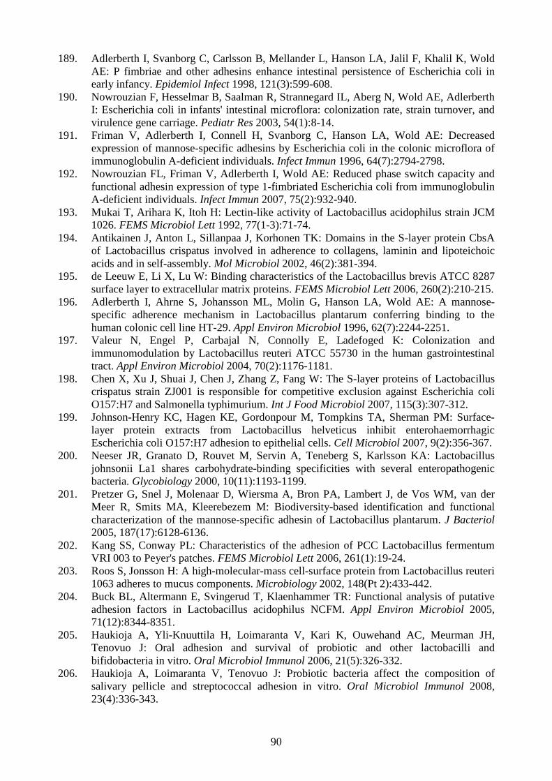
90
189. Adlerberth I, Svanborg C, Carlsson B, Mellander L, Hanson LA, Jalil F, Khalil K, Wold AE: P fimbriae and other adhesins enhance intestinal persistence of Escherichia coli in early infancy. Epidemiol Infect 1998, 121(3):599-608.
190. Nowrouzian F, Hesselmar B, Saalman R, Strannegard IL, Aberg N, Wold AE, Adlerberth I: Escherichia coli in infants' intestinal microflora: colonization rate, strain turnover, and virulence gene carriage. Pediatr Res 2003, 54(1):8-14.
191. Friman V, Adlerberth I, Connell H, Svanborg C, Hanson LA, Wold AE: Decreased expression of mannose-specific adhesins by Escherichia coli in the colonic microflora of immunoglobulin A-deficient individuals. Infect Immun 1996, 64(7):2794-2798.
192. Nowrouzian FL, Friman V, Adlerberth I, Wold AE: Reduced phase switch capacity and functional adhesin expression of type 1-fimbriated Escherichia coli from immunoglobulin A-deficient individuals. Infect Immun 2007, 75(2):932-940.
193. Mukai T, Arihara K, Itoh H: Lectin-like activity of Lactobacillus acidophilus strain JCM 1026. FEMS Microbiol Lett 1992, 77(1-3):71-74.
194. Antikainen J, Anton L, Sillanpaa J, Korhonen TK: Domains in the S-layer protein CbsA of Lactobacillus crispatus involved in adherence to collagens, laminin and lipoteichoic acids and in self-assembly. Mol Microbiol 2002, 46(2):381-394.
195. de Leeuw E, Li X, Lu W: Binding characteristics of the Lactobacillus brevis ATCC 8287 surface layer to extracellular matrix proteins. FEMS Microbiol Lett 2006, 260(2):210-215.
196. Adlerberth I, Ahrne S, Johansson ML, Molin G, Hanson LA, Wold AE: A mannose-specific adherence mechanism in Lactobacillus plantarum conferring binding to the human colonic cell line HT-29. Appl Environ Microbiol 1996, 62(7):2244-2251.
197. Valeur N, Engel P, Carbajal N, Connolly E, Ladefoged K: Colonization and immunomodulation by Lactobacillus reuteri ATCC 55730 in the human gastrointestinal tract. Appl Environ Microbiol 2004, 70(2):1176-1181.
198. Chen X, Xu J, Shuai J, Chen J, Zhang Z, Fang W: The S-layer proteins of Lactobacillus crispatus strain ZJ001 is responsible for competitive exclusion against Escherichia coli O157:H7 and Salmonella typhimurium. Int J Food Microbiol 2007, 115(3):307-312.
199. Johnson-Henry KC, Hagen KE, Gordonpour M, Tompkins TA, Sherman PM: Surface-layer protein extracts from Lactobacillus helveticus inhibit enterohaemorrhagic Escherichia coli O157:H7 adhesion to epithelial cells. Cell Microbiol 2007, 9(2):356-367.
200. Neeser JR, Granato D, Rouvet M, Servin A, Teneberg S, Karlsson KA: Lactobacillus johnsonii La1 shares carbohydrate-binding specificities with several enteropathogenic bacteria. Glycobiology 2000, 10(11):1193-1199.
201. Pretzer G, Snel J, Molenaar D, Wiersma A, Bron PA, Lambert J, de Vos WM, van der Meer R, Smits MA, Kleerebezem M: Biodiversity-based identification and functional characterization of the mannose-specific adhesin of Lactobacillus plantarum. J Bacteriol 2005, 187(17):6128-6136.
202. Kang SS, Conway PL: Characteristics of the adhesion of PCC Lactobacillus fermentum VRI 003 to Peyer's patches. FEMS Microbiol Lett 2006, 261(1):19-24.
203. Roos S, Jonsson H: A high-molecular-mass cell-surface protein from Lactobacillus reuteri 1063 adheres to mucus components. Microbiology 2002, 148(Pt 2):433-442.
204. Buck BL, Altermann E, Svingerud T, Klaenhammer TR: Functional analysis of putative adhesion factors in Lactobacillus acidophilus NCFM. Appl Environ Microbiol 2005, 71(12):8344-8351.
205. Haukioja A, Yli-Knuuttila H, Loimaranta V, Kari K, Ouwehand AC, Meurman JH, Tenovuo J: Oral adhesion and survival of probiotic and other lactobacilli and bifidobacteria in vitro. Oral Microbiol Immunol 2006, 21(5):326-332.
206. Haukioja A, Loimaranta V, Tenovuo J: Probiotic bacteria affect the composition of salivary pellicle and streptococcal adhesion in vitro. Oral Microbiol Immunol 2008, 23(4):336-343.
91
207. Stamatova I, Kari K, Vladimirov S, Meurman JH: In vitro evaluation of yoghurt starter lactobacilli and Lactobacillus rhamnosus GG adhesion to saliva-coated surfaces. Oral Microbiol Immunol 2009, 24(3):218-223.
208. Darwin KH, Miller VL: Molecular basis of the interaction of Salmonella with the intestinal mucosa. Clin Microbiol Rev 1999, 12(3):405-428.
209. Baumler AJ, Tsolis RM, Heffron F: Contribution of fimbrial operons to attachment to and invasion of epithelial cell lines by Salmonella typhimurium. Infect Immun 1996, 64(5):1862-1865.
210. Gillor O, Etzion A, Riley MA: The dual role of bacteriocins as anti- and probiotics. Appl Microbiol Biotechnol 2008, 81(4):591-606.
211. De Vuyst L, Leroy F: Bacteriocins from lactic acid bacteria: production, purification, and food applications. J Mol Microbiol Biotechnol 2007, 13(4):194-199.
212. Lin CK, Tsai HC, Lin PP, Tsen HY, Tsai CC: Lactobacillus acidophilus LAP5 able to inhibit the Salmonella choleraesuis invasion to the human Caco-2 epithelial cell. Anaerobe 2008, 14(5):251-255.
213. Sang Y, Blecha F: Antimicrobial peptides and bacteriocins: alternatives to traditional antibiotics. Anim Health Res Rev 2008, 9(2):227-235.
214. Topping DL, Clifton PM:The . Physiol Rev 2001, 81(3):1031-1064. 215. Van der Waaij D, Van der Waaij BD: The colonization resistance of the digestive tract in
different animal species and in man; a comparative study. Epidemiol Infect 1990, 105:237-243.
216. Collins FM, Carter PB: Growth of salmonellae in orally infected germfree mice. Infect Immun 1978, 21(1):41-47.
217. Alam MS: Modulation of gastrointestinal cell kinetics and endocrine cell populations by the microflora and endogenous prostaglandins. Thesis the Karolinska institute: Stockholm 1995.
218. Chowdhury SR, King DE, Willing BP, Band MR, Beever JE, Lane AB, Loor JJ, Marini JC, Rund LA, Schook LB et al: Transcriptome profiling of the small intestinal epithelium in germfree versus conventional piglets. BMC Genomics 2007, 8:215.
219. Adlerberth I, Wold AE, Establishment of the gut microbiota in Western infants, Acta pediatrica 2009, 98:229-238.
220. Stappenbeck TS, Hooper LV, Gordon JI: Developmental regulation of intestinal angiogenesis by indigenous microbes via Paneth cells. Proc Natl Acad Sci U S A 2002, 99(24):15451-15455.
221. Nicaise P, Gleizes A, Sandre C, Forestier F, Kergot R, Quero AM, Labarre C: Influence of intestinal microflora on murine bone marrow and spleen macrophage precursors. Scand J Immunol 1998, 48(6):585-591.
222. Shroff KE, Meslin K, Cebra JJ: Commensal enteric bacteria engender a self-limiting humoral mucosal immune response while permanently colonizing the gut. Infect Immun 1995, 63(10):3904-3913.
223. Zocco MA, Ainora ME, Gasbarrini G, Gasbarrini A: Bacteroides thetaiotaomicron in the gut: molecular aspects of their interaction. Dig Liver Dis 2007, 39(8):707-712.
224. Hooper LV, Midtvedt T, Gordon JI: How host-microbial interactions shape the nutrient environment of the mammalian intestine. Annu Rev Nutr 2002, 22:283-307.
225. Backhed F, Ding H, Wang T, Hooper LV, Koh GY, Nagy A, Semenkovich CF, Gordon JI: The gut microbiota as an environmental factor that regulates fat storage. Proc Natl Acad Sci U S A 2004, 101(44):15718-15723.
226. Whitehead RH, Young GP, Bhathal PS: Effects of short chain fatty acids on a new human colon carcinoma cell line (LIM1215). Gut 1986, 27(12):1457-1463.
227. Scheppach W, Christl SU, Bartram HP, Richter F, Kasper H: Effects of short-chain fatty acids on the inflamed colonic mucosa. Scand J Gastroenterol Suppl 1997, 222:53-57.
92
228. Yajima T: Contractile effect of short-chain fatty acids on the isolated colon of the rat. J Physiol 1985, 368:667-678.
229. Ostman S, Rask C, Wold AE, Hultkrantz S, Telemo E: Impaired regulatory T cell function in germ-free mice. Eur J Immunol 2006, 36(9):2336-2346.
230. Penders J, Stobberingh EE, van den Brandt PA, Thijs C: The role of the intestinal microbiota in the development of atopic disorders. Allergy 2007, 62(11):1223-1236.
231. Frank DN, St Amand AL, Feldman RA, Boedeker EC, Harpaz N, Pace NR: Molecular-phylogenetic characterization of microbial community imbalances in human inflammatory bowel diseases. Proc Natl Acad Sci U S A 2007, 104(34):13780-13785.
232. Turnbaugh PJ, Ley RE, Mahowald MA, Magrini V, Mardis ER, Gordon JI: An obesity-associated gut microbiome with increased capacity for energy harvest. Nature 2006, 444(7122):1027-1031.
233. Hogenauer C, Hammer HF, Krejs GJ, Reisinger EC: Mechanisms and management of antibiotic-associated diarrhea. Clin Infect Dis 1998, 27(4):702-710.
234. Beaugerie L, Flahault A, Barbut F, Atlan P, Lalande V, Cousin P, Cadilhac M, Petit JC: Antibiotic-associated diarrhoea and Clostridium difficile in the community. Aliment Pharmacol Ther 2003, 17(7):905-912.
235. Croswell A, Amir E, Teggatz P, Barman M, Salzman NH: Prolonged impact of antibiotics on intestinal microbial ecology and susceptibility to enteric Salmonella infection. Infect Immun 2009, 77(7):2741-2753.
236. McFarland LV: Antibiotic-associated diarrhea: epidemiology, trends and treatment. Future Microbiol 2008, 3:563-578.
237. Bartlett JG: Clinical practice. Antibiotic-associated diarrhea. N Engl J Med 2002, 346(5):334-339.
238. Deshpande A, Pant C, Jain A, Fraser TG, Rolston DD: Do fluoroquinolones predispose patients to Clostridium difficile associated disease? A review of the evidence. Curr Med Res Opin 2008, 24(2):329-333.
239. Barbut F, Lalande V, Burghoffer B, Thien HV, Grimprel E, Petit JC: Prevalence and genetic characterization of toxin A variant strains of Clostridium difficile among adults and children with diarrhea in France. J Clin Microbiol 2002, 40(6):2079-2083.
240. Wistrom J, Norrby SR, Myhre EB, Eriksson S, Granstrom G, Lagergren L, Englund G, Nord CE, Svenungsson B: Frequency of antibiotic-associated diarrhoea in 2462 antibiotic-treated hospitalized patients: a prospective study. J Antimicrob Chemother 2001, 47(1):43-50.
241. Wullt M, Johansson Hagslatt ML, Odenholt I, Berggren A: Lactobacillus plantarum 299v Enhances the Concentrations of Fecal Short-Chain Fatty Acids in Patients with Recurrent Clostridium difficile-Associated Diarrhea. Dig Dis Sci 2007.
242. Lupp C, Robertson ML, Wickham ME, Sekirov I, Champion OL, Gaynor EC, Finlay BB: Host-mediated inflammation disrupts the intestinal microbiota and promotes the overgrowth of Enterobacteriaceae. Cell Host Microbe 2007, 2(3):204.
243. Fujita K, Kaku M, Yanagase Y, Ezaki T, Furuse K, Ozawa A, Saidi SM, Sang WK, Waiyaki PG: Physicochemical characteristics and flora of diarrhoeal and recovery faeces in children with acute gastro-enteritis in Kenya. Ann Trop Paediatr 1990, 10(4):339-345.
244. Neal KR, Hebden J, Spiller R: Prevalence of gastrointestinal symptoms six months after bacterial gastroenteritis and risk factors for development of the irritable bowel syndrome: postal survey of patients. BMJ 1997, 314(7083):779-782.
245. Barbara G, Stanghellini V, Berti-Ceroni C, De Giorgio R, Salvioli B, Corradi F, Cremon C, Corinaldesi R: Role of antibiotic therapy on long-term germ excretion in faeces and digestive symptoms after Salmonella infection. Aliment Pharmacol Ther 2000, 14(9):1127-1131.
93
246. Spiller RC, Jenkins D, Thornley JP, Hebden JM, Wright T, Skinner M, Neal KR: Increased rectal mucosal enteroendocrine cells, T lymphocytes, and increased gut permeability following acute Campylobacter enteritis and in post-dysenteric irritable bowel syndrome. Gut 2000, 47(6):804-811.
247. Pimentel M, Chow EJ, Lin HC: Eradication of small intestinal bacterial overgrowth reduces symptoms of irritable bowel syndrome. Am J Gastroenterol 2000, 95(12):3503-3506.
248. King TS, Elia M, Hunter JO: Abnormal colonic fermentation in irritable bowel syndrome. Lancet 1998, 352(9135):1187-1189.
249. Balsari A, Ceccarelli A, Dubini F, Fesce E, Poli G: The fecal microbial population in the irritable bowel syndrome. Microbiologica 1982, 5(3):185-194.
250. Malinen E, Rinttila T, Kajander K, Matto J, Kassinen A, Krogius L, Saarela M, Korpela R, Palva A: Analysis of the fecal microbiota of irritable bowel syndrome patients and healthy controls with real-time PCR. Am J Gastroenterol 2005, 100(2):373-382.
251. Neal KR, Barker L, Spiller RC: Prognosis in post-infective irritable bowel syndrome: a six year follow up study. Gut 2002, 51(3):410-413.
252. FAO-WHO, ftp://ftp.fao.org/docrep/fao/009/a0512e/a0512e00.pdf FaAOotUNaWHOF-W: Probiotics in food. Health and nutritional properties and guidelines for evaluation. 2006.
253. Parry CM, Threlfall EJ: Antimicrobial resistance in typhoidal and nontyphoidal salmonellae. Curr Opin Infect Dis 2008, 21(5):531-538.
254. Martin FP, Wang Y, Sprenger N, Yap IK, Lundstedt T, Lek P, Rezzi S, Ramadan Z, van Bladeren P, Fay LB et al: Probiotic modulation of symbiotic gut microbial-host metabolic interactions in a humanized microbiome mouse model. Mol Syst Biol 2008, 4:157.
255. Rochet V, Rigottier-Gois L, Sutren M, Krementscki MN, Andrieux C, Furet JP, Tailliez P, Levenez F, Mogenet A, Bresson JL et al: Effects of orally administered Lactobacillus casei DN-114 001 on the composition or activities of the dominant faecal microbiota in healthy humans. Br J Nutr 2006, 95(2):421-429.
256. Marzotto M, Maffeis C, Paternoster T, Ferrario R, Rizzotti L, Pellegrino M, Dellaglio F, Torriani S: Lactobacillus paracasei A survives gastrointestinal passage and affects the fecal microbiota of healthy infants. Res Microbiol 2006, 157(9):857-866.
257. Wheeler JG, Bogle ML, Shema SJ, Shirrell MA, Pittler AJ, Burks AW, Helm RM: Immune and clinical impact of Lactobacillus acidophilus on asthma. Ann Allergy Asthma immunol 1997, 79:229-233.
258. Schiffrin EJ, Rochat F, Link-Amster H, Aeschlimann JM, Donnet-Hughes A: Immunomodulation of human blood cells following the ingestion of lactic acid bacteria. J Dairy Sci 1995, 78(3):491-497.
259. Donnet-Hughes A, Rochat F, Serrant P, Aeschlimann JM, Schiffrin EJ: Modulation of nonspecific mechanisms of defense by lactic acid bacteria: effective dose. J Dairy Sci 1999, 82(5):863-869.
260. Arunachalam K, Gill HS, Chandra RK: Enhancement of natural immune function by dietary consumption of Bifidobacterium lactis (HN019). Eur J Clin Nutr 2000, 54(3):263-267.
261. Pelto L, Isolauri E, Lilius EM, Nuutila J, Salminen S: Probiotic bacteria down-regulate the milk-induced inflammatory response in milk-hypersensitive subjects but have an immunostimulatory effect in healthy subjects. Clin Exp Allergy 1998, 28(12):1474-1479.
262. Fang H, Elina T, Heikki A, Seppo S: Modulation of humoral immune response through probiotic intake. FEMS Immunol Med Microbiol 2000, 29(1):47-52.
263. Korhonen R, Korpela R, Moilanen E: Signalling mechanisms involved in the induction of inducible nitric oxide synthase by Lactobacillus rhamnosus GG, endotoxin, and lipoteichoic acid. Inflammation 2002, 26(5):207-214.
94
264. Kekkonen RA, Lummela N, Karjalainen H, Latvala S, Tynkkynen S, Jarvenpaa S, Kautiainen H, Julkunen I, Vapaatalo H, Korpela R: Probiotic intervention has strain-specific anti-inflammatory effects in healthy adults. World J Gastroenterol 2008, 14(13):2029-2036.
265. Bernet MF, Brassart D, Neeser JR, Servin AL: Lactobacillus acidophilus LA 1 binds to cultured human intestinal cell lines and inhibits cell attachment and cell invasion by enterovirulent bacteria. Gut 1994, 35(4):483-489.
266. Mack DR, Ahrne S, Hyde L, Wei S, Hollingsworth MA: Extracellular MUC3 mucin secretion follows adherence of Lactobacillus strains to intestinal epithelial cells in vitro. Gut 2003, 52(6):827-833.
267. Caballero-Franco C, Keller K, De Simone C, Chadee K: The VSL#3 probiotic formula induces mucin gene expression and secretion in colonic epithelial cells. Am J Physiol Gastrointest Liver Physiol 2007, 292(1):G315-322.
268. Guandalini S, Pensabene L, Zikri MA, Dias JA, Casali LG, Hoekstra H, Kolacek S, Massar K, Micetic-Turk D, Papadopoulou A et al: Lactobacillus GG administered in oral rehydration solution to children with acute diarrhea: a multicenter European trial. J Pediatr Gastroenterol Nutr 2000, 30(1):54-60.
269. Dubey AP, Rajeshwari K, Chakravarty A, Famularo G: Use of VSL[sharp]3 in the treatment of rotavirus diarrhea in children: preliminary results. J Clin Gastroenterol 2008, 42 Suppl 3 Pt 1:S126-129.
270. Surawicz CM, Elmer GW, Speelman P, McFarland LV, Chinn J, van Belle G: Prevention of antibiotic-associated diarrhea by Saccharomyces boulardii: a prospective study. Gastroenterology 1989, 96(4):981-988.
271. Arvola T, Laiho K, Torkkeli S, Mykkanen H, Salminen S, Maunula L, Isolauri E: Prophylactic Lactobacillus GG reduces antibiotic-associated diarrhea in children with respiratory infections: a randomized study. Pediatrics 1999, 104(5):e64.
272. Hickson M, D'Souza AL, Muthu N, Rogers TR, Want S, Rajkumar C, Bulpitt CJ: Use of probiotic Lactobacillus preparation to prevent diarrhoea associated with antibiotics: randomised double blind placebo controlled trial. Bmj 2007, 335(7610):80.
273. Nase L, Hatakka K, Savilahti E, Saxelin M, Ponka A, Poussa T, Korpela R, Meurman JH: Effect of long-term consumption of a probiotic bacterium, Lactobacillus rhamnosus GG, in milk on dental caries and caries risk in children. Caries Res 2001, 35(6):412-420.
274. Olah A, Belagyi T, Issekutz A, Gamal ME, Bengmark S: Randomized clinical trial of specific lactobacillus and fibre supplement to early enteral nutrition in patients with acute pancreatitis. Br J Surg 2002, 89(9):1103-1107.
275. McFarland LV, Surawicz CM, Greenberg RN, Elmer GW, Moyer KA, Melcher SA, Bowen KE, Cox JL: Prevention of beta-lactam-associated diarrhea by Saccharomyces boulardii compared with placebo. Am J Gastroenterol 1995, 90(3):439-448.
276. Kotowska M, Albrecht P, Szajewska H: Saccharomyces boulardii in the prevention of antibiotic-associated diarrhoea in children: a randomized double-blind placebo-controlled trial. Aliment Pharmacol Ther 2005, 21(5):583-590.
277. Vanderhoof JA, Whitney DB, Antonson DL, Hanner TL, Lupo JV, Young RJ: Lactobacillus GG in the prevention of antibiotic-associated diarrhea in children. J Pediatr 1999, 135(5):564-568.
278. Armuzzi A, Cremonini F, Bartolozzi F, Canducci F, Candelli M, Ojetti V, Cammarota G, Anti M, De Lorenzo A, Pola P et al: The effect of oral administration of Lactobacillus GG on antibiotic-associated gastrointestinal side-effects during Helicobacter pylori eradication therapy. Aliment Pharmacol Ther 2001, 15(2):163-169.
279. Siitonen S, Vapaatalo H, Salminen S, Gordin A, Saxelin M, Wikberg R, Kirkkola AL: Effect of Lactobacillus GG yoghurt in prevention of antibiotic associated diarrhoea. Ann Med 1990, 22(1):57-59.
95
280. Wullt M, Hagslatt ML, Odenholt I: Lactobacillus plantarum 299v for the treatment of recurrent Clostridium difficile-associated diarrhoea: a double-blind, placebo-controlled trial. Scand J Infect Dis 2003, 35(6-7):365-367.
281. Gorbach SL, Chang TW, Goldin B: Successful treatment of relapsing Clostridium difficile colitis with Lactobacillus GG [letter]. Lancet 1987, 2(8574):1519.
282. Surawicz CM, McFarland LV, Greenberg RN, Rubin M, Fekety R, Mulligan ME, Garcia RJ, Brandmarker S, Bowen K, Borjal D et al: The search for a better treatment for recurrent Clostridium difficile disease: use of high-dose vancomycin combined with Saccharomyces boulardii. Clin Infect Dis 2000, 31(4):1012-1017.
283. Ruszczynski M, Radzikowski A, Szajewska H: Clinical trial: effectiveness of Lactobacillus rhamnosus (strains E/N, Oxy and Pen) in the prevention of antibiotic-associated diarrhoea in children. Aliment Pharmacol Ther 2008, 28(1):154-161.
284. Sokol H, Pigneur B, Watterlot L, Lakhdari O, Bermudez-Humaran LG, Gratadoux JJ, Blugeon S, Bridonneau C, Furet JP, Corthier G et al: Faecalibacterium prausnitzii is an anti-inflammatory commensal bacterium identified by gut microbiota analysis of Crohn disease patients. Proc Natl Acad Sci U S A 2008, 105(43):16731-16736.
285. Gionchetti P, Rizzello F, Venturi A, Brigidi P, Matteuzzi D, Bazzocchi G, Poggioli G, Miglioli M, Campieri M: Oral bacteriotherapy as maintenance treatment in patients with chronic pouchitis: a double-blind, placebo-controlled trial [see comments]. Gastroenterology 2000, 119(2):305-309.
286. Niedzielin K, Kordecki H, Birkenfeld B: A controlled, double-blind, randomized study on the efficacy of Lactobacillus plantarum 299V in patients with irritable bowel syndrome. Eur J Gastroenterol Hepatol 2001, 13(10):1143-1147.
287. Guyonnet D, Chassany O, Ducrotte P, Picard C, Mouret M, Mercier CH, Matuchansky C: Effect of a fermented milk containing Bifidobacterium animalis DN-173 010 on the health-related quality of life and symptoms in irritable bowel syndrome in adults in primary care: a multicentre, randomized, double-blind, controlled trial. Aliment Pharmacol Ther 2007, 26(3):475-486.
288. Uehara S, Monden K, Nomoto K, Seno Y, Kariyama R, Kumon H: A pilot study evaluating the safety and effectiveness of Lactobacillus vaginal suppositories in patients with recurrent urinary tract infection. Int J Antimicrob Agents 2006, 28 Suppl 1:S30-34.
289. Lin HC, Su BH, Chen AC, Lin TW, Tsai CH, Yeh TF, Oh W: Oral probiotics reduce the incidence and severity of necrotizing enterocolitis in very low birth weight infants. Pediatrics 2005, 115(1):1-4.
290. Bin-Nun A, Bromiker R, Wilschanski M, Kaplan M, Rudensky B, Caplan M, Hammerman C: Oral probiotics prevent necrotizing enterocolitis in very low birth weight neonates. J Pediatr 2005, 147(2):192-196.
291. Kalliomaki M, Salminen S, Poussa T, Isolauri E: Probiotics during the first 7 years of life: a cumulative risk reduction of eczema in a randomized, placebo-controlled trial. J Allergy Clin Immunol 2007, 119(4):1019-1021.
292. McFarland LV: Meta-analysis of probiotics for the prevention of antibiotic associated diarrhea and the treatment of Clostridium difficile disease. Am J Gastroenterol 2006, 101(4):812-822.
293. Graul T, Cain AM, Karpa KD: Lactobacillus and bifidobacteria combinations: a strategy to reduce hospital-acquired Clostridium difficile diarrhea incidence and mortality. Med Hypotheses 2009, 73(2):194-198.
294. Gorbach SL: Probiotics and gastrointestinal health. Am J Gastroenterol 2000, 95(1 Suppl):S2-4.
295. Mortensen PB, Clausen MR: Short-chain fatty acids in the human colon: relation to gastrointestinal health and disease. Scand J Gastroenterol Suppl 1996, 216:132-148.
96
296. Pascual M, Hugas M, Badiola JI, Monfort JM, Garriga M: Lactobacillus salivarius CTC2197 prevents Salmonella enteritidis colonization in chickens. Appl Environ Microbiol 1999, 65(11):4981-4986.
297. Casey PG, Gardiner GE, Casey G, Bradshaw B, Lawlor PG, Lynch PB, Leonard FC, Stanton C, Ross RP, Fitzgerald GF et al: A five-strain probiotic combination reduces pathogen shedding and alleviates disease signs in pigs challenged with Salmonella enterica Serovar Typhimurium. Appl Environ Microbiol 2007, 73(6):1858-1863.
298. Silva AM, Barbosa FH, Duarte R, Vieira LQ, Arantes RM, Nicoli JR: Effect of Bifidobacterium longum ingestion on experimental salmonellosis in mice. J Appl Microbiol 2004, 97(1):29-37.
299. La Ragione RM, Narbad A, Gasson MJ, Woodward MJ: In vivo characterization of Lactobacillus johnsonii FI9785 for use as a defined competitive exclusion agent against bacterial pathogens in poultry. Lett Appl Microbiol 2004, 38(3):197-205.
300. Alm L: The effect of Lactobacillus acidophilus administration upon the survival of Salmonella in randomly selected human carriers. Prog Food Nutr Sci 1983, 7(3-4):13-17.
301. Jin LZ, Ho YW, Ali MA, Abdullah N, Jalaludin S: Effect of adherent Lactobacillus spp. on in vitro adherence of salmonellae to the intestinal epithelial cells of chicken. J Appl Bacteriol 1996, 81(2):201-206.
302. Candela M, Perna F, Carnevali P, Vitali B, Ciati R, Gionchetti P, Rizzello F, Campieri M, Brigidi P: Interaction of probiotic Lactobacillus and Bifidobacterium strains with human intestinal epithelial cells: adhesion properties, competition against enteropathogens and modulation of IL-8 production. Int J Food Microbiol 2008, 125(3):286-292.
303. Makras L, Triantafyllou V, Fayol-Messaoudi D, Adriany T, Zoumpopoulou G, Tsakalidou E, Servin A, De Vuyst L: Kinetic analysis of the antibacterial activity of probiotic lactobacilli towards Salmonella enterica serovar Typhimurium reveals a role for lactic acid and other inhibitory compounds. Res Microbiol 2006, 157(3):241-247.
304. Ushijima T, Seto A: Selected faecal bacteria and nutrients essential for antagonism of Salmonella typhimurium in anaerobic continuous flow cultures. J Med Microbiol 1991, 35(2):111-117.
305. Vinderola G, Matar C, Perdigon G: Milk fermented by Lactobacillus helveticus R389 and its non-bacterial fraction confer enhanced protection against Salmonella enteritidis serovar Typhimurium infection in mice. Immunobiology 2007, 212(2):107-118.
306. Lin WH, Yu B, Lin CK, Hwang WZ, Tsen HY: Immune effect of heat-killed multistrain of Lactobacillus acidophilus against Salmonella typhimurium invasion to mice. J Appl Microbiol 2007, 102(1):22-31.
307. Wagner RD, Johnson SJ, Kurniasih Rubin D: Probiotic bacteria are antagonistic to Salmonella enterica and Campylobacter jejuni and influence host lymphocyte responses in human microbiota-associated immunodeficient and immunocompetent mice. Mol Nutr Food Res 2009, 53(3):377-388.
308. Johansson ML, Nobaek S, Berggren A, Nyman M, Bjorck I, Ahrne S, Jeppsson B, Molin G: Survival of Lactobacillus plantarum DSM 9843 (299v), and effect on the short-chain fatty acid content of faeces after ingestion of a rose-hip drink with fermented oats. Int J Food Microbiol 1998, 42(1-2):29-38.
309. Goossens D, Jonkers D, Russel M, Stobberingh E, Van Den Bogaard A, StockbrUgger R: The effect of Lactobacillus plantarum 299v on the bacterial composition and metabolic activity in faeces of healthy volunteers: a placebo-controlled study on the onset and duration of effects. Aliment Pharmacol Ther 2003, 18(5):495-505.
310. Liu Q, Nobaek S, Adawi D, Mao Y, Wang M, Molin G, Ekelund M, Jeppsson B: Administration of Lactobacillus plantarum 299v reduces side-effects of external radiation on colon anastomotic healing in an experimental model. Colorectal Dis 2001, 3(4):245-252.
97
311. Osman N, Adawi D, Ahrne S, Jeppsson B, Molin G: Modulation of the effect of dextran sulfate sodium-induced acute colitis by the administration of different probiotic strains of Lactobacillus and Bifidobacterium. Dig Dis Sci 2004, 49(2):320-327.
312. Mangell P, Nejdfors P, Wang M, Ahrne S, Westrom B, Thorlacius H, Jeppsson B: Lactobacillus plantarum 299v inhibits Escherichia coli-induced intestinal permeability. Dig Dis Sci 2002, 47(3):511-516.
313. Nobaek S, Johansson ML, Molin G, Ahrne S, Jeppsson B: Alteration of intestinal microflora is associated with reduction in abdominal bloating and pain in patients with irritable bowel syndrome. Am J Gastroenterol 2000, 95(5):1231-1238.
314. Sen S, Mullan MM, Parker TJ, Woolner JT, Tarry SA, Hunter JO: Effect of Lactobacillus plantarum 299v on colonic fermentation and symptoms of irritable bowel syndrome. Dig Dis Sci 2002, 47(11):2615-2620.
315. Rayes N, Seehofer D, Hansen S, Boucsein K, Muller AR, Serke S, Bengmark S, Neuhaus P: Early enteral supply of lactobacillus and fiber versus selective bowel decontamination: a controlled trial in liver transplant recipients. Transplantation 2002, 74(1):123-127.
316. Klarin B, Wullt M, Palmquist I, Molin G, Larsson A, Jeppsson B: Lactobacillus plantarum 299v reduces colonisation of Clostridium difficile in critically ill patients treated with antibiotics. Acta Anaesthesiol Scand 2008, 52(8):1096-1102.
317. Snydman DR: The safety of probiotics. Clin Infect Dis 2008, 46 Suppl 2:S104-111; discussion S144-151.
318. Cassone M, Serra P, Mondello F, Girolamo A, Scafetti S, Pistella E, Venditti M: Outbreak of Saccharomyces cerevisiae subtype boulardii fungemia in patients neighboring those treated with a probiotic preparation of the organism. J Clin Microbiol 2003, 41(11):5340-5343.
319. Borriello SP, Hammes WP, Holzapfel W, Marteau P, Schrezenmeir J, Vaara M, Valtonen V: Safety of probiotics that contain lactobacilli or bifidobacteria. Clin Infect Dis 2003, 36(6):775-780.
320. Ishibashi N, Yamazaki S: Probiotics and safety. Am J Clin Nutr 2001, 73(2 Suppl):465S-470S.
321. Husni RN, Gordon SM, Washington JA, Longworth DL: Lactobacillus bacteremia and endocarditis: review of 45 cases. Clin Infect Dis 1997, 25(5):1048-1055.
322. Cannon JP, Lee TA, Bolanos JT, Danziger LH: Pathogenic relevance of Lactobacillus: a retrospective review of over 200 cases. Eur J Clin Microbiol Infect Dis 2005, 24(1):31-40.
323. Gevers D, Huys G, Swings J: In vitro conjugal transfer of tetracycline resistance from Lactobacillus isolates to other Gram-positive bacteria. FEMS Microbiol Lett 2003, 225(1):125-130.
324. Mathur S, Singh R: Antibiotic resistance in food lactic acid bacteria--a review. Int J Food Microbiol 2005, 105(3):281-295.
325. Jacobsen L, Wilcks A, Hammer K, Huys G, Gevers D, Andersen SR: Horizontal transfer of tet(M) and erm(B) resistance plasmids from food strains of Lactobacillus plantarum to Enterococcus faecalis JH2-2 in the gastrointestinal tract of gnotobiotic rats. FEMS Microbiol Ecol 2007, 59(1):158-166.
326. Toomey N, Monaghan A, Fanning S, Bolton D: Transfer of antibiotic resistance marker genes between lactic acid bacteria in model rumen and plant environments. Appl Environ Microbiol 2009, 75(10):3146-3152.
327. Hanson LA: Selective IgA deficiency, vol. Edited by nataro JP, Blaser MJ, Cunnigham-Rundles S. Edinburgh: Churchill Livingstone; 1983.
328. Grimont PAD, Weill FX: Antigenic Formulas of the Salmonella Serovars, 9th revision. WHO collaborating Centre for Reference and Research on Salmonella, Institut Pasteur, Paris 2007.
98
329. Rogosa M, Mitchell JA, Wiseman RF: A selective medium for the isolation and enumeration of oral and fecal lactobacilli. J Bacteriol 1951:132-133.
330. Efthymiou C, Hansen PA: An antigenic analysis of Lactobacillus acidophilus. J Infect Dis 1962, 110:258-267.
331. Antonsson M, Molin G, Ardo Y: Lactobacillus strains isolated from Danbo cheese as adjunct cultures in a cheese model system. Int J Food Microbiol 2003, 85(1-2):159-169.
332. Roy D, Sirois S: Molecular differentiation of Bifidobacterium species with amplified ribosomal DNA restriction analysis and alignment of short regions of the ldh gene. FEMS Microbiol Lett 2000, 191(1):17-24.
333. Fritzell P, Bergstrom T, Welinder-Olsson C: Detection of bacterial DNA in painful degenerated spinal discs in patients without signs of clinical infection. Eur Spine J 2004, 13(8):702-706.
334. Johnson S, Sambol SP, Brazier JS, Delmee M, Avesani V, Merrigan MM, Gerding DN: International typing study of toxin A-negative, toxin B-positive Clostridium difficile variants. J Clin Microbiol 2003, 41(4):1543-1547.
335. National Clostridium difficile Standards Group: Report to the Department of Health. J Hosp Infect 2004, 56 Suppl 1:1-38.
336. Hallander H, Thore M, Wollin R: Referensmetodik för laboratoriediagnostik vid kliniskt mikrobiologiska laboratorier; I Infektionsdiagnostik; II Infektioner i mag-tarmkanalen. 2002.
337. Burton P, Gurrin L, Sly P: Extending the simple linear regression model to account for correlated responses: an introduction to generalized estimating equations and multi-level mixed modelling. Stat Med 1998, 17(11):1261-1291.
338. Hanley JA, Negassa A, Edwardes MD, Forrester JE: Statistical analysis of correlated data using generalized estimating equations: an orientation. Am J Epidemiol 2003, 157(4):364-375.
339. Witkin SS, Linhares IM, Giraldo P: Bacterial flora of the female genital tract: function and immune regulation. Best Pract Res Clin Obstet Gynaecol 2007, 21(3):347-354.
340. Ellis-Pegler RB, Crabtree C, Lambert HP: The faecal flora of children in the United Kingdom. J Hyg (Lond) 1975, 75(1):135-142.
341. Sjogren YM, Jenmalm MC, Bottcher MF, Bjorksten B, Sverremark-Ekstrom E: Altered early infant gut microbiota in children developing allergy up to 5 years of age. Clin Exp Allergy 2009, 39(4):518-526.
342. Mikelsaar M, Annuk H, Shchepetova J, Mändar R, Sepp E, Bjorksten B: Intestinal Lactobacilli of Estonian and Swedish Children. Microbial ecology in health and disease 2002, 14:75-80.
343. Huurre A, Kalliomaki M, Rautava S, Rinne M, Salminen S, Isolauri E: Mode of delivery - effects on gut microbiota and humoral immunity. Neonatology 2008, 93(4):236-240.
344. Tanaka S, Kobayashi T, Songjinda P, Tateyama A, Tsubouchi M, Kiyohara C, Shirakawa T, Sonomoto K, Nakayama J: Influence of antibiotic exposure in the early postnatal period on the development of intestinal microbiota. FEMS Immunol Med Microbiol 2009, 56(1):80-87.
345. Mevissen-Verhage EA, Marcelis JH, Harmsen-van Amerongen WC, de Vos NM, Berkel J, Verhoef J: Effect of iron on neonatal gut flora during the first week of life. Eur J Clin Microbiol 1985, 4(1):14-18.
346. Harmsen HJ, Wildeboer-Veloo AC, Raangs GC, Wagendorp AA, Klijn N, Bindels JG, Welling GW: Analysis of intestinal flora development in breast-fed and formula-fed infants by using molecular identification and detection methods. J Pediatr Gastroenterol Nutr 2000, 30(1):61-67.
99
347. Rinne M, Kalliomaki M, Arvilommi H, Salminen S, Isolauri E: Effect of probiotics and breastfeeding on the bifidobacterium and lactobacillus/enterococcus microbiota and humoral immune responses. J Pediatr 2005, 147(2):186-191.
348. Simhon A, Douglas JR, Drasar BS, Soothill JF: Effect of feeding on infants' faecal flora. Arch Dis Child 1982, 57(1):54-58.
349. Lundequist B, Nord CE, Winberg J: The composition of the faecal microflora in breastfed and bottle fed infants from birth to eight weeks. Acta Paediatr Scand 1985, 74(1):45-51.
350. Palmer C, Bik EM, Digiulio DB, Relman DA, Brown PO: Development of the Human Infant Intestinal Microbiota. PLoS Biol 2007, 5(7):e177.
351. Martin R, Langa S, Reviriego C, Jiminez E, Marin ML, Xaus J, Fernandez L, Rodriguez JM: Human milk is a source of lactic acid bacteria for the infant gut. J Pediatr 2003, 143(6):754-758.
352. Perez PF, Dore J, Leclerc M, Levenez F, Benyacoub J, Serrant P, Segura-Roggero I, Schiffrin EJ, Donnet-Hughes A: Bacterial imprinting of the neonatal immune system: lessons from maternal cells? Pediatrics 2007, 119(3):e724-732.
353. Zhou X, Bent SJ, Schneider MG, Davis CC, Islam MR, Forney LJ: Characterization of vaginal microbial communities in adult healthy women using cultivation-independent methods. Microbiology 2004, 150(Pt 8):2565-2573.
354. Azcarate-Peril MA, Altermann E, Goh YJ, Tallon R, Sanozky-Dawes RB, Pfeiler EA, O'Flaherty S, Buck BL, Dobson A, Duong T et al: Analysis of the genome sequence of Lactobacillus gasseri ATCC 33323 reveals the molecular basis of an autochthonous intestinal organism. Appl Environ Microbiol 2008, 74(15):4610-4625.
355. Tanasupawat S, Okada S, Komagata K: Lactic acid bacteria found in fermented fish in Thailand. J Gen Appl Microbiol 1998, 44(3):193-200.
356. Rodas AM, Ferrer S, Pardo I: Polyphasic study of wine Lactobacillus strains: taxonomic implications. Int J Syst Evol Microbiol 2005, 55(Pt 1):197-207.
357. Van Houte J, Green DB: Relationship between the concentration of bacteria in saliva and the colonization of teeth in humans. Infect Immun 1974, 9(4):624-630.
358. Sakki T, Knuuttila M: Controlled study of the association of smoking with lactobacilli, mutans streptococci and yeasts in saliva. Eur J Oral Sci 1996, 104(5-6):619-622.
359. Narhi TO, Kurki N, Ainamo A: Saliva, salivary micro-organisms, and oral health in the home-dwelling old elderly--a five-year longitudinal study. J Dent Res 1999, 78(10):1640-1646.
360. Kristoffersson K, Birkhed D: Effects of partial sugar restriction for 6 weeks on numbers of Streptococcus mutans in saliva and interdental plaque in man. Caries Res 1987, 21(1):79-86.
361. Matee MI, Mikx FH, Maselle SY, Van Palenstein Helderman WH: Mutans streptococci and lactobacilli in breast-fed children with rampant caries. Caries Res 1992, 26(3):183-187.
362. Rodis OM, Matsumura S, Kariya N, Okazaki Y, Ogata S, Reissmann DR: Culture-based PCR analysis of plaque samples of Japanese school children to assess the presence of six common cariogenic bacteria and its association with caries risk. Mol Cell Probes 2009, 23(6):259-263.
363. Chung J, Ha ES, Park HR, Kim S: Isolation and characterization of Lactobacillus species inhibiting the formation of Streptococcus mutans biofilm. Oral Microbiol Immunol 2004, 19(3):214-216.
364. Cebra JJ: Influences of microbiota on intestinal immune system development. Am J Clin Nutr 1999, 69(5):1046S-1051S.
365. Velez MP, Keersmaecker SC, Vanderleyden J: Adherence factors of Lactobacillus in the human gastrointestinal tract. FEMS Microbiol Lett 2007.
100
366. Brandtzaeg P, Baekkevold ES, Farstad IN, Jahnsen FL, Johansen FE, Nilsen EM, Yamanaka T: Regional specialization in the mucosal immune system: what happens in the microcompartments? Immunol Today 1999, 20(3):141-151.
367. Johansson ML, Molin G, Jeppsson B, Nobaek S, Ahrne S, Bengmark S: Administration of different Lactobacillus strains in fermented oatmeal soup: in vivo colonization of human intestinal mucosa and effect on the indigenous flora. Appl Environ Microbiol 1993, 59(1):15-20.
368. McFarland LV, Clarridge JE, Beneda HW, Raugi GJ: Fluoroquinolone use and risk factors for Clostridium difficile-associated disease within a Veterans Administration health care system. Clin Infect Dis 2007, 45(9):1141-1151.
369. McFarland LV: Epidemiology of infectious and iatrogenic nosocomial diarrhea in a cohort of general medicine patients. Am J Infect Control 1995, 23(5):295-305.
370. Diop L, Guillou S, Durand H: Probiotic food supplement reduces stress-induced gastrointestinal symptoms in volunteers: a double-blind, placebo-controlled, randomized trial. Nutr Res 2008, 28(1):1-5.
371. Kim KH, Fekety R, Batts DH, Brown D, Cudmore M, Silva J, Jr., Waters D: Isolation of Clostridium difficile from the environment and contacts of patients with antibiotic-associated colitis. J Infect Dis 1981, 143(1):42-50.
372. Roberts K, Smith CF, Snelling AM, Kerr KG, Banfield KR, Sleigh PA, Beggs CB: Aerial dissemination of Clostridium difficile spores. BMC Infect Dis 2008, 8:7.
373. McFarland LV, Mulligan ME, Kwok RY, Stamm WE: Nosocomial acquisition of Clostridium difficile infection. N Engl J Med 1989, 320(4):204-210.
374. Goossens D, Jonkers D, Russel M, Thijs A, van den Bogaard A, Stobberingh E, Stockbrugger R: Survival of the probiotic, L. plantarum 299v and its effects on the faecal bacterial flora, with and without gastric acid inhibition. Dig Liver Dis 2005, 37(1):44-50.
375. Goossens DA, Jonkers DM, Russel MG, Stobberingh EE, Stockbrugger RW: The effect of a probiotic drink with Lactobacillus plantarum 299v on the bacterial composition in faeces and mucosal biopsies of rectum and ascending colon. Aliment Pharmacol Ther 2006, 23(2):255-263.
376. Biller JA, Katz AJ, Flores AF, Buie TM, Gorbach SL: Treatment of recurrent Clostridium difficile colitis with Lactobacillus GG. J Pediatr Gastroenterol Nutr 1995, 21(2):224-226.
377. McFarland LV, Surawicz CM, Greenberg RN, Fekety R, Elmer GW, Moyer KA, Melcher SA, Bowen KE, Cox JL, Noorani Z et al: A randomized placebo-controlled trial of Saccharomyces boulardii in combination with standard antibiotics for Clostridium difficile disease [published erratum appears in JAMA 1994 Aug 17;272(7):518]. Jama 1994, 271(24):1913-1918.
378. Sazawal S, Hiremath G, Dhingra U, Malik P, Deb S, Black RE: Efficacy of probiotics in prevention of acute diarrhoea: a meta-analysis of masked, randomised, placebo-controlled trials. Lancet Infect Dis 2006, 6(6):374-382.
379. Higashikawa F, Noda M, Awaya T, Nomura K, Oku H, Sugiyama M: Improvement of constipation and liver function by plant-derived lactic acid bacteria: A double-blind, randomized trial. Nutrition 2009.
380. Gross G, van der Meulen J, Snel J, van der Meer R, Kleerebezem M, Niewold TA, Hulst MM, Smits MA: Mannose-specific interaction of Lactobacillus plantarum with porcine jejunal epithelium. FEMS Immunol Med Microbiol 2008, 54(2):215-223.
381. Gibson GR, Roberfroid MB: Dietary modulation of the human colonic microbiota: introducing the concept of prebiotics. J Nutr 1995, 125(6):1401-1412.
382. Drzikova B, Dongowski G, Gebhardt E: Dietary fibre-rich oat-based products affect serum lipids, microbiota, formation of short-chain fatty acids and steroids in rats. Br J Nutr 2005, 94(6):1012-1025.
101
383. Kedia G, Vazquez JA, Charalampopoulos D, Pandiella SS: In vitro fermentation of oat bran obtained by debranning with a mixed culture of human fecal bacteria. Curr Microbiol 2009, 58(4):338-342.
384. Anthony JP, Fyfe L, Stewart D, McDougall GJ, Smith HV: The effect of blueberry extracts on Giardia duodenalis viability and spontaneous excystation of Cryptosporidium parvum oocysts, in vitro. Methods 2007, 42(4):339-348.
385. Osman N, Adawi D, Ahrne S, Jeppsson B, Molin G: Probiotics and blueberry attenuate the severity of dextran sulfate sodium (DSS)-induced colitis. Dig Dis Sci 2008, 53(9):2464-2473.
386. Teff KL, Alavi A, Chen J, Pourdehnad M, Townsend RR: Muscarinic blockade inhibits gastric emptying of mixed-nutrient meal: effects of weight and gender. Am J Physiol 1999, 276(3 Pt 2):R707-714.
387. Reller ME, Tauxe RV, Kalish LA, Molbak K: Excess salmonellosis in women in the United States: 1968-2000. Epidemiol Infect 2008, 136(8):1109-1117.
388. el-Radhi AS, Rostila T, Vesikari T: Association of high fever and short bacterial excretion after salmonellosis. Arch Dis Child 1992, 67(4):531-532.
389. Caron J, Loredo-Osti JC, Morgan K, Malo D: Mapping of interactions and mouse congenic strains identified novel epistatic QTLs controlling the persistence of Salmonella Enteritidis in mice. Genes Immun 2005, 6(6):500-508.
390. Caron J, Lariviere L, Nacache M, Tam M, Stevenson MM, McKerly C, Gros P, Malo D: Influence of Slc11a1 on the outcome of Salmonella enterica serovar Enteritidis infection in mice is associated with Th polarization. Infect Immun 2006, 74(5):2787-2802.
391. Jouanguy E, Doffinger R, Dupuis S, Pallier A, Altare F, Casanova JL: IL-12 and IFN-gamma in host defense against mycobacteria and salmonella in mice and men. Curr Opin Immunol 1999, 11(3):346-351.
392. Liva SM, Voskuhl RR: Testosterone acts directly on CD4+ T lymphocytes to increase IL-10 production. J Immunol 2001, 167(4):2060-2067.
393. Wilcoxen SC, Kirkman E, Dowdell KC, Stohlman SA: Gender-dependent IL-12 secretion by APC is regulated by IL-10. J Immunol 2000, 164(12):6237-6243.
394. Bouman A, Heineman MJ, Faas MM: Sex hormones and the immune response in humans. Hum Reprod Update 2005, 11(4):411-423.
395. Spiller R, Garsed K: Infection, inflammation, and the irritable bowel syndrome. Dig Liver Dis 2009.
396. Gonsalkorale WM, Perrey C, Pravica V, Whorwell PJ, Hutchinson IV: Interleukin 10 genotypes in irritable bowel syndrome: evidence for an inflammatory component? Gut 2003, 52(1):91-93.
397. van der Veek PP, van den Berg M, de Kroon YE, Verspaget HW, Masclee AA: Role of tumor necrosis factor-alpha and interleukin-10 gene polymorphisms in irritable bowel syndrome. Am J Gastroenterol 2005, 100(11):2510-2516.