msu/cse fall 20141 computer vision: imaging devices summary of several common imaging devices...
TRANSCRIPT

MSU/CSE Fall 2014 1
Computer Vision: Imaging Devices
Summary of several common imaging devices covered ( Chapter 2 of Shapiro and
Stockman)

MSU/CSE Fall 2014 2
Major issues What radiation is sensed? Is motion used to scan the scene or are
all sensing elements static? How fast can sensing occur? What are the sensing elements? How is resolution determined? -- in intensity -- in space

MSU/CSE Fall 2014 3
CCD type camera Array of small
fixed elements Can read faster
than TV rates Can add
refracting elts to get color in 2x2 neighborhoods
8-bit intensity common

MSU/CSE Fall 2014 4
Blooming Problem with Arrays Difficult to
insulate adjacent sensing elements.
Charge often leaks from hot cells to neighbors, making bright regions larger.

MSU/CSE Fall 2014 5
8-bit intensity can be clipped
Dark grid intersections at left were actually brightest of scene.
In A/D conversion the bright values were clipped to lower values.

MSU/CSE Fall 2014 6
Lens distortion distorts image “Barrel distortion” of
rectangular grid is common for cheap lenses ($50). “Pin cushion” opposite.
Precision lenses can cost $1000 or more.
Zoom lenses often show severe distortion.

MSU/CSE Fall 2014 7
Other important effects/problems
Discrete pixel effect: signal is integrated
Mixed pixel effect: materials mix
Thin features can be missed
Measurements in pixels have error
Region or material A
Region or material B

MSU/CSE Fall 2014 8
Orbiting satellite scanner View earth 1 pixel
at a time (through a straw)
Prism produces multispectral pixel
Image row by scanning boresight
All rows by motion of satellite in orbit
Scanned area of earth is a parallelogram, not a rectangle
MSU/CSE Fall 2014 9
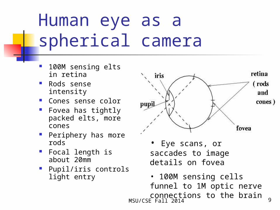
Human eye as a spherical camera 100M sensing elts in
retina Rods sense intensity Cones sense color Fovea has tightly
packed elts, more cones
Periphery has more rods
Focal length is about 20mm
Pupil/iris controls light entry
• Eye scans, or saccades to image details on fovea
• 100M sensing cells funnel to 1M optic nerve connections to the brain

MSU/CSE Fall 2014 10
Surface data (2.5D) sensed by structured light sensor
Projector projects plane of light on object
Camera sees bright points along an imaging ray
Compute 3D surface point via line-plane intersection
REF: new Minolta Vivid 910 camera

Structured light
MSU/CSE Fall 2014 11
http://www.laserfocusworld.com/articles/2011/01/lasers-bring-gesture-recognition-to-the-home.html
MSU/CSE Fall 2014 12
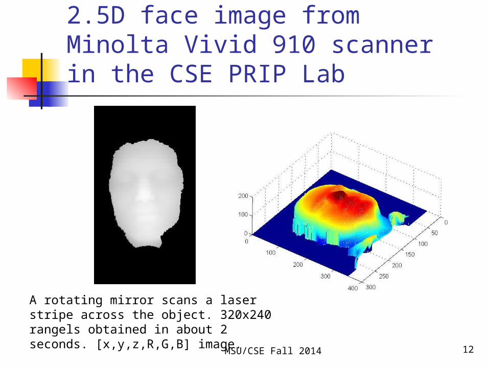
2.5D face image from Minolta Vivid 910 scanner in the CSE PRIP Lab
A rotating mirror scans a laser stripe across the object. 320x240 rangels obtained in about 2 seconds. [x,y,z,R,G,B] image.

MSU/CSE Fall 2014 13
LIDAR also senses surfaces
Single sensing element scans scene
Laser light reflected off surface and returned
Phase shift codes distance
Brightness change codes albedo

MSU/CSE Fall 2014 15
What about human vision? Do we compute the surfaces in our
environment? Do we represent them in our
memory as we move around or search?
Do we save representations of familiar objects?
(See David Marr, Vision, 1982; Aloimonus and Rosenfeld 1991.)

MSU/CSE Fall 2014 16
3D scanning technology
3D image of voxels obtained Usually computationally expensive
reconstruction of 3D from many 2D scans (CAT computer-aided-tomography)
More info later in the course.

MSU/CSE Fall 2014 17
Magnetic Resonance Imaging
Sense density of certain chemistry
S slices x R rows x C columns
Volume element (voxel) about 2mm per side
At left is shaded 2D image created by “volume rendering” a 3D volume: darkness codes depth
http://en.wikipedia.org/wiki/File:MRI-Philips.JPG

MSU/CSE Fall 2014 18
Single slice through human head
MRIs are computed structures, computed from many views.
At left is MRA (angiograph), which shows blood flow.
CAT scans are computed in much the same manner from X-ray transmission data.

MSU/CSE Fall 2014 19
Other variations Microscopes, telescopes, endoscopes, … X-rays: radiation passes through objects
to sensor elements on the other side Fibers can carry image around curves;
in bodies, in machine tools Pressure arrays create images
(fingerprints, butts) Sonar, stereo, focus, etc can be used for
range sensing (see Chapters 12 and 13)

MSU/CSE Fall 2014 20
Summary of some digital imaging problems. Mixed pixel problem: mixed material in the
world/scene map to a single pixel Loss of thin/small features due to resolution Variations in area or perimeter due to
variation in object location and thresholding Path problems: refraction, dispersion, etc.
takes radiation off straight path (blue light bends more or less than red light going through lens?)