mri of cervical cancer: staging, prognostic … · demonstrates restricted diffusion. prognostic...
TRANSCRIPT

Patricia Balcacer, MD
Mahan Mathur, MD
Margarita Revzin, MD
Michael Spektor, MD
MRI OF CERVICAL CANCER: STAGING,
PROGNOSTIC IMPLICATIONS AND PITFALLS
Department of Diagnostic Radiology
Yale-New Haven Hospital

MRI OF CERVICAL CANCER: STAGING,
PROGNOSTIC IMPLICATIONS AND PITFALLS
Goals/Objectives:
Provide an overview of the revised FIGO staging system for cervical cancer with relevant examples of each stage on MRI.
Review prognostic factors of cervical cancer, treatment implications, and criteria for adequate treatment response.
Present some of the pitfalls of cervical cancer imaging on MRI.
Target Audience:
Radiology residents, Body Imaging fellows and attendings
Disclosures:
None

INTRODUCTION – CERVICAL CANCER
• Most common gynecologic malignancy worldwide and third most common
gynecologic malignancy in the United States.
• Introduction of cervical cancer screening programs and improved treatment
strategies have caused a reduction in mortality rates in industrialized nations 1.
• Staging recommended by International Federation of Gynecology and Obstetrics
(FIGO) is widely adopted both for therapy planning and post-therapy follow-up.
• However FIGO staging, usually based on only clinical assessment, has shown to be
inaccurate in the estimation of the actual tumor extent2.
• Cervical carcinoma is a relatively slow-growing disease, usually invading the vagina
and the paracervical space along the parametrium and uterosacral ligaments.
Bladder, rectum, pelvic and para-aortic lymph nodes may be invaded in later stages.
USE OF MRI IN CERVICAL CANCER
• MRI of the pelvis is the most reliable imaging modality for staging, treatment
planning and follow-up of cervical cancer. Often complementary to clinical
assessment, which currently remains the reference standard.
• MRI can accurately evaluate the extent of disease because of its high spatial and
contrast resolution for pelvic tissues and organs.
• Since clinical implications and therapeutic strategies for cervical cancer treatment
vary tremendously according to the degree of tumor extension, familiarity with
relevant MRI findings is essential for radiologists.

NORMAL CERVIX
Trilaminar pattern of signal intensity:
1. High signal intensity endocervical mucosal glands
2. Low signal intensity stroma
3. Intermediate signal intensity smooth muscle
T2 Fat-sat
REVISED FIGO STAGING OF CERVICAL CANCER3
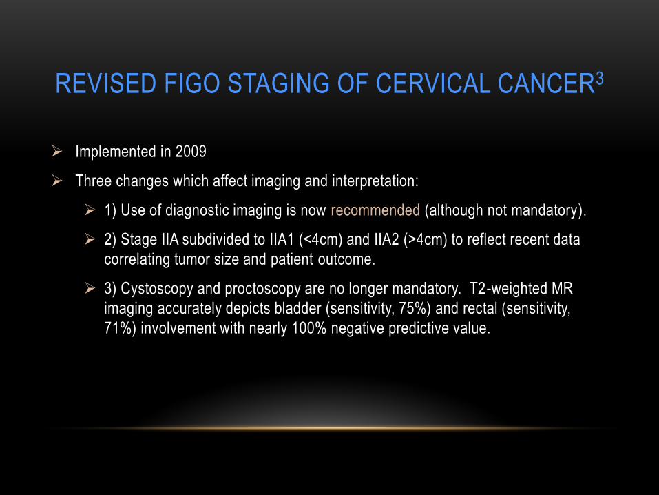
Implemented in 2009
Three changes which affect imaging and interpretation:
1) Use of diagnostic imaging is now recommended (although not mandatory).
2) Stage IIA subdivided to IIA1 (<4cm) and IIA2 (>4cm) to reflect recent data
correlating tumor size and patient outcome.
3) Cystoscopy and proctoscopy are no longer mandatory. T2-weighted MR
imaging accurately depicts bladder (sensitivity, 75%) and rectal (sensitivity,
71%) involvement with nearly 100% negative predictive value.
REVISED FIGO STAGING
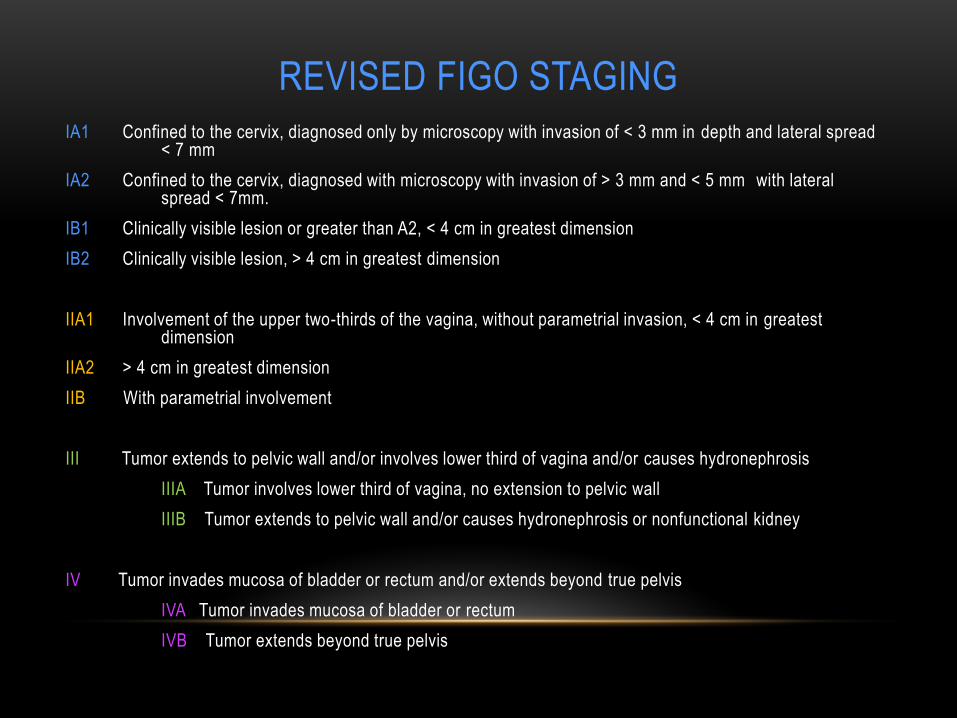
IA1 Confined to the cervix, diagnosed only by microscopy with invasion of < 3 mm in depth and lateral spread < 7 mm
IA2 Confined to the cervix, diagnosed with microscopy with invasion of > 3 mm and < 5 mm with lateral spread < 7mm.
IB1 Clinically visible lesion or greater than A2, < 4 cm in greatest dimension
IB2 Clinically visible lesion, > 4 cm in greatest dimension
IIA1 Involvement of the upper two-thirds of the vagina, without parametrial invasion, < 4 cm in greatest dimension
IIA2 > 4 cm in greatest dimension
IIB With parametrial involvement
III Tumor extends to pelvic wall and/or involves lower third of vagina and/or causes hydronephrosis
IIIA Tumor involves lower third of vagina, no extension to pelvic wall
IIIB Tumor extends to pelvic wall and/or causes hydronephrosis or nonfunctional kidney
IV Tumor invades mucosa of bladder or rectum and/or extends beyond true pelvis
IVA Tumor invades mucosa of bladder or rectum
IVB Tumor extends beyond true pelvis
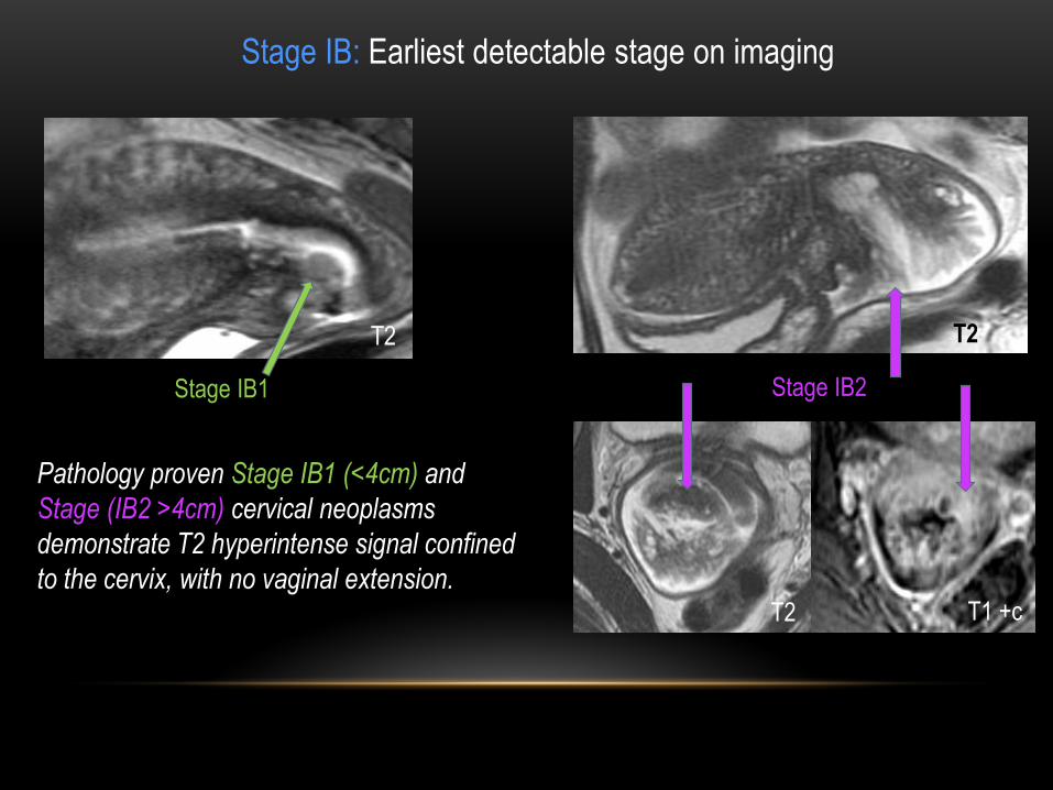
Pathology proven Stage IB1 (<4cm) and
Stage (IB2 >4cm) cervical neoplasms
demonstrate T2 hyperintense signal confined
to the cervix, with no vaginal extension.
T2
T1 +c T2
Stage IB: Earliest detectable stage on imaging
Stage IB1 Stage IB2
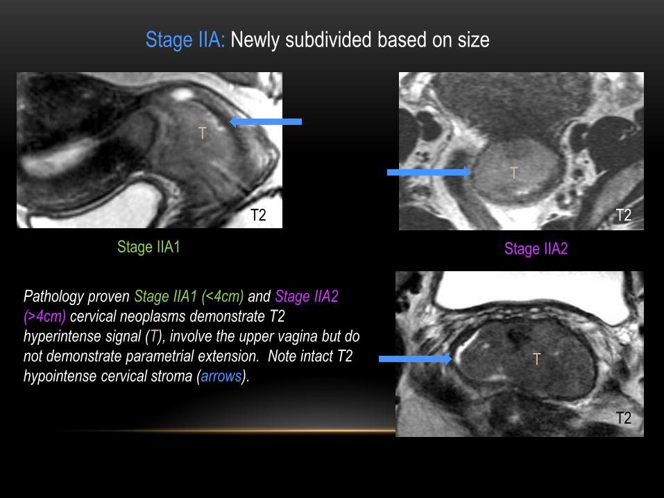
Stage IIA: Newly subdivided based on size
Pathology proven Stage IIA1 (<4cm) and Stage IIA2
(>4cm) cervical neoplasms demonstrate T2
hyperintense signal (T), involve the upper vagina but do
not demonstrate parametrial extension. Note intact T2
hypointense cervical stroma (arrows).
Stage IIA1 Stage IIA2
T2 T2
T2
T
T
T
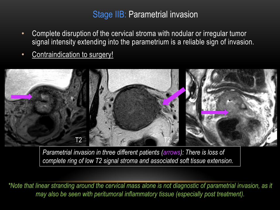
• Complete disruption of the cervical stroma with nodular or irregular tumor signal intensity extending into the parametrium is a reliable sign of invasion.
• Contraindication to surgery!
Stage IIB: Parametrial invasion
*Note that linear stranding around the cervical mass alone is not diagnostic of parametrial invasion, as it
may also be seen with peritumoral inflammatory tissue (especially post treatment).
Parametrial invasion in three different patients (arrows): There is loss of
complete ring of low T2 signal stroma and associated soft tissue extension.
T2 T2 T2

Stage IIIA
IIIA: Tumor involves lower third of
vagina, no extension to pelvic wall
For the purposes of staging, invasion of the lower one-third of the vagina implies modification of
radiation therapy strategy.
T
T2
T2 sagittal image demonstrates cervical
tumor (T) with invasion of the lower one-third
of the vagina (arrow).

Stage IIIB
IIIB: Tumor extends to either pelvic wall and/or causes hydronephrosis or nonfunctional kidney
Axial T2 and post-contrast T1 images
demonstrate tumor extension to left pelvic
sidewall (arrows)
Axial and coronal T2 images in a different
patient demonstrate left hydroureter (arrows)
caused by cervical mass encasement (arrow)
T2
T2
T2
T1 +c
Ureteral obstruction at the level of the tumor is considered to be an indication of pelvic wall invasion.

Stage IVA: Tumor invades mucosa of bladder or rectum
Bladder or rectal invasion can be diagnosed when disruption of their normal
hypointense walls is seen at T2-weighted imaging, with or without a mass
protruding into the lumen.
Sagittal T2 image
demonstrates a bulky cervical
mass invading the posterior
bladder wall (arrow).
Vagina
Free fluid T2

Stage IVA: Tumor invades mucosa of bladder or rectum
Rectal invasion in two different patients:
Top image: Sagittal T2 demonstrates invasion of
anterior rectal wall (arrow).
Bottom image: Axial T2 demonstrates loss of a fat
plane between cervical neoplasm and anterior rectal
wall (arrow)
Although invasion of the bladder or rectum is rare in
cervical carcinoma, mandatory endoscopic assessment
of these organs was part of the standard FIGO clinical
staging system.
According to the revised FIGO system, cystoscopy or
proctoscopy are no longer routine. The absence of
bladder or rectal invasion can be diagnosed confidently
with MRI. T2
T2

The incidence of distant metastases
increases with the stage of disease: 3% in
stage IA to 75% in stage IVA4.
Common hematogenous metastases to
lungs, liver, brain, and bones.
Stage IVB: Tumor extends beyond true pelvis
56-year-old female with cervical
neoplasm (not shown) demonstates brain
and mediastinal metastases (arrows).
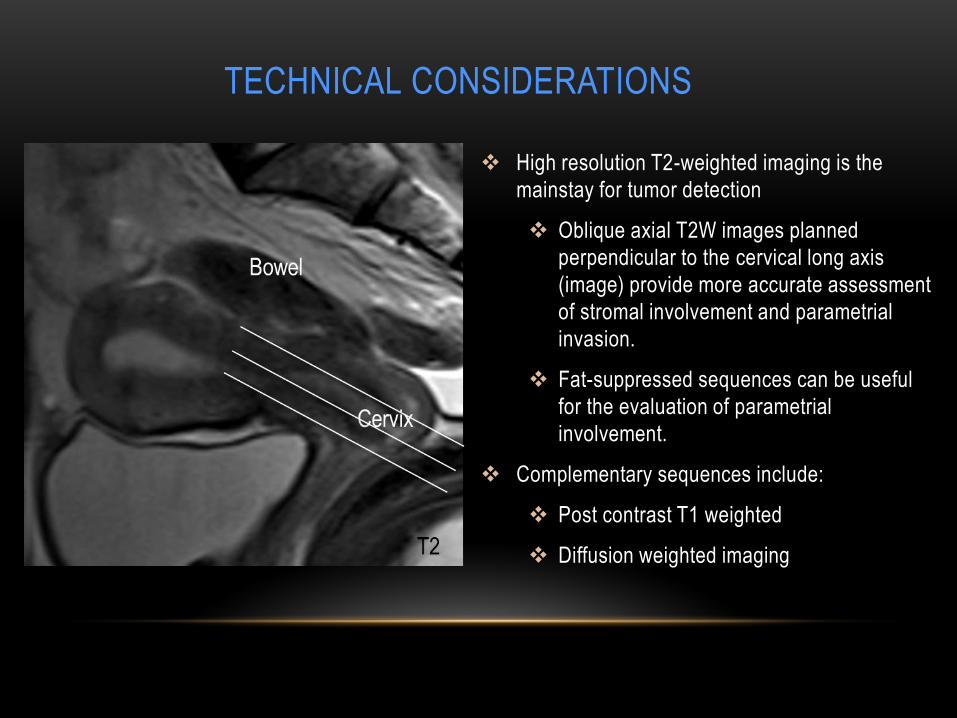
TECHNICAL CONSIDERATIONS
High resolution T2-weighted imaging is the
mainstay for tumor detection
Oblique axial T2W images planned
perpendicular to the cervical long axis
(image) provide more accurate assessment
of stromal involvement and parametrial
invasion.
Fat-suppressed sequences can be useful
for the evaluation of parametrial
involvement.
Complementary sequences include:
Post contrast T1 weighted
Diffusion weighted imaging
Cervix

Wide variation in the literature regarding use of IV contrast.
Cervical carcinoma is better defined at T2WI, however detection of small tumors may be improved with IV contrast secondary due to their early enhancement relative to the cervical stroma5.
Improves accuracy of diagnosing bladder and rectal invasion.
Useful in the post-treatment setting to differentiate residual or recurrent tumor from radiation fibrosis.
Helps to delineate complications of treatment, such as fistulas.
Role of IV contrast
Axial T2 (top) and post contrast T1 (bottom) images
demonstrate a poorly defined cervical tumor (arrows) that is
subtle, mildly hyperintense on T2 and more readily visualized
after administering IV contrast.
T2
T1 + C

DWI can be applied to the detection of
cervical cancer because of its superior
disease contrast with normal tissue6.
It is complimentary to T2WI in recognizing
early-stage disease.
ADC values can be a useful tool in
monitoring response to therapy.
Role of Diffusion Weighted Imaging
ADC
ADC
DWI
DWI
Top Images: Normal cervix demonstrates
uniformly hypointense intact cervical stroma
(arrows).
Bottom Images: Cervical neoplasm (arrows)
demonstrates restricted diffusion.
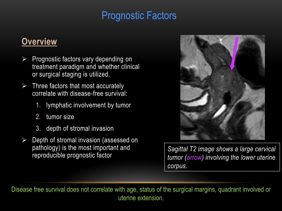
Prognostic factors vary depending on treatment paradigm and whether clinical or surgical staging is utilized.
Three factors that most accurately correlate with disease-free survival:
1. lymphatic involvement by tumor
2. tumor size
3. depth of stromal invasion
Depth of stromal invasion (assessed on pathology) is the most important and reproducible prognostic factor
Overview
Sagittal T2 image shows a large cervical
tumor (arrow) involving the lower uterine
corpus.
Prognostic Factors
Disease free survival does not correlate with age, status of the surgical margins, quadrant involved or
uterine extension.
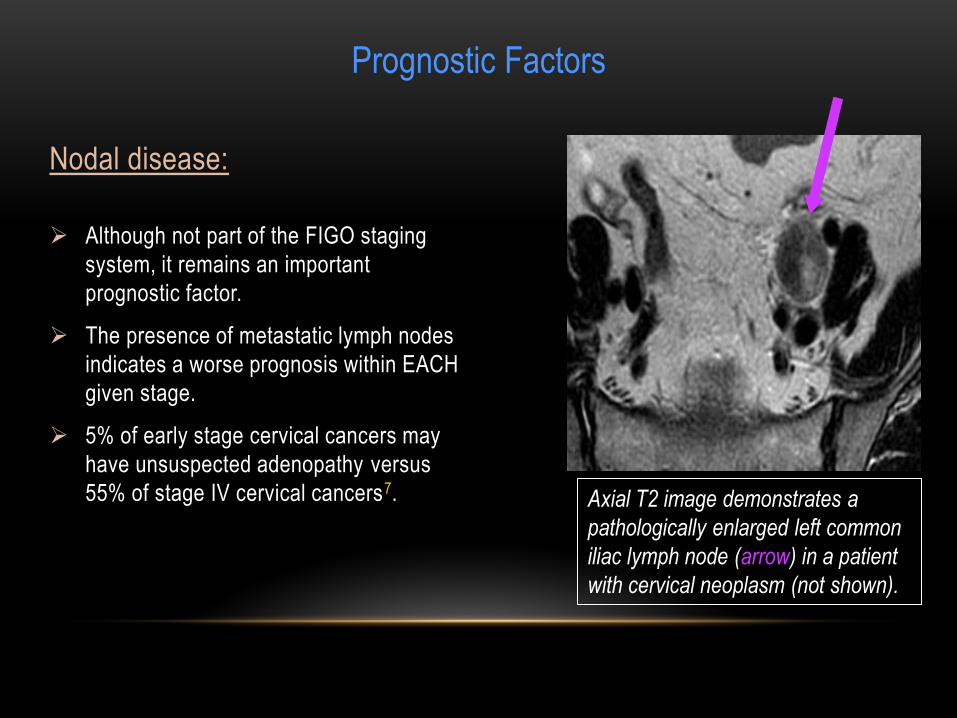
Although not part of the FIGO staging
system, it remains an important
prognostic factor.
The presence of metastatic lymph nodes
indicates a worse prognosis within EACH
given stage.
5% of early stage cervical cancers may
have unsuspected adenopathy versus
55% of stage IV cervical cancers7.
Nodal disease:
Prognostic Factors
Axial T2 image demonstrates a
pathologically enlarged left common
iliac lymph node (arrow) in a patient
with cervical neoplasm (not shown).

Tumor size:
The shape and direction of growth should
be noted as they are important for
brachytherapy planning.
The size of the lesion may at times be
overestimated on T2-weighted imaging
due to inflammation or edema; post
contrast images may be used.
The craniocaudal diameter of the tumor is
a critical factor in predicting prognosis
after radiation therapy.
Fertility sparing surgery is possible with
tumors < 2 cm, whereas tumors > 4 cm
may undergo chemo radiotherapy.
C
T
Prognostic Factors
Sagittal T2 image demonstrates a bulky tumor
(T) extending into the superior two thirds of the
vagina and obstructing the uterine corpus (C).
T2

TREATMENT RESPONSE
MR imaging is 78% accurate in evaluation of tumor response; in 22% of
patients, however, benign conditions are not distinguishable from tumor8.
There is no consensus in the reviewed literature regarding indication of MRI
for routine follow-up of cervical carcinoma after chemoradiation or surgery.
After trachelectomy, MRI at 6 months and 1 year is advised due to high
recurrence rate.
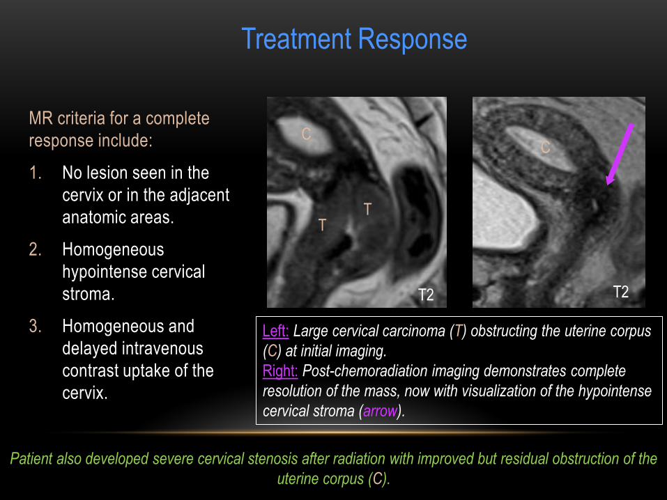
MR criteria for a complete
response include:
1. No lesion seen in the
cervix or in the adjacent
anatomic areas.
2. Homogeneous
hypointense cervical
stroma.
3. Homogeneous and
delayed intravenous
contrast uptake of the
cervix.
Left: Large cervical carcinoma (T) obstructing the uterine corpus
(C) at initial imaging.
Right: Post-chemoradiation imaging demonstrates complete
resolution of the mass, now with visualization of the hypointense
cervical stroma (arrow).
Treatment Response
T
C C
T
T2 T2
Patient also developed severe cervical stenosis after radiation with improved but residual obstruction of the
uterine corpus (C).
PITFALLS
False positive parametrial extension
Axial T2WI showing large cervical
mass extending into the right
parametrium with corresponding avid
FDG uptake on PET/CT (arrows).
Soft tissue stranding in the cervix,
radiation vs. recurrent tumor.
PET/CT shortly after demonstrates no
corresponding FDG activity, consistent
with treatment changes (arrows).
Please note brachytherapy seeds.
T2 PET/CT
PET/CT T2
Post chemoradiation:

False positive parametrial extension
PITFALLS
Top images: T2 hyperintense,
enhancing mass involving the right
posterolateral cervix (arrows). Bottom
images: Post-radiation, hazy soft tissue
in the same region, equivocal for
residual tumor versus radiation change
(arrows).
PET-CT performed shortly after
demonstrates no residual tumor.
T2
T2
T1 + c
T1 + c

Cervical tumor (T) in the posterior wall
of the exocervix with loss of the
adjacent cervical stroma (arrow).
PITFALLS
Importance of intact cervical stroma post treatment
Good treatment response after
radiation, now with visualization of the
T2 hypointense cervical stroma (arrow).
Pitfall: ‘’Burned out’’ endometriosis along the posterior vaginal fornix which was mistaken for
residual tumor post-treatment and seen in retrospect on the initial image (arrows).
T

PITFALLS
Large vascular mass centered
in the cervix on the initial
ultrasound (arrow). Please note
displaced nabothian cysts (thin
arrow).
Coronal T2 image
demonstrates a large
hyperintense mass in the
cervical canal (arrow).
However, note intact cervical
stroma (arrow).
Sagittal T2 demonstrates a
polypoid mass originating from
the junctional zone (arrow)
protruding into the cervix.
Pathology proven polypoid adenomyoma.
T2 T2
Alternate Diagnoses
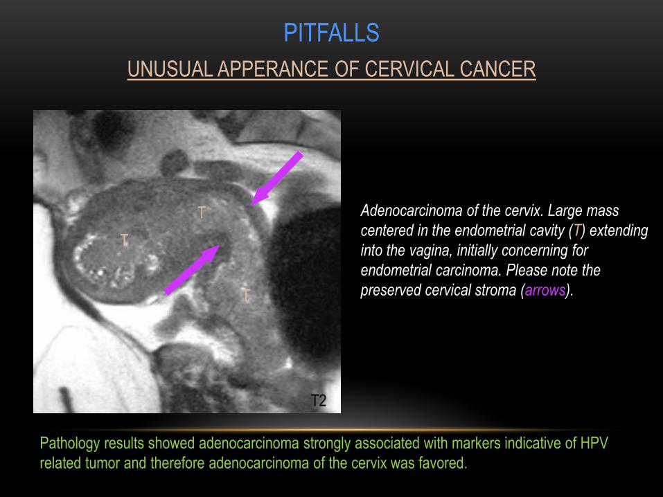
Adenocarcinoma of the cervix. Large mass
centered in the endometrial cavity (T) extending
into the vagina, initially concerning for
endometrial carcinoma. Please note the
preserved cervical stroma (arrows).
PITFALLS
UNUSUAL APPERANCE OF CERVICAL CANCER
T
T
T
Pathology results showed adenocarcinoma strongly associated with markers indicative of HPV
related tumor and therefore adenocarcinoma of the cervix was favored.
T2

TAKE HOME POINTS:
Although FIGO does not require MRI in the staging of cervical cancer, imaging is now
recommended to supplement clinical assessment, often replacing cystocopy and
proctoscopy.
MRI is the imaging modality of choice for staging primary cervical tumor, evaluating
response to treatment and detecting tumor recurrence and potential complications.
High resolution T2-weighted imaging is the mainstay for tumor detection with post
contrast T1 and DWI serving important complementary roles.
T2 hypointense intact cervical stroma is a critical finding when staging cervical cancer and
assessing treatment response.
Three factors that most accurately correlate with disease-free survival are lymphatic
involvement, tumor size, and depth of stromal invasion.
PET/CT can often help in evaluating equivocal findings on post-treatment cervical MRI.

REFERENCES
1. Ferlay F, Bray F, Pisanni P, Parkin DM. Globocan 2002: Cancer incidence, mortality and prevalence worldwide. IARC CancerBase No. 5, version 2.0. Lyon: IARC Press, 2004.
2. Narayan K, Mckenzie AF, Hicks RJ, Fisher R, Bernshaw D, Bau S. Relation between FIGO stage, primary tumor volume, and presence of lymph node metastases in cervical cancer patients referred for radiotherapy. Int J Gynecol Cancer 2003;13:657–663.
3. Bipat S, Glas AS, van der Velden J, Zwinderman AH, Bossuyt PM, Stoker J. Computed tomography and magnetic resonance imaging in staging of uterine cervical carcinoma: a systematic review. Gynecol Oncol 2003;91(1):59–66.
4. Balleyguier C: Staging of uterine cervical cancer with MRI: guidelines of the European Society of Urogenital Radiology.
5. Kinkel K, Brown M, Sirlin C. Malignant disorders of the female pelvis. In: Edelman RR, Hesselink JR, Zlatkin MB, Crues JV, editors. Clinical Magnetic Resonance Imaging. 3 rd ed. Philadelphia: Saunders Elsevier; 2005. p. 5580-7.
6. Chen, J, Zhang Y, Yang Z The utility of diffusion-weighted MR imaging in cervical cancer. Eur J Radiol, 2010 Jun; 74(3):e101-6
7. Freeman S: The Revised FIGO staging system for Uterine malignancies: Implications for MR Imaging. Radiographics 2012 32:6 , 1805-1827.
8. Int J Gynaecol Obstet. 2008 May;101(2):205-10. doi: 10.1016/j.ijgo.2007.11.004. Epub 2008 Jan 15.
9. Rockall, AG: Can MRI rule out bladder and rectal invasion in cervical cancer to help select patients for limited EUA? Gynecologic Oncology,May 2006.
10. Harmeet K: Diagnosis, staging and surveillnace of cervical carcinoma AJR June 2003
Correspondence: [email protected]