mrcp part 2 - beta revision.. rheumatology
TRANSCRIPT

MRCP Part 2 - beta revision.. Rheumatology
A 74-year-old woman complains of neck pain and stiffness. This has gradually developed over the past
few years and is now at a point where she only has limited movement. A cervical spine film is requested:
© Image used on license from Radiopaedia
Based on the cervical spine film, what is the most likely diagnosis?
Rheumatoid arthritis
Osteoarthritis
Ankylosing spondylitis
Multiple myeloma
Paget's disease
The x-ray shows complete fusion of the anterior and posterior element resulting in a 'bamboo spine'.
Ankylosing spondylitis: investigation and management
Ankylosing spondylitis is a HLA-B27 associated spondyloarthropathy. It typically presents in males (sex
ratio 3:1) aged 20-30 years old.
Investigation
Inflammatory markers (ESR, CRP) are typically raised although normal levels do not exclude ankylosing
spondylitis.
HLA-B27 is of little use in making the diagnosis as it is positive in:
90% of patients with ankylosing spondylitis
10% of normal patients
Plain x-ray of the sacroiliac joints is the most useful investigation in establishing the diagnosis.
Radiographs may be normal early in disease, later changes include:
sacroilitis: subchondral erosions, sclerosis
squaring of lumbar vertebrae
'bamboo spine' (late & uncommon)
syndesmophytes: due to ossification of outer fibers of annulus fibrosus
chest x-ray: apical fibrosis
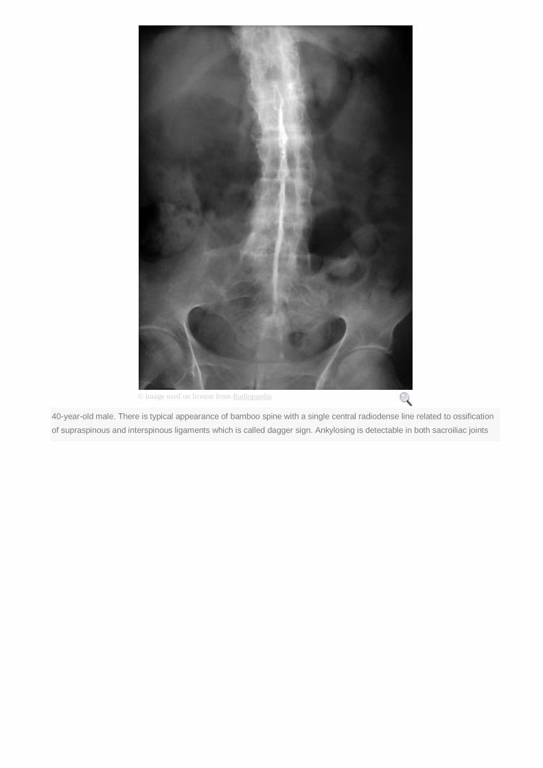
© Image used on license from Radiopaedia
40-year-old male. There is typical appearance of bamboo spine with a single central radiodense line related to ossification
of supraspinous and interspinous ligaments which is called dagger sign. Ankylosing is detectable in both sacroiliac joints

© Image used on license from Radiopaedia
Ankylosing spondylitis with well formed syndesmophytes
© Image used on license from Radiopaedia

Lateral cervical spine. Complete fusion of anterior and posterior elements in ankylosing spondylitis, so called bamboo
spine
© Image used on license from Radiopaedia
Fusion of bilateral sacroiliac joints. Sacroiliitis may present as sclerosis of joint margins which can be asymmetrical at early
stage of disease, but is bilateral and symmetrical in late disease
© Image used on license from Radiopaedia

Syndesmophytes and squaring of vertebral bodies. Squaring of anterior vertebral margins is due to osteitis of anterior
corners. Syndesmophytes are due to ossification of outer fibers of annulus fibrosus
Spirometry may show a restrictive defect due to a combination of pulmonary fibrosis, kyphosis and
ankylosis of the costovertebral joints.
Management
The following is partly based on the 2010 EULAR guidelines (please see the link for more details):
encourage regular exercise such as swimming
physiotherapy
NSAIDs are the first-line treatment
the disease-modifying drugs which are used to treat rheumatoid arthritis (such as sulphasalazine)
are only really useful if there is peripheral joint involvement
the 2010 EULAR guidelines suggest: 'Anti-TNF therapy should be given to patients with
persistently high disease activity despite conventional treatments'
research is ongoing to see whether anti-TNF therapies such as etanercept and adalimumab
should be used earlier in the course of the disease
A 51 year old man presents to Accident and Emergency with a 2 week history of lumbar back pain. He
has a background of asthma, hypertension and benign prostatic hypertrophy (BPH). He has no history of
trauma. Apart from a recent exacerbation of asthma, he is otherwise well.
On examination you find a man of large body habitus. He is able to mobilise with some discomfort. He is
afebrile, with a heart rate of 75 beats per minute and blood pressure 120/82mmHg. He has some spinal
tenderness at L4, and discomfort on extension of the spine. On neurological examination he has no
muscle wasting or fasciculations. He has full power in all limbs and normal tone. Reflexes are
symmetrical and plantars downgoing. Sensation is intact and he has normal rectal tone, with no saddle
anaesthesia.
Blood tests show:
Hb 14.1 g/dl
Platelets 245 * 109/l
WBC 8.0 * 109/l
Na+ 137 mmol/l
K+ 4.0 mmol/l
Urea 5.1 mmol/l
Creatinine 82 µmol/l

Bilirubin 14 µmol/l
ALP 42 u/l
ALT 17 u/l
CRP 3 mg/L
PSA 3.1ng/mL
What is the most likely diagnosis?
Pagets disease
Infective discitis
Spinal metastases
Vertebral compression fracture
Lumbar radiculopathy
Next question
This man has a crush fracture of the lumbar spine. He has had repeated courses of steroid treatment for
his asthma, which has led to the development of osteoporosis. A plain X-ray would reveal the diagnosis.
Pagets disease is unlikely given the normal ALP. Back pain due to metastases can be a presenting
feature of prostate carcinoma. Although this man has BPH, his normal PSA and ALP make metastatic
prostate cancer unlikely. He does not have neurological signs or symptoms suggestive of lumbar
radiculopathy. Raised inflammatory markers would be expected in infective discitis.
Other side effects of prolonged corticosteroid use include:
Peptic ulcer
Skin thinning
Mood and sleep disturbance
Central obesity
Myopathy
Avascular necrosis of bone
Cataracts
Osteoporosis: glucocorticoid-induced
Patients who take the equivalent of prednisolone 7.5 mg or more each day for 3 months or longer should
be assessed and where necessary given prophylactic treatment
Assessment for treatment - patients taking the equivalent of prednisolone 7.5 mg or more each day for 3
months, and one of the following
are over the age of 65 years
have a history of a fragility fracture
have a T-score less than - 1.5 SD
Treatment
first-line: oral bisphosphonate
second-line: alfacalcidol or calcitriol
A 38-year-old Armenian visitor presents with 3 day history of pyrexia, shortness of breath, chest pain and
abdominal pain, associated with temperature of 38.5 degrees. She has no other known past medical
history and reports at least 2 other episodes of similar pain, both times spontaneously resolving without
treatment or diagnosis. On examination, she has a pleural rub and a swollen, tender left 3rd metcarpal-
phalangeal joint. Her mother has recently been admitted for similar symptoms last month. Her blood tests
are as follow:
Hb 14.5 g/dl
Platelets 560 * 109/l
WBC 17.8 * 109/l
Na+ 143 mmol/l
K+ 4.6 mmol/l
Urea 5.2 mmol/l
Creatinine 78 µmol/l
CRP 78 mg/l
A chest radiograph demonstrates mild bilateral pleural effusions with no significant focus of consolidation,
her Mantoux test is negative. Urine dip is negative, urine MC+S grows no organisms, urinary
porphobilinogen is negative. A rheumatology review was requested regarding the synovitis and
colchicine prescribed. She responds well with resolution of all symptoms within 24 hours. An infectious
diseases opinion and induced sputum is awaited. What is the most likely diagnosis?

Tuberculosis
Acute intermittent porphyria (AIP)
Coxsackie B virus infection
Familial mediterranean fever
Systemic lupus erythematous (SLE)
Next question
The patient is of Mediterranean descent is experiencing an acute attack of abdominal pain, chest pain,
synovitis and pyrexia with an acute phase response. There appears to be a previous history of similar
symptoms and a possible genetic element. Familial Mediterranean fever would fit with all these
symptoms, with almost all patients presenting with abdominal pain, pleuritis and synovitis, associated
with fever greater than 38 degrees. The main differentials in this case are with SLE and AIP: during an
acute event, urinary porphobilinogen is likely positive in AIP. The distinguishing feature against SLE is
the resolution of symptoms with colchicine, which is a key diagnostic feature1.
1. Livneh A, Langevitz P, Zemer D et al. Criteria for the diagnosis of familial Mediterranean fever. Arthritis
Rheum. 1997;40(10):1879
Familial Mediterranean Fever
Familial Mediterranean Fever (FMF, also known as recurrent polyserositis) is an autosomal recessive
disorder which typically presents by the second decade. It is more common in people of Turkish,
Armenian and Arabic descent
Features - attacks typically last 1-3 days
pyrexia
abdominal pain (due to peritonitis)
pleurisy
pericarditis
arthritis
erysipeloid rash on lower limbs
Management
colchicine may help

**************************************************************************
A 68 year old female diagnosed with rheumatoid arthritis four years ago presents gradually increasing
tenderness in the small joints of both hands over the past 5 months. She continues to work as a legal
secretary, involving significant amounts of time at a computer. She is currently on maximum doses of
methotrexate and sulphasalazine on diagnosis and maintained on the same doses since. Her DAS score
today is 5.8, it was 4.7 when you saw her in clinic last 1 month ago. What is the next management step?
Continue methotrexate and sulphasalazine. Short-course oral prednisolone
Stop current DMARDs. Start etanercept
Stop current DMARDs. Start infliximab
Admit for pulsed intravenous methylprednisolone
Prescribe regular long-term celocoxib in addition to methotrexate and sulphasalazine
Next question
Current NICE guidelines recommend the starting of biologic therapy when the patient has been on at
least two DMARDs, including methotrexate, reporting two DAS 28 scores of greater than 5.1 at least one
month apart1. A short course of oral prednisolone may be appropriate for flares for symptomatic control.
However, this is not an option if the patient does not wish to take any steroids. Intravenous pulsed
steroids or long-term treatments are not appropriate. Regular COX-2 inhibitors are not recommended by
NICE guidelines. NSAIDs are appropriate for short-term symptomatic control but only at lowest doses for
as short a period as possible.
1. NICE Clinical Guideline 79. The management of rheumatoid arthritis in adults. Jan 2009
Rheumatoid arthritis: management
The management of rheumatoid arthritis (RA) has been revolutionised by the introduction of disease-
modifying therapies in the past decade. NICE has issued a number of technology appraisals on the
newer agents and released general guidelines in 2009.
Patients with evidence of joint inflammation should start a combination of disease-modifying drugs
(DMARD) as soon as possible. Other important treatment options include analgesia, physiotherapy and
surgery.
Initial therapy
in the 2009 NICE guidelines it is recommend that patients with newly diagnosed active RA start a
combination of DMARDs (including methotrexate and at least one other DMARD, plus short-term
glucocorticoids)
DMARDs
methotrexate is the most widely used DMARD. Monitoring of FBC & LFTs is essential due to the
risk of myelosuppression and liver cirrhosis. Other important side-effects include pneumonitis
sulfasalazine
leflunomide
hydroxychloroquine
TNF-inhibitors
the current indication for a TNF-inhibitor is an inadequate response to at least two DMARDs
including methotrexate
etanercept: recombinant human protein, acts as a decoy receptor for TNF-α, subcutaneous
administration, can cause demyelination, risks include reactivation of tuberculosis
infliximab: monoclonal antibody, binds to TNF-α and prevents it from binding with TNF receptors,
intravenous administration, risks include reactivation of tuberculosis
adalimumab: monoclonal antibody, subcutaneous administration
Rituximab
anti-CD20 monoclonal antibody, results in B-cell depletion
two 1g intravenous infusions are given two weeks apart
infusion reactions are common
Abatacept
fusion protein that modulates a key signal required for activation of T lymphocytes
leads to decreased T-cell proliferation and cytokine production
given as an infusion
not currently recommend by NICE
A 6-year-old girl is reviewed due to persistent pain in her hips. On examination she is noted to have an
antalgic gait. There is also a limited range of hip movement secondary to pain. An x-ray is requested:

© Image used on license from Radiopaedia
What is the most likely diagnosis?
Osteosarcoma
Acute lymphoblastic leukaemia
Perthes disease
Slipped upper femoral epiphysis
Developmental dysplasia of the hip
Next question
Perthes disease
Perthes disease is a degenerative condition affecting the hip joints of children, typically between the ages
of 4-8 years. It is due to avascular necrosis of the femoral head
Perthes disease is 5 times more common in boys. Around 10% of cases are bilateral
Features
hip pain: develops progressively over a few weeks

limp
stiffness and reduced range of hip movement
x-ray: early changes include widening of joint space, later changes include decreased femoral
head size/flattening
Complications
osteoarthritis
premature fusion of the growth plates
© Image used on license from Radiopaedia
Perthes disease - both femoral epiphyses show extensive destruction, the acetabula are deformed

© Image used on license from Radiopaedia
Perthes disease - bilateral disease
A 74 year old female was admitted to the medical ward initially for treatment of a CURB = 4 community
acquired pneumonia. She is now awaiting discharge but since her illness, she has not returned to her
pre-morbid state. Her past medical history include two previous myocardial infarctions, hypertension,
type 2 diabetes mellitus, duodenal ulcer and obesity. In addition, the physiotherapists report significant
right knee pain to be contributing to poor mobility. On questioning, the patient reports that the pain is
chronic and has been progressively worsening for about 3 years.
Her GP had sent her for two X rays previously that demonstrated cartilage loss and osteophyte
formation, with reduction in joint space. On examination, you note significant crepitus in the right knee,
with reduced range of movements in flexion and extension. You also note boney outgrowths in the
proximal interphalangeal joints of her second and third digits of her right hand. She had successfully lost
9kg in weight and had previously taken 1g paracetamol four times a day regularly but neither measure
seemed to help her pain.
What is the most appropriate next step?
Increase 500mg paracetamol as required
Oral ibuprofen
Topical diclofenac
Oramorph as required

Glucosamine
Next question
The patient describes osteoarthritic symptoms that have persisted despite non-pharmacological
therapies (weight loss) and paracetamol on a regular basis. Her past medical history of duodenal ulcer
should make you wary of oral NSAIDs while her previous MIs indicate selective COX-2 inhibitors should
be used with caution. Opiods are not the second line therapies for osteoarthritis. The evidence for
glucosamine is limited and is not recommended for use under the NHS. Topical NSAIDs such as
diclofenac or topical capsaicin as an adjunct are reasonable options in this setting. A 3rd line possibility if
topical NSAIDs are not efficacious and in this case, where oral NSAIDs are contraindicated, are
intraarticular steroid injections, which have been demonstrated to produce significant symptomatic
improvements in the knee joint when compared against placebo (although evidence is weaker for other
joints). Interestingly, an inflammatory element is not required for symptomatic benefits.
Osteoarthritis: management
NICE published guidelines on the management of osteoarthritis (OA) in 2014
all patients should be offered help with weight loss, given advice about local muscle
strengthening exercises and general aerobic fitness
paracetamol and topical NSAIDs are first-line analgesics. Topical NSAIDs are indicated only for
OA of the knee or hand
second-line treatment is oral NSAIDs/COX-2 inhibitors, opioids, capsaicin cream and intra-
articular corticosteroids. A proton pump inhibitor should be co-prescribed with NSAIDs and COX-
2 inhibitors. These drugs should be avoided if the patient takes aspirin
non-pharmacological treatment options include supports and braces, TENS and shock absorbing
insoles or shoes
if conservative methods fail then refer for consideration of joint replacement
What is the role of glucosamine?
normal constituent of glycosaminoglycans in cartilage and synovial fluid
a systematic review of several double blind RCTs of glucosamine in knee osteoarthritis reported
significant short-term symptomatic benefits including significantly reduced joint space narrowing
and improved pain scores
more recent studies have however been mixed
the 2008 NICE guidelines suggest it is not recommended
a 2008 Drug and Therapeutics Bulletin review advised that whilst glucosamine provides modest
pain relief in knee osteoarthritis it should not be prescribed on the NHS due to limited evidence of
cost-effectiveness
A 45 year old woman was referred to Rheumatology clinic after experiencing widespread aches and
pains felt throughout her body. The pains were felt particularly in her arms and legs in addition to

significant pain throughout the patients spinal column. The patient could not recall a precise onset of her
symptoms but she felt they had been present for at least 12 months, possibly longer. In addition, the
patient reported on-going feelings of tiredness and lethargy. Despite going to bed around 10 pm each
evening, the patient reported waking in the morning still feeling exhausted. She denied any history of hot
or tender joints, skin rashes, hair loss, swallowing difficulties or dry eyes. The patients appetite was
described as normal for her with no significant change in weight.
There was no previous past medical history and the patient took no regular medications except for a non-
prescription multi-vitamin. Family history was remarkable for hypothyroidism affecting her mother and
elder sister. The patient worked as an accountant and lived with her two teenage children. She had
separated from her ex-husband 18 months previously.
Examination did not demonstrate any evidence of active sinovitis of the hands or feet with no other
inflamed or deformed joints. Palpation of the muscles of the upper arms and legs as well as the
paraspinal muscles was exquisitely tender. Neurological examination of the arms and legs was
unremarkable. Cardiovascular and respiratory examination was unremarkable with no skin rashes.
During clinic interaction the patient appeared tired and stressed but had good rapport and maintained
good eye contact. She denied any significant low mood but was anxious that her symptoms represented
a serious underlying illness.
Investigations requested following clinic are listed below.
Haemoglobin 12.9 g / dL
White cell count 7.2 * 109/l
Platelets 332 * 109/l
Mean cell volume 87 fL
Sodium 140 mmol / L
Potassium 3.6 mmol / L
Urea 3.5 mmol / L
Creatinine 68 micromol / L
Erythrocyte sedimentation rate 11 mm / h
Rheumatoid factor Negative
Anti-nuclear antigen Weak positive
B12 324 pmol / L (reference 74-516)
Folate 30 nmol / L (reference 7-36)
Serum immunoglobulin Normal electrophoresis strip
Thyroid stimulating hormone 0.9 microU / mL (reference 0.4-5.0)
X-rays of hands: some minor degenerative change in right index proximal interphalangeal joint but
otherwise unremarkable with no boney erosion or deformity

What is the cause for the patients pain?
Fibromyalgia
Systemic lupus erythematous
Chronic regional pain syndrome
Generalised anxiety disorder
Depression
Next question
The patient has chronic widespread pain (>3 months) associated with lethargy, non-refreshing sleep and
multiple tender points on palpation. Basic blood tests are essentially normal and there is no history or
examination to suggest connective tissue disease or other pathology. This presentation is consistent with
fibromyalgia, the diagnostic label used to describe chronic widespread pain associated with multiple
muscular tender points or associated symptoms of fatigue, non-refreshing sleep or cognitive dysfunction.
Please note that many healthy individuals have weakly positive anti-nuclear antigen results and this does
not imply a diagnosis of systemic lupus erythematous in the absence of symptoms and signs of the
disease. It may be that requesting immunological tests was inappropriate in this patient given the lack of
clinical evidence of connective tissue disease.
Chronic regional pain syndrome is associated with persistent burning pain in one limb, usually after a
minor injury. The brief mental state examination documented does not suggest evidence of significant
depression or generalised anxiety.
Carnes D, Underwood M, Rahman A. Fibromyalgia. BMJ 2014;348:g474.
Fibromyalgia
Fibromyalgia is a syndrome characterised by widespread pain throughout the body with tender points at
specific anatomical sites. The cause of fibromyalgia is unknown.
Epidemiology
women are 10 times more likely to be affected
typically presents between 30-50 years old

Features
pain: at multiple site, sometimes 'pain all over'
lethargy
sleep disturbance, headaches, dizziness are common
Diagnosis is clinical and sometimes refers to the American College of Rheumatology
classification criteria which lists 9 pairs of tender points on the body. If a patient is tender in at least 11 of
these 18 points it makes a diagnosis of fibromyalgia more likely
The management of fibromyalgia is often difficult and needs to be tailored to the individual patient. A
psychosocial and multidisciplinary approach is helpful. Unfortunately there is currently a paucity of
evidence and guidelines to guide practice. The following is partly based on consensus guidelines from
the European League against Rheumatism (EULAR) published in 2007 and also a BMJ review in 2014.
explanation
aerobic exercise: has the strongest evidence base
cognitive behavioural therapy
medication: pregabalin, duloxetine, amitriptylin
A 60 year old woman attended her General Practitioner and reported a three month history of bilateral
shoulder muscle and bilateral hip girdle aches and pain. She also experienced stiffness affecting these
areas that lasted for up to two hours each morning. These symptoms were limiting her day to day
activities and were unresponsive to simple analgesics.
The patient denied symptoms of headache, visual disturbance or jaw claudication. Intermittent episodes
of dry mouth and dry eyes had been present for several years. There was no history of unexplained skin
rashes. Past medical history included coeliac disease diagnosed twenty years previously that was well
controlled on a gluten-free diet. The patient was a non-smoker and drank alcohol only occasionally.
Examination revealed mild muscular tenderness across the shoulder and hip girdles although with no
other inflamed or tender joints. Cardiovascular and respiratory examination was unremarkable.
Blood tests requested by her GP demonstrated an elevated ESR of 65. A diagnosis of PMR was made
and a course of 20 mg prednisolone daily prescribed. However 6 weeks later the patients symptoms had
not significantly improved and she was referred to rheumatology clinic. Repeat blood tests and other
investigations are listed below.
Haemoglobin 110 g / dL
White cell count 8.9 * 109/l
Neutrophils 7.8 * 109/l
Platelets 456 * 109/l
Urea 6.2 mmol / L
Creatinine 87 micromol / L

Sodium 138 mmol / L
Potassium 4.1 mmol / L
Ferritin 180 ng / mL
Erythrocyte sedimentation rate 75 mm / h
Rheumatoid factor Negative
Connective tissue ANA Negative
Anti-CCP antibodies 58 EU (reference < 20)
Creatinine kinase 89 U / L (reference 5-130)
X-ray hands: minor degenerative change in multiple inter-phalangeal joints of both hands; no evidence of
erosive arthropathy
What is correct diagnosis?
Polymyalgia rheumatica
Rheumatoid arthritis
Polymyositis
Sjorgren's syndrome
Systemic lupus erythematous
Next question
Rheumatoid arthritis can present with a polymyalgic syndrome prior to clinically detectable sinovitis. In
this case this is suggested by the lack of response to trial of prednisolone and the positive anti-CCP
antibody. Observational studies have shown a greater clinical and laboratory response to steroids in
polymyalgia rheumatica than polymyalgic onset rheumatoid arthritis. Anti-CCP antibodies are rarely
present in polymyalgia rheumatica but are strongly associated with rheumatoid arthritis.
Sjorgren's syndrome and SLE are unlikely given the lack of anti-nuclear antibodies. Polymyositis is
excluded by the normal CK.
Mackie S, Mallen C. Polymyalgia rheumatica. BMJ 2013;347:f6937.
Rheumatoid arthritis: diagnosis

NICE have stated that clinical diagnosis is more important than criteria such as those defined by the
American College of Rheumatology.
2010 American College of Rheumatology criteria
Target population. Patients who
1) have at least 1 joint with definite clinical synovitis
2) with the synovitis not better explained by another disease
Classification criteria for rheumatoid arthritis (add score of categories A-D;
a score of 6/10 is needed definite rheumatoid arthritis)
Key
RF = rheumatoid factor
ACPA = anti-cyclic citrullinated peptide antibody
Factor Scoring
A. Joint involvement
1 large joint 0
2 - 10 large joints 1
1 - 3 small joints (with or without involvement of large joints)
2
4 - 10 small joints (with or without
involvement of large joints)
3
10 joints (at least 1 small joint) 5
B. Serology (at least 1 test result is needed for
classification)
Negative RF and negative ACPA 0

Factor Scoring
Low-positive RF or low-positive ACPA 2
High-positive RF or high-positive ACPA 3
C. Acute-phase reactants (at least 1 test result is
needed for classification)
Normal CRP and normal ESR 0
Abnormal CRP or abnormal ESR 1
D. Duration of symptoms
< 6 weeks 0
> 6 weeks 1
A 62 year old lady is seen in the rheumatology clinic. She was diagnosed with rheumatoid arthritis 16
years ago. Her symptoms were relatively well controlled with a combination of methotrexate 20mg once
per week, folic acid 5mg once per week and azathioprine 100mg once per day until the last few months
when she complained of increasing joint pain with stiffness. Since then her methotrexate dose was
gradually uptitrated to the current dose of 25mg per week. She reported that her joints were less painful
and stiff in the morning. Unfortunately she was also complained of increasing tiredness with an
increasing quantity of respiratory tract infections, requiring antibiotics twice in the last six months. She
also noted that she bruised more easily of late.
Examination revealed a slender 62 year old systemically well lady. She was haemodynamically normal
and afebrile. Cardiovascular and respiratory examinations were unremarkable, and abdominal
examination revealed a mass arising from the left upper quadrant. Clinical examination of her joints
revealed no evidence of synovitis or swelling.
Routine blood investigations prior to attending clinic were as follows:
Hb 11.5 g/dl
MCV 84 fl
Platelets 82 * 109/l
WBC 3.5 * 109/l
Neutrophils 1.6 * 109/l

Lymphocytes 1.0 * 109/l
Eosinophils 0.9 * 109/l
Na+ 141 mmol/l
K+ 3.9 mmol/l
Urea 7.0 mmol/l
Creatinine 81 µmol/l
Bilirubin 12 µmol/l
ALP 99 u/l
ALT 13 u/l
Albumin 39 g/l
What is the single most likely cause of the clinical and haematological abnormalities?
Myelodyspastic syndrome
Chronic lymphocytic leukaemia
Marrow aplasia secondary to drug therapy
Feltys syndrome
Myelodysplasia
Next question
Feltys syndrome is a complication of Rheumatoid Arthritis (RA). It consists of a combination of
rheumatoid arthritis, neutropaenia and splenomegaly, and tends to affect RA of longstanding duration.
The main differential diagnosis is drug induced marrow aplasia; however this would not easily account for
the presence of splenomegaly.
Rheumatoid arthritis: complications
A wide variety of extra-articular complications occur in patients with rheumatoid arthritis (RA):

respiratory: pulmonary fibrosis, pleural effusion, pulmonary nodules, bronchiolitis obliterans,
methotrexate pneumonitis, pleurisy
ocular: keratoconjunctivitis sicca (most common), episcleritis, scleritis, corneal ulceration,
keratitis, steroid-induced cataracts, chloroquine retinopathy
osteoporosis
ischaemic heart disease: RA carries a similar risk to type 2 diabetes mellitus
increased risk of infections
depression
Less common
Felty's syndrome (RA + splenomegaly + low white cell count)
amyloidosis
A 47-year-old lady presented with a three-week history of pain in her fingers. She had noticed her hands
were getting extremely cold when she went outside and turned a 'funny colour'. When she came back
inside her hands were very painful as they began to warm up. She had managed in the past by wearing
gloves outside but now had ulcers on her fingertips which she had never experienced before. She also
complained of epigastric pain and had longstanding shortness of breath.
Her past medical history included pulmonary fibrosis and hypertension. Her medications included
propranolol, amlodipine, simvastatin and omeprazole.
On examination the skin over her hands was dry and shiny and there was severe digital ulceration on
three fingertips of the left hand. There was no exudate or erythema. The fingertips were dusky in colour
and extremely tender. The skin over the upper arms and chest appeared normal. On auscultation of the
lungs there were fine bibasal inspiratory crepitations which did not alter in character upon coughing.
Heart sounds were normal with no added murmurs. There was a left ventricular heave
Which of the following is the most appropriate management plan for this lady?
Start flucloxacillin and stop all anti-hypertensive medications
Educate this lady about the use of gloves and hand-warmers and increase her amlodipine
dose
Stop amlodipine and refer for an urgent dermatology assessment
Stop propranolol and admit for an iloprost infusion
Start high dose oral prednisolone
Next question

This patient has secondary Raynauds associated with an underlying diagnosis of Limited Systemic
Sclerosis. Her disease is 'limited' as opposed to 'diffuse' as she does not have skin changes proximal to
the elbows. She clearly has systemic involvement with pulmonary fibrosis and oesophageal dysmotility
(she has epigastric pain and is taking omeprazole). Other features that were not mentioned in the
question but may be present in such patients include telangiectasia, typically over mucosal surfaces, a
'beak-like' nose and microstomia, calcinosis and renal impairment.
Severe digital ulceration in such patients can be treated with infusion of a prostacyclin analogue such as
iloprost. Prompt treatment is required to avoid gangrene and loss of digits. Drugs such as beta blockers
and the oral contraceptive pill can exacerbate Raynauds phenomenon by causing vascular spasm and
should therefore be avoided.
There is no indication for flucloxacillin if the ulcers are not infected.
Gloves and hand-warmers can be very helpful for patients with Raynauds phenomenon. Calcium channel
blockers such as amlodipine and nifidepine cause vasodilation in peripheral arterioles are also used to
treat Raynauds phenomenon. However, if there is severe ulceration admission for an iloprost infusion is
required.
Raynaud's
Raynaud's phenomena may be primary (Raynaud's disease) or secondary (Raynaud's phenomenon)
Raynaud's disease typically presents in young women (e.g. 30 years old) with symmetrical attacks
Factors suggesting underlying connective tissue disease
onset after 40 years
unilateral symptoms
rashes
presence of autoantibodies
features which may suggest rheumatoid arthritis or SLE, for example arthritis or recurrent
miscarriages
digital ulcers, calcinosis
very rarely: chilblains
Secondary causes
connective tissue disorders: scleroderma (most common), rheumatoid arthritis, SLE
leukaemia
type I cryoglobulinaemia, cold agglutinins
use of vibrating tools
drugs: oral contraceptive pill, ergot
cervical rib

Management
first-line: calcium channel blockers e.g. nifedipine
IV prostacyclin infusions: effects may last several weeks/months
A 41-year-old man presents with lower back pain. The pain has been getting gradually worse over the
past nine months and is located in the lower lumbar spine. Other than a slightly pigmented sclera no
other abnormalities are found on examination. A number of x-rays are obtained:
© Image used on license from Radiopaedia
What is the most likely diagnosis?
Fabry's disease
Alkaptonuria
Homocystinuria
Osteopetrosis
Ankylosing spondylitis
Next question
The x-rays show multi-level intervertebral disc calcification with disc space narrowing.

Alkaptonuria
Alkaptonuria (ochronosis) is a rare autosomal recessive disorder of phenylalanine and tyrosine
metabolism caused by a deficiency of homogentisic acid.
Alkaptonuria is generally a benign and often asymptomatic condition. Possible features include:
pigmented sclera
urine turns black if left exposed to the air
intervertebral disc calcification may result in back pain
renal stones
Treatment
high-dose vitamin C
dietary restriction of phenylalanine and tyrosine
**************************************************************************************************************
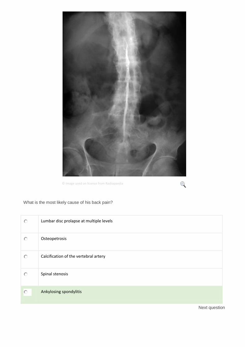
A 40-year-old man is investigated for back. For the past few months he has been troubled with pain in his
lower back which is typically worse in the morning and better by the end of the day. There is some
radiation of pain to the right buttock but no leg pains. An x-ray of his lumbar spine is shown below
© Image used on license from Radiopaedia
What is the most likely cause of his back pain?
Lumbar disc prolapse at multiple levels
Osteopetrosis
Calcification of the vertebral artery
Spinal stenosis
Ankylosing spondylitis
Next question

This image shows the typical appearance of bamboo spine with a single central radiodense line related
to ossification of supraspinous and interspinous ligaments which is called dagger sign. Ankylosing is
detectable in both sacroiliac joints.
Note the history of morning pain is typical for an inflammatory arthritis such as ankylosing spondylitis.
Ankylosing spondylitis: investigation and management
Ankylosing spondylitis is a HLA-B27 associated spondyloarthropathy. It typically presents in males (sex
ratio 3:1) aged 20-30 years old.
Investigation
Inflammatory markers (ESR, CRP) are typically raised although normal levels do not exclude ankylosing
spondylitis.
HLA-B27 is of little use in making the diagnosis as it is positive in:
90% of patients with ankylosing spondylitis
10% of normal patients
Plain x-ray of the sacroiliac joints is the most useful investigation in establishing the diagnosis.
Radiographs may be normal early in disease, later changes include:
sacroilitis: subchondral erosions, sclerosis
squaring of lumbar vertebrae
'bamboo spine' (late & uncommon)
syndesmophytes: due to ossification of outer fibers of annulus fibrosus
chest x-ray: apical fibrosis

© Image used on license from Radiopaedia
40-year-old male. There is typical appearance of bamboo spine with a single central radiodense line related to ossification
of supraspinous and interspinous ligaments which is called dagger sign. Ankylosing is detectable in both sacroiliac joints
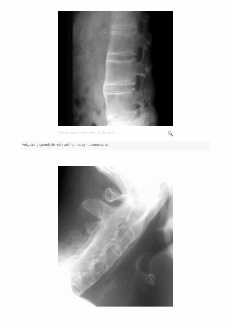
© Image used on license from Radiopaedia
Ankylosing spondylitis with well formed syndesmophytes

© Image used on license from Radiopaedia
Lateral cervical spine. Complete fusion of anterior and posterior elements in ankylosing spondylitis, so called bamboo
spine
© Image used on license from Radiopaedia
Fusion of bilateral sacroiliac joints. Sacroiliitis may present as sclerosis of joint margins which can be asymmetrical at early
stage of disease, but is bilateral and symmetrical in late disease

© Image used on license from Radiopaedia
Syndesmophytes and squaring of vertebral bodies. Squaring of anterior vertebral margins is due to osteitis of anterior
corners. Syndesmophytes are due to ossification of outer fibers of annulus fibrosus
Spirometry may show a restrictive defect due to a combination of pulmonary fibrosis, kyphosis and
ankylosis of the costovertebral joints.
Management
The following is partly based on the 2010 EULAR guidelines (please see the link for more details):
encourage regular exercise such as swimming
physiotherapy
NSAIDs are the first-line treatment
the disease-modifying drugs which are used to treat rheumatoid arthritis (such as sulphasalazine)
are only really useful if there is peripheral joint involvement
the 2010 EULAR guidelines suggest: 'Anti-TNF therapy should be given to patients with
persistently high disease activity despite conventional treatments'
research is ongoing to see whether anti-TNF therapies such as etanercept and adalimumab
should be used earlier in the course of the disease
A 56-year-old man presents with buttock pain. This has been present for many years but has recently
become. The pain is usually worse in the early part of the day and often eases by the late afternoon. An

x-ray is requested:
© Image used on license from Radiopaedia
What is the most likely underlying diagnosis?
Ankylosing spondylitis
Alkaptonuria
Multiple myeloma
Peripheral arterial disease
Bilateral hip osteoarthritis
Next question
Fusion of bilateral sacroiliac joints consistent with ankylosing spondylitis. Sacroiliitis may present as
sclerosis of joint margins which can be asymmetrical at early stage of disease, but is bilateral and
symmetrical in late disease.
Pain that is worse in the morning is consistent with an inflammatory condition such as ankylosing
spondylitis.

Ankylosing spondylitis: investigation and management
Ankylosing spondylitis is a HLA-B27 associated spondyloarthropathy. It typically presents in males (sex
ratio 3:1) aged 20-30 years old.
Investigation
Inflammatory markers (ESR, CRP) are typically raised although normal levels do not exclude ankylosing
spondylitis.
HLA-B27 is of little use in making the diagnosis as it is positive in:
90% of patients with ankylosing spondylitis
10% of normal patients
Plain x-ray of the sacroiliac joints is the most useful investigation in establishing the diagnosis.
Radiographs may be normal early in disease, later changes include:
sacroilitis: subchondral erosions, sclerosis
squaring of lumbar vertebrae
'bamboo spine' (late & uncommon)
syndesmophytes: due to ossification of outer fibers of annulus fibrosus
chest x-ray: apical fibrosis
A 32 year old gentleman presented to his GP with an 8 week history of debilitating pain in his oral cavity
and in his groin area. He had presented on numerous occasions to his GP with episodes of tiredness and
non specific malaise which each time was put down to non specific viral illness. Eight months ago he was
investigated by a gastroenterologist having presented with bloody diarrhoea and abdominal pain. He was
diagnosed with a non specific colitis of unknown origin which resolved spontaneously. He also suffered a
solitary DVT of his left leg 6 years ago which was treated with oral anticoagulation. He smoked 20
cigarettes per day and consumed 20 units of alcohol per week. He was on no regular medication. Upon
specific questioning he denied joint pain or swelling. He also denied the presence of back pain. He was
unaware of any family history as he was adopted from birth.
On examination he appeared pale. His heart rate was 88 and blood pressure 118/78 mmHg. Examination
of his cardiovascular system was unremarkable. Examination of his abdomen was likewise
unremarkable. Examination of his oral mucosa revealed the presence of multiple apthous ulceration.
Examination of his external genitalia likewise revealed the presence of multiple shallow ulcers within his
groin region. Examination of his joints was unremarkable.
Initial investigations revealed the following results:
Hb 13.9 g/dl
Platelets 333 * 109/l

WBC 5.1 * 109/l
ESR 22 mm/hr
CRP 28 mg/l
Rheumatoid factor negative
Anti CCP negative
ANA negative
HLA B27 positive
What is the most likely underlying diagnosis?
Seronegative arthritis
Disseminated gonococcal infection
Crohn's disease
Coeliac's disease
Behcets syndrome
Next question
This gentleman presents with a combination of malaise, oral and genital ulceration, colitis and iritis. Of
the above options Behcets syndrome is the only option that unifies this combination. Note that HLA B27
is positive in 10 % of the population in the absence of seronegative arthritis. Crohns disease may present
with a colitis and apthous ulceration as well as iritis, but it would be difficult to account for the past deep
vein thrombosis.
Behcet's syndrome
Behcet's syndrome is a complex multisystem disorder associated with presumed autoimmune mediated
inflammation of the arteries and veins. The precise aetiology has yet to be elucidated however. The
classic triad of symptoms are oral ulcers, genital ulcers and anterior uveitis
Epidemiology
more common in the eastern Mediterranean (e.g. Turkey)
more common in men (complicated gender distribution which varies according to country.
Overall, Behcet's is considered to be more common and more severe in men)
tends to affect young adults (e.g. 20 - 40 years old)
associated with HLA B5* and MICA6 allele
around 30% of patients have a positive family history
Features
classically: 1) oral ulcers 2) genital ulcers 3) anterior uveitis
thrombophlebitis
arthritis
neurological involvement (e.g. aseptic meningitis)
GI: abdo pain, diarrhoea, colitis
erythema nodosum, DVT
Diagnosis
no definitive test
diagnosis based on clinical findings
positive pathergy test is suggestive (puncture site following needle prick becomes inflamed with
small pustule forming)
*more specifically HLA B51, a split antigen of HLA B5
A 23 year old Sri Lankan male presents with 6 months of gradual onset low back pain, worse before
waking. He describes increasing stiffness in his right wrist and left third metacarpal joints. On
examination, you note reduced spinal movements in lateral spinal flexion and rotation, and a positive
Schobers test. He has received any previous treatments and no other past medical history. He is keen to
start drugs he has read to be 'miracle cure antibodies that act against your own body'. What is the most
appropriate management?
Start sulphasalazine
Start infliximab
Start etanercept
Physiotherapy and NSAIDs
No treatment
Next question
The patient gives a classic description of new onset anklylosing spondylitis. He presents from the typical
age group of between 15-25. NSAIDs and physiotherapy should be the first line treatment for all
symptomatic AS patients, allowing up to 4 weeks for assessment of effect. Up to 70% of AS patients
receive sufficient symptomatic relief with NSAIDs alone, with the most recent EULAR guidelines
recommending continuous NSAIDs therapy for those with active persistent symptoms1. There is also
evidence that this reduces radiological progression of disease.
Systemic glucocorticoids have no place for AS management but intraarticular steroid injections may be
indicated in peripheral joints or enthesitis. Of traditional DMARDs, sulphasalazine is the only DMARD
with evidence of efficiacy in peripheral joint involvement but is not effective in those with axial joint
involvement2. TNF alpha inhibitors are recommended on those with AS symptoms insufficiently controlled
by NSAIDs alone. There appears to be no difference in efficiacy between eternacept, infliximab or
adalimumab3.
1. Zochling J, van der Heijde D, Burgos-Vargas R et al. ASAS/EULAR recommendations for the
management of ankylosing spondylitis. Ann Rheum Dis. 2010;65(4):442
2. Van der Heijde D, Sieper J, Maksymowych WP et al. 2010 Update of the international ASAS
recommendations for the use of anti-TNF agents in patients with axial spondyloarthritis. Ann Rheum Dis.
2011;70(6):905
3. McLeod C, Bagust A, Boland A et al. Adalimumab, etanercept and infliximab for the treatment of
ankylosing spondylitis: a systematic review and economic evaluation. Health Technol Assess.
2007;11(28):1
Ankylosing spondylitis: investigation and management
Ankylosing spondylitis is a HLA-B27 associated spondyloarthropathy. It typically presents in males (sex
ratio 3:1) aged 20-30 years old.
Investigation
Inflammatory markers (ESR, CRP) are typically raised although normal levels do not exclude ankylosing
spondylitis.
HLA-B27 is of little use in making the diagnosis as it is positive in:
90% of patients with ankylosing spondylitis
10% of normal patients
Plain x-ray of the sacroiliac joints is the most useful investigation in establishing the diagnosis.
Radiographs may be normal early in disease, later changes include:
sacroilitis: subchondral erosions, sclerosis
squaring of lumbar vertebrae

'bamboo spine' (late & uncommon)
syndesmophytes: due to ossification of outer fibers of annulus fibrosus
chest x-ray: apical fibrosis
Spirometry may show a restrictive defect due to a combination of pulmonary fibrosis, kyphosis and ankylosis of the costovertebral joints. Management The following is partly based on the 2010 EULAR guidelines (please see the link for more details):
encourage regular exercise such as swimming
physiotherapy
NSAIDs are the first-line treatment
the disease-modifying drugs which are used to treat rheumatoid arthritis (such as sulphasalazine)
are only really useful if there is peripheral joint involvement
the 2010 EULAR guidelines suggest: 'Anti-TNF therapy should be given to patients with
persistently high disease activity despite conventional treatments'
research is ongoing to see whether anti-TNF therapies such as etanercept and adalimumab
should be used earlier in the course of the disease
A 55-year-old man is referred to rheumatology for management of severe tophaceous gout. The patient
had been experiencing intermittent gout attacks over the previous few years, typically affecting the first
metatarsophalangeal joints of both feet. However, during the last two months the patient had developed
inflammation of multiple small joints of his hand preventing the patient from continuing his work as a train
driver. A trial of Colchicine prescribed by the patient's General Practitioner had been discontinued after
the patient experienced severe diarrhoea. Past medical history included an upper GI bleed secondary to
a duodenal ulcer six months previously.
Examination demonstrated severe asymmetrical inflammation of multiple metacarpalphalangeal, distal
interphalangeal and proximal interphalangeal joints across both hands. Yellow-white tophi were present
across the inflamed joints. Blood tests taken prior to clinic attendance are listed below.
Hb 15.2 g/dl
Platelets 265 * 109/l
WBC 6.5 * 109/l
Na+ 134 mmol/l
K+ 4.2 mmol/l
Urea 9.5 mmol/l
Creatinine 175 µmol/l
eGFR 62 ml/min
Urate 370 µmol/l
What is the best treatment for this patient's acute gout?

Intra-articular steroid injection
Naproxen
Allopurinol
Febuxostat
Short course prednisolone
Next question
The best option in this case is a short course of oral prednisolone (30 mg daily for five days). Two
randomised controlled trials have shown this treatment to have a similar efficacy compared to NSAIDs.
Intra-articular steroid injection is felt to be an effective treatment for acute gout affecting large joints, but
is less appropriate for treatment of multiple small joints of the hands. Naproxen would be contra-indicated
in this case due to the history of peptic ulceration and renal impairment.
Allopurinol and Febuxostat are both used to lower serum urate levels as part of gout prophylaxis and
have no role in the treatment of an acute attack.
Roddy E, Mallen C, Doherty M. Gout. BMJ 2013;347:5648.
Gout: management
Gout is a form of microcrystal synovitis caused by the deposition of monosodium urate monohydrate in
the synovium. It is caused by chronic hyperuricaemia (uric acid > 450 µmol/l)
Acute management
NSAIDs
intra-articular steroid injection
colchicine* has a slower onset of action. The main side-effect is diarrhoea
if the patient is already taking allopurinol it should be continued
Allopurinol prophylaxis - see indications below
allopurinol should not be started until 2 weeks after an acute attack has settled as it may
precipitate a further attack if started too early
initial dose of 100 mg od, with the dose titrated every few weeks to aim for a serum uric acid of <
300 µmol/l
NSAID or colchicine cover should be used when starting allopurinol
Indications for allopurinol**
recurrent attacks - the British Society for Rheumatology recommend 'In uncomplicated gout uric
acid lowering drug therapy should be started if a second attack, or further attacks occur within 1
year'
tophi
renal disease
uric acid renal stones
prophylaxis if on cytotoxics or diuretics
Lifestyle modifications
reduce alcohol intake and avoid during an acute attack
lose weight if obese
avoid food high in purines e.g. Liver, kidneys, seafood, oily fish (mackerel, sardines) and yeast
products
*inhibits microtubule polymerization by binding to tubulin, interfering with mitosis. Also inhibits neutrophil
motility and activity
**patients with Lesch-Nyhan syndrome often take allopurinol for life
*******************************************************************************88
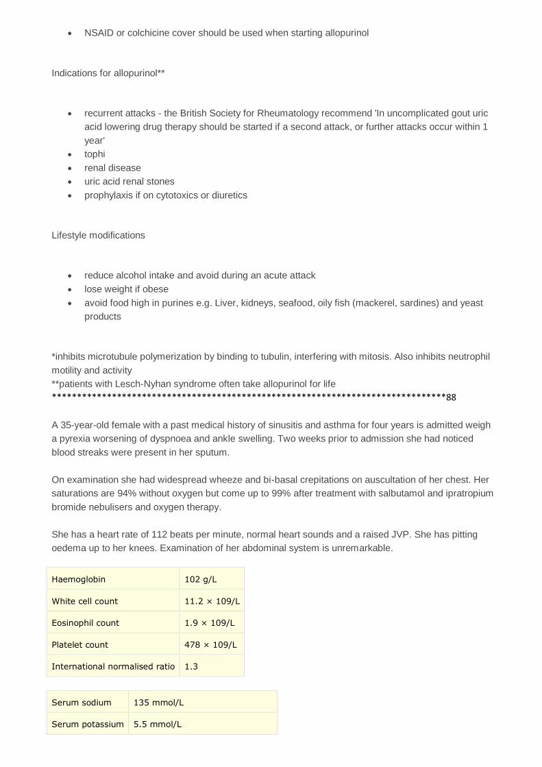
A 35-year-old female with a past medical history of sinusitis and asthma for four years is admitted weigh
a pyrexia worsening of dyspnoea and ankle swelling. Two weeks prior to admission she had noticed
blood streaks were present in her sputum.
On examination she had widespread wheeze and bi-basal crepitations on auscultation of her chest. Her
saturations are 94% without oxygen but come up to 99% after treatment with salbutamol and ipratropium
bromide nebulisers and oxygen therapy.
She has a heart rate of 112 beats per minute, normal heart sounds and a raised JVP. She has pitting
oedema up to her knees. Examination of her abdominal system is unremarkable.
Haemoglobin 102 g/L
White cell count 11.2 × 10⁹/L
Eosinophil count 1.9 × 10⁹/L
Platelet count 478 × 109/L
International normalised ratio 1.3
Serum sodium 135 mmol/L
Serum potassium 5.5 mmol/L

Serum urea 21 mmol/L
Serum creatinine 211 mol/L
CRP 22 mg/l
Blood cultures Coagulase-negative staphylococci
ANCA positive immunostaining awaited
Urine dip- positive for protein and blood
CXR: Bi-basal non specific shadowing
What is the most likely diagnosis?
Churg Strauss syndrome
Systematic Inflammatory Response Syndrome
Systematic Lupus Erythematous
Wegeners granulamatosis
Microscopic polyangiitis
Next question
This patient has essentially presented with acute kidney injury and is ANCA positive. ANCA may be
positive in Wegeners granulomatosis, Churg Strauss syndrome and microscopic polyangitis all of which
can affect the kidneys. However the history of asthma and sinusitis coupled with a raised eosinophil
count make Churg Strauss syndrome the most likely option.
Systematic Inflammatory response syndrome is unlikely to explain the entire clinical picture. Additionally,
although blood cultures are positive this is most likely to be a contaminant considering the organism and
the trivial C-reactive protein.
Churg-Strauss syndrome
Churg-Strauss syndrome is an ANCA associated small-medium vessel vasculitis.
Features
asthma
blood eosinophilia (e.g. > 10%)
paranasal sinusitis
mononeuritis multiplex
pANCA positive in 60%
Leukotriene receptor antagonists may precipitate the disease
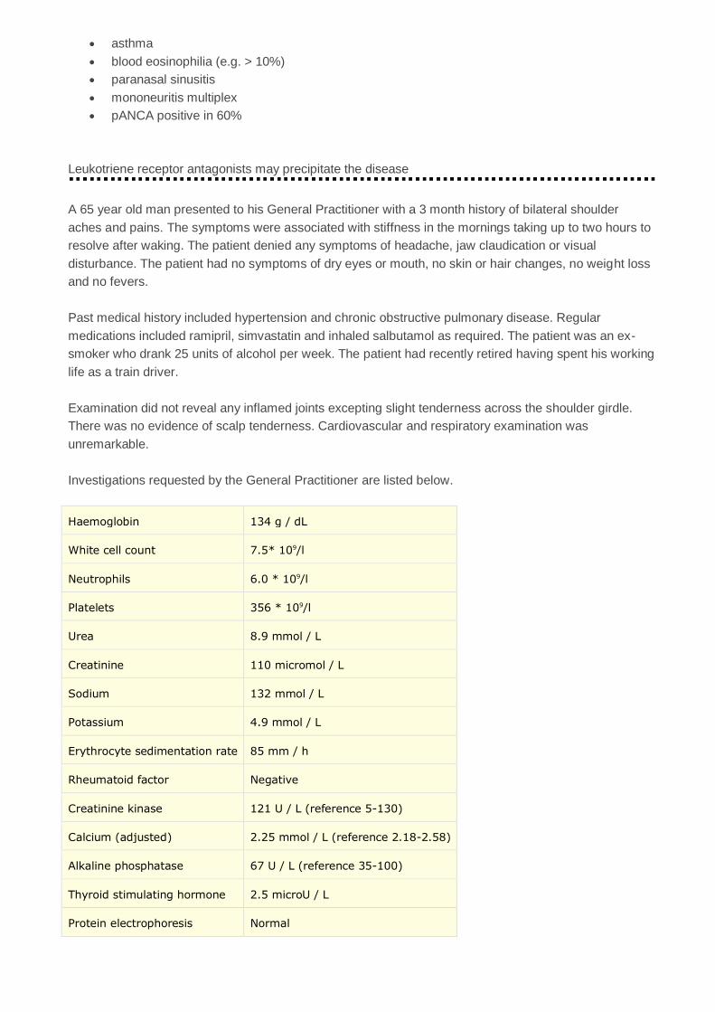
A 65 year old man presented to his General Practitioner with a 3 month history of bilateral shoulder
aches and pains. The symptoms were associated with stiffness in the mornings taking up to two hours to
resolve after waking. The patient denied any symptoms of headache, jaw claudication or visual
disturbance. The patient had no symptoms of dry eyes or mouth, no skin or hair changes, no weight loss
and no fevers.
Past medical history included hypertension and chronic obstructive pulmonary disease. Regular
medications included ramipril, simvastatin and inhaled salbutamol as required. The patient was an ex-
smoker who drank 25 units of alcohol per week. The patient had recently retired having spent his working
life as a train driver.
Examination did not reveal any inflamed joints excepting slight tenderness across the shoulder girdle.
There was no evidence of scalp tenderness. Cardiovascular and respiratory examination was
unremarkable.
Investigations requested by the General Practitioner are listed below.
Haemoglobin 134 g / dL
White cell count 7.5* 109/l
Neutrophils 6.0 * 109/l
Platelets 356 * 109/l
Urea 8.9 mmol / L
Creatinine 110 micromol / L
Sodium 132 mmol / L
Potassium 4.9 mmol / L
Erythrocyte sedimentation rate 85 mm / h
Rheumatoid factor Negative
Creatinine kinase 121 U / L (reference 5-130)
Calcium (adjusted) 2.25 mmol / L (reference 2.18-2.58)
Alkaline phosphatase 67 U / L (reference 35-100)
Thyroid stimulating hormone 2.5 microU / L
Protein electrophoresis Normal

What is the appropriate next management step for this patient?
Stop statin therapy and review in 6 weeks
Ultrasound study of shoulders and hips
Referral for specialist rheumatology opinion
Prednisolone 15 mg daily with dose tapering over 2 years
Prednisolone 40 mg daily with dose tapering over 1 year
Next question
This patient presents with a classical history for polymyalgia rheumatica and a raised ESR. There are no
factors in the history or investigations that suggest an alternative diagnosis (for example, giant cell
arteritis, other connective tissue disease, myeloma, malignancy or occult infection). The normal CK would
make statin-induced myopathy unlikely.
In such cases, a trial of steroid therapy for likely polymyalgia rheumatica is appropriate. The diagnosis
will be confirmed by rapid resolution of symptoms following initiation of treatment. Observational studies
suggest a typical starting dose of prednisolone around 15 mg with a median time to stopping therapy of
two years.
Musculoskeletal ultrasound often identifies inflammation around the shoulders and hips although these
findings are not unique to polymyalgia rheumatica. The usefulness of this technique is yet to be
determined outside of specialist settings.
Mackie S, Mallen C. Polymyalgia rheumatica. BMJ 2013;347:f6937
Polymyalgia rheumatica
Pathophysiology
overlaps with temporal arteritis
histology shows vasculitis with giant cells, characteristically 'skips' certain sections of affected
artery whilst damaging others
muscle bed arteries affected most in polymyalgia rheumatica
Features

typically patient > 60 years old
usually rapid onset (e.g. < 1 month)
aching, morning stiffness in proximal limb muscles (not weakness)
also mild polyarthralgia, lethargy, depression, low-grade fever, anorexia, night sweats
Investigations
ESR > 40 mm/hr
note CK and EMG normal
reduced CD8+ T cells
Treatment
prednisolone e.g. 15mg/od - dramatic response
A 35 year old woman presents to rheumatology clinic with a 2 month history of symmetrical swelling of
the ankles and fingers. She also complains of joint pain and stiffness. The stiffness is primarily worse in
the early morning and eases with use. Apart from a recent sore throat, she is otherwise well. She has a
family history of type 1 diabetes mellitus. She does not take any prescribed medication, but has found
herself relying on over-the-counter analgesics to get through the day.
On examination, she has bilateral swelling of the index, ring and middle fingers and bilateral ankle
swelling. She has a full range of movement in the fingers, wrists and ankles. There is marked swelling
and tenderness to palpation at the distal interphalangeal joints in the index, middle and ring fingers on
both sides. There are no skin changes, but yellowing and pitting of the nails is noted.
Blood tests show:
Hb 11.1 g/dl
Platelets 305 * 109/l
WBC 7.8 * 109/l
Na+ 141 mmol/l
K+ 4.2 mmol/l
Urea 5.8 mmol/l
Creatinine 64 µmol/l

Bilirubin 13 µmol/l
ALP 83 u/l
ALT 15 u/l
ESR 50mm/hr
CRP 39 mg/L
Rheumatoid factor negative
Hand X-ray shows mild erosion at the distal interphalangeal joints of the index, middle and ring fingers on
both hands.
What is the diagnosis?
Rheumatoid arthritis
Reiters syndrome
Ankylosing spondylitis
Yellow nail syndrome
Psoriatic arthritis
Next question
Although the patient does not have a psoriatic rash, she has classic symptoms of psoriatic arthritis. She
has dactylitis and distal interphalangeal swelling, as well as ankle involvement. Nail signs are well-
documented in psoriasis. The diagnosis is clinched by the negative rheumatoid factor and raised
inflammatory markers. A slightly low haemaglobin is also a common feature of the disease. The pattern
of joint involvement points more towards psoriatic arthritis than rheumatoid arthritis, in which the
metacarpophalangeal joints and wrists are more commonly affected. Reiters syndrome is a reactive
arthritis that typically follows a gastrointestinal or venereal infection. Conjunctivitis and urethritis are seen
alongside arthritis. Back pain would be expected to accompany ankylosing spondylitis. Yellow nail
syndrome is a rare disorder of uncertain pathogenesis. It presents with nail discolouration, lymphoedema
and pleural effusions.
Psoriatic arthritis can manifest in the absence of skin signs, particularly if the patient has a family history
of psoriasis. The patient may develop a rash later, or have signs limited to the nails.

Psoriatic arthropathy
Psoriatic arthropathy correlates poorly with cutaneous psoriasis and often precedes the development of
skin lesions. Around 10% percent of patients with skin lesions develop an arthropathy with males and
females being equally affected
© Image used on license from DermNet NZ
Notice the nail changes on this image as well
Types*
rheumatoid-like polyarthritis: (30-40%, most common type)
asymmetrical oligoarthritis: typically affects hands and feet (20-30%)
sacroilitis
DIP joint disease (10%)
arthritis mutilans (severe deformity fingers/hand, 'telescoping fingers')

© Image used on license from DermNet NZ
Management
treat as rheumatoid arthritis
but better prognosis
*Until recently it was thought asymmetrical oligoarthritis was the most common type, based on data from
the original 1973 Moll and Wright paper. Please see the link for a comparison of more recent studies
A 8-year-old boy is complains of progressively worsening pain in both groin areas. He has no past
medical history of note and his immunisations are up-to-date. There is no recent history of trauma. On
examination he walks with a limp. An x-ray is requested:

© Image used on license from Radiopaedia
What is the most likely diagnosis?
Developmental dysplasia of the hip
Slipped upper femoral epiphysis
Osteosarcoma
Acute lymphoblastic leukaemia
Perthes disease
Next question
Perthes disease
Perthes disease is a degenerative condition affecting the hip joints of children, typically between the ages

of 4-8 years. It is due to avascular necrosis of the femoral head
Perthes disease is 5 times more common in boys. Around 10% of cases are bilateral
Features
hip pain: develops progressively over a few weeks
limp
stiffness and reduced range of hip movement
x-ray: early changes include widening of joint space, later changes include decreased femoral
head size/flattening
Complications
osteoarthritis
premature fusion of the growth plates
© Image used on license from Radiopaedia
Perthes disease - both femoral epiphyses show extensive destruction, the acetabula are deformed

© Image used on license from Radiopaedia
Perthes disease - bilateral disease
A 32 year old female with known rheumatoid arthritis presents to clinic and would like some advice. She
would like to start a family with her partner. Her rheumatoid arthritis is current well-controlled on
methotrexate and sulphasalazine, she has not required changing of doses for 2 years. She is reluctant to
stop medications unless she has to, she had a number of flares when doses were reduced 3 years ago.
What would you advice regarding her plans for pregnancy?
She should reconsider her plans for pregnancy. Stopping medications would make her
disease uncontrollable and continuing medications will affect her child
Continue sulphasalazine and methotrexate
Stop sulphasalazine, continue methotrexate
Continue sulphasalazine and stop methotrexate
Stop both sulphasalazine and methotrexate
Next question
This is a relatively common scenario in rheumatology clinics: rheumatoid arthritis has a pre-ponderance

for females and a large number of RA patients are of child-bearing age1. The first consideration is the
need for treatment, then balancing the risks of disease flares and medical foetal toxicity. The majority of
RA patients, as in most autoimmune disorders, experience improvements in their condition during
pregnancy. Methotrexate is highly teratogenic and should be stopped between one and three months
before pregnancy. Hydroxychloroquine and sulphasalazine are generally accepted to be DMARDs that
can be continued during pregnancy. Sulphasalazine should however be avoided in male patients
attempting conception due to the risks of oligospermia. Glucocorticoids cross the placenta in low doses
and can be used after 14 weeks of pregnancy. Before this cut-off, there is an increased risk of cleft palate
and gestational hypertension. Low doses of prednisolone is an option should flares occur when off
methotrexate.
1. Dugowson CE, Koepsell TD, Voigt LF et al. Rheumatoid arthritis in women. Incidence rates in group
health cooperative, Seattle, Washington, 1987-1989. Arthritis Rheum. 1991;34(12):1502
Rheumatoid arthritis: management
The management of rheumatoid arthritis (RA) has been revolutionised by the introduction of disease-
modifying therapies in the past decade. NICE has issued a number of technology appraisals on the
newer agents and released general guidelines in 2009.
Patients with evidence of joint inflammation should start a combination of disease-modifying drugs
(DMARD) as soon as possible. Other important treatment options include analgesia, physiotherapy and
surgery.
Initial therapy
in the 2009 NICE guidelines it is recommend that patients with newly diagnosed active RA start a
combination of DMARDs (including methotrexate and at least one other DMARD, plus short-term
glucocorticoids)
DMARDs
methotrexate is the most widely used DMARD. Monitoring of FBC & LFTs is essential due to the
risk of myelosuppression and liver cirrhosis. Other important side-effects include pneumonitis
sulfasalazine
leflunomide
hydroxychloroquine
TNF-inhibitors
the current indication for a TNF-inhibitor is an inadequate response to at least two DMARDs
including methotrexate
etanercept: recombinant human protein, acts as a decoy receptor for TNF-α, subcutaneous
administration, can cause demyelination, risks include reactivation of tuberculosis
infliximab: monoclonal antibody, binds to TNF-α and prevents it from binding with TNF receptors,
intravenous administration, risks include reactivation of tuberculosis
adalimumab: monoclonal antibody, subcutaneous administration
Rituximab
anti-CD20 monoclonal antibody, results in B-cell depletion
two 1g intravenous infusions are given two weeks apart
infusion reactions are common
Abatacept
fusion protein that modulates a key signal required for activation of T lymphocytes
leads to decreased T-cell proliferation and cytokine production
given as an infusion
not currently recommend by NICE
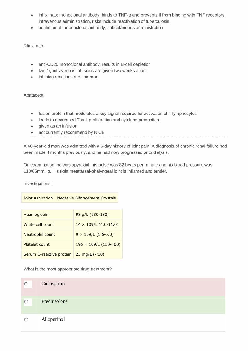
A 60-year-old man was admitted with a 6-day history of joint pain. A diagnosis of chronic renal failure had
been made 4 months previously, and he had now progressed onto dialysis.
On examination, he was apyrexial, his pulse was 82 beats per minute and his blood pressure was
110/65mmHg. His right metatarsal-phalyngeal joint is inflamed and tender.
Investigations:
Joint Aspiration Negative Bifringement Crystals
Haemoglobin 98 g/L (130-180)
White cell count 14 × 109/L (4.0-11.0)
Neutrophil count 9 × 10⁹/L (1.5-7.0)
Platelet count 195 × 109/L (150-400)
Serum C-reactive protein 23 mg/L (<10)
What is the most appropriate drug treatment?
Ciclosporin
Prednisolone
Allopurinol

Naproxen
Colchicine
Next question
The most appropriate drug treatment is Prednisolone. This gentleman has gout as shown by the
characteristic joint involvement (big toe). His treatment options are limited due to his dialysis.
Prednisolone is the safest drug to be used for a short course.
Naproxen is a known nephrotoxic and shouldnt be used in end stage renal failure. Colchicine is
contraindicated when patients are on dialysis. Allopurinol should not be started while a patient is suffering
with acute flare of gout.
Gout: management
Gout is a form of microcrystal synovitis caused by the deposition of monosodium urate monohydrate in
the synovium. It is caused by chronic hyperuricaemia (uric acid > 450 µmol/l)
Acute management
NSAIDs
intra-articular steroid injection
colchicine* has a slower onset of action. The main side-effect is diarrhoea
if the patient is already taking allopurinol it should be continued
Allopurinol prophylaxis - see indications below
allopurinol should not be started until 2 weeks after an acute attack has settled as it may
precipitate a further attack if started too early
initial dose of 100 mg od, with the dose titrated every few weeks to aim for a serum uric acid of <
300 µmol/l
NSAID or colchicine cover should be used when starting allopurinol
Indications for allopurinol**
recurrent attacks - the British Society for Rheumatology recommend 'In uncomplicated gout uric
acid lowering drug therapy should be started if a second attack, or further attacks occur within 1
year'
tophi
renal disease

uric acid renal stones
prophylaxis if on cytotoxics or diuretics
Lifestyle modifications
reduce alcohol intake and avoid during an acute attack
lose weight if obese
avoid food high in purines e.g. Liver, kidneys, seafood, oily fish (mackerel, sardines) and yeast
products
*inhibits microtubule polymerization by binding to tubulin, interfering with mitosis. Also inhibits neutrophil
motility and activity
**patients with Lesch-Nyhan syndrome often take allopurinol for life