morphological characteristics of black aspergilli isolated...
TRANSCRIPT

Songklanakarin J. Sci. Technol. 41 (1), 181-191, Jan. - Feb. 2019
Original Article
Morphological characteristics of black aspergilli
isolated from clinical wastes
Efaq A. N.1*, Al-Gheethi A. A.2 , Nik Norulaini Nik Ab. Rahman3,
Nagao H.4, and Ab. Kadir M. O.5
1 Department of Applied Microbiology, Faculty of Applied Sciences, Taiz University, Yemen
2 Faculty of Civil and Environmental Engineering, University Tun Hussein Onn Malaysia, Johor, 86400 Malaysia
3 School of Distance Education, University Science Malaysia, Penang, 11800 Malaysia
4 School of Biological Science, University Science Malaysia, Penang, 11800 Malaysia
5 School of Industrial Technology, University Science Malaysia, Penang, 11800 Malaysia
Received: 10 November 2015; Revised: 19 April 2017; Accepted: 17 October 2017
Abstract
The present study aimed to recognize the microscopic characteristics of black aspergilli species which exhibit similar
characteristics on culture media. Forty eight black aspergilli isolates were obtained from clinical wastes and purified using single
spore technique on six different culture media. The ultrastructure of fungal conidiophore and spores was detected by using
Scanning Electron Microscope (SEM). The fungal isolates were identified within five species included A. niger, A. tubingensis,
Aspergillus section Nigri, A. violaceofuscus, A. neoniger. Besides, two isolates identified as Aspergillus sp. strain no. 39,
Aspergillus sp. strain no. 53 appear as new strains based on the structure of conidiophore and spores. The fungi species have
similar culture characteristics. However, SEM observation demonstrated that they have quite different conidiophore and spores
morphology. The study revealed that the microstructure of the fungal spores and conidiophores plays an important role in the
identification of fungi species based on the phenotypic method.
Keywords: black aspergilli, SEM, ultrastructure, single spore technique
1. Introduction
Many fungi can be found in hospitals and other
clinical facilities or laboratories, and form a composite in the
wastes, generated from these premises (Efaq et al., 2015a).
The clinical wastes present an appropriate and fertile envi-
ronment for the reproduction of fungi due to the right tem-
perature and proper nutrients as well as the adequate moisture
and pH of the clinical wastes are favourable for their growth
(Efaq et al., 2017; United States Environmental Protection
Agency [U.S. EPA], 1990). On the other hand, the identifica-
tion using phenotypic techniques depends on the culture and
microscopic characteristics. Fungi have high diversity in their
conidiophore and spores, which are useful to identify of fun-
gal to species level (Efaq et al., 2016). Aspergillus section Ne-
gri complex have many common species which are known as
the black aspergilli (Balajee et al., 2009; Klich, 2009; Silva et
al., 2011). These species are often misidentified as A. niger
owing to the culture characteristics such as colony surface and
reverse colour are similar (Samson et al., 2007). The develop-
*Corresponding author
Email address: [email protected]; [email protected]

182 E. A. N. et al. / Songklanakarin J. Sci. Technol. 41 (1), 181-191, 2019
ments for Scanning Electronic Microscopy (SEM), Fluores-
cent Microscopy and Flow Cytometry have enhanced the
recognition of several details of phenotypic taxonomic
significance (Cole & Samson, 1979; Guarro et al., 1999). The
SEM has the ability to show the slight differences in the spore
structure to classify of fungi to the varieties level.
The present work aimed to describe the morpholo-
gical and culture characteristics of black aspergillli on dif-
ferent culture media included V8A, Potato Dextrose Agar, Sa-
bouraud dextrose agar, Czapek-Dox Agar, Czapek Yeast Ex-
tract Agar and Malt Extract Agar. The study focused on the
role SEM in the identification of these species. The ultra-
structure of conidiophores and spores included shape, texture,
spore surface ornamentation which cannot be observed using
light microscope were recognized by SEM.
2. Materials and Methods
2.1 Isolation and purification of fungi from clinical
waste samples
Clinical waste samples were collected from five di-
visions of Wellness Centre located within University Science
Malaysia (USM). The samples were collected in a biohazards
clinical waste bags and then transported to the laboratory in-
side the polystyrene box containing ice. The fungi isolates
were recovered from the clinical waste samples using direct
plate method on V8 juice agar medium (V8A) (Yu, 2010) and
then purified using single spore technique on PDA medium
according to Dr. Nagao technique (Efaq et al., 2015b). In
brief; the fungal colony grown on V8A was placed under
stereo light microscope (4X, Olympus SZ51, Japan). A spot
inoculation of the fungal spores (100-500 spores) was trans-
ferred onto a new PDA medium by using sterilized scalpel.
The inoculum was spread on the surface of the culture me-
dium by using stainless steel spreaders and with 0.1 mL steri-
lized distilled water (SDW). The plates were placed inside the
laminar flow 10 min to dry and then incubated at 28˚C for 16-
18 hours. Thereafter, a small piece (0.2×0.2 cm) of agar me-
dium containing a germinated spore was cut out aseptically by
using scalpel and under the light microscope to insure the
observation of the spore germinated tube and transferred onto
PDA medium. The plates were incubated at 28˚C for 7 days
and the fungal growth was noted daily.
2.2 Identification of fungal isolates
The identification of fungal isolates was performed
according to the culture and microscopic characterization. The
following references were used in the identification process;
Ellis and Martin (1985); Barnett and Hunter (1998); Watanabe
(2002); Samson et al. (2007); Robert et al. (2011); Silva et al.
(2011); Campbell et al. (2013) and National Mycology Re-
ference Centre (NMRC, 2015).
2.2.1 Cultural characteristics
The culture characteristics for each fungal isolate
was investigated on V8A, Potato Dextrose Agar (PDA, Oxoid,
UK), Czapek-Dox Agar (CZ, R&M Marketing, UK), Sabou-
raud dextrose agar (SDA, Hi Media, India), Malt Extract Agar
(MEA, Merck, Germany) and Czapek Yeast Extract Agar
(CYA, Oxoid, UK). The fungal cultures were separated into
groups based on their morphological characteristics including
growth diameter of the colonies (mm), surface and reverse co-
lour, texture, zonation on PDA and sporulation as described
by Promputtha et al. (2005). These characteristics were exa-
mined after 7 days of the incubation period at 28˚C.
2.2.2 Microscopic characteristics
The morphological characteristics for each fungal
isolate were observed under light microscope (100X, Olym-
pus, BX53F-CCD, Serial No. 1A589796, JAPAN). A small
mycelia part from the centre and edge of each colony was
placed onto glass slide contained one drop of distilled water
and covered by a cover slip. The characteristics of conidio-
phores and spores shapes and size were described for each
fungal isolates. The mean for 25 spores size determination by
using Cell Sens Standard (Version 1.4.1) programme was used
to give the average of the spore size for each fungus.
2.3 Scanning electron microscope (SEM) analysis
The microstructure of the conidiophore and spore
shapes was observed using SEM. Pure culture of each fungal
isolate sub-cultured on new PDA medium was sealed with
parafilm and incubated at 28˚C for 2 days. Thereafter, the pure
culture was sent under aseptic conditions to SEM laboratory at
Biological School, USM. A small piece (0.2×0.2 cm) from the
edge of grown colony was taken into aluminium petri dish.
The sample was dried using liquid nitrogen. The sample was
then coated with gold metal and observed using SEM (Zeiss
Supra 50 VP, Germany).
3. Results and Discussion
3.1 Recovery of fungal isolates from the clinical
waste samples
The first isolation of fungi from clinical waste sam-
ples was performed on V8A medium, which was chosen due
to the high fungal growth observed on this medium during the
preliminary study in comparison to CZ, CYA, PDA, SDA and
MEA media. PDA is the most common isolation medium for
the fungi from the samples. However, V8A exhibited here
more efficiency to improve the Aspergillus spp. growth. It has
been mentioned that the V8A is superior for the fungi which
need a complex medium for their growth and sporulation
(Choi et al. 1999). This might be due to the high contents of
the natural nutrients which enrich the fungal growth. Nonethe-
less, the reverse colour for the fungal growth on V8A was
unclear. Therefore, the morphology of fungal colonies was
described on PDA, CZ, CYA, MEA and SDA as presented in
Section 3.2.
3.2 Description of culture and morphological
characteristics of black aspergilli species
The identification of black aspergilli species in the
present study was conducted using phenotypic methods. This
technique represent a good method to best understand the
similarity and differences between the morphological and cul-
ture among the aspergilli species. Moreover, the micro-
E. A. N. et al. / Songklanakarin J. Sci. Technol. 41 (1), 181-191, 2019 183
structure of the fungal conidiophores and spores were
determined using SEM. The culture characteristics and sporu-
lation of fungal species obtained from clinical waste samples
were described on V8A, PDA, CZ, CYA, MEA and SDA. It
can be observed that the fungal growth and culture charac-
teristics of each fungal isolate rely on the culture medium.
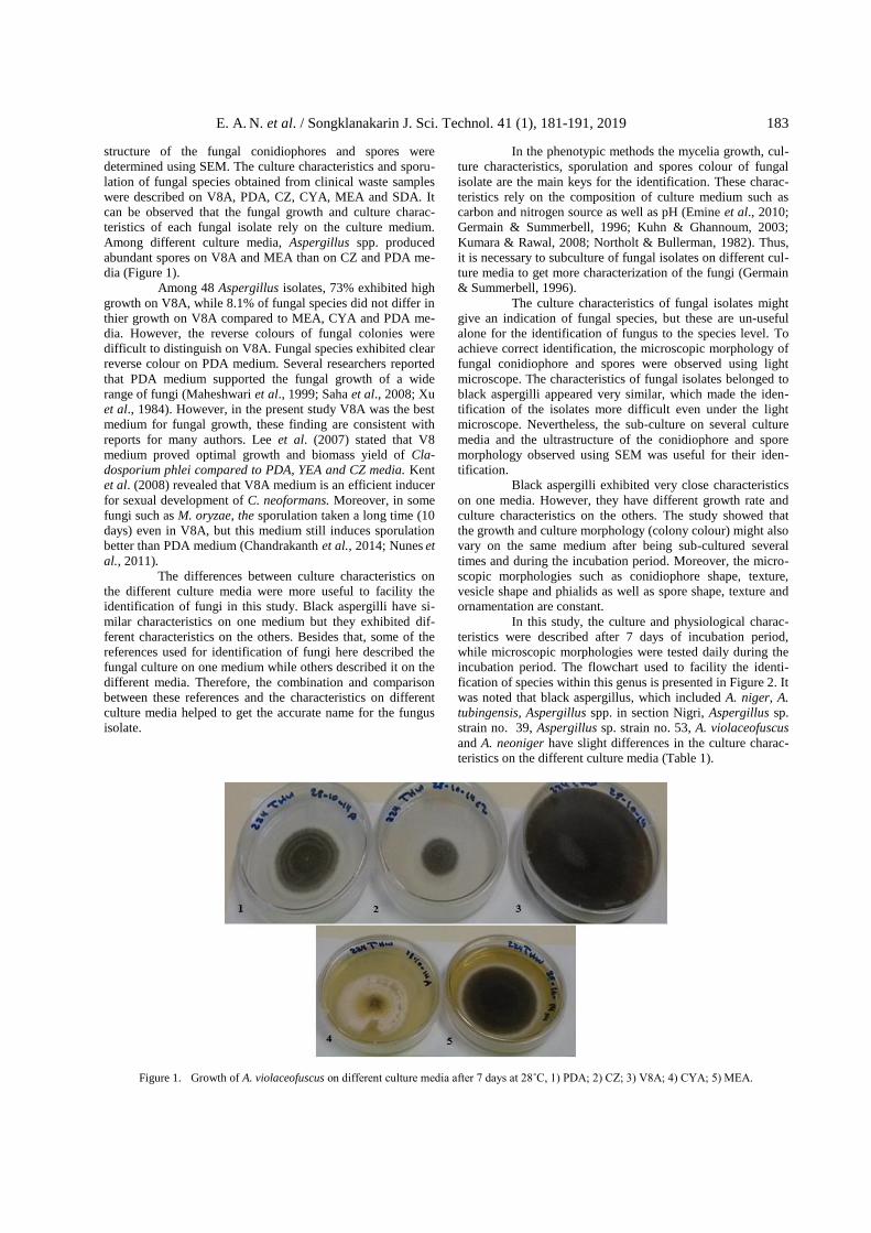
Among different culture media, Aspergillus spp. produced
abundant spores on V8A and MEA than on CZ and PDA me-
dia (Figure 1).
Among 48 Aspergillus isolates, 73% exhibited high
growth on V8A, while 8.1% of fungal species did not differ in
thier growth on V8A compared to MEA, CYA and PDA me-
dia. However, the reverse colours of fungal colonies were
difficult to distinguish on V8A. Fungal species exhibited clear
reverse colour on PDA medium. Several researchers reported
that PDA medium supported the fungal growth of a wide
range of fungi (Maheshwari et al., 1999; Saha et al., 2008; Xu
et al., 1984). However, in the present study V8A was the best
medium for fungal growth, these finding are consistent with
reports for many authors. Lee et al. (2007) stated that V8
medium proved optimal growth and biomass yield of Cla-
dosporium phlei compared to PDA, YEA and CZ media. Kent
et al. (2008) revealed that V8A medium is an efficient inducer
for sexual development of C. neoformans. Moreover, in some
fungi such as M. oryzae, the sporulation taken a long time (10
days) even in V8A, but this medium still induces sporulation
better than PDA medium (Chandrakanth et al., 2014; Nunes et
al., 2011).
The differences between culture characteristics on
the different culture media were more useful to facility the
identification of fungi in this study. Black aspergilli have si-
milar characteristics on one medium but they exhibited dif-
ferent characteristics on the others. Besides that, some of the
references used for identification of fungi here described the
fungal culture on one medium while others described it on the
different media. Therefore, the combination and comparison
between these references and the characteristics on different
culture media helped to get the accurate name for the fungus
isolate.
In the phenotypic methods the mycelia growth, cul-
ture characteristics, sporulation and spores colour of fungal
isolate are the main keys for the identification. These charac-
teristics rely on the composition of culture medium such as
carbon and nitrogen source as well as pH (Emine et al., 2010;
Germain & Summerbell, 1996; Kuhn & Ghannoum, 2003;
Kumara & Rawal, 2008; Northolt & Bullerman, 1982). Thus,
it is necessary to subculture of fungal isolates on different cul-
ture media to get more characterization of the fungi (Germain
& Summerbell, 1996).
The culture characteristics of fungal isolates might
give an indication of fungal species, but these are un-useful
alone for the identification of fungus to the species level. To
achieve correct identification, the microscopic morphology of
fungal conidiophore and spores were observed using light
microscope. The characteristics of fungal isolates belonged to
black aspergilli appeared very similar, which made the iden-
tification of the isolates more difficult even under the light
microscope. Nevertheless, the sub-culture on several culture
media and the ultrastructure of the conidiophore and spore
morphology observed using SEM was useful for their iden-
tification.
Black aspergilli exhibited very close characteristics
on one media. However, they have different growth rate and
culture characteristics on the others. The study showed that
the growth and culture morphology (colony colour) might also
vary on the same medium after being sub-cultured several
times and during the incubation period. Moreover, the micro-
scopic morphologies such as conidiophore shape, texture,
vesicle shape and phialids as well as spore shape, texture and
ornamentation are constant.
In this study, the culture and physiological charac-
teristics were described after 7 days of incubation period,
while microscopic morphologies were tested daily during the
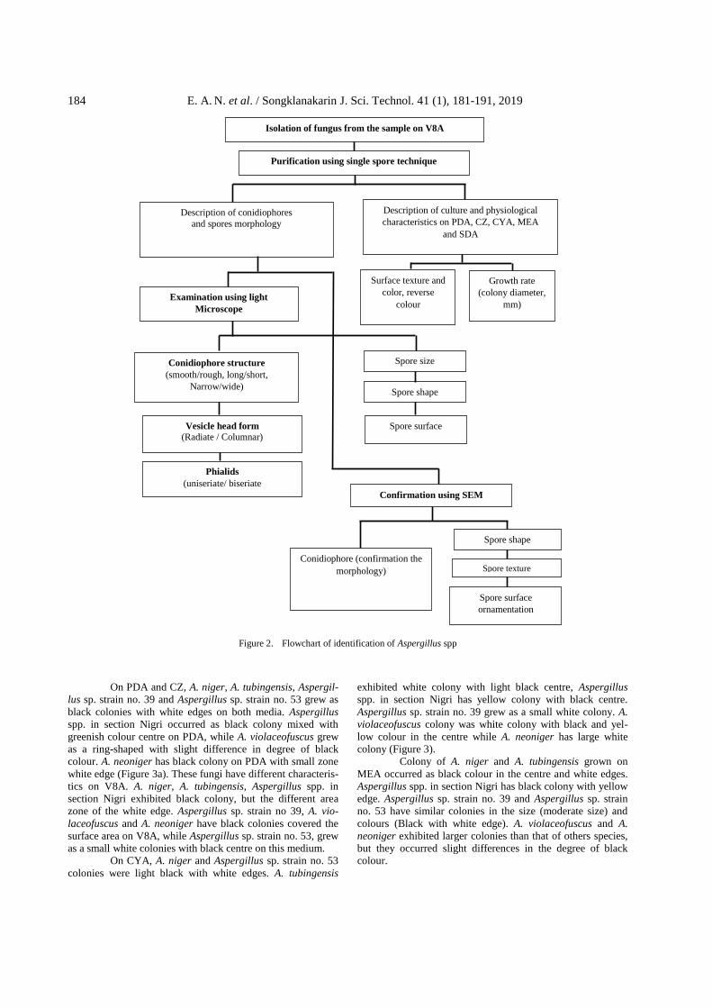
incubation period. The flowchart used to facility the identi-
fication of species within this genus is presented in Figure 2. It
was noted that black aspergillus, which included A. niger, A.
tubingensis, Aspergillus spp. in section Nigri, Aspergillus sp.
strain no. 39, Aspergillus sp. strain no. 53, A. violaceofuscus
and A. neoniger have slight differences in the culture charac-
teristics on the different culture media (Table 1).
Figure 1. Growth of A. violaceofuscus on different culture media after 7 days at 28˚C, 1) PDA; 2) CZ; 3) V8A; 4) CYA; 5) MEA.
184 E. A. N. et al. / Songklanakarin J. Sci. Technol. 41 (1), 181-191, 2019
Figure 2. Flowchart of identification of Aspergillus spp
On PDA and CZ, A. niger, A. tubingensis, Aspergil-
lus sp. strain no. 39 and Aspergillus sp. strain no. 53 grew as
black colonies with white edges on both media. Aspergillus
spp. in section Nigri occurred as black colony mixed with
greenish colour centre on PDA, while A. violaceofuscus grew
as a ring-shaped with slight difference in degree of black
colour. A. neoniger has black colony on PDA with small zone
white edge (Figure 3a). These fungi have different characteris-
tics on V8A. A. niger, A. tubingensis, Aspergillus spp. in
section Nigri exhibited black colony, but the different area
zone of the white edge. Aspergillus sp. strain no 39, A. vio-
laceofuscus and A. neoniger have black colonies covered the
surface area on V8A, while Aspergillus sp. strain no. 53, grew
as a small white colonies with black centre on this medium.
On CYA, A. niger and Aspergillus sp. strain no. 53
colonies were light black with white edges. A. tubingensis
exhibited white colony with light black centre, Aspergillus
spp. in section Nigri has yellow colony with black centre.
Aspergillus sp. strain no. 39 grew as a small white colony. A.
violaceofuscus colony was white colony with black and yel-
low colour in the centre while A. neoniger has large white
colony (Figure 3).
Colony of A. niger and A. tubingensis grown on
MEA occurred as black colour in the centre and white edges.
Aspergillus spp. in section Nigri has black colony with yellow
edge. Aspergillus sp. strain no. 39 and Aspergillus sp. strain
no. 53 have similar colonies in the size (moderate size) and
colours (Black with white edge). A. violaceofuscus and A.
neoniger exhibited larger colonies than that of others species,
but they occurred slight differences in the degree of black
colour.
Isolation of fungus from the sample on V8A
Purification using single spore technique
Description of culture and physiological
characteristics on PDA, CZ, CYA, MEA
and SDA
Description of conidiophores
and spores morphology
Surface texture and
color, reverse
colour
Growth rate
(colony diameter,
mm) Examination using light
Microscope
Confirmation using SEM
Conidiophore structure
(smooth/rough, long/short,
Narrow/wide)
Vesicle head form
(Radiate / Columnar)
Phialids
(uniseriate/ biseriate
Spore size
Spore shape
Spore surface
Conidiophore (confirmation the
morphology)
Spore shape
Spore texture
Spore surface
ornamentation

E. A. N. et al. / Songklanakarin J. Sci. Technol. 41 (1), 181-191, 2019 185
Table 1. Culture characteristics of black aspergilli on different culture media after 7 days of incubation period at 28˚C
Fungus Media type
Colony
Diameter (mm)
Colony character
Zon
atio
n
(Mar
gin
)
Sp
oru
lati
on
Texture Surface colour
Reverse colour
A.niger
V8A 73±5 crisp/ floccose dark black in
centre not clear white high
CZ 44±9 velvety centre/ floccose
centre black colour-less white moderate
CYA 38±5 annular velvety dark brown to
black
light
yellow yellow moderate
MEA 34±4.5 crisp/ floccose black colour-less white high
PDA 45±2.5 velvety/ crisp dark black colour-less white high
SDA 55±3.4 velvety black black white high
A. tubingensis
V8A 78±4.5 crisp/ floccose black not clear white high
CZ 49.3±4.5 velvety centre/ floccose
centre black colourless white high
CYA 66±0.8 velvety/ floccose/ sulcate light yellow
centre colourless white moderate
MEA 30±3 crisp/ floccose black colourless white high
PDA 44±4.3 velvety/ crisp black colourless white high
SDA 70±2.5 velvety/ floccose black black white high
Aspergillus spp.
in section Nigri
V8A 77±1.7 velvety black not clear yellow/
white high
CZ 25.5±0.5 velvety black yellow white moderate
CYA 75±1.5 velvety yellow/ black
centre yellow yellow moderate
MEA 45±7.5 velvety centre/ sulcate
edge black brown yellow high
PDA 41±1.8 velvety black
greenish yellow white moderate
SDA 80 ±0.0 velvety centre/ sulcate
edge black
yellow/
brown yellow high
Aspergillus sp. strain no. 39
V8A 80±0.0 velvety black not clear black high
CZ 21±4 velvety black colourless white high
CYA 19±6.7 creamy growth/ velvety
and radially centre white centre colourless
beige/
white less
MEA 32±4.8 velvety centre/ sulcate
edge black colourless white high
PDA 34±7.2 velvety/annular black light yellow white high
SDA 37.1±2.8 sulcate black black white High
Aspergillus sp.
strain no. 53
V8A 20±3.1 velvety, wrinkled, sulcate black colourless white low
CZ 22±1 velvety black colourless white high
CYA 70±8.3 velvety, wrinkled/ sulcate brown centre colourless white moderate
MEA 48±6.8 velvety/sulcate black light brown white high
PDA 29±10 velvety black colourless white High

186 E. A. N. et al. / Songklanakarin J. Sci. Technol. 41 (1), 181-191, 2019
Table 1. Continued
Fungus Media type
Colony
Diameter
(mm)
Colony character
Zon
atio
n
(Mar
gin
)
Sp
oru
lati
on
Texture Surface Reverse
A.
viola
ceofu
scu
s
V8A 80±0.0 velvety black not clear black high
CZ 24.5±1.5 velvety light black colourless white moderate
CYA 46±2.5 floccose/ wrinkled yellow-ish/
brown orange white low
MEA 62±5.2 amaranthine black colourless white high
PDA 32±7.2 velvety annular black light yellow white moderate
SDA 36±0.8 amaranthine black colourless white high
A. neoniger
V8A 80±0.0 velvety black not clear black high
CZ 26±10 velvety centre/ floccose edge
black colourless white high
CYA 28±8.0 floccose/sulcate light yellow
centre colourless to light yellow
white moderate
MEA 68±2.5 velvety centre /
sulcate edge black
colourless to light yellow
white high
PDA 39±1.3 velvety/annular dark black yellow white high
SDA 80±0.0 velvety/ sulcate black black white high
Figure 3. Culture characteristics of Aspergillus spp. on different culture media after incubation period for 7 days at 28˚C, A) Aspergillus sp. strain no. 39; B) Aspergillus sp. strain no. 53; C) A. violaceofuscus; D) A. neoniger; 1) PDA; 2) CZ; 3) V8A; 4) CYA; 5) MEA
E. A. N. et al. / Songklanakarin J. Sci. Technol. 41 (1), 181-191, 2019 187
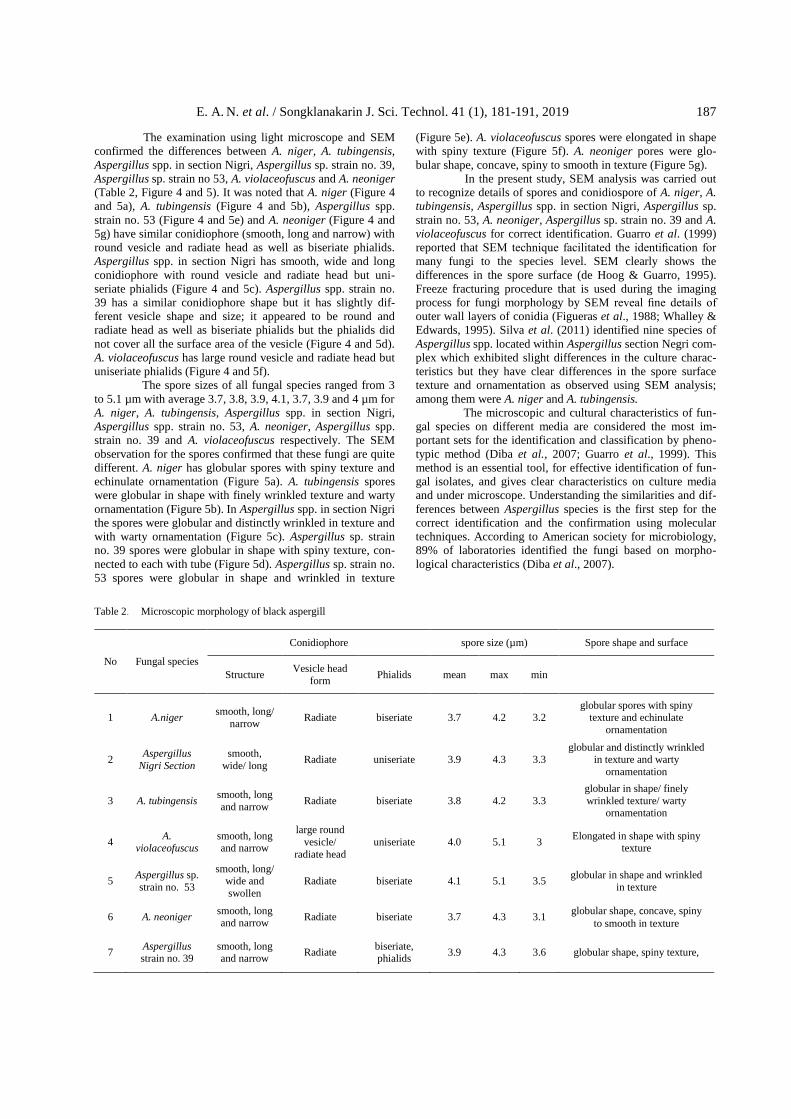
The examination using light microscope and SEM
confirmed the differences between A. niger, A. tubingensis,
Aspergillus spp. in section Nigri, Aspergillus sp. strain no. 39,
Aspergillus sp. strain no 53, A. violaceofuscus and A. neoniger
(Table 2, Figure 4 and 5). It was noted that A. niger (Figure 4
and 5a), A. tubingensis (Figure 4 and 5b), Aspergillus spp.
strain no. 53 (Figure 4 and 5e) and A. neoniger (Figure 4 and
5g) have similar conidiophore (smooth, long and narrow) with
round vesicle and radiate head as well as biseriate phialids.
Aspergillus spp. in section Nigri has smooth, wide and long
conidiophore with round vesicle and radiate head but uni-
seriate phialids (Figure 4 and 5c). Aspergillus spp. strain no.
39 has a similar conidiophore shape but it has slightly dif-
ferent vesicle shape and size; it appeared to be round and
radiate head as well as biseriate phialids but the phialids did
not cover all the surface area of the vesicle (Figure 4 and 5d).
A. violaceofuscus has large round vesicle and radiate head but
uniseriate phialids (Figure 4 and 5f).
The spore sizes of all fungal species ranged from 3
to 5.1 µm with average 3.7, 3.8, 3.9, 4.1, 3.7, 3.9 and 4 µm for
A. niger, A. tubingensis, Aspergillus spp. in section Nigri,
Aspergillus spp. strain no. 53, A. neoniger, Aspergillus spp.
strain no. 39 and A. violaceofuscus respectively. The SEM
observation for the spores confirmed that these fungi are quite
different. A. niger has globular spores with spiny texture and
echinulate ornamentation (Figure 5a). A. tubingensis spores
were globular in shape with finely wrinkled texture and warty
ornamentation (Figure 5b). In Aspergillus spp. in section Nigri
the spores were globular and distinctly wrinkled in texture and
with warty ornamentation (Figure 5c). Aspergillus sp. strain
no. 39 spores were globular in shape with spiny texture, con-
nected to each with tube (Figure 5d). Aspergillus sp. strain no.
53 spores were globular in shape and wrinkled in texture
(Figure 5e). A. violaceofuscus spores were elongated in shape
with spiny texture (Figure 5f). A. neoniger pores were glo-
bular shape, concave, spiny to smooth in texture (Figure 5g).
In the present study, SEM analysis was carried out
to recognize details of spores and conidiospore of A. niger, A.
tubingensis, Aspergillus spp. in section Nigri, Aspergillus sp.
strain no. 53, A. neoniger, Aspergillus sp. strain no. 39 and A.
violaceofuscus for correct identification. Guarro et al. (1999)
reported that SEM technique facilitated the identification for
many fungi to the species level. SEM clearly shows the
differences in the spore surface (de Hoog & Guarro, 1995).
Freeze fracturing procedure that is used during the imaging
process for fungi morphology by SEM reveal fine details of
outer wall layers of conidia (Figueras et al., 1988; Whalley &
Edwards, 1995). Silva et al. (2011) identified nine species of
Aspergillus spp. located within Aspergillus section Negri com-
plex which exhibited slight differences in the culture charac-
teristics but they have clear differences in the spore surface
texture and ornamentation as observed using SEM analysis;
among them were A. niger and A. tubingensis.
The microscopic and cultural characteristics of fun-
gal species on different media are considered the most im-
portant sets for the identification and classification by pheno-
typic method (Diba et al., 2007; Guarro et al., 1999). This
method is an essential tool, for effective identification of fun-
gal isolates, and gives clear characteristics on culture media
and under microscope. Understanding the similarities and dif-
ferences between Aspergillus species is the first step for the
correct identification and the confirmation using molecular
techniques. According to American society for microbiology,
89% of laboratories identified the fungi based on morpho-
logical characteristics (Diba et al., 2007).
Table 2. Microscopic morphology of black aspergill
No Fungal species
Conidiophore spore size (µm) Spore shape and surface
Structure Vesicle head
form Phialids mean max min
1 A.niger smooth, long/
narrow Radiate biseriate 3.7 4.2 3.2
globular spores with spiny texture and echinulate
ornamentation
2 Aspergillus
Nigri Section
smooth,
wide/ long Radiate uniseriate 3.9 4.3 3.3
globular and distinctly wrinkled in texture and warty
ornamentation
3 A. tubingensis smooth, long
and narrow Radiate biseriate 3.8 4.2 3.3
globular in shape/ finely
wrinkled texture/ warty ornamentation
4 A.
violaceofuscus smooth, long and narrow
large round
vesicle/
radiate head
uniseriate 4.0 5.1 3 Elongated in shape with spiny
texture
5 Aspergillus sp.
strain no. 53
smooth, long/ wide and
swollen
Radiate biseriate 4.1 5.1 3.5 globular in shape and wrinkled
in texture
6 A. neoniger smooth, long and narrow
Radiate biseriate 3.7 4.3 3.1 globular shape, concave, spiny
to smooth in texture
7 Aspergillus strain no. 39
smooth, long and narrow
Radiate biseriate, phialids
3.9 4.3 3.6 globular shape, spiny texture,

188 E. A. N. et al. / Songklanakarin J. Sci. Technol. 41 (1), 181-191, 2019
Figure 4a. Light microscope micrographs of A. niger,
A) conidiophore; B) spores
Figure 4b. Light microscope micrographs of A. tubingensis, A) conidiophore; B) spores
Figure 4c. Light microscope micrographs of Aspergillus spp. in
section Nigri, A) conidiophore; B) spores
Figure 4d. Light microscope micrographs of Aspergillus sp. new
strain 39, A) conidiophore; B) spores
Figure 4e. Light microscope micrographs of Aspergillus sp. new strain 53, A) conidiophore; B) spores
Figure 5a. Scanning electron micrographs of A. niger showing the
conidiophore (A), which occurs as smooth, long and
narrow with round vesicles and radiate head as well as biseriate phialids, and spores (B), which occur as glo-
bular spores with spiny texture and echinulate orna-
mentation. The magnification is 300x for conidiophore and 10.00 kx for the spores
Figure 5b. Scanning electron micrographs of A. tubingensis showing
the conidiophore (A), which occurs as smooth, long and
narrow with round vesicles and radiate head as well as
biseriate phialids, and spores (B), which occur globular in shape/ finely wrinkled texture/ warty ornamentation.
The magnification is 401x for the conidiophore and 3.00
kx for the spores.
Figure 5c. Scanning electron micrographs of Aspergillus spp. in section Nigri showing the conidiophore (A), which oc-curs as smooth, wide and long conidiophore with round
vesicles and radiate head as well as uniseriate phialids,
and spores B), which occur as globular and distinctly wrinkled in texture and warty ornamentation. The
magnification was 700x for the conidiophore and 6.00
kx for the spores.
Figure 5d. Scanning electron micrographs of Aspergillus sp. strain no. 39 showing the conidiophore (A), which occurs as
smooth, wide and long with round and radiate head
vesicles as well as biseriate phialids but the phialids have not covered all surface area of the vesicles, and spores
B), which occur as globular shape and spiny texture. The
magnification is 561x for the conidiophore and 5.01 kx for the spores.

E. A. N. et al. / Songklanakarin J. Sci. Technol. 41 (1), 181-191, 2019 189
Figure 5e. Scanning electron micrographs of Aspergillus sp. strain
no. 53 showing the conidiophore (A), which occurs as smooth, long and narrow with round and swollen vesi-
cles and radiate head as well as biseriate phialids, and
spores (B), which occur globular in shape and wrinkled in texture. The magnification is 500x for the conidio-
phore and 5.00 kx for the spores.
Figure 5f . Scanning electron micrographs of A. violaceofuscus
showing the conidiophore (A) which occurs as smooth,
wide and long with large round vesicles and radiate head but uniseriate phialids, and spores B), which are elon-
gated in shape with spiny texture. The magnification is
589x for the conidiophore and 5.00 kx for the spores.
Figure 5g. Scanning electron micrographs of A. neoniger showing
the conidiophore (A), which occurs as smooth, long and
narrow with round vesicle and radiate head as well as
biseriate phialids, and spores (B), which occur as glo-
bular shape, concave, spiny to smooth in texture. The
magnification is 636x for the conidiophore and 5.11 kx for the spores.
The phenotypic method for the identification of
fungi is usually easy. However, the culture and microscopic
observation using light microscope often create a miscon-
ception about its value for the detection some species such as
Aspergillus spp. especially within black aspergilli as noted in
this study. Therefore, SEM observation was used for the
determination of the ultrastructure of conidiophore and spores
of Aspergillus spp. SEM has been previously used as a
technique for study and identification of fungi based on the
spore structure (Clarke & Griffiths, 1970; Eduard et al., 19
85). Gao et al. (2007) determined two strains of A. flavus (L
and S strain) based on the microscopic observation of coni-
diophores structures. Mares et al. (2008) identified 3 new
species of Aspergillus spp. including A. quitensis; E19C, A.
amazonicus; E19D, and A. ecuadorensis; E19F based on SEM
analysis of conidiophore and spores, and DNA analysis con-
firmed that these fungi are quite different. Zhang (2009) des-
cribed a new species of Aspergillus sp. in China based on
morphological characteristics using light and SEM observa-
tion. Lately, Vestlund et al. (2014) used SEM for classifi-
cation of bioaerosols from composting which included fungal
spores. In this study, two new isolates within black aspergilli
were identified based on culture and microscopic morpho-
logies as well as ultrastructure determined using SEM. These
strains were Aspergillus sp. strain no. 39, Aspergillus sp.
strain no. 53. However, the confirmation for both isolates will
be conducted based on molecular analysis in the possible
future work.
4. Conclusions
The high diversity in the structure of conidiophore
and spores of black aspergilli is more useful for their iden-
tification. However, these structures might be difficult to re-
cognize using light microscopy. SEM clearly recognize the
slight differences in the spore structure, which includes spore
texture and surface ornamentation. Both parameters play an
important role in the identification process by phenotypic
method.
Acknowledgements
The authors gratefully acknowledge the Ministry of
Science Technology and Innovation (MOSTI) for the research
project financial support under FRGS Grant No. 203/PTE
KIND/6711438) and APEX Grant 1002/PJJIUH/910324).
References
Balajee, S. A., Kano, R., Baddley, J. W., Moser, S. A., Marr,
K. A., Alexander, B. D., . . . Chiller, T. (2009).
Molecular Identification of Aspergillus Species Col-
lected for the Transplant-Associated Infection Sur-
veillance Network. Journal of Clinical Microbio-
logy, 47(10), 3138–3141.
Barnett, H. L., & Hunter, B. B. (1998). Illustrated genera of
imperfect fungi; A comprehensive resource for re-
cognizing, identifying, and learning various aspects
of imperfect fungi 4th Edition. Saint Paul, MN: APS
Press. Campbell, C. K., Johnson, E. M., & Warnock, D. W. (2013).
Identification of pathogenic fungi 2nd Edition. Lon-
don, England: A John Wiley & Sons.
Chandrakanth, R., Jagadeesh, D., & Devaki, N. S. (2014).
Black light mediated growth and sporulation of
magnaporthe oryzae. International Journal of Agri-
culture Science and Research, 4, 25-30.
Choi, Y. W., Hyde, K. D., & Ho, W. H. (1999). Single spore
isolation of fungi. Fungal Diversity, 3, 29-38.
Clarke, J. H., & Griffiths, D. A. (1970). Ascospores of some
common species of Eurotium (Aspergillus glaucus)
as shown by scanning electron microscopy. Tran-
sactions of the British Mycological Society, 55, 117–
122.

190 E. A. N. et al. / Songklanakarin J. Sci. Technol. 41 (1), 181-191, 2019
Cole, G. T., & Samson, R. A. (1979). Patterns of development
in conidial fungi. London, England: Pitman.
de Hoog, G. S., & Guarro, J. (1995). Atlas of clinical fungi.
Baarn, The Netherlands: Centraalbureau voor
Schimmelcultures.
Diba, K., Kordbacheh, P., Mirhendi, S. H., Rezaie, S., & Mah-
moudi, M. (2007). Identification of Aspergillus
species using morphological characteristics. Pakis-
tan Journal of Medical Science, 23(6), 867-872.
Eduard, W., Sandven, P., Johansen, B. V., & Bruun, R. (1985).
Identification and quantification of mould spores by
scanning electron microscopy (SEM): analysis of
filter samples collected in Norwegian Saw Mills.
Proceedings of an International Symposium and
Workshop on Lung Dosimetry Organised by the Bri-
tish Occupational Hygiene Society in Co-Operation
with the Commission of the European Communities,
Cambridge, England. 447–453.
Efaq, A. N., Nik, N. N. A. R., Nagao, H., Al-Gheethi, A. A.,
Md, S., & Ab. Kadir, M. O. (2015a). Supercritical
carbon dioxide as non-thermal alternative techno-
logy for safe handling of clinical wastes. Journal of
Environmental Processes, 2(4), 797–822.
Efaq A. N., Nik, N. N. A. R., Nagao, H., Al-Gheethi, A. A., &
Ab. Kadir, M. O. (2017). Inactivation of Aspergillus
Spores in Clinical Wastes by Supercritical Carbon
Dioxide. Arabian Journal for Science and Engi-
neering, 42(1), 39-51.
Efaq, A.N., Al-Gheethi, A. A., Nik, N. N. A. R., Nagao, H., &
Ab. Kadir, M. O. (2016). Assessment of relevant
fungal species in clinical solid wastes. Environ-
mental Science and Pollution Research, 23(19),
19806–19824. doi:10.1007/s11356-016-7161-8
Efaq, A. N., Nagao, H., Nik, N. N. A., Al-Gheethi, A. A., &
Ab. Kadir, M. O. (2015b). Survival of opportunistic
fungi in clinical wastes. 4th International Conference
on Environmental Research and Technology, Pe-
nang, Malaysia.
Ellis, M. B. (1971). Dematiaceous Hyphomycetes. London,
England: Kew: Commonwealth Mycological Insti-
tute.
Emine, S., Kambol, R., & Zainol, N. (2010). Morphological
Characterization of Soil Penicillium sp. Strains –
Potential Producers of Statin. Biotechnology Sym-
posium IV, Universiti Malaysia Sabah, Sabah, Ma-
laysia.
Figueras, M. J., Guarro, J., & Dijk, F. (1988). Rodlet structure
on the surface of Chaetomium spores. Journal of
Microbios, 53, 101–107.
Gao, J., Liu, Z. and Yu, J. 2007. Identification of Aspergillus
section Flavi in maize in northeastern China. Myco-
pathology Journal, 64, 91–99.
Germain, S. G., & Summerbell, R. (1996). Identifying Fila-
mentous Fungi–A Clinical Laboratory Handbook 1st
Edition. Belmont, California: Star Publishing.
Guarro, J., Gene, J., & Stchigel, A. M. (1999). Developments
in Fungal Taxonomy. Clinical Microbiology Review,
12(3), 454–500.
Kent, C. R., Bermúdez, P. O., Giles, S. S., & Hull, C. M.
(2008). Formulation of a Defined V8 Medium for
Induction of Sexual Development of Cryptococcus
neoformans. Appled Environmental Microbiology,
74(20), 6248–6253.
Klich, M. A. (2009). Health effects of Aspergillus in food and
air. Toxicology Industrial Health 25, 657–667.
Kuhn, D. M., & Ghonnoum, M. A. (2003). Indoor mold, toxi-
genic fungi and Stachybotrys chartarum: Infectious
disease perspective. Clinical Microbiology Review,
16(1), 144-172.
Kumara, K. L. W., & Rawal, R. D. (2008). Influence of car-
bon, nitrogen, temperature and pH on the growth
and sporulation of some Indian isolates of Colleto-
trichum gloeosporioides causing anthracnose disease
of papaya (Carrica papaya L). Tropical Agriculture
Research Experiment, 11, 7-12.
Lee, J. K., Kim, B. T., Kim, J. A., Chung, H. J., Park, S. M.,
Yang, M. S., . . . Kim, D. H. (2007). Cultural cha-
racteristics and extraction of the fungal pigment
phleichrome from the phytopathogenic fungus Cla-
dosporium phlei. Biotechnology Bioprocess Engi-
neering, 12(5), 508-515.
Maheshwari, S. K., Singh, D. V., & Sahu, A. K. (1999). Effect
of several nutrient media, pH and carbon sources
on growth and sporulation of Alternaria alternata.
Journal of Mycopathology Research, 37, 21-23.
Mares, D., Andreotti, E., Maldonado, M. E., Pedrini, P.,
Colalongo, C., & Romagnoli, C. (2008). Three New
Species of Aspergillus from Amazonian Forest Soil
(Ecuador). Current Microbiology, 57, 222–229.
National Mycology Reference Centre. (2015, January 2). My-
cology Online. National Mycology Reference Cen-
tre, the University of Adelaide. Retrieved from http:
//www.mycology.adelaide.edu.au
Northolt, M. D., & Bullerman, L. B. (1982). Prevention of
mould growth and toxin production through control
of environmental condition. Journal of Food Pro-
tection, 6, 519-526.
Nunes, C. C., Gowda, M., Sailsbery, J., Xue, M., Chen, F.,
Brown, D. E., . . . Dean, R. A. (2011). Diverse and
tissue-enriched small RNAs in the plant pathogenic
fungus, Magnaporthe oryzae. BMC Genomics, 12
(288), 2-20.
Promputtha, I., Jeewon, R., Lumyong, S., McKenzie, E. H. C.,
& Hyde, K. D. (2005). Ribosomal DNA
fingerprinting in the identification of non
sporulating endophytes from Magnolia liliifera
(Magnoliaceae). Fungal Diversity, 20, 167-186.
Robert, A. S., János, V., & Christian, F. J. (2011). Taxonomic
studies on the genus Aspergillus- DTU Orbit. CBS-
KNAW Fungal Biodiversity Centre, Utrecht, Ne-
therlands.
Saha, A., Mandal, P., Dasgupta, S., & Saha, D. (2008). In-
fluence of culture media and environmental fac-tors
on mycelial growth and sporulation of Lasiodiplodia
theobromae (Pat.) Griffon and Maubl. Journal of
Environmental Biology, 29(3), 407-410.

E. A. N. et al. / Songklanakarin J. Sci. Technol. 41 (1), 181-191, 2019 191
Samson, R. A., Houbraken, J., Thrane, U., Frisvad, J. C., &
Andersen, B. (2010). Food and indoor fungi. CBS-
KNAW Fungal Biodiversity Centre, Utrecht, Ne-
therlands.
Silva, D. M., Batista, L. R., Rezende, E. F., Fungaro, M. H. P.,
Sartori, D., & Alves, E. (2011). Identification of
fungi of the genus Aspergillus section Nigri using
polyphasic taxonomy. Brazilian Journal of Micro-
biology, 42, 761-773.
United States Environmental Protection Agency. (1990).
Summary of potential risks from hospital waste in-
cineration: Pathogens in air emission and residues.
EACO-R-0238. Retrieved from https://nepis.epa.
gov/
Vestlund, A. T., Al-Ashaab, R., Tyrre, S. F, Longhurst, P. J.,
Pollard, S. J. T., & Drewa, G. H. (2014). Morpho-
logical classification of bioaerosols from composting
using scanning electron microscopy. Waste Manage-
ment, 34, 1101–1108.
Watanabe, T. (2002). Pictorial atlas of soil and seed fungi;
Morphologies of cultured fungi and key to species
2nd Edition. Boca Raton, FL: CRC Press.
Whalley, A. J. S., & Edwards, R. L. (1995). Secondary meta-
bolites and systematic arrangement within the
Xylariaceae. Canadian Journal of Botany, 73, 802–
810.
Xu, S. O., Yuan, S. Z., & Chen, X. C. (1984). Studies on pa-
thogenic fungus (Alternaria tennuis Nees) of poplar
leaf blight. Journal of North East Forestry Institute,
12, 56-64.
Yu, J. (2010). Identification of fungi and bacteria associated
with internally discolored horseradish roots (Mas-
ter’s thesis, Graduate College, University of Illinois,
IL).
Zhang, Z. (2009). A new species of Aspergillus. International
Journal of biology, 1(2), 78-80.