molybdenum sequestration in brassica species. a role for
TRANSCRIPT
Molybdenum Sequestration in Brassica Species.A Role for Anthocyanins?1
Kerry L. Hale, Steve P. McGrath, Enzo Lombi, Stephen M. Stack, Norman Terry, Ingrid J. Pickering,Graham N. George, and Elizabeth A.H. Pilon-Smits*
Department of Biology, Anatomy/Zoology Building, Colorado State University, Fort Collins, Colorado 80523(K.L.H., S.M.S., E.A.H.P.-S.); Agriculture and Environment Division, Institute of Arable Crops Research-Rothamsted, Harpenden, Hertfordshire AL5 2JQ, United Kingdom (S.P.M., E.L.); Department of Plant andMicrobial Biology, 111 Koshland Hall, University of California, Berkeley, California 94270 (N.T.); andStanford Synchrotron Research Laboratory (SSRL), Stanford Linear Accelerator Center, P.O. Box 20450,Stanford, California 94309 (I.J.P., G.N.G.)
To elucidate plant mechanisms involved in molybdenum (Mo) sequestration and tolerance, Brassica spp. seedlings weresupplied with molybdate, and the effects on plant physiology, morphology, and biochemistry were analyzed. Whensupplied with (colorless) molybdate Indian mustard (Brassica juncea) seedlings accumulated water-soluble blue crystals intheir peripheral cell layers. Energy dispersive x-ray analysis showed that Mo accumulated predominantly in the vacuolesof the epidermal cells. Therefore, the blue crystals are likely to be a Mo compound. The x-ray absorption spectrum of theplant-accumulated Mo was different than that for molybdate, indicating complexation with a plant molecule. Because theblue compound was water soluble and showed a pH-dependent color change, possible involvement of anthocyanins wasinvestigated. An anthocyanin-less mutant of Brassica rapa (“fast plants”) was compared with varieties containing normal orhigh anthocyanin levels. The anthocyanin-less mutant did not show accumulation of a blue compound when supplied withmolybdate. In the anthocyanin-containing varieties, the blue compound colocalized with anthocyanins in the peripheral celllayers. Mo accumulation by the three B. rapa varieties was positively correlated with anthocyanin content. Addition ofmolybdate to purified B. rapa anthocyanin resulted in an in vitro color change from pink to blue. Therefore, Mo appears tobe sequestered in vacuoles of the peripheral cell layers of Brassica spp. as a blue compound, probably a Mo-anthocyanincomplex.
Molybdenum (Mo) is an essential micronutrient forplants, bacteria, and animals. Mo-deficient plants ex-hibit poor growth and low chlorophyll and ascorbicacid content (Marschner, 1995). Mo is also a compo-nent of some bacterial nitrogenases, and therefore isespecially important for plants that live in symbiosiswith nitrogen-fixing bacteria. The major metabolicfunction for Mo in eukaryotic organisms is as anessential component of the mononuclear Mo en-zymes (Hille, 1996), which play roles in many keymetabolic processes such as sulfur detoxification, pu-rine catabolism, nitrate assimilation, and phytohor-
mone synthesis in plants (Stallmeyer et al., 1999).These enzymes usually (but not always) catalyzetwo-electron redox reactions that are coupled to thetransfer of an oxygen atom to or from substrate andthe metal, during which the Mo cycles between theMo(VI) and Mo(IV) oxidation states. In these sys-tems, Mo is bound to the dithiolene of a novel pyr-anopterin cofactor that is known as molybdopterin(Hille, 1996). Although a low-Mr species containingboth Mo and molybdopterin has never been properlycharacterized, this complex is known as the Mo co-factor (Moco). These enzymes are unrelated to theMo-containing nitrogenase of nitrogen-fixing bacte-ria that contain a Mo-iron-sulfur cluster.
A mutational block in the early steps of Mocobiosynthesis leads to the combined loss of function ofMo enzymes. As a result, plant cells can no longerassimilate inorganic nitrogen and have altered levelsof certain phytohormones (Seo et al., 1998). However,a change in proteins catalyzing the last step of Mocosynthesis, i.e. the transfer and incorporation of Mointo molybdopterin, leads to a molybdate repairablephenotype (Falciani et al., 1994), whereby the addi-tion of molybdate is sufficient to overcome the neg-ative effects of the mutation. Therefore, molybdate isthought to be the source of Mo for incorporation intoMoco (Kuper et al., 2000).
1 This work was supported by the National Science Foundation(Career Development Grant no. 9982432) and by the EnvironmentalProtection Agency (Research Grant no. G8A11586 to E.A.H.P.-S.).The XAS experiments were made possible through SSRL Synchro-tron beam time (granted to N.T.). SSRL is funded by the U.S.Department of Energy, Offices of Basic Energy Sciences and Bio-logical and Environmental Research; by the National Institutes ofHealth; by the National Center for Research Resources, by theBiomedical Technology Program; and by the National Instituteof General Medical Sciences. The Institute of Arable CropsResearch-Rothamsted receives grant-aided support from the Bio-technology and Biological Sciences Research Council of the UK.
* Corresponding author; e-mail: [email protected];fax 970 – 491– 0649.
Plant Physiology, August 2001, Vol. 126, pp. 1391–1402, www.plantphysiol.org © 2001 American Society of Plant Biologists 1391
In plants, Mo is readily mobile in xylem andphloem for long-distance transport (Kannan and Ra-mani, 1978), though little is known of the mecha-nisms involved in Mo homeostasis. Molybdate com-petes with sulfate for uptake at the root surface,suggesting a common uptake mechanism (Stout etal., 1951). However, it has also been suggested thatmolybdate is taken up by a phosphate transporter(Heuwinkel et al., 1992). Furthermore, a recent studyby Palmgren and Harper (1999) suggests the pres-ence of a Mo-specific metal transporter, AMA1, inArabidopsis. T-DNA knockout mutants, whereAMA1 was rendered ineffective, showed 5-fold re-duced Mo accumulation, but no reduction in anyother essential micronutrients.
The Mo content of plants is a direct reflection of thebioavailability of Mo in the soil. The relation of Moinflux versus concentration is approximately linear insoil systems (Barber, 1995). Bioavailability of Mo ispositively correlated with soil pH (Karmian and Cox,1978). In acidic soils, Mo deficiency in plants is com-mon. On the other hand, in some soils of high pH,plants may accumulate enough Mo for ruminantsfeeding on the plants to develop molybdenosis. Mo-lybdenosis is caused by an imbalance of Mo andcopper in the ruminant diet, which induces a copperdeficiency (Stark and Redente, 1990). Plants suppliedwith adequate Mo usually contain 1 to 2 mg kg�1 Mo;plant material containing an excess of 5 mg kg�1 Mois sufficient to cause molybdenosis in ruminant ani-mals (Barber, 1995). Molybdenosis in livestock occursin the western U.S., often in soils with poor drainageand high organic matter (Gupta and Lipsett, 1981).Mo pollution due to mining and stainless steel indus-try poses a serious environmental problem at severallocations in the U.S., including several Superfundsites (polluted sites in the U.S. designated by the U.S.Environmental Protection Agency for high priorityremediation). Plant samples from a mining site nearEmpire, CO were found to contain up to 400 mg kg�1
Mo (Trlica and Brown, 2000). Phytoremediation, theuse of plants to remediate environmental pollution,may prove to be a viable strategy for remediating Moin these areas, either via phytoextraction (accumula-tion in harvestable plant parts) or phytostabilization(in situ immobilization).
To use plants optimally for Mo remediation, weneed to know which processes are involved andwhere rate limitations may occur. The goal of thisstudy was to obtain a better understanding of plantmechanisms that control accumulation, tolerance,and biotransformation of Mo in plants.
RESULTS
When Indian mustard (Brassica juncea) seedlingswere grown on agar medium containing differentconcentrations of colorless ammonium molybdate,they showed blue coloration, especially at the base of
the hypocotyl and around the petioles (Fig. 1, A–C).Cross sections of the Mo-treated seedlings showedthat the blue compound was present in a crystal-likeform (Fig. 1, C and D), localized predominantly in theepidermal and subepidermal tissues (Fig. 1E). Seed-lings grown without Mo did not show this blue col-oration (Fig. 1F). A similar distribution was observedwhen mature plants were supplied with ammoniummolybdate, with the blue coloration being most pro-nounced in the stem and near vascular tissue in theleaves (results not shown).
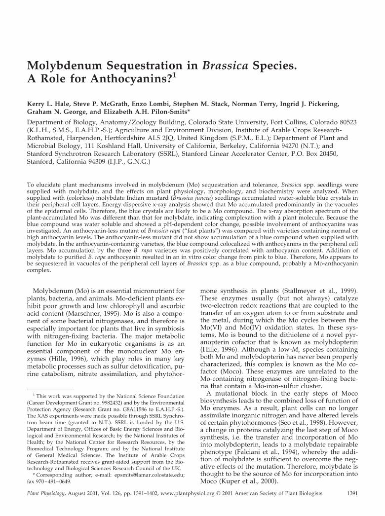
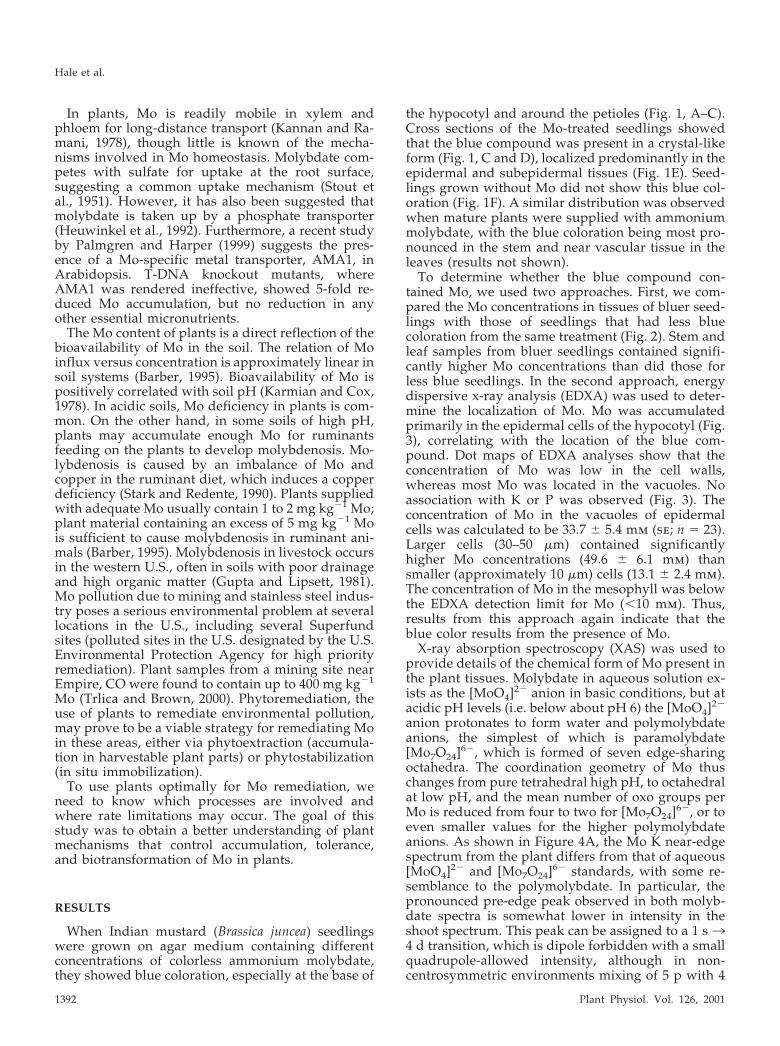
To determine whether the blue compound con-tained Mo, we used two approaches. First, we com-pared the Mo concentrations in tissues of bluer seed-lings with those of seedlings that had less bluecoloration from the same treatment (Fig. 2). Stem andleaf samples from bluer seedlings contained signifi-cantly higher Mo concentrations than did those forless blue seedlings. In the second approach, energydispersive x-ray analysis (EDXA) was used to deter-mine the localization of Mo. Mo was accumulatedprimarily in the epidermal cells of the hypocotyl (Fig.3), correlating with the location of the blue com-pound. Dot maps of EDXA analyses show that theconcentration of Mo was low in the cell walls,whereas most Mo was located in the vacuoles. Noassociation with K or P was observed (Fig. 3). Theconcentration of Mo in the vacuoles of epidermalcells was calculated to be 33.7 � 5.4 mm (se; n � 23).Larger cells (30–50 �m) contained significantlyhigher Mo concentrations (49.6 � 6.1 mm) thansmaller (approximately 10 �m) cells (13.1 � 2.4 mm).The concentration of Mo in the mesophyll was belowthe EDXA detection limit for Mo (�10 mm). Thus,results from this approach again indicate that theblue color results from the presence of Mo.
X-ray absorption spectroscopy (XAS) was used toprovide details of the chemical form of Mo present inthe plant tissues. Molybdate in aqueous solution ex-ists as the [MoO4]2� anion in basic conditions, but atacidic pH levels (i.e. below about pH 6) the [MoO4]2�
anion protonates to form water and polymolybdateanions, the simplest of which is paramolybdate[Mo7O24]6�, which is formed of seven edge-sharingoctahedra. The coordination geometry of Mo thuschanges from pure tetrahedral high pH, to octahedralat low pH, and the mean number of oxo groups perMo is reduced from four to two for [Mo7O24]6�, or toeven smaller values for the higher polymolybdateanions. As shown in Figure 4A, the Mo K near-edgespectrum from the plant differs from that of aqueous[MoO4]2� and [Mo7O24]6� standards, with some re-semblance to the polymolybdate. In particular, thepronounced pre-edge peak observed in both molyb-date spectra is somewhat lower in intensity in theshoot spectrum. This peak can be assigned to a 1 s34 d transition, which is dipole forbidden with a smallquadrupole-allowed intensity, although in non-centrosymmetric environments mixing of 5 p with 4
Hale et al.
1392 Plant Physiol. Vol. 126, 2001

d levels confers significant dipole-allowed intensity(Kutzler et al., 1980). The intensity of this feature hasbeen observed to correlate with the number of Mo �O (oxo) ligands to Mo, which is consistent with thesmaller intensity observed for [Mo7O24]6� versus[MoO4]2�. In the plant spectrum, its reduced pres-ence suggests a smaller number of oxo groups (e.g.around two).
The EXAFS spectrum (Fig. 4B) of Mo in plantshoots confirms that the Mo is modified from thesimple [MoO4]2� structure, which consists of fourMo � O interactions at 1.76 Å. The shoot spectrumindicates several different interactions in the firstshell, and there is also evidence for longer rangeinteractions at �3 Å. Curve fitting of the data (TableI) indicates three different Mo � O distances in the
Figure 1. Indian mustard seedlings grown for 7 d in agar medium, with (A–E) or without (F) 60 mg L�1 ammoniummolybdate. A, Seedling. B, Close-up of petiole. C, Close-up of hypocotyl. D, Longitudinal section of hypocotyl at highermagnification to show irregular blue precipitate in some epidermal cells. The black line connects the precipitate at low andhigh magnification. E, Cross section of hypocotyls showing blue color in the peripheral cell layers. F, Hypocotyl cross sectionof a seedling grown without Mo.
Molybdenum Sequestration in Brassica Sp.
Plant Physiol. Vol. 126, 2001 1393
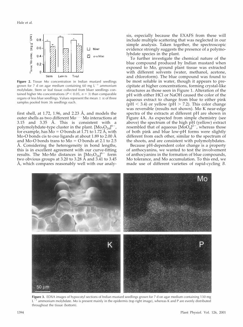
first shell, at 1.72, 1.96, and 2.23 Å, and models theouter shells as two different Mo. . . . Mo interactions at3.15 and 3.35 Å. This is consistent with apolymolybdate-type cluster in the plant. [Mo7O24]6�,for example, has Mo � O bonds at 1.71 to 1.72 Å, withMo-O bonds cis to oxo ligands at about 1.89 to 2.00 Åand Mo-O bonds trans to Mo � O bonds at 2.1 to 2.5Å. Considering the heterogeneity in bond lengths,this is in excellent agreement with our curve-fittingresults. The Mo-Mo distances in [Mo7O24]6� formtwo obvious groups at 3.20 to 3.28 Å and 3.41 to 3.45Å, which compares reasonably well with our analy-
sis, especially because the EXAFS from these willinclude multiple scattering that was neglected in oursimple analysis. Taken together, the spectroscopicevidence strongly suggests the presence of a polymo-lybdate species in the plant.
To further investigate the chemical nature of theblue compound produced by Indian mustard whenexposed to Mo, ground plant tissue was extractedwith different solvents (water, methanol, acetone,and chloroform). The blue compound was found tobe most soluble in water, though it appears to pre-cipitate at higher concentrations, forming crystal-likestructures as those seen in Figure 1. Alteration of thepH with either HCl or NaOH caused the color of theaqueous extract to change from blue to either pink(pH � 3.4) or yellow (pH � 7.2). This color changewas reversible (results not shown). Mo K near-edgespectra of the extracts at different pH are shown inFigure 4A. As expected from simple chemistry (seeabove) the spectrum of the high pH (yellow) extractresembled that of aqueous [MoO4]2�, whereas thoseof both pink and blue low-pH forms were slightlydifferent from each other, similar to the spectrum ofthe shoots, and are consistent with polymolybdates.
Because pH-dependent color change is a propertyof anthocyanins, we wanted to test the involvementof anthocyanins in the formation of blue compounds,Mo tolerance, and Mo accumulation. To this end, wemade use of different varieties of rapid-cycling B.
Figure 2. Tissue Mo concentration in Indian mustard seedlingsgrown for 7 d on agar medium containing 60 mg L�1 ammoniummolybdate. Stem or leaf tissue collected from bluer seedlings con-tained higher Mo concentrations (P � 0.05, n � 3) than comparableorgans of less blue seedlings. Values represent the mean � SE of threesamples pooled from 36 seedlings each.
Figure 3. EDXA images of hypocotyl sections of Indian mustard seedlings grown for 7 d on agar medium containing 130 mgL�1 ammonium molybdate. Mo is present mainly in the epidermis (top right image), whereas K and P are evenly distributedthroughout the tissue (bottom).
Hale et al.
1394 Plant Physiol. Vol. 126, 2001
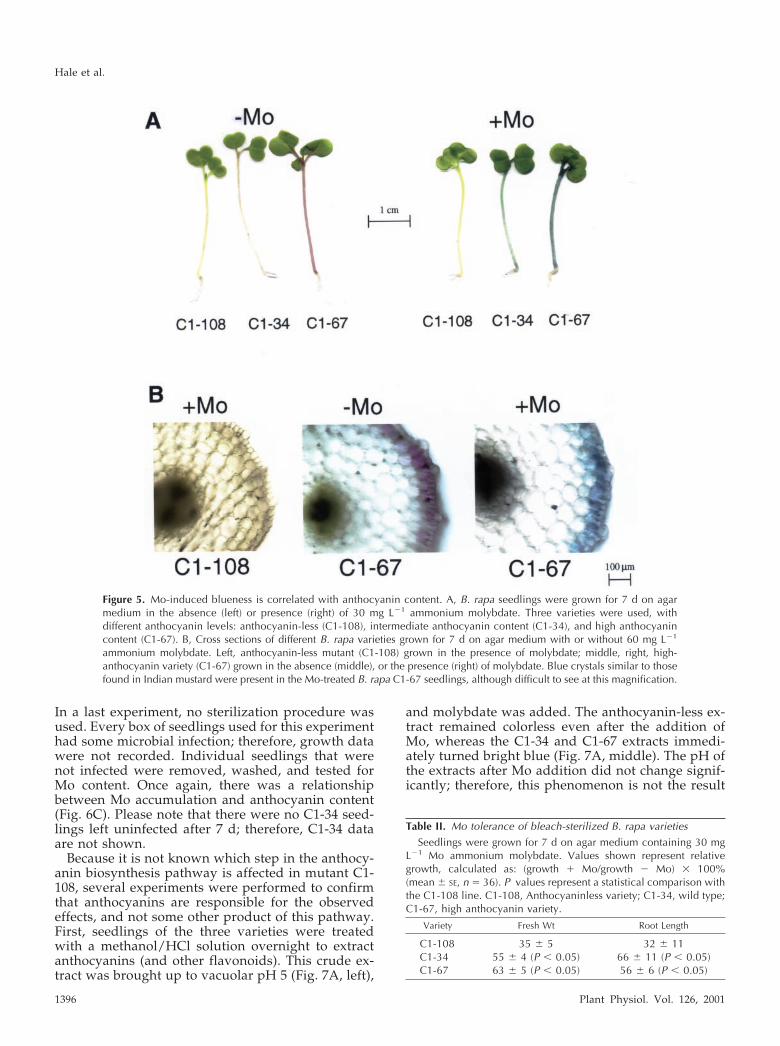
rapa (“fast plants”) that vary in anthocyanin produc-tion. A mutant unable to produce anthocyanin (C1-108) was compared with intermediate anthocyanin(C1-34) and high anthocyanin (C1-67) varieties.When grown in the presence of Mo, the anthocyanin-less mutant did not show the blue coloration, and thehigh anthocyanin variety was bluer than the varietycontaining intermediate anthocyanin levels (Fig. 5A).Thus, the degree of blueness was related to anthocy-anin content. The Mo K near-edge spectrum of shoottissue of the B. rapa anthocyanin-less mutant (Fig. 4A)appeared similar to that of the Indian mustard shoottissue, but with a less pronounced pre-edge peak.
To visualize the distribution of the colored com-pounds, B. rapa varieties grown with and withoutMo were cross-sectioned and the presence of bluecompounds and anthocyanins were compared. an-thocyanin-less mutants exposed to Mo showed noaccumulation of the blue compound (Fig. 5B, left).There was no observable difference betweentreated and non-treated (not shown) cross sectionsof anthocyanin-less plants. In the anthocyanin-containing varieties, the anthocyanin was shown tobe concentrated in the peripheral cell layers (Fig. 5B,middle), as was the blue compound that was pro-duced when the plants were treated with Mo (Fig.5B, right). Therefore, the tissue distribution of an-thocyanin is similar to that of the blue compound.
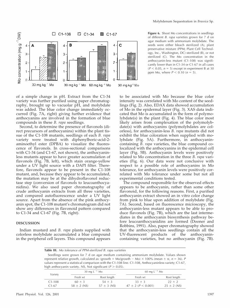
To determine the possible role of anthocyanins inMo accumulation and tolerance, we compared shootMo concentrations, fresh weights, and root lengths ofthe three B. rapa varieties. In a first experiment, seedswere sterilized using a standard procedure with eth-anol and bleach. The results showed a distinct rela-tionship between anthocyanin content and Mo toler-ance (Table II) and accumulation (Fig. 6A). There wasno difference in tolerance to the bleach treatmentalone; therefore, it is unlikely that the relationshipsbetween anthocyanin and Mo tolerance or accumu-lation is an artifact of differential bleach tolerance.However, because germination frequencies wererather low after the bleach and ethanol treatment(especially for C1-108), a second experiment wasdone where seeds were sterilized using plant preser-vative mixture (PPM) to deter infection without thepotential harm that bleach and ethanol may cause.Again, there was a significant difference in Moaccumulation between the anthocyanin-less and an-thocyanin-containing varieties (Fig. 6B). However,there was no clear correlation between Mo toleranceand anthocyanin content, except for fresh weight at60 mg L�1 Mo (Table III) Please note that because wewere unable to grow C1-34 seedlings without infec-tion on PPM, no data are available for that genotype.
Figure 4. XAS of Mo in Indian mustard seedlings grown for 7 d on agarmedium containing 60 mg L�1 ammonium molybdate. A, Mo K x-rayabsorption near-edge spectra of: a, Molybdate in aqueous solution atpH 8 (solid line) and pH 4 (dashed line); b, Indian mustard seedlings;c, Brassica rapa anthocyanin-less mutant; and d, aqueous extracts ofIndian mustard at pH 8 (dashed line), pH 6 (solid line), and pH 4(dot-dashed line). B, Results of extended x-ray absorption fine structure(EXAFS) curve fitting of shoots of Indian mustard seedlings. Inset, Thek3-weighted Mo K-edge EXAFS oscillations for the experimental data(solid line) and the best fit (dashed line) according to the parameters inTable I. The main panel shows the corresponding Fourier transforms,phase corrected for the first shell Mo-O interactions.
Table I. EXAFS curve fitting of molybdate and molybdate trans-formed in plant shoots
Coordination no. was fitted to the nearest integer. The nos. inparentheses after the distances and Debye-Waller factors indicate theprecisions, expressed as three times the estimated SDs (obtained fromthe diagonal elements of the covariance matrix) in the last digit(s) ofthe values. The accuracies will be larger than, and related to, theprecisions, and will typically be less than �0.02 Å for interatomicdistances.
Sample No. and Type Distance Debye-Waller Factor
Å Å2
Molybdate 4 Mo-O 1.767 (3) 0.0025 (3)Mo-treated shoots 3 Mo-O 1.719 (5) 0.0048 (5)
2 Mo-O 1.964 (8) 0.0041 (10)1 Mo-O 2.23 (2) 0.004 (2)
1 Mo. . .Mo 3.15 (1) 0.0068 (12)1 Mo. . .Mo 3.35 (2) 0.0068 (12)
Molybdenum Sequestration in Brassica Sp.
Plant Physiol. Vol. 126, 2001 1395
In a last experiment, no sterilization procedure wasused. Every box of seedlings used for this experimenthad some microbial infection; therefore, growth datawere not recorded. Individual seedlings that werenot infected were removed, washed, and tested forMo content. Once again, there was a relationshipbetween Mo accumulation and anthocyanin content(Fig. 6C). Please note that there were no C1-34 seed-lings left uninfected after 7 d; therefore, C1-34 dataare not shown.
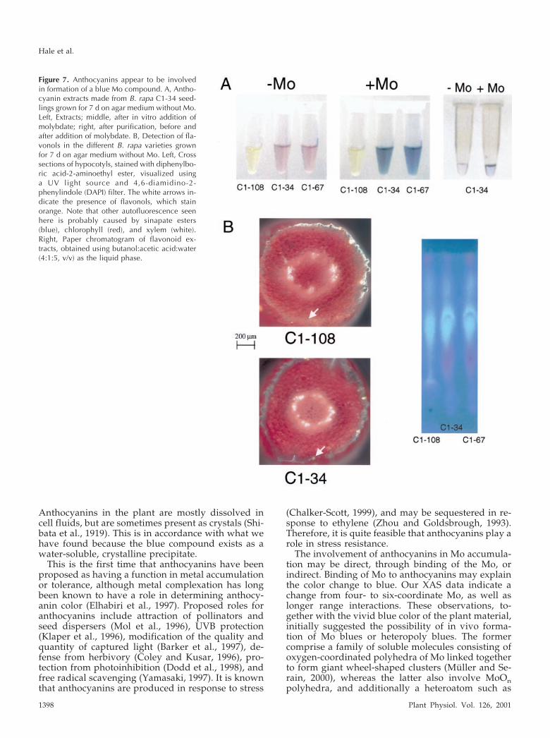
Because it is not known which step in the anthocy-anin biosynthesis pathway is affected in mutant C1-108, several experiments were performed to confirmthat anthocyanins are responsible for the observedeffects, and not some other product of this pathway.First, seedlings of the three varieties were treatedwith a methanol/HCl solution overnight to extractanthocyanins (and other flavonoids). This crude ex-tract was brought up to vacuolar pH 5 (Fig. 7A, left),
and molybdate was added. The anthocyanin-less ex-tract remained colorless even after the addition ofMo, whereas the C1-34 and C1-67 extracts immedi-ately turned bright blue (Fig. 7A, middle). The pH ofthe extracts after Mo addition did not change signif-icantly; therefore, this phenomenon is not the result
Figure 5. Mo-induced blueness is correlated with anthocyanin content. A, B. rapa seedlings were grown for 7 d on agarmedium in the absence (left) or presence (right) of 30 mg L�1 ammonium molybdate. Three varieties were used, withdifferent anthocyanin levels: anthocyanin-less (C1-108), intermediate anthocyanin content (C1-34), and high anthocyanincontent (C1-67). B, Cross sections of different B. rapa varieties grown for 7 d on agar medium with or without 60 mg L�1
ammonium molybdate. Left, anthocyanin-less mutant (C1-108) grown in the presence of molybdate; middle, right, high-anthocyanin variety (C1-67) grown in the absence (middle), or the presence (right) of molybdate. Blue crystals similar to thosefound in Indian mustard were present in the Mo-treated B. rapa C1-67 seedlings, although difficult to see at this magnification.
Table II. Mo tolerance of bleach-sterilized B. rapa varieties
Seedlings were grown for 7 d on agar medium containing 30 mgL�1 Mo ammonium molybdate. Values shown represent relativegrowth, calculated as: (growth � Mo/growth � Mo) � 100%(mean � SE, n � 36). P values represent a statistical comparison withthe C1-108 line. C1-108, Anthocyaninless variety; C1-34, wild type;C1-67, high anthocyanin variety.
Variety Fresh Wt Root Length
C1-108 35 � 5 32 � 11C1-34 55 � 4 (P � 0.05) 66 � 11 (P � 0.05)C1-67 63 � 5 (P � 0.05) 56 � 6 (P � 0.05)
Hale et al.
1396 Plant Physiol. Vol. 126, 2001
of a simple change in pH. Extract from the C1-34variety was further purified using paper chromatog-raphy, brought up to vacuolar pH, and molybdatewas added. The blue color change immediately oc-curred (Fig. 7A, right) giving further evidence thatanthocyanins are involved in the formation of bluecompounds in these B. rapa seedlings.
Second, to determine the presence of flavonols (di-rect precursors of anthocyanins) within the plant tis-sue of the C1-108 mutants, seedlings of each B. rapavariety were treated with diphenylboric-acid-2-aminoethyl ester (DPBA) to visualize the fluores-cence of flavonols. In cross-sectional comparisonswith C1-34 (and C1-67, not shown), the anthocyanin-less mutants appear to have greater accumulation offlavonols (Fig. 7B, left), which stain orange-yellowunder a UV light source with a DAPI filter. There-fore, flavonols appear to be present in the C1-108mutant, and, because they appear to be accumulated,the mutation may be at the dihydroflavonol reduc-tase step (conversion of flavonols to leucoanthocya-nidins). We also used paper chromatography ofcrude anthocyanin extracts from all three varieties,and compared autofluorescence under a UV lightsource. Apart from the absence of the pink anthocy-anin spot, the C1-108 mutant’s chromatogram did notshow any differences in flavonoid pattern comparedto C1-34 and C1-67 (Fig. 7B, right).
DISCUSSION
Indian mustard and B. rapa plants supplied withcolorless molybdate accumulated a blue compoundin the peripheral cell layers. This compound appears
to be associated with Mo because the blue colorintensity was correlated with Mo content of the seed-lings (Fig. 2). Also, EDAX data showed accumulationof Mo in the epidermal layer (Fig. 3). XAS data indi-cated that Mo is accumulated in the form of polymo-lybdate(s) in the plant (Fig. 4). The blue color mostlikely arises from complexation of the polymolyb-date(s) with anthocyanins (polymolybdates are col-orless), for anthocyanin-less B. rapa mutants did notexhibit the blue coloration when supplied with mo-lybdate (Fig. 5A). Furthermore, in anthocyanin-containing B. rapa varieties, the blue compound co-localized with the anthocyanins in the epidermal celllayer (Fig. 5B). Anthocyanin levels were positivelyrelated to Mo concentration in the three B. rapa vari-eties (Fig. 6). Our data were not conclusive withrespect to a possible role of anthocyanins in Motolerance, for anthocyanin levels were positively cor-related with Mo tolerance under some but not allexperimental conditions tested.
The compound responsible for the observed effectsappears to be anthocyanin, rather than some otherflavonoid, for the following reasons. First, a purifiedanthocyanin extract showed an in vitro color changefrom pink to blue upon addition of molybdate (Fig.7A). Second, based on fluorescence microscopy, theanthocyanin-less mutant appears to be able to pro-duce flavonols (Fig. 7B), which are the last interme-diates in the anthocyanin biosynthesis pathway be-fore leucoanthocyanidins are formed (Dooner andRobbins, 1991). Also, paper chromatography showedthat the anthocyanin-less seedlings contain all theUV-fluorescent products of the anthocyanin-containing varieties, but no anthocyanin (Fig. 7B).
Figure 6. Shoot Mo concentrations in seedlingsof different B. rapa varieties grown for 7 d onagar medium with ammonium molybdate. Theseeds were either bleach sterilized (A), plantpreservative mixture (PPM; Plant Cell Technol-ogy, Inc., Washington, DC) sterilized (B), or notsterilized (C). The Mo concentration in theanthocyanin-less mutant (C1-108) was signifi-cantly lower than in C1-34 or C1-67 in all cases(P � 0.05, n � 5) except in experiment B at 30ppm Mo, where P � 0.10 (n � 5).
Table III. Mo tolerance of PPM-sterilized B. rapa varieties
Seedlings were grown for 7 d on agar medium containing ammonium molybdate. Values shownrepresent relative growth, calculated as: (growth � Mo/growth � Mo) � 100% (mean � SE, n � 36). Pvalues represent a statistical comparison with the C1-108 line. C1-108, Anthocyaninless variety; C1-67,high anthocyanin variety. NS, Not significant (P � 0.05).
Variety30 mg L�1 Mo 60 mg L�1 Mo
Fresh wt Root length Fresh wt Root length
C1-108 60 � 3 54 � 3 33 � 3 22 � 2C1-67 58 � 2 (NS) 57 � 3 (NS) 47 � 2 (P � 0.001) 25 � 2 (NS)
Molybdenum Sequestration in Brassica Sp.
Plant Physiol. Vol. 126, 2001 1397
Anthocyanins in the plant are mostly dissolved incell fluids, but are sometimes present as crystals (Shi-bata et al., 1919). This is in accordance with what wehave found because the blue compound exists as awater-soluble, crystalline precipitate.
This is the first time that anthocyanins have beenproposed as having a function in metal accumulationor tolerance, although metal complexation has longbeen known to have a role in determining anthocy-anin color (Elhabiri et al., 1997). Proposed roles foranthocyanins include attraction of pollinators andseed dispersers (Mol et al., 1996), UVB protection(Klaper et al., 1996), modification of the quality andquantity of captured light (Barker et al., 1997), de-fense from herbivory (Coley and Kusar, 1996), pro-tection from photoinhibition (Dodd et al., 1998), andfree radical scavenging (Yamasaki, 1997). It is knownthat anthocyanins are produced in response to stress
(Chalker-Scott, 1999), and may be sequestered in re-sponse to ethylene (Zhou and Goldsbrough, 1993).Therefore, it is quite feasible that anthocyanins play arole in stress resistance.
The involvement of anthocyanins in Mo accumula-tion may be direct, through binding of the Mo, orindirect. Binding of Mo to anthocyanins may explainthe color change to blue. Our XAS data indicate achange from four- to six-coordinate Mo, as well aslonger range interactions. These observations, to-gether with the vivid blue color of the plant material,initially suggested the possibility of in vivo forma-tion of Mo blues or heteropoly blues. The formercomprise a family of soluble molecules consisting ofoxygen-coordinated polyhedra of Mo linked togetherto form giant wheel-shaped clusters (Muller and Se-rain, 2000), whereas the latter also involve MoOnpolyhedra, and additionally a heteroatom such as
Figure 7. Anthocyanins appear to be involvedin formation of a blue Mo compound. A, Antho-cyanin extracts made from B. rapa C1-34 seed-lings grown for 7 d on agar medium without Mo.Left, Extracts; middle, after in vitro addition ofmolybdate; right, after purification, before andafter addition of molybdate. B, Detection of fla-vonols in the different B. rapa varieties grownfor 7 d on agar medium without Mo. Left, Crosssections of hypocotyls, stained with diphenylbo-ric acid-2-aminoethyl ester, visualized usinga UV light source and 4,6-diamidino-2-phenylindole (DAPI) filter. The white arrows in-dicate the presence of flavonols, which stainorange. Note that other autofluorescence seenhere is probably caused by sinapate esters(blue), chlorophyll (red), and xylem (white).Right, Paper chromatogram of flavonoid ex-tracts, obtained using butanol:acetic acid:water(4:1:5, v/v) as the liquid phase.
Hale et al.
1398 Plant Physiol. Vol. 126, 2001
four-coordinate phosphorus. However, the presenceof these complexes is not consistent with the pH-dependent reversible color changes of the aqueousplant extracts observed in the present work (Mullerand Serain, 2000). These observations are more con-sistent with the formation of a Mo-catecholate com-plex, such as a Mo-anthocyanin complex, in whichthere may be binding of one or more Mo of a poly-molybdate anion at the ortho-dihydroxyl group onthe B ring on the anthocyanin. A similar complex-ation of metals with anthocyanins has been reportedfor magnesium (Kondo et al., 1992), iron (Everest andHall, 1921), and aluminum (Takeda et al., 1985). Ineach case, binding of these metals also resulted in acolor change to blue. Therefore, it is likely that theanthocyanins directly bind the Mo, resulting in a blueproduct. In fact, molybdate is used in colorimetricassays of phenolics or lipids, in which the resultingproduct turns blue (Rouser et al., 1970). The involve-ment of anthocyanin is not expected to be obvious inthe XAS data because both Mo-OH and Mo-O-(anthocyanin) would be expected to have verysimilar bond lengths, and only one Mo in the poly-molybdate need be involved for effective complexformation.
The location of the Mo in the vacuoles of the epi-dermis (EDXA, 33 mm) corresponds with the intra-cellular location of anthocyanins (Alfenito et al.,1998). Other studies have also shown accumulationof metals in the vacuoles of the epidermis. For exam-ple, Heath et al. (1997) reported similar localizationof nickel in the epidermis of Thlaspi montanum varsikiyouense leaves and Kupper et al. (1999) found zincto compartmentalize in the epidermal vacuoles of thezinc hyperaccumulator, Thlaspi caerulescens. The ob-served correlation between dimension of cells andMo content is also in agreement with results concern-ing Zn accumulation in T. caerulescens leaves, as re-ported by Kupper et al. (1999), who propose thatvacuolation of epidermal cells may promote prefer-ential Zn accumulation. Thus, sequestration of excessmetals in the vacuoles of epidermal cells appears tobe a common mechanism of metal accumulation andmay play a role in tolerance. It is not clear whatchelating agents are involved in accumulating vari-ous metals in the epidermal vacuoles. So far, com-pounds shown to be involved in the chelation of toxicmetals are mostly peptides or organic acids (e.g. Ste-ffens, 1990; Salt et al., 1995; Zenk, 1996; Larsen et al.,1998; Sagner et al., 1998; von Wiren et al., 1999). Toour knowledge, this is the first time that a role foranthocyanin is proposed in metal sequestration.
The anthocyanin-containing B. rapa varieties accu-mulated more Mo in their shoots than anthocyanin-less plants because their Mo concentration washigher, and their biomass was equal or greater. It isfeasible that anthocyanins facilitate vacuolar seques-tration of Mo, thereby allowing plants to separate Mofrom vital biochemical processes in other cell com-
partments. This separation reduces metal toxicity,resulting in better growth. Faster growth, in turn, islikely to enhance Mo accumulation because metaltranslocation through the xylem is thought to bedriven by transpiration (Salt et al., 1995).
There are still many questions surrounding plantMo uptake and metabolism. The requirement ofplants for Mo is lower than that for any of the othermineral nutrients, except nickel (Marschner, 1995).Still, plants are fairly tolerant to Mo. Therefore, theremust be some mechanism in plants whereby the toxiceffects of Mo are reduced. This study suggests antho-cyanin to be a part of that mechanism, and therebysheds new light on both Mo metabolism and antho-cyanin function in plants.
MATERIALS AND METHODS
Plant Material
Indian mustard (Brassica juncea) seeds (accession no.173874) were obtained from the North Central RegionalPlant Introduction Station (Ames, IA). Brassica rapa seeds(accession nos. C1-108, C1-34, and C1-67) were obtainedfrom the Crucifer Genetics Cooperative Department ofPlant Pathology (University of Wisconsin, Madison). Theaccession nos. correspond accordingly: C1-108 �anthocyanin-less variety, C1-34 � wild type, and C1-67 �high-anthocyanin variety.
Seedling Growth Experiments
Indian mustard seeds were sterilized by rinsing in 96%(v/v) ethanol for 30 s, then in 0.65% (w/v) hypochloritesolution for 30 min, and subsequently in sterile deionizedwater for 5 � 10 min, all on a rocking platform (Pilon-Smitset al., 1999).
B. rapa seeds were either sterilized using the proceduredescribed above, or by soaking the seeds in 2% (w/v) PPMand incorporating 0.1% (w/v) PPM into the growthmedium.
Fifty seeds were sown in a grid pattern in Magenta boxes(Sigma, St. Louis) on one-half-strength Murashige andSkoog medium with 10 g L�1 Suc and 4 g L�1 Agargel(Sigma), pH 5.8, with or without ammonium heptamolyb-date tetrahydrate [(NH4)6Mo7O24] (Aldrich, Milwaukee,WI). Please note that one-half-strength Murashige andSkoog contains 0.125 mg L�1 Mo; therefore, the untreatedcontrols were not Mo depleted. Indian mustard seedlingswere supplied with 60 mg L�1 Mo and B. rapa seed-lings were supplied with 30 or 60 mg L�1 Mo. Mo concen-trations were chosen to give approximately 50% growthinhibition. For Indian mustard, this concentration was 60mg L�1 Mo. B. rapa seedlings were more sensitive to Mo:50% inhibition was reached at 30 mg L�1 Mo. Still, someexperiments with B. rapa were carried out using 60 mg L�1
Mo to compare with Indian mustard. After 7 d at 25°C at16-h-light and 8-h-dark photoperiod, individual seedlingswere harvested, washed, and weighed, and the root length
Molybdenum Sequestration in Brassica Sp.
Plant Physiol. Vol. 126, 2001 1399

was measured. Roots were removed and the shoots weredried overnight at 70°C for elemental analysis.
Mo Analysis
For the analysis of Mo content, dried plant samples (30mg) were acid digested according to the method ofZarcinas et al. (1987). Mo concentrations were analyzed inthe acid digests using inductively coupled plasma atomicemission spectrometry according to the method of Fassel(1978).
EDXA
Seeds of Indian mustard were germinated under sterileconditions as described above, on agar medium containing130 mg L�1 Mo. This Mo concentration is higher than thatused for other experiments with Indian mustard becauseEDXA requires high levels of Mo in the plant tissue foruseful dotmaps to be collected. The seedlings were grownin a controlled environment under the following condi-tions: 16-h day length with a light intensity of 350 �molphotons m�2 s�1 supplied by fluorescent tubes, 20°C/16°Cday/night temperature, and 60% to 70% relative humidity.After 10 d the seedlings were removed and x-ray micro-analysis was performed on the stems (n � 3).
Sections of plant stems were excised and mounted in anAl vice. Samples were then rapidly (within less than 1 minafter excision) frozen in liquid nitrogen and transferredto a fracturing chamber cooled to �170°C. A blade wasused to cut through the cells. Samples subsequently werecoated evaporatively with carbon. EDXA analysis was per-formed in a scanning electron microscope (XL 40, Philips,Eindhoven, The Netherlands) on a cryostage (�160°Cto �180°C), using an acceleration voltage of 30 kV and aworking distance of 10 mm. Spectra from 0 to 20 keV werecollected at increments of 10 eV per channel with theelectron beam focused on a rectangular area in the center ofselected cells. The spectra were analyzed using the pro-gram Superquant (EDAX, San Francisco). A calibrationbetween peak/background ratios for specific elements andtheir concentrations in the standard solutions was used toquantify the data recorded (Boekestein et al., 1984; VanSteveninck and Van Steveninck, 1991). The distribution ofselected elements across a section of a sample was mea-sured semiquantitatively by displaying the count ratewithin a narrow spectrum window within its peak (0.6�peak one-half width) along a line transect. A two-dimensional distribution pattern was also recorded byscanning an area of the specimen repeatedly for up to 2 hand integrating the counts for Mo, P, and K within theirrespective spectrum windows into dot maps.
XAS Analysis
Shoot and root tissues were collected from 7-d-old In-dian mustard seedlings supplied with 60 mg L�1 Mo. Thesamples were frozen in liquid nitrogen, ground, and storedat �80°C. Comprehensive XAS of frozen plant tissues was
carried out on beam line 4-3 of the SSRL, with a Si(220)double crystal monochromator, an upstream aperture of 1mm, and no focusing optics. Samples, either ground plantmaterial or dilute solutions of standards, were transferredto lucite cuvettes with mylar tape for windows. Duringdata collection, the cuvettess were maintained at approxi-mately 15 K in a flowing liquid helium cryostat. X-rayabsorption spectra were measured in fluorescence using a13-element germanium detector. Energy calibration wasachieved by collecting the spectrum of elemental Mo intransmittance simultaneously with the data; the first en-ergy inflection of the Mo K edge was assumed to be20,003.9 eV.
XAS data were analyzed using the EXAFSPAK suite ofprograms (http://www-ssrl.slac.stanford.edu/exafspak).Extended EXAFS data were quantitatively fit (Pickering etal., 1999) using phase and amplitude functions generatedusing the program feff7 (Mustre de Leon et al., 1991; Rehret al., 1991).
Plant Extractions and Flavonoid Analysis
To extract the blue compound, Indian mustard seedlingstreated with 60 mg�1 L Mo were ground in liquid nitrogenusing a mortar and pestle. Samples (0.5 g) were transferredto microcentrifuge tubes. One-milliliter aliquots of varioussolvents (methanol, chloroform, and water) were added toindividual samples. The solutions were mixed, centrifugedto remove cell debris, and the supernatant was transferredto a new tube. We considered this solution to be a crudecell extract. The blue compound was most soluble in water;therefore, aqueous extracts were used for pH titration ex-periments. The pH of the solutions was changed graduallyby adding dilute solutions of HCl or KOH, and colorchanges were monitored.
Anthocyanins and other flavonoids were extracted over-night at room temperature from fresh, untreated 7-d-old B.rapa seedlings in a 70% (v/v) methanol/1% (w/v) HClsolution. Plant material was removed via centrifugation,and the solutions were brought up to pH 5, using KOH, tomimic vacuolar conditions.
The resulting crude anthocyanin extracts of each B. rapavariety were spotted on chromatography paper (#1 What-man Inc., Clifton, NJ). The chromatogram was developedin butanol:acetic acid:water (4:1:5, v/v), and viewed usinga UV light source. Anthocyanins from C1-34 seedlingswere isolated using the same procedure. The pink spot onthe paper was cut out and placed in 1 mL of 70% (v/v)methanol/1% (w/v) HCL solution to allow the anthocya-nins to elute. The purity of this anthocyanin isolate wasconfirmed by paper chromatography, using 1% (w/v) HClas the liquid phase. To test the capacity of anthocyanins toform a blue product with Mo, ammonium molybdate crys-tals were added to the crude or pure anthocyanin extracts,and color changes were monitored visually.
Flavonol staining was done using 0.25% (w/v) DPBAand 0.02% (v/v) Triton X-100 solution (Murphy et al.,2000). The solution was mixed with gentle agitation for48 h at room temperature (25°C) prior to use. Cross sections
Hale et al.
1400 Plant Physiol. Vol. 126, 2001
of seedlings were stained with the DPBA solution imme-diately before visualization under a UV light source with aDAPI filter (excitation 340–380 nm, suppression 430 nm).
Statistical Analysis
Student’s t tests were performed using the statistical soft-ware program JMP-IN from the SAS Institute (Cary, NC).
ACKNOWLEDGMENTS
We thank Lorrie Anderson for her help making themicroscopic images. We also thank Jan Maas, Wendy Peer,and Angus Murphy for their helpful advice concerningflavonoid detection. We thank Mark de Souza, Adel Zayed,and Steve Whiting for their help with the XAS analyses,and the Crucifer Genetics Cooperative, Department ofPlant Pathology (University of Wisconsin, Madison) forsupplying seeds of the B. rapa varieties. We also thankMarinus Pilon and Dr. Edward I. Stiefel for helpful sug-gestions and for critically reading the manuscript.
Received January 22, 2001; returned for revision April 9,2001; accepted April 30, 2001.
LITERATURE CITED
Alfenito MR, Souer E, Goodman CD, Buell R, Mol J, KoesR, Walbot V (1998) Functional complementation of an-thocyanin sequestration in the vacuole by widely diver-gent glutathione S-transferase. Plant Cell 10: 1135–1149
Barber SA (1995) Soil Nutrient Bioavailability: A Mecha-nistic Approach. John Wiley & Sons, Inc., New York
Barker DH, Seaton GGR, Robinson SA (1997) Internal andexternal photoprotection in developing leaves of theCAM plant Cotyledon orbiculata. Plant Cell Environ 20:617–624
Boekestein A, Thiel F, Stols ALH, Aouw E, StadhoudersAM (1984) Surface roughness and the use of a peak tobackground ration in the x-ray analysis of bio-organicbulk specimen. J Microsc 134: 327–333
Chalker-Scott L (1999) Environmental significance of an-thocyanins in plant stress response. Photochem Photo-biol 70: 1–9
Coley PD, Kusar TA (1996) Anti-herbivore defenses ofyoung tropical leaves: physiological constraints and eco-logical tradeoffs. In SS Mulkey, RL Shazdon, AP Smith,eds, Tropical Forest Plant Ecophysiology. Chapman andHall, New York, pp 305–335
Dodd IC, Critchley C, Woodall GS, Stewart GR (1998)Photoinhibition in differently colored juvenile leaves ofSyzgium species. Exp Bot 49: 1437–1445
Dooner HK, Robbins TP (1991) Genetic and developmen-tal control of anthocyanin biosynthesis. Annu Rev Genet25: 173–199
Elhabiri M, Figueiredo P, Toki K, Saito N, Brouillard R(1997) Anthocyanin-aluminum and -gallium complexesin aqueous solution. J Chem Soc Perkin Trans 2: 355–362
Everest AE, Hall AJ (1921) Anthocyanins and anthocyani-dins: Part IV. Observations on (a) anthocyan colors in
flowers and (b) the formation of anthocyans in plants.Proc R Soc B 92: 150–162
Falciani F, Terao M, Goldwurm S, Ronchi A, Gatti A,Minoia C, Li Calzi M, Salmona M, Cazzaniga G, Ga-rattini E (1994) Molybdenum (VI) salts convert the xan-thene oxidoreductase apoprotein into the active enzymein mouse L929 fibroblastic cells. Biochem J 298: 69–77
Fassel VA (1978) Quantitative elemental analysis byplasma emission spectroscopy. Science 202: 183–191
Gupta UC, Lipsett J (1981) Molybdenum in soils, plantsand amimals. Adv Agron 34: 73–115
Heath SM, SouthwortheD, D’Allura JA (1997) Localiza-tion of nickel in epidermal subsidiary cells of leaves ofThlaspi montanum var sikiyouense (Brassicaceae) usingenergy-dispersive X-ray microanalysis. Int J Plant Sci158: 184–188
Heuwinkel H, Kirkby EA, Bot JL, Marschner H (1992)Phosphorous deficiency enhances molybdenum uptakeby tomato plants. Plant Nutr 15: 549–568
Hille R (1996) The mononuclear molybdenum enzymes.Chem Rev 96: 2757–2816
Kannan S, Ramani S (1978) Studies on molybdenum ab-sorption and transport in bean and rice. Plant Physiol 62:179–181
Karmian N, Cox FR (1978) Adsorption and extractability ofmolybdenum in relation to some chemical properties ofsoils. Soil Sci Soc Am J 42: 757–761
Klaper R, Frankel S, Barenbaum MR (1996) Anthocyanincontent and UVB sensitivity in Brassica rapa. PhotochemPhotobiol 63: 811–813
Kondo T, Yoshida K, Nakagawa A, Kawai T, Tamura H,Goto T (1992) Structural basis of blue-color developmentin flower petals from Commelina communis. Nature 358:515–518
Kuper J, Palmer T, Mendel R, Schwarz G (2000) Mutationsin the molybdenum cofactor biosynthetic protein Cnx1Gfrom Arabidopsis thaliana define functions for molybdop-terin binding, molybdenum insertion, and molybdenumcofactor stabilization. Proc Natl Acad Sci USA 97:6475–6480
Kupper H, Zhao F, McGrath SP (1999) Cellular compart-mentation of zinc in leaves of the hyperaccumulatorThlaspi caerulescens. Plant Physiol 119: 305–311
Kutzler FW, Natoli CR, Misemer DK, Doniach S, Hodg-son KO (1980) Use of one-electron theory for the inter-pretation of near edge structure in K-shell x-ray absorp-tion spectra of transition metal complexes. J Chem Phys73: 3274–3288
Larsen PB, Degenhardt J, Tai C-Y, Stenzler LM, HowellSH, Kochian LV (1998) Aluminum-resistant Arabidopsismutants that exhibit altered patterns of aluminum accu-mulation and organic acid release from roots. PlantPhysiol 117: 9–17
Marschner H (1995) Mineral Nutrition of Higher Plants.Academic Press, San Diego
Mol J, Jenkins G, Schafer E, Weiss D (1996) Signal per-ception, transduction, and gene expression involved inanthocyanin biosynthesis. Crit Rev Plant Sci 15: 525–557
Muller A, Serain C (2000) Soluble molybdenum blues:“des Pudels Kern.” Accounts Chem Res 33: 2–10
Molybdenum Sequestration in Brassica Sp.
Plant Physiol. Vol. 126, 2001 1401
Murphy AS, Peer WA, Taiz L (2000) Regulation of auxintransport by aminopeptidases and endogenous fla-vonoids. Planta 211: 315–324
Mustre de Leon J, Rehr JJ, Zabinsky SI, Albers RC (1991)Ab initio curved-wave x-ray-absorption fine structure.Phys Rev B44: 4146–4156
Palmgren MG, Harper JF (1999) Pumping with plantP-type ATPases. J Exp Bot 50: 883–893
Pickering IJ, Prince RC, George GN, Rauser WE, Wickra-masinghe WA, Watson AA, Dameron CT, Dance IG,Fairlie DP, Salt DE (1999) X-ray absorption spectroscopyof cadmium phytochelatin and model systems. BiochimBiophys Acta 1429: 351–364
Pilon-Smits EAH, Hwang S, Lytle CM, Zhu Y, Tai JC,Bravo RC, Chen Y, Leustek T, Terry N (1999) Overex-pression of ATP sulfurylase in Indian mustard leads toincreased selenate uptake, reduction, and tolerance.Plant Physiol 119: 123–132
Rehr JJ, Mustre de Leon J, Zabinsky SI, Albers RC (1991)Theoretical x-ray absorption fine structure standards.J Am Chem Soc 113: 5135–5140
Rouser G, Fleisher S, Yamamoto A (1970) Two-dimensional thin layer chromatographic separation ofpolar lipids and determination of phospholipids byphosphorous analysis of spots. Lipids 5: 494–496
Sagner S, Kneer R, Wanner G, Cosson J-P, Deus-Neumann B, Zenk MH (1998) Hyperaccumulation, com-plexation and distribution of nickel in Sebertia acuminata.Phytochemistry 47: 339–347
Salt DE, Price RC, Pickering IJ, Raskin I (1995) Mecha-nisms of cadmium mobility and accumulation in Indianmustard. Plant Physiol 109: 1427–1433
Seo M, Akaba S, Oritani T, Delarue M, Bellini C, Cab-oche M, Koshiba T (1998) Higher activity of an aldehydeoxidase in the auxin-overproducing superroot1 mutant ofArabidopsis thaliana. Plant Physiol 116: 687–693
Shibata K, Shibata Y, Kasiwagi I (1919) Studies on antho-cyanins: color variation in anthocyanins. J Am Chem Soc41: 208–220
Stallmeyer B, Schwarz G, Schulze J, Nerlich A, Kirsh J,Mendel RR (1999) The neurotransmitter receptor-anchoring protein gephyrin reconstitutes molybdenumcofactor biosynthesis in bacteria, plants and mammaliancells. Proc Natl Acad Sci USA 96: 1333–1338
Stark JM, Redente EF (1990) Copper fertilization to pre-vent molybdenosis on retorted oil shale disposal piles. JEnviron Qual 19: 50–504
Steffens JC (1990) The heavy metal-binding peptides ofplants. Annu Rev Plant Physiol Mol Biol 41: 553–575
Stout PR, Meagher WR, Pearson GA, Johnson CM (1951)Molybdenum nutrition of plant crops: I. The influence ofphosphate and sulfate on the absorption of molybdenumfrom soils and solution cultures. Plant Soil 3: 51–87
Takeda K, Kariuda M, Itoi H (1985) Blueing of sepal colorof Hydrangea macrophylla. Phytochemistry 24: 2251–2254
Trlica MJ, Brown LF (2000) Reclamation of URAD molyb-denum tailing: 20 years of monitoring change. In WKeammerer, ed, Proceedings of the High Altitude Reveg-etation Workshop, No. 14, Fort Collins, CO. CooperativeExtension Resource Center, Colorado State University,Fort Collins, CO, pp 82–133
Van Steveninck RFM, Van Steveninck ME (1991) Micro-analysis. In JL Hall, C Hawes, eds, Electron Microscopyof Plant Cells. Acad Press, London, pp 415–455
von Wiren N, Sukhbinder K, Bansal S, Briat J-F, Khodr H,Shiori T, Leigh RA, Hider RC (1999) Nicotianaminechelates both FeIII and FeII: implications for metal trans-port in plants. Plant Physiol 119: 1107–1114
Yamasaki H (1997) A function of color. Trends Plant Sci2: 7–8
Zarcinas BA, Cartwright B, Spouncer LR (1987) Nitric aciddigestion and multi-element analysis of plant material byinductively coupled plasma spectrometry. Comm SoilSci Plant Anal 18: 131–146
Zenk MH (1996) Heavy metal detoxification in higherplants: a review. Gene 179: 21–30
Zhou J, Goldsbrough PB (1993) An Arabidopsis gene withhomology to glutathione S-transferase is regulated byethylene. Plant Mol Biol 22: 517–523
Hale et al.
1402 Plant Physiol. Vol. 126, 2001