molecular modeling of an antigenic complex between a viral ... · between a viral peptide and a...
TRANSCRIPT
PROTEINS: Structure, Function, and Genetics 13:70-85 (1992)
Molecular Modeling of an Antigenic Complex Between a Viral Peptide and a Class I Major Histocompatibility Glycoprotein Didier Rognan,' Matthias J. Reddehase? Ulrich H. Koszinowski? and Gerd Folkers' 'Institute for Pharmacy, University of Tubingen, 7400 Tubingen, Federal Republic of Germany, and 'Department of Virology, Institute for Microbiology, University of Ulm, 7900 Ulm, Federal Republic of Germany
ABSTRACT Computer simulation of the conformations of short antigenic peptides (&lo residues) either free or bound to their receptor, the major histocompatibility complex (MHC)- encoded glycoprotein H-2 Ld, was employed to explain experimentally determined differences in the antigenic activities within a set of related peptides. Starting for each sequence from the most probable conformations disclosed by a pattern-recognition technique, several energy- minimized structures were subjected to molec- ular dynamics simulations (MD) either in vacuo or solvated by water molecules. Notably, anti- genic potencies were found to correlate to the peptides propensity to form and maintain an overall a-helical conformation through regular i,i + 4 hydrogen bonds. Accordingly, less active or inactive peptides showed a strong tendency to form i,i+3 hydrogen bonds at their N- terminal end. Experimental data documented that the C-terminal residue is critical for inter- action of the peptide with H-2 Ld. This finding could be satisfactorily explained by a 3-D Q.S.A.R. analysis postulating interactions be- tween ligand and receptor by hydrophobic forces. A 3-D model is proposed for the complex between a high-affinity nonapeptide and the H- 2 Ld receptor. First, the H-2 Ld molecule was built from X-ray coordinates of two homolo- gous proteins: HLA-A2 and HLA-Aw68, energy- minimized and studied by MD simulations. With HLA-A2 as template, the only realistic simula- tion was achieved for a solvated model with mi- nor deviations of the MD mean structure from the X-ray conformation. Water simulation of the H-2 Ld protein in complex with the antigenic nonapeptide was then achieved with the tem- plate-derived optimal parameters. The bound peptide retains mainly its a-helical conforma- tion and binds to hydrophobic residues of H-2 Ld that correspond to highly polymorphic posi- tions of MHC proteins. The orientation of the nonapeptide in the binding cleft is in accor- dance with the experimentally determined dis- tribution of its MHC receptor-binding residues (agretope residues). Thus, computer simulation
0 1992 WILEY-LISS, INC.
was successfully employed to explain func- tional data and predicts a-helical conformation for the bound peptide. o 1992 Wiley-Liss, Inc.
Key words: major histocompatibility complex, antigenic peptide, molecular dy- namics
INTRODUCTION As opposed to B lymphocytes, T lymphocytes do
not recognize a protein antigen in its native confor- mation, but recognize short peptides derived thereof by intracellular processing and bound to cell surface glycoproteins encoded by the major histocompatibil- ity complex (MHC).lP5 Thus, in molecular terms, an- tigen recognition by T lymphocytes can be described as binding of the T cell receptor (TCR) to a molecular complex composed of an antigenic peptide and an MHC molecule on the surface of a target cell. This concept is supported by experimental evidence: (1) Intracellular antigen processing can be bypassed by extracellular addition of synthetic pep tide^.^,^ (2) Synthetic antigenic peptides bind directly to MHC molecules.a~9 (3) Peptide ligand binding is involved in assembly and intracellular transport of MHC class I molecules" and (4) naturally processed anti- genic peptides can be isolated biochemically from cells synthesizing the respective antigenic pro- teins. ''J'
Much less is known, however, of the structural features that determine the antigenic quality of a free peptide and about the geometry of the peptide when bound to its MHC receptor. Crystal structures are now available for two human MHC (HLA) mol- ecules and have revealed a conformational groove a t the surface as a putative peptide binding site with
Received December 29, 1990; revision accepted June 24, 1991.
Address reprint requests to Gerd Folkers, Department Phar- mazie, ETH Zentrum, Clausisstrasse 25, CH-8092 Zurich, Switzerland.
Present address of D. Rognan and G. Folkers: Departement Pharmazie, ETH Zentrum, Clausiusstrasse 25, CH-8092 Zii- rich, Switzerland.

MOLECULAR MODELING OF AN ANTIGENIC COMPLEX 71
TABLE I. Antigenic Properties for Cytolytic T Lymphocytes (Clone IE1)
Peptide Name Sequence Recognition concentration* Nona YPHFMPTNL + 10-9 Nonar PHFMPTNLG + 10-3 Nonal MYPHFMPTN - Octar PHFMPTNL + lop4 Octal YPHFMPTN - Heptar PHFMPTN + 10-7 Heptal YPHFMPT - Penta HFMPT + 10-3
*Detection limit concentration (M) leading to lysis of H-2Ld expressing target cells by CTL clone IE1.
specificity pockets for the interaction with amino acid side chains of the antigenic peptide.13.14 Yet, to date, a peptideMHC complex could not be vizual- ized. There is currently a debate about the most likely conformation of antigenic peptides: argu- ments have been collected for proposing an a- helix,15-17, a P-sheet,” or an extended conformation incorporating a 3,, helical turn.”
The present study combines data from immuno- logical measurements with molecular mechanics and dynamics simulations as well as 3-D Q.S.A.R. studies in order to explain observed functional prop- erties of an antigenic sequence and to propose a likely 3-D model of a peptide-MHC complex. The particular peptide used for this study is the non- apeptide 168YPHFMPTNL176 (one-letter code) that is derived from the 595 aa immediate-early (IE) pro- tein pp89 of murine cytomegalovirus.20,21 This pep- tide is the optimal ligand bound to murine class I MHC molecule Ld (H-2Ld) and recognized by the TCR of the cytolytic T lymphocyte (CTL) clone IE1.22
A valid structural model must explain known functional characteristics of an antigenic peptide. We show here that a a-helical conformation of both the free and the bound IE1 nonapeptide could ex- plain the different antigenic potencies of a set of peptide analogs as well as the experimentally deter- mined distribution of MHC-binding (agretope) and TCR-binding (epitope) residues.
COMPUTATIONAL METHODS Generation of Starting Conformers for Peptides
Since no experimental structures were available for the peptides, we used a pattern-recognition tech- nique to generate a set of starting conformations for each sequence in Table I. The PRISM (Pattern-Rec- ognition Importance Sampling-Minimization) pro- ~edure’~- ’~ was chosen because of its advantage to propose 2-D conformational probabilities in the Ra- machandran map for a peptide chain that can be
converted into 3-D conformations using an adequate set of phi,psi angles.
Ten parameters were taken into account for each amino acid,26 resulting in 64 possible states occur- ring from 6,773 different tripeptide fragments of the Protein Brookhaven Databank.23 Starting from the primary structure of each sequence, 2-D conforma- tional probabilities were first computed for tripep- tides (each residue and its neighbours) from the N- terminal to the C-terminal end, so that at the end of the process 2-D chain state probabilities were dis- closed. The 15 first conformations with the highest probability of existence were then extracted and transformed into 3-D structures using a state- related +,+ d a t a ~ e t . ’ ~ All conformations were en- ergy-minimized and those three with the lowest energy were submitted to molecular dynamics sim- ulations.
In Vacuo Computations (Free Peptides) Free antigens were simulated using the AMBER
all atom force field.27 To avoid splitting dipoles, non- bonded interactions were calculated within a resi- due-based cutoff of 10 A and the pair list updated every 50 steps. A distance-dependent dielectric con- stant (function of Rij) was chosen without scaling 1,4 nonbonded interactions. After 10 steps of steepest descent, a conjugate gradient energy minimization (EM) was achieved until convergence within a 0.01 kcaUmo1.A value for the root mean square gradient.
Minimized coordinates were used as starting point for a molecular dynamics (MD) simulation in vacuo. Initial velocities were taken from a Maxwellian dis- tribution at a temperature of To = 300 K. To pre- vent abrupt changes in temperature, the system was weakly coupled to a heat bath at 300 K with a tem- perature relaxation time of T~ = 0.4 psec. The MD simulation covered 40 psec of time with a time step of 1 fsec. The values of the potential energy and root mean square (rms) deviation from the initial struc- tures were monitored to follow equilibration of the system. Coordinates, energies and velocities were saved every 100 steps (0.1 psec) and analyzed after 10 psec. The simulation of a nonapeptide took 1 cen- tral processing unit (CPU) hour on a CONVEX c220.
Water Simulations (Proteins and Protein-Antigen Complex)
AMBER united atom force field parameters were used for the protein in addition to the TIP3P modelz8 for simulating water. Since explicit water solvent was taken into account, the dielectric constant was kept fixed to 1.0 and 1,4 nonbonded interactions were scaled (factor 8 for Lennard-Jones and 2 for electrostatic interactions) in order to reproduce “Big Amber” parameters.” The solute was then solvated by a bag of 1764 Monte Carlo water molecules. Any water molecule closer than 1.5 A or farther than 5 A

72 D. ROGNAN ET AL.
TABLE 11. Energy Minimization of Nona
Conformer Probability* 1 0.163 2 0.140 3 0.052 4 0.049 5 0.042 6 0.042 7 0.039 8 0.031 9 0.026
10 0.025 11 0.023 12 0.021 13 0.020 14 0.018 15 0.017
State+ 022222220 0222221 10 022221 11 0 01 1222220 01 12221 10 02222221 0 022222120 021222220 0212221 10 022221 120 021 121 120 01 11 1221 0 01 11 11 11 0 01 11 11220 01221 1130
AE* 25.01 23.79 14.01 29.28 17.19 35.12 18.32 35.78 23.87 24.66 29.50 2.98
5.58 13.38
-
rms§ 3.42 4.05 3.37 3.80 2.84 4.64 4.21 4.01 3.40 3.19 4.05 1.36
1.80 3.50
-
*2-D probability order. 'PRISM states in the Ramachandran map for the Nona resi- dues (YPHFMPTNL), 1: 160<@<360", -120<T<40"; 2: 160<@<360", 40<Q<240"; 3: 0<@<160, -9O<T<ll0"; 4: 0<@<160", llO<T<<270". Probabilities are not calculated on the N- and C-terminal residues. See ref. 25 for attribution of @,T angles depending on tripeptide conformational states (e.g., conformation #13, residue Met-5: state 111, @ = -66", T =
- 38"). *Energy deviation (in kcal/mol) from the minimum #13. $Root mean square deviation (in A) from the minimum #13 (on backbone atoms).
away from any protein atom was discarded from the calculation. After 100 steps of steepest descent, 1,000 steps of conjugate gradient minimization were performed to provide minimized coordinates used for water MD simulation. The other MD parameters were the same as previously described for the in vacuo minimization and were used for a 30 psec sim- ulation (15 psec of equilibrium and 15 psec of anal- ysis). It covered 60 CPU hours on a CONVEX C220 for each computation.
3-D Q.S.A.R. Analysis The CoMFA (Comparative Molecular Field Anal-
~ s i s ) ~ ' module of the SYBYL molecular modeling package31 was used, running on a micro VAX 3500 with graphic display on an E&S PS390 terminal. The analysis was carried out on 9 peptides (see Table IV) with AMBER all atom charges. A three-dimen- sional lattice with a 2 A grid resolution was defined by overlapping the overall molecular volumes of tab- ulated molecules by 4 A along all axes. Steric and electrostatic fields exerted by peptides on a probe atom (sp3 carbon with a charge of + 2 esu) were computed at each point of the lattice with a 30 kcal/ mol cutoff for splitting each field contribution. They were then fit to a reference field (see text) using a field-fit process30 and compared by using a partial least-squares (PLS) ana ly~is .~ ' Any CoMFA column whose standard deviation (minimum sigma factor) was less than 2.0 was excluded from the analysis to
save computational time, for a calculation that took 20 CPU minutes.
RESULTS AND DISCUSSION Simulation of Antigens
Conformational analysis A recent study has identified the nonapeptide
YPHFMPTNL (from now on referred to as Nona) as the peptide with the highest biological activity in the assay used, that is the peptide for which the lowest concentration in solution is needed to get suf- ficient binding to H-2 Ld on the target cell surface for measuring target cell lysis with the CTL clone IE1 as probe." Furthermore, from biochemical pep- tide isolation," Nona appears to be the naturally processed antigenic peptide presented by cells syn- thesizing pp89 (U.H. Koszinowski, unpublished ob- servations). Activity is dramatically reduced and even lost when the tested sequence is shifted to the right (Nonar) and to the left (Nonal) along the nat- ural sequence, respectively, and essentially the same is true after N-terminal (Octar) and C- terminal (Octal) truncation. Notably, antigenic ac- tivity is largely retained after biterminal truncation (Heptar) and even the core pentapeptide H F M P T is more active than Nonal and Octal, although both encompass this pentapeptide sequence entirely (Table I).'' Until now, there was no structural model that could explain these empirical findings. This problem therefore provided a good test for the valid- ity of our conformational analysis. After energy minimization of 15 starting coordinates for each of the peptides in Table I, a-helical structures were preferred, in particular in the case of sequences with high functional activity. When Nona was taken as template, the a-helical conformation (#13) proved to be more stable than any other of the sampled conformations with energy deviations AE rang- ing from 2.98 to 35.78 kcallmol (Table 11). Conform- ers #12 and #14 differed from the a-helix only by some N-terminal backbone modifications as re- flected by their small rms deviation from the minimized structure #13. In fact, after molecular dynamics simulation, these three conformers con- verged toward a stable mean a-helical structure. Based on this experience, the relative stability of the a-helical conformation of the sequences was consid- ered as a reference parameter. Our prime observa- tion was fully confirmed by molecular dynamics. Only antigenic sequences were observed to maintain and stabilize during the 40 psec simulation time an overall a-helical conformation through regular hy- drogen bonds involving residues i,i, + 4. As can be seen in Figure la, four a-helical hydrogen bonds were disclosed during the 40 psec dynamics simula- tion of Nona. The reported average mean hydrogen bond distances between residues i,i + 4 fluctuate around a 2 A value illustrating the permanent ex-
MOLECULAR MODELING OF AN ANTIGENIC COMPLEX 73
TABLE 111. Hydrogen Bonding Pattern of Free Peptides*
Antigenic Number Number potency+ i,i + 4 i,i+3
Name (MI H-bonds H-bonds rms* Nona 10-9 4 0 Nonar 10-3 3 1 0.23 Octar 10-4 3 1 0.26 Heptar lop7 3 1 0.17 Penta 10-3 1 0 0.16 Nonal 0 2 1 0.45 Octar 0 1 2 0.81 Heptal 0 0 1 1.80 *Results of 40 psec molecular dynamics runs, averaged over 300 conformations. 'See Table I. *Root mean square deviation (in A) of mean conformations from the Nona mean structure.
-
TABLE IV. Observed and Predicted Activities of a Series of Nonapeptides*
Peptide Observed Predicted number Xaa activity+ activity+ 1 Leu 9.0 8.9 2 Ala 3.0 3.0 3 Nle 10.0 9.9 4 Ile 5.0 5.1 5 Phe 9.0 8.9 6 Met 8.0 8.0 7 Val 3.0 3.1
9 - <3.0 0.0 10 Phg5 7.0 5.1 11 Hpas 8.0 7.6
8 As: <3.0 0.0
12 TYr 5.0 7.5 13 R P <3.0 7.9 *Sequence: Tyr-Pro-His-Phe-Met-Pro-Thr-Asn-Xaa. 'Expressed in -log M (see Table I). *No residue (corresponding octapeptide: Octal). "hg, phenylglycine; Hpa, homophenylalanine.
istance of 4 hydrogen bonds during the simulation. The dynamics trajectories could be analyzed using our own program ADAPTU33 on an E&S PS 390 graphic terminal. Shown in Figure l b are 10 frames corresponding to 10 conformations at 10 different simulation times. They all share an a-helical back- bone with only expected minor modifications occur- ring on side chains. For less antigenic right-shifted peptides, only one i,i + 3 hydrogen bond (Pro-l-Met- 4) was found to disrupt the a-helix (Table 111). For the inactive left-shifted peptides less i,i + 4 and in the case of Octal two i , i+3 hydrogen bonds were disclosed during dynamics simulation. Their mean structure (averaged over 300 conformations) dif- fered markedly from the optimal a-helix bound for Nona, as can be seen clearly from the rms deviation of all dynamics mean structures to the Nona mean
Tyrl(0) - Met5(HN)
3 .2 i
1.64 5 10 15 20 25 30 35 40
time [ps]
Hia!.3(0) - Thr7(Hh') I
4.0-1 1
5 10 15 20 26 30 35 40 time [ps]
PheJ(0) - AsnB(HN)
2.81 1 - 2.6 3 ly 2.4
2 2.2 Y
Y)
3 2.0
1.8
1.64 5 10 15 20 25 30 35 40
time [ps]
M e t S ( 0 ) - LeuS(1TN)
II I $ 2.4
$ 2.2
2.0
1.8 i
5 10 15 20 25 30 35 40 time [ps]
Fig. 1 a. Legend appears on page 74
74 D. ROGNAN ET AL.
Fig. 1. (a) Hydrogen bonding pattern of free conformations of Nona, during a 40 psec in vacuo molecular dynamics simulation; (b) representation of 10 conformations (frames) of Nona at 10 different simulation times. The ADAPTU program33 was used to analyze and visualize AMBER" atomic trajectories.
Fig. 2. Relaxed dynamics minimum of Nona. Agretopes are colored in blue, epitopes in red, and spacer residues in yellow.

MOLECULAR MODELING OF AN ANTIGENIC COMPLEX 75
Fig. 3. Orthogonal views of three HLA-A2 backbone conformations: X-ray (top), D50 mean conformation (middle), DW mean conformation (bottom).
TABLE V. Averaged Topology of the HLA-A2 Binding Site*
Residue Residue A B Helices X-Rav <MD50>' <MDW>*
Lys-66 Tyr-159 01-ct2 14.11 7.58 13.67 Trp-51 Tyr-84 al-ctl 42.84 39.82 42.38 Met-138 Leu-179 oz-az 41.64 35.01 40.43
*Inter-C, distances (in A) over 150 conformations. 'Mean structure after molecular dynamics in vacuo. *Mean structure after molecular dynamics in water.
conformation. Notably, high rms deviations corre- spond to low antigenic activity (Table 111). The fact that only right-shifted peptides retain some activity might be explained in part by the nature of the N- terminal residue (proline), which has limited confor- mational flexibility so that structural modifications occurring at the N-terminal end are less pro- nounced. Furthermore, the new Pro-1-Met-4 hydro- gen bond found for right-shifted peptides substitutes in part for the precluded bond with the i + 4 residue,

76 D. ROGNAN ET AL.
Fig. 4. Building a loop using the SYBYL3’ Loop Search algorithm. Proposed loop backbone conformations are represented in red. The one selected, depicted in green, showed the minimum rms deviation from the loop-defining inter-(=, distance (Ser-12-Glu-18).
TABLE VI. Averaged Topology of the H-2LdBinding Site*
Residue A Residue B Helices MIN50t M I N W ~ <MD50>5 <MDW>** Ile-65 Tyr-158 a1-% 13.70 13.81 8.22 13.50 ‘Ikp-50 Tyr-83 al-al 42.58 42.24 41.21 42.18 Met-137 Leu- 178 az-az 40.91 41.40 40.11 42.49 *Distances between C, atoms (in A) over 150 conformations. +In vacuo energy minimization. *Energy minimization in water. *Mean structure after molecular dynamics in vacuo. **Mean structure after molecular dynamics in water.
which is also a proline. Since Tyr-1 is missing, this appears the best way of stabilizing the N-terminal end.
The relaxed dynamics minimum conformation of Nona (Fig. 2) represented with a special color cod- ing is fully compatible with competitive binding experiment^^^ classifying residues as agretopes (binding to the MHC protein), epitopes (binding to the T-cell receptor), and spacers (not critically in- volved in binding). The conformation found after molecular dynamics locates agretopes (Tyr-1, Pro-2, and Leu-9; colored in blue) on the same side of the helix, and epitopes (Hid-3 and Phe-4; colored in red) on the opposite side so that both can interact with
their respective partner in the ternary complex. In this respect, our example resembles the model pro- posed by Allen et al.17 and, since hydrophobic and hydrophilic residues segregate accordingly, it is also in agreement with antigenicity prediction algorithm based on amphiphatic sequence^.'^.^^ Regardless of whether regular structures of short peptides ever form in solution, our calculations have revealed that the propensity to adopt a a-helical conformation might be higher for the antigenic forms within the set of peptides tested, and for a 40 psec MD simula- tion. Stronger statements on conformational tenden- cies for this series of peptides should demand much longer simulation times. However, the present

MOLECULAR MODELING OF A N ANTIGENIC COMPLEX 77
A
Fig. 5. Orthogonal views of two H-2Ld mean backbone conformations observed after 30 psec of molecular dynamics simulation: D50 structure (top), DW structure (bottom).
model offers a satisfactory explanation for func- tional data that could not be explained before.' It not only explained the alternation of activity in the series of the nonapeptide to the pentapeptide, but even the differential effects of C- and N-terminal truncations.
Importance of the C-terminal residue: 3-0 Q.S.A.R. analysis
In addition to conformational properties, the na- ture of the C-terminal residue appears to be critical. Starting from the original Nona (Xaa = Leu, Table IV), modification of the last residue was shown to influence strongly the antigenic activity, going from more active (Xaa = Nle) to equipotent (Xaa = Phe, Met) or totally inactive (Xaa = Asn, H). Since none of the residue variation involved a charged amino acid that could weaken the stability of an a-helix by interacting with the C-terminal end as well as the helix m a ~ r o d i p o l e , ~ ~ it is possible that the experi- mentally observed structure-activity relationships can be explained solely by the nature of the C-ter- minal residue and its binding to the H-2Ld molecule.
A 3-D Q.S.A.R. study was therefore performed to correlate these data by using the CoMFA module of the SYBYL modeling package. Steric and electro- static fields were computed for all C-terminal Nona analogs. These fields were fit together and used for a PLS analysis. The reference field for field fitting was a combination of fields induced by the authentic and an equipotent peptide (#1 and #5, see Table IV). When cross-validation was applied over pep- tides #1 through #9 (with 9 cross-validation groups) a predictive ? of 0.632 was obtained (press = 7.040). The corresponding final analysis without cross-val- idation and with an optimal number of components gave the following results:
12 = 0.999, s = 0.001 F values (n, = 7, n2 = 1): 1.42 e+06
Prob. of? = 0:O.OOl.
The derived model is able to almost perfectly explain experimental data within the original set of analogs tested (peptides # 1 through #9). The predominance of the steric field contribution in the analysis (70%) is intriguing and suggests that the interaction of the
78 D. ROGNAN ET AL.
47 ..i
5 10 15 20 25 Tzme [PSI
I Y ”
1
5 I0 15 20 25 Tzme [ps ]
Fig. 6. Time course of two inter-C, distances (top: Tyr-50- Tyr-83, bottom: lle-65-Tyr-158) observed during molecular dy- namics simulation of free H-2Ld. The first distance represents the length of the a-helical part of domain a,, the second the interheli- ces distance at their central part between a, and a2 domains (antigen-binding site width).
C-terminal residue is mainly driven by hydrophobic forces. Yet, the predictive power of our 3-D model built from peptides #1 through #9 proved to be lim- ited, as the prediction of the antigenic activity for a set of designed peptides (#lo to #13, Table IV) was less accurate. In particular, the complete inactivity of the tryptophan analog (peptide #13) was unex- pected from our model and is probably due to steric hindrance within the active site. Thus, in this ex- ample, the 3-D model derived by Q.S.A.R. studies was not valid for extrapolating to the biological ac- tivity of molecules that differ sterically or electro- statically from the set used originally to build the three-dimensional box. In addition, adaptation of the ligand to its receptor site as well as induced-fit process cannot be taken into account in Q.S.A.R. studies so that modeling the peptide-MHC complex became necessary for explaining the observed dis- crepancies.
Simulation of MHC Proteins Use of €€LA&? as template
Because the three-dimensional structure of the H- 2Ld molecule is unknown, a model was built from
the X-ray coordinates of the human HLA-A2 MHC protein at 2.6 A resolution36 (entry in the Brookhaven Protein Databank: 3HLA), which shares 70% amino acid homology with the Ld mole- cule in the a, and a2 domains (182 residues) that form the peptide binding site.13 HLA-A2 was con- sidered as a good template since comparison of sim- ulated structures with the experimental X-ray con- formation could be achieved. The crystal coordinates of HLA-A2 were first refined to remove crystal pack- ing forces. Using three different dielectric functions, particular attention was given to the treatment of electrostatics effects: (1) a low dielectric model (DR) with a distance-dependent dielectric function (of Rij), (2) a high dielectric model (D50) with a dielec- tric constant of 50, previously used to simulate im- plicit water s ~ l v a t i o n , ~ ~ and (3) a high dielectric model (DW) with explicit water molecules and a di- electric constant set to 1.
The minimized structures closest to the averaged X-ray conformation were obtained by using high di- electric models. The rms deviations (on backbone at- oms) from the crystal coordinates were 0.44 and 0.57 A for the D50 and DW minimized structures, respec- tively. Use of a distance-dependent dielectric func- tion caused more significant modifications (rms = 0.75 A) occuring on nonregular structures imput- able to overestimation of salt bridge formation at the protein surface. Therefore, the two high dielec- tric models were used for a 30 psec MD simulation. Surprisingly, major discrepancies were found in the two simulations, possibly attributable to influence of electrostatic interactions. After averaging 150 conformations, the rms deviation from the X-ray structure was much higher for the D50 (3.90 A) than for the DW mean conformation (2.11 A). Only the water simulation was found to be realistic with re- spect to the solid-state conformation. In fact, an ar- tificial reorganization of the binding site (delimited by two long a-helices) was found during the in vacuo D50 simulation (Table V). The distance between the two helices in their central parts was reduced by 50%. Moreover, both helices were found to be con- stricted significantly by 3 A €or helix 1 and 6.6 A for
Fig. 7. CPK representations of the H-2Ld molecule with the Fauchere-Pliska hydrophobic scores.36 Magenta: Trp, Ile, Phe, Leu, Cys (scores form 2.85 to 1.54); cyan: Met, Val, Tyr, Pro (scores form 1.23 to 0.72); white: Ala, Thr, His, Gly, Ser (scores from 0.31 to -0.04); yellow: Gln, Asn, Glu (score from -0.22 to -0.64); red: Asp, Lys, Arg (scores from -0.77 to -1.01).
Fig. 8. Minimized conformation of a complex between H-2Ld (in cyan) and Nona (in red). For the sake of clarity, water molecules are omitted.
Fig. 9. Interaction of Nona (red) with H-2Ld residues (cyan). van der Waals volumes of Nona agretope residues (yellow) are rep- resented embedded in the Ld hydrophobic pockets (in blue): (a) Tyr-I, (b) Pro-2, (c) Leu-9.

MOLECULAR MODELING OF A N ANTIGENIC COMPLEX
Figs. 7-9. Legends appear on page 78.
79

80 D. ROGNAN ET AL.
helix 2 (Table V). In this case, the antigen binding site would be far too narrow to accomodate any an- tigenic peptide (Fig. 3). Such in vacuo behavior may be explained by hydrophobic packing forces tending to reduce the inter-a-helical distances, and illus- trates the inability of in vacuo simulations to pre- serve the integrity of large hydrophobic cavities, which then tends to minimize their surface areas.
In contrast, by using explicit water molecules, mo- tions were found to fluctuate around the crystal con- formation, thus leading to averaged topologies close to the solid state situation (Table V). The mean DW conformation (Fig. 3), like the X-ray structure, should remain sterically able to accommodate an a- helical peptide.
Alternatively, the observed binding site constric- tion could be explained by retarded conformational sampling, when explicit solvent is included. How- ever, current work in progress has demonstrated that the HLA-A2 solvated structure was stable over a longer simulation time (D. Rognan, unpublished observations).
Building the H-2Ld molecule The DW minimized coordinates of HLA-A2 were
then used to build the Ld molecule with the same dielectric parameters. Adequate residue modifica- tions corresponding to the polymorphic positions were performed only on the side chains while main- taining the backbone conformation. The deletion oc- curring in a loop led us to rebuild this part of the protein by using a Loop-Search p r ~ c e d u r e . ~ ~ The loop was defined from Ser-12 to Glu-18 (H-2Ld nu- meration) as a Ser-Arg-Arg-Gly-Leu-Gly-Glu se- quence (Fig. 4) 25 possible loop backbone conforma- tions were disclosed by searching in the Brookhaven Databank for C, distances between defining-loop residues close to the examined Ser-12-Glu-18 inter- C, distance. The one selected (represented in green, Fig. 4) was extracted from a serine protease (PDB entry: 3RP2) for a Ser-Tyr-Asn-Ser-Val-Pro-Glu se- quence whose backbone showed a 0.38 A rms devi- ation from the original loop-defining inter-C, dis- tance. Side chains were then added and the whole Ld structure minimized. MD simulations were per- formed by using the previously described pa- rameters starting from the D50 and DW minimized structures. Again, the same conformational rear- rangement of the peptide binding cleft was observed during the in vacuo computation (Table VI) with the same spectacular narrowing of the binding site (Fig. 5). Water simulation yielded a 2.40 A mean devia- tion from the initial conformation as compared to 3.60 A for the D50 model. The DW mean structure was in fact analogous to that of HLA-A2 and showed periodic fluctuations around the starting conforma- tion (Fig. 5). It is of particular interest that the dis- tance between the two a-helices that flank the pu- tative binding cleft was found to fluctuate within
the 30 psec simulation time around an averaged value with a same periodicity as the motion of helix 1 (Fig. 6). When helix 1 is elongated (from 8 to 24 psec), the inter-a-helices distance (featuring the an- tigen binding width) is reduced and vice versa. Ob- viously, longer computations over many cycles should give more informations about such hypothet- ical periodic fluctuations of the Ld structure.
Simulation of a Complex The more significant MD mean structure (DW
model) of free H-2Ld was used in order to model the interaction with the antigenic peptide Nona (see Ta- ble I) for which a large set of data is a ~ a i l a b l e . ~ ~ , ~ ~
Docking statement Previous competitive binding experiments have
already identified agretope, epitope, and spacer res- idues for N ~ n a . ~ ~ Taking these data into consider- ation, agretopes (Tyr-1, Pro-2, Leu-9) must bind to the H-2Ld protein, epitopes (His-3, Phe-4) must face the TCR (or solvent, since the TCR as third partner cannot be taken into account as yet) and spacer res- idues (Metd, Pro-6, Thr-7, Asn-8) should not be crit- ically involved in binding to either partner. As an- choring residue, we chose the C-terminal amino acid (Leu-91, which may interact with a well-delimited hydrophobic pocket of the protein as suggested by our previous 3-D Q.S.A.R. studies. We had to find a pocket in the H-2Ld molecule, large enough to ac- commodate Leu, Nle, or Phe residues but excluding Trp (see Table IV). As a graphical help, we used a color-coded representation of the target protein em- ploying the FauchBre-Plisca hydrophobicity scale.38 These scores were used mainly because they are ex- perimentally derived and were successfully used previously for predicting antigenic sites on pro- teins.16 Color coding went from red (hydrophilic res- idues) to yellow, white, cyan, and magenta for the more hydrophobic residues (Fig. 7). As to be ex- pected, hydrophilic residues (in red and yellow) are mainly located a t the protein surface exposed to sol- vent, whereas hydrophobic amino acids (cyan, ma- genta) are more buried in the three-dimensional structure of the receptor protein. Of particular in- terest was one hydrophobic pocket (in magenta, cen- ter of the protein structure) that fulfilled our expec- tations. It was indeed large enough for binding Leu or Nle side chains but too narrow (in such a confor- mation) for a Trp residue. This choice is also sup- ported by the two X-ray studies done on HLA pro- teins (HLA-A2 and H L A - A w ~ ~ ) ' ~ , ~ ~ , ~ ~ that have detected extra electron densities in this region (known as 74 pocket) supposedly resulting from co- crystallized peptide. For all these reasons, this pe- culiar pocket was selected to anchor Nona through
its C-terminal end, using the minimized a-helical conformation proposed by MD simulations on free peptides (Fig. 2).
MOLECULAR MODELING OF AN ANTIGENIC COMPLEX 81
Molecular dynamics of the complex
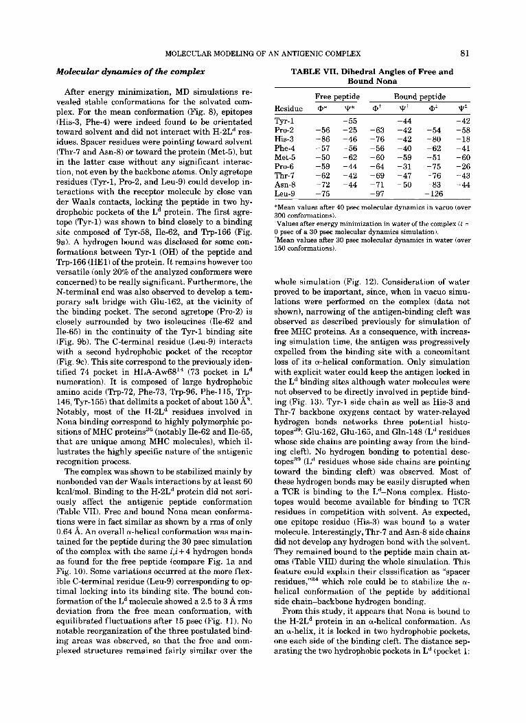
After energy minimization, MD simulations re- vealed stable conformations for the solvated com- plex. For the mean conformation (Fig. 81, epitopes (His-3, Phe-4) were indeed found to be orientated toward solvent and did not interact with H-2Ld res- idues. Spacer residues were pointing toward solvent (Thr-7 and Asn-8) or toward the protein (Met-51, but in the latter case without any significant interac- tion, not even by the backbone atoms. Only agretope residues (Tyr-1, Pro-2, and Leu-9) could develop in- teractions with the receptor molecule by close van der Waals contacts, locking the peptide in two hy- drophobic pockets of the Ld protein. The first agre- tope (Tyr-1) was shown to bind closely to a binding site composed of Tyr-58, Ile-62, and Trp-166 (Fig. 9a). A hydrogen bound was disclosed for some con- formations between Tyr-1 (OH) of the peptide and Trp-166 (HE1) of the protein. It remains however too versatile (only 20% of the analyzed conformers were concerned) to be really significant. Furthermore, the N-terminal end was also observed to develop a tem- porary salt bridge with Glu-162, a t the vicinity of the binding pocket. The second agretope (Pro-2) is closely surrounded by two isoleucines (Ile-62 and Ile-65) in the continuity of the Tyr-1 binding site (Fig. 9b). The C-terminal residue (Leu-9) interacts with a second hydrophobic pocket of the receptor (Fig. 9c). This site correspond to the previously iden- tified 74 pocket in HLA-Aw6814 (73 pocket in Ld numeration). It is composed of large hydrophobic amino acids (Trp-72, Phe-73, Trp-96, Phe-115, Trp- 146, Tyr-155) that delimits a pocket of about 150 A3. Notably, most of the H-2Ld residues involved in Nona binding correspond to highly polymorphic po- sitions of MHC proteins36 (notably Ile-62 and Ile-65, that are unique among MHC molecules), which il- lustrates the highly specific nature of the antigenic recognition process.
The complex was shown to be stabilized mainly by nonbonded van der Waals interactions by at least 60 kcalimol. Binding to the H-2Ld protein did not seri- ously affect the antigenic peptide conformation (Table VII). Free and bound Nona mean conforma- tions were in fact similar as shown by a rms of only 0.64 A. An overall a-helical conformation was main- tained for the peptide during the 30 psec simulation of the complex with the same i,i + 4 hydrogen bonds as found for the free peptide (compare Fig. l a and Fig. 10). Some variations occurred at the more flex- ible C-terminal residue (Leu-9) corresponding to op- timal locking into its binding site. The bound con- formation of the Ld molecule showed a 2.5 to 3 A rms deviation from the free mean conformation, with equilibrated fluctuations after 15 psec (Fig. 11). No notable reorganization of the three postulated bind- ing areas was observed, so that the free and com- plexed structures remained fairly similar over the
TABLE VII. Dihedral Angles of Free and Bound Nona
Free peptide Bound peptide Residue @* T* at qt @* T* Tyr- 1 Pro-2 His-3 Phe-4 Met-5 Pro-6 Thr-7 Asn-8 Leu-9
- 55 -56 -25 -86 -46 -57 -56 -50 -62 -59 -44 -62 -42 -72 -44 - 75
-63 - 76 - 56 - 60 - 64 - 69 -71 - 97
-44 - 42 - 42 - 40 -59 -31 - 47 - 50
- 54 -80 - 62 -51 -75 - 76 -83 - 126
-42 -58 -18 -41 -60 -26 - 43 - 44
*Mean values after 40 psec molecular dynamics in vacuo (over 300 conformations). ‘Values after energy minimization in water of the complex ( t = 0 psec of a 30 psec molecular dynamics simulation). *Mean values after 30 psec molecular dynamics in water (over 150 conformations).
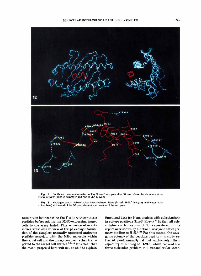
whole simulation (Fig. 12). Consideration of water proved to be important, since, when in vacuo simu- lations were performed on the complex (data not shown), narrowing of the antigen-binding cleft was observed as described previously for simulation of free MHC proteins. As a consequence, with increas- ing simulation time, the antigen was progressively expelled from the binding site with a concomitant loss of its a-helical conformation. Only simulation with explicit water could keep the antigen locked in the Ld binding sites although water molecules were not observed to be directly involved in peptide bind- ing (Fig. 13). Tyr-1 side chain as well as His-3 and Thr-7 backbone oxygens contact by water-relayed hydrogen bonds networks three potential histo- topes3’: Glu-162, Glu-165, and Gln-148 (Ld residues whose side chains are pointing away from the bind- ing cleft). No hydrogen bonding to potential dese- topes3’ (Ld residues whose side chains are pointing toward the binding cleft) was observed. Most of these hydrogen bonds may be easily disrupted when a TCR is binding to the Ld-Nona complex. Histo- topes would become available for binding to TCR residues in competition with solvent. As expected, one epitope residue (His-3) was bound to a water molecule. Interestingly, Thr-7 and Asn-8 side chains did not develop any hydrogen bond with the solvent. They remained bound to the peptide main chain at- oms (Table VIII) during the whole simulation. This feature could explain their classification as “spacer residues,”34 which role could be to stabilize the a- helical conformation of the peptide by additional side chain-backbone hydrogen bonding.
From this study, it appears that Nona is bound to the H-2Ld protein in an a-helical conformation. As an a-helix, it is locked in two hydrophobic pockets, one each side of the binding cleft. The distance sep- arating the two hydrophobic pockets in Ld (pocket 1:

82 D. ROGNAN ET AL.
Round Nona : Tyrl(O)-MetS(HN)
3.4 1 3.2 n l - 3.0 s 2.8
r" 2.6
5 2.4 Q 7 7 _.-
2.0 1.8
I 5 10 15 20 25
3.50
3.25
3 3.00 -
(Y r" 2.75 d 3 2.50
2.25
2.00
1.75
B a n d Nona P h e 4 ( H N ) - A d ( H N )
Time Lps]
Bound Nona : Hid3(0)-Thr7(HN)
5 10 15 20 25 Time [ps]
3.50 3 .751 nl
5 10 15 20 25 Time [ps]
Bound Nona: MetS(O)-LeuS(HN)
3.2 3.4 1 - 3 0 3
2 8
2 6 3 2.4
2.2 2 0 1.8
5 10 15 20 25 Time [ps]
Fig. 10. Hydro2en-bonding pattern of Nona conformations bound to the H-2L proteln during a 30 psec molecular dynamics simulation in water.
1.5
1.0
0 .5 { , , , 5 10 15 20 25
Time bs] Fig. 11. Time course of rms fluctuations (in A) of peptide-
bound H-2Ld conformations from the mean free structure.
Tyr-58, Ile-62, Ile-65, Trp-166; pocket 2: Trp-72, Phe-73, Trp-96, Phe-115, Trp-146, Tyr-155) was found in fluctuate around 20 A. It allows an optimal binding of Nona, which, in an a-helical conforma- tion, spans 19.3 A as measured between Tyr-1 (OH) and Leu-9 (CD2). In contrast, as a minimized p- sheet, Nona would have a length of 25.5 A and would therefore have to span over one of these pock- ets, which would preclude proper interaction of Tyr- 1, Leu-9, or both. The topology of the binding cleft thus makes it unlikely that Nona could interact with Ld in any regular conformation other than a- helical.
Interestingly, the model provides also an immedi- ate explanation for the inactivity of the C-terminal Trp analog of Nona, that 3-D Q.S.A.R. studies have failed to explain (recall Table IV). The binding pocket into which the authentic Nona is proposed to be anchored by Leu-9 has an averaged diameter be- tween Trp-96 and Tyr-155 of 4.5 A, which is simply too narrow to accommodate the bulky Trp. Thus, the modeled MHC-peptide complex has resolved the dis- crepancies between 3-D Q.S.A.R. prediction and ob- served activity, and is in remarkable agreement with all functional data available so far for Nona and its C-terminally substituted analogs.
Our findings with a particular MHC-peptide pair do not exclude, of course, that other peptide confor- mations are favored in other MHC-peptide com- plexes. In particular, the nature of MHC binding pockets (hydrophilic or hydrophobic, neutral or charged) and the intersite distances, which may vary in different MHC molecules, are likely to dic- tate the optimal length and conformation of anti- genic peptides. One may criticize that any model of antigen recognition must necessarily be incomplete as long as the contribution of the TCR in the ternary MHC-peptide-TCR complex cannot be included in the simulation. Yet, to this argument, the fact has to be recalled that formation of the ternary complex shows polarity. The peptide must first bind to the MHC molecule before the MHC-peptide binary com- plex is recognized by a TCR. Attempts to get peptide
MOLECULAR MODELING OF AN ANTIGENIC COMPLEX 83
Fig. 12. Backbone mean conformation of the Nona-Ld complex after 30 psec molecular dynamics simu-
Fig. 13. Hydrogen bonds (yellow broken lines) between Nona (in red), H-2Ld (in cyan), and water mole-
lation in water (nona is colored in red and H-2Ld in cyan).
cules (blue) at the end of the 30 psec dynamics simulation of the complex.
recognition by incubating the T cells with synthetic peptides before adding the MHC-expressing target cells to the assay failed. This sequence of events makes sense also in view of the physiologic forma- tion of the complex: naturally processed antigenic peptides associate with the MHC molecule within the target cell and the binary complex is then trans- ported to the target cell surface.''-" It is clear that the model proposed here will not be able to explain
functional data for Nona analogs with substitutions in epitope positions (His-3, Phe-4).34 In fact, all sub- stitutions or truncations of Nona considered in this report were shown by functional assays to affect pri- mary binding to H-ZLd.34 For this reason, the anti- genic potency of the peptides used in this study re- flected predominantly, if not exclusively, their capability of binding to H-2Ld, which reduced the three-molecular problem to a two-molecular inter-

84 D. ROGNAN ET AL.
TABLE VIII. Nona-Water-Ld Hydrogen Bonds Network*
D . . . A D-H . . . A Donor AcceDtor distance' angle*
Met-5 (NH) Tyr-1 (0) 2.89 168.7 Thr-7 (NH) His-3 (0) 2.99 176.4 Thr-7 (OG1) His-3 (0) 2.78 165.0 Asn-8 (NH) Phe-4 (0) 2.78 158.4 Asn-8 (ND2) Phe-4 (0) 2.85 147.3 Leu-9 (NH) Met-5 (0) 2.81 150.1 Tyr-1 (OH) Wat-452 (OW) 2.80 159.3 Wat-452 (OW) Wat-332 (OW) 2.77 163.9 Wat-332 (OW) Glu-165 (OE2) 2.69 159.7 His-3 (NH) Wat-484 (OW) 2.86 154.5 Wat-484 (OW) Wat-461 (OW) 2.61 168.8 Wat-461 (OW) Wat-341 (OW) 3.28 162.0 Wat-341 (OW) Wat-350 (OW) 2.85 158.9 Wat-350 (OW) Glue-162 (OEl) 2.58 146.4 His-3 (ND1) Wat-489 (OW) 2.86 171.0 Gln-148 (NE2) Wat-787 (OW) 2.89 172.4 Wat-787 (OW) Wat-512 (OW) 2.89 157.3 Wat-512 (OW) Thr-7 (0) 2.80 132.3 Wat-790 (OW) Wat-512 (OW) 2.76 161.3 Wat-790 (OW) Leu-9 (0) 2.83 136.5 Wat-796 (OW) Leu-9 (0) 2.77 159.1
*Situation after 30 psec of molecular dhynamics of the complex in water. 'Donnor (D)-acceptor (A) distance in A. *Donnor (Dbhydrogen (Hbacceptor (A) angle in degrees.
action. As Nona represents the bona fide naturally processed peptide, it is worth emphasizing that its N- and C-terminal amino acids are both critically involved in binding to H-2Ld and, according to the model presented here, these terminal residues are buried within hydrophobic pockets of the receptor. An hypothesis that proposes trimming of longer pep- tides to the optimal length by peptidases after bind- ing to the MHC molecule40 is therefore difficult to envisage. Thus, our model predicts that antigenic peptides should be finally processed a t least at ei- ther of their termini before interaction with the MHC molecule.
CONCLUSION From molecular dynamics simulations based on
biological data, we propose a mode of binding of a set of related antigenic peptides to the H-2Ld major his- tocompatibility protein. Free peptides and free MHC molecule as well as the complex between both were studied and revealed the following features: (1) For free peptides, antigenicity was observed to corre- spond to the aptitude to maintain an a-helical con- formation through i,i + 4 hydrogen bonds. Less ac- tive and inactive peptides showed a disruption of the a-helical conformation at the N-terminal end. In ad- dition, the nature of the C-terminal residue was shown to be determinative for an optimal binding to the H-2Ld protein, driven mainly by hydrophobic forces. (2) A free conformation of the murine MHC
molecule H-2Ld is proposed, built from X-ray coor- dinates of the homologous human MHC molecule HLA-A2 It revealed the crucial importance of elec- trostatics in molecular dynamics simulations since realistic computations were achieved only by using a model fully solvated with water. In contrast, in vacuo simulations, regardless of which dielectric functions were employed, were unable to maintain the integrity of the large antigen-binding cleft and led to dramatic narrowing of the binding site. (3) Binding of an antigenic peptide to H-2Ld is achieved in an a-helical conformation by different van der Waals interactions with two distinct hydrophobic pockets of the protein. Polymorphic residues of MHC proteins were involved in the binding process, which illustrates the specific nature of antigenic peptide association with its receptor.
The proposed model is not only fully compatible with functional data but also offers a reasonable ex- planation for previously unexplained experimental findings: alternation of antigenic potency during systematic biterminal truncation of a highly active nonapeptide,22 differential effects of N- and C-ter- minal truncations," distribution of agretope, epi- tope, and spacer residues within the sequence,34 and effects of amino acid side chain substitutions at the C-terminus (this report). The model highlights the largely hydrophobic nature of peptid+Ld interac- tion and will aid the design of new experiments for studying peptide presentation through the H-2Ld molecule.
ACKNOWLEDGMENTS The authors thank Dr. K. Burke for critical read-
ing of the manuscript, and Drs. U. Weber and H. Kalbacher for peptide synthesis. Gerd Helms is gratefully acknowledged for its technical assistance. This work was supported by FOURNIER Laborato- ries (Dijon, France) and by the Deutsche Forschungs- gemeinschaft SFB 322.
REFERENCES 1. Zinkernagel, R.M., Doherty, P.C. MHC-restricted cyto-
toxic T cells: Studies on the biological role of polymorphic major transplantation antigens determining T cell restric- tion specificity, function and responsiveness. Adv. Immun. 2752-177, 1979.
2. Schwartz, R.H. T lymphocyte recognition of antigen in as- sociation with gene products of the major histocompatibil- ity complex. Annu. Rev. Immunol. 3:237-261, 1985.
3. Buus, S., Sette, A,, Grey, H.M. The interaction between protein-derived immunogenic peptides and Ia. Immunol. Rev. 98:115-141, 1987.
4. Kourilsky, P., Claverie, J.M. MHC-antigen interactions: What does the T cell receptor see? Ad. Immunol. 45107- 193, 1989.
5. Townsend, A.R.M., Bodmer, H. Antigen recognition by class I-restricted T lymphocytes. Annu. Rev. Immunol. 7: 601-624,1989.
6. Unanue, E.R., Beller, D.I., Lu, C.Y., Allen, P.M. Antigen presentation: Comments on its regulation and mechanism. J. Immunol. 132:l-5, 1984.
7. Townsend, A.R.M., Rothbard, J., Gotch, F.M., Bahadur, G., Wraith, D., McMichael, A.J. The epitopes of influenza nu- cleoprotein recognized by cytolytic T lymphocytes can be
MOLECULAR MODELING OF AN ANTIGENIC COMPLEX 85
defined with short synthetic peptides. Cell 44:959-968, 1986.
Unanue, E.R. Binding of immunogenic peptides to Ia his- tocompatibility molecules. Nature (London) 317:359-361, 1985.
9. Chen, B.P., Parham, P. Direct binding of influenza pep- tides to class I HLA molecules. Nature (London) 337:743- 745, 1989.
10. Townsend, A.R.M., Ohlen, C., Bastin, J., Ljunggreen, H.G., Foster, L., Kare, K. Association of class I major his- tocompatibility heavy and light chains induced by viral peptides. Nature (London) 340:443-448, 1989.
11. Van Bleek, G.M., Nathenson, S.J. Isolation of an endoge- nously processed immunodominant viral peptide from the class I H-2Kb molecule. Nature (London) 348:213-216, 1990.
12. Riitzschke, O., Falk, K., Deres, K., Schild, H., Norda, M., Metzger, J., Jung, G., Rammensee, H.G. Isolation and analysis of naturally processed viral peptides as recog- nized by cytotoxic T cells. Nature (London) 348:252-253, 1990.
13. Bjorkman, P.J., Saper, M.A., Samraoui, B., Bennett, W.S., Strominger, J.L., Wiley, D.C. Structure of the human class I histocompatibility antigen, HLA-A2. Nature (London) 329506-512, 1987.
14. Garrett, T.P.J., Saper, M.A., Bjorkman, P.J., Strominger, J.L., Wiley, D.C. Specificity pockets for the side chains of peptide antigens in HLA-AW68. Nature (London) 342: 692-696, 1989.
15. DeLisi, C., Berzofsky, J.A. T-cell antigenic sites tend to be amphipatic structures. Proc. Natl. Acad. Sci. U.S.A. 82: 7048-7052, 1985. 63144, U.S.A.
16. Margalit, H., Spouge, J.L., Cornette, J.L., Cease, K.B., DeLisi, C., Berzofsky, J.A. Prediction of immunodominant helper T cell antigenic sites from the primary structure. J. Immunol. 138:2213-2229, 1987.
17. Allen, P.M., Matsueda, G.R., Evans, R.J., Dunbar, J.B., Jr . , Marshall, G.R., Unanue, E.M. Identification of the T- cell and Ia contact residues of a T-cell antigenic epitope. Nature (London) 327:713-715, 1987.
18. Sette, A., Buus, S., Colon, S., Smith, J.A., Miles, c., G ~ ~ ~ , H.M. Structural characteristics of an antigen required for its interaction with Ia and recognition by T cells. Nature (London) 328:395-399, 1987.
19. Maryanski, J.L., Verdini, A.S., Weber, P.C., Salemme, F.R., Corradin, G, Competitor analogs for defined T cell antigens: Peptides incorporating a putative binding profile and polypro~ine or polyglycine spacers, Cell 60:63-72, 1990.
virus immediate early gene expression in cellular immu- nity to cytomegalovirus infection. Nature (London) 312: 369-371, 1984.
21. Del Val, M., Volkmer, H., Rothbard, J.B., Jonijic, S., Mes- serle, M., Schickendanz, J., Reddehase, M.J., Koszinowski, U.H. Molecular basis for CflolYtic T-lYmPhocYte reCo@i- tion of the murine cytomegalovirus immediate-early pro- tein pp89. J . Virol. 62:3965-3972, 1988.
22. Reddehase, M.J., Rothbard, J.B., Koszinowski, U.H. A pentapeptide as minimal antigenic determinant for MHC class I-restricted T lymphocytes. Nature (London) 337:
23. Lambert, M.H., Sheraga, H.A. Pattern-recognition in the
prediction of protein structure. I. Tripeptide conforma- tional probabilities calculated from the amino acid se-
24. Lambert, M.H., Sheraga, H.A., Pattern-recognition in the prediction of protein structure. 11. Chain conformation from a probability-directed search procedure. J . Comput. Chem. 10:798-816, 1989.
25. Lambert, M.H., Sheraga, H.A., Pattern-recognition in the prediction of protein structure. 111. An importance-sam- pling mimimization procedure. J . Comput. Chem. 10:817- 831, 1989.
26. Kidera, A., Konishi, A., Oka, M., Ooi, T., Sheraga, H.A. Statistical analysis of the physical properties of the 20 naturally occuring amino acids. J . Protein. Chem. 4:23- 55, 1985.
27. Weiner, S.J., Kollman, P.A., Case, D.A., Singh, U.C., Ghio, C., Alagona, G., Profeta, S., Jr., Weiner, P. A new force field for molecular mechanical simulation of nucleic acids and proteins. J. Am. Chem. Soc. 106:765-784, 1984.
28. Jorgensen, W.L., Chandrasekhar, J., Madura, J.D., Impey, R.W., Klein, M.L. Comparison of simple potential func- tions for simulating liquid water. J . Chem. Phys. 70:926- 935, 1983.
29. Tirado-Rives, J., Jorgensen, W.L. Molecular dynamics of proteins with the OPLS potential function. Simulations of the third domain of siver pheasant ovomucoid in water. J . Am. Chem. SOC. 112:2773-2781, 1990.
30. Cramer, R.D., 111, Patterson, D.E., Bunce, J.D. Compara- tive molecular field analysis (CoMFA). 1. Effect of shape on binding of steroids to carrier proteins. J. Am. Chem. SOC. 110:5959-5967, 1988.
31. SYBYL 5.3 release, n ipos Assoc., Inc., St-Louis, MO
32. Wold, S., Albano, C., Dunn, W.J., 111, Esbensen, K., Hell- berg, S., Johansson, E., Lindberg, W., Sjoestroem, M. Mod- eling data files by principal component and PLS: Class patterns and quantitative predictive relations. Analusis 12:477-485, 1984.
33. Krug, M., Folkers, G. ADAPTU: Animated Dynamics Analysis Program at the Tiibingen University. J . Mol. Graph. 9:119-121,1991.
34. Reddehase, M.J., Koszinowski, U.H. Redistribution ofcrit- ical major histocornpatability complex and T cell receptor- binding functions of residues in an antigenic sequence after biterminal substitution. Eur. J. Immunol. 21:1697- 1701.
35. Semano, L., Fersht, A. Capping and alpha helix stability. Nature (London) 342:296-299, 1989.
36. Saper, M.A., Bjorkman, P.J., Wiley, D.C. Refinement of the human histocompatibility antigen HLA-A2 at 2.6 A resolution. J. Mol. Biol. 219:277-319, 1991.
fects on protein electrostatics. Proteins 5313-321, 1989. 38, FauchBre, J.L., pliska, V. Hydrophobic parameters of
amino acids side chains from the partitioning of N-acetyl- amino-acid amides. Eur. J . Med. Chem. 18:369-375, 1983.
In: “Immune Recognition.” D.E. Male, ed. Oxford: IRL Press, 1988: pp. 1-16.
40. Falk, K., Rotzschke, O., Rammensee, H.G. Cellular pep- tide composition governed by major histocompatibility complex class I molecules. Nature (London) 348:248-251,
8. Babbit, B.P., Allen, P.M., Matsueda, G., Haber, E., quence. J. Comput. Chem. 10:770-797,1989.
20. Reddehase, M.J., Koszinowskl U.H. Significance of herpes 37, Wendoloski, J. J,, Matthew, J,B, Molecular dynamics ef-
39. Owen, M.J., Lamb, J.R. General principles of recognition.
651-653, 1989. 1990.