molecular insights into beneficial effects of tea-plant
TRANSCRIPT

Applied Microbiology: Theory & TechnologyVolume 2 Issue 2|2021| 37
Research Article
Molecular Insights into Beneficial Effects of Tea-Plant Growth and Selenium Enrichment by Herbaspirillum camelliae
Wei Cheng, Xuejing Yu, Xingguo Wang*
State Key Laboratory of Biocatalysis and Enzyme Engineering, the Faculty of Life Science, Hubei University, P.R.ChinaE-mail: [email protected]
Received: 7 November 2020; Revised: 19 July 2021; Accepted: 19 July 2021
Abstract: Herbaspirillum camelliae WT00C, as a tea-plant endophytic bacterium, not only colonizes specifically in tea plants but also promotes tea-plant growth and selenium enrichment. Different from diazotrophic endophytes H. seropedicae, H. frisingense and H. rubrisubalbicans, H. camelliae WT00C does not display nitrogen-fixing activity. To understand the molecular mechanisms of promoting the growth of tea plant and Se-enrichment, we sequenced and annotated the genome of H. camelliae WT00C. The results showed that the genome was composed of 6,079,821 base pairs with a total of 5,537 genes. The genomic survey also revealed that H. camelliae WT00C was a multifunctional bacterium metabolizing a variety of carbon and nitrogen sources and defending against biotic and abiotic stress. Although this bacterium did not have intact nitrogen-fixing genes, its genome held the genes responsible for indole-3-acetic acid (IAA) biosynthesis, 1-aminocyclopropane-1-carboxylate (ACC) deamination, siderophore synthesis, ammonia formation, urea metabolism, glutathione and selenocompound metabolisms. Biosynthesis of IAA, siderophore, ammonia, urea and ACC deaminase could explain why two bacterial strains promote tea-plant growth and development. Selenocompound metabolism in this bacterium might also benefit tea-plant growth and Se-enrichment. In addition, the genome of H. camelliae also contained a multitude of protein secretion systems T1SS, T3SS, T4SS and T6SS, in which T4SS did not exhibit in other members of the genus Herbaspirillum.
Keywords: Herbaspirillum, endophytes, genome annotation, selenocompound metabolism, secretion system
1. IntroductionHerbaspirillum camelliae WT00C and WT00F were two gram-negative, endophytic bacterial strains isolated from
the tea plant in Wuhan city, China [1]. Two strains were already identified as a novel species of the genus Herbaspirillum based on their physiochemical and genetic characteristics as well as 16S rRNA gene sequences [2]. Differences between the strains WT00C and WT00F were that (a) the strain WT00C displayed catalase activity but the strain WT00F did not, and (b) the strain WT00F decomposed lactose to produce acid but the strain WT00C did not [1]. Unlike H. seropedicae, H. rubrisubalbicans, H. frisingense and H. lusitanum [3-5], H. camelliae was unable to fix nitrogen. However, biochemical assays showed that these two strains produced ammonia, siderophores and indole-3-acetic acid (IAA) [1]. Production of ammonia, siderophores and IAA implied that H. camelliae might have the potential to promote the growth and development of tea plants. Further studies found that H. camelliae entered plants via plant vulnus, and only colonized in
Copyright ©2021 Xingguo Wang, et al. DOI: https://doi.org/10.37256/amtt.222021713This is an open-access article distributed under a CC BY license (Creative Commons Attribution 4.0 International License)https://creativecommons.org/licenses/by/4.0/
Applied Microbiology: Theory & Technologyhttp://ojs.wiserpub.com/index.php/AMTT/

Applied Microbiology: Theory & Technology 38 | Xingguo Wang, et al.
tea plant. Inoculating the bacterial cells to tea cuttings by socking for an hour markedly stimulated lateral root formation and the growth of newborn shoots. Sprinkling the bacterial cells to the upper incision of tea twigs in the field also enhanced the growth of newborn shoots. More importantly, both strains WT00C and WT00F did not cause plant disease symptoms in tea plants. Thus, H. camelliae was thought to be potential bioaccelerator for tea cultivation [6]. Two strains also had a strong capability of reducing selenate/selenite to form red elemental selenium (Se0) and improving the Se-enrichment of tea plants [7]. Nevertheless, the detailed molecular mechanism of promoting tea-plant growth and Se-enrichment by two bacterial strains has not been fully understood.
In an attempt to understand bacterial effects on promoting tea-plant growth and Se-enrichment, we sequenced the genome of H. camelliae WT00C and analyzed its metabolic profiles. In this paper, we reported those metabolic pathways involving probably in promoting tea-plant growth and Se-enrichment, and explained why H. camelliae could act as a bioaccelerator stimulating tea-plant growth and improving Se-enrichment in tea plants. Our study not only helps us to deeply understand the biological functions and properties of H. camelliae as a novel bioaccelerator at the molecular level, but also benefits us to further exploit new purposes of two strains in agriculture or other aspects.
2. Materials and methods2.1 Bacterial strain
H. camelliae WT00C, deposited in CCTCC (AB 2018017T) and KCTC (62527T), was routinely grown in the nutrient broth (NB) containing 0.5% peptone, 0.3% yeast extract and 0.5% NaCl (pH 6.8) at 28 °C, 200 rpm. This strain was also able to grow at 37 °C, 200 rpm in LB medium (1% tryptone, 0.5% yeast extract, and 0.5% NaCl, pH 7.2). The detailed process was described by Wang et al. [1] and Liu et al. [2].
2.2 Preparation of genomic DNA
To prepare genomic DNA, H. camelliae WT00C was activated by inoculating into 5 ml NB broth and growing at 28 °C overnight. Then, 1 ml of the bacterial culture was put into 100 ml of fresh LB broth (10 g/L tryptone, 5 g/L yeast extract and 10 g/L NaCl) and incubated at 37 °C until OD600 of 0.8 recorded on a spectrophotometer (Shimadzu UV-2550). The bacterial culture was collected by centrifugation at 8000 rpm at 4 °C. DNA was isolated and purified by using the phenol-chloroform method after bacterial cells were lysed with lysozyme and SDS. The purified DNA was sent to BGI (Wuhan, China) for sequencing.
2.3 Genome sequencing and annotation
As stated by the manufacturer’s instructions, the genomic DNA of H. camelliae WT00C was sequenced with an Illumina HiSeq 2000 instrument. The genomic DNA prepared from H. camelliae WT00C was used to construct two random sequencing libraries containing small fragments (500 bp) and large fragments (6 kb). In both 500 bp and 6,000 bp libraries, the mean read length was 90 bp. These reads were then filtered and assembled into contigs by using SOAPdenovo v1.05 (http://soap.genomics.org.cn). 4 scaffolds consisting of 11 contigs were finally constructed step by step using all the paired-end information of reads. Repeat sequences and gene islands were predicted via Tandem repeat finder (TRF) [8], IslandPath-DIOMB and SIGI-HMM software [9]. Open reading frames (ORFs) were identified by an integrated automatic annotation platform with Glimmer 3.0 [10] and Blast softwares [11]. The function of translation product for each ORF was inferred by using the Blast package to search the public databases NR (non-redundant) (NCBI), UniProtKB/Swiss-Port (EMBL-EBI), Inter Pro [12], Gene Ontology (GO) [13] and Kyoto Encyclopedia of Genes and Genomes (KEGG) [14]. The genomic data of the strain WT00C was finally deposited in the GenBank database (accession numbers: KV880769.1).

Applied Microbiology: Theory & TechnologyVolume 2 Issue 2|2021| 39
3. Results and discussion3.1 General features for the genome of H. camelliae WT00C
Percent of genes
8
683
81
10
642
665100
1882
16
1
42
8658
373
287
1397
1397
132
107
36
1
1
5
1599
1750
4291
310
2253
196
0 35 352 3526
Number of genes
Go Standard
biological process
biological adhesionbiological regulation
cellular component organization or biogenesiscellular process
metabolic process
multi-organism process
multicellular organismal process
negative regulation of biological processnitrogen utilization
positive regulation of biological process
regulation of biological processresponse to stimulussignaling
cellcell part
macromolecular complexorganelleorganelle partvirionvirion part
antioxidant activitybindingcatalytic activity
enzyme regulator activity
molecular transducer activitystructural molecule activity
transporter activity
protein binding transcription factor activity
nucleic acid binding transcription factor activity
developmental process
establishment of localizationlocalizationlocomotion
cellular component
molecular function
all
1893
0.1 1 10 100
(a)

Applied Microbiology: Theory & Technology 40 | Xingguo Wang, et al.
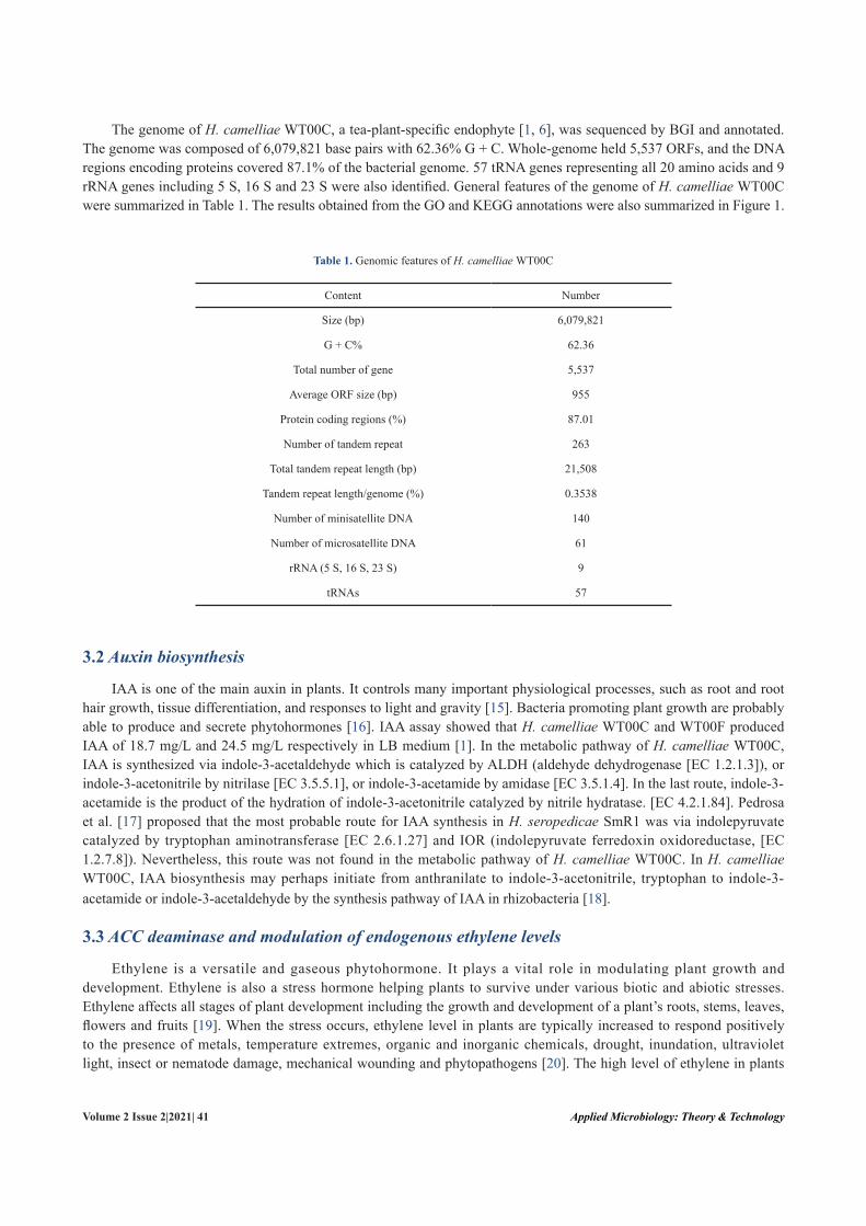
Figure 1. Genome information of H. camelliae WT00C. (a) Gene ontology annotation; (b) KEGG pathway classification
KEG
G pathw
ay classification
Organism
al Systems--N
ervous SystemO
rganismal System
s--Imm
une SystemO
rganismal System
s--Excretory SystemO
rganismal System
s--Environmental A
daptationO
rganismal System
s--Endocrine SystemO
rganismal System
s--Digestive System
Organism
al Systems--C
irculatory SystemM
etabolism--X
enobiotics Biodegradation and M
etabolismM
etabolism--N
ucleotide Metabolism
Metabolism
--Metabolism
of Terpenoids and PolyketidesM
etabolism--M
etabolism of O
ther Am
ino Acids
Metabolism
--Metabolism
of Cofactors and V
itamins
Metabolism
--Lipid Metabolism
Metabolism
--Glycan B
iosynthesisan Metabolism
Metabolism
--Enzyme Fam
iliesM
etabolism--Energy M
etabolismM
etabolism--C
arbohydrate Metabolism
Metabolism
--Biosynthesis of O
ther Secondary Metabolites
Metabolism
--Am
ino Acid M
etabolismH
uman D
iseases--Neurodegenerative D
iseasesH
uman D
iseases--Metabolic D
iseasesH
uman D
iseases--Infectious Diseases
Hum
an Diseases--Im
mune System
Diseases
Hum
an Diseases--C
ardiovascular Diseases
Hum
an Diseases--C
ancersG
enetic Information Processing--Translation
Genetic Inform
ation Processing--TranscriptionG
enetic Information Processing--R
eplication and Repair
Genetic Inform
ation Processing--Folding, Sorting and Degradation
Environmental Inform
ation Processing--Signaling Molecules and Interaction
Environmental Inform
ation Processing--Signal TransductionEnvironm
ental Information Processing--M
embrane Transport
Celluar Processes--Transport and C
atabolismC
elluar Processes--Cell M
otilityC
elluar Processes--Cell G
rowth and D
eath
33351623
342112
95100164
2138195
211
48
213
27319
194155
26792
58
38
0 200 400 600 800
336Num
ber of matched genes
342863
491
480
(b)
Applied Microbiology: Theory & TechnologyVolume 2 Issue 2|2021| 41
The genome of H. camelliae WT00C, a tea-plant-specific endophyte [1, 6], was sequenced by BGI and annotated. The genome was composed of 6,079,821 base pairs with 62.36% G + C. Whole-genome held 5,537 ORFs, and the DNA regions encoding proteins covered 87.1% of the bacterial genome. 57 tRNA genes representing all 20 amino acids and 9 rRNA genes including 5 S, 16 S and 23 S were also identified. General features of the genome of H. camelliae WT00C were summarized in Table 1. The results obtained from the GO and KEGG annotations were also summarized in Figure 1.
Table 1. Genomic features of H. camelliae WT00C
Content Number
Size (bp) 6,079,821
G + C% 62.36
Total number of gene 5,537
Average ORF size (bp) 955
Protein coding regions (%) 87.01
Number of tandem repeat 263
Total tandem repeat length (bp) 21,508
Tandem repeat length/genome (%) 0.3538
Number of minisatellite DNA 140
Number of microsatellite DNA 61
rRNA (5 S, 16 S, 23 S) 9
tRNAs 57
3.2 Auxin biosynthesis
IAA is one of the main auxin in plants. It controls many important physiological processes, such as root and root hair growth, tissue differentiation, and responses to light and gravity [15]. Bacteria promoting plant growth are probably able to produce and secrete phytohormones [16]. IAA assay showed that H. camelliae WT00C and WT00F produced IAA of 18.7 mg/L and 24.5 mg/L respectively in LB medium [1]. In the metabolic pathway of H. camelliae WT00C, IAA is synthesized via indole-3-acetaldehyde which is catalyzed by ALDH (aldehyde dehydrogenase [EC 1.2.1.3]), or indole-3-acetonitrile by nitrilase [EC 3.5.5.1], or indole-3-acetamide by amidase [EC 3.5.1.4]. In the last route, indole-3-acetamide is the product of the hydration of indole-3-acetonitrile catalyzed by nitrile hydratase. [EC 4.2.1.84]. Pedrosa et al. [17] proposed that the most probable route for IAA synthesis in H. seropedicae SmR1 was via indolepyruvate catalyzed by tryptophan aminotransferase [EC 2.6.1.27] and IOR (indolepyruvate ferredoxin oxidoreductase, [EC 1.2.7.8]). Nevertheless, this route was not found in the metabolic pathway of H. camelliae WT00C. In H. camelliae WT00C, IAA biosynthesis may perhaps initiate from anthranilate to indole-3-acetonitrile, tryptophan to indole-3-acetamide or indole-3-acetaldehyde by the synthesis pathway of IAA in rhizobacteria [18].
3.3 ACC deaminase and modulation of endogenous ethylene levels
Ethylene is a versatile and gaseous phytohormone. It plays a vital role in modulating plant growth and development. Ethylene is also a stress hormone helping plants to survive under various biotic and abiotic stresses. Ethylene affects all stages of plant development including the growth and development of a plant’s roots, stems, leaves, flowers and fruits [19]. When the stress occurs, ethylene level in plants are typically increased to respond positively to the presence of metals, temperature extremes, organic and inorganic chemicals, drought, inundation, ultraviolet light, insect or nematode damage, mechanical wounding and phytopathogens [20]. The high level of ethylene in plants

Applied Microbiology: Theory & Technology 42 | Xingguo Wang, et al.
obviously exacerbates the effects of the stress triggering ethylene responses. Thus, lowering the magnitude of stress and ethylene level simultaneously decreases plant damage caused by the stress [20].
In plants, ethylene is synthesized via the catalysis of ACC synthase and ACC oxidase. ACC synthase activated by IAA under biotic and abiotic stress conditions catalyses L-methione to form S-adenosylmethionine (SAM) and then to form ACC (1-aminocyclopropane 1-carboxylate). ACC is further converted to ethylene by ACC oxidase [20, 21]. In the genome of H. camelliae WT00C, the gene encoding ACC deaminase [EC 3.5.99.7] was cloned. The cloned ACC deaminase gene was composed of 1082 bp, and its protein with a molecular mass of 38.5 kDa contained 355 amino acids. The assay of enzyme activity showed that the purified enzyme catalyzed ACC to form 2-oxobutanoate and ammonium. ACC deaminase was thought to modulate ethylene levels in plants, decrease the stress response caused by ethylene and allow plant growth under stress conditions [22]. Thus, ACC deaminase in bacterial cells has an obvious competitive effect against ACC oxidase in plants. As shown in Figure 2, decomposition of ACC by ACC deaminase in H. camelliae WT00C likely reduces the synthesis of ethylene in tea plant. IAA and ACC deaminase synthesized coordinatively in H. camelliae WT00C may play an important role in promoting plant growth and development, as shown in Burkholderia phytofirmans PsJN [23].
Figure 2. A schematic model for the growth promotion of ACC deaminase and IAA in H. camelliae WT00C. ACC: 1-aminocyclopropane-1-carboxylate; IAA: indole-3-acetic acid; SAM, S-adenosyl methionine
3.4 Siderophore production
Siderophores are small iron-chelating compounds. In bacteria, Fe2+-dependent repressors can bind to the upstream of genes responsible for siderophore synthesis when intracellular iron concentrations are high. At low concentrations, Fe2+ dissociates from the repressor, and then the repressor, in turn, dissociates from the upstream of genes, which finally leads to transcription of those genes. The ferric uptake regulator (Fur) or diphtheria toxin repressor (DtxR) usually
Stress
IAA IAA
SAM
Stress
Stressresponse
Tea plant tissue
Herbaspirllum sp WTOOC
Ethylene
Auxin responsefactoes
ACCACC
ACCdeaminase
Ammonia +α-ketobutyrate
ACCoxidase
Plant growth
Plant IAAsynthesis

Applied Microbiology: Theory & TechnologyVolume 2 Issue 2|2021| 43
regulates Fe2+ dissociation from the Fe2+-repressor complex. Meanwhile, the whole Fe-siderophore complex, in many cases, transports actively across cell membranes. In gram-negative bacteria, the Fe-siderophore complex is transported into the periplasm via TonB-dependent receptors. Thus, ferric ion in Fe-siderophore complex may be reduced to Fe2+
outside the cells. Siderophores are also able to chelate other metals, such as copper, zinc, lead, aluminium, cadmium, vanadium, plutonium, manganese, indium, uranium and gallium chromium [24-27].
Universal chemical assay [28] demonstrated that both H. camelliae WT00C and WT00F strains were capable of producing siderophore [1]. KEGG annotation revealed that H. camelliae WT00C might perhaps synthesize the siderophore enterochelin because three subunits VibB, EntB and DhbB of isochorismate lyase/aryl carrier protein [EC 3.3.2.1] and EntF (the component F of enterobactin synthetase [EC 2.7.7.-]) were present. In addition, the genome survey also found a very large DNA fragment (> 27 kb, similar to the Hsero_2343 of H. seropedicae). This large DNA fragment was located in downstream of TonB-dependent siderophore receptor CirA and the sigma factor PrfI encoding for two non-ribosomal peptide synthases and an amino acid adenylation protein. These enzymes might be involved in the non-ribosomal synthesis of siderophores in H. camelliae WT00C. The genome of H. camelliae WT00C also held 20 TonB-dependent siderophore receptors and a ABC-type hydroxamate-type ferric siderophore uptake system. Pedrosa et al. [17] suggested that iron uptake occurred through active transport mechanisms by means of an ABC-type system and TonB/ExbB/ExbD. In the rice-endophyte Azoarcus, a plethora of TonB-dependent siderophore receptors were also found [29, 30]. Those iron receptors may endow H. camelliae WT00C with strong competitiveness in iron-limited environments so that this bacterium can effectively compete with other bacteria. Furthermore, siderophores were also thought to improve plant growth through a direct effect on the plant or the control of noxious organisms or some other routes [31].
3.5 Nitrogen metabolism and ammonia formation
Figure 3. A schematic diagram showing nitrogen metabolic pathways of H. camelliae WT00C. The number in each square box represents EC number assigned by European community to each enzyme
Cyanoamino acidmetabolism
Glyoxylate metabolism
Methane metabolism
1-aminocyclopropane-1-carboxylate (ACC) α-Amino acids
(Nitrogenouscompounds)
Nitroalkane
NitriteNitrate Ammonia
Nitriles
Urea
+ H2O
CO2
Formamide
L-Aspartate
Formate
3.5.1.49
3.5.99.73.5.1.38
L-Glutamine
L-Asparagine
L-Glutamate
Alanine, aspartate andglutamate metabolism
4.3.1.31.4.99.1
3.5.51.1
1.4.1.31.4.1.131.4.1.14
6.3.5.41.4.7.1
6.3.1.23.5.1.23.5.1.38
4.4.1.8
1.7.1.4
3.5.1.5
3.5.5.1
1.13.12.16
1.7.99.4

Applied Microbiology: Theory & Technology 44 | Xingguo Wang, et al.
As an important source of nitrogen for many plant species, ammonia is the dominant nutrient content for plant growth and development and is easily absorbed by plants. Interestingly, two strains of H. camelliae held the capability of producing ammonia under the culture conditions [1]. In H. seropedicae, ammonia mainly comes from atmospheric nitrogen via nitrogen fixation by nitrogenases, because it holds nitrogen-fixing genes (nif ) such as nifA, nif B, nif D, nif E, nif H, nif K, nif N, nif Q, nif S, nif U, nif V, nif W, nif X, nif Z, and nif Z1 [17]. In contrast, H. camelliae did not show any nitrogen-fixing activity. Amplification of nif H and nif D genes by polymerase chain reaction (PCR) was also failed when the specific primer pairs were used [1]. In the genome of H. camelliae WT00C, only two analogues of nifA and nif U genes relevant to nitrogen fixation are found. As a consequence, H. camelliae does not have a nitrogen-fixing capability.
In accordance with the metabolic pathways of H. camelliae WT00C, ammonia mainly comes from amino acid metabolism. For instance, ammonia formation occurs via L-aspartate catalyzed by glutaminase-asparaginase [EC 3.5.1.38], L-glutamin by glutaminase [EC 3.5.1.2], L-asparagine by asparaginase [EC 3.5.1.1] and L-glutamate deamination by glutamate dehydrogenase [EC 1.4.1.3] (see Figure 3). In addition, ammonia also comes from formamide catalyzed by formamidase [EC 3.5.1.49], 1-aminocyclopropane-1-carboxylate (ACC) by ACC deaminase [EC 3.5.99.7], nitrite by nitrite reductase [EC 1.7.1.4], and urea or nitriles by urease [EC 3.5.1.5] or nitrelase [EC 3.5.5.1]. As shown in Figure 3, nitrite is the production of nitrate and nitroalkane catalyzed by nitrate reductase [EC 1.7.99.4] and nitronate monooxygenase (NMO EC 1.13.12.16).
Figure 3 also showed that ammonia was used to synthesize L-glutamate by glutamate dehydrogenase (GDH EC 1.4.1.3) or L-glutamine by glutamine synthetase [EC 6.3.1.2]. L-glutamine is then catalyzed by NAD(P)H-glutamate synthase [EC 1.4.1.13/14], ferredoxin-glutamate synthase [EC 1.4.7.1] or asparagine synthase [EC 6.3.5.4] to form L-glutamate. Finally, L-glutamate enters the metabolic pathway of amino acids. Thus, ammonia is excreted from the interior to the exterior by the bacterium only when it becomes overabundant.
3.6 Urea metabolism
Urea, as a nitrogen-release fertilizer, is synthesized in the body of many organisms as part of the urea cycle. Like H. seropedicae, H. camelliae was capable of synthesizing and degrading urea. In our previous study, both strains displayed urease activities [1]. In the metabolic pathways of H. camelliae WT00C, there is a complete urea cycle for arginine biosynthesis. Four key enzymes ornithine carbamoyltransferase [EC 2.1.3.3], argininosuccinate synthase [EC 6.3.4.5], argininosuccinate lyase [EC 4.3.2.1] and arginase [EC 3.5.3.1] in the urea cycle are present in the bacterial genome. In the last step of the urea cycle, arginase converts L-argininr into L-ornithine and urea. Then urea is degraded into carbon dioxide and ammonia by urease [EC 3.5.1.5] or by urea carboxylase [EC 6.3.4.6] and allophanate hydrolase [EC 3.5.1.54].
Like the H. seropedicae, Janthinobacterium and Corynebacterium glutamicum [32], the urease operon in the genome of H. camelliae WT00C contains ureA, ureB and ureC genes and 6 accessory genes ureD, ureE, ureF, ureG, ureH, and ureJ. The operon constitution is similar to that of H. seropedicae SmR1 [17], except that ureH does not occur in the latter operon. In the genome of H. camelliae WT00C, ABC-type urea transport operon (urtABCDE) was also found in the upstream of the ure gene cluster. In view of the fact that a σ54-dependent promoter is located in the upstream of urtA and both urt and ure genes are expressed under nitrogen deprivation, the urt and ure genes are probably controlled by the nitrogen regulatory (Ntr) system [33].
3.7 Glutathione and selenocompound metabolisms
In plants, animals, fungi and some bacteria, glutathione (GSH) is an important antioxidant, which prevents damage to cellular components caused by reactive oxygen species including free radicals, heavy metals, peroxides and lipid peroxides [34]. Glutathione is also crucial for biotic and abiotic stress management in plants. In the glutathione-ascorbate cycle, glutathione is a pivotal component for the precursor of phytochelatins and glutathione oligomers chelating heavy metals as well as reducing poisonous hydrogen peroxide [35, 36]. Glutathione is required to defend efficiently against plant pathogens [37]. 5′-Adenylylsulfate (APS) reductase in the sulfur assimilation pathway uses glutathione as electron donor, and glutaredoxin also uses glutathione as substrate. These oxidoreductases participate in flower development and plant defense signaling [38].
The genome of H. camelliae WT00C held a complete pathway for glutathione biosynthesis. As shown in Figure

Applied Microbiology: Theory & TechnologyVolume 2 Issue 2|2021| 45
4, glutathione is synthesized from L-glutamate, L-cysteine and L-glycine by glutamate-cysteine ligase [EC 6.3.2.2] and glutathione synthase [EC 6.3.2.3]. Meanwhile, glutathione (GSH) can be converted to glutathione disulfide (GSSG) by glutathione peroxidase [EC 1.11.1.9], and glutathione disulfide can be reversely converted to GSH by NADPH-dependent glutathione reductase [EC 1.8.1.7]. GSH can decompose into L-glutamate, L-cysteine, L-glycine or to form L-γ-glutamyl-L-amino acid or R-S-cysteine via four routes (see Figure 4). In this bacterium, glutathione appears to involve the conversion of putrescine to spermidine under the catalysis of spermidine synthase [EC 2.5.1.16]. Spermidine regulates plant growth and inhibits nitric oxide synthase (NOS). In addition, spermidine is the precursor for other polyamines (e.g. spermine and thermospermine), some of which are involved in plant tolerance to drought and salinity [39].
Figure 4. A schematic diagram showing glutathione metabolism of H. camelliae WT00C. The number in each square box stands for EC number of an enzyme.
PepA: cytosol aminopeptidase; PepN: aminoendopeptidase; LAP3: leucine aminopeptidase 3
Glutathione is also involved in selenocompound metabolism in H. camelliae WT00C. As shown in Figure 5, selenate or selenite is converted to hydrogen selenide by both enzymatic reactions and glutathione. Whereafter, selenium can be incorporated into bacterial proteins to form selenoproteins via seleno-methionayl-tRNA or L-seleno-cysteinyl-tRNA during protein synthesis. In addition, glutathione interacts with toxic selenite to form selenodiglutathion and then converts to non-toxic selenium (Se0) under the catalysis of GSSG reductase [EC 1.8.1.7]. When selenate was added into the bacterial culture, H. camelliae was indeed able to convert selenate/selenite to red elemental selenium (Se0) [7]. As an essential micronutrient for animals or trace element nutrient in humans, selenium functions as a cofactor for reducting antioxidant enzymes (e.g. glutathione peroxidases and certain forms of thioredoxin reductase). Selenium is
L-Amino acid L-Cysteine
NADPH NADP+
RX
Glycine
PepN
PepN
PepA3.4.11.2
2.3.2.2
2.3.2.26.3.2.3
2.5.1.18 2.3.2.23.4.11.2
PepA
LAP3
1.8.1.7
1.1.1.421.1.1.49
1.11.1.9
2.5.1.16
LAP3
6.3.2.2
L-Cysteiny-glycine
Glutathione(GSH)
R-S-Cysteinyl-
glycine
R-S-Glutathione
Spermidine
L-Glutamate
L-γ-Glutamyl-L-amino acid
L-γ-Glutamylcysteine
L-Glutamate
R-S-Cysteine
Putrescine
Glycine
Cysteine andmethioninemetabolism
Taurine andhypotaurinemetabolism Glutamate
metabolism
Cyanoamino acidmetabolism
Glutathionedisulfide(GSSG)

Applied Microbiology: Theory & Technology 46 | Xingguo Wang, et al.
also important for the function of thyroid gland. An increase of dietary selenium intakes reduces the effects of mercury toxicity [17]. Selenium deficiency occurs in a number of severe or chronic diseases (e.g. cancer, diabetes, HIV/AIDS and tuberculosis) [40-44]. Selenium-rich tea was thought to be a good choice for selenium-deficient patients as dietary supply. The conversion of selenate into non-toxic elemental selenium or incorporation of selenium into bacterial proteins to form selenoproteins in H. camelliae WT00C facilitated selenium accumulation in tea when the bacterium colonized in tea plant [7]. Red elemental selenium (Se0) purified from H. camelliae WT00C cells improved immunoactivities of crucian carp and zebrafish against spring viraemia of carp virus [45].
Figure 5. A diagram showing selenocompound metabolism in H. camelliae WT00C. The number in each square box represents EC number. MET: methionine synthase; CBL: cystathionine beta lyase
3.8 Bacterial secretion systems and protein secretion
Eight types of secretion systems (T1SS, T2SS, T3SS, T4SS, T5SS, T6SS, T7SS and T8SS) have been reported in prokaryotic bacteria, in which only T7SS occurs in Gram-positive bacteria [46-49]. T2SS, T5SS, T7SS and T8SS use a two-step process to perform secretion, which includes a stopover in the periplasm with the help of Sec or Tat export machinery. In contrast, T1SS, T3SS, T4SS and T6SS use a one-step mechanism to deliver exoproteins. T1SS, T2SS and T5SS deliver proteins into the extracellular medium, and T3SS, T4SS and T6SS deliver proteins into host cells [47, 50]. In pathogenic bacteria, T3SS, T4SS and T6SS are implicated in the delivery of toxic effector proteins into the cytoplasm of eukaryotic cells [17]. The genomic survey revealed that H. camelliae WT00C held T1SS (ABC transporters), T3SS, T4SS, T4SS pili and T6SS. The detailed gene information for each secretion system was shown in Figure 6. In addition, Sec-related genes ftsY, yidC, f f h, secA, secB, secY, secE, yajC, secD, secF and secG were also found in the bacterial genome except for secM (secretion monitor). Different from the Sec system, only tatA, tatB and tatC genes for Tat translocase systems were found in the genome of H. camelliae WT00C. The absence of tatE gene encoding a component of Tat export machinery implied that the Tat system might not function properly. Although partial T2SS gene analogues including gspD, gspF, gspG, gspE, gspA and gspH were found, these genes dispersed over different locations and did not occur as a gene cluster.
Seleno-methionyl-tRNA (Met) Seleno-
methionine
Seleno-homocysteine
Seleno-cystathionine
L-Seleno-cysteinyl-
tRNA(Sec)
L-Seryl-tRNA(Sec)
Seleno-phosphate
Hydrogenselenide
6.1.1.10
MET
Protein
CBL
1.1.1.49
1.1.1.42
2.9.1.1
SelenocysteineGlutathio-
selenol Selenodi-glutathione
Se0
NADP+ NADPH
Selenate
2.7.9.3
1.8.1.7
2.7.7.4
Adenosine
Selenitespontaneous spontaneous
5'-phosphoselenate
1.8.1.9

Applied Microbiology: Theory & TechnologyVolume 2 Issue 2|2021| 47
Figure 6. The gene clusters of the secretion systems in H. camelliae WT00C
hlyB hlyD tolC
hrcV hrpQ
hrcN hrcQ
hrcR hrcS hrpX hrcU hrpB hrcJ hrpD
hrpE hrcC
virB10 virB9 virB5 virB6 trbJ virB4 virB3 virB2 virB11 copG virD
4 virD2 virD
4 traF virB6 virB10 virB9 virB5 virB4 virB3 virB2 virB11 ppkA traF virB10 virB9 virB5 virB6 trbJ virB4 virB2 virB3 virB11 copG virD
4 virD2
pilB pilC pilA pilQ pilN pilO
pilP gspE bfpE pilS pilV virB1 pilT cesT pilL pilZ flhB2 fliK fliT fliS fliD flaG
fliC pilV pilWprotein pilE fim
T pilL pilJ pill
vgr impk im
pJ lip impB im
pC hcp impF im
pG im
pH clpV vgr im
pA motB im
pM im
pL vgr
ftsY yidC ffh secA secB secY secE yajC secD secF secG
Sec-SRP
assembly
(icmF)
(dotU)
T1SS
T3SS
T4SS
T4SS pili
T6SS

Applied Microbiology: Theory & Technology 48 | Xingguo Wang, et al.
Effector proteins secreted via T3SS were thought to control host metabolism and circumvent plant defense mechanisms [17]. T3SS might also optimize beneficial interactions between host and bacterial cells because effector proteins secreted by T3SS responded to flavonoids exudated by plant roots. The biologic effects of those effectors, either enhancing or diminishing nodulation, depended on the host legume [51-53]. Interestingly, H. camelliae WT00C held an intact T4SS and T4SS pili genes in its genome, whereas H. seropedicae only held T4SS pili without T4SS. T4SS was initially found in Agrobacterium tumefaciens. A. tumefaciens used T4SS to deliver T-DNA (a portion of Ti plasmid) into the host leguminous plant. This secretion system mediated intracellular transfer of macromolecules including DNA and proteins via a mechanism that was ancestrally related to one of bacterial conjugation machineries [54, 55]. Two effector proteins AnkA and ATS-1 were also secreted via T4SS [56]. Moreover, VirE2 (a type IV secretion substrate) was found to interact with the VirD4 (T4SS-coupling proteins) and had six functions in A. tumefaciens [57, 58]. T6SS was initially identified in Pseudomonas aeruginosa and Vibrio cholerae [59, 60], and then found in 1/4 proteobacterial genomes including plant, animal and human pathogens as well as environmental, soil or marine bacteria [61, 62]. Although most studies of T6SS secretion in the early stage focused mainly on its pathogenesic role in higher organisms, more recent studies suggested that T6SS played a broader physiological role in defense against simple eukaryotic predators and interactions between bacteria [63, 64]. In the H. camelliae WT00C genome, the T6SS gene clusters contained 17 genes, in which Hcp and VgrG proteins were thought to be the substrates secreted universally by T6SS. Structural analysis revealed a striking similarity between those proteins and the tail spike of T4 phage, and the activity of this system in function was thought to resemble phage infection [65]. Thus, T3SS, T4SS and T6SS in non-pathogenic bacteria (e.g. H. seropedicae SmR1 and H. camelliae WT00C) are mainly involved in microbe–plant recognition and interaction.
4. Concluding remarksThe genomic survey has revealed that H. camelliae WT00C is a metabolically versatile bacterium able to produce
plant-growth modulators auxin, siderophore enterochelin, ammonia and other metabolites involving in nutrient metabolism and improvement of stress tolerance in tea plants. Although H. camelliae is devoid of nitrogen-fixing genes, its plant-growth-promoting capacity may mainly derive from IAA biosynthesis, urea and ammonia metabolism, ACC deamination and its modulation of endogenous ethylene levels in tea plant as well as other metabolites involving in plant stress tolerance. The formation of ammonia mainly depends on the many genes for amino acid metabolism, NO3
- and NO2
- assimilation. Whereas ACC deaminase may play a crucial role in modulating the production of ethylene in plants so that tea plant can thrive under biotic or abiotic stress. In addition, glutathione, siderophore, glycogen, trehalose, spermine, peroxidases may also play a role in enhancing stress tolerance or promoting the growth and development of tea plants. Different from H. seropedicae, H. rubrisubalbicans and H. frisingense isolated from gramineous plants, H. camelliae colonizes only in tea plants of Camellia genus, but not in gramineous and brassicaceous plants [6]. H. camelliae is devoid of glycohydrolases (e.g. polymethyl galacturonase [EC 3.1.1.11], ß-1,3-glucanase [EC 3.2.1.4]) involved in plant cell wall degradation. This may explain why this bacterium invades tea plant only through plant vulnus other than other manners (e.g. irrigation and sprinkling) [6]. H. camelliae also displays an impressive variety of protein secretion systems that may benefit bacterial invasion, colonization and its endophytic life. H. camelliae holds a typical T4SS, which is absent in other members in the genus Herbaspirillum. T4SS of gram-negative bacteria usually delivers DNA or protein substrates from bacterial cells to host cells through conjugation machinery [54]. Exchange of substance between the bacterium and its host plant via T4SS may perhaps help H. camelliae to establish its endophytic lifestyle in tea plants. In addition, this bacterium has an intact pathway of selenocompound metabolism as shown in Figure 5. Selenate reduction and selenoprotein synthesis will benefit not only red elemental selenium and selenoprotein synthesis but also selenium enrichment in tea plants.
AcknowledgementsThis work was supported financially by the grant (2015CFA089) from Science and Technology Department of
Hubei Province and the innovation-driven power program from Hubei Association for Science and Technology, China.

Applied Microbiology: Theory & TechnologyVolume 2 Issue 2|2021| 49
References[1] Wang T, Yang S, Chen Y, Hu L, Tu Q, Zhang L, et al. Microbiological properties of two endophytic bacteria
isolated from tea (Camellia sinensis L.). Acta Microbiologica Sinica. 2014; 54(4): 424-432.[2] Liu X, Zhou J, Tian J, Cheng W, Wang X. Herbaspirillum camelliae sp. nov., a novel endophytic bacterium isolated
from Camellia sinensis L. Archives of Microbiology. 2020; 202(7): 1801-1807.[3] Baldani JI, Baldani VLD, Seldin L, Dobereiner J. Characterization of Herbaspirillum seropedicae gen. nov., sp.
Nov., a root-associated nitrogen-fixing bacterium. International Journal of Systematic Bacteriologyl. 1986; 36(1): 86-93.
[4] Kirchhof G, Eckert B, Stoffels M, Baldani JI, Reis VM, Hartmann A. Herbaspirillum frisingense sp.Nov., a new nitrogen-fixing bacterial species that occurs in C4-fibre plants. International Journal of Systematic and Evolutionary Microbiologyl. 2001; 51(1): 157-168.
[5] Valverde A, Velazquez E, Gutierrez C, Cervantes E, Ventosa A, Igual JM. Herbaspirillum lusitanum sp. Nov., a novel nitrogen-fixing bacterium associated with root nodules of Phaseolus vulgaris. International Journal of Systematic Bacteriologyl. 2003; 53(6): 1979-1983.
[6] Zhan G, Cheng W, Liu W, Li Y, Ding K, Rao H, et al. Infection, colonization and growth-promoting effects of tea plant (Camellia sinensis L.) by the endophytic bacterium Herbaspirillum sp. WT00C. African Journal of Agricultural Research. 2016; 11(3): 130-138.
[7] Xu X, Cheng W, Liu X, You H, Wu G, Ding K, et al. Selenate reduction and selenium enrichment of tea by the endophytic Herbaspirillum sp. Strain WT00C. Current Microbiology. 2020; 77(4): 588-601.
[8] Benson G. Tandem repeats finder: A program to analyze DNA sequences. Nucleic Acids Research. 1999; 27: 573-580.
[9] Langille MGI, Hsiao WWL, Brinkman FSL. Detecting genomic islands using bioinformatics approaches. Nature Reviews Microbiology. 2010; 8: 373-382.
[10] Delcher AL, Bratke KA, Powers EC, Salzberg SL. Identifying bacterial genes and endosymbiont DNA with Glimmer. Bioinformatics. 2007; 23: 673-679.
[11] Altschul SF, Madden TL, Schäffer AA, Zhang J, Zhang Z, Webb M, et al. Gapped BLAST and PSI-BLAST: A new generation of protein database search programs. Nucleic Acids Research. 1997; 25: 3389-3402.
[12] Alex M, Chang HY, Louise D, Matthew F, Sarah H, Rodrigo L, et al. The InterPro protein families database: The classification resource after 15 years. Nucleic Acids Research. 2015; 43: D213-221.
[13] Tatusov RL, Fedorova ND, Jackson JD, Jacobs AR, Kiryutin B, Koonin EV, et al. The COG database: An updated version includes eukaryotes. BMC Bioinformatics. 2003; 4: 1-41.
[14] Kanehisa M, Goto S, Hattori M, Hattori M, Aoki-Kinoshita KF, Hirakawa M. From genomics to chemical genomics: New developments in KEGG. Nucleic Acids Research. 2006; 34: 354-357.
[15] Shahab S, Ahmed N, Khan NS. Indole acetic acid production and enhanced plant growth promotion by indigenous PSBs. African Journal of Agricultural Research. 2009; 4(11): 1312-1316.
[16] Bastiăn F, Cohen A, Piccoli P, Luna V, Bottini R, Baraldi R, et al. Production of indole-3-acetic acid and gibberellins A1 and A3 by Acetobacter diazotrophicus and Herbaspirillum seropedicae in chemically-defined culture media. Plant Growth Regulation. 1998; 24: 7-11.
[17] Pedrosa FO, Monteiro RA, Wassem R, Cruz LM, Ayub RA, Colauto NB, et al. Genome of Herbaspirillum seropedicae strain SmR1, a specialized diazotrophic endophyte of tropical grasses. PLos Genetics. 2011; 7(5): e1002064.
[18] Ahemad M, Kibret M. Mechanisms and applications of plant growth promoting rhizobacteria: Current perspective. Journal of King Saud University-Science. 2014; 26: 1-20.
[19] Abeles FB, Morgan PW, Saltveit ME Jr. Ethylene in Plant Biology. 2nd ed. New York Academic Press; 1992.[20] Glick BR. Bacteria with ACC deaminase can promote plant growth and help to feed the world. Microbiological
Research. 2014; 169: 30-39.[21] Wang K, Li H, Ecker JR. Ethylene biosynthesis and signaling networks. The Plant Cell. 2002; 14: s131-151.[22] Glick BR, Todorovic B, Czarny J, Cheng Z, Duan J, McConkey B. Promotion of plant growth by bacterial ACC
deaminase. Critical Reviews in Plant Sciences. 2007; 26: 227-242.[23] Sun Y, Cheng Z, Glick BR. The presence of a 1-aminocyclopropane-1-carboxylate (ACC) deaminase deletion
mutation alters the physiology of the endophytic plant growth-promoting bacterium Burkholderia phytofirmans PsJN. FEMS Microbiology Letters. 2009; 296: 131-136.
[24] Olmo AD, Caramelo C, SanJose C. Fluorescent complex of pyoverdin with aluminum. Journal of Inorganic

Applied Microbiology: Theory & Technology 50 | Xingguo Wang, et al.
Biochemistry. 2003; 97(4): 384-387.[25] Carrillo-Castañeda G, Juárez Muños J, Peralta-Videa JR, Gomez E, Tiemannb KJ, Duarte-Gardea M, et al. Alfalfa
growth promotion by bacteria grown under iron limiting conditions. Advances in Environmental Research. 2002; 6(3): 391-399.
[26] Hider RC, Hall AD. Clinically useful chelators of tripositive elements. Progress in Medicinal Chemistry. 1991; 28: 41-137.
[27] Seth GJ, Ruggiero CE, Hersman LE, Tung CS, Neu MP. Siderophore mediated plutonium accumulation by microbacterium flavescens (JG-9). International Journal of Environmental Science and Technology. 2001; 35(14): 2942-2948.
[28] Schwyn B, Neilands JB. Universal chemical assay for the detection and determination of siderophores. Analytical Biochemistry. 1987; 160: 47-56.
[29] Krause A, Ramakumar A, Bartels D, Battistoni F, Bekel T, Boch J, et al. Complete genome of the mutualistic, N2-fixing grass endophyte Azoarcus sp. strain BH72. Nature Biotechnology. 2008; 24: 1385-1391.
[30] Haunberg L, Schmidt F, Scharf C, Dörr J, Völker U, Reinhold-Hurek B. Proteomic characterization of a pilR regulatory mutant of Azoarcus sp. Strain BH72 with the aid of gel-based and gel-free approaches. Proteomics. 2010; 10: 458-469.
[31] Neilands JB. Siderophores: Structure and function of microbial iron transport compounds. Journal of Biological Chemistry. 1995; 270(45): 26723-26726.
[32] Beckers G, Bendt AK, Krämer R, Burkovski A. Molecular identification of the urea uptake system and transcriptional analysis of urea transporter and urease-encoding genes in Corynebacterium glutamicum. Journal of Bacteriology. 2004; 186: 7645-7652.
[33] Schwab S, Ramos HJ, Souza EM, Pedrosa FO, Yates MG, Rigo CLU. Identification of NH4-regulated genes of Herbaspirillum seropedicae by random insertional mutagenesis. Archives of Microbiology. 2007; 187: 379-386.
[34] Pompella A, Visvikis A, Paolicchi A, Tata V, Casini AF. The changing faces of glutathione, a cellular protagonist. Biochemical Pharmacology. 2003; 66(8): 1499-1503.
[35] Graham N, Christine FH. Ascorbate and glutathione: Keeping active oxygen under control. Annual Review of Plant Physiology and Plant Molecular Biology. 1998; 49(1): 249-279.
[36] Ha S-B, Smith AP, Howden R, Dietrich WM, Bugg S, O’Connell MJ, et al. Phytochelatin synthase genes from arabidopsis and the yeast Schizosaccharomyces pombe. The Plant Cell. 1999; 11(6): 1153-1164.
[37] Parisy V, Poinssot B, Owsianowski L, Buchala A, Glazebrook J, Mauch F. Identification of PAD2 as a γ-glutamylcysteine synthetase highlights the importance of glutathione in disease resistance of Arabidopsis. The Plant Journal. 2006; 49(1): 159-172.
[38] Rouhier N, Lemaire SD, Jacquot JP. The role of glutathione in photosynthetic organisms: Emerging functions for glutaredoxins and glutathionylation. Annual Review of Plant Biology. 2008; 59(1): 143-166.
[39] Martin-Tanguy J. Metabolism and function of polyamines in plants: Recent development (new approaches). Plant Growth Regulation. 2001; 34: 135-148.
[40] Ip C. Lessons from basic research in selenium and cancer prevention. The Journal of Nutrition. 1998; 128(11): 1845-1854.
[41] Rayman P. The importance of selenium to human health. The Lancet. 2000; 356(9225): 233-241.[42] Patrick L. Nutrients and HIV: Part one-beta carotene and selenium. Alternative Medicine Review. 1999; 4(6):
403-413.[43] Stone CA, Kawai K, Kupka R, Fawzi WW. Role of selenium in HIV infection. Nutrition Reviews. 2010; 68(11):
671-681.[44] Bleys J, Navas-Acien A, Guallar E. Serum selenium and diabetes in U.S. adults. Diabetes Care. 2007; 30(4): 829-
834.[45] Tian J, Zhang Y, Zhu R, Wu Y, Liu X, Wang X. Red elemental selenium (Se0) improves the immunoactivities of
EPC cells, crucian carp and zebrafish against spring viremia of carp virus. Journal of Fish Biology. 2021; 98: 208-218.
[46] Abdallah AM, Verboom T, Weerdenburg EM, Gey van Pittius NC, Mahasha PW, Jimenez C, et al. PPE and PE PGRS proteins of Mycobacterium marinum are transported via the type VII secretion system ESX-5. Molecular Microbiology. 2009; 73: 329-340.
[47] Desvaux M, Hébraud M, Talon R, Henderson IR. Secretion and subcellular localizations of bacterial proteins: A semantic awareness issue. Trends in Microbiology. 2009; 17: 139-145.
[48] Economou A, Christie PJ, Fernandez RC, Palmer T, Plano GV, Pugsley AP. Secretion by numbers: Protein traffic in

Applied Microbiology: Theory & TechnologyVolume 2 Issue 2|2021| 51
prokaryotes. Molecular Microbiology. 2006; 62: 308-319.[49] Warne B, Harkins CP, Harris SR, Vatsiou A, Stabley-Wall N, Parkhill J, et al. The Ess/Type VII secretion system of
Staphylococcus aureus shows unexpected genetic diversity. BMC Genomics. 2016; 17: 222-234. [50] Bleves S, Viarre V, Salachaa R, Michel GPF, Filloux A, Voulhoux R. Protein secretion systems in Pseudomonas
aeruginosa: A wealth of pathogenic weapons. International Journal of Medical Microbiology. 2010; 300: 534-543.[51] Deakin WJ, Broughton WJ. Symbiotic use of pathogenic strategies: Rhizobial protein secretion. Nature Reviews
Microbiology. 2009; 7: 312-320.[52] Marie C, Deakin WJ, Viprey V, Kopciñska J, Golinowski W, Krishnan HB, et al. Characterization of Nops,
nodulation outer proteins, secreted via the type III secretion system of NGR234. Molecular Plant-Microbe Interactions. 2003; 16(9): 743-751.
[53] Viprey V, Del Greco A, Golinowski W, Broughton WJ, Perret X. Symbiotic implications of type III protein secretion machinery in Rhizobium. Molecular Microbiology. 1998; 28: 1381-1389.
[54] Christie PJ. Type IV secretion: The agrobacterium VirB/D4 and related conjugation systems. Biochimica et Biophysica Acta-Molecular Cell Research. 2004; 1694(1-3): 219-234.
[55] Yeo H, Yuan Q, Beck, MR, Baron C, Waksman G. Structural and functional characterization of the VirB5 protein from the type IV secretion system encoded by the conjugative plasmid pKM101. PNAS. 2003; 100(26): 15947-15952.
[56] Rikihisa Y, Lin M, Niu H. Type IV secretion in the obligatory intracellular bacterium Anaplasma phagocytophilum. Cellular Microbiology. 2010; 12(9): 1213-1221.
[57] Ward DV, Zambryski PC. The six functions of Agrobacterium VirE2. PNAS. 2001; 98: 385-386.[58] Atmakuri K, Ding Z, Christie PJ. VirE2, a type IV secretion substrate, interacts with the VirD4 transfer protein at
cell poles of Agrobacterium tumefaciens. Molecular Microbiology. 2003; 49: 1699-1713.[59] Pukatzki S, Ma AT, Sturtevant D, Krastins B, Sarracino D, Nelson WC, et al. Identification of a conserved bacterial
protein secretion system in Vibrio cholerae using the dictyostelium host model system. PNAS. 2006; 103(5): 1528-1533.
[60] Mougous JD, Cuff ME, Raunser S, Shen A, Zhou M, Gifford CA, et al. A virulence locus of Pseudomonas aeruginosa encodes a protein secretion apparatus. Science. 2006; 312(5779): 1526-1530.
[61] Bingle LEH, Bailey CM, Pallen MJ. Type VI secretion: A beginner’s guide. Current Opinion in Microbiology. 2008; 11(1): 3-8.
[62] Cascales E. The type VI secretion toolkit. EMBO Reports. 2008; 9(8): 735-741.[63] Schwarz S, Hood RD, Mougous JD. What is type VI secretion doing in all those bugs. Trends in Microbiology.
2010; 18(12): 531-537.[64] Coulthurst S. The type VI secretion system-A widespread and versatile cell targeting system. Research in
Microbiology. 2013; 164(6): 640-654. [65] Silverman J, Brunet Y, Cascales EP, Mougous J. Structure and regulation of the type VI secretion system. Annual
Review of Microbiology. 2012; 66: 453-472.