molecular biology of prion protein and its first homolo- gous...
TRANSCRIPT
INTRODUCTION
The normal cellular isoform of prion protein, des-ignated PrPC, is a membrane glycoprotein abundantlyexpressed in the central nervous system (CNS),particularly in neurons (1). Its structural counter-part, the abnormally folded, amyloidogenic isoform,termed PrPSc, is specifically present in the tissuesaffected by prion diseases (1). Prion diseases are a
group of fatal neurodegenerative disorders includ-ing Creutzfeldt-Jakob disease in humans and scra-pie and bovine spongiform encephalopathy in ani-mals (2). The causative agents of the diseases, theso-called prions, are very different from conventionalpathogens, such as bacteria and viruses (3). Prionslack a nucleic acid genome (3). According to thewidely accepted protein-only hypothesis, prions areassumed to consist of PrPSc alone (4). However, theexact nature of prions still remains controversial.Here, I will discuss the nature of prions and the rolesof PrP in the pathogenesis of prion diseases.
I will also discuss the normal functions of PrPC
and its antagonistic function to the first identifiedPrP-like protein, termed PrPLP/doppel (Dpl) (5).
REVIEW
Molecular biology of prion protein and its first homolo-gous protein
Suehiro Sakaguchi
Division of Molecular Neurobiology, The Institute for Enzyme Research, The University of Tokushima,
Japan
Abstract : Conformational conversion of the normal cellular isoform of prion protein, PrPC,a glycoprotein anchored to the cell membrane by a glycosylphosphatidylinositol moiety,into the abnormally folded, amyloidogenic prion protein, PrPSc, plays a pivotal role in thepathogenesis of prion diseases. It has been suggested that PrPC might be functionally dis-turbed by constitutive conversion to PrPSc due to either the resulting depletion of PrPC
or the dominant negative effects of PrPSc on PrPC or both. Consistent with this, we andothers showed that mice devoid of PrPC (PrP-/-) spontaneously developed abnormal phe-notypes very similar to the neurological abnormalities of prion diseases, supporting theconcept that functional loss of PrPC might at least be partly involved in the pathogenesisof the diseases. However, no neuronal cell death could be detected in PrP-/- mice, indi-cating that the functional loss of PrPC alone might not be enough to induce neuronal celldeath, one of major pathological hallmarks of prion diseases. Interestingly, it was recentlyshown that the first identified PrP-like protein, termed PrPLP/Doppel (Dpl), is neuro-toxic in the absence of PrPC, causing Purkinje cell degeneration in the cerebellum of mice.Although it is not understood if PrPSc could have a neurotoxic potential similar to PrPLP/Dpl, it is very interesting to speculate that accumulation of PrPSc and the functional dis-turbance of PrPC, both of which are caused by constitutive conversion, might be requiredfor the neurodegeneration in prion diseases. J. Med. Invest. 54 : 211-223, August, 2007
Keywords : prion, prion protein, prion protein-like protein, knockout mice, neurodegeneration
Received for publication June 15, 2007 ; accepted July 16, 2007.
Address correspondence and reprint requests to Suehiro Sakaguchi,Division of Molecular Neurobiology, The Institute for Enzyme Re-search, The University of Tokushima, Kuramoto-cho, Tokushima770-8503, Japan and Fax : +81-88-633-7440.
The Journal of Medical Investigation Vol. 54 2007
211

PrPLP/Dpl is neurotoxic when ectopically expressedin neurons, causing Purkinje cell degeneration inmice, and PrPC antagonizes the neurotoxicity, res-cuing the mice from neurodegeneration (6, 7). How-ever, unlike PrPC, PrPLP/Dpl seems to have no po-tential to convert to a PrPSc-like infectious isoform(6, 8).
NORMAL AND ABNORMAL ISOFORMS OFPRPNormal isoform of PrP
The gene for PrP, designated Prnp , is located onchromosomes 2 and 20 in human and mouse, re-spectively (9). Human and hamster Prnp consists oftwo exons (9). On the other hand, mouse, sheep,and rat Prnp contain three exons with exon 3 analo-gous to exon 2 of human and hamster Prnp . Theprotein coding sequence is present in the last sin-gle exon in all mammals. Prnp is constitutively ex-pressed in various tissues, with highest expressionin brain, particularly in neurons, and, to a lesser ex-tent, in others including spleen, kidney, lung, andheart (10).
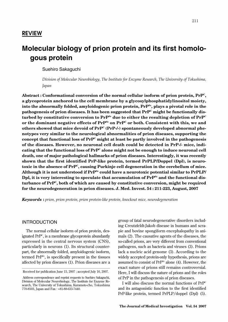
Mouse Prnp encodes the precursor protein consist-ing of 254 amino acids (Fig. 1). The 22 N-terminalhydrophobic amino acids are removed as a signal
peptide when the nascent precursor protein entersthe endoplasmic reticulum (ER) (Fig. 1). The 23 C-terminal hydrophobic amino acids are also cleavedas a glycosylphosphatidylinositol (GPI) -anchor sig-nal sequence in the ER and, at the same time, a GPIanchor moiety is attached to the C-terminus of theserine residue at position 231 (Fig. 1) (1). Then, theprotein is transported to the Golgi apparatus, wheretwo asparagine residues at positions 181 and 197 un-dergo N-glycosylation (Fig. 1), and finally to the cellsurface along the secretory pathway. As a result,mature PrPC, consisting of the amino acid residues23-231, is expressed as a membrane glycoproteinanchored to the cell surface via a GPI moiety.
Abnormal isoform of PrP
The abnormal isoform, PrPSc, is specifically pro-duced within prion infected cells, particularly in neu-rons. We and others showed that PrPSc failed to beproduced in mice devoid of PrPC (PrP-/-) (11-14),indicating that PrPC is essential for the generationof PrPSc. Moreover, it was reported that PrPSc-likePrP could be generated from PrPC in certain condi-tions in vitro (15). Therefore, it is thought that PrPSc
is produced from PrPC. However, it remains un-known whether or not PrPSc is produced from eithermature PrPC or immature unfolded PrP, or both.
PrPSc is identical to PrPC in amino acid sequence.However, structural analysis of PrPC and PrPSc us-ing circular dichroism revealed marked differencesin the protein structures of both proteins (Table 1).PrPC has a lower content of β-sheet strands (3%)but a higher α-helix content (42%) (16). In contrast,PrPSc has a higher β-sheet content (43%) (16). It istherefore postulated that the transition of α-helicesinto β-sheet strands within PrP might be a key stepin the generation of PrPSc.
Biochemical and structural properties of PrPs
PrPC and PrPSc possess markedly different bio-chemical properties from each other, particularlyin detergent solubility and resistance to proteinasedigestion, probably due to the structural differences.PrPC is highly soluble and easily digested by pro-teinase K whereas PrPSc readily aggregates to form
Fig. 1. Schematic structures of PrP and PrPLP/Dpl. α and βindicate α-helix and β-strand, respectively. S-S and Y indicate adisulfide bond and N-glycosylation, respectively. Arabic figuresrepresent amino acid positions.
Table 1 Different biochemical and structural properties of PrP isoforms
PrP isoformssecondary structure content detergent
solubilityproteinase K
digestionα-helix β-sheetNormal isoform
(PrPC) 42% 3% soluble sensitive
Abnormal isoform(PrPSc) 30% 43% insoluble relatively
resistant
S. Sakaguchi PrP and PrP-like protein in neurodegeneration212

amyloid fibers and is relatively resistant to the di-gestion (Table 1) (1). These different biochemicalproperties are useful to distinguish PrPSc from PrPC.
PrPSc remains structurally unresolved due to itspredisposition to form aggregates. In contrast, theprotein structure of PrPC was resolved by nuclearmagnetic resonance (NMR) analysis. According toNMR analysis, the N-terminal domain of PrPC ishighly flexible and lacks identifiable secondary struc-ture while the C-terminal domain forms a globularstructure with three α-helices and two short anti-parallel β-strands (Fig. 1) (17). The second andthird helices are linked by a disulfide bond (Fig. 1)(17).
The N-terminal domain includes a PrP-specific re-gion, the so-called octapeptide repeat (OR) region,in which 8 amino acids are repeated 5 times in tan-dem (Fig. 1). This region is considered to bind Cu2+
via histidine residues and mediate anti-oxidative ac-tivities by activating Cu2+-dependent antioxidant en-zymes such as superoxide dismutase via transfer ofbound Cu2+ to the enzymes (18, 19). However, theexact function of this region in anti-oxidative activi-ties remains to be elucidated.
THE FIRST PRP-LIKE PROTEIN (PRPLP/DPL)PrPLP/Dpl and PrP
We and others isolated a novel gene, termed Prnd ,encoding the first PrP-like protein, PrPLP/Dpl, about16-kb downstream of the PrP gene, Prnp (5, 7).PrPLP/Dpl is also a GPI-anchored membrane gly-coprotein (5). Like PrPC, PrPLP/Dpl is first trans-lated into the precursor protein consisting of 179amino acids and then undergoes several modifica-tions, including cleavage of the 23 N-terminal and25 C-terminal hydrophobic residues as a signalpeptide and a GPI-anchor signal, respectively, N-glycosylation at two sites, and formation of two di-sulfide bonds (Fig. 1) (20).
PrPLP/Dpl and PrP share �23% identical aminoacids (5, 7). However, PrPLP/Dpl lacks a region cor-responding to the N-terminal part of PrPC (5, 7). Theprotein structural analysis clearly showed that PrPLP/Dpl is a structural homologue of the C-terminal globu-lar domain of PrPC, composed of three α-helices andtwo short antiparallel β-strands (Fig. 1) (21).
Normal functions of PrPLP/Dpl
PrPLP/Dpl mRNA is expressed in various tissues
of adult wild-type mice, including the testis, heart,spleen and skeletal muscle (22). To investigatethe physiological functions of PrPLP/Dpl in mice,Behrens, et al. produced mice devoid of PrPLP/Dpl, designated Prnd neo/neo mice (23). Interestingly,male mutant mice were sterile whereas female mu-tant mice were fertile. The testes in these mutantmice were macroscopically normal. However, thenumber of spermatozoa and motility of mutant spermwere significantly decreased. Moreover, the mutantsperm exhibited abnormal morphologies and im-paired acrosome function. Consistently, it has beenreported that PrPLP/Dpl is expressed in spermatidsin mice and spermatozoa and Sertoli cells in hu-mans (23, 24). These results indicate that PrPLP/Dpl is involved in spermatogenesis.
In contrast to PrPC, PrPLP/Dpl was undetectablein the brains of adult wild-type mice (22). However,in neonatal mice, we found substantial expressionof PrPLP/Dpl mRNA in their brains, preferentiallyin blood vessel endothelial cells. PrPLP/Dpl mRNAwas already expressed 1 day after birth, peaked byaround 1 week, and then decreased to an undetect-able level by at least 8 weeks (22). Therefore, suchdevelopmental regulation of PrPLP/Dpl expressionin brain blood vessels suggests that PrPLP/Dpl maybe involved in the development of brain blood ves-sels and/or blood-brain barrier. However, no patho-logical abnormalities were detected in the tissuesof Prnd neo/neo mice, including the brain, heart, spleenand skeletal muscle (23).
PRION RESEARCH IN MICE DEVOID OFPRPProtein-only hypothesis and mice devoid of PrP
The protein-only hypothesis postulates that a prionis constituted of PrPSc alone (25). According to thehypothesis, a prion or PrPSc interacts with PrPC ex-pressed on the cell surface and induces changes inthe conformation of the interacting PrPC into thatof PrPSc, resulting in generation of a new PrPSc mole-cule, or propagation of a prion (Fig. 2). However,the hypothesis is controversial. If the hypothesis istrue, prions cannot propagate when PrPC is absent.In contrast, if prions can propagate without PrPC, thehypothesis is clearly negated. In other words, ifPrP-/- mice can support prion propagation, the hy-pothesis is wrong, and vise versa.
To investigate validity of the protein-only hypothe-sis, we generated a line of PrP-/- mice, referred to
The Journal of Medical Investigation Vol. 54 August 2007 213

as Ngsk PrP-/- mice, and intracerebrally inoculatedthem with a mouse-adapted Fukuoka-1 prion (14).Wild-type (PrP+/+) mice developed disease-specificsymptoms at 138�12 days and died 143�14 daysafter inoculation (Table 2). Microscopic examina-tions of the brains of these diseased mice showedprofound vacuolation and gliosis, both of which arehallmarks of the pathological changes in prion dis-eases. In addition, PrPSc was markedly accumulatedand prions were propagated in their brains. In con-trast, no Ngsk PrP-/- mice showed such specificsymptoms. They were all alive at least by 460 daysafter inoculation (Table 2). No disease-specific pa-thologies were observed in the brains of Ngsk PrP-/-mice sacrificed 400 days after inoculation. Moreover,neither accumulation of PrPSc nor prion propagationcould be detected in their brains. Other investiga-tors also showed similar results using other lines ofPrP-/- mice (11-13). These results indicate that PrPC
is essentially required for prion propagation, andthat prion propagation is linked to accumulation ofPrPSc, strongly supporting the protein-only hypothe-sis.
Prolonged incubation times and less accumulationof PrP Sc in mice heterozygous for PrP
We also inoculated Ngsk PrP+/- mice with theFukuoka-1 prion (14). Compared with PrP+/+ mice,Ngsk PrP+/- mice developed the disease with con-siderably retarded incubation times of 259�27 daysand died 269�27 days after inoculation (Table 2).The clinical symptoms and pathological changes indiseased Ngsk PrP+/- mice were indistinguishable
from those of diseased PrP+/+ mice. However, verystrangely, amounts of PrPSc accumulated in thebrains of terminal PrP+/- mice were only half ofthose in terminal PrP+/+ mice (14). These resultsindicate that the expression levels of PrPC prior toinfection affect the timing of onset of disease andthe accumulation levels of PrPSc but not the final se-verity and pathology of disease, and that PrPSc levelsare not correlated with disease progression.
NORMAL FUNCTIONS OF PRP IN NEU-RONS AND GLIAHigher brain functions and PrP
Since PrPC is abundantly expressed in pyramidalneurons of the hippocampus, in which learning andmemory processes are integrated, it has been sug-gested that PrPC might be involved in learning andmemory processes. Büeler, et al. produced a lineof PrP-/- mice, Zrch I PrP-/-, and subjected themto behavioral tasks, such as a swimming navigationtest and a Y-maze discrimination test (26). However,no different performance could be detected in thesetests between the mutant mice and control PrP+/+mice (26). On the other hand, Nishida, et al. re-ported poor performance in Ngsk PrP-/- mice us-ing other behavioral tests, including a water-findingtest and a conditioned passive-avoidance test (27).Thus, it may be possible that PrPC is involved in cer-tain types of learning and memory.
Collinge, et al. showed that long-term potentiation(LTP), a form of synaptic plasticity that is thoughtto be important for memory formation, was impairedin the hippocampal CA1 neurons of Zrch I PrP-/-mice using electrophysiological studies (28). Similarresults were reported in another line of PrP-/- mice,Npu PrP-/- mice (29). Therefore, these results seemto support that PrPC is involved in the processes oflearning and memory. However, other investigatorsreported no such electrophysiological abnormalitiesin the hippocampus of Zrch I PrP-/- mice (30).
It was also shown that PrPC is involved in the regu-lation of circadian rhythm (31). In both Zrch I PrP-/-and Npu PrP-/- mice, much more fragmented sleepwas observed than in PrP+/+ mice (31). Moreover,
Fig. 2. A prion replication model according to the protein-only hypothesis. A prion is constituted of the abnormal isofromof PrP, PrPSc, interacts with the normal isoform of PrP, PrPC,and induces the conformational changes of the interacting PrPC
to produce a new molecule of PrPSc or a prion. The newly pro-duced PrPSc or prion also converts another PrPC into PrPSc orprion in the same way.
Table 2 Ngsk PrP-/- mice were resistant to prion disease.
Mouse genotype Incubation time (mean�SD days) Survival time (mean�SD days)Wild-type 138�12 143�14
Ngsk PrP+/- 259�27 269�27Ngsk PrP-/- �460 �460
S. Sakaguchi PrP and PrP-like protein in neurodegeneration214

the mutant mice exhibited a much longer activityperiod of 23.9 h under constant darkness, comparedto 23.3 h in PrP+/+ mice (31).
Axonal myelination and PrP
We found many vacuoles in the spinal cord andperipheral nervous system of Ngsk PrP-/- and ZrchI PrP-/- mice (32). Most of the vacuoles were sur-rounded by an enlarged myelin sheath, but in somecases splits within a myelin sheath formed vacuoles(32). In addition, large myelinated fibers were re-duced in number and remaining axons were thinlymyelinated (32). Subsequently, we could rescueNgsk PrP-/- mice from the demyelination by trans-genically expressing mouse PrPC (32). These resultsindicate that PrPC is involved in the organization ofthe myelin sheath.
PrPC is expressed on the surface of oligodendro-cytes and Schwann cells (33, 34), both of which formmyelin sheaths in the CNS and the peripheral nerv-ous system, respectively. It is therefore conceivablethat PrPC functions as an adhesion molecule withina myelin sheath and/or between a myelin sheathand an axon to form a tightly compacted myelinsheath. This might be consistent with the result thatsome vacuoles were formed due to splits within mye-lin sheaths. It is alternatively possible that PrPC
could be a trophic factor for these glial cells.
PRP AND PRPLP/DPL IN NEURODEGEN-ERATIONPurkinje cell degeneration among different lines ofmice devoid of PrP
No neuropathological abnormalities were reportedin Zrch I PrP-/- and Npu PrP-/- mice (26, 35). How-ever, very strangely, we noticed that Ngsk PrP-/-mice showed ataxic gait around 70 weeks after birth(36). In these ataxic mice, cerebellar Purkinje cellswere dramatically decreased in number due to theirdegeneration and the molecular layer also becamevery thin, probably due to the loss of the dendritictrees of Purkinje cells (Fig. 3) (36). In contrast, noPurkinje cell degeneration could be detected inyounger Ngsk PrP-/- mice and old PrP+/+ and NgskPrP+/- mice (36). We also confirmed that the ataxiaand Purkinje cell degeneration in Ngsk PrP-/- micecould be successfully rescued by introduction ofthe transgene encoding PrPC (Fig. 3) (32). Similarcerebellar degeneration was subsequently reportedin other lines of PrP-/- mice, such as Rcm0 PrP-/-
and Zrch II PrP-/- mice (5, 37). Taken together,these results indicated that the functional loss ofPrPC is responsible for Purkinje cell degeneration,although it remained unknown why the neurode-generation was discrepant among different lines ofPrP-/- mice.
In Zrch I PrP-/- mice, a part of the PrP open read-ing frame (ORF) was replaced with the neomycinphosphotransferase (neo) gene (Fig. 4) (26). In NpuPrP-/- mice, the neo gene was simply inserted intoa unique site in the PrP-coding sequence (Fig. 4)(35). In contrast, in the ataxic lines of Ngsk PrP-/-,Rcm0 PrP-/-, and Zrch II PrP-/- mice, the entireORF was completely deleted (Fig. 4) (5, 36, 37). Itwas therefore conceivable that in non-ataxic linesof Zrch I PrP-/- and Npu PrP-/- mice, some as-pects of the normal function of PrPC might remainintact because of incomplete disruption of the PrPallele. Consistently, it was reported that a fusedmRNA consisting of the neo and the residual Prnpsequences was produced in their brains (26). Al-ternatively, it might be possible that the loss of PrPC
alone may not be enough to induce Purkinje cell de-generation, and that other factor(s), which are spe-cifically associated with the ataxic lines of PrP-/-mice, together with the loss of PrPC, are involvedin the neurodegeneration.
Ectopic expression of PrPLP/Dpl associated withPurkinje cell degeneration
We found that, in the brains of Ngsk PrP-/- micebut not in Zrch I PrP-/- and PrP+/+ mice, the PrPLP/Dpl-coding exons were ectopically expressed as chi-meric mRNAs with the residual non-coding Prnpexons 1 and 2 due to an abnormal intergenic splic-ing taking place between Prnp and Prnd (7). InNgsk PrP-/- mice, due to lack of the 3’ part of in-tron 2 including a splice acceptor, the pre-mRNAtranscribed from the residual Prnp promoter couldnot efficiently undergo cleavage/polyadenylationat the end of Prnp (Fig. 5B). The unsuccessfullycleaved pre-mRNA was then elongated until the last
Fig. 3. Purkinje cell degeneration in Ngsk PrP-/- mice andPurkinje cells rescued in Ngsk PrP-/- mice transgenic for PrPC.Purkinje cells are immunohistochemically stained using anti-calbindin antibodies and can be observed as brownish dots.
The Journal of Medical Investigation Vol. 54 August 2007 215

Fig. 4. Targeted PrP alleles among different lines of PrP-/- mice. In Ngsk PrP-/- mice, a 2.1-kb genomic DNA segment including0.9-kb of intron 2, 10-bp of 5’ untranslated region (UTR) of exon 3, the entire PrP ORF, and 0.45-kb of 3’ UTR is replaced by the neogene under the control of the mouse phosphoglycerate kinase (PGK) promoter. Rcm0 PrP-/- mice were generated by a similar targetingstrategy utilized in Ngsk PrP-/- mice. The hypoxanthine phosphoribosyltransferase gene under control of the PGK promoter was usedin Rcm0 PrP-/- mice as a selectable marker. In Zrch II PrP-/- mice, 0.27-kb of intron 2, the entire exon 3, and 0.6-kb of the 3’ flank-ing DNA segment were targeted by a specific 34-bp loxP sequence. In these lines of PrP-/- mice, the entire PrP ORF is completelydeleted. In contrast, Zrch I PrP-/- mice were generated by replacement of PrP codons 4-187 among a total of 254 codons with theneo gene under the control of the herpes simplex virus thymidine kinase promoter. Npu PrP-/- mice contain the disrupted Prnp al-leles, in which the neo gene under the control of the mouse metalothioneine promoter was simply inserted into a unique Knp I sitein the PrP-coding sequence.
Fig. 5. Mechanism for the generation of PrPLP/Dpl-encoding chimeric mRNAs in Ngsk PrP-/- mice. In wild-type mice, the PrPpre-mRNA is normally cleaved and polyadenylated at the last exon of the Prnp (A). However, in Ngsk PrP-/- mice, due to lack of the3’ part of intron 2, the pre-mRNA transcribed from the Prnp promoter could not efficiently undergo cleavage/polyadenylation at thelast exon of Prnp , being further elongated until the last exon of Prnd , and subjected to intergenic splicing between the residual Prnpexons 1/2 and the PrPLP/Dpl-coding exon (B). As a result, PrPLP/Dpl became abnormally expressed under the control of the Prnppromoter, leading to the ectopic expression of PrPLP/Dpl in the brains of ataxic lines of PrP-/- mice.
S. Sakaguchi PrP and PrP-like protein in neurodegeneration216

exon of Prnd and subjected to intergenic splicingbetween the residual Prnp exon 2 and the PrPLP/Dpl-coding exon, producing chimeric mRNAs con-sisting of the Prnp exons 1 and 2 and the PrPLP/Dpl-coding exons (Fig. 5B). As a result, Prnd becameabnormally regulated under the control of the Prnppromoter in Ngsk PrP-/- mice and PrPLP/Dpl wasectopically expressed in the brains, especially inneurons and glial cells where the promoter is veryactive (7). Similar ectopic expression of PrPLP/Dpl was subsequently reported in other ataxic linesof PrP-/- mice, Rcm0 PrP-/- and Zrch II PrP-/-mice (5, 37). Taken together, these results indi-cate that the ectopic expression of PrPLP/Dpl in theabsence of PrPC might be responsible for Purkinjecell degeneration in the ataxic lines of PrP-/- mice.
PrPLP/Dpl in the absence of PrP causes Purkinjecell degeneration
To investigate the possibility that ectopic expres-sion of PrPLP/Dpl in the absence of PrPC couldcause the Purkinje cell degeneration, we generatedtransgenic mice, referred to as tg(N-PrPLP/Dpl)mice, in which PrPLP/Dpl was specifically expressedin nearly all neurons including Purkinje cells underthe control of the neuron-specific enolase promoter,and subsequently crossed them with the non-ataxicline of Zrch I PrP-/- mice (38). Tg(N-PrPLP/Dpl)32 mice expressed PrPLP/Dpl in the cerebellumat a level about 1-2 times more than that of NgskPrP-/- mice, developing ataxia at 58�15 days on theZrch I PrP-/- background (Table 3). Purkinje cellswere markedly decreased in number due to the de-generation in these ataxic tg mice. In contrast, nei-ther ataxia nor Purkinje cell degeneration could bedetected in the tg mice carrying the PrP+/+ back-ground (Table 3). Another tg(N-PrPLP/Dpl)25mouse line, in which PrPLP/Dpl was expressed inthe cerebellum at a level less than a quarter that ofNgsk PrP-/- mice, also developed ataxia and Purk-inje cell degeneration at 359�52 days on the ZrchI PrP-/- background but not on the PrP+/+ back-ground (Table 3). These results clearly showed thatthe ectopic expression of PrPLP/Dpl is neurotoxic
in the absence of PrPC, causing the Purkinje cell de-generation, and that the neurotoxicity of PrPLP/Dplis antagonized by PrPC.
We also produced another type of tg mice, tg(P-PrPLP/Dpl) mice (38). In these tg mice, the ex-pression of PrPLP/Dpl was specifically targeted toPurkinje cells under the control of the Purkinje cellprotein-2 promoter (38). Like tg(N-PrPLP/Dpl)mice, tg(P-PrPLP/Dpl) mice developed ataxia andPurkinje cell degeneration on the Zrch I PrP-/- back-ground but not on the PrP+/+ background (38).Tg(P-PrPLP/Dpl)26 and 27 mice showed ataxia at268�28 and 167�13 days after birth, respectively,on the Zrch I PrP-/- background (Table 3). Theseresults clearly indicate that PrPLP/Dpl ectopicallyexpressed on Purkinje cells is itself neurotoxic tothe cells.
Stoichiometrical antagonism between PrP and PrPLP/Dpl in neurodegeneration
The times to the onset of ataxia in tg(N-PrPLP/Dpl) mice were inversely correlated with the ex-pression levels of PrPLP/Dpl (38). Tg(N-PrPLP/Dpl)32 mice expressed more PrPLP/Dpl in the cere-bellum and developed the ataxia earlier than tg(N-PrPLP/Dpl)25 mice (Table 3). In contrast, the lev-els of PrPC were correlated with the times of the on-set. Tg(N-PrPLP/Dpl)32 and 25 mice showed sig-nificantly retarded onset of the ataxia on the Zrch IPrP+/- background, compared with the Zrch I PrP-/- background (Table 3). Thus, these results indi-cate that PrPLP/Dpl and PrPC stoichiometrically an-tagonize each other to induce Purkinje cell degen-eration.
N-terminal domain of PrP antagonistic for PrPLP/Dpl
PrPC possesses the OR region-containing N-terminaldomain whereas PrPLP/Dpl lacks the correspond-ing domain. It is therefore conceivable that the N-terminal domain might be important for PrPC toantagonize against the PrPLP/Dpl neurotoxcity.To investigate the possibility, we introduced PrPwith a deletion of the N-terminal residues 23-88
Table 3 PrPLP/Dpl stoichiometrically antagonizes PrPC to induce ataxia
Tg linesOnset of ataxia on different genetic backgrounds (mean�SD days)
Zrch I PrP-/- Zrch I PrP+/- Wild-type
Tg(N-PrPLP/Dpl)25 359�52 495�86 �60032 58�15 259�48 �600
Tg(P-PrPLP/Dpl)26 268�28 463�81 �60027 167�13 391�108 �600
The Journal of Medical Investigation Vol. 54 August 2007 217

into Ngsk PrP-/- mice (39). As expected, the dele-tion mutant PrP failed to rescue Ngsk PrP-/- micefrom ataxia and Purkinje cell degeneration (39).Ngsk PrP-/- mice expressing the deletion mutantdeveloped ataxia and Purkinje cell degenerationon a time course identical to that of non-transgenicNgsk PrP-/- mice (39). These clearly indicate thatthe N-terminal residues 23-88 are important for PrPC
to antagonize the neurotoxicity of PrPLP/Dpl. TheN-terminal residues 23-88 include most of the ORregion. Thus, it is suggested that this OR regioncould be important for PrPC to protect Purkinje cellsfrom PrPLP/Dpl-induced degeneration, although itremains to be investigated which region in the de-leted N-terminal domain could be essential for theneuroprotection of PrPC against PrPLP/Dpl.
We also introduced PrP carrying a familial priondisease-associated mutation (E199K) into NgskPrP-/- mice (39). Interestingly, these mice devel-oped no ataxia and Purkinje cell degeneration (39),showing that the mutant PrP was fully functionalfor antagonizing the PrPLP/Dpl-induced neurotox-icity, suggesting that other disease-associated mu-tant PrPs are also functionally competent.
PRP AND PRPLP/DPL IN ISCHEMIC NEU-RONAL CELL DEATH
To assess whether PrPC and PrPLP/Dpl could beinvolved in other types of neuronal cell death, wesubjected Zrch I PrP-/- and Ngsk PrP-/- mice totransient forebrain ischemia (40). Interestingly, maleZrch I PrP-/- mice were very susceptible to the ische-mia compared to control PrP+/+ mice, developingmarked apoptosis in the hippocampal CA1 region(40). McLennan, et al. also reported that perma-nent occlusion of the middle cerebral artery in-creased the infarction volume in male Npu PrP-/-mice without no ectopic expression of PrPLP/Dplin neurons (41). However, no apoptotic cell deathcould be detected in the CA 1 of female Zrch I PrP-/-mice (40). Taken together, these results indicatethat PrPC is involved in neuroprotection againstbrain ischemia, and that the neuroprotective func-tion of PrPC is masked by female-specific neuropro-tective factor(s).
We also showed that, in contrast to Zrch I PrP-/-mice, both male and female Ngsk PrP-/- mice ex-hibited severe ischemic damage to CA1 neurons(40). Since Ngsk PrP-/- mice ectopically expressPrPLP/Dpl in neurons, it is therefore conceivable
that PrPLP/Dpl might counteract the female-specificneuroprotective function, thereby increasing thesusceptibility of PrPC-deficient neurons to ischemicinsults.
ROLES OF PRP AND PRPLP/DPL IN NEU-RODEGENERATIONNeurotoxic PrPs
PrPLP/Dpl is a homologue of the C-terminal partof PrPC. Interestingly, it was shown that the N-terminally truncated PrPs, PrP�32-121 and PrP�32-134, induced ataxia and cerebellar degenerationcharacterized by marked granule cell death in ZrchI PrP-/- mice and the neurotoxicity of these trun-cated PrPs was antagonized by the expression offull-length PrPC (42). No Purkinje cell degenerationwas observed in these mice because the truncatedPrPs were not expressed in Purkinje cells of thesemice due to the limited activity of the promoter used(42). Consistently, it was demonstrated that ataxiaand Purkinje cell loss could be induced in Zrch IPrP-/- mice when PrP�32-134 was targeted to Purk-inje cells (43). PrP�32-121 and PrP�32-134 encom-pass the homologous C-terminal part of PrPC toPrPLP/Dpl. It is therefore very likely that PrPLP/Dpl and the truncated PrPs might use the same ora very similar molecular mechanism to execute theneurotoxicity.
Cis- and trans-neuroprotective function of PrPagainst PrPLP/Dpl
In contrast to neurotoxic PrP�32-121 and PrP�32-134, it was shown that PrP�23-88 was not neu-rotoxic, causing no Purkinje cell degeneration inZrch I PrP-/- mice (39), suggesting that the neuro-toxicity of the C-terminal domain of PrPC is blockedby a cis-element(s) present in the region between theresidues 89 and 121. This region, overlapping withthe central hydrophobic part, is reported to com-prise part of the binding sites for the heat shockprotein, stress-inducible protein 1, and the extra-cellular matrix constituent glycosaminoglycans (44,45). It is therefore possible that interaction of PrPC
with these molecules might be involved in the cis-inhibition of the neurotoxicity of the C-terminaldomain of PrPC. In contrast, PrPLP/Dpl and thetruncated PrPs are unable to interact with thesemolecules, therefore acting as neurotoxic proteins.
PrPC also neutralizes the neurotoxicity of PrPLP/Dpl and the truncated PrPs in trans, rescuing from
S. Sakaguchi PrP and PrP-like protein in neurodegeneration218

the Purkinje cell degeneration. Interestingly, weshowed that PrP�23-88 has no such trans-neuropro-tective activity, unable to antagonize against PrPLP/Dpl (39). It is therefore suggested that N-terminalresidues 23-88 are involved in the trans-neuropro-tection of PrPC against PrPLP/Dpl and probably thetruncated PrPs as well.
Antagonistic signals of PrP and PrPLP/Dpl inneuronal cell death
Kuwahara, et al. previously reported that hip-pocampal neuronal cells from PrP-/- mice easilyundergo apoptosis after withdrawal of serum, whichcan be prevented by either re-introduction of PrPC orby expressing the anti-apoptotic protein, Bcl-2 (46).Bounhar, et al. also showed that PrPC protected hu-man primary neurons from the apoptosis inducedby the pro-apoptotic protein Bax (47). It is thereforesuggested that PrPC might be an anti-apoptotic pro-tein while PrPLP/Dpl might be pro-apoptotic.
It was shown that the primary cultured cerebel-lar neurons from non-ataxic Npu PrP-/- mice weremore sensitive to oxidative stress than those fromPrP+/+ mice (19), indicating that PrPC might beinvolved in mitigation of oxidative stress. Interest-ingly, Wong, et al. showed that oxidative stress wasmuch more elevated in the brains of ataxic Rcm0PrP-/- mice than in those of non-ataxic Npu PrP-/-mice (48). It is therefore suggested that, in contrastto PrPC, PrPLP/Dpl aggravates oxidative stress. Oxi-dative stress is associated with overproduction ofradical oxygens, such as superoxide and nitric oxide(NO). Interestingly, it was shown that enzymaticactivity of superoxide dismutase, a superoxide-detoxifying enzyme, was significantly decreased inPrP-/- mice (18). Moreover, it was reported thatrecombinant PrPLP/Dpl was toxic to the culturedcerebellar neurons of non-ataxic Zrch I PrP-/- miceand that this PrPLP/Dpl-neurotoxicity could be pre-vented by L-N -acetyl methyl ester, a pharmacologi-cal inhibitor of NO synthases (49). It is thereforepossible that PrPLP/Dpl might aggravate oxidativestress by overproducing NO and superoxide, therebycausing Purkinje cell death, and that PrPC coulddetoxify it, preventing PrPLP/Dpl-induced neurode-generation.
It was previously shown that, in Zrch I PrP-/-mice, Ca2+-activated K+ currents in hippocampal py-ramidal neurons were abnormal and intracellular Ca2+
contents in cerebellar granule cells were altered (50).Moreover, we showed that the T- and L-type Ca2+-antagonist, flunarizine, significantly reduced the
PrPLP/Dpl-aggravated ischemic neuronal apoptosisin Ngsk PrP-/- mice (40). Since excess of intracel-lular Ca2+ has been shown to be toxic to neurons(51), it is alternatively possible that PrPC could re-duce the intracellular Ca2+ load in neurons, therebybeing neuroprotective, and that, in contrast, PrPLP/Dpl increases the intracellular Ca2+ load in neurons,thereby enhancing the susceptibility of neurons toapoptosis.
A proposed model of antagonistic interaction be-tween PrP and PrPLP/Dpl
Weissmann and colleagues have proposed an in-teresting hypothesis, in which two conjectural mole-cules, protein π, which is another PrP-like protein,and a cognate ligand for PrPC, are introduced (42,52). In normal mice, PrPC binds its cognate ligand toelicit a neuroprotective signal, thereby Purkinje cellsare able to survive. But, in ataxic lines of PrP-/- mice,PrPLP/Dpl or the neurotoxic truncated PrPs com-pete with PrPC for the ligand and blocks the signal,resulting in Purkinje cell degeneration. In non-ataxiclines of PrP-/- mice, instead of PrPC, the protein πcould elicit the same neuroprotective signal via bind-ing to the ligand, thereby causing no Purkinje celldegeneration. However, this hypothesis can be veri-fied only when the putative molecules are identified.
FINDINGS IN PRP-NULL MICE AND PATHO-GENESIS OF PRION DISEASES
The molecular pathogenesis of prion diseases re-mains elusive. Since PrP-/- mice were resistant tothese diseases (11-14), it is considered that the con-formational conversion of PrPC to PrPSc plays an es-sential role in the pathogenesis of the diseases. How-ever, the exact nature of this role has not been fullyunderstood. PrPSc is markedly accumulated in in-fected brains due to constitutive conversion. Forloni,et al. showed that an amyloidogenic PrP peptide(PrP106-126) was highly toxic to primary culturedneurons (53). It is therefore conceivable that PrPSc
might itself be neurotoxic. In contrast, Yokoyama,et al. showed that the PrPC-specific immunoreactiv-ity was decreased in the brain regions where PrPSc
had accumulated in experimentally infected mice(54), indicating that PrPC is reduced in the infectedneurons due to conversion. It is therefore alterna-tively possible that PrPC might be functionally dis-turbed due to its marked reduction, thereby result-ing in the nerodegeneration. Or, both the accumu-
The Journal of Medical Investigation Vol. 54 August 2007 219

lation of PrPSc and the functional loss of PrPC mightbe required for the neuronal cell death.
PrP-/- mice without ectopic expression of PrPLP/Dpl exhibited neurological abnormalities, includingimpairment of LTP, alteration in sleep and circadianrhythm, as well as demyelination in the spinal cordand peripheral nervous system (28, 31, 32). LTP isa form of synaptic plasticity that is thought to un-derlie memory formation. Memory loss or demen-tia is a common symptom in prion diseases (55).Alteration in sleep and circadian rhythms is also asymptom characteristic for the inherited human priondisease, fetal familial insomnia (55). Moreover, de-myelinating peripheral neuropathy has been reportedin some cases of inherited prion disease (56, 57).Taken together, these results strongly support thatthe functional loss of PrPC is involved in these patho-genic changes in the diseases. However, no neuro-nal cell degeneration could be detected in mice de-void of PrPC alone, indicating that loss of PrPC alonemight not be enough to induce neuronal cell deathor that other neurotoxic mechanisms might asso-ciate with neuronal cell death in prion diseases.
Interestingly, the ectopic expression of PrPLP/Dpl, PrP�32-121, or PrP�32-134 in the absence ofPrPC caused degeneration of Purkinje cells and gran-ule cells (6, 38, 42). Purkinje cells and granule cellsare markedly degenerative in human prion diseases(55). It is therefore conceivable that PrPLP/Dpland the truncated PrPs might be involved in thepathogenesis of the diseases. However, PrPLP/Dplitself is unlikely to be involved in the pathogenesisof prion diseases because PrPLP/Dpl could not bedetected in the brains affected by experimental priondiseases (6, 8). Instead, PrPSc is markedly accumu-lated and its fragmented C-terminal products areoften observed in the affected brains (58). It is there-fore very interesting to speculate that PrPSc or itsfragmented products possess a neurotoxic poten-tial equivalent to that of PrPLP/Dpl, PrP�32-121,or PrP�32-134. Thus, elucidation of the molecularmechanism for the Purkinje cell degeneration inataxic lines of PrP-/- mice might be useful for un-derstanding of the molecular pathogenesis of priondiseases.
ACKNOWLEDGMENTS
I would like to thank Dr. S. Katamine, Dr. K.Shigematsu, Dr. N. Nishida, Dr. R. Atarashi, Dr. A.Li, Dr. N. Yamaguchi, Dr. Y. Sakurai-Yamashita,
and Mr. N. Okimura. This study is partly supportedfrom the Ministry of Health, Labour and Welfare,Japan.
REFERENCES
1. Prusiner SB, Scott MR, DeArmond SJ, CohenFE : Prion protein biology. Cell 93 : 337-48, 1998
2. Prusiner SB, Hsiao KK : Human prion diseases.Ann Neurol 35 : 385-95, 1994
3. Prusiner SB : Prions. Proc Natl Acad Sci USA95 : 13363-83, 1998
4. Prusiner SB : Prions : novel infectious patho-gens. Adv Virus Res 29 : 1-56, 1984
5. Moore RC, Lee IY, Silverman GL, Harrison PM,Strome R, Heinrich C, Karunaratne A, PasternakSH, Chishti MA, Liang Y, Mastrangelo P,Wang K, Smit AF, Katamine S, Carlson GA,Cohen FE, Prusiner SB, Melton DW, TremblayP, Hood LE, Westaway D : Ataxia in prion pro-tein (PrP)-deficient mice is associated withupregulation of the novel PrP-like protein dop-pel. J Mol Biol 292 : 797-817, 1999
6. Moore RC, Mastrangelo P, Bouzamondo E,Heinrich C, Legname G, Prusiner SB, HoodL, Westaway D, DeArmond SJ, Tremblay P :Doppel-induced cerebellar degeneration in trans-genic mice. Proc Natl Acad Sci USA 98 : 15288-93, 2001
7. Li A, Sakaguchi S, Atarashi R, Roy BC, NakaokeR, Arima K, Okimura N, Kopacek J, ShigematsuK : Identification of a novel gene encoding aPrP-like protein expressed as chimeric tran-scripts fused to PrP exon 1/2 in ataxic mouseline with a disrupted PrP gene. Cell Mol Neu-robiol 20 : 553-67, 2000
8. Tuzi NL, Gall E, Melton D, Manson JC : Ex-pression of doppel in the CNS of mice doesnot modulate transmissible spongiform encepha-lopathy disease. J Gen Virol 83 : 705-11, 2002
9. Prusiner SB : Molecular biology of prion dis-eases. Science 252 : 1515-22, 1991
10. Oesch B, Westaway D, Walchli M, McKinleyMP, Kent SB, Aebersold R, Barry RA, TempstP, Teplow DB, Hood LE, Prusiner SB, WeissmannC : A cellular gene encodes scrapie PrP 27-30protein. Cell 40 : 735-46, 1985
11. Bueler H, Aguzzi A, Sailer A, Greiner RA,Autenried P, Aguet M, Weissmann C : Micedevoid of PrP are resistant to scrapie. Cell 73 :1339-47, 1993
S. Sakaguchi PrP and PrP-like protein in neurodegeneration220

12. Prusiner SB, Groth D, Serban A, Koehler R,Foster D, Torchia M, Burton D, Yang SL,DeArmond SJ : Ablation of the prion protein(PrP) gene in mice prevents scrapie and facili-tates production of anti-PrP antibodies. ProcNatl Acad Sci USA 90 : 10608-12, 1993
13. Manson JC, Clarke AR, McBride PA, McConnellI, Hope J : PrP gene dosage determines thetiming but not the final intensity or distributionof lesions in scrapie pathology. Neurodegen-eration 3 : 331-40, 1994
14. Sakaguchi S, Katamine S, Shigematsu K,Nakatani A, Moriuchi R, Nishida N, KurokawaK, Nakaoke R, Sato H, Jishage K, Kuno J, NodaT, Miyamoto T : Accumulation of proteinaseK-resistant prion protein (PrP) is restricted bythe expression level of normal PrP in mice in-oculated with a mouse-adapted strain of theCreutzfeldt-Jakob disease agent. J Virol 69 :7586-92, 1995
15. Kocisko DA, Come JH, Priola SA, ChesebroB, Raymond GJ, Lansbury PT, Caughey B :Cell-free formation of protease-resistant prionprotein. Nature 370 : 471-4, 1994
16. Pan KM, Baldwin M, Nguyen J, Gasset M,Serban A, Groth D, Mehlhorn I, Huang Z,Fletterick RJ, Cohen FE, Prusiner SB : Con-version of alpha-helices into beta-sheets fea-tures in the formation of the scrapie prion pro-teins. Proc Natl Acad Sci USA 90 : 10962-6,1993
17. Riek R, Hornemann S, Wider G, Billeter M,Glockshuber R, Wuthrich K : NMR structure ofthe mouse prion protein domain PrP(121-321).Nature 382 : 180-2, 1996
18. Brown DR, Qin K, Herms JW, Madlung A,Manson J, Strome R, Fraser PE, Kruck T,von Bohlen A, Schulz-Schaeffer W, Giese A,Westaway D, Kretzschmar H : The cellularprion protein binds copper in vivo. Nature 390 :684-7, 1997
19. Brown DR, Nicholas RS, Canevari L : Lack ofprion protein expression results in a neuronalphenotype sensitive to stress. J Neurosci Res67 : 211-24, 2002
20. Silverman GL, Qin K, Moore RC, Yang Y,Mastrangelo P, Tremblay P, Prusiner SB, CohenFE, Westaway D : Doppel is an N-glycosylated,glycosylphosphatidylinositol-anchored protein.Expression in testis and ectopic production inthe brains of Prnp(0/0) mice predisposed toPurkinje cell loss. J Biol Chem 275 : 26834-41,
200021. Mo H, Moore RC, Cohen FE, Westaway D,
Prusiner SB, Wright PE, Dyson HJ : Two dif-ferent neurodegenerative diseases caused byproteins with similar structures. Proc Natl AcadSci USA 98 : 2352-7, 2001
22. Li A, Sakaguchi S, Shigematsu K, Atarashi R,Roy BC, Nakaoke R, Arima K, Okimura N,Kopacek J, Katamine S : Physiological expres-sion of the gene for PrP-like protein, PrPLP/Dpl,by brain endothelial cells and its ectopic expres-sion in neurons of PrP-deficient mice ataxic dueto Purkinje cell degeneration. Am J Pathol 157 :1447-52, 2000
23. Behrens A, Genoud N, Naumann H, RulickeT, Janett F, Heppner FL, Ledermann B, AguzziA : Absence of the prion protein homologueDoppel causes male sterility. Embo J 21 : 3652-8, 2002
24. Peoc’h K, Serres C, Frobert Y, Martin C,Lehmann S, Chasseigneaux S, Sazdovitch V,Grassi J, Jouannet P, Launay JM, LaplancheJL : The human “prion-like” protein Doppel isexpressed in both Sertoli cells and spermato-zoa. J Biol Chem 277 : 43071-8, 2002
25. Prusiner SB : Novel proteinaceous infectiousparticles cause scrapie. Science 216 : 136-44,1982
26. Bueler H, Fischer M, Lang Y, Bluethmann H,Lipp HP, DeArmond SJ, Prusiner SB, AguetM, Weissmann C : Normal development andbehaviour of mice lacking the neuronal cell-surface PrP protein. Nature 356 : 577-82, 1992
27. Nishida N, Katamine S, Shigematsu K, NakataniA, Sakamoto N, Hasegawa S, Nakaoke R,Atarashi R, Kataoka Y, Miyamoto T : Prion pro-tein is necessary for latent learning and long-term memory retention. Cell Mol Neurobiol17 : 537-45, 1997
28. Collinge J, Whittington MA, Sidle KC, SmithCJ, Palmer MS, Clarke AR, Jefferys JG : Prionprotein is necessary for normal synaptic func-tion. Nature 370 : 295-7, 1994
29. Manson JC, Hope J, Clarke AR, Johnston A,Black C, MacLeod N : PrP gene dosage andlong term potentiation. Neurodegeneration 4 :113-4, 1995
30. Lledo PM, Tremblay P, DeArmond SJ, PrusinerSB, Nicoll RA : Mice deficient for prion proteinexhibit normal neuronal excitability and synap-tic transmission in the hippocampus. Proc NatlAcad Sci USA 93 : 2403-7, 1996
The Journal of Medical Investigation Vol. 54 August 2007 221

31. Tobler I, Gaus SE, Deboer T, Achermann P,Fischer M, Rulicke T, Moser M, Oesch B,McBride PA, Manson JC : Altered circadian ac-tivity rhythms and sleep in mice devoid of prionprotein. Nature 380 : 639-42, 1996
32. Nishida N, Tremblay P, Sugimoto T, ShigematsuK, Shirabe S, Petromilli C, Erpel SP, NakaokeR, Atarashi R, Houtani T, Torchia M, SakaguchiS, DeArmond SJ, Prusiner SB, Katamine S : Amouse prion protein transgene rescues micedeficient for the prion protein gene from purk-inje cell degeneration and demyelination. LabInvest 79 : 689-97, 1999
33. Moser M, Colello RJ, Pott U, Oesch B : Devel-opmental expression of the prion protein genein glial cells. Neuron 14 : 509-17, 1995
34. Follet J, Lemaire-Vieille C, Blanquet-GrossardF, Podevin-Dimster V, Lehmann S, Chauvin JP,Decavel JP, Varea R, Grassi J, Fontes M,Cesbron JY : PrP expression and replication bySchwann cells : implications in prion spreading.J Virol 76 : 2434-9, 2002
35. Manson JC, Clarke AR, Hooper ML, AitchisonL, McConnell I, Hope J : 129/Ola mice carry-ing a null mutation in PrP that abolishes mRNAproduction are developmentally normal. MolNeurobiol 8 : 121-7, 1994
36. Sakaguchi S, Katamine S, Nishida N, MoriuchiR, Shigematsu K, Sugimoto T, Nakatani A,Kataoka Y, Houtani T, Shirabe S, Okada H,Hasegawa S, Miyamoto T, Noda T : Loss ofcerebellar Purkinje cells in aged mice homozy-gous for a disrupted PrP gene. Nature 380 :528-31, 1996
37. Rossi D, Cozzio A, Flechsig E, Klein MA,Rulicke T, Aguzzi A, Weissmann C : Onset ofataxia and Purkinje cell loss in PrP null miceinversely correlated with Dpl level in brain.Embo J 20 : 694-702, 2001
38. Yamaguchi N, Sakaguchi S, Shigematsu K,Okimura N, Katamine S : Doppel-induced Purk-inje cell death is stoichiometrically abrogated byprion protein. Biochem Biophys Res Commun319 : 1247-52, 2004
39. Atarashi R, Nishida N, Shigematsu K, Goto S,Kondo T, Sakaguchi S, Katamine S : Deletionof N-terminal residues 23-88 from prion protein(PrP) abrogates the potential to rescue PrP-deficient mice from PrP-like protein/doppel-induced Neurodegeneration. J Biol Chem 278 :28944-9, 2003
40. Sakurai-Yamashita Y, Sakaguchi S, Yoshikawa
D, Okimura N, Masuda Y, Katamine S, NiwaM : Female-specific neuroprotection againsttransient brain ischemia observed in mice de-void of prion protein is abolished by ectopic ex-pression of prion protein-like protein. Neuro-science 136 : 281-7, 2005
41. McLennan NF, Brennan PM, McNeill A, DaviesI, Fotheringham A, Rennison KA, Ritchie D,Brannan F, Head MW, Ironside JW, WilliamsA, Bell JE : Prion protein accumulation andneuroprotection in hypoxic brain damage. AmJ Pathol 165 : 227-35, 2004
42. Shmerling D, Hegyi I, Fischer M, Blattler T,Brandner S, Gotz J, Rulicke T, Flechsig E,Cozzio A, von Mering C, Hangartner C, AguzziA, Weissmann C : Expression of amino-termi-nally truncated PrP in the mouse leading toataxia and specific cerebellar lesions. Cell 93 :203-14, 1998
43. Flechsig E, Hegyi I, Leimeroth R, Zuniga A,Rossi D, Cozzio A, Schwarz P, Rulicke T, GotzJ, Aguzzi A, Weissmann C : Expression of trun-cated PrP targeted to Purkinje cells of PrPknockout mice causes Purkinje cell death andataxia. Embo J 22 : 3095-101, 2003
44. Zanata SM, Lopes MH, Mercadante AF, HajjGN, Chiarini LB, Nomizo R, Freitas AR, CabralAL, Lee KS, Juliano MA, de Oliveira E, JachieriSG, Burlingame A, Huang L, Linden R, BrentaniRR, Martins VR : Stress-inducible protein 1 isa cell surface ligand for cellular prion that trig-gers neuroprotection. Embo J 21 : 3307-16, 2002
45. Warner RG, Hundt C, Weiss S, Turnbull JE :Identification of the heparan sulfate bindingsites in the cellular prion protein. J Biol Chem277 : 18421-30, 2002
46. Kuwahara C, Takeuchi AM, Nishimura T,Haraguchi K, Kubosaki A, Matsumoto Y, SaekiK, Matsumoto Y, Yokoyama T, Itohara S,Onodera T : Prions prevent neuronal cell-linedeath. Nature 400 : 225-6, 1999
47. Bounhar Y, Zhang Y, Goodyer CG, LeBlanc A :Prion protein protects human neurons againstBax-mediated apoptosis. J Biol Chem 276 :39145-9, 2001
48. Wong BS, Liu T, Paisley D, Li R, Pan T, ChenSG, Perry G, Petersen RB, Smith MA, MeltonDW, Gambetti P, Brown DR, Sy MS : Induc-tion of HO-1 and NOS in doppel-expressingmice devoid of PrP : implications for doppelfunction. Mol Cell Neurosci 17 : 768-75, 2001
49. Cui T, Holme A, Sassoon J, Brown DR : Analy-
S. Sakaguchi PrP and PrP-like protein in neurodegeneration222

sis of doppel protein toxicity. Mol Cell Neuro-sci 23 : 144-55, 2003
50. Colling SB, Collinge J, Jefferys JG : Hippocam-pal slices from prion protein null mice : dis-rupted Ca(2+)-activated K+ currents. NeurosciLett 209 : 49-52, 1996
51. Pisani A, Bonsi P, Calabresi P : Calcium sig-naling and neuronal vulnerability to ischemiain the striatum. Cell Calcium 36 : 277-84, 2004
52. Weissmann C, Aguzzi A : Perspectives : neu-robiology. PrP’s double causes trouble. Sci-ence 286 : 914-5, 1999
53. Forloni G, Angeretti N, Chiesa R, Monzani E,Salmona M, Bugiani O, Tagliavini F : Neuro-toxicity of a prion protein fragment. Nature362 : 543-6, 1993
54. Yokoyama T, Kimura KM, Ushiki Y, Yamada S,Morooka A, Nakashiba T, Sassa T, Itohara S : Invivo conversion of cellular prion protein to patho-
genic isoforms, as monitored by conformation-specific antibodies. J Biol Chem 276 : 11265-71, 2001
55. DeArmond SJ, Prusiner SB : Etiology and patho-genesis of prion diseases. Am J Pathol 146 :785-811, 1995
56. Kitamoto T, Amano N, Terao Y, Nakazato Y,Isshiki T, Mizutani T, Tateishi J : A new inher-ited prion disease (PrP-P105L mutation) show-ing spastic paraparesis. Ann Neurol 34 : 808-13,1993
57. Neufeld MY, Josiphov J, Korczyn AD : Demye-linating peripheral neuropathy in Creutzfeldt-Jakob disease. Muscle Nerve 15 : 1234-9, 1992
58. Chen SG, Teplow DB, Parchi P, Teller JK,Gambetti P, Autilio-Gambetti L : Truncatedforms of the human prion protein in normalbrain and in prion diseases. J Biol Chem 270 :19173-80, 1995
The Journal of Medical Investigation Vol. 54 August 2007 223