molecular biology 101 - aphl · 1. molecular biology 101 1.1 introduction notes: welcome to the...
TRANSCRIPT

Molecular Biology 101

1. Molecular Biology 101
1.1 Introduction
Notes:
Welcome to the Association of Public Health Laboratories Essentials for the Mycobacteriology Laboratory Promoting Quality Practices. This presentation is Molecular Biology 101.

1.2 Overview
Notes:
This module will provide a brief overview of the basics of molecular testing methods and will cover four areas: principals of molecular biology, applying the concepts of molecular biology, workflow, contamination, safety and regulation and considerations for TB molecular testing.

2. Principles of Molecular Biology
Notes:
First, let's discuss principles of molecular biology.
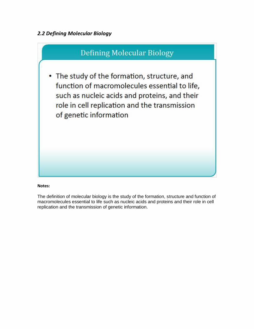
2.2 Defining Molecular Biology
Notes:
The definition of molecular biology is the study of the formation, structure and function of macromolecules essential to life such as nucleic acids and proteins and their role in cell replication and the transmission of genetic information.

2.3 Central Dogma of Molecular Biology
Notes:
The central dogma of molecular biology explains the flow of genetic information in a biological system. In general, we talk about DNA that makes RNA that makes proteins. So through the process of replication DNA can make additional copies of DNA. Through the process of transcription the DNA can be made into RNA and that RNA is then translated into proteins.

2.4 Deoxyribonucleic Acid (DNA)
Notes:
The first macromolecule we'll discuss is DNA, deoxyribonucleic acid. DNA is a polynucleotide chain made up of four bases A, T, G, and C: Adenine, Thymine, Guanine, and Cytosine. A nucleotide is composed of a base an A, C, T or G; a sugar and phosphate group as shown in the diagram here. In the TB genome there are approximately four million base pairs which translates into approximately 4000 genes. DNA exists as a double stranded molecule where the strands are held together through complementary base pairing A to T and G to C. The two strands are complementary to one another and run in an anti-parallel fashion, so five prime to three prime on this strand in the downward direction and five prime to three prime in the other strand in the upward direction. Replication of the DNA also occurs in this five prime to three prime direction.

2.5 Different Types of Bacterial DNA
Notes:
There are several types of DNA but we will focus on these three today. Bacterial Chromosomal DNA shown by this large molecule here exists as a double stranded circular chromosome. The length of the chromosome varies between different genera and even between species within a genus. The chromosome generally contains the genes that the bacteria need for survival. Plasmids can be found inside bacteria. They are small circular DNA in this picture. They also exist as circular, double stranded DNA. They replicate in the organism independent of the bacterial chromosome and are generally smaller than the bacterial chromosome. In general, they also contain genes that are not required for bacterial survival. Insertion sequences are small segments of DNA in the bacterial chromosome. They are flanked by inverted repeat sequences that allow the insertion sequence to be inserted at varying points in the genome depending on the pressures for rearrangement. They can interrupt coding sequences and block gene expression depending on where they're inserted in the genome.

2.6 Ribonucleic Acid (RNA)
Notes:
The next macromolecule we will discuss is RNA, ribonucleic acid. It is also a polynucleotide like DNA and formed by four bases but we have Adenine, Cytosine, Guanine and Uracil. There's no thymine in RNA. It's a single stranded molecule and as we talked about in the central dogma of molecular biology, RNA is formed by transcription of DNA using an RNA polymerase. RNA can form hydrogen bonds with multiple molecules including DNA, other RNA and itself. RNA is a less stable molecule than DNA because it contains more reactive hydroxyl group on the two prime carbon as you'll see in the next slide.
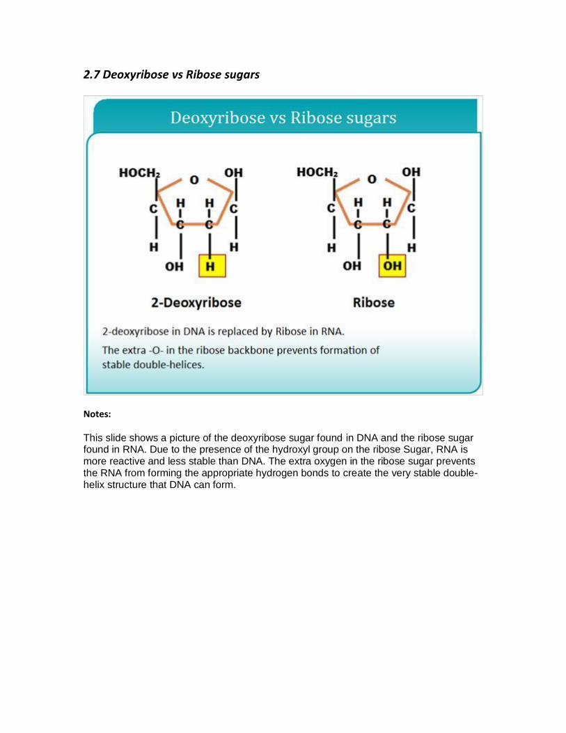
2.7 Deoxyribose vs Ribose sugars
Notes:
This slide shows a picture of the deoxyribose sugar found in DNA and the ribose sugar found in RNA. Due to the presence of the hydroxyl group on the ribose Sugar, RNA is more reactive and less stable than DNA. The extra oxygen in the ribose sugar prevents the RNA from forming the appropriate hydrogen bonds to create the very stable double-helix structure that DNA can form.

2.8 Important Definitions
Notes:
There are a few definitions we should talk about specific to DNA. First is gene. This is a specific sequence of nucleotides along a segment of DNA that provide the coded instructions for synthesizing a protein or a RNA molecule. Below are two different sequences of a gene. Within a gene we can talk about a locus or a specific region of interest within a gene. The DNA sequence of a gene is broken up then into codons or three base segments of a gene that code for a specific amino acid. We also talk about wild type frequently. Wild type or a wild type sequence is the sequence of a gene as it is found in nature and the normal gene sequence at a specific locus.

2.9 Amino Acids
Notes:
The third macromolecule we will discuss are the amino acids. Amino acids are the building blocks of proteins. In the picture here we can see a long segment of DNA that contains three separate genes. Let's take a closer look at gene 1. The first thing we see is the DNA sequence in the five prime to three prime direction. Through transcription, that DNA will be transcribed into an RNA sequence. Then the RNA will be translated using those three base codons into the amino acids coded for by each codon. So for this codon here UGG, the amino acid is tryptophan. For the codon UUU the amino acid is phenylalanine and so on for glycine and serine.

2.10 Universal Genetic Code
Notes:
So let's talk about the genetic code for a little bit. We talked about how the three base codons indicate which amino acid should be added in a growing polypeptide chain. This table will help us navigate that genetic code. So for any codon you want to start with the first base shown on the vertical axis of this table. If the first space is an A then you're going to proceed in the third row of the table. If the second base of your codon is an A then you're in the third row and third column of the table. Now within this box you will select the third position of your codon. You can see here that whether your third position is an A or a G it codes for a lysine residue. If your third position is a T or a C then it codes for asparagine residue. There are many codons that code for the same amino acid and this demonstrates redundancy in the genetic code. The third positon of the codons sometimes referred to as the wobble positon can help maintain some consistency in the resulting protein, even when the DNA sustains a mutation due to this redundancy.

2.11 Proteins
Notes:
So using this genetic code each codon of a gene is then translated into an amino acid and all of those amino acids are linked together to form a protein. This image shows the RNA molecule being translated by the ribosome three bases at a time. The UGG codon pulls in a tryptophan amino acid. The AAA codon pulls in a lysine amino acid. The GAU codon pulls in an aspartic acid and so on and so forth until the whole protein is created. Sometimes the word peptide will be used instead of protein. A protein is a polypeptide that can be hundreds or thousands of amino acids in length.

2.12 Mutations
Notes:
When we talk about DNA we have to talk about mutations. For the definition of mutation it is a change in the DNA or RNA sequence that results in variation from previous generations and that can be transmitted to subsequent generations. These are changes that alter the transcription and translation and are carried on in the replication of the DNA within an organism. They can range in size from a single base all the way up to large segments of the DNA. Mutations can come about through a variety of mechanisms including mutagens like UV light, selective pressures like the presence of antibiotics or they can be spontaneous due to errors in the replication process.

2.13 Types of Mutations
Notes:
There are a few different types of mutations. A point mutation is a mutation in a single base in the sequence. These are sometimes referred to as single nucleotide polymorphisms or SNIPS. A mutation can be in the form of a deletion of a single base or set of bases that are deleted from the sequence. A mutation can also be in the form of an insertion where a single base or set of bases are inserted into the sequence.

2.14 Influence of Mutations
Notes: The influence of a mutation depends on the way in which it has changed the DNA. A silent or a synonymous mutation is one in which the DNA sequence is changed but the resulting amino acid sequence is not changed and this will likely not change the structure and function of the protein. In the table below at the DNA level, we have the codon TTC which codes for a lysine. A silent mutation would be one where the wobble position is mutated to the T but the codon TTT still codes for a lysine. So the amino acid is the same. The protein is the same and the structure and function should remain unchanged. A nonsense mutation is a mutation that results in a stop codon where protein synthesis stops and the resulting protein is truncated and may have severely altered structure and function so our codon TTC is now ATC and the stop codon causes protein synthesis to halt. A missense or non-synonymous mutation is one in which the mutation results in a different amino acid and may have a small or large effect on the structure and the function of the protein. So our wild type codon TTC is now TGC. Instead of a lysine we get a [threanine?] which could significantly change the structure and function of our protein and have detrimental effects on the protein. A frameshift mutation is one by which the reading frame of the gene changes. I will demonstrate this on the next slide but it can also severely alter the resulting protein.

2.15 Mutations Illustrated (2)
Notes:
This slide will help demonstrate the effects of mutations on a coding sequence. So if we have a normal gene that starts here with the start code on "the" and ends here with a stop code on "end" and in the middle are all of these three letter codons and words the man saw the dog hit the can end. If we have a point mutation and a sequence such as this G being mutated to a T then the definition of the word changes. Instead of, the man saw the dog hit the can end, it is, the man saw the dot hit the can end. If we have a deletion of a whole codon then the sentence becomes, the man saw the hit the can end. Clearly, we've lost something there. If we have an insertion this can change the entire effect of the words as well and we get, the man saw the fat dog hit the can end. Again, we've made a significant change to the intent of the words. And finally, a frameshift mutation, where we have a single base deleted that causes the rest of the bases or words to shift in that three letter code system. Instead of, the man saw the dog hit the can end, we have, the man saw a bunch of gibberish compared to the expected sentence or sequence and you can see how a frameshift can wreak havoc on the entire sequence of the letters.

2.16 Naming Mutations
Notes: In order for a mutation to be understood no matter who's reading the interpretation a common naming scheme must be used. Mutations can be named at the DNA level by describing the base changes that have occurred or at the protein level by describing the amino acid changes that have occurred. If you are naming mutations at the DNA level in the coding sequence of a gene you need to indicate the base position where the change occurred along with the base change. So for a substitution as listed here, you would indicate that at position 76 an A was changed to a C using this notation. For the insertion we have indicated that between bases 76 and 77 a T was inserted and for a deletion we can show that the base between 76 and 78 was deleted. If the mutation occurs outside of the coding region maybe in a regulatory region before the start codon of a gene, you would indicate base positions using a negative number to indicate how many bases upstream from the start of the coding region that the mutation occurs. So in the TB genome, the inhA promoter region has a mutation found at 15 bases upstream from the start codon. That mutation is a C being replaced by a T at the minus 15 position and this is indicated by the notation here C minus 15 T. When describing mutations specific to the amino acid sequence you indicate the wild type amino acid sequence, the codon number and the resulting amino acid based on the mutation. So for example, a histidine at position 526 is changed to an aspartic acid. His 526 Asp or H 526 D. A silent mutation where the coding sequence is different but the amino acid is unchanged can be indicated like this Phe 514 Phe or F 514 F.

3. Applying the Concepts of Molecular Biology
Notes:
Next we will talk about applying these concepts of molecular biology.

3.2 Definitions
Notes:
Again, a few definitions before we proceed. A clinical specimen is a specimen taken directly from the patient such as sputum, pleural fluid, CSF. An isolate is an organism isolated from culture of a clinical specimen. Direct detection is the detection of RNA or DNA sequences of interest in organisms present in a clinical specimen. Nucleic acid amplification is the exponential amplification of a specific sequence of nucleic acid. An amplicon is the product of nucleic acid amplification. And a laboratory developed test is an in vitro diagnostic test that is intended for clinical use and is designed, manufactured, validated and used within a single laboratory.

3.3 Nucleic Acid Amplification
Notes:
Nucleic acid amplification is a method by which we can increase the concentration of nucleic acids in a sample for analysis. This can increase sensitivity and specificity of an assay especially in samples where you have very little organism present. There are two main types of nucleic acid amplification polymerase chain reaction or PCR which utilizes thermal cycling to assist in the steps necessary to increase the DNA concentration. This technique uses DNA polymerase and DNA templates. Transcription mediated amplification or TMA on the other hand is isothermal. It doesn't require the repeated cycling that PCR does and it uses RNA as a template and RNA polymerase to increase the concentration of nucleic acid in the sample. In the TB laboratory GeneXpert and many lab-developed tests make use of PCR technology while Gen-Probe MTD Assay makes use of TMA.

3.4 Polymerase Chain Reaction (PCR)
Notes:
So let's talk a little bit more about PCR. PCR is a technique in which specific regions of DNA are amplified by DNA polymerase. It requires repeated cycling of heating and cooling. This can be performed automatically in the thermocycler but you will still talk to many folks who remember the days of moving tubes from one heat block to another in order to accomplish this cycling of temperatures. Throughout the process of PCR the DNA concentration doubles with each amplification cycle so that a small amount of DNA present in a sample initially can be increased to a larger amount of DNA to work with. Following the PCR you can analyze your amplicon or product through a variety of methods. We mentioned that DNA is the starting material for PCR however, if you are interested in amplifying RNA you can perform reverse transcriptase PCR or RT PCR. This is a technique for generating DNA from an RNA template. In order for RNA to serve as a template RNA is first transcribed into cDNA by an RNA dependent DNA polymerase or a reverse transcriptase. The cDNA is then used as the target for PCR.

3.5 Components of a PCR Reaction
Notes: There are many components of a PCR reaction. They include the template, sometimes referred to as the target which is the DNA containing your region of interest. You also need primers, sometimes referred to as oligonucleotides or oligos and these are short pieces of single stranded DNA complementary to and flanking the target. Two primers are required. One is complementary to the sense strand and one is complementary to the antisense strand of the template DNA. They should be specific for the target and are required for initiation of synthesis. A probe is used for realtime PCR and it's a short, single-stranded oligonucleotide specific for region of interest that may be added for detection of amplification. DNA polymerase is the enzyme responsible for DNA synthesis. Taq polymerase is one of the most commonly used polymerases for PCR. Other polymerases may have certain enhanced characteristics such as decreased error rate. The specific polymerase used dictates the extension temperature and time used for the reaction. Taq polymerase works best at 72 degrees and typically extends at a rate of one kb of DNA per minute. PCR reactions also require buffer which contains the chemical environment necessary for the enzyme to function, deoxynucleotide triphosphate or dNTPs the building blocks of DNA. DATP, DGTP, DTTP and DCTP are required for extension of the DNA and magnesium chloride a divalent cation neutralizes repulsion between the primers and the template. The concentration of magnesium chloride used in a reaction can be optimized to improve primary annealing for specificity. A master mix can be prepared including all of the components except the DNA template. Commercial master mixes are available which often include buffer, dNTPs, magnesium chloride and DNA polymerase. Primers, the optional probe and template must be added by the end user.

3.6 Elements of a PCR Cycle
Notes:
Next we will discuss the specific steps of the PCR process. First we start with our PCR reaction in a tube including the Taq, primers, buffer and dNTPs and place it in a thermocycler. The first step is denaturation which occurs at a high temperature. This breaks our double-stranded DNA into two single strands. The next step is annealing which allows primers to bind their complementary sequences on the target DNA. The ideal annealing temperature is about three to five degrees below the melting temperature of the primers. Higher annealing temperatures increase specificity of the priming reaction. Finally, an extension step where the polymerase adds dNTPs to the three prime end of the primer in a template dependent manner resulting in a copy of the target. Denaturation, annealing and extension steps together make up a cycle. PCR reactions are typically carried out for 20 to 40 cycles. Newly generated DNA copies serve as the template for the subsequent rounds of amplification resulting in exponential amplification of the target and a final extension step is included following the last cycle to ensure that all DNA copies are fully extended. Once the thermocycling is completed a small portion of the PCR product can be visualized on an agarose gel to ensure that amplification was successful.

3.7 Real-time PCR
Notes:
Another way to perform PCR is using realtime PCR where you can detect and quantify the DNA present in the sample in realtime. Realtime PCR is also referred to as quantitative or qPCR but it is not RT-PCR which refers to a reverse transcriptase PCR. In realtime PCR you use all of the same reagents as a conventional PCR but add in a fluorescent reporter. This is called the Probe and it is an oligo that is complementary to the PCR product you are generating. It has a fluorescent label on it that allows it to be detected during the PCR. Realtime PCR is performed in a specialized thermocycler that can visualize the fluorescence during each cycle. The amount of fluorescence in the PCR reaction is proportional to the quantity of the target present.

3.8 Real-time PCR: Detection
Notes:
There are several ways in which the amplification in a realtime PCR reaction can be visualized. One of those is using molecular beacons. Molecular beacons are small oligos that are complementary to your target sequence. They exist in this hairpin loop structure with a fluorescent dye or reporter on the five prime end and a quencher molecule on the three prime end. When it is in this hairpin loop structure the quencher keeps the fluorescence from being emitted. However, during amplification of the target DNA the molecular beacon opens up, binds to the target sequence and with the quencher distance from the fluorescent reporter the fluorescence can be detected. A TaqMan assay is very similar in that you have a small oligo that is complementary to your target sequence. The oligo has a fluorescent reporter and a quencher. The proximity of the quencher to the fluorescent reporter inhibits the fluorescence. However, during amplification the polymerase degrades the probe which separates the reporter from the quencher allowing the fluorescence to be detected.
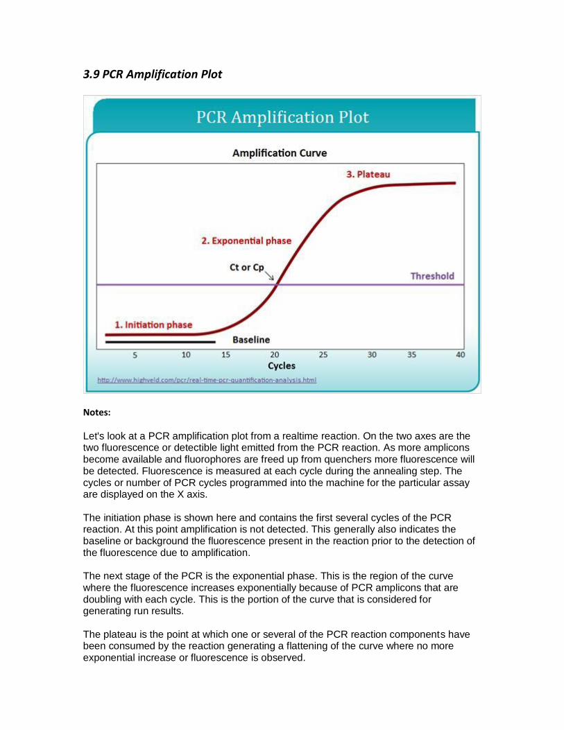
3.9 PCR Amplification Plot
Notes:
Let's look at a PCR amplification plot from a realtime reaction. On the two axes are the two fluorescence or detectible light emitted from the PCR reaction. As more amplicons become available and fluorophores are freed up from quenchers more fluorescence will be detected. Fluorescence is measured at each cycle during the annealing step. The cycles or number of PCR cycles programmed into the machine for the particular assay are displayed on the X axis.
The initiation phase is shown here and contains the first several cycles of the PCR reaction. At this point amplification is not detected. This generally also indicates the baseline or background the fluorescence present in the reaction prior to the detection of the fluorescence due to amplification.
The next stage of the PCR is the exponential phase. This is the region of the curve where the fluorescence increases exponentially because of PCR amplicons that are doubling with each cycle. This is the portion of the curve that is considered for generating run results.
The plateau is the point at which one or several of the PCR reaction components have been consumed by the reaction generating a flattening of the curve where no more exponential increase or fluorescence is observed.

The threshold is the line that crosses the curve of a PCR run at the exponential phase. To set the threshold look for the region of the PCR curve where amplification is exponential and set the threshold just above the background. The threshold should always be placed in the exponential phase. If the assay calls for a pre- determined threshold setting check that its placement is in the exponential phase of the curve and above the background.
The CT or CP is the cycle threshold or crossing point. This is the cycle of the PCR reaction at which the threshold line crosses the PCR curve. Results from PCR runs are communicated with this number. As in the above picture, the results from the PCR run would be at CT 20 or CP 20. This allows for results to be comparable from run to run.

3.10 Nucleic Acid Sequencing
Notes:
Another application of molecular biology is nucleic acid sequencing. This is the process for determining the specific order of nucleotides within a DNA molecule. There are several methods for DNA sequences including Sanger sequencing, pyrosequencing and next generation sequencing.

3.11 Sanger Sequencing
Notes:
Sanger sequencing takes advantage of the use of dideoxynucleotides. In the image you can see that a standard deoxynucleotide has a hydroxyl group on this carbon here, whereas the dideoxynucleotide is lacking that oxygen and only has a hydrogen. This makes the nucleotide a chain terminator in that when a dideoxynucleotide is incorporated into a DNA strand no additional bases can be added after that base.

3.12 Sanger Sequencing
Notes:
So the dideoxynucleotides are also labeled with a fluorescent dye. Each base A, C, T and G all have different colored dyes. Much like PCR you put into a reaction together a DNA template, a primer, native dNTPs as well as dideoxynucleotides and a polymerase. Through many cycles of a PCR many new strands of DNA will be created. Some will have incorporated one or two or three or many native dNTPs and randomly the dideoxynucleotides will be incorporated. When the dideoxynucleotide are incorporated the DNA strand is no longer able to be elongated. These new strands that are created are also unable to serve as templates strands for the next round of PCR. The final mixture following the PCR cycles will contain many strands of DNA each ending with a fluorescently labeled dideoxynucleotide. It is not an orderly process like the image portrays but you should end up with every base of the DNA sequence being labeled with a chain terminator.

3.13 Sanger Sequencing: Detection
Notes:
After you have performed your cycle sequencing or labeling reaction the samples are then separated by capillary electrophoresis according to their size. Fragments pass in front of a laser which excites the fluorescent dye on each DNA fragment. Depending on the wavelength of light emitted, data analysis software determines the identity of the dideoxynucleotide at the end of each DNA fragment. The length of the DNA fragment in combination with this specific fluorescence detected reveals the sequence of a DNA fragment. As those bases are detected computer software will interpret the fluorescence and generate a chromatogram that shows in a graphical representation the DNA sequence of your DNA of interest.

3.14 Pyrosequencing
Notes:
Pyrosequencing is another method of sequencing DNA. It is sometimes referred to as sequencing by synthesis because you obtain the DNA sequence as the new DNA molecule is created. In pyrosequencing you have your target DNA as a single stranded molecule, a sequencing primer, a polymerase as well as additional enzymes. In a one at a time fashion nucleotides are added to the pyrosequencing reaction in a pre-determined order. If the nucleotide is incorporated into the growing DNA strand pyrophosphate is given off and converted into a light signal. This light data is captured by the instrument and recorded. Any remaining nucleotides are degraded and the next nucleotide is added to the reaction. The light signal data is presented in the form of a pyrogram as shown here. You can see that there is a peak of light for the G nucleotide but nothing for the C or the A. The T incorporated so there is a peak but not for the G or the C. The A has a peak that is about twice the height of the preceding G and T incorporations so it demonstrates that two A bases were being incorporated into the strand.

3.15 Sanger vs. Pyrosequencing
Notes:
Sanger sequencing is generally better for longer sequence reads such as approximately 500 base pairs. It's very easy to customize and it does have the ability to detect mixed sequences if their present in the sample at approximately 30 percent or more. The actual DNA sequence is determined and the output the visualized by a chromatogram or the data analyze software will analyze and give you a DNA sequence. Pyrosequencing on the other hand is much better for short sequence reads. It cannot sequence large segments of DNA. The dispensational order of the nucleotides is customizable and the through-put may be greater than for Sanger sequencing. It does have a limited ability to detect mixed sequences but again the actual DNA sequence is determined and the output is visualized by a pyrogram with built-in software to analyze.

3.16 Next Generation Sequencing (NGS)
Notes:
Next generation sequencing is a collection of techniques and technologies for high through-put sequencing also known as massively parallel sequencing. It includes sequencing of genomes as well as targeted sequencing. There are currently several different technologies and instruments available for this process. The results are large, complex amounts of data and they require intense bioinformatic analysis. Data storage can also be problematic with nextgen sequencing.

3.17 NGS Instrumentation
Notes:
Here are three common next generation sequencing instruments the Illumina MiSeq the PacBio RS and the Ion Torrent Personal Genome Machine or PGM. Now let's talk a little bit about how they work.

3.18 NGS Methodology
Notes:
In general, next generation sequencing is a method in which you take genomic DNA or cDNA and fragment it into smaller pieces. Adapters are added to those fragments that allow the next steps of the process to find your DNA fragments. Then specific to which technology you have your fragments are amplified. For the Ion Torrent PGM instrument your small adapter ligated fragments are put through an emulsion PCR in little oil microspheres or if you have the Illumina MiSeq then clusters are generated using a bridge PCR technique.
The next step is the sequencing of all of those amplified fragments. On the Ion Torrent PGM this occurs on a small silicon semi-conductor chip. Your fragments are dispersed across millions of wells on the chip and the nucleotides are added one at a time. If they are incorporated the pH of the well changes and that signal is recorded. If they are not incorporated then they are washed away and the next base is applied for as many bases as are needed to sequence the entire fragment length you've created. On the Illumina instrument the sequencing occurs in a very similar way to Sanger sequencing with the capture of fluorescence data as bases are incorporated. All of the bases then have to be realigned to the full sequence rather than these small fragments. This can be done against a reference genome as a scaffold for where the sequences go or it can be done in the absence of a reference genome called de novo alignment.

3.19 Applications of NGS
Notes:
Nextgen sequencing provides a high through-put alternative for sequencing. Its applications include basic science as well as clinical diagnostics. It can be used for molecular typing such as pathogen identification and characterization, detection of mutations associated with drug resistance and identification of novel mechanism of resistance. It can also be used for molecular epidemiology in genotyping such as outbreak detection and surveillance.

3.20 Line Probe Assay
Notes:
Another application of molecular biology relating to TB is the line probe assay. In a line probe assay DNA is extracted from the patient specimen or culture isolate then you use PCR to amplify DNA. The genes you amplify depend on the strip you are using but it is usually the 16S genes for identification and rpoB gene for resistance information. The PCR products are hybridized to probes on the strip and are then labeled and detected using a color matrix assay. Bands are compared to standard patterns. The test amplicon anneals to any oligos for which it is an exact match. If the test amplicon differs from the strip oligo it should not anneal. A mutant amplicon would not anneal to a wild type oligo and vice versa. Absence of a reaction would indicate a difference between the oligo and the amplicon. Using line probe assays you can get identification information and resistance information in one test. However, it cannot detect unknown or novel mutations.

3.21 MALDI-TOF MS
Notes:
Another application of molecular biology is MALDI-TOF or matrix assisted laser desorption ionization time of flight mass spectrometry. This technology can use protein composition to identify bacterial species. In the mycobacteria lab it is mostly used for identification of non-tubercular mycobacteria. It cannot yet reliably differentiate between species within the MTB complex.

3.22 MALDI-TOF MS (2)
Notes:
In the clinical lab a MALDI-TOF instrument is used for identification of bacteria and fungi. The isolate is applied to a chip or a slide and a matrix is added then the slider chip is placed in the instrument and a laser is fired at the organism. The energy from the laser is absorbed and the sample is ionized. The positive ions accelerate through a vacuum toward the detector. Their speed or time of flight is proportional to the ion's mass so smaller ions reach the detector first and the larger ions take longer.
This data is collected and interpreted as a spectrograph. The spectrograph for the organism is then compared to a database and identification can be made based on matching spectrographs.

4. Workflow, Contamination, Safety, and Regulation
Notes:
Next we will cover some general information regarding work flow, contamination, safety and regulatory considerations.

4.2 What is PCR Contamination?
Notes:
So what is PCR contamination? Contamination of a PCR is the unintentional presence of a nucleic acid target. Because PCR is so sensitive, it is possible to detect even a single copy of a contaminant which compromises the results of all of your reactions in a run. You can have contamination from one sample to another or carry over from a previous PCR run of the same target where amplified DNA gets into your new reactions.

4.3 How to avoid contamination—Unidirectional workflow
Notes:
Some work flow considerations for implementing molecular testing. You need distinct areas or rooms for each step in the process including reagent preparation, specimen preparation or nucleic acid extraction, amplification and product analysis. You must have a uni-directional work flow working from clean to dirty areas and never in the opposite direction. Clean areas are those areas that do not have nucleic acids either extracted or amplified in them such as your master mix room where only master mix components are stored and used. Dirty areas are those areas where nucleic acids are used, amplified products are analyzed and where potential contaminants are housed. You want to minimize the risk of amplicon cross-contamination and have dedicated equipment including refrigerators, freezers, pipettes, aerosol resistant tips, lab coats, pens and notebooks in each area and gloves must be worn and changed often.

4.4 Workflow Through Laboratory Rooms
Notes:
This slide shows a work flow diagram through the laboratory rooms where you would start out in a reagent preparation area and move to sample preparation but never in the reverse direction. From sample preparation you could then move to amplification and product handling rooms but never in the reverse direction. And you should never go from the amplification and product area back to reagent preparation. In an ideal laboratory you would have separate rooms for each of these steps. While that is not always possible good molecular biology work can be done even in a single space by designating clean and dirt areas, by separating where the steps are performed and by observing uni-directional work flow at all times.

4.5 Other Potential Sources of Contamination
Notes:
There are many potential sources of contamination. You can have template or amplicon transfer from aerosol generation, tube breakage, inadequate plate sealing, inadequate cleaning procedures or pipette and reagent contamination. You can also get contamination through noncompliance with uni-directional work flow such as movement of individuals from dirty to clean areas or transfer of reagents and materials from dirty to clean areas.

4.6 Detection of Contamination
Notes:
One way to constantly monitor for contamination is to include a no template control or NTC in every PCR run you perform. This is a reaction that contains all of the PCR components except for the DNA template. This reaction should always be negative. If you detect amplification in your NTC you likely have a contamination issue that you need to investigate. Some things to consider when trying to determine the source of contamination are reagents and equipment. Performing environmental testing of the areas where you work can help determine where the DNA exists in the lab spaces. You can clean suspect areas with ten percent bleach to degrade the DNA and any corrosion print surfaces should then be cleaned with ethanol. You may need to change some of your institutional practices to reduce contamination events and you must not report out results from a run with a positive NTC. Once the contamination has been found and cleaned up you must repeat your assay.
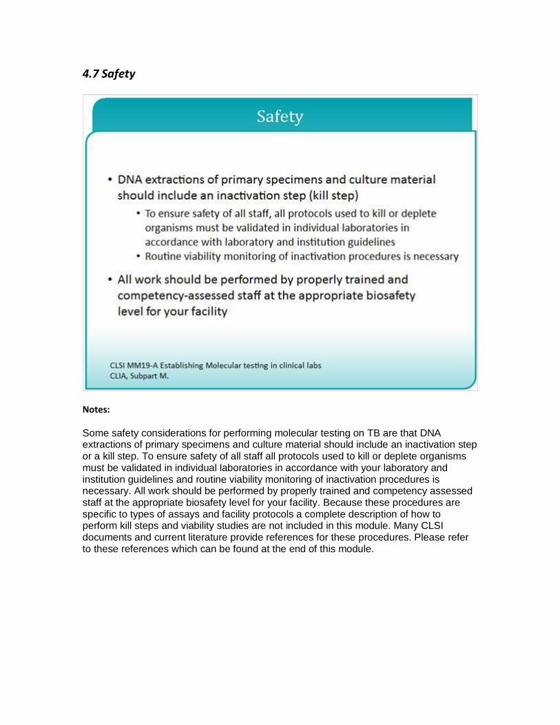
4.7 Safety
Notes:
Some safety considerations for performing molecular testing on TB are that DNA extractions of primary specimens and culture material should include an inactivation step or a kill step. To ensure safety of all staff all protocols used to kill or deplete organisms must be validated in individual laboratories in accordance with your laboratory and institution guidelines and routine viability monitoring of inactivation procedures is necessary. All work should be performed by properly trained and competency assessed staff at the appropriate biosafety level for your facility. Because these procedures are specific to types of assays and facility protocols a complete description of how to perform kill steps and viability studies are not included in this module. Many CLSI documents and current literature provide references for these procedures. Please refer to these references which can be found at the end of this module.

4.8 Considerations for Molecular Testing
Notes:
In addition, you should confer with your institution's compliance director or quality assurance officer before bringing on new technologies. All newly implemented assays require some level of validation even those that are FDA approved. Consult the CLIA and FDA websites for detailed information. Please see the references and is resources at the end of this module as well.

4.9 Regulatory Classification of Assays
Notes:
All molecular testing in the Mycobacteriology laboratory falls under FDA and CLIA regulation. CLIA regulates laboratories that perform testing on patient specimens in order to ensure accurate and reliable test results. FDA regulates manufacturers and devices to ensure devices intended for use in the diagnosis of disease are reasonably safe and effective. Here are some molecular assays for TB that are cleared, approved or authorized by FDA.
The Hologic Amplified MTD for Detection of mycobacterium in tuberculosis complex, the Cepheid GeneXpert MTB/RIF for detection of mycobacterium tuberculosis complex and mutations associated with rifampin resistance and the Hologic Accuprobe for ID of Mycobacterium tuberculosis complex from culture. Use of laboratory developed tests or assays labeled as research use only or investigational use only require proper validation and compliance with the regulations of your accrediting body such as CLIA or CAP.

4.10 Why the Increased Interest in Molecular Diagnostics for TB?
Notes:
There are several reasons for the increased interest in molecular diagnostics for TB. Molecular diagnostics allow for rapid detection of mycobacterium tuberculosis complex and mutations associated with drug resistance. Some assays even allow for simultaneous detection. Molecular diagnostics can be performed at a lower cost on a variety of platforms and they also allow for rapid outbreak detection.

4.11 Use of Molecular Assays in the TB Laboratory
Notes:
Molecular assays are used in the TB lab for direct detection of mycobacterium tuberculosis complex, by nucleic acid amplification testing in clinical specimens and isolates, identification of mycobacteria and detection of drug mutations. These will be covered in detection and identification of mycobacteria and the molecular detection of drug resistance modules.

4.12 CLIA Program
Regulatory Information Resources
Notes:
The new few slides include references and resources associated with this module and can be found on the APHL website.

4.13 FDA Regulatory Resources

4.14 CLSI References

4.15 Conclusion
Notes:
This concludes the Molecular Biology 101 presentation which is part of a series from the APHL Essentials for Mycobacteriology Laboratory, Promoting Quality Practices. Please see the CDC and APHL website for more information on these topics.