modulation of the antigenic peptide transporter tap by ... · modulation of the antigenic peptide...
TRANSCRIPT

doi:10.1016/j.jmb.2007.02.102 J. Mol. Biol. (2007) 369, 95–107
Modulation of the Antigenic Peptide Transporter TAP byRecombinant Antibodies Binding to the Last FiveResidues of TAP1
Gabriele Plewnia1, Katrin Schulze1, Carola Hunte2, Robert Tampé1
and Joachim Koch1⁎
1Institute of Biochemistry,Biocenter, Johann WolfgangGoethe-University,Max-von-Laue-Strasse 9,D-69438 Frankfurt a. M.,Germany2Max-Planck-Instituteof Biophysics,Max-von-Laue-Strasse 3,D-60438 Frankfurt a. M.,GermanyAbbreviations used: ABC, ATP-bicomplementarity determining regionreticulum; ICP, infected cell protein;antibody; MHC, major histocompatnucleotide-binding domain; PLC, pecomplex; TAP, transporter associatedprocessing; TBS-T, Tris-buffered saliTMD, transmembrane domain; US,E-mail address of the correspondi
0022-2836/$ - see front matter © 2007 E
The transporter associatedwith antigen processing (TAP) plays a pivotal rolein the major histocompatibility complex (MHC) class I mediated immuneresponse against infected or malignantly transformed cells. It belongs to theATP-binding cassette (ABC) superfamily and consists of TAP1 (ABCB2) andTAP2 (ABCB3), each of which possesses a transmembrane and a nucleotide-binding domain (NBD). Here we describe the generation of recombinant Fvand Fab antibody fragments to human TAP from a hybridoma cell lineexpressing the TAP1-specific monoclonal antibody mAb148.3. The epitopeof the antibody was mapped to the very last five C-terminal amino acidresidues of TAP1 on solid-supported peptide arrays. The recombinantantibody fragments were heterologously expressed in Escherichia coli andpurified to homogeneity from periplasmic extracts by affinity chromato-graphy. The monoclonal and recombinant antibodies bind with nanomolaraffinity to the last five C-terminal amino acid residues of TAP1 asdemonstrated by ELISA and surface plasmon resonance. Strikingly, therecombinant antibody fragments confer thermal stability to the hetero-dimeric TAP complex. At the same time TAP is arrested in a peptidetransport incompetent conformation, although ATP and peptide binding toTAP are not affected. Based on our results we suggest that the C terminus ofTAP1 modulates TAP function presumably as part of the dimer interface ofthe NBDs.
© 2007 Elsevier Ltd. All rights reserved.
Keywords: ABC transporter; adaptive immune system; antigen processing;epitope mapping; SPOT
*Corresponding authorIntroduction
Major histocompatibility complex (MHC) class Imolecules present endogenous peptides on thesurface of nucleated cells to cytotoxic T-lymphocytes(CD8+), which scan these peptide-MHC I complexesto eliminate infected or malignantly transformed
nding cassette; CDR,; ER, endoplasmicmAb, monoclonalibility complex; NBD,ptide-loadingwith antigen
ne with Tween 20;unique short.ng author:
lsevier Ltd. All rights reserve
cells.1,2 The majority of antigenic peptides arederived from proteolytic processing of poly-ubiqui-tylated proteins by the proteasome complex in thecytoplasm. These peptides are translocated into theendoplasmic reticulum (ER) via the transporterassociated with antigen processing (TAP) and aresubsequently loaded onto MHC I molecules.3,4 Thisprocess requires a sophisticated macromolecularmachinery, the peptide-loading complex (PLC).5–9
The TAP-dependent peptide supply is crucial in theassembly and quality control of peptide-MHC Icomplexes, since only peptide-loaded MHC Imolecules can leave the ER for transport to the cellsurface.10–12
DNA viruses have evolved manifold strategies toescape the host's immune response leading topersistence and repeated reactivation. The PLCrepresents a major target for viral effector pro-teins.13,14 Two viral inhibitors of human TAP have
d.

96 Antibodies Modulating TAP Function
been characterized in detail. ICP47 of herpes sim-plex virus 1 (HSV-1) acts as a high-affinity inhibitorin the cytosol by blocking peptide binding toTAP.15,16 By contrast, US6 of human cytomegalo-virus (HCMV) binds to TAP in the ER and inhibitspeptide translocation by blocking ATP but notpeptide binding.17–21 Both proteins proved usefulto investigate TAP function in vivo and in vitro.Beside these fascinating strategies of viral evasion,
synthetic or protein engineered inhibitors have notbeen described so far. Antibodies, which recognizediscrete sites within TAP are promising tools to shedlight on the functional and structural dynamics ofthe transport complex. One of the candidates, thehuman TAP1-specific monoclonal antibody (mAb)148.3, has been used for indirect immunofluores-cence, immunoblotting, co-immunoprecipitationand purification of the heterodimeric TAP-complex.Moreover, the mAb148.3 allowed for the identifica-tion and isolation of the macromolecular peptide-loading complex composed of TAP1, TAP2, tapasin,MHC class I heavy chain, β2-microglobulin, calreti-culin, and ERp57.5,7,9Since impaired TAP expression is directly linked to
down-regulation of MHC class I antigen presenta-tion and immunevasion of many tumors the TAPheterodimer represents an important diagnostic tar-get in tumorigenesis.14,22,23 However, to date, theproduction of TAP-specific recombinant antibodiessuch as Fv and Fab fragments has not been reportedalthough recombinant antibodies are superior to full-length antibodies in a variety of applications (forrecent review see Schaedel & Reiter24).Here, we generated for the first time recombinant
antibody fragments (Fv and Fab) to TAP. Therecombinant antibody fragments were produced inEscherichia coli and purified from periplasmicextracts by affinity chromatography. The epitopeof the antibodies was mapped to the last fiveC-terminal amino acid residues of TAP1 and theiraffinity binding constants were determined. Strik-ingly, the recombinant antibodies inhibit the TAP-specific peptide transport in semi-permeabilizedcells and at the same time confer thermal stabilityto the TAP complex. These antibodies are thereforeimportant tools to investigate the functional role ofthe C terminus of TAP1 and provide the possibilityto modulate the activity of the TAP complex.Furthermore, the variable heavy (VH) and variablelight (VL) chains are now accessible for geneticengineering, thus allowing for functionalizationand modulation of their binding properties, desir-able features for therapy and diagnostic imaging.
Results
The mAb148.3 binds to the very last fiveC-terminal amino acid residues of human TAP1
So far, only three monoclonal antibodies to humanTAP have been generated.25,26 The mAb148.3, whichis specific for human TAP1, is an important diag-
nostic tool to investigate the antigen processingmachinery in tumor development and viral escapemechanisms.21,23 Moreover, the mAb148.3 has led tothe identification and subunit mapping of themacromolecular peptide-loading complex in theER membrane;7,9 however, up to date its biochem-ical properties have not been addressed.Here we have determined by immunological sub-
typing that the mAb148.3 belongs to the immuno-globulin G1 (IgG1) κ-chain subtype (data notshown). To map the epitope of the mAb148.3 wehave used peptide arrays of overlapping oligopep-tides (15mers off-set by three amino acid residues)covering the entire sequence of human TAP1 andTAP2 (Figure 1). In accordance with the fact that aC-terminal peptide of TAP1 was used for immuni-zation of the mice to establish a hybridoma, theepitope of the mAb148.3 is located within the last 15C-terminal amino acid residues of human TAP1(Figure 1). However, most importantly no otherpeptide derived of TAP1 or TAP2 is recognized. Thesame results were obtained in mapping experimentson peptide arrays with one amino acid off-set (datanot shown). To determine the precise epitope andcritical residues for epitope recognition, system-atically permutated variants and arrays of N andC-terminal truncations of the identified peptidewereprobed with hybridoma supernatant (Figure 2). Alinear epitope was identified corresponding to thevery last five C-terminal amino acid residues of TAP1(744ADAPE748) (Figure 2(b) and (c)). Here, thebinding efficiency was fully preserved when com-pared to the 15mer peptide. Further mutationalanalyses demonstrate that the residues D745 andE748 are mandatory for recognition, as they cannotbe replaced by alanine, whereas A744 has less impact(Figure 2(a)). By constrast, exchange of A746 andP747 did not affect epitope recognition (Figure 2(a)).Surprisingly, binding of the mAb148.3 is not depen-dent on a free carboxyl group at the C terminus, sincethe peptides were linked via a peptide bond to thePEG-spacer on the cellulose membrane. Notably,three independently synthesized peptides corre-sponding to the last 15 amino acid residues of TAP1(Figure 2, always first spot on the peptide arrays)showabout the same signal intensitywhen incubatedwith equal amounts of mAb148.3, demonstratingquantitative reproducibility of the assay. In sum-mary, the epitope of the mAb148.3 recognizesspecifically the very last five C-terminal amino acidresidues ADAPE (essential amino acids underlined)of human TAP1.
Cloning, expression, and purification ofrecombinant antibody fragments ofthe mAb148.3
To convert the TAP1-specific mAb148.3 into Fvand Fab formats, cDNA was prepared from hybri-doma cells and the VH and VL chain genes werePCR-amplified using a set of degenerate primers.After sequencing, the complementarity determiningregions (CDR) and framework regions (FR) of the

Figure 1. Epitope mapping of the monoclonal antibody mAb148.3. (a) Schematic representation of the TAPheterodimer. The transmembrane domains (TMDs) of TAP1 and TAP2 and the nucleotide-binding domains (NBDs) areshown in dark and light grey, respectively. The TMDs are subdivided into a core-domain, which was recently shown tocomprise the peptide translocation pore and N-terminal domains, which recruit the adapter protein tapasin.6,53 (b) Arraysof overlapping oligopeptides (15mers, off-set three amino acids) of human TAP1 and TAP2 were generated by Fmocchemistry on PEG-functionalized cellulose membranes and probed with hybridoma supernatant (mAb148.3). Boundantibodies were detected with a goat anti-mouse HRP-conjugated antibody and visualized by chemiluminescenceimaging. The areas on the membrane that correspond to the TMDs and the NBDs of TAP are shown in grey and as openboxes, respectively.
97Antibodies Modulating TAP Function
antibody chains were assigned as described byKabat et al.27 (Figure 3). The genes of the identifiedVH and VL chains were subsequently cloned into theexpression vectors pASK68 and pASK85 for peri-plasmic production of Fv and Fab fragments,respectively. Recombinant Fv and Fab fragmentswere produced at different levels in E. coli (Figure 4).Both antibody fragments were detected after 1 h ofinduction as shown by SDS-PAGE and immunoblot-ting with tag-specific antibodies (Figure 4). Therecombinant antibody fragments were initiallypurified via the C-terminal Strep-tag of the Fv-VHchain and the C-terminal His6-tag of the Fab-VHCH1chain. However, the Fv fragment displayed atendency to dimerize, leading to reduced affinityto its epitope (data not shown). Therefore, therecombinant antibody fragments were purified byaffinity chromatography employing the peptideCYWAMVQAPADAPE (epitope underlined). Here,the Fv fragment was purified to homogeneity in asingle affinity chromatography step as shown bySDS-PAGE (silver-stained) and immunoblottingusing a Streptactin-HRP conjugate (Figure 5(a)).By contrast, the purity as well as the total amount
of purified Fab fragment was considerably lowerthan for the Fv fragment (Figure 5(b)). The Fabfragment was however clearly detectable byimmunoblotting using an Fab-specific antibody(Figure 5(b)). Heterodimer assembly of the purifiedFab fragment was confirmed by SDS-PAGE undernon-reducing conditions. Here, the Fab fragmenthas an apparent molecular mass of 50 kDa due tothe intermolecular disulfide bridge at the C terminiof the constant regions (between CH1 and CL) (datanot shown). The yields of purified Fv and Fabfragments were 100 μg and 20 μg per liter of E. coliculture, respectively. Notably, comparable amountsof the Fab fragment were purified by IMAC.Correct folding and monodispersity of the purifiedFv fragment was demonstrated by analytical sizeexclusion chromatography (Figure 5(c)). The Fvfragment appears as a single peak with a molecularmass of 28 kDa. A proteolytic Fab fragment(50 kDa), which was generated by papain digestionof mAb148.3, eluted shortly before the Fv fragmentand was used as an internal standard (Figure 5(c)).Notably, the proteolytic Fab fragment appears as amonomeric peak with an additional sub-fraction
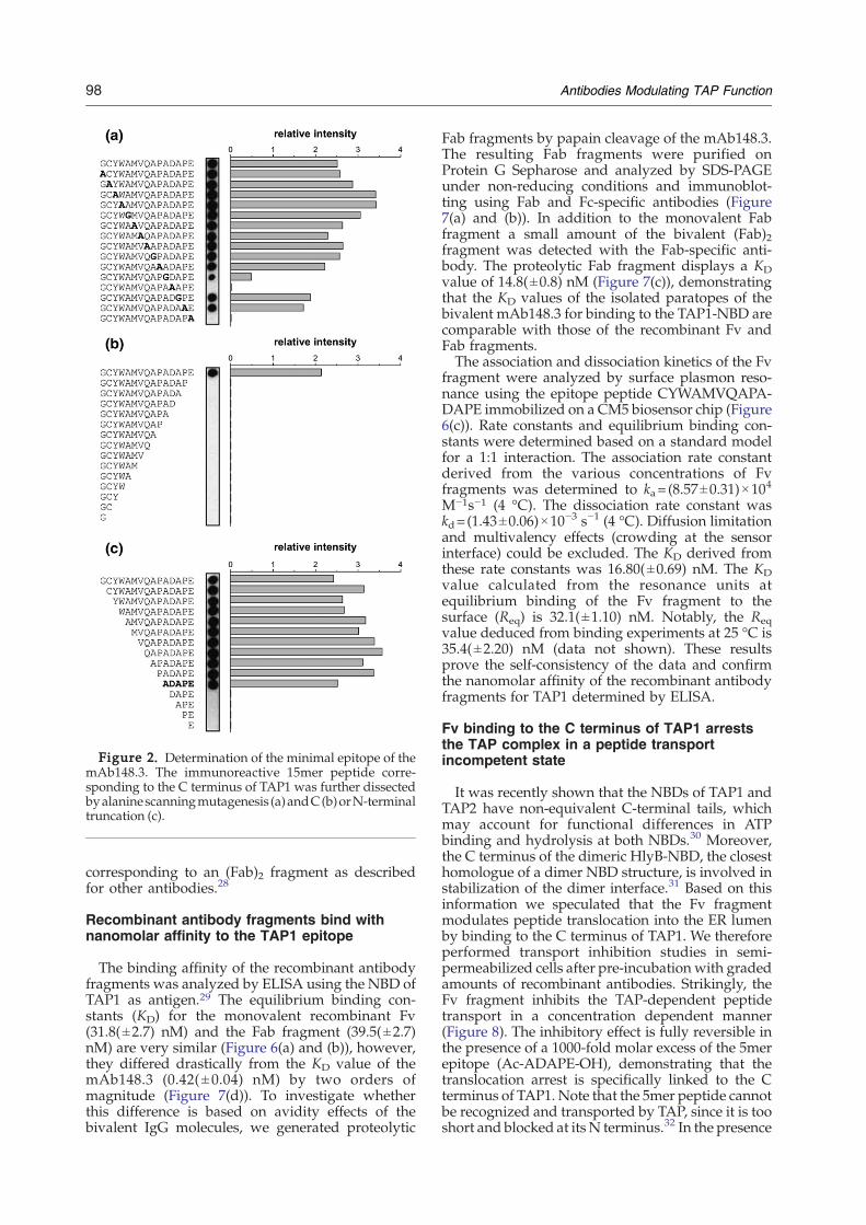
Figure 2. Determination of the minimal epitope of themAb148.3. The immunoreactive 15mer peptide corre-sponding to the C terminus of TAP1 was further dissectedbyalaninescanningmutagenesis (a)andC(b)orN-terminaltruncation (c).
98 Antibodies Modulating TAP Function
corresponding to an (Fab)2 fragment as describedfor other antibodies.28
Recombinant antibody fragments bind withnanomolar affinity to the TAP1 epitope
The binding affinity of the recombinant antibodyfragments was analyzed by ELISA using the NBD ofTAP1 as antigen.29 The equilibrium binding con-stants (KD) for the monovalent recombinant Fv(31.8(±2.7) nM) and the Fab fragment (39.5(±2.7)nM) are very similar (Figure 6(a) and (b)), however,they differed drastically from the KD value of themAb148.3 (0.42( ± 0.04) nM) by two orders ofmagnitude (Figure 7(d)). To investigate whetherthis difference is based on avidity effects of thebivalent IgG molecules, we generated proteolytic
Fab fragments by papain cleavage of the mAb148.3.The resulting Fab fragments were purified onProtein G Sepharose and analyzed by SDS-PAGEunder non-reducing conditions and immunoblot-ting using Fab and Fc-specific antibodies (Figure7(a) and (b)). In addition to the monovalent Fabfragment a small amount of the bivalent (Fab)2fragment was detected with the Fab-specific anti-body. The proteolytic Fab fragment displays a KDvalue of 14.8(±0.8) nM (Figure 7(c)), demonstratingthat the KD values of the isolated paratopes of thebivalent mAb148.3 for binding to the TAP1-NBD arecomparable with those of the recombinant Fv andFab fragments.The association and dissociation kinetics of the Fv
fragment were analyzed by surface plasmon reso-nance using the epitope peptide CYWAMVQAPA-DAPE immobilized on a CM5 biosensor chip (Figure6(c)). Rate constants and equilibrium binding con-stants were determined based on a standard modelfor a 1:1 interaction. The association rate constantderived from the various concentrations of Fvfragments was determined to ka= (8.57±0.31)×10
4
M−1s−1 (4 °C). The dissociation rate constant waskd=(1.43±0.06)×10
−3 s−1 (4 °C). Diffusion limitationand multivalency effects (crowding at the sensorinterface) could be excluded. The KD derived fromthese rate constants was 16.80(±0.69) nM. The KDvalue calculated from the resonance units atequilibrium binding of the Fv fragment to thesurface (Req) is 32.1(±1.10) nM. Notably, the Reqvalue deduced from binding experiments at 25 °C is35.4(±2.20) nM (data not shown). These resultsprove the self-consistency of the data and confirmthe nanomolar affinity of the recombinant antibodyfragments for TAP1 determined by ELISA.
Fv binding to the C terminus of TAP1 arreststhe TAP complex in a peptide transportincompetent state
It was recently shown that the NBDs of TAP1 andTAP2 have non-equivalent C-terminal tails, whichmay account for functional differences in ATPbinding and hydrolysis at both NBDs.30 Moreover,the C terminus of the dimeric HlyB-NBD, the closesthomologue of a dimer NBD structure, is involved instabilization of the dimer interface.31 Based on thisinformation we speculated that the Fv fragmentmodulates peptide translocation into the ER lumenby binding to the C terminus of TAP1. We thereforeperformed transport inhibition studies in semi-permeabilized cells after pre-incubation with gradedamounts of recombinant antibodies. Strikingly, theFv fragment inhibits the TAP-dependent peptidetransport in a concentration dependent manner(Figure 8). The inhibitory effect is fully reversible inthe presence of a 1000-fold molar excess of the 5merepitope (Ac-ADAPE-OH), demonstrating that thetranslocation arrest is specifically linked to the Cterminus of TAP1. Note that the 5mer peptide cannotbe recognized and transported by TAP, since it is tooshort and blocked at its N terminus.32 In the presence
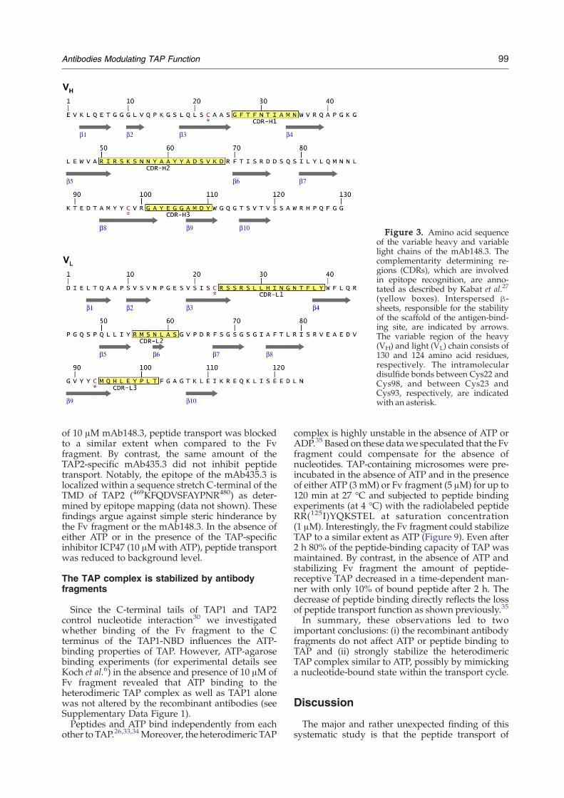
Figure 3. Amino acid sequenceof the variable heavy and variablelight chains of the mAb148.3. Thecomplementarity determining re-gions (CDRs), which are involvedin epitope recognition, are anno-tated as described by Kabat et al.27
(yellow boxes). Interspersed β-sheets, responsible for the stabilityof the scaffold of the antigen-bind-ing site, are indicated by arrows.The variable region of the heavy(VH) and light (VL) chain consists of130 and 124 amino acid residues,respectively. The intramoleculardisulfide bonds between Cys22 andCys98, and between Cys23 andCys93, respectively, are indicatedwith an asterisk.
99Antibodies Modulating TAP Function
of 10 μM mAb148.3, peptide transport was blockedto a similar extent when compared to the Fvfragment. By contrast, the same amount of theTAP2-specific mAb435.3 did not inhibit peptidetransport. Notably, the epitope of the mAb435.3 islocalized within a sequence stretch C-terminal of theTMD of TAP2 (469KFQDVSFAYPNR480) as deter-mined by epitope mapping (data not shown). Thesefindings argue against simple steric hinderance bythe Fv fragment or the mAb148.3. In the absence ofeither ATP or in the presence of the TAP-specificinhibitor ICP47 (10 μMwith ATP), peptide transportwas reduced to background level.
The TAP complex is stabilized by antibodyfragments
Since the C-terminal tails of TAP1 and TAP2control nucleotide interaction30 we investigatedwhether binding of the Fv fragment to the Cterminus of the TAP1-NBD influences the ATP-binding properties of TAP. However, ATP-agarosebinding experiments (for experimental details seeKoch et al.6) in the absence and presence of 10 μM ofFv fragment revealed that ATP binding to theheterodimeric TAP complex as well as TAP1 alonewas not altered by the recombinant antibodies (seeSupplementary Data Figure 1).Peptides and ATP bind independently from each
other to TAP.26,33,34 Moreover, the heterodimeric TAP
complex is highly unstable in the absence of ATP orADP.35 Based on these datawe speculated that the Fvfragment could compensate for the absence ofnucleotides. TAP-containing microsomes were pre-incubated in the absence of ATP and in the presenceof either ATP (3 mM) or Fv fragment (5 μM) for up to120 min at 27 °C and subjected to peptide bindingexperiments (at 4 °C) with the radiolabeled peptideRR(125I)YQKSTEL at saturation concentration(1 μM). Interestingly, the Fv fragment could stabilizeTAP to a similar extent as ATP (Figure 9). Even after2 h 80% of the peptide-binding capacity of TAP wasmaintained. By contrast, in the absence of ATP andstabilizing Fv fragment the amount of peptide-receptive TAP decreased in a time-dependent man-ner with only 10% of bound peptide after 2 h. Thedecrease of peptide binding directly reflects the lossof peptide transport function as shown previously.35
In summary, these observations led to twoimportant conclusions: (i) the recombinant antibodyfragments do not affect ATP or peptide binding toTAP and (ii) strongly stabilize the heterodimericTAP complex similar to ATP, possibly by mimickinga nucleotide-bound state within the transport cycle.
Discussion
The major and rather unexpected finding of thissystematic study is that the peptide transport of

Figure 4. Expression of recombinant antibodies. E. coli were transformed with expression vectors pASK68 (Fv) andpASK85 (Fab). Expression was induced with IPTG (pASK68) or AHT (pASK85) for 3 h. Aliquots of the bacterial celllysates (0 h, 1 h, 2 h, 3 h), the supernatant after harvesting (sn), the periplasmic fraction (pp, 1/25 aliquot), and thecytoplasmic fraction (cp) were analyzed by reducing Tricine-SDS-PAGE ((a), 16%, expression of Fv; (d), 12%, expression ofFab). The VH (b) and VL (c) chains of the Fv fragment were detected with tag-specific antibodies at 14.4 kDa and 13.8 kDa,respectively. (e) Heavy chain (VH-CH1) of the Fab fragment at 25 kDa. Molecular mass standard in kDa.
100 Antibodies Modulating TAP Function
human TAP can be inhibited by antibodies thatspecifically recognize a discrete linear epitopecomprised of the very last five C-terminal aminoacid residues of TAP1 (744ADAPE748). In addition,these antibodies strongly stabilize the heterodimericTAP complex against rapid thermal inactivation inthe absence of ATP. Since no other region within TAPwas recognized by the antibodies both effects areexclusively linked to the C terminus of TAP1.Here, we generated recombinant Fv and Fab
fragments by genetic engineering from hybridomacells expressing the mAb148.3. Recombinant anti-body fragments were heterologously expressed inE. coli and secreted into the periplasm. Both, the Fvand Fab fragments were purified to homogeneityandmonodispersity as demonstrated by SDS-PAGE,immunoblotting and size exclusion chromatogra-phy. Based on kinetic and thermodynamic analysesthe epitope–paratope interaction was characterizedin detail and compared with the parental antibodydemonstrating nanomolar affinity of the monova-lent antibody fragments for TAP1. The equilibrium
binding constants of the recombinant antibodies aswell as of the mAb148.3 were determined by ELISAand surface plasmon resonance using the isolatedNBD and the C-terminal epitope peptide CYWAMV-QAPADAPE of TAP1, respectively, as antigen. TheKD values for the monovalent recombinant Fv andthe Fab fragments are very similar. Moreover, the KDof the monovalent proteolytic Fab fragment of themAb148.3 for the NBD of TAP1 is in the same rangeas for the recombinant antibody fragments.Surprisingly, the recombinant Fv fragment inhi-
bits TAP-dependent peptide translocation into theER similar to viral factors such as ICP47 of herpessimplex virus or US6 of human cytomegalovirus.Transport inhibition of the monovalent Fv fragmentand the intact mAb148.3 was equally efficient. Thisobservation demonstrates that inhibition resultsfrom a direct interaction of the paratopes with theC terminus of TAP1 and not from steric effectscaused by the Fc regions of the intact antibodies orcross-linking of adjacent TAP heterodimers becauseof bivalent interactions of the mAb148.3.

Figure 5. Affinity purification of recombinant antibody fragments. Fv and Fab fragments were affinity-purified via theepitope peptide CYWAMVQAPADAPE of TAP1. Aliquots of the periplasmic fraction (pp), the flow through (ft), thewashing step (w), and the elution fractions (1–5) were analyzed. (a) Purification profile of the Fv fragment analyzed byTricine-SDS-PAGE (16%, silver-stained). The VH chain was detected with a Streptactin-HRP conjugate. (b) Purification ofthe Fab fragment was monitored by Tricine-SDS-PAGE (12%, silver-stained). The Fab fragment was detected with an Fab-specific antibody. Molecular mass standard in kDa. (c) The affinity-purified Fv and proteolytic Fab fragments wereseparated by size exclusion chromatography (Superdex 200 PC3.2/30). Proteins (0.3 mg total protein) were monitored at280 nm. The void volume (V0=0.85 ml) and the total volume (Vt=2.4 ml) are indicated by arrows in the graph.
101Antibodies Modulating TAP Function
To elucidate the inhibition mechanism we con-sidered interference with ATP or peptide binding tothe TAP complex. However, we could not detect anadverse effect on ATP or peptide binding in thepresence of the Fv fragment, illustrating that theoverall integrity of the heterodimeric TAP complexis maintained. Strikingly, the recombinant antibodyfragments stabilize the TAP complex similar to ATP.The peptide-binding capacity of TAP was main-tained over 2 h, whereas only 10% of remaining TAPactivity was observed in the absence of Fv fragment.These results are reminiscent of observations madeduring characterization of the recently identifiedTAP-specific viral inhibitor UL49.5 from bovineherpesvirus 1 (BHV-1). This protein employs twomechanisms to inhibit peptide translocation: (i)arrest of the transporter in a functionally incompe-tent conformation and (ii) induction of proteasomaldegradation of components of the peptide-loadingcomplex.36 Interestingly, the TAP complex wasresistant to proteasomal degradation when greenfluorescent protein was fused to the C terminus ofTAP1. However, this effect could be explained by
either protection from proteolytic degradation orstabilization of the TAP complex.By binding to the C terminus of TAP1, the
antibodies presented in our study arrest TAP in atransport-incompetent state. Several lines of argu-mentation have to be considered: The X-ray crystalstructure of the isolated NBD of human TAP1 boundto ADP-Mg2+ was solved at a resolution of 2.5 Å.37
However, the structure of the last six C-terminalamino acid residues, which contain the epitope of themAb148.3, was not resolved, indicating high flex-ibility of this region in the ADP-bound monomericstate of the NBD. Based on truncation studies andchimeras of rat TAP1 and TAP2, Knittler andcolleagues could show that the C-terminal tails ofTAP1 and TAP2 are non-equivalent.30 It became alsoevident that the control of nucleotide interaction intheNBDof TAP1 ismore complex than in theNBDofTAP2. Although nucleotide binding to the NBDs canbe modulated by both C-terminal tails, the exchangeof ADP to ATP within the NBD of TAP1 is restrictedto the intrinsic C-terminal tail. Interestingly, the C-terminal tail of the NBD of TAP1 can force the NBD

Figure 6. Affinity binding constants of the recombi-nant antibodies. ELISA plates were coated with purifiedTAP1-NBD (10 μg/ml) and probed with various concen-trations of the affinity-purified Fv (a) and Fab fragments(b) (for details see Materials and Methods). The experi-mental data were fitted to a 1:1 Langmuir binding model.(c) The binding kinetics of the Fv fragment to the epitopepeptide CYWAMVQAPADAPE were analyzed by surfaceplasmon resonance at 4 °C. Rate constants and equili-brium binding constants were determined based on astandard model for a 1:1 interaction.
102 Antibodies Modulating TAP Function
of TAP2 to adapt the sameATP binding properties astheNBDof TAP1, and the transporter containing twoC-terminal tails of TAP1 has a remaining peptidetransport activity of only 30% when compared towild-type TAP.30 More recently, it was suggestedthat the so-calledα6/β10-loops close to the C terminiof TAP1 and TAP2 determine the non-synonymousnucleotide binding of the two NBDs.38 However,there is no indication from structural or biochemicalstudies that the α6/β10-loop region makes direct
contact with bound ATP or ADP, suggesting that theα6/β10-loops contribute to the regulation of nucleo-tide binding in a conformational rather than a se-quence-specific manner.The recombinant antibody fragments generated in
our study did not affect ATP binding to TAP.However, peptide translocation was inhibited by60%. Considering the potential importance of theα6/β10-loop, the distance between this region andthe epitope of the antibodies at the C terminus ofTAP1 might be too large to fully disturb theintramolecular cross-talk within the NBD of TAP1.Inhibition of peptide transport was highly specific,since the mAb435.3 directed against TAP2 showedabsolutely no effect on peptide transport even at aconcentration of 10 μM. If the affinity of mAb435.3were to be 1000-fold lower than that of mAb148.3we would still have expected to see an effect onpeptide transport. Furthermore, the fact thatmAb435.3 is widely used in co-immunoprecipita-tion studies of TAP and the peptide-loading com-plex, demonstrates that the native conformation ofTAP2 is recognized with a reasonable affinity.Notably, the TAP-specific viral inhibitor ICP47,which displays a similar equilibrium binding con-stant (50 nM39) to the Fv fragment (32 nM), inhibitspeptide translocation to a similar extent (75%) whenused at a concentration of 10 μM.It is commonly accepted that ATP binding to the
NBDs of ABC-transporters induces the formation ofa composite dimer, which in turn is required forproductive ATP hydrolysis.40 Several sequencemotifs of both NBD monomers contribute to dimerformation, most importantly theWalker Amotif andthe C-loop.31,41,42 The crystal structure of the HlyB-NBD dimer bound to ATP-Mg2+provides evidencethat also the C terminus of the NBDs contributes tothe dimer interface.31 This suggests one possibleexplanation of our data, namely that the antibody-mediated inhibition of the TAP-dependent peptidetransport is due to interference with the formation ofa stable dimer interface between the NBDs of TAP1and TAP2. Notably, alignment of HlyB and TAP1shows little conservation of the C-terminal 20residues. However, of the few dimeric NBD struc-tures of ABC transporters available, HlyB is theclosest relative to TAP. At this stage, therefore, wecan only suggest a direct contribution of the Cterminus of TAP1 to the dimer interface. In order toprovide strong experimental proof we have to relyon high-resolution 3D structures of a TAP hetero-dimer, which are not available yet.Within the current study a detailed analysis of the
inhibition mechanism of TAP-dependent peptidetransport is provided (peptide binding, ATP bind-ing). The only open issue is whether TAP is inhibitedby steric hindrance of the recombinant antibodyfragments or by neutralizing a possible directfunctional role of the C terminus of TAP1. In thisrespect, truncation of the C-terminal residues ofTAP1 will not provide experimental proof todistinguish between these two inhibition modes.This distinction might, if at all, be possible on the

Figure 7. Affinity binding constant of the proteolytic Fab fragment. After papain cleavage, the proteolytic Fabfragment was affinity-purified on Protein G Sepharose and analyzed by non-reducing SDS-PAGE (10%). The Fab (a) andFc fragments (b) were detected with specific antibodies. The binding constants of the proteolytic Fab fragment (c) and themAb148.3 (d) for the NBD of TAP1 were determined by ELISA (for details see Materials and Methods). The experimentaldata were fitted to a 1:1 Langmuir binding model.
103Antibodies Modulating TAP Function
level of TAP NBDs dimerization, which cannot beanalyzed within the context of a full-length trans-porter since its transmembrane domains conferheterodimerization of TAP1 and TAP2. Therefore,studies with the isolated NBDs would be required.However, nobody in the field has so far managed toform a non-artificial heterodimer of the NBDs ofTAP1 and TAP2 in solution. Finally, even if available,it remains questionable whether the isolated NBDdimer represents a valid model system for the TAPheterodimer within the ER membrane.In conclusion, the recombinant antibody frag-
ments generated here proved useful to analyze thefunctional importance of the very last amino acidresidues at the C terminus of TAP1. Moreover, theantibodies represent valuable tools to modulate TAPfunction and might prove useful in the structuralanalysis and 3D-crystallization of the TAP complex.
Material and Methods
Subtyping of monoclonal antibodies
The Fc subtype of the human TAP1-specific mousemonoclonal antibody mAb148.325 was determined using
the mouse hybridoma subtyping kit (Roche) according tothe manufacturer's instructions.
Epitope mapping
Oligopeptides covering the entire sequence of humanTAP1A (accession no. L21204) and TAP2E (accessionno. Z22936) were synthesized on activated cellulosemembranes employing the automated SPOT-robotASP222 (Intavis) as described.43 15mer oligopeptidearrays of TAP1 and TAP2 (off-set by one or threeamino acids), mutated peptides with sequential ex-change of all amino acids by alanine or glycine, and Nor C-terminally truncated peptides were generated.After saturation of non-specific binding sites with 2%(w/v) skimmed milk powder in TBS (20 mM Tris-HCl(pH 8.0), 250 mM NaCl), the arrays were probed withhybridoma supernatant (mAb148.3, 1:10 dilution inblocking buffer) for 2 h at 20 °C. The cellulosemembranes were washed three times for 5 min withTBS supplemented with 0.2% Tween 20 (TBS-T(0.2%))and three times with TBS. Bound antibodies weredetected with a goat anti-mouse HRP-conjugated anti-body (Sigma) and quantified by chemiluminescenceimaging (Lumi-Imager F1™, Roche). Analogous experi-ments were performed for the mAb435.3, a mousemonoclonal antibody recognizing the NBD of humanTAP2.26

Figure 8. Inhibition of intracellular peptide transportin semi-permeabilized cells. Transport assays wereperformed in semi-permeabilized cells with fluorescein-labeled peptide (500 nM of RRYQNSTC(F)L) for 3 min at32 °C in the presence of ATP (10 mM). For inhibitionstudies the samples were pre-incubated with either ICP47(10 μM), Fv fragment (10 μM) together with thecompetitor peptide Ac-ADAPE-OH (10 mM), differentconcentrations of Fv fragment (0.01–10 μM), mAb148.3(10 μM) or mAb435.3 (10 μM). After cell lysis, N-coreglycosylated (transported) peptides were bound to Con-canavalin A-Sepharose and quantified after specificelution. Background transport activity was determinedby replacing ATP with apyrase (one unit). Data representthe mean of triplicate measurements after subtraction ofbackground transport activity.
Figure 9. Stabilization of the TAP complex. TAP-containing microsomes (15 μg protein) were pre-incu-bated with 3 mM of ATP or 5 μM of Fv fragment for theindicated periods at 27 °C. Peptide binding wasperformed with 1 μM of radiolabeled peptide for15 min at 4 °C. TAP-associated peptides were quantifiedby γ-counting. Measurements without ATP and Fvfragment (a), in the presence of ATP (b), and in thepresence of Fv fragment but without ATP (c). Datarepresent the mean of triplicate measurements and werenormalized to the peptide-binding data of TAP withoutadditives (0 min).
104 Antibodies Modulating TAP Function
cDNA synthesis, PCR amplification, and cloning ofantibody genes
Total RNA of hybridoma cells expressing the mAb148.3was extracted with the RNAeasy Mini Kit (Qiagen)followed by poly(A)-mRNA isolation with the OligotexDirect mRNA Mini Kit (Qiagen). The VL and VH chaingenes were PCR-amplified using a set of degenerateprimers,44 cloned into the pDrive vector (Invitrogen) andsequenced. To determine genetic heterogeneity, sequenceswere compared with antibody genes in the Kabatdatabase.27 The identified VH and VL chain genes werePCR-amplified and subsequently cloned into the vectorspASK6845 and pASK8546 for periplasmic expression of Fvand Fab fragments in E. coli. The following PCR primerpairswere used:VH,for 5′-AGGTGAAGCTGCAGGAGAC-TG-3′ and VH,back 5′-GGAGACGGTGACCGAGGTTCCT-TGAC-3′, VL,for 5′-TTGAGCTCAC TCAGGCTGCACCC-TCTG-3′ and VL,back 5′-TCAGCTCGAGCTTGGTCCCAG-CACCGAACGTGAG-3′ (introduced restriction sites areunderlined). The identity of the VH andVL chain geneswasconfirmed by DNA sequencing.
Preparation of periplasmic extracts
Antibody fragments were expressed in E. coli (strainJM83) based on published procedures.47,48 Briefly, anovernight culture (grown at 30 °C in 2YT mediumsupplemented with ampicillin (100 μg/ml) and strepto-mycin (30 μg/ml)) was diluted 40-fold in 2 l of the samemedium without streptomycin and grown at 22.5 °C tillmid exponential phase (A550 nm=0.5). Protein expressionwas induced by adding isopropyl-β-D-thiogalactopyrano-
side (IPTG, 1 mM, pASK68) or anhydrotetracyclin (AHT,0.2 mg/l, pASK85). Growth was continued for 3 h at22.5 °C and 200 rpm. Cells were harvested (5000g, 15 min,4 °C), resuspended in 20 ml of periplasm extraction buffer(100 mMTris-HCl (pH 8.0), 500 mM sucrose, 1 mM EDTA)and incubated on ice for 30 min. Spheroblasts wereremoved (5000g, 45 min, 4 °C) and the periplasmic fractionwas stored at −20 °C.

105Antibodies Modulating TAP Function
SDS-PAGE and immunoblotting
Proteins were separated by Tricine-SDS-PAGE49 andelectro-transferred onto nitrocellulose membranes. Non-specific binding sites were saturated with 2% (w/v)skimmed milk powder in TBS-T(0.05%) (blots with Fabfragments) or 3% (w/v) BSA in TBS-T(0.05%) supple-mented with 0.5% (w/v) avidin (blots with Fv frag-ments). All washing steps were performed with TBS-T(0.05%). The Strep-tag of the Fv-VH chain was detectedwith a Streptactin-HRP conjugate (Sigma), the myc-tag ofthe Fv-VL chain with the myc-specific monoclonal anti-body 9E10, and the histidine-tag of the Fab-VHCH1 chainwith a His6-specific monoclonal antibody (Novagen).Bound antibodies were detected with a goat-anti mouseHRP-conjugated antibody (Sigma) and visualized bychemiluminescence imaging.
Affinity purification of monoclonal and recombinantantibodies
The peptide CYWAMVQAPADAPE corresponding tothe C terminus of human TAP1 was coupled to UltralinkIodoacetyl Gel (Pierce) via the N-terminal cysteineaccording to the manufacturer's instructions. Afterequilibration of the column with equilibration buffer(50 mM sodium phosphate (pH 7.0), 150 mMNaCl), 50 mlof hybridoma supernatant (mAb148.3, adjusted to pH 7.0)or periplasmic fractions of the recombinant antibodies (Fvand Fab fragments, dialyzed against equilibration buffer)were loaded. After washing, bound antibodies wereeluted with 100 mM glycine-HCl (pH 2.7). The antibodieswere immediately buffered to neutral pH (1 M Tris-HCl,pH 9.0), concentrated by ultrafiltration (Amicon, Milli-pore) and quantified by Micro BCA Protein Assay(Pierce).
Generation of proteolytic Fab fragments
Papain cleavage of the mAb148.3 was carried out asdescribed50 with some modification. Digestion wasperformed in 50 μl aliquots of reaction buffer (10 μMmAb in 100 mMNaOAc, 1 mM EDTA, pH 5.5). To activatethe protease, 500 ng papain (Sigma) were pre-incubated inreaction buffer supplemented with 100 mM cysteine(Sigma) for 15 min at 20 °C. The reaction was started byadding the mAb and continued for 16 h at 35 °C. Thedigest was terminated with 70 mM iodoacetamide for30 min at 20 °C. After neutralization (1 M Tris-HCl, pH9.0) the mixture was loaded onto Protein G Sepharose(Amersham Biosciences) pre-equilibrated with ten columnvolumes of equilibration buffer (50 mM sodium phos-phate, pH 7.0) and subsequently washed with ten columnvolumes of the same buffer. The Fab fragments wereeluted with 100 mM glycine-HCl (pH 2.7), immediatelybuffered to neutral pH (1 M Tris-HCl, pH 9.0), andconcentrated by ultrafiltration (Amicon, Millipore).
Size exclusion chromatography
Size exclusion chromatography was performed via aSuperdex 200 PC3.2/30 column (GE Healthcare) on aSMART system (GE Healthcare) in running buffer (50 mMsodium phosphate (pH 7.0), 150 mM NaCl) with a flowrate of 40 μl/min at 4 °C. To determine the molecular massof the purified proteins carbonic anhydrase (29 kDa) andalbumin (66 kDa) were used as standards.
ELISA assay
96well plates (MaxiSorb,Nunc)were coatedwith 1 μg ofsolubleNBDof TAP129 in coating buffer (1MNaHCO3 (pH9.0); 100 μl/well) for 16 h at 4 °C. After saturation of non-specific binding sites with 3% (w/v) BSA in TBS-T(0.05%),the mAb148.3 or the antibody fragments were bound for1 h at 25 °C and detected with a goat anti-mouse HRP-conjugated antibody (Sigma). All washing steps wereperformed with TBS-T(0.05%). Bound antibodies werevisualized with 3,3′,5,5′-tetramethylbenzidine (TMB, Pro-gen) and quantified in an ELISA reader at a wavelength of450 nm. All experiments were performed in triplicate.
Surface plasmon resonance measurements
Binding kinetics were determined by SPR on a CM5 chip(Biacore) using a Biacore® T100 (Biacore) surface plasmonspectrometer. The peptide epitope CYWAMVQAPADAPEwas coupled to the dextran matrix in citric acid buffer(0.1 M, pH 2.5) using a thiol coupling kit (Biacore). Allexperiments were carried out at a flow rate of 30 μl/minusing HBS buffer (20 mM Hepes (pH 7.5), 150 mM NaCl,0.05% surfactant P20). Different concentrations of recom-binant antibody were injected onto the chip and dissocia-tion was followed in a constant flow of HBS buffer withoutantibody. The surface was regenerated with 0.1 M glycine-HCl (pH 2.7). The analysis of the binding data was carriedout using the BIAevaluation software (Biacore) and Origin(OriginalLab Corporation, Northampton, U. S. A.) basedon a standard model for a 1:1 interaction.
Cell culture
Insect cells (Spodoptera frugiperda (Sf9)) were grown inSf900II medium (Invitrogen) following standard proce-dures. Infection with recombinant baculovirus, encodinghuman TAP1 andTAP2,was performed as described.25 Forinfections, a multiplicity of infection (MOI) of 3 was used.
Peptide transport assay
2.5×10⁶ Sf 9 cells were semi-permeabilized in 50 μl ofAP-buffer (5 mM MgCl2 in PBS, pH 7.0) supplementedwith saponin (0.05% (w/v)) for 1 min at 20 °C. Afterwashing with AP-buffer, the cells were pre-incubated withthe mAb148.3 (10 μM), the mAb435.3 (10 μM) or differentconcentrations of the Fv fragment for 20 min at 4 °C andthen mixed with fluorescein-labeled peptide (500 nM,RRYQNSTC(F)L, N-core glycosylation site underlined;52)and ATP (10 mM) in 100 μl of AP-buffer. The transportassay was performed for 3 min at 32 °C and terminatedwith 1 ml of ice-cold PBS supplemented with 10 mMEDTA. After centrifugation, the cells were solubilized in1 ml of lysis buffer (50 mM Tris-HCl (pH 7.5), 150 mMNaCl, 5 mMKCl, 1 mMCaCl2, 1 mMMnCl2, 1% (v/v) NP-40) for 15 min at 20 °C. Insoluble debris was removed bycentrifugation. N-core glycosylated and therefore trans-ported peptides were bound to 60 μl of Concanavalin A(ConA) Sepharose (50% (v/v) in lysis buffer) for 16 h at4 °C. After three times washing of the beads with 1 ml oflysis buffer, peptides were eluted with 200 mM methyl α-D-mannopyranoside for 30min at 4 °C and quantifiedwitha fluorescence plate reader (λex/em=485/520 nm). Back-ground transport activity was determined by replacingATP with 1 unit of apyrase (Sigma). Specific inhibition ofthe Fv fragment was determined in the presence of a 1000-

106 Antibodies Modulating TAP Function
fold molar excess of 5mer epitope peptide (Ac-ADAPE-OH). ICP47 was used as a control at identical conditions asdescribed for the Fv fragment. All experiments wereperformed in triplicate and corrected for backgroundtransport activity.
Peptide binding assay
Peptide binding to TAP was measured in filter assays asdescribed.51 TAP-containing microsomes (15 μg totalprotein) were pre-incubated in the absence of ATP, withATP (3 mM) or Fv fragment (5 μM) for different periods at27 °C. Subsequently, the microsomes were incubated withthe radiolabeled peptide RR(125I)YQKSTEL (1 μM) in 50 μlof AP-buffer for 15 min at 4 °C. Microsomes weretransferred to the filter plate and washed three timeswith 100 μl of ice-cold AP-buffer. Peptides bound to thefilters were quantified by γ-counting. The data werecorrected for background binding determined in thepresence of a 100-fold molar excess of unlabeled peptide.All experiments were performed in triplicate.
Acknowledgements
We thank Dr Arne Skerra for providing thepASK68 and pASK85 vectors, Eckhard Linker fortechnical assistance in cell culture, Dr David Parcejfor stimulating discussions, and Klaus Hoffmeier forhelp in preparing Figure 3. This work was sup-ported by the Deutsche Forschungsgemeinschaft(SFB 628) and the Center for Membrane Proteomics(Frankfurt/M.).
Supplementary Data
Supplementary data associated with this articlecan be found, in the online version, at doi:10.1016/j.jmb.2007.02.102
References
1. Lehner, P. J. & Cresswell, P. (2004). Recent develop-ments in MHC-class-I-mediated antigen presentation.Curr. Opin. Immunol. 16, 82–89.
2. Yewdell, J. W. &Haeryfar, S. M. (2005). Understandingpresentation of viral antigens to CD8+ T cells in vivo:the key to rational vaccine design.Annu. Rev. Immunol.23, 651–682.
3. Rock, K. L., Gramm, C., Rothstein, L., Clark, K., Stein,R., Dick, L. et al. (1994). Inhibitors of the proteasomeblock the degradation of most cell proteins and thegeneration of peptides presented on MHC class Imolecules. Cell, 78, 761–771.
4. Abele,R.&Tampé,R. (2004).TheABCsof immunology:structureandfunctionofTAP,thetransporterassociatedwith antigen processing. Physiology (Bethesda), 19,216–224.
5. Koch, J. & Tampé, R. (2006). The macromolecularpeptide-loading complex in MHC class I-dependentantigen presentation. Cell. Mol. Life Sci. 63, 653–662.
6. Koch, J., Guntrum, R., Heintke, S., Kyritsis, C. &Tampé, R. (2004). Functional dissection of the
transmembrane domains of the transporter associatedwith antigen processing (TAP). J. Biol. Chem. 279,10142–10147.
7. Bangia, N. & Cresswell, P. (2005). Stoichiometrictapasin interactions in the catalysis of major histo-compatibility complex class I molecule assembly.Immunology, 114, 346–353.
8. Antoniou, A. N., Ford, S., Pilley, E. S., Blake, N. &Powis, S. J. (2002). Interactions formed by individuallyexpressed TAP1 and TAP2 polypeptide subunits.Immunology, 106, 182–189.
9. Ortmann, B., Copeman, J., Lehner, P. J., Sadasivan, B.,Herberg, J. A., Grandea,A.G. et al. (1997). A critical rolefor tapasin in the assembly and function of multimericMHC class I-TAP complexes. Science, 277, 1306–1309.
10. Townsend, A., Ohlen, C., Bastin, J., Ljunggren, H. G.,Foster, L. & Karre, K. (1989). Association of class Imajor histocompatibility heavy and light chainsinduced by viral peptides. Nature, 340, 443–448.
11. York, I. A., Roop, C., Andrews, D. W., Riddell, S. R.,Graham, F. L. & Johnson, D. C. (1994). A cytosolicherpes simplex virus protein inhibits antigen presen-tation to CD8+ T lymphocytes. Cell, 77, 525–535.
12. Hughes, E. A., Hammond, C. & Cresswell, P. (1997).Misfolded major histocompatibility complex class Iheavy chains are translocated into the cytoplasm anddegraded by the proteasome. Proc. Natl Acad. Sci.USA, 94, 1896–1901.
13. Loch, S. & Tampé, R. (2005). Viral evasion of the MHCclass I antigen-processing machinery. Pflugers. Arch.451, 409–417.
14. McCluskey, J., Rossjohn, J. & Purcell, A.W. (2004). TAPgenes and immunity.Curr. Opin. Immunol. 16, 651–659.
15. Tomazin, R., Hill, A. B., Jugovic, P., York, I., vanEndert, P., Ploegh, H. L. et al. (1996). Stable binding ofthe herpes simplex virus ICP47 protein to the peptidebinding site of TAP. EMBO J. 15, 3256–3266.
16. Ahn, K., Meyer, T. H., Uebel, S., Sempe, P., Djaballah,H., Yang, Y. et al. (1996). Molecular mechanism andspecies specificity of TAP inhibition by herpes simplexvirus ICP47. EMBO J. 15, 3247–3255.
17. Ahn, K., Gruhler, A., Galocha, B., Jones, T. R., Wiertz,E. J., Ploegh, H. L. et al. (1997). The ER-luminal domainof the HCMV glycoprotein US6 inhibits peptidetranslocation by TAP. Immunity, 6, 613–621.
18. Hengel, H., Koopmann, J. O., Flohr, T., Muranyi, W.,Goulmy, E., Hämmerling, G. J. et al. (1997). A viralER-resident glycoprotein inactivates the MHC-enco-ded peptide transporter. Immunity, 6, 623–632.
19. Lehner, P. J., Karttunen, J. T., Wilkinson, G. W. &Cresswell, P. (1997). The human cytomegalovirus US6glycoprotein inhibits transporter associated withantigen processing-dependent peptide translocation.Proc. Natl Acad. Sci. USA, 94, 6904–6909.
20. Hewitt, E. W., Gupta, S. S. & Lehner, P. J. (2001). Thehuman cytomegalovirus gene product US6 inhibitsATP binding by TAP. EMBO J. 20, 387–396.
21. Kyritsis, C., Gorbulev, S., Hutschenreiter, S., Paw-litschko, K., Abele, R. & Tampé, R. (2001). Molecularmechanism and structural aspects of transporterassociated with antigen processing inhibition by thecytomegalovirus protein US6. J. Biol. Chem. 276,48031–48039.
22. Spies, T. & DeMars, R. (1991). Restored expression ofmajor histocompatibility class I molecules by genetransfer of a putative peptide transporter. Nature, 351,323–324.
23. Seliger, B., Ritz, U., Abele, R., Bock, M., Tampé, R.,Sutter, G. et al. (2001). Immune escape of melanoma:

107Antibodies Modulating TAP Function
first evidence of structural alterations in two distinctcomponents of the MHC class I antigen processingpathway. Cancer Res. 61, 8647–8650.
24. Schaedel, O. & Reiter, Y. (2006). Antibodies and theirfragments as anti-cancer agents. Curr. Pharm. Des. 12,363–378.
25. Meyer, T. H., van Endert, P. M., Uebel, S., Ehring, B. &Tampé, R. (1994). Functional expression and purifica-tion of the ABC transporter complex associated withantigen processing (TAP) in insect cells. FEBS Letters,351, 443–447.
26. van Endert, P. M., Tampé, R., Meyer, T. H., Tisch, R.,Bach, J. F. & McDevitt, H. O. (1994). A sequentialmodel for peptide binding and transport by thetransporters associated with antigen processing.Immunity, 1, 491–500.
27. Kabat, E. A., Wu, T. T., Reid-Miller, M., Perry, H. M. &Gottesmann, K. (1987). Sequences of Proteins of Immu-nological Interest. Public Health Science, NationalInstitutes of Health, Bethesda, Maryland, USA.
28. Better, M., Chang, C. P., Robinson, R. R. & Horwitz, A.H. (1988). Escherichia coli secretion of an activechimeric antibody fragment. Science, 240, 1041–1043.
29. Müller, K. M., Ebensperger, C. & Tampé, R. (1994).Nucleotide binding to the hydrophilic C-terminaldomain of the transporter associated with antigenprocessing (TAP). J. Biol. Chem. 269, 14032–14037.
30. Bouabe, H. & Knittler, M. R. (2003). The distinctnucleotide binding states of the transporter associatedwith antigen processing (TAP) are regulated by thenonhomologous C-terminal tails of TAP1 and TAP2.Eur. J. Biochem. 270, 4531–4546.
31. Zaitseva, J., Jenewein, S., Jumpertz, T., Holland, I. B. &Schmitt, L. (2005). H662 is the linchpin of ATPhydrolysis in the nucleotide-binding domain of theABC transporter HlyB. EMBO J. 24, 1901–1910.
32. Uebel, S., Kraas,W., Kienle, S.,Wiesmüller, K. H., Jung,G. & Tampé, R. (1997). Recognition principle of theTAP transporter disclosed by combinatorial peptidelibraries. Proc. Natl Acad. Sci. USA, 94, 8976–8981.
33. Androlewicz, M. J., Anderson, K. S. & Cresswell, P.(1993). Evidence that transporters associated withantigen processing translocate a major histocompat-ibility complex class I-binding peptide into theendoplasmic reticulum in an ATP-dependent manner.Proc. Natl Acad. Sci. USA, 90, 9130–9134.
34. Uebel, S., Meyer, T. H., Kraas, W., Kienle, S., Jung, G.,Wiesmüller, K. H. & Tampé, R. (1995). Requirementsfor peptide binding to the human transporter asso-ciated with antigen processing revealed by peptidescans and complex peptide libraries. J. Biol. Chem. 270,18512–18516.
35. van Endert, P. M. (1999). Role of nucleotides andpeptide substrate for stability and functional state ofthe human ABC family transporters associated withantigen processing. J. Biol. Chem. 274, 14632–14638.
36. Koppers-Lalic, D., Reits, E. A., Ressing,M. E., Lipinska,A. D., Abele, R., Koch, J. et al. (2005). Varicellovirusesavoid T cell recognition by UL49.5-mediated inactiva-tion of the transporter associated with antigen proces-sing. Proc. Natl Acad. Sci. USA, 102, 5144–5149.
37. Gaudet, R. &Wiley, D. C. (2001). Structure of the ABCATPase domain of human TAP1, the transporter
associated with antigen processing. EMBO J. 20,4964–4972.
38. Ehses, S., Leonhardt, R. M., Hansen, G. & Knittler,M. R. (2005). Functional role of C-terminal sequenceelements in the transporter associated with antigenprocessing. J. Immunol. 174, 328–339.
39. Beinert, D., Neumann, L., Uebel, S. & Tampé, R. (1997).Structure of the viral TAP-inhibitor ICP47 induced bymembrane association. Biochemistry, 36, 4694–4700.
40. Jones, P. M. & George, A. M. (2004). The ABCtransporter structure and mechanism: perspectiveson recent research. Cell. Mol. Life Sci. 61, 682–699.
41. Smith, P. C., Karpowich, N., Millen, L., Moody, J. E.,Rosen, J., Thomas, P. J. & Hunt, J. F. (2002). ATPbinding to the motor domain from an ABC transporterdrives formation of a nucleotide sandwich dimer.Mol.Cell, 10, 139–149.
42. Chen, J., Lu, G., Lin, J., Davidson, A. L. & Quiocho,F. A. (2003). A tweezers-like motion of the ATP-binding cassette dimer in an ABC transport cycle.Mol.Cell, 12, 651–661.
43. Koch, J. & Mahler, M. (2002). Peptide Arrays onMembrane Supports. Springer, Heidelberg, Germany.
44. Hunte, C. & Müncke, C. (2004). Application ofantibody-fragments as crystallization enhancers. InMolecular Biology in Medicinal Chemistry (Dingermann,T., Steinhilber, D. & Folkers, C., eds), vol. 21, pp.Wiley-VCH, Weinheim, Germany.
45. Kleymann, G., Ostermeier, C., Ludwig, B., Skerra, A.&Michel, H. (1995). Engineered Fv fragments as a toolfor the one-step purification of integral multisubunitmembrane protein complexes. Biotechnology (N.Y.), 13,155–160.
46. Skerra, A. (1994). Use of the tetracycline promoter forthe tightly regulated production of a murine antibodyfragment in Escherichia coli. Gene, 151, 131–135.
47. Skerra, A. & Plückthun, A. (1988). Assembly of afunctional immunoglobulin Fv fragment in Escherichiacoli. Science, 240, 1038–1041.
48. Fiedler, M. & Skerra, A. (2001). proBA complementa-tion of an auxotrophic E. coli strain improves plasmidstability and expression yield during fermenter pro-duction of a recombinant antibody fragment. Gene,274, 111–118.
49. Schägger, H. (2003). SDS electrophoresis techniques. InMembrane Purification and Crystallization (Hunte, C.,von Jargow, G. & Schägger, H. eds), 2nd edit., ElsevierAcademic Press, Burlington, Maryland, USA.
50. Parham, P. (1986). Preparation and purification ofactive fragments from mouse monoclonal antibodies.In Cellular Immunology (Weir, D. M., ed.), 4th edit.Blackwell Scientific Publications, California, USA.
51. Chen,M.,Abele,R.&Tampé,R. (2003). Peptides induceATP hydrolysis at both subunits of the transporterassociated with antigen processing. J. Biol. Chem. 278,29686–29692.
52. Neumann, L. & Tampé, R. (1999). Kinetic analysis ofpeptide binding to the TAP transport complex:evidence for structural rearrangements induced bysubstrate binding. J. Mol. Biol. 294, 1203–1213.
53. Koch, J.,Guntrum,R.&Tampé,R. (2005). Exploring theminimal functional unit of the transporter associatedwith antigen processing. FEBS Letters, 579, 4413–4416.
Edited by I. Wilson
(Received 14 September 2006; received in revised form 19 February 2007; accepted 23 February 2007)Available online 15 March 2007