modulation of cell-cell adherens junctions by surface ... · plate and the following...
TRANSCRIPT

Modulation of Cell–Cell Adherens Junctions by Surface Clusteringof the N-Cadherin Cytoplasmic Tail
Ben-Zion Katz,1 Shulamit Levenberg,* Kenneth M. Yamada, and Benjamin Geiger*
National Institute of Dental Research, National Institutes of Health, Bethesda, Maryland 20892-4370;and *Department of Molecular Cell Biology, The Weizmann Institute of Science, Rehovot 76100, Israel
Cadherins mediate the formation of cell–cell adher-ens junctions (AJ) by homophilic interactions throughtheir extracellular domains as well as by interactingwith the actin cytoskeleton via their cytoplasmic por-tions. Cadherin clustering initiates cytoplasmic sig-naling that results in the assembly of structural com-ponents into cell–cell AJ. To elucidate the function ofthe cytoplasmic tail of cadherins in initiating the as-sembly signal, we generated and characterized a chi-meric cadherin tail fused to an inert transmembraneanchor. The chimera enabled us to cluster the cad-herin cytoplasmic tail in the absence of extracellularportions of the molecule. The transfected cadherin tailchimera localized to cell–cell AJ of epithelial cells,indicating that the submembrane junctional plaquehas the capacity to recruit additional cadherins, withno involvement of their extracellular domains. Ex-pression of the chimera in cells of mesenchymal originresulted in dominant negative effects on the formationof cell–cell AJ. Surface clustering of cadherin cyto-plasmic tails induced the recruitment of componentsand structural assembly of cell–cell AJ, thereby re-versing the initial dominant–negative effects. We con-clude that the cadherin cytoplasmic tail contains in-formation required to direct the molecule to cell–cellAJ. Its function as modulator of cell–cell AJ dependson cell type and on whether the tail is clustered. © 1998
Academic Press
Key Words: cadherin; cytoplasmic domain; cluster-ing; assembly.
INTRODUCTION
Cadherins are transmembrane adhesion moleculeswhich mediate cell-to-cell homophilic interactions[1–4]. The capacity of a cadherin to form stable adhe-sions depends not only on its specific homophilic bind-
ing site in the extracellular portion of the molecule, butalso on interactions with the actin cytoskeleton via itscytoplasmic domain [3, 5, 6]. Recent studies indicatethat cell adhesion plays a central role not only in theformation of cell–cell adherens junctions (AJ) but alsoin the generation of specific transmembrane signalswhich affect cell behavior and fate [7, 8]. To studypossible signaling events triggered by cadherins, wehave recently investigated the effects of cadherin clus-tering induced by reacting cells expressing N-cadherinwith synthetic beads covalently linked to the extracel-lular domain of N-cadherin [9]. The results indicatedthat cadherin clustering by these beads induced spe-cific global enhancement of cadherin junction assembly[9]. This enhancement apparently depended on ty-rosine phosphorylation associated with the AJ in theinduced cells [9]. While clustering of an intact cadherinmay result in a long-range assembly signal, a recentstudy demonstrated that experimental clustering ofthe C-cadherin ectodomain results in an increase ofcell–cell binding strength, independent of the cytoplas-mic associations of the cadherin molecule [10].
Our main objective in the present study was to de-termine whether local clustering of the cadherin cyto-plasmic domain, per se, can result in transduction ofcell–cell AJ assembly signals. To test this hypothesisdirectly, cells expressing low levels of N-cadherin weretransfected with a chimeric molecule that contains theextracellular and transmembrane domains of the non-signaling a subunit of the IL-2 receptor (IL-2R), toserve as an inert transmembrane anchor, and the cy-toplasmic domain of N-cadherin. We show that whilethe IL-2R/N-cadherin tail chimera segregated into cell-–cell AJ and perturbed them (especially when overex-pressed in mesenchymal cells), clustering of the chi-meric molecules, using beads coated with a monoclonalanti IL-2R antibody, dramatically enhanced the assem-bly of cell–cell AJ. This study indicates that the cyto-plasmic portion of cadherin can transduce a cytoplas-mic signal upon clustering, in analogy with signalsdelivered by aggregation of integrin cytoplasmic tails.
1 To whom reprint requests should be addressed at CraniofacialDevelopmental Biology and Regeneration Branch, Building 30,Room 421, NIDR, NIH, 30 Convent Drive, MSC 4370, Bethesda, MD20892-4370.
EXPERIMENTAL CELL RESEARCH 243, 415–424 (1998)ARTICLE NO. EX984194
0014-4827/98 $25.00415Copyright © 1998 by Academic Press
All rights of reproduction in any form reserved.
MATERIALS AND METHODS
Cells. NIH 3T3 cells were kindly provided by J. Silvio Gutkind(NIDR, NIH, Bethesda, MD). CaCo-2 cells were obtained from theAmerican Type Culture Collection (Rockville, MD). NBT-II ratbladder carcinoma cells were kindly provided by Pierre Savagner(NIDR, NIH, Bethesda, MD). CHO cells stably expressing chickenN-cadherin (CHO-Ncad cells line FL4) were previously described[9]. These cells were transfected with pIL-2R/N-cadherin to gen-erate stable cell lines expressing the chimera. All the cells used inthis study were maintained in Dulbecco’s modified Eagle’s me-dium containing 10% fetal calf serum, at 37°C under 5% CO2 and95% air.
Generation of the IL-2R/N-cadherin cytoplasmic domain chimera.The cDNA coding for the cytoplasmic domain of chicken N-cadherinwas amplified using full-length chicken N-cadherin cDNA as a tem-plate and the following oligonucleotides: the sense primer 59-AAG-CTTAAGCGCCGTGATAAGGAG-39 containing the HindIII re-striction site followed by a sequence coding for the first six aminoacids of chicken N-cadherin cytoplasmic domain (underlined) andthe antisense primer 59-TCTAGATCAGTCATCACCTCCACC-39containing the XbaI restriction site followed by a complementarysequence coding for a termination codon and the last six aminoacids of chicken N-cadherin (underlined). The PCR product wasrestricted with HindIII/XbaI and ligated into a HindIII/XbaI re-stricted pCMV/IL-2R expression vector. This vector is coding forthe nonsignaling subunit of the IL-2R and was used in previousstudies [11].
Antibodies. Monoclonal mouse anti IL-2Ra and mouse anti-phosphotyrosine (4G10) antibodies were purchased from UpstateBiotechnology (Lake Placid, NY). Monoclonal mouse anti b-cate-nin antibody was a gift from Dr. M. Wheelock (Department ofBiology, University of Toledo). Monoclonal mouse anti N-cadherincytoplasmic tail antibody (CH-19) was purchased from Sigma (St.Louis, MO).
Preparation and application of IL-2R reactive and ConA coatedbeads. Polybead amino microspheres (mean diameter 6 mm;Polysciences Inc., PA), were coated with either mouse anti IL-2Rmonoclonal antibody or ConA (Worthington Biochemical, USA) bywashing 108 beads with PBS at pH 7.4, incubating with 8% glu-taraldehyde for 16 h with gentle mixing, washing with PBS,followed by incubation with 500 mg/ml antibody or ConA for 5 h.The beads were then incubated with 0.5 Methanolamine in PBSfor 30 min, followed by incubation with 10 mg/ml BSA for 30 minand then resuspended in a storage buffer containing 10 mg/mlBSA, 0.1% NaN3, and 5% glycerol in PBS, pH 7.4.
Indirect immunofluorescence. Cultured cells were stained byindirect immunofluorescence. Cells were fixed with 4% formal-dehyde with 5% sucrose in PBS for 20 min and permeabilizedwith 0.5% Triton X-100 in PBS for 5 min. The cells were thenincubated for 1 h with the indicated antibodies in PBS at 10 mg/mland then stained with a 1:100 dilution of either fluorescein- orrhodamine-conjugated goat F(ab9)2 anti-mouse or anti-rabbit IgG(Biosource International, Camarillo, CA). Coverslips weremounted in Gel-mount (Biomeda Corp., Foster City, CA) contain-ing p-phenylenediamine (Fluka, Ronkonkoma, NY) at 1 mg/mlto inhibit photobleaching, and viewed at 6303 magnification us-ing a Zeiss Axiophot photomicroscope equipped with epifluores-cence. In some cases the cells were examined by confocal micro-scopy (Leica, model TCS 4D; Deerfield, IL) at 10003 magnifica-tion. Immunofluorescence was photographed using Kodak TMAX3200 film.
Digital immunofluorescence microscopy. The computerized mi-croscopic system used here was based on the design of Agard et al.[12], consisting of a Zeiss Axioscope and Micro VAX III worksta-
tion which controls image acquisition, light shutters, filter wheels,and focus. Images were recorded with a cooled, scientific grade,charge-coupled device (CCD) camera (Photometrics, Tucson, AZ).Images were recorded into an array processor (Mercury ComputerSystems, Chelmsford, MA) which calculated on-the-fly pixel-per-pixel correction for illumination and CCD sensitivity and scaledand deconvoluted images, essentially in real time [12].
Calculation of fluorescence intensities. Cell– cell AJ (identifiedby labeling for associated molecules, i.e., cadherin and b-catenin)were manually marked by polygons enclosing the entire relevantarea, and the net intensity of labeling (minus background) in themwas determined. The average intensity values of the enclosedpixels displaying signal above the threshold level was then calcu-lated. The effects mediated by the anti IL-2R antibody- and ConA-coated beads on cell– cell AJ was examined in five independentexperiments. Variations between experiments were typicallylower than 20%. In each experiment, data were collected from15–20 microscopic images containing a total number of about 50marked cell– cell adhesions, all in selected beads-associated cells.To compare the results obtained following treatment with IL-2R-reactive beads to those found in control cells (associated withConA beads), the Student t test was performed and significancevalues were calculated for each pair.
RESULTS
Expression and Junctional Localization of IL-2R/N-Cadherin Tail Chimera in Epithelial Cells
The intracellular tail of the cadherin molecule pro-vides the linkage between the extracellular portionthat is mediating the cell–cell adhesion response andthe physiological machinery in the cytoplasm. Thebinding of the cadherin cytoplasmic tail to catenins isessential for the association of the cadherin with thecytoskeleton and is also important for the extracellularcadherin/cadherin association [3, 5, 13, 14].
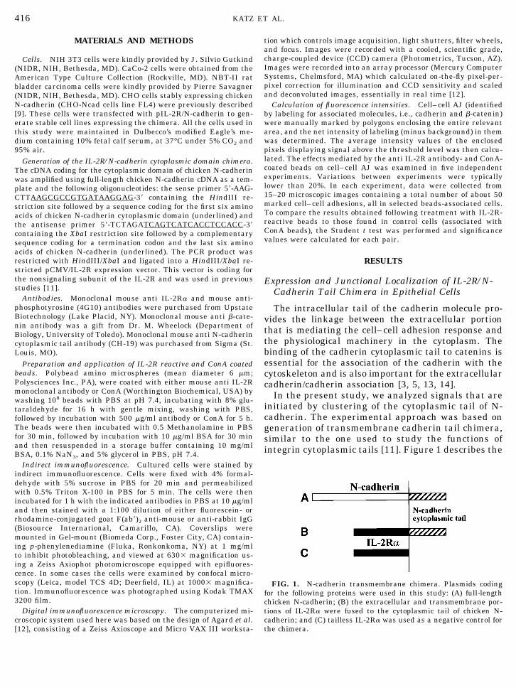
In the present study, we analyzed signals that areinitiated by clustering of the cytoplasmic tail of N-cadherin. The experimental approach was based ongeneration of transmembrane cadherin tail chimera,similar to the one used to study the functions ofintegrin cytoplasmic tails [11]. Figure 1 describes the
FIG. 1. N-cadherin transmembrane chimera. Plasmids codingfor the following proteins were used in this study: (A) full-lengthchicken N-cadherin; (B) the extracellular and transmembrane por-tions of IL-2Ra were fused to the cytoplasmic tail of chicken N-cadherin; and (C) tailless IL-2Ra was used as a negative control forthe chimera.
416 KATZ ET AL.
different versions of the cadherin constructs thatwere used in this study. Full-length N-cadherin (Fig.1A) was stably expressed in CHO cells as previouslydescribed [9]. The cytoplasmic tail of N-cadherin wasfused to the extracellular and transmembrane do-mains of the nonsignaling a subunit of the IL-2R(Fig. 1B). The same portions of the IL-2R, withoutany cytoplasmic domain (Fig. 1C), served as a nega-tive control in the following experiments.
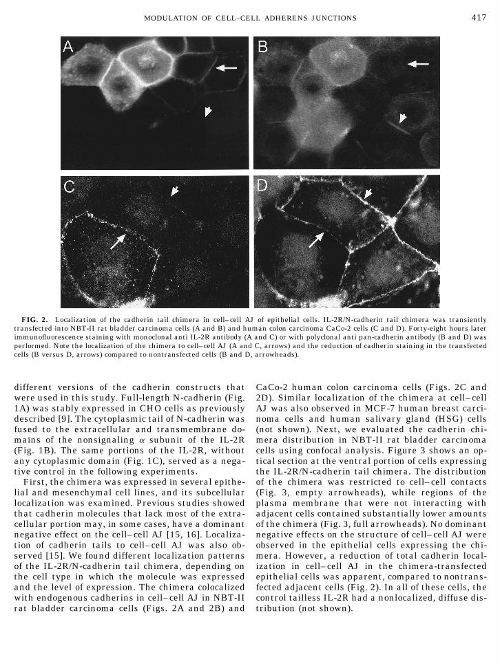
First, the chimera was expressed in several epithe-lial and mesenchymal cell lines, and its subcellularlocalization was examined. Previous studies showedthat cadherin molecules that lack most of the extra-cellular portion may, in some cases, have a dominantnegative effect on the cell– cell AJ [15, 16]. Localiza-tion of cadherin tails to cell– cell AJ was also ob-served [15]. We found different localization patternsof the IL-2R/N-cadherin tail chimera, depending onthe cell type in which the molecule was expressedand the level of expression. The chimera colocalizedwith endogenous cadherins in cell– cell AJ in NBT-IIrat bladder carcinoma cells (Figs. 2A and 2B) and
CaCo-2 human colon carcinoma cells (Figs. 2C and2D). Similar localization of the chimera at cell– cellAJ was also observed in MCF-7 human breast carci-noma cells and human salivary gland (HSG) cells(not shown). Next, we evaluated the cadherin chi-mera distribution in NBT-II rat bladder carcinomacells using confocal analysis. Figure 3 shows an op-tical section at the ventral portion of cells expressingthe IL-2R/N-cadherin tail chimera. The distributionof the chimera was restricted to cell– cell contacts(Fig. 3, empty arrowheads), while regions of theplasma membrane that were not interacting withadjacent cells contained substantially lower amountsof the chimera (Fig. 3, full arrowheads). No dominantnegative effects on the structure of cell– cell AJ wereobserved in the epithelial cells expressing the chi-mera. However, a reduction of total cadherin local-ization in cell– cell AJ in the chimera-transfectedepithelial cells was apparent, compared to nontrans-fected adjacent cells (Fig. 2). In all of these cells, thecontrol tailless IL-2R had a nonlocalized, diffuse dis-tribution (not shown).
FIG. 2. Localization of the cadherin tail chimera in cell–cell AJ of epithelial cells. IL-2R/N-cadherin tail chimera was transientlytransfected into NBT-II rat bladder carcinoma cells (A and B) and human colon carcinoma CaCo-2 cells (C and D). Forty-eight hours laterimmunofluorescence staining with monoclonal anti IL-2R antibody (A and C) or with polyclonal anti pan-cadherin antibody (B and D) wasperformed. Note the localization of the chimera to cell–cell AJ (A and C, arrows) and the reduction of cadherin staining in the transfectedcells (B versus D, arrows) compared to nontransfected cells (B and D, arrowheads).
417MODULATION OF CELL–CELL ADHERENS JUNCTIONS

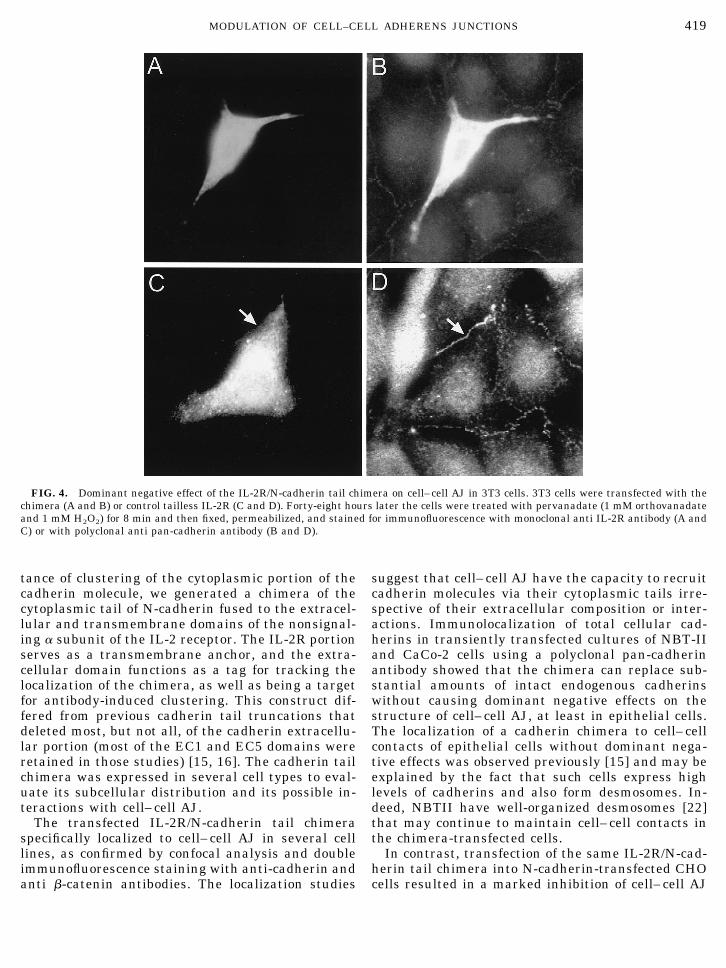
Possible dominant negative effects of the chimeraon cell– cell AJ may depend on the levels of endoge-nous cadherin expression. These levels are high inepithelial cells; therefore, the chimera may be incor-porated into these sites with only minor structuraleffects. We examined the localization of the chimerain cells of mesenchymal origin that express rela-tively low levels of cadherins [17]. As shown in Fig.4B, 3T3 cells form cell– cell AJ upon short-term (8min) induction with 1 mM pervanadate, as previ-ously reported [17]. Expression of the cadherin tailchimera in pervanadate-induced 3T3 cells resultedin detachment of the transfected cells from the mono-layer (Fig. 4B), and no localization of the chimera tocell– cell AJ was observed. The control tailless IL-2Rdid not localize to cell– cell AJ and did not affect cellmorphology (Figs. 4C and 4D).
Clustering of IL-2R/N-Cadherin Tail Results in theEnhancement of Cadherin and Catenin Labelingwithin Cell–Cell AJ of the Same Cells
In a previous study we demonstrated that surfaceclustering of intact N-cadherin in CHO cells (CHO-Ncad cells stably expressing N-cadherin) resulted ina major increase in the levels of AJ-associated cad-herin and b-catenin [9]. To examine whether cluster-ing of the cadherin cytoplasmic tail can also result inintracellular long-range effects on cell– cell AJ, we
first expressed the chimera in CHO-Ncad cells. Asshown in Fig. 5, a cell line stably expressing theIL-2R/N-cadherin tail chimera was generated. Thechimera localized to cell– cell AJ in these cells (Fig.5B); however, a notable dominant negative effectwas observed with respect to the number, size, andcadherin staining of these sites (Fig. 5D), comparedto nontransfected cells (Fig. 5C). As shown in Fig. 5(inserts), expression of the chimera did not resultin any reduction of endogenous cadherin levels intransfected compared to control cells.
While transfection of the IL-2R/N-cadherin tailchimera into CHO-Ncad cells resulted in the inhibi-tion of AJ formation, incubation of the transfectedcultures with monoclonal anti IL-2R antibody-coatedbeads dramatically enhanced the formation of cad-herin and b-catenin containing junctions. As shownin Fig. 6, clustering of the cadherin tail chimeraresulted in the accumulation of cadherins in cell– cellAJ. Also, the sizes of these sites in the induced cellsincreased (Fig. 6F) compared to cells incubated withConA-coated beads (Fig. 6B). The accumulation ofcadherins in cell– cell AJ upon clustering of the cad-herin tail was accompanied by increases in quanti-ties of b-catenin at these sites (Fig. 7F). No increasein b-catenin accumulation was observed in cells in-cubated with ConA-coated beads (Fig. 7B) or in con-trol cells expressing the tailless IL-2R (Figs. 7A, 7Cand 7E).
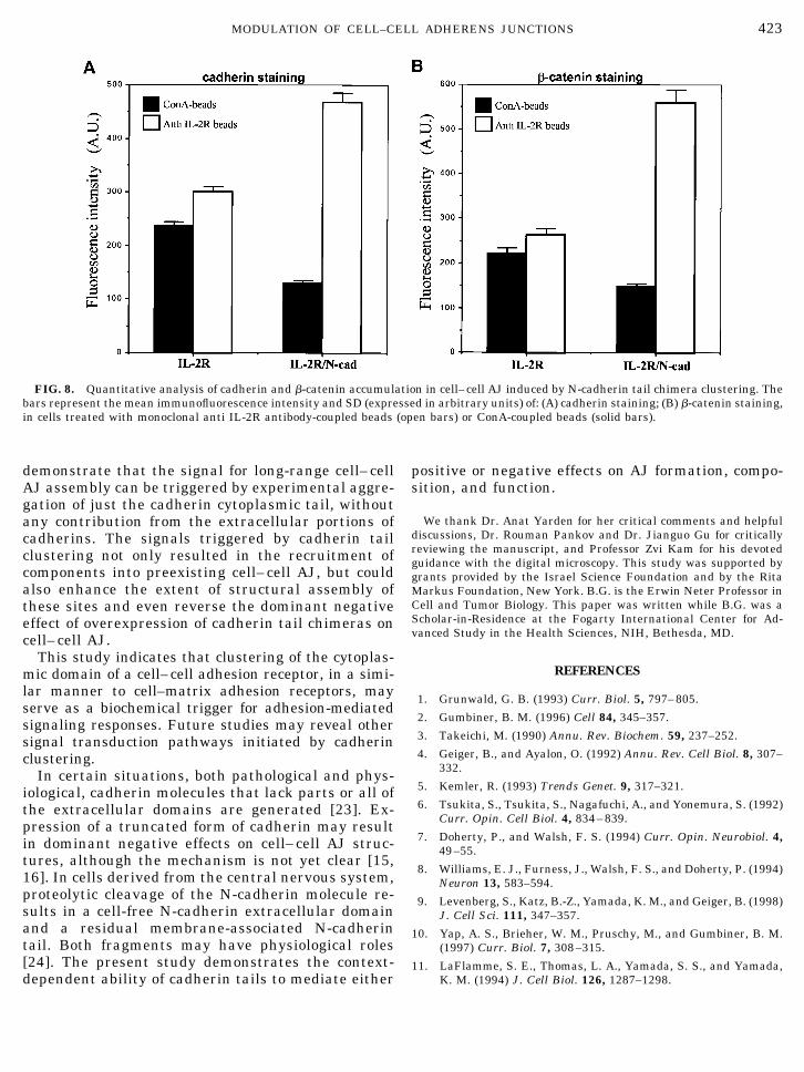
To obtain quantitative information about the changesin junction labeling following the clustering of the IL-2R/N-cadherin chimera, we have determined the inten-sity of junctional fluorescence labeling for cadherin andb-catenin, using digital microscopy as previously per-formed [9]. The results indicate a significant (P ,0.001), fourfold increase in cadherin (Fig. 8A) andb-catenin (Fig. 8B) immunofluorescence staining in-tensity in AJ of cells induced by the clustering of theIL-2R/N-cadherin tail chimera, using IL-2R beads com-pared to ConA beads (Figs. 8A and 8B). No such in-crease was observed in control cells expressing thetailless IL-2R (Figs. 8A and 8B).
DISCUSSION
Clustering of cytokine and growth factor receptorsis a critical event in generating a variety of signaltransduction responses [18, 19]. Similarly, cluster-ing of b-integrin cytoplasmic tails, even in the ab-sence of any extracellular integrin domain, promotestyrosine phosphorylation of focal adhesion kinase[20]. Clustering also appears to be important for theinitiation of cadherin-mediated signaling, which re-sults in tyrosine phosphorylation and assembly ofcell– cell junctions [9, 21]. To investigate the impor-
FIG. 3. Confocal analysis of the IL-2R/N-cadherin tail chimeradistribution in NBT-II rat bladder carcinoma cells. IL-2R/N-cadherintail chimera was transiently transfected into NBT-II rat bladdercarcinoma cells. Forty-eight hours later immunofluorescence stain-ing with monoclonal anti IL-2R antibody was performed. An opticalsection at the bottom of the cells is shown. Note that the chimera islocalized at cell–cell contacts (empty arrowheads), and absent fromregions of the cell membrane that do not associate with adjacent cells(full arrowheads).
418 KATZ ET AL.
tance of clustering of the cytoplasmic portion of thecadherin molecule, we generated a chimera of thecytoplasmic tail of N-cadherin fused to the extracel-lular and transmembrane domains of the nonsignal-ing a subunit of the IL-2 receptor. The IL-2R portionserves as a transmembrane anchor, and the extra-cellular domain functions as a tag for tracking thelocalization of the chimera, as well as being a targetfor antibody-induced clustering. This construct dif-fered from previous cadherin tail truncations thatdeleted most, but not all, of the cadherin extracellu-lar portion (most of the EC1 and EC5 domains wereretained in those studies) [15, 16]. The cadherin tailchimera was expressed in several cell types to eval-uate its subcellular distribution and its possible in-teractions with cell– cell AJ.
The transfected IL-2R/N-cadherin tail chimeraspecifically localized to cell– cell AJ in several celllines, as confirmed by confocal analysis and doubleimmunofluorescence staining with anti-cadherin andanti b-catenin antibodies. The localization studies
suggest that cell– cell AJ have the capacity to recruitcadherin molecules via their cytoplasmic tails irre-spective of their extracellular composition or inter-actions. Immunolocalization of total cellular cad-herins in transiently transfected cultures of NBT-IIand CaCo-2 cells using a polyclonal pan-cadherinantibody showed that the chimera can replace sub-stantial amounts of intact endogenous cadherinswithout causing dominant negative effects on thestructure of cell– cell AJ, at least in epithelial cells.The localization of a cadherin chimera to cell– cellcontacts of epithelial cells without dominant nega-tive effects was observed previously [15] and may beexplained by the fact that such cells express highlevels of cadherins and also form desmosomes. In-deed, NBTII have well-organized desmosomes [22]that may continue to maintain cell– cell contacts inthe chimera-transfected cells.
In contrast, transfection of the same IL-2R/N-cad-herin tail chimera into N-cadherin-transfected CHOcells resulted in a marked inhibition of cell– cell AJ
FIG. 4. Dominant negative effect of the IL-2R/N-cadherin tail chimera on cell–cell AJ in 3T3 cells. 3T3 cells were transfected with thechimera (A and B) or control tailless IL-2R (C and D). Forty-eight hours later the cells were treated with pervanadate (1 mM orthovanadateand 1 mM H2O2) for 8 min and then fixed, permeabilized, and stained for immunofluorescence with monoclonal anti IL-2R antibody (A andC) or with polyclonal anti pan-cadherin antibody (B and D).
419MODULATION OF CELL–CELL ADHERENS JUNCTIONS

formation. The localization pattern and dominantnegative effects of the IL-2R/N-cadherin tail chimerawere similar to those previously described usingtruncated cadherin molecules [15, 16]. Therefore,cadherin replacement may be the mechanism for thechimera-induced dominant negative effect, in a man-ner similar to the actions of previously describedtruncated cadherins [16]. The effects of the IL-2R/N-cadherin tail chimera on cell– cell AJ differ markedlyfrom the dominant negative effects on cell-substrateadhesion of our IL-2R/integrin tail chimeras de-scribed previously [11].
Subsequent effects of membrane clustering of thechimera were studied by incubating transfected cellswith beads coated with anti IL-2R monoclonal anti-body. This experimental approach is similar to thatused previously to induce surface clustering of intactcadherins, which resulted in the induction of long-range recruitment of components to cell– cell AJ [9].We observed a similar induction of accumulation ofcadherins and b-catenin in cell– cell AJ of cells afterbead-induced clustering of chimeras containing onlythe cadherin cytoplasmic tail. Control ConA-coatedbeads had no effects on AJ structure. These data
FIG. 5. Expression of the IL-2R/N-cadherin tail chimera in CHO cells expressing N-cadherin. Nontransfected cells (A) and cells from aclone expressing the IL-2R/N-cadherin tail chimera (B) cultured on coverslips were fixed, permeabilized, and immunostained with amonoclonal anti IL-2R antibody followed by rhodamine-labeled goat anti-mouse IgG. Note that the chimera is located on the cell membraneat a cell–cell contact. Nontransfected cells (C) and cells from a clone expressing the IL-2R/N-cadherin tail chimera (D) cultured on coverslipswere fixed, permeabilized, and immunostained with a monoclonal anti N-cadherin antibody that reacts with the extracellular domain ofN-cadherin, followed by rhodamine-labeled anti-mouse IgG. Insets: Expression of the IL-2R/N-cadherin tail chimera in N-cadherin express-ing CHO cells. Equal amounts of total cell protein from nontransfected cells or from cells of a positive clone were analyzed by SDS–PAGEand Western blotting with a monoclonal mouse anti N-cadherin antibody that reacts with the intracellular domain of N-cadherin. Note thedominant negative effect of the IL-2R/N-cadherin tail chimera on N-cadherin staining in cell–cell junctions.
420 KATZ ET AL.

FIG. 6. Experimental clustering of the IL-2R/N-cadherin cytoplasmic tail induces the accumulation of cadherins in cell–cell AJ.CHO-Ncad cells expressing IL-2R/N-cadherin tail chimera (B, D, F) or tailless IL-2R as a control (A, C, E) were plated on glass coverslips,and 24 h later beads coated with monoclonal anti IL-2R antibody (E, F) or ConA (A–D) were added to the cells. Following 20 min of incubationat 37°C, the cells were fixed and stained with anti N-cadherin antibody. A and B pictures were taken using concurrent phase contrast underlow illumination in order to see the ConA beads.
421MODULATION OF CELL–CELL ADHERENS JUNCTIONS
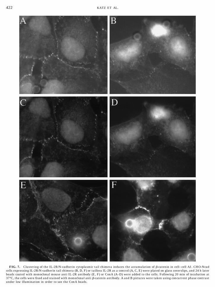
FIG. 7. Clustering of the IL-2R/N-cadherin cytoplasmic tail chimera induces the accumulation of b-catenin in cell–cell AJ. CHO-Ncadcells expressing IL-2R/N-cadherin tail chimera (B, D, F) or tailless IL-2R as a control (A, C, E) were plated on glass coverslips, and 24 h laterbeads coated with monoclonal mouse anti IL-2R antibody (E, F) or ConA (A–D) were added to the cells. Following 20 min of incubation at37°C, the cells were fixed and stained with monoclonal anti b-catenin antibody. A and B pictures were taken using concurrent phase contrastunder low illumination in order to see the ConA beads.
422 KATZ ET AL.
demonstrate that the signal for long-range cell– cellAJ assembly can be triggered by experimental aggre-gation of just the cadherin cytoplasmic tail, withoutany contribution from the extracellular portions ofcadherins. The signals triggered by cadherin tailclustering not only resulted in the recruitment ofcomponents into preexisting cell– cell AJ, but couldalso enhance the extent of structural assembly ofthese sites and even reverse the dominant negativeeffect of overexpression of cadherin tail chimeras oncell– cell AJ.
This study indicates that clustering of the cytoplas-mic domain of a cell–cell adhesion receptor, in a simi-lar manner to cell–matrix adhesion receptors, mayserve as a biochemical trigger for adhesion-mediatedsignaling responses. Future studies may reveal othersignal transduction pathways initiated by cadherinclustering.
In certain situations, both pathological and phys-iological, cadherin molecules that lack parts or all ofthe extracellular domains are generated [23]. Ex-pression of a truncated form of cadherin may resultin dominant negative effects on cell– cell AJ struc-tures, although the mechanism is not yet clear [15,16]. In cells derived from the central nervous system,proteolytic cleavage of the N-cadherin molecule re-sults in a cell-free N-cadherin extracellular domainand a residual membrane-associated N-cadherintail. Both fragments may have physiological roles[24]. The present study demonstrates the context-dependent ability of cadherin tails to mediate either
positive or negative effects on AJ formation, compo-sition, and function.
We thank Dr. Anat Yarden for her critical comments and helpfuldiscussions, Dr. Rouman Pankov and Dr. Jianguo Gu for criticallyreviewing the manuscript, and Professor Zvi Kam for his devotedguidance with the digital microscopy. This study was supported bygrants provided by the Israel Science Foundation and by the RitaMarkus Foundation, New York. B.G. is the Erwin Neter Professor inCell and Tumor Biology. This paper was written while B.G. was aScholar-in-Residence at the Fogarty International Center for Ad-vanced Study in the Health Sciences, NIH, Bethesda, MD.
REFERENCES
1. Grunwald, G. B. (1993) Curr. Biol. 5, 797–805.
2. Gumbiner, B. M. (1996) Cell 84, 345–357.
3. Takeichi, M. (1990) Annu. Rev. Biochem. 59, 237–252.
4. Geiger, B., and Ayalon, O. (1992) Annu. Rev. Cell Biol. 8, 307–332.
5. Kemler, R. (1993) Trends Genet. 9, 317–321.
6. Tsukita, S., Tsukita, S., Nagafuchi, A., and Yonemura, S. (1992)Curr. Opin. Cell Biol. 4, 834–839.
7. Doherty, P., and Walsh, F. S. (1994) Curr. Opin. Neurobiol. 4,49–55.
8. Williams, E. J., Furness, J., Walsh, F. S., and Doherty, P. (1994)Neuron 13, 583–594.
9. Levenberg, S., Katz, B.-Z., Yamada, K. M., and Geiger, B. (1998)J. Cell Sci. 111, 347–357.
10. Yap, A. S., Brieher, W. M., Pruschy, M., and Gumbiner, B. M.(1997) Curr. Biol. 7, 308–315.
11. LaFlamme, S. E., Thomas, L. A., Yamada, S. S., and Yamada,K. M. (1994) J. Cell Biol. 126, 1287–1298.
FIG. 8. Quantitative analysis of cadherin and b-catenin accumulation in cell–cell AJ induced by N-cadherin tail chimera clustering. Thebars represent the mean immunofluorescence intensity and SD (expressed in arbitrary units) of: (A) cadherin staining; (B) b-catenin staining,in cells treated with monoclonal anti IL-2R antibody-coupled beads (open bars) or ConA-coupled beads (solid bars).
423MODULATION OF CELL–CELL ADHERENS JUNCTIONS
12. Agard, D. A., Hiraoka, Y., Shaw, P., and Sedat, J. W. (1989)Methods Cell Biol. 30, 353–377.
13. Nagafuchi, A., Ishihara, S., and Tsukita, S. (1994) J. Cell Biol.127, 235–245.
14. Aberle, H., Schwartz, H., and Kemler, R. (1996) J. Cell. Bio-chem. 61, 514–523.
15. Fujimori, T., and Takeichi, M. (1993) Mol. Biol. Cell 4, 37–47.16. Kintner, C. (1992) Cell 69, 225–236.17. Michalides, R., Volberg, T., and Geiger, B. (1994) Cell. Adhes.
Commun. 2, 481–490.18. Taga, T., and Kishimoto, T. (1995) Curr. Opin. Immunol. 7,
17–23.
19. Heldin, C. H. (1995) Cell 80, 213–223.20. Akiyama, S. K., Yamada, S. S., Yamada, K. M., and LaFlamme,
S. E. (1994) J. Biol. Chem. 269, 15961–15964.21. Kinch, M. S., Petch, L., Zhong, C., and Burridge, K. (1997) Cell
Adhes. Commun. 4, 425–437.22. Savanger, P., Yamada, K. M., and Thiery, J. P. (1997) J. Cell
Biol. 137, 1403–1419.23. Becker, K.-H., Atkinson, J. A., Reich, U., Becker, I., Nekarada,
H., Siewert, J. R., and Hofler, H. (1996) Cancer Res. 54, 3845–3852.
24. Paradies, N. E., and Grunwald, G. B. (1995) J. Neurosci. Res.36, 33–45.
Received April 10, 1998Revised version received June 24, 1998
424 KATZ ET AL.