mitochondria death/survival signaling pathways in cardiotoxicity
TRANSCRIPT

Hindawi Publishing CorporationBiochemistry Research InternationalVolume 2012, Article ID 951539, 12 pagesdoi:10.1155/2012/951539
Review Article
Mitochondria Death/Survival SignalingPathways in Cardiotoxicity Induced by Anthracyclinesand Anticancer-Targeted Therapies
David Montaigne, Christopher Hurt, and Remi Neviere
Department of Physiology (EA4484), Faculty of Medicine, University of Lille 1, Place de Verdun, 59045 Lille, France
Correspondence should be addressed to Remi Neviere, [email protected]
Received 16 November 2011; Revised 4 January 2012; Accepted 9 January 2012
Academic Editor: Catherine Brenner
Copyright © 2012 David Montaigne et al. This is an open access article distributed under the Creative Commons AttributionLicense, which permits unrestricted use, distribution, and reproduction in any medium, provided the original work is properlycited.
Anthracyclines remain the cornerstone of treatment in many malignancies but these agents have a cumulative dose relationshipwith cardiotoxicity. Development of cardiomyopathy and congestive heart failure induced by anthracyclines are typically dose-dependent, irreversible, and cumulative. Although past studies of cardiotoxicity have focused on anthracyclines, more recentlyinterest has turned to anticancer drugs that target many proteins kinases, such as tyrosine kinases. An attractive model to explainthe mechanism of this cardiotoxicity could be myocyte loss through cell death pathways. Inhibition of mitochondrial transitionpermeability is a valuable tool to prevent doxorubicin-induced cardiotoxicity. In response to anthracycline treatment, activationof several protein kinases, neuregulin/ErbB2 signaling, and transcriptional factors modify mitochondrial functions that determinecell death or survival through the modulation of mitochondrial membrane permeability. Cellular response to anthracyclines is alsomodulated by a myriad of transcriptional factors that influence cell fate. Several novel targeted chemotherapeutic agents have beenassociated with a small but worrying risk of left ventricular dysfunction. Agents such as trastuzumab and tyrosine kinase inhibitorscan lead to cardiotoxicity that is fundamentally different from that caused by anthracyclines, whereas biological effects converge tothe mitochondria as a critical target.
1. Introduction
Cardiotoxicity is a term often used to describe a broad rangeof adverse effects on heart function induced by therapeuticmolecules. These effects may either emerge early in preclin-ical studies or become apparent latter in the clinical settingafter the drug has already been licensed for clinical use. Theuse of several chemotherapeutics for the treatment of canceris associated with a risk of cardiovascular complications [1–3]. They present as a defect in cardiac function that can beeither symptomatic or not. The classic example of this issueis the use of anthracyclines such as doxorubicin, which iscommonly prescribed to treat hematological malignanciesand solid tumors [4, 5].
Potential cardiovascular toxicities linked to anticancerdrugs include increases of QT duration, arrhythmias,and myocardial ischemia (antimetabolite compounds) [6],
hypertension and thromboembolic complications (antian-giogenic agents) [7], and myocardial dysfunction [1]. Thelatter, variable in severity, can be reversible or not and canoccur during treatment or later on. For example, the clinicaluse of anthracyclines such as doxorubicin is hampered bythe development of cardiomyopathy and congestive heartfailure, which are typically dose-dependent and cumulative.While acute cardiotoxicity occurs, the most troublesomeform manifests late after treatment and is characterized bystructural changes of the human heart [8], leading to de-creases in the left ventricle wall thickness and myocardiummass, as well as reduced ventricular compliance [9]. Unlikeacute toxicity, the delayed manifestation of anthracycline useoften presents as symptomatic heart failure and is consideredlargely irreversible [1, 5, 10].
Although past studies of cardiotoxicity have focusedon anthracyclines, more recently interest has turned to

2 Biochemistry Research International
anticancer drugs that target many proteins kinases, suchas tyrosine kinases [2]. Targeted therapeutics, particularlythose that inhibit the activity of protein kinases that are mu-tated and/or overexpressed in cancer, have revolutionizedthe treatment of some cancers and improved survival inmany others [11, 12]. Unexpected cardiotoxicity induced bytargeted drugs has been related to the existence of numer-ous parallels between signaling pathways that drive tumori-genesis and those that regulate survival of cardiomyocytes[12–15]. For example, on-target heart toxicity of tras-tuzumab, a monoclonal antibody against the ErbB2 receptor[16] revealed that human epidermal growth factor receptor 2signaling also interfered with survival pathway in cardiomy-ocyte, a terminally differentiated cell [16, 17]. At this point,it can be hypothesized that mitochondrial dysfunction andATP depletion are the main contributors to targeted therapy-induced cardiac toxicity [15].
An attractive model to explain the mechanism of this car-diotoxicity could be myocyte loss through cell death path-ways [18, 19]. Given the limited regenerative capacity ofthe heart, cumulative toxicity may be explained by the pro-gressive increase of cardiac cell loss. Cardiac cell stress (speci-fically oxidative stress induced by anthracyclines and manykinase inhibitors) activates apoptosis and necrosis via a mito-chondrial pathway [1, 10]. As mitochondria are a centralcomponent of intrinsic apoptotic and necrotic pathways,mitochondrial “effects” of anticancer drugs are to be an ex-pected outcome of adverse interactions between the drug andcells [20].
2. Overview of Anthracycline Cardiotoxicity
2.1. Clinical Picture of Cardiac Toxicity. Anthracycline-in-duced cardiotoxicity has been categorized into acute, early-onset chronic progressive and late-onset chronic progressiveforms [21]. Acute cardiotoxicity occurs during or shortlyafter drug infusion and includes nonspecific EKG changesand arrhythmias, which may be accompanied in some pa-tients by heart failure and pericarditis-myocarditis syndrome[9]. These complications are typically reversible, not dose-dependent and do not preclude further anthracycline use.Single cases of acute cardiac failure and sudden death werealso reported [9, 22]. The subacute cardiac toxicity occurswithin a few weeks, clinically resembles myocarditis (withedema and thickening of the left ventricle LV walls), and isassociated with 60% mortality [22]. Acute cardiotoxicity oc-cur in 1% of patients, whereas the subacute form occurs in1.4–2% of patients [9]. Clinically the most significant effectof anthracyclines is chronic cardiac toxicity that may lead toLV dysfunction and congestive heart failure [23, 24]. Late-onset chronic progressive cardiotoxicity usually appears atleast one year after completion of therapy and manifestsclinically in 1.6–5% of patients [24]. Late-onset chronic pro-gressive may not become evident until 10 to 20 years afterthe first dose of cancer treatment. The prognosis in anthra-cycline-related heart failure is poor, with 50% 2-year mor-tality in untreated established LV dysfunction [24, 25]. Themost important risk factor for late cardiac toxicity is the cu-mulative anthracycline dose [25].
It is believed that each anthracycline dose causes struc-tural changes to cardiomyocytes, which ultimately lead tocardiomyocyte death. These defects are balanced by com-pensatory mechanisms until a certain threshold, above whichventricular remodeling common to multiple forms of cardiacinjury is triggered. The estimated risk of chronic heart fail-ure for doxorubicin dose greater than 400 mg/m2 rangesfrom 3% to 5%, for 550 mg/m2 from 7% to 26%, and for700 mg/m2 from 18% to 48% [1, 10]. In clinical practice,however, it seems wise to estimate the risk as being in theupper limit of the given ranges and adapt maximum cumula-tive doses accordingly [26]. Despite a considerable variabilityin individual dose-response relationship for cardiac toxicity,the maximum lifetime cumulative dose for doxorubicin is400 to 550 mg/m2 [1, 10]. The long-term follow-up dataof patients treated with adjuvant anthracycline-based regi-mens demonstrated increased incidence of symptomatic andasymptomatic left ventricular dysfunction and decrease inthe mean LVEF value, suggesting that the incidence and se-verity of postanthracycline heart damage increases with time[26]. Overall, these data stress the importance of cardiacfollow-up well beyond the treatment period.
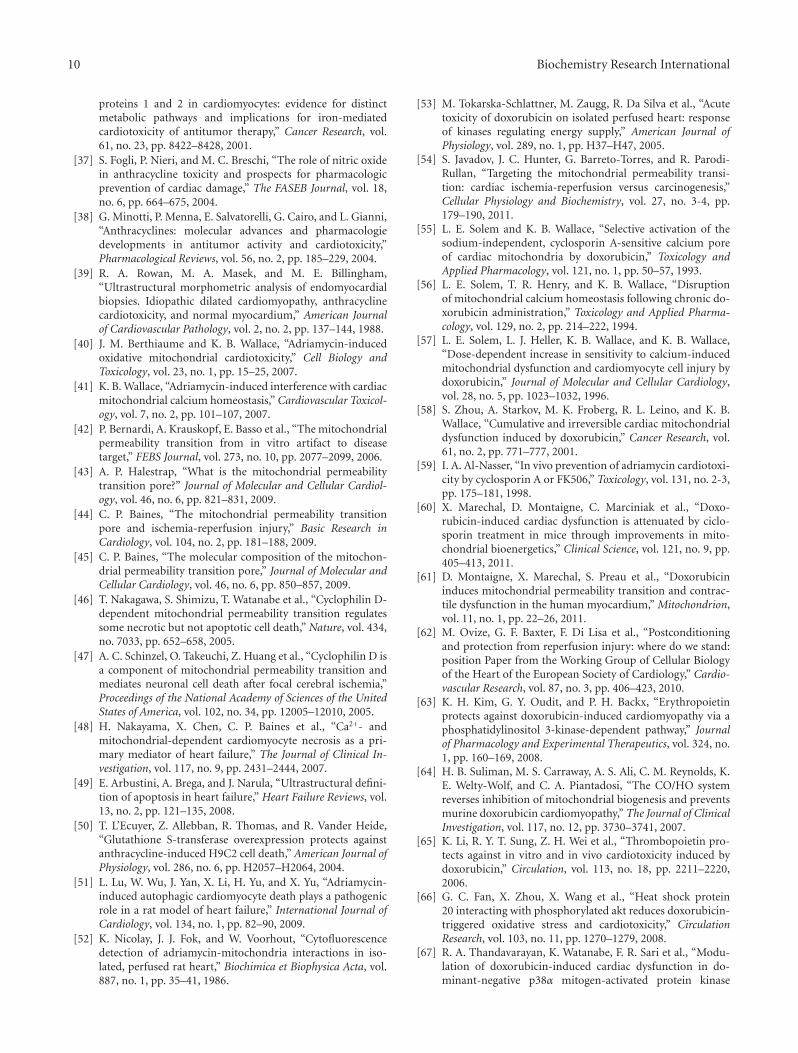
2.2. Proposed Pathophysiology. Despite a remarkably exten-sive literature on many aspects of cardiotoxicity, a singleunifying theory for the deleterious effects of anthracyclineson the heart is still lacking. Anthracycline-induced myocar-dial damage has long been regarded as occurring primarilythrough the generation of reactive oxygen species and freeradicals [27]. This oxidative stress model is supported bymany studies showing ROS formation, especially in thesetting of increased intracellular iron levels, in response toanthracycline treatment [27, 28]. Recent studies, however,have suggested that the ROS model is inadequate to accountfor all features of anthracycline cardiotoxicity [27]. Indeed,there is strong evidence that anthracycline cardiotoxicitystems from (at least partially) ROS-independent mecha-nisms, such as cardiomyocyte apoptosis or necrosis, disrup-tion of normal sarcomere structure and altered energeticsimpairing the cardiac cell ability to generate adequate co-ntraction [18, 29–31]. Recent studies have highlighted theeffects of anthracyclines on compensatory prosurvival mech-anisms, such as neuregulin/heregulin-Erb/HER2 and cell sal-vage kinase pathways, which may modulate the developmentof heart failure [32, 33]. Figure 1 provides a summary ofthe different signaling pathways involved in anthracycline-in-duced cardiotoxicity.
2.3. Oxidative Stress Pathway and Iron Hypothesis. The chem-ical structure of anthracyclines is complex: these drugs arecomposed of an aglycone and a sugar. The aglycone consistsof a tetracyclic ring with adjacent quinone-hydroquinonemoieties. Quinone moieties have toxicological importancebecause of their involvement in both reductive and oxidativebiotransformation leading to highly reactive species involvedin cardiotoxicity [27, 34].
One-electron reduction of the tetracyclic ring of anthra-cyclines leads to the formation of a semiquinone free radical.This radical is relatively stable in an anoxic environment,

Biochemistry Research International 3
DNA damageRNA
ER stress
Antracyclines
ROS
NO
Cell death
Sarcopenia
Bax
Bad
GATA4
MAPKJAPK
p300p53
Pi3K
Caspase-3
Cas
pase
-12
Caspase-9
ROS
ATP
Antracyclines
Sarcomere disarray
JNKp38
Nuclear and mitochondrialgene expression
Cell injury
AM
Pki
nase
Akt/G
SK3
Neureg
ulin
Ca 2+overload
Ca
2+ov
erlo
ad
ErbB2
protein synthesis
ROS
Cel
l sur
viva
l Cyt cmPTP
ONOO−
Δψm
Figure 1: Potential signaling pathways involved in anthracycline-induced cardiomyocyte injury. Anthracycline-induced cell death isbalanced by intracellular survival signaling which is linked to neuregulin/ErbB2 and Akt activation. The suggested principal mechanismof anthracycline damage is via generation of reactive oxygen species ROS by iron-anthracycline complexes, leading to lipid peroxidationand membrane damage. Oxidative stress (ROS, nitric oxide NO, and peroxynitrite ONOO–) causes activation of kinase pathways (mitogen-activated protein kinase MAPK, stress-activated protein kinase SAPK, c-Jun N-terminal kinases JNK) modulating response to anthracyclinesand linking to apoptotic pathway. In mitochondria, ROS and calcium overload lead to the release of cytochrome c (cyt c) from mitochondriainto cytoplasm, via mitochondrial permeability transition pore opening (mPTP), which results in membrane potential dissipation (delta psim), activation of caspases and apoptosis. Other putative mechanisms include damage to nuclear DNA, disruption of sarcomeric protein,suppression of transcription factors (GATA-4, p300, p53) that regulate cell survival and sarcomeric protein synthesis, and disturbance ofenergy metabolism.
but under normoxic conditions, its unpaired electron is do-nated to oxygen, forming superoxide radicals. Suitable flavo-proteins such as complex I catalyze the formation of reducedsemiquinone radicals by accepting electrons from NADHor NADPH and donating them to anthracyclines. This se-quence of reactions, known as “redox cycling,” can be highlydamaging, because a relatively small amount of drug is suf-ficient for the formation of numerous superoxide radicals.The redox cycling of anthracyclines has been described in cy-toplasm, mitochondria, and sarcoplasmic reticulum [27].The first targets of anthracycline-mediated free-radical dam-age are various cellular membranes, which are rich in lipidsprone to peroxidation. This radical damage results in pro-duction of many stable and highly toxic aldehydes, which fur-ther attack macromolecular targets. Although formation ofROS is induced by the quinone moiety of anthracyclines, oxi-dative stress can also occur via induction of nitric oxide syn-thase, leading to superoxide anion, nitric oxide and perox-ynitrite formation [18, 27].
Promotion of myocardial oxidative stress remains by farthe most frequently proposed mechanisms of anthracycline-induced cardiotoxicity [27]. Production of ROS that followsnuclear binding of the drug results in injuries to DNA as
well as to cell membranes and mitochondria. ROS produc-tion is involved in a vast variety of cardiotoxicity inducingmechanisms, including impaired expression of cardiac pro-teins, disruption of cellular and mitochondrial calcium ho-meostasis, induction of mitochondrial DNA lesions, dis-ruption of mitochondrial bioenergetics and ATP transfersystems, and degradation of myofilament and cytoskeletonproteins [18, 29–31]. Involvement of oxidative stress inthe pathogenesis of anthracycline cardiotoxicity has beensupported by several approaches, that is, isolated cardiac cellsdisplaying perimitochondrial ROS production in response toanthracycline exposition, cultured cell and animal modelsshowing that antioxidant prevented anthracycline-inducedcardiotoxicity, and resistance of transgenic mice overexpress-ing the mitochondrial manganese-superoxide dismutase toanthracycline-induced cardiotoxicity [35].
Anthracyclines may promote the formation of ROSthrough redox cycling of their aglycones as well as theiranthracycline-iron complexes. Indeed, unless adequately se-questered within the cells, iron can dramatically promoteROS production by the Fenton and Haber-Weiss reactionsand the formation of reactive anthracycline-iron complexes[27, 34]. Cellular iron (Fe) level is tightly regulated by the

4 Biochemistry Research International
transferring receptor and storage regulating ferritin, both ofwhich are, themselves regulated at the posttranscriptionallevel by interactions of Fe-regulatory-protein (IRP-1) withspecific motifs iron-responsive-elements (IREs) in targetgenes. Doxorubicin and its metabolites can disrupt the Fe-S cluster of cytoplasmic aconitase and inhibit IRP-1. In do-xorubicin-treated cardiomyocytes, increased IRP-1 inhibi-tion leads to intracellular Fe accumulation causing increasedoxidative stress [36].
Consistent results suggesting the involvement of oxida-tive and nitrosative oxidant stresses in anthracycline-inducedcardiotoxicity provide a rationale for cardioprotection withantioxidants in humans [27, 37]. Unfortunately, use of differ-ent antioxidant agents have failed to provide protection inboth preclinical experiments and clinical studies [3]. Firstgeneration antioxidant molecules such as N-acetylcystein,vitamins D, E have been investigated on the basis of someprotective effects observed in animal models [27]. None ofthese antioxidant approaches has yet shown consistent effi-ciency. The only compound found to reduce long-term car-diac dysfunction is dexrazoxane, although whether its effi-ciency is related to its antioxidant properties or other mech-anisms remains under debate [3]. For example, underlyingmechanisms of dexrazoxane include prevention of iron accu-mulation, which is implicated in increased ROS productionand anthracycline-induced cardiotoxicity.
2.4. Mitochondrial Apoptosis and Necrosis Pathway. Cardiacside effects of anthracyclines involve two main mechanisms,which interact with each other, oxidative stress and apoptosis[18, 27]. Most of the cellular events induced by ROS genera-tion contribute to cardiomyocyte death, which has beenshown to be a primary mechanism for anthracycline-inducedcardiomyopathy. Indeed, cardiac myocyte loss following acti-vation of both apoptotic and necrotic pathways provide anattractive explanation for anthracycline-induced cardiotoxi-city [18, 20, 38]. Studies in animals have demonstrated thatapoptotic cell death occurs after in vivo exposure to anthra-cyclines [38]. Experimental cell cultures have also shown thatanthracyclines induce both apoptotic and necrotic cell death[38]. Evidence of mitochondrial injuries and hallmarks ofapoptosis have been found in endomyocardial biopsies ofpatients treated with anthracyclines [8, 39]. Overall, cardiacmyocyte death following anthracycline administration typi-cally presents with biochemical features of apoptosis and themorphological aspect of cell necrosis [18, 20, 38].
Anthracycline-induced apoptosis in the heart appears toinvolve a mitochondrial pathway, which requires Bax, cyto-chrome c and caspase-3 [20]. Typically, anthracycline treat-ment increases mitochondrial oxidative stress and disruptsintracellular calcium levels [40, 41]. Increased intracellularcalcium, favored by calcium flux aberrations, eventuallyraises mitochondrial calcium levels. Above a certain thresh-old, this calcium overload triggers permeability transition ofthe mitochondria, resulting in the dissipation of transmem-brane potential, as well as mitochondrial swelling and in-creased permeability of its outer membrane to apoptotic fac-tors such as cytochrome c [20]. In the cytosol, cytochrome cforms a complex with the adaptor protein apoptosis protease
activator protein-1 (Apaf-1), dATP, and caspase-9, so-calledapoptosome, which in turn activates caspase-9. The intrinsicpathway converges then to the downstream executionercaspases [20].
The current hypothesis is that necrotic and some formsof apoptotic cell death involve prolonged opening of a largeconductance pore in the mitochondria, known as the mito-chondrial permeability transition pore mPTP [36]. In itsfully open state, the mPTP has been reported to allow un-restricted movement of solutes of <1.5 kDa. The activation ofthe mPTP in isolated mitochondria has been shown to lead tomitochondrial swelling, which is commonly used as an assayfor mPTP opening. In spite of the great recent interest con-cerning mPTP and its apparent importance in cell death, itsmolecular identity is unknown. It has been proposed thatthe mPTP is formed through conformational change in theassociation of the adenine nucleotide translocator (ANT)with the voltage dependent anion channel (VDAC) contactsites between the inner and outer mitochondrial membranes[42, 43]. Cyclophilin D is thought to regulate the openingof the pore via its interactions with ANT. These interactionsare inhibited by cyclosporine A supporting the idea thatcyclophilin D play a role in pore opening [42, 43]. Recentstudies have shown that genetic ablation of either ANT orVDAC isoforms did not result in the absence of mPTP,suggesting that neither of these proteins is an obligatorycomponent [43]. In contrat, ablation of cyclophilin Dreduces ischemia-reperfusion-induced cell death, suggestinga role for cyclophilin in the mPTP [44, 45]. Growing evidencesuggests that the phosphate carrier is a critical component ofthe mPTP and that interaction between ANT and the phos-phate carrier can modulate mPTP opening [45].
For many years, it has been put forward that the mPTPcontributed mainly to apoptotic cell death as a protagonist ofmitochondrial permeabilization. Recent data suggests, how-ever, that an increase in mitochondrial membrane perme-abilization is one of the key events in both apoptotic andnecrotic cell death [43, 44]. This information is importantsince necrosis occurs in many forms of adult human heartinjuries, including the cardiotoxic effects of anticancer drugs.Indeed, in the mid-2000s, new experimental studies suggest-ed that mPTP did not initiate apoptosis and that this complexinstead played a central role in necrosis, especially in theheart. In this line of reasoning, it has been shown that cy-closporine A (CsA), a known inhibitor of the mPTP, can re-duce the occurrence of cardiac and brain cell necrosis dur-ing ischemia reperfusion injury. Studies of cyclophilin-D-de-ficient mice have also provided consistent evidence that themPTP plays a crucial role in cell necrosis [46, 47]. In thesemice, mPTP was still functional but cyclophilin-D ablationincreased the amount of calcium required for mPTP openingand abolished the sensitivity to CsA [46, 48]. Cyclophilin-D-deficient mice had increased resistance against necroticstimuli such as calcium overload, whereas these animals stilldied in response to treatments with classical apoptotic induc-ers such as staurosporine or etoposide. Overall, this data in-dicates that mPTP opening is chiefly involved in cardiac cellnecrosis rather than in triggering cytochrome c release dur-ing early apoptosis.

Biochemistry Research International 5
2.5. Alternative Types of Cell Death: Oncosis and Autophagy.Features of cell oncosis, which is typically associated in car-diomyocytes with mitochondrial and cytoplasmic swelling,coagulated sarcomere and early rupture of the plasma mem-brane [49] have been described in anthracycline-inducedcardiac cell damage [50]. As mentioned above, recent studieshave shown that this form of cell death can be well con-trolled and programmed through mPTP-dependent mech-anisms. The rationale is that increased ROS leads to mito-chondrial calcium overloading, promotes mPTP opening,causes mitochondrial swelling and ATP depletion, and hencetriggers necrotic cell death [20]. Autophagy has evolved as aconserving process that uses bulk degradation and recyclingof cytoplasmic components, such as long-lived proteins andorganelles. In the heart, autophagy is important for the turn-over of organelles at low basal levels under normal condi-tions and it is upregulated in response to stresses such asischemia/reperfusion and in cardiovascular diseases such asheart failure. Recent evidence suggests that autophagic celldeath may play a significant role in the myocardial dys-function induced by doxorubicin [51]. Overall, it could bestated that in anthracyclin cardiotoxicity, mitochondria is thecrossroad for apoptosis, necrosis and autophagy processes,which may converge in dying cells in response to differentpathways including ROS production, calcium overload, andDNA lesions.
3. Mitochondria-RelatedSurvival/Death Pathways
If apoptotic and necrotic cell death are central to the featureof anthracycline-induced cardiotoxicity, then the underlyingmechanisms in play are worth exploring, as they may leadto cytoprotective strategies. Likewise, better understandingof activation of cell survival pathways in response to anthra-cycline exposition may also provide valuable knowledge thatwould help in the development of new cytoprotective stra-tegies.
3.1. Mitochondrial Permeability Transition. Bioenergetic fail-ure, enzyme inhibitions, lipid peroxidations, induction ofmembrane disorders as well as the initiation of oxidativestress are being attributed to the accumulation of anthracy-clines at or inside mitochondria. From heart tissue perfusedwith anthracyclines two distinct cellular sites of drug accu-mulation were the nuclei and mitochondria, which becomelabeled with the drug [52, 53]. Hence, it has been commonlyproposed that deleterious signals related to anthracycline ex-posure converge to the mitochondria to favor mPTP-mediat-ed cell death [54]. In addition, if this hypothesis is correct,an understanding cardioprotective mechanisms is intimatelylinked to an understanding of the mechanisms by which mi-tochondria regulate cell death.
Disruption of mitochondrial calcium homeostasis fol-lowing chronic doxorubicin administration can be demon-strated by using cardiac mitochondria isolated from doxo-rubicin-treated animals [40, 41]. For example, activation ofthe selective cyclosporine- (CsA-)sensitive calcium channelof cardiac mitochondria by doxorubicin occurs both in vitro
[55] and in cardiac mitochondria isolated in rats having un-dergone chronic in vivo treatment with doxorubicin [56]. Inthe latter protocol of exposition (2 mg/kg/week doxorubicintreatment for 13 weeks), isolated mitochondria have a lowerrespiratory control ratio and exhibit an enhanced CsA-sen-sitive release of mitochondrial calcium. Associated with thiswas a calcium-induced loss of membrane potential, whichmay be inhibited by either cyclosporine A or rutheniumred. Further experiments have demonstrated that doxoru-bicin treatment in vivo causes a dose-dependent and irre-versible interference of mitochondrial calcium transportand calcium-dependent regulation of membrane potentialindicative of an induction of the mPTP and of an increasedsensitivity to calcium-induced loss of cell viability [57, 58].Implication of the mPTP in the cardiotoxicity of doxorubicinhas been explored in cyclophilin-D-deficient mice, cyclo-philin-D being a mitochondrial matrix peptidyl-prolyl iso-merase known to modulated mitochondrial transition poreopening. The result that cyclophilin-D deficiency in miceinhibited doxorubicin-induced cardiomyocyte necrosis andheart failure suggests that mPTP is involved in doxorubicin-induced cardiotoxicity [48]. This contention is also support-ed by animal studies showing prevention of doxorubicincardiotoxicity by in vivo CsA or FK506 treatment [59, 60].In human atrial trabeculae, our group also demonstratedthat cyclosporine A prevented doxorubicin-induced mito-chondrial dysfunction and impaired contractile performanceinduced by doxorubicin [61]. These findings reinforce therationale that mPTP is involved in the development of do-xorubicin cardiotoxicity in the human myocardium.
3.2. Survival Protein Kinase Signaling. Accumulating evi-dence indicates that several protein kinases (i.e., Akt, PKCs,EKR, GSK-3b, hexokinase) receive extra mitochondrial sig-nals and modify mitochondrial proteins that determine celldeath or survival, such as the mPTP [62]. Activities ofsome of these kinases are mutually regulated, and phospho-rylation of GSK-3β and hexokinase in mitochondria appearsto directly modify the mPTP, elevating its opening threshold[62]. Doxorubicin may induce inhibition of Akt phospho-rylation, which increases active glycogen synthase kinase-3b (GSK-3b) [63]. GSK-3b is a protein kinase linked tothe regulation of a variety of cellular functions within themyocardium, including glycogen metabolism, gene expres-sion and cellular survival [62]. GSK-3b phosphorylation,and, therefore inhibition, could confer cardioprotectionthrough its potential mitochondrial effects on the mPTP.Strategies that prevent GSK-3b activation via upstreamkinase activation have been shown to be protective againstdoxorubicin treatment. For example, pretreatment with var-ious therapeutic molecules (erythropoietin, thrombopoietin,CO/HO1) can protect the myocardium against doxorubicin-induced impaired heart function and cardiomyocyte apop-tosis by activating Pi3k-Akt cell survival pathways [63–65].In contrast, upregulation of Ser/Thr phosphatase PP1 bydoxorubicin may be involved in the Akt dephosphorylation,resulting in executioner caspase activation and cell death[66].

6 Biochemistry Research International
Cellular stress and specifically oxidative stress has beenshown to activate mitogen/stress activated protein kinases(MAPKs and SAPKs) that appear to be important indetermining cell fate. MAPKs and SAPKs pathways modulatethe response of the heart to anthracycline exposure [67] andhave also been proposed as cellular mediators linking anthra-cyclines to the apoptotic cell death pathway [18, 30]. Undertreatment with anthracyclines that significantly inducesmyocyte apoptosis in the primary cultures of neonatal mousecardiomyocytes, p38 MAPK is dramatically activated. Thatp38 MAPK may be involved at least in part in the anthra-cycline-induced myocyte apoptosis is demonstrated bytwo important observations. First, the time-course analysisrevealed that p38 MAPK activation typically precedes theonset of apoptosis. Second, application of inhibitors of p38MAPK significantly inhibits anthracycline-induced myocyteapoptosis [68]. Although most studies have focused onthe ERK member of the MAPKs, other members of theMAPKs/SAPKs family have been associated with cardiopro-tection through the modulation of the mPTP. For example,JNK and p38 are activated by doxorubicin and linked mPTPto cardiac myocyte apoptosis [69]. Strategies reducing activa-tion of the MPAKs/SAPKs pathways are typically protectiveagainst doxorubicin cardiotoxicity.
3.3. Genomic Analyses and Cellular Energetic Deficits. Dox-orubicin typically causes selective downregulation of manynuclear genes that encode for proteins with mitochondrialfunction [70, 71]. The depressive effect on the expression ofgenes that comprise the mitochondrial proteome is persistentand can be observed weeks after prolonged administration ofdoxorubicin [70]. Previous studies suggest that a prominentfeature of doxorubicin-induced cardiotoxicity is a profoundalteration in the abundance of transcripts related to energymetabolism and mitochondrial performance. Consistent evi-dence suggests that cellular energy deficits related to decreasein fatty acid and glucose oxidation could play a criticalrole in the development of the cardiomyopathy induced byanthracyclines [29]. For example, oxidation of the long-chain fatty acid palmitate is inhibited by doxorubicin withinminutes in isolated cardiomyocyte preparation, as well asin chronic situation in which cardiomyocytes are isolatedfrom doxorubicin-treated rats. In these studies, impairmentof carnitine palmitoyl transferase I and depletion of its sub-strate l-carnitine by doxorubicin was demonstrated [72].Reduction in fatty acid oxidation is not accompanied by up-regulation of glucose utilization as a compensatory response[73]. Rather, doxorubicin-induced cardiomyopathy is asso-ciated with a decreased utilization of both fatty acids andglucose substrates, which has been related to the effects ofdoxorubicin on cellular glucose supply [74] and the impair-ment of phosphofructokinase, the rate-limiting enzyme ofglycolysis [75].
Furthermore, the selective effects of doxorubicin on sup-pression of mitochondria gene expression is accompanied bya coordinate and adaptive response of energy-sensing mole-cules [76], such as AMP-activated protein kinase (AMPK),hypoxia-inducible factor 1 (HIF1), nuclear respiratoryfactors (Nrf) and proliferator-activated receptor gamma
coactivator1 (PGC1) [77]. Proteomic analyses revealed con-sistent changes in proteins involved in mitochondria energyproduction, energy channeling and mitochondrial antioxi-dant protection [78, 79]. Overall, this information is in linewith doxorubicin-induced mitochondrial defects at differentstages of cardiac energy metabolism, including reduction ofoxidative capacity, changes in the profile of energy substrateutilization, disruption of energy transfer systems such asmitochondrial CK, and AMPK-dependent energy signalingpathways.
3.4. Anthracyclines Induce Sarcomere Functional and Struc-tural Changes. Functional and structural changes to car-diomyocyte sarcomeres have been observed in both exper-imental experiments and in endomyocardial biopsies of pa-tients treated with anthracyclines. Loss of myofibrils, disarrayof myofibrils, swelling of mitochondria and dilation of thesarcoplasmic reticulum were observed [80, 81]. Breakdownof sarcomeres typically involved early-onset degradation ofthe giant myofilament protein, titin. As titin maintains sar-comere integrity, its accelerated degradation via calpain pro-teolytic activity in response to doxorubicin can rapidly leadto sarcomere disorganization and progressive cardiomyocytecontractile dysfunction [82].
Several lines of evidence indicate that an abnormal cal-cium handling of myocardial cells may explain, at least inpart, the cardiac dysfunction seen in doxorubicin-inducedcardiomyopathy. For example, doxorubicin has been shownto inhibit the gene transcription of the sarcoplasmic retic-ulum Ca2+-ATPase [83] and to activate cardiac calciumrelease channels (ryanodine receptors) [84]. A decrease ofsarcoplasmic reticulum calcium load and hence calcium-in-duced calcium release has been observed with doxorubicinin isolated guinea pig ventricular myocytes [85]. The mech-anism by which doxorubicin affects calcium homeostasis ofcardiac myocytes has not been fully defined but may involvean iron-catalyzed direct effect of doxorubicin, doxorubicin-induced formation of reactive oxygen intermediates [83],and conversion of doxorubicin to the toxic alcohol metabo-lites [86].
3.5. Transcriptional Factors. Several lines of evidence suggestthat progressive anthracycline-induced cardiac injury resultsfrom effects on myocyte differentiation programs therebyimpeding myocyte survival and the cardiac adaptativeresponse. Genes with anthracycline inhibited expression in-clude genes encoding, transcriptional factors [71, 73]. Forexample, anthracyclines can disrupt expression and activityof the transcription factor GATA-4 [87]. Transcriptionalfactor GATA-4 is a member of a zinc finger transcriptionalfactor family that is critical for regulating differentiation, sar-comere synthesis and survival signaling. GATA-4 is expressedin the heart and regulates several specific cardiac genes,including antiapoptotic genes, making it a key regulatorof heart development. This important survival factor israpidly depleted in response to doxorubicin treatment [64,88]. Anthracyclines downregulate GATA-4 expression incardiac myocytes and upregulation of GATA-4 can suppressdoxorubicin-induced myocyte apoptosis and drug-induced

Biochemistry Research International 7
cardiotoxicity [88, 89]. These prosurvival effects have beenlinked to the effects of GATA-4 on the upstream activatorof the antiapoptotic gene Bcl-Xl. Since the overexpression ofGATA-4 can attenuate the incidence of apoptosis induced byanthracyclines, GATA-4 may serve as an antiapoptotic factorin the heart. Moreover, GATA-4 also regulates expression ofseveral cardiac specific genes that are involved in sarcomeresynthesis, such as cardiac troponin C and I and myosin lightchain-3 [90]. Hence, one potential mechanism by whichanthracycline may induced myocardial dysfunction is viasuppression of sarcomere protein expression and sarcopeniain response to GATA-4 reduction [91].
In addition to GATA-4, the cardiac ankyrin repeat pro-tein transcriptional regulator CARP and the transcriptionalcoactivating factor p300 have been implicated in the car-diotoxicity of anthracyclines. CARP is rapidly degraded inmyocytes after anthracycline exposure [91, 92]. Suppressionof CARP expression using short-interference RNA is suffi-cient to induce myofibrillar disarray and cell dysfunction[93]. Similar to CARP, p300 is degraded after doxorubicinexposure through p38 kinases alpha and beta and is as-sociated with apoptosis in neonatal cardiomyocytes [93, 94].In these experiments, restoration of p300 inhibited doxo-rubicin-induced cell death.
Doxorubicin treatment has been associated with in-creased expression and activation of p53 tumor suppressorprotein, which activates the intrinsic mitochondrial apopto-tic pathway [95]. Consistently, p53-knockout mice and adultmouse hearts expressing cardiac myocyte-restricted domin-ant-interfering p53 are partially protected against doxo-rubicin-induced cell death and myocardial dysfunction [96].In addition, activation of p53 may also mediate anthra-cycline-induced cardiotoxicity through other pathways inde-pendent of cardiomyocyte apoptosis. For example, p53-med-iated inhibition of mammalian target of rapamycin signaling(mTOR) may contribute to the cardiac mass reduction andmyocardial dysfunction observed in doxorubicin-treatedmice [96]. Hence, acute doxorubicin-induced toxicity couldresult from p53-dependent modulation of mTOR activity. Itmay be thus of considerable interest to determine whetherupstream effectors that activate mTOR pathway would becardioprotective against doxorubicin-induced cardiac toxi-city.
3.6. Neuregulin/ErbB2 Cardioprotective Program. Unexpect-ed cardiac side effect of ErbB2 antagonists, such as tras-tuzumab, has sparked great scientific efforts to elucidate therole of Neuregulin/ErbB2 pathway in cardiomyocyte func-tional and structural integrity [16, 17, 97]. Before this obser-vation, ErbB2 signaling was only recognized as being indis-pensable to normal fetal cardiac development. Subsequentstudies have demonstrated that stimulation of the ErbB2signaling by ErbB-receptor ligands improves cardiomyocytefunction and survival in the heart [97].
The first evidence regarding the protective effects ofthe ErbB2 signaling in the adult heart came from clinicaltrials in breast cancer patients using trastuzumab, a mon-oclonal antibody that blocks the ErbB2 receptor [1, 2].The incidence of clinical heart failure increased five-fold in
patients treated concurrently with chemotherapy drug doxo-rubicin and trastuzumab compared to those treated withdoxorubicin alone [1]. Mostly based on the analogy betweenErbB2 knockout-induced cardiomyopathy and trastuzumab-induced heart failure, many studies have concluded thattrastuzumab causes heart failure by blocking the physiolog-ical actions of ErbB2 in the heart [32, 33]. The synergisticincrease of heart failure incidence has been related to thefact that ErbB2 expression is upregulated following doxo-rubicin administration, while trastuzumab inhibits theErbB2 downstream pathways, which is essential for cell re-pair, survival, and function. Thus, if trastuzumab inhibits theErbB2 cardioprotective pathways during a vulnerable periodafter anthracycline injury, the anthracycline damage could beaugmented, resulting in increased cell death [2].
Overall, these results suggest that ErbB2 inhibition resultin mitochondrial apoptotic signaling in cardiomyocytes lead-ing to increased cell loss in the heart. Therefore, up-regu-lation of the cardiac neuregulin/ErbB2 pathway may be onestrategy to limit myocardial anthracycline injury. Experi-mental work on both animals and humans has demonstratedthat exercise is a potent activator of neuregulin release withsubsequent activation of ErbB2 activation [98]. The fact thatexercise protects against calcium-induced cardiac mitochon-drial permeability transition and reduces cell death followingdoxorubicin administration could be related to the neureg-ulin/ErbB2 survival pathway activation [99].
3.7. Lessons from Targeted Chemotherapy. Several novel “tar-geted” agents have been associated with a small but wor-risome risk of heart dysfunction [2, 12–14]. These agentsinclude the tyrosine kinase inhibitors sunitinib, lapatinib,and imatinib, which are members of a growing class of target-ed chemotherapy agents. Clues as to the nature of thecardiotoxicity due to these agents are beginning to emergethat point to the mitochondria. To date, the only approv-ed kinase inhibitor that is clearly associated with clinical car-diotoxicity is sunitinib, whereas the extent of imatinib-in-duced cardiotoxicity is still under scrutiny [12].
On-target cardiac toxicity is inherent to kinase inhibitionand quickly becomes apparent since many of the pathwaysthat regulate cancer cell survival also regulate essential pro-cesses in cardiomyocytes, including contractile function andsurvival [14, 17]. The ATP binding pocket represents the keyregion of the kinase targeted by most inhibitors. Conserva-tion of that ATP binding pocket among kinases means thatinhibitors can also inhibit unintended kinases, and if anyof these kinases serve important functions in the heart, off-target cardiotoxicity can occur. An additional issue is thattargeting of kinases will achieve entire pathway targeting.For example, the inhibition of multiple components of thePi3kinase/Akt pathway is a viable strategy for cancers, butthis pathway also maintains cardiomyocyte homeostasis andprotects cardiomyocytes from death [100]. Eventually, kinaseinhibitors could mediate toxicity through the inhibition ofnon-kinase targets, such an enzyme requiring ATP to per-form its function [12, 13].
To illustrate the complexities inherent in identifying me-chanisms of kinase inhibitor-induced cardiac toxicity, suni-

8 Biochemistry Research International
tinib-induced cardiac injury will be discussed. Cardiac dys-function was first related to systemic hypertension secondaryto VEGFR2 and PDGFRbeta inhibition by sunitinib [101].As several patients developed cardiotoxicity in absence ofhypertension, additional mechanisms were explored. Inthis study, endomyocardial biopsy samples from two pa-tients who presented with profound sunitinib-induced heartfailure were obtained. Abnormal histopathological changesincluded marked mitochondrial swelling, which could indi-cate mPTP and energetic failure [101, 102]. In culturedcardiomyocytes, the same mitochondrial abnormalities wereobserved and were associated with apoptotic cell death [101,102]. Studies in cardiomyocytes have confirmed that energycompromise was involved in sunitinib-induced cardiac tox-icity but surprisingly was not associated with activation ofAMP kinase, the master fuel control of the cell [102]. Lackof response to this energy loss was the result of direct in-hibition of AMP kinase by sunitinib, so-called off-targettoxicity. AMP kinase inhibition has also been reported in iso-lated heart exposed to doxorubicin [73], whereas recentstudies demonstrated that AMPK gene expression and en-zyme activities were acutely increased [71]. These resultsare important since AMPK is implicated in many survivalpathways, including Pi3k/Akt/mTOR axis [103].
Likewise, imatinib causes a modest but consistent declinein left ventricular function, which was associated with lossof myocardial mass and increased cell death [104]. Studiesin cardiomyocytes showed that imatinib leads to signifi-cant mitochondrial dysfunction with mitochondria swelling,mitochondrial membrane potential collapse followed bycytochrome c release and energetic failure [15, 104]. Thisprocess was associated with a cell death that has the biochem-ical features of apoptosis and the morphological aspect of cellnecrosis. Mitochondria isolated from hearts of mice treatedwith imatinib showed enhanced calcium-induced swellingand mitochondrial permeability transition mPTP. As is thecase with trastuzumab, mitochondrial dysfunction plays acentral role in the cardiotoxic response to imatinib, but themechanism seems to be the induction of endoplasmic retic-ulum ER stress by the drug [15, 104]. ER stress in responseto imatinib exposition has been related to the downstreamactivation of the c-Jun N-terminal kinase (JNK) family ofstress MAPKs [105]. Similarly, ER stress-mediated apopto-tic pathway has also been reported to mediate cardiac celldeath induced by doxorubicin [20]. In this case, caspase 12,which resides in the ER, is an essential caspase to initiate ER-mediated apoptosis that is activated in doxorubicin-treatedhearts [106].
The Pi3k/Akt/mTOR pathway more than any other epit-omizes the similarity between cancer cell signaling and sur-vival signaling in cardiac cells [54]. The cautionary note isthat Pi3k/Akt/mTOR pathway is also critical for cardiomy-ocyte integrity and survival. Hence, inhibition of multiplecomponents of the pathway would jeopardize cardiomyocyteintegrity and survival [100]. Conversely, activation of thissignaling cascade, together with other Akt-activated mole-cules (such as GSK-3β, mTOR and p70S6 kinase), elicits pro-survival and cardiovascular protective effects, which aremediated by inhibition of opening of the mPTP [62].
4. Conclusion
Cardiac dysfunction is the most severe side effects of anthra-cycline treatment. The major mechanism of anthracyclinedamage involves the generation of reactive oxygen speciesROS by iron-anthracycline complexes, leading to lipid perox-idation and membrane damage. In mitochondria, ROS andcalcium overload lead to mitochondrial permeability transi-tion pore opening (mPTP), which is associated with therelease of cytochrome c (cyt c) from mitochondria into cyto-plasm and cell death. Cardiac myocyte loss following acti-vation of both apoptotic and necrotic pathways provide anattractive explanation for anthracycline-induced cardiotox-icity. There is evidence that the stressed cardiac myocytesurvival may rely on both growth and survival pathways thatare altered in the anthracycline-exposed myocardium. Acti-vation of ErbB2 and Pi3k/Akt/mTOR pathways represents amajor adaptative mechanism for cardioprotection, which arealtered by anthracyclines. The delicate balance between pro-and antiapoptosis signaling that relies on kinase-regulatedpathways creates a cause for concern when one attempts touse anticancer molecules, that is, targeted therapies that canimpair the coordinated function of this kinase network.
Abbreviations
(HER2 or EbrB2): Human epidermal growth factorreceptor 2
Pi3k: Phosphatidylinositol-3-KinaseAkt: Serine/threonine protein kinaseGSK3: Glycogen Synthase Kinase 3;BCL: B Cell LymphomaBax: (BCL)-associated XER stress: Endoplasmic reticulumGATA: Family name is derived from their
ability to bind to the consensus DNAsequence (A/T) GATA (A/G)
p53: Tumor suppressor p53p300: Transcription factor p300ΔΨ: Mitochondrial membrane potential.
Acknowledgments
Authors thank Dr. Hurt for English editing skills, Dr.Marechal for his advice and the members of EA4484 researchteam for its support. The authors also thank university ofLille, France, for the financial support (Grant no. EA4484).
References
[1] T. Eschenhagen, T. Force, M. S. Ewer et al., “Cardiovascularside effects of cancer therapies: a position statement fromthe Heart Failure Association of the European Society ofCardiology,” European Journal of Heart Failure, vol. 13, no.1, pp. 1–10, 2011.
[2] M. S. Ewer and S. M. Ewer, “Cardiotoxicity of anticancertreatments: what the cardiologist needs to know,” NatureReviews Cardiology, vol. 7, no. 10, pp. 564–575, 2010.
[3] E. C. van Dalen, H. N. Caron, H. O. Dickinson, and L. C.Kremer, “Cardioprotective interventions for cancer patients

Biochemistry Research International 9
receiving anthracyclines,” Cochrane Database of SystematicReviews, no. 1, Article ID CD003917, 2005.
[4] A. L. A. Ferreira, L. S. Matsubara, and B. B. Matsubara, “An-thracycline-induced cardiotoxicity,” Cardiovascular and He-matological Agents in Medicinal Chemistry, vol. 6, no. 4, pp.278–281, 2008.
[5] J. J. Monsuez, J. C. Charniot, N. Vignat, and J. Y. Artigou,“Cardiac side-effects of cancer chemotherapy,” InternationalJournal of Cardiology, vol. 144, no. 1, pp. 3–15, 2010.
[6] M. W. Saif, M. M. Shah, and A. R. Shah, “Fluoropyrimidine-associated cardiotoxicity: revisited,” Expert Opinion on DrugSafety, vol. 8, no. 2, pp. 191–202, 2009.
[7] S. M. Gressett and S. R. Shah, “Intricacies of bevacizumab-induced toxicities and their management,” Annals of Phar-macotherapy, vol. 43, no. 3, pp. 490–501, 2009.
[8] M. T. Meinardi, W. T. A. Van Der Graaf, D. J. Van Veldhuisen,J. A. Gietema, E. G. E. De Vries, and D. T. Sleijfer, “Detectionof anthracycline-induced cardiotoxicity,” Cancer TreatmentReviews, vol. 25, no. 4, pp. 237–247, 1999.
[9] A. U. Buzdar, C. Marcus, T. L. Smith, and G. R. Blu-menschein, “Early and delayed clinical cardiotoxicity ofdoxorubicin,” Cancer, vol. 55, no. 12, pp. 2761–2765, 1985.
[10] E. T. Yeh and C. L. Bickford, “Cardiovascular complicationsof cancer therapy: incidence, pathogenesis, diagnosis, andmanagement,” Journal of the American College of Cardiology,vol. 53, no. 24, pp. 2231–2247, 2009.
[11] S. Aggarwal, “Targeted cancer therapies,” Nature ReviewsDrug Discovery, vol. 9, no. 6, pp. 427–428, 2010.
[12] T. Force and K. L. Kolaja, “Cardiotoxicity of kinase inhibitors:the prediction and translation of preclinical models toclinical outcomes,” Nature Reviews Drug Discovery, vol. 10,no. 2, pp. 111–126, 2011.
[13] T. Force, D. S. Krause, and R. A. Van Etten, “Molecularmechanisms of cardiotoxicity of tyrosine kinase inhibition,”Nature Reviews Cancer, vol. 7, no. 5, pp. 332–344, 2007.
[14] H. Cheng and T. Force, “Molecular mechanisms of cardio-vascular toxicity of targeted cancer therapeutics,” CirculationResearch, vol. 106, no. 1, pp. 21–34, 2010.
[15] M. H. Chen, R. Kerkela, and T. Force, “Mechanisms ofcardiac dysfunction associated with tyrosine kinase inhibitorcancer therapeutics,” Circulation, vol. 118, no. 1, pp. 84–95,2008.
[16] S. A. Crone, Y. Y. Zhao, L. Fan et al., “ErbB2 is essential in theprevention of dilated cardiomyopathy,” Nature Medicine, vol.8, no. 5, pp. 459–465, 2002.
[17] G. W. De Keulenaer, K. Doggen, and K. Lemmens, “Thevulnerability of the heart as a pluricellular paracrine organ:lessons from unexpected triggers of heart failure in targetedErbB2 anticancer therapy,” Circulation Research, vol. 106, no.1, pp. 35–46, 2010.
[18] D. B. Sawyer, X. Peng, B. Chen, L. Pentassuglia, and C. C. Lim,“Mechanisms of anthracycline cardiac injury: can we identifystrategies for cardioprotection?” Progress in CardiovascularDiseases, vol. 53, no. 2, pp. 105–113, 2010.
[19] M. H. Chen, S. D. Colan, and L. Diller, “Cardiovasculardisease: cause of morbidity and mortality in adult survivorsof childhood cancers,” Circulation Research, vol. 108, no. 5,pp. 619–628, 2011.
[20] Y. W. Zhang, J. Shi, Y. J. Li, and L. Wei, “Cardiomyocytedeath in doxorubicin-induced cardiotoxicity,” Archivum Im-munologiae et Therapiae Experimentalis, vol. 57, no. 6, pp.435–445, 2009.
[21] M. Ryberg, D. Nielsen, G. Cortese, G. Nielsen, T. Skovsgaard,and P. K. Andersen, “New insight into epirubicin cardiac
toxicity: competing risks analysis of 1097 breast cancerpatients,” Journal of the National Cancer Institute, vol. 100,no. 15, pp. 1058–1067, 2008.
[22] B. N. Bernaba, J. B. Chan, C. K. Lai, and M. C. Fishbein,“Pathology of late-onset anthracycline cardiomyopathy,”Cardiovascular Pathology, vol. 19, no. 5, pp. 308–311, 2010.
[23] H. De Graaf, W. V. Dolsma, P. H. B. Willemse et al., “Cardio-toxicity from intensive chemotherapy combined with radio-therapy in breast cancer,” British Journal of Cancer, vol. 76,no. 7, pp. 943–945, 1997.
[24] B. M. P. Aleman, A. W. Van Den Belt-Dusebout, M. L. DeBruin et al., “Late cardiotoxicity after treatment for Hodgkinlymphoma,” Blood, vol. 109, no. 5, pp. 1878–1886, 2007.
[25] P. A. Ganz, M. A. Hussey, C. M. Moinpour et al., “Latecardiac effects of adjuvant chemotherapy in breast cancersurvivors treated on Southwest Oncology Group protocolS8897,” Journal of Clinical Oncology, vol. 26, no. 8, pp. 1223–1230, 2008.
[26] S. M. Swain, F. S. Whaley, and M. S. Ewer, “Congestive heartfailure in patients treated with doxorubicin: a retrospectiveanalysis of three trials,” Cancer, vol. 97, no. 11, pp. 2869–2879, 2003.
[27] T. Simunek, M. Sterba, O. Popelova, M. Adamcova, R.Hrdina, and V. Gersi, “Anthracycline-induced cardiotoxicity:overview of studies examining the roles of oxidative stressand free cellular iron,” Pharmacological Reports, vol. 61, no.1, pp. 154–171, 2009.
[28] E. Raschi, V. Vasina, M. G. Ursino, G. Boriani, A. Martoni,and F. de Ponti, “Anticancer drugs and cardiotoxicity:insights and perspectives in the era of targeted therapy,” Phar-macology and Therapeutics, vol. 125, no. 2, pp. 196–218, 2010.
[29] M. Tokarska-Schlattner, M. Zaugg, C. Zuppinger, T. Walli-mann, and U. Schlattner, “New insights into doxorubicin-induced cardiotoxicity: the critical role of cellular energetics,”Journal of Molecular and Cellular Cardiology, vol. 41, no. 3,pp. 389–405, 2006.
[30] B. Chen, X. Peng, L. Pentassuglia, C. C. Lim, and D. B.Sawyer, “Molecular and cellular mechanisms of anthracyclinecardiotoxicity,” Cardiovascular Toxicology, vol. 7, no. 2, pp.114–121, 2007.
[31] V. A. Sardao, P. J. Oliveira, J. Holy, C. R. Oliveira, and K. B.Wallace, “Morphological alterations induced by doxorubicinon H9c2 myoblasts: nuclear, mitochondrial, and cytoskeletaltargets,” Cell Biology and Toxicology, vol. 25, no. 3, pp. 227–243, 2009.
[32] D. Rayson, D. Richel, S. Chia, C. Jackisch, S. van der Vegt, andT. Suter, “Anthracycline-trastuzumab regimens for HER2/neu-overexpressing breast cancer: current experience andfuture strategies,” Annals of Oncology, vol. 19, no. 9, pp. 1530–1539, 2008.
[33] T. Horie, K. Ono, H. Nishi et al., “Acute doxorubicin car-diotoxicity is associated with miR-146a-induced inhibition ofthe neuregulin-ErbB pathway,” Cardiovascular Research, vol.87, no. 4, pp. 656–664, 2010.
[34] H. G. Keizer, H. M. Pinedo, G. J. Schuurhuis, and H. Joenje,“Doxorubicin (adriamycin): a critical review of free radical-dependent mechanisms of cytotoxicity,” Pharmacology andTherapeutics, vol. 47, no. 2, pp. 219–231, 1990.
[35] L. Wojnowski, B. Kulle, M. Schirmer et al., “NAD(P)Hoxidase and multidrug resistance protein genetic polymor-phisms are associated with doxorubicin-induced cardiotoxi-city,” Circulation, vol. 112, no. 24, pp. 3754–3762, 2005.
[36] G. Minotti, R. Ronchi, E. Salvatorelli, P. Menna, and G.Cairo, “Doxorubicin irreversibly inactivates iron regulatory
10 Biochemistry Research International
proteins 1 and 2 in cardiomyocytes: evidence for distinctmetabolic pathways and implications for iron-mediatedcardiotoxicity of antitumor therapy,” Cancer Research, vol.61, no. 23, pp. 8422–8428, 2001.
[37] S. Fogli, P. Nieri, and M. C. Breschi, “The role of nitric oxidein anthracycline toxicity and prospects for pharmacologicprevention of cardiac damage,” The FASEB Journal, vol. 18,no. 6, pp. 664–675, 2004.
[38] G. Minotti, P. Menna, E. Salvatorelli, G. Cairo, and L. Gianni,“Anthracyclines: molecular advances and pharmacologiedevelopments in antitumor activity and cardiotoxicity,”Pharmacological Reviews, vol. 56, no. 2, pp. 185–229, 2004.
[39] R. A. Rowan, M. A. Masek, and M. E. Billingham,“Ultrastructural morphometric analysis of endomyocardialbiopsies. Idiopathic dilated cardiomyopathy, anthracyclinecardiotoxicity, and normal myocardium,” American Journalof Cardiovascular Pathology, vol. 2, no. 2, pp. 137–144, 1988.
[40] J. M. Berthiaume and K. B. Wallace, “Adriamycin-inducedoxidative mitochondrial cardiotoxicity,” Cell Biology andToxicology, vol. 23, no. 1, pp. 15–25, 2007.
[41] K. B. Wallace, “Adriamycin-induced interference with cardiacmitochondrial calcium homeostasis,” Cardiovascular Toxicol-ogy, vol. 7, no. 2, pp. 101–107, 2007.
[42] P. Bernardi, A. Krauskopf, E. Basso et al., “The mitochondrialpermeability transition from in vitro artifact to diseasetarget,” FEBS Journal, vol. 273, no. 10, pp. 2077–2099, 2006.
[43] A. P. Halestrap, “What is the mitochondrial permeabilitytransition pore?” Journal of Molecular and Cellular Cardiol-ogy, vol. 46, no. 6, pp. 821–831, 2009.
[44] C. P. Baines, “The mitochondrial permeability transitionpore and ischemia-reperfusion injury,” Basic Research inCardiology, vol. 104, no. 2, pp. 181–188, 2009.
[45] C. P. Baines, “The molecular composition of the mitochon-drial permeability transition pore,” Journal of Molecular andCellular Cardiology, vol. 46, no. 6, pp. 850–857, 2009.
[46] T. Nakagawa, S. Shimizu, T. Watanabe et al., “Cyclophilin D-dependent mitochondrial permeability transition regulatessome necrotic but not apoptotic cell death,” Nature, vol. 434,no. 7033, pp. 652–658, 2005.
[47] A. C. Schinzel, O. Takeuchi, Z. Huang et al., “Cyclophilin D isa component of mitochondrial permeability transition andmediates neuronal cell death after focal cerebral ischemia,”Proceedings of the National Academy of Sciences of the UnitedStates of America, vol. 102, no. 34, pp. 12005–12010, 2005.
[48] H. Nakayama, X. Chen, C. P. Baines et al., “Ca2+- andmitochondrial-dependent cardiomyocyte necrosis as a pri-mary mediator of heart failure,” The Journal of Clinical In-vestigation, vol. 117, no. 9, pp. 2431–2444, 2007.
[49] E. Arbustini, A. Brega, and J. Narula, “Ultrastructural defini-tion of apoptosis in heart failure,” Heart Failure Reviews, vol.13, no. 2, pp. 121–135, 2008.
[50] T. L’Ecuyer, Z. Allebban, R. Thomas, and R. Vander Heide,“Glutathione S-transferase overexpression protects againstanthracycline-induced H9C2 cell death,” American Journal ofPhysiology, vol. 286, no. 6, pp. H2057–H2064, 2004.
[51] L. Lu, W. Wu, J. Yan, X. Li, H. Yu, and X. Yu, “Adriamycin-induced autophagic cardiomyocyte death plays a pathogenicrole in a rat model of heart failure,” International Journal ofCardiology, vol. 134, no. 1, pp. 82–90, 2009.
[52] K. Nicolay, J. J. Fok, and W. Voorhout, “Cytofluorescencedetection of adriamycin-mitochondria interactions in iso-lated, perfused rat heart,” Biochimica et Biophysica Acta, vol.887, no. 1, pp. 35–41, 1986.
[53] M. Tokarska-Schlattner, M. Zaugg, R. Da Silva et al., “Acutetoxicity of doxorubicin on isolated perfused heart: responseof kinases regulating energy supply,” American Journal ofPhysiology, vol. 289, no. 1, pp. H37–H47, 2005.
[54] S. Javadov, J. C. Hunter, G. Barreto-Torres, and R. Parodi-Rullan, “Targeting the mitochondrial permeability transi-tion: cardiac ischemia-reperfusion versus carcinogenesis,”Cellular Physiology and Biochemistry, vol. 27, no. 3-4, pp.179–190, 2011.
[55] L. E. Solem and K. B. Wallace, “Selective activation of thesodium-independent, cyclosporin A-sensitive calcium poreof cardiac mitochondria by doxorubicin,” Toxicology andApplied Pharmacology, vol. 121, no. 1, pp. 50–57, 1993.
[56] L. E. Solem, T. R. Henry, and K. B. Wallace, “Disruptionof mitochondrial calcium homeostasis following chronic do-xorubicin administration,” Toxicology and Applied Pharma-cology, vol. 129, no. 2, pp. 214–222, 1994.
[57] L. E. Solem, L. J. Heller, K. B. Wallace, and K. B. Wallace,“Dose-dependent increase in sensitivity to calcium-inducedmitochondrial dysfunction and cardiomyocyte cell injury bydoxorubicin,” Journal of Molecular and Cellular Cardiology,vol. 28, no. 5, pp. 1023–1032, 1996.
[58] S. Zhou, A. Starkov, M. K. Froberg, R. L. Leino, and K. B.Wallace, “Cumulative and irreversible cardiac mitochondrialdysfunction induced by doxorubicin,” Cancer Research, vol.61, no. 2, pp. 771–777, 2001.
[59] I. A. Al-Nasser, “In vivo prevention of adriamycin cardiotoxi-city by cyclosporin A or FK506,” Toxicology, vol. 131, no. 2-3,pp. 175–181, 1998.
[60] X. Marechal, D. Montaigne, C. Marciniak et al., “Doxo-rubicin-induced cardiac dysfunction is attenuated by ciclo-sporin treatment in mice through improvements in mito-chondrial bioenergetics,” Clinical Science, vol. 121, no. 9, pp.405–413, 2011.
[61] D. Montaigne, X. Marechal, S. Preau et al., “Doxorubicininduces mitochondrial permeability transition and contrac-tile dysfunction in the human myocardium,” Mitochondrion,vol. 11, no. 1, pp. 22–26, 2011.
[62] M. Ovize, G. F. Baxter, F. Di Lisa et al., “Postconditioningand protection from reperfusion injury: where do we stand:position Paper from the Working Group of Cellular Biologyof the Heart of the European Society of Cardiology,” Cardio-vascular Research, vol. 87, no. 3, pp. 406–423, 2010.
[63] K. H. Kim, G. Y. Oudit, and P. H. Backx, “Erythropoietinprotects against doxorubicin-induced cardiomyopathy via aphosphatidylinositol 3-kinase-dependent pathway,” Journalof Pharmacology and Experimental Therapeutics, vol. 324, no.1, pp. 160–169, 2008.
[64] H. B. Suliman, M. S. Carraway, A. S. Ali, C. M. Reynolds, K.E. Welty-Wolf, and C. A. Piantadosi, “The CO/HO systemreverses inhibition of mitochondrial biogenesis and preventsmurine doxorubicin cardiomyopathy,” The Journal of ClinicalInvestigation, vol. 117, no. 12, pp. 3730–3741, 2007.
[65] K. Li, R. Y. T. Sung, Z. H. Wei et al., “Thrombopoietin pro-tects against in vitro and in vivo cardiotoxicity induced bydoxorubicin,” Circulation, vol. 113, no. 18, pp. 2211–2220,2006.
[66] G. C. Fan, X. Zhou, X. Wang et al., “Heat shock protein20 interacting with phosphorylated akt reduces doxorubicin-triggered oxidative stress and cardiotoxicity,” CirculationResearch, vol. 103, no. 11, pp. 1270–1279, 2008.
[67] R. A. Thandavarayan, K. Watanabe, F. R. Sari et al., “Modu-lation of doxorubicin-induced cardiac dysfunction in do-minant-negative p38α mitogen-activated protein kinase

Biochemistry Research International 11
mice,” Free Radical Biology and Medicine, vol. 49, no. 9, pp.1422–1431, 2010.
[68] M. Khan, S. Varadharaj, L. P. Ganesan et al., “C-phycocyaninprotects against ischemia-reperfusion injury of heart throughinvolvement of p38 MAPK and ERK signaling,” AmericanJournal of Physiology, vol. 290, no. 5, pp. H2136–H2145,2006.
[69] E. Bartha, I. Solti, A. Szabo et al., “Regulation of kinasecascade activation and heat shock protein expression bypoly(ADP-ribose) polymerase inhibition in doxorubicin-in-duced heart failure,” Journal of Cardiovascular Pharmacology,vol. 58, no. 4, pp. 380–391, 2011.
[70] J. M. Berthiaume and K. B. Wallace, “Persistent alterations tothe gene expression profile of the heart subsequent to chronicdoxorubicin treatment,” Cardiovascular Toxicology, vol. 7, no.3, pp. 178–191, 2007.
[71] A. V. Pointon, T. M. Walker, K. M. Phillips et al., “Dox-orubicin in vivo rapidly alters expression and translation ofmyocardial electron transport chain genes, leads to ATP lossand caspase 3 activation,” PloS One, vol. 5, no. 9, Article IDe12733, 2010.
[72] S. Abdel-Aleem, M. M. El-Merzabani, M. Sayed-Ahmed, D.A. Taylor, and J. E. Lowe, “Acute and chronic effects ofadriamycin on fatty acid oxidation in isolated cardiac my-ocytes,” Journal of Molecular and Cellular Cardiology, vol. 29,no. 2, pp. 789–797, 1997.
[73] S. Wakasugi, A. J. Fischman, J. W. Babich et al., “Myocardialsubstrate utilization and left ventricular function in adri-amycin cardiomyopathy,” Journal of Nuclear Medicine, vol.34, no. 9, pp. 1529–1535, 1993.
[74] S. Hrelia, D. Fiorentini, T. Maraldi et al., “Doxorubicininduces early lipid peroxidation associated with changes inglucose transport in cultured cardiomyocytes,” Biochimica etBiophysica Acta, vol. 1567, pp. 150–156, 2002.
[75] R. Jeyaseelan, C. Poizat, H. Y. Wu, and L. Kedes, “Molec-ular mechanisms of doxorubicin-induced cardiomyopathy.Selective suppression of Reiske iron-sulfur protein, ADP/ATPtranslocase, and phosphofructokinase genes is associatedwith ATP depletion in rat cardiomyocytes,” The Journal ofBiological Chemistry, vol. 272, no. 9, pp. 5828–5832, 1997.
[76] K. L. Thompson, B. A. Rosenzweig, J. Zhang et al., “Earlyalterations in heart gene expression profiles associated withdoxorubicin cardiotoxicity in rats,” Cancer Chemotherapyand Pharmacology, vol. 66, no. 2, pp. 303–314, 2010.
[77] P. J. Fernandez-Marcos and J. Auwerx, “Regulation of PGC-1α, a nodal regulator of mitochondrial biogenesis,” AmericanJournal of Clinical Nutrition, vol. 93, no. 4, pp. 884S–890S,2011.
[78] M. Sterba, O. Popelova, J. Lenco et al., “Proteomic insightsinto chronic anthracycline cardiotoxicity,” Journal of Molecu-lar and Cellular Cardiology, vol. 50, no. 5, pp. 849–862, 2011.
[79] S. N. Kumar, E. A. Konorev, D. Aggarwal, and B. Kalyanara-man, “Analysis of proteome changes in doxorubicin-treatedadult rat cardiomyocyte,” Journal of Proteomics, vol. 74, no. 5,pp. 683–697, 2011.
[80] R. L. Jones and D. W. Miles, “Use of endomyocardial bio-psy to assess anthracycline-induced cardiotoxicity,” LancetOncology, vol. 6, no. 2, p. 67, 2005.
[81] B. Mackay, M. S. Ewer, C. H. Carrasco, and R. S. Benjamin,“Assessment of anthracycline cardiomyopathy by endomy-ocardial biopsy,” Ultrastructural Pathology, vol. 18, no. 1-2,pp. 203–211, 1994.
[82] C. C. Lim, C. Zuppinger, X. Guo et al., “Anthracyclines in-duce calpain-dependent titin proteolysis and necrosis in car-
diomyocytes,” The Journal of Biological Chemistry, vol. 279,no. 9, pp. 8290–8299, 2004.
[83] M. Arai, A. Yoguchi, T. Takizawa et al., “Mechanism of doxo-rubicin-induced inhibition of sarcoplasmic reticulum Ca2+-ATPase gene transcription,” Circulation Research, vol. 86, no.1, pp. 8–14, 2000.
[84] S. R. M. Holmberg and A. J. Williams, “Patterns of interac-tion between anthraquinone drugs and the calcium-releasechannel from cardiac sarcoplasmic reticulum,” CirculationResearch, vol. 67, no. 2, pp. 272–283, 1990.
[85] Y. X. Wang and M. Korth, “Effects of doxorubicin onexcitation-contraction coupling in guinea pig ventricularmyocardium,” Circulation Research, vol. 76, no. 4, pp. 645–653, 1995.
[86] R. D. Olson, P. S. Mushlin, D. E. Brenner et al., “Doxorubicincardiotoxicity may be caused by its metabolite, doxorubici-nol,” Proceedings of the National Academy of Sciences of theUnited States of America, vol. 85, no. 10, pp. 3585–3589, 1988.
[87] Y. Kim, A. G. Ma, K. Kitta et al., “Anthracycline-induced sup-pression of GATA-4 transcription factor: implication in theregulation of cardiac myocyte apoptosis,” Molecular Pharm-acology, vol. 63, no. 2, pp. 368–377, 2003.
[88] A. Aries, P. Paradis, C. Lefebvre, R. J. Schwartz, and M.Nemer, “Essential role of GATA-4 in cell survival and drug-induced cardiotoxicity,” Proceedings of the National Academyof Sciences of the United States of America, vol. 101, no. 18, pp.6975–6980, 2004.
[89] S. Kobayashi, P. Volden, D. Timm, K. Mao, X. Xu, and Q.Liang, “Transcription factor GATA4 inhibits doxorubicin-in-duced autophagy and cardiomyocyte death,” The Journal ofBiological Chemistry, vol. 285, no. 1, pp. 793–804, 2010.
[90] S. Pikkarainen, H. Tokola, R. Kerkela, and H. Ruskoaho,“GATA transcription factors in the developing and adultheart,” Cardiovascular Research, vol. 63, no. 2, pp. 196–207,2004.
[91] L. Li, G. Takemura, Y. Li et al., “Preventive effect of ery-thropoietin on cardiac dysfunction in doxorubicin-inducedcardiomyopathy,” Circulation, vol. 113, no. 4, pp. 535–543,2006.
[92] R. Jeyaseelan, C. Poizat, R. K. Baker et al., “A novel car-diac-restricted target for doxorubicin. CARP, a nuclear mo-dulator of gene expression in cardiac progenitor cells and car-diomyocytes,” The Journal of Biological Chemistry, vol. 272,no. 36, pp. 22800–22808, 1997.
[93] T. Arimura, J. M. Bos, A. Sato et al., “Cardiac ankyrin repeatprotein gene (ANKRD1) mutations in hypertrophic car-diomyopathy,” Journal of the American College of Cardiology,vol. 54, no. 4, pp. 334–342, 2009.
[94] C. Poizat, P. L. Puri, Y. Bai, and L. Kedes, “Phosphorylation-dependent degradation of p300 by doxorubicin-activated p38mitogen-activated protein kinase in cardiac cells,” Molecularand Cellular Biology, vol. 25, no. 7, pp. 2673–2687, 2005.
[95] T. L’Ecuyer, S. Sanjeev, R. Thomas et al., “DNA damage is anearly event in doxorubicin-induced cardiac myocyte death,”American Journal of Physiology, vol. 291, no. 3, pp. H1273–H1280, 2006.
[96] W. Zhu, M. H. Soonpaa, H. Chen et al., “Acute doxorubicincardiotoxicity is associated with p53-induced inhibition ofthe mammalian target of rapamycin pathway,” Circulation,vol. 119, no. 1, pp. 99–106, 2009.
[97] K. Lemmens, K. Doggen, and G. W. De Keulenaer, “Roleof neuregulin-1/ErbB signaling in cardiovascular physiologyand disease: implications for therapy of heart failure,” Cir-culation, vol. 116, no. 8, pp. 954–960, 2007.

12 Biochemistry Research International
[98] A. Guma, V. Martınez-Redondo, I. Lopez-Soldado, C. Canto,and A. Zorzano, “Emerging role of neuregulin as a modulatorof muscle metabolism,” American Journal of Physiology, vol.298, no. 4, pp. E742–E750, 2010.
[99] X. Peng, B. Chen, C. C. Lim, and D. B. Sawyer, “The car-diotoxicology of anthracycline chemotherapeutics: translat-ing molecular mechanism into preventative medicine,” Mole-cular Interventions, vol. 5, no. 3, pp. 163–171, 2005.
[100] H. Cheng and T. Force, “Why do kinase inhibitors causecardiotoxicity and what can be done about it?” Progress inCardiovascular Diseases, vol. 53, no. 2, pp. 114–120, 2010.
[101] T. F. Chu, M. A. Rupnick, R. Kerkela et al., “Cardiotoxicityassociated with tyrosine kinase inhibitor sunitinib,” TheLancet, vol. 370, no. 9604, pp. 2011–2019, 2007.
[102] R. Kerkela, K. C. Woulfe, J. B. Durand et al., “Sunitinib-induced cardiotoxicity is mediated by off-target inhibitionof AMP-activated protein kinase,” Clinical and TranslationalScience, vol. 2, no. 1, pp. 15–25, 2009.
[103] M.-B. Chen, X.-Y. Wu, J.-H. Gu, Q.-T. Guo, W.-X. Shen, andP.-H. Lu, “Activation of AMP-activated protein kinase con-tributes to doxorubicin-induced cell death and apoptosis incultured myocardial H9c2 cells,” Cell Biochemistry and Bio-physics, vol. 60, no. 3, pp. 311–322, 2011.
[104] R. Kerkela, L. Grazette, R. Yacobi et al., “Cardiotoxicityof the cancer therapeutic agent imatinib mesylate,” NatureMedicine, vol. 12, no. 8, pp. 908–916, 2006.
[105] A. Fernandez, A. Sanguino, Z. Peng et al., “An anticancer C-Kit kinase inhibitor is reengineered to make it more activeand less cardiotoxic,” The Journal of Clinical Investigation, vol.117, no. 12, pp. 4044–4054, 2007.
[106] J. L. V. Reeve, E. Szegezdi, S. E. Logue et al., “Distinct mech-anisms of cardiomyocyte apoptosis induced by doxorubicinand hypoxia converge on mitochondria and are inhibited byBcl-xL,” Journal of Cellular and Molecular Medicine, vol. 11,no. 3, pp. 509–520, 2007.

Submit your manuscripts athttp://www.hindawi.com
Hindawi Publishing Corporation http://www.hindawi.com Volume 2013Hindawi Publishing Corporation http://www.hindawi.com Volume 2013
The Scientific World Journal
Hindawi Publishing Corporationhttp://www.hindawi.com
Nucleic AcidsJournal of
Volume 2013
ArchaeaHindawi Publishing Corporationhttp://www.hindawi.com Volume 2013
ISRN Biotechnology
Hindawi Publishing Corporationhttp://www.hindawi.com Volume 2013
Hindawi Publishing Corporationhttp://www.hindawi.com
GenomicsInternational Journal of
Volume 2013
Evolutionary BiologyInternational Journal of
Hindawi Publishing Corporationhttp://www.hindawi.com Volume 2013
Hindawi Publishing Corporationhttp://www.hindawi.com Volume 2013
Advances in
Virolog y
ISRN Microbiology
Hindawi Publishing Corporationhttp://www.hindawi.com Volume 2013
Marine BiologyJournal of
Hindawi Publishing Corporationhttp://www.hindawi.com Volume 2013
BioMed Research International
Hindawi Publishing Corporationhttp://www.hindawi.com Volume 2013
ISRN Zoology
Hindawi Publishing Corporationhttp://www.hindawi.com Volume 2013
Hindawi Publishing Corporationhttp://www.hindawi.com Volume 2013
Signal TransductionJournal of
ISRN Cell Biology
Hindawi Publishing Corporationhttp://www.hindawi.com Volume 2013
Hindawi Publishing Corporationhttp://www.hindawi.com Volume 2013
BioinformaticsAdvances in
PeptidesInternational Journal of
Hindawi Publishing Corporationhttp://www.hindawi.com Volume 2013
Hindawi Publishing Corporationhttp://www.hindawi.com Volume 2013
Enzyme Research
Hindawi Publishing Corporationhttp://www.hindawi.com Volume 2013
Biochemistry Research International
ISRN Molecular Biology
Hindawi Publishing Corporationhttp://www.hindawi.com Volume 2013
Stem CellsInternational
Hindawi Publishing Corporationhttp://www.hindawi.com Volume 2013