mineral trioxide aggregate material use in endodontic
TRANSCRIPT

R
Mt
Ha
b
Mc
d
a
A
R
R
2
A
K
H
P
B
P
A
R
P
E
G
W
M
M
mD
0d
d e n t a l m a t e r i a l s 2 4 ( 2 0 0 8 ) 149–164
avai lab le at www.sc iencedi rec t .com
journa l homepage: www. int l .e lsev ierhea l th .com/ journa ls /dema
eview
ineral trioxide aggregate material use in endodonticreatment: A review of the literature�
oward W. Robertsa,∗, Jeffrey M. Tothb, David W. Berzinsc, David G. Charltond
USAF Dental Evaluation and Consultation Service, Dental Biomaterials Evaluation, Great Lakes, IL, United StatesMedical College of Wisconsin, Department of Orthopedics; Marquette University School of Dentistry, Graduate Dental Biomaterials;ilwaukee WI, USAMarquette University School of Dentistry, Graduate Dental Biomaterials; Milwaukee WI, USAUS Navy Institute for Dental and Biomedical Research, Great Lakes IL, USA
r t i c l e i n f o
rticle history:
eceived 26 July 2005
eceived in revised form
3 April 2007
ccepted 30 April 2007
eywords:
ydroxyapatite
ortland cement
iocompatibility
ulp-capping
pexification
oot-end filling
ulpotomy
ndodontics
MTA
MTA
TA
a b s t r a c t
Objective. The purpose of this paper was to review the composition, properties, biocompati-
bility, and the clinical results involving the use of mineral trioxide aggregate (MTA) materials
in endodontic treatment.
Methods. Electronic search of scientific papers from January 1990 to August 2006 was accom-
plished using PubMed and Scopus search engines (search terms: MTA, GMTA, WMTA,
mineral AND trioxide AND aggregate).
Results. Selected exclusion criteria resulted in 156 citations from the scientific, peer-reviewed
dental literature. MTA materials are derived from a Portland cement parent compound and
have been demonstrated to be biocompatible endodontic repair materials, with its biocom-
patible nature strongly suggested by its ability to form hydroxyappatite when exposed to
physiologic solutions. With some exceptions, MTA materials provide better microleakage
protection than traditional endodontic repair materials using dye, fluid filtration, and bac-
terial penetration leakage models. In both animal and human studies, MTA materials have
been shown to have excellent potential as pulp-capping and pulpotomy medicaments but
studies with long-term follow-up are limited. Preliminary studies suggested a favorable MTA
material use as apical and furcation restorative materials as well as medicaments for apex-
ogenesis and apexification treatments; however, long-term clinical studies are needed in
these areas.
Conclusion. MTA materials have been shown to have a biocompatible nature and have excel-
ineral trioxide aggregate lent potential in endodontic use. MTA materials are a refined Portland cement material and
the substitution of Portland cement for MTA products is presently discouraged. Existing
human studies involving MTA materials are very promising, however, insufficient random-
ized, double-blind clinica
clinical indications. Furth
© 2007 Academy
� None of the authors have any financial interests in any of the prodanuscript are solely those of the authors and do not represent the oepartment of Defense, or the United States Government.∗ Corresponding author. Present address: 310C B Street, Building 1H, Gre
E-mail address: [email protected] (H.W. Roberts).109-5641/$ – see front matter © 2007 Academy of Dental Materials. Puoi:10.1016/j.dental.2007.04.007
l studies of sufficient duration exist involving MTA for all of its
er clinical studies are needed in these areas.
of Dental Materials. Published by Elsevier Ltd. All rights reserved.
ucts mentioned in this manuscript. The opinions stated in thispinion of the United States Air Force, the United States Navy, the
at Lakes, IL 60088, USA. Tel.: +1 847 688 7670; fax: +1 847 688 7667.
blished by Elsevier Ltd. All rights reserved.

150 d e n t a l m a t e r i a l s 2 4 ( 2 0 0 8 ) 149–164
Contents
1. Introduction . . . . . . . . . . . . . . . . . . . . . . . . . . . . . . . . . . . . . . . . . . . . . . . . . . . . . . . . . . . . . . . . . . . . . . . . . . . . . . . . . . . . . . . . . . . . . . . . . . . . . . . . . . . . . . . . . . 1502. Chemical, physical, and mechanical properties . . . . . . . . . . . . . . . . . . . . . . . . . . . . . . . . . . . . . . . . . . . . . . . . . . . . . . . . . . . . . . . . . . . . . . . . . . . . 1503. Microleakage studies. . . . . . . . . . . . . . . . . . . . . . . . . . . . . . . . . . . . . . . . . . . . . . . . . . . . . . . . . . . . . . . . . . . . . . . . . . . . . . . . . . . . . . . . . . . . . . . . . . . . . . . . . 153
3.1. In vitro dye/fluid filtration method leakage studies . . . . . . . . . . . . . . . . . . . . . . . . . . . . . . . . . . . . . . . . . . . . . . . . . . . . . . . . . . . . . . . . . . 1533.2. In vitro bacterial leakage studies. . . . . . . . . . . . . . . . . . . . . . . . . . . . . . . . . . . . . . . . . . . . . . . . . . . . . . . . . . . . . . . . . . . . . . . . . . . . . . . . . . . . . . 1533.3. Biocompatibility studies . . . . . . . . . . . . . . . . . . . . . . . . . . . . . . . . . . . . . . . . . . . . . . . . . . . . . . . . . . . . . . . . . . . . . . . . . . . . . . . . . . . . . . . . . . . . . . 154
3.3.1. In vitro studies . . . . . . . . . . . . . . . . . . . . . . . . . . . . . . . . . . . . . . . . . . . . . . . . . . . . . . . . . . . . . . . . . . . . . . . . . . . . . . . . . . . . . . . . . . . . . . . . 1543.3.2. In vivo studies . . . . . . . . . . . . . . . . . . . . . . . . . . . . . . . . . . . . . . . . . . . . . . . . . . . . . . . . . . . . . . . . . . . . . . . . . . . . . . . . . . . . . . . . . . . . . . . . 155
3.4. Characterization of MTA biocompatibility . . . . . . . . . . . . . . . . . . . . . . . . . . . . . . . . . . . . . . . . . . . . . . . . . . . . . . . . . . . . . . . . . . . . . . . . . . . 1564. Clinical applications of mineral trioxide aggregate materials . . . . . . . . . . . . . . . . . . . . . . . . . . . . . . . . . . . . . . . . . . . . . . . . . . . . . . . . . . . . . 157
4.1. Pulp-capping . . . . . . . . . . . . . . . . . . . . . . . . . . . . . . . . . . . . . . . . . . . . . . . . . . . . . . . . . . . . . . . . . . . . . . . . . . . . . . . . . . . . . . . . . . . . . . . . . . . . . . . . . . 1574.1.1. Animal models . . . . . . . . . . . . . . . . . . . . . . . . . . . . . . . . . . . . . . . . . . . . . . . . . . . . . . . . . . . . . . . . . . . . . . . . . . . . . . . . . . . . . . . . . . . . . . . 157
4.2. Human studies . . . . . . . . . . . . . . . . . . . . . . . . . . . . . . . . . . . . . . . . . . . . . . . . . . . . . . . . . . . . . . . . . . . . . . . . . . . . . . . . . . . . . . . . . . . . . . . . . . . . . . . . 1574.2.1. Pulp-capping . . . . . . . . . . . . . . . . . . . . . . . . . . . . . . . . . . . . . . . . . . . . . . . . . . . . . . . . . . . . . . . . . . . . . . . . . . . . . . . . . . . . . . . . . . . . . . . . . 1574.2.2. Pulpotomy dressing . . . . . . . . . . . . . . . . . . . . . . . . . . . . . . . . . . . . . . . . . . . . . . . . . . . . . . . . . . . . . . . . . . . . . . . . . . . . . . . . . . . . . . . . . . 1574.2.3. Other MTA material use . . . . . . . . . . . . . . . . . . . . . . . . . . . . . . . . . . . . . . . . . . . . . . . . . . . . . . . . . . . . . . . . . . . . . . . . . . . . . . . . . . . . . 159
5. Conclusion. . . . . . . . . . . . . . . . . . . . . . . . . . . . . . . . . . . . . . . . . . . . . . . . . . . . . . . . . . . . . . . . . . . . . . . . . . . . . . . . . . . . . . . . . . . . . . . . . . . . . . . . . . . . . . . . . . . . 160Acknowledgements . . . . . . . . . . . . . . . . . . . . . . . . . . . . . . . . . . . . . . . . . . . . . . . . . . . . . . . . . . . . . . . . . . . . . . . . . . . . . . . . . . . . . . . . . . . . . . . . . . . . . . . . . . 160References . . . . . . . . . . . . . . . . . . . . . . . . . . . . . . . . . . . . . . . . . . . . . . . . . . . . . . . . . . . . . . . . . . . . . . . . . . . . . . . . . . . . . . . . . . . . . . . . . . . . . . . . . . . . . . . . . . . . 160
1. Introduction
It is estimated that over 24 million endodontic proceduresare performed on an annual basis, with up to 5.5% of thoseprocedures involving endodontic apical surgery, perforationrepair, and apexification treatment [1]. Endodontic surgeryis performed to resolve inflammatory processes that cannotbe successfully treated by conventional techniques, whichmay be due to complex canal and/or apical anatomy andexternal inflammatory processes [2]. Surgical procedures mayalso be indicated for the resolution of procedural misadven-tures, to include root perforation that may occur either duringcanal instrumentation or post-space preparation [2,3]. Surgi-cal treatment usually involves the placement of a materialdesigned to seal the root canal contents from the peri-radicular tissues and repair root defects [2]. Understandably,this material should demonstrate the ability to form a sealwith dental tissues while also exhibiting biocompatible behav-ior with the periodontal tissues [3].
An ideal endodontic repair material ideally would adhere totooth structure, maintain a sufficient seal, be insoluble in tis-sue fluids, dimensionally stable, non-resorbable, radiopaque,and exhibit biocompatibility if not bioactivity [2,4,5]. A numberof materials have historically been used for retrograde fillingsand perforation repair, such as amalgam, zinc-oxide-eugenolcements, composite resin, and glass-ionomer cements [4,6].Unfortunately, none of these materials have been able to sat-isfy the total requirements of an ideal material [4,5].
Mineral trioxide aggregate (MTA) is a biomaterial that hasbeen investigated for endodontic applications since the early1990s. MTA was first described in the dental scientific liter-ature in 1993 [7] and was given approval for endodontic use
of this article is to present a systematic review of the physi-cal properties, biocompatibility testing, and pertinent clinicalstudies involving MTA materials.
A structured literature review was performed for articlespublished between January 1990 and August 2006. The Inter-net database PubMed (www.ncbi.nlm.nih.gov/entrez) andScopus (www.scopus.com) was used to search for the key-words MTA, GMTA, WMTA, and mineral AND trioxide ANDaggregate. For further refinement, the following exclusioncriteria were defined: Publications were limited to thoseof English language and from the scientific, peer-reviewedliterature. Furthermore, publications possessing a question-able peer-review process (e.g., manufacturer-supported) wereexcluded for consideration. Although clinical case reportswere included, only clinical studies involving appropriatenumber, sufficient controls and analysis were given seriousconsideration [9]. Using the search keywords limited to dentalpublications produced a total of 245 results, of which applica-tion of inclusion criteria produced the 156 citations that formsthe basis for this review (Fig. 1).
2. Chemical, physical, and mechanicalproperties
MTA materials are a mixture of a refined Portland cement andbismuth oxide, and are reported to contain trace amountsof SiO2, CaO, MgO, K2SO4, and Na2SO4 [10–12]. The majorcomponent, Portland cement, is a mixture of dicalcium sil-icate, tricalcium silicate, tricalcium aluminate, gypsum, andtetracalcium aluminoferrite [10–12]. Gypsum is an importantdeterminant of setting time, as is tetracalcium aluminoferrate,
by the U.S. Food and Drug Administration in 1998 [8]. As itwill soon follow, MTA materials are derived from a Portlandcement parent compound: it is interesting that no informationhas been published regarding to any investigations that led tothe precise delineation of the present MTA materials. The aim
although to a lesser extent [12]. MTA products may contain
approximately half the gypsum content of Portland cement, aswell as smaller amounts of aluminum species, which providesa longer working time than Portland cement. Although it maybe inferred that Portland cement could serve as a MTA substi-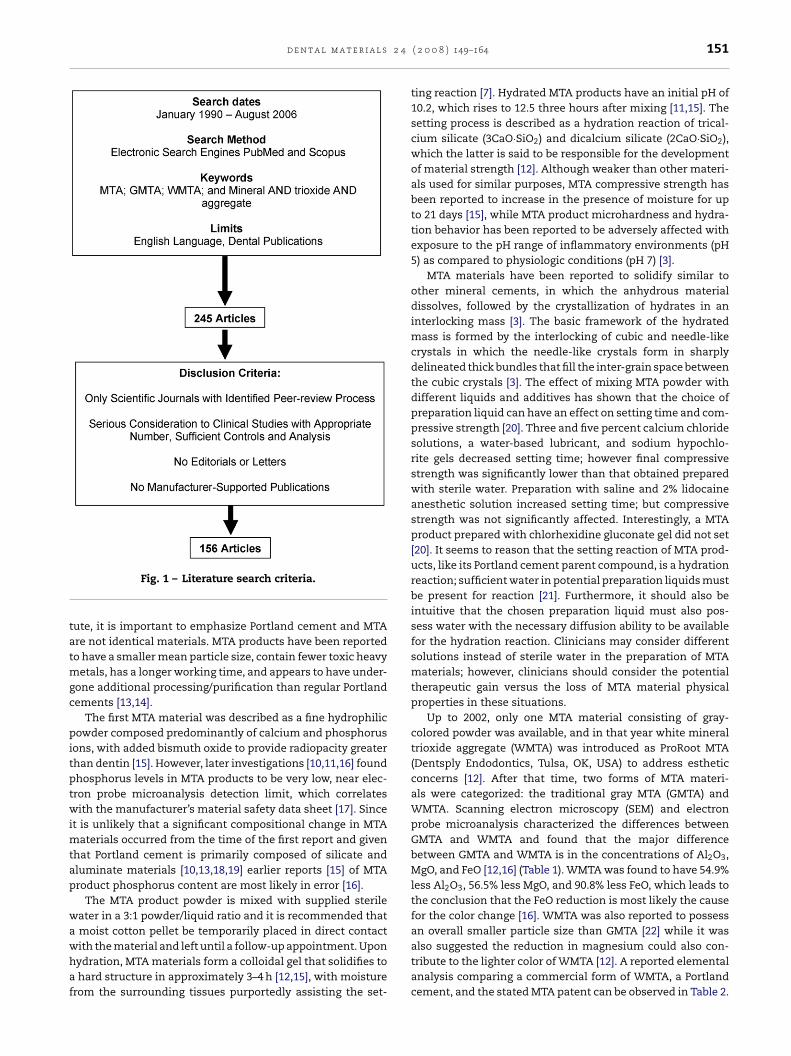
d e n t a l m a t e r i a l s 2 4
tatmgc
pitptwimtap
wawhaf
Fig. 1 – Literature search criteria.
ute, it is important to emphasize Portland cement and MTAre not identical materials. MTA products have been reportedo have a smaller mean particle size, contain fewer toxic heavy
etals, has a longer working time, and appears to have under-one additional processing/purification than regular Portlandements [13,14].
The first MTA material was described as a fine hydrophilicowder composed predominantly of calcium and phosphorus
ons, with added bismuth oxide to provide radiopacity greaterhan dentin [15]. However, later investigations [10,11,16] foundhosphorus levels in MTA products to be very low, near elec-ron probe microanalysis detection limit, which correlatesith the manufacturer’s material safety data sheet [17]. Since
t is unlikely that a significant compositional change in MTAaterials occurred from the time of the first report and given
hat Portland cement is primarily composed of silicate andluminate materials [10,13,18,19] earlier reports [15] of MTAroduct phosphorus content are most likely in error [16].
The MTA product powder is mixed with supplied sterileater in a 3:1 powder/liquid ratio and it is recommended thatmoist cotton pellet be temporarily placed in direct contact
ith the material and left until a follow-up appointment. Uponydration, MTA materials form a colloidal gel that solidifies tohard structure in approximately 3–4 h [12,15], with moisturerom the surrounding tissues purportedly assisting the set-
( 2 0 0 8 ) 149–164 151
ting reaction [7]. Hydrated MTA products have an initial pH of10.2, which rises to 12.5 three hours after mixing [11,15]. Thesetting process is described as a hydration reaction of trical-cium silicate (3CaO·SiO2) and dicalcium silicate (2CaO·SiO2),which the latter is said to be responsible for the developmentof material strength [12]. Although weaker than other materi-als used for similar purposes, MTA compressive strength hasbeen reported to increase in the presence of moisture for upto 21 days [15], while MTA product microhardness and hydra-tion behavior has been reported to be adversely affected withexposure to the pH range of inflammatory environments (pH5) as compared to physiologic conditions (pH 7) [3].
MTA materials have been reported to solidify similar toother mineral cements, in which the anhydrous materialdissolves, followed by the crystallization of hydrates in aninterlocking mass [3]. The basic framework of the hydratedmass is formed by the interlocking of cubic and needle-likecrystals in which the needle-like crystals form in sharplydelineated thick bundles that fill the inter-grain space betweenthe cubic crystals [3]. The effect of mixing MTA powder withdifferent liquids and additives has shown that the choice ofpreparation liquid can have an effect on setting time and com-pressive strength [20]. Three and five percent calcium chloridesolutions, a water-based lubricant, and sodium hypochlo-rite gels decreased setting time; however final compressivestrength was significantly lower than that obtained preparedwith sterile water. Preparation with saline and 2% lidocaineanesthetic solution increased setting time; but compressivestrength was not significantly affected. Interestingly, a MTAproduct prepared with chlorhexidine gluconate gel did not set[20]. It seems to reason that the setting reaction of MTA prod-ucts, like its Portland cement parent compound, is a hydrationreaction; sufficient water in potential preparation liquids mustbe present for reaction [21]. Furthermore, it should also beintuitive that the chosen preparation liquid must also pos-sess water with the necessary diffusion ability to be availablefor the hydration reaction. Clinicians may consider differentsolutions instead of sterile water in the preparation of MTAmaterials; however, clinicians should consider the potentialtherapeutic gain versus the loss of MTA material physicalproperties in these situations.
Up to 2002, only one MTA material consisting of gray-colored powder was available, and in that year white mineraltrioxide aggregate (WMTA) was introduced as ProRoot MTA(Dentsply Endodontics, Tulsa, OK, USA) to address estheticconcerns [12]. After that time, two forms of MTA materi-als were categorized: the traditional gray MTA (GMTA) andWMTA. Scanning electron microscopy (SEM) and electronprobe microanalysis characterized the differences betweenGMTA and WMTA and found that the major differencebetween GMTA and WMTA is in the concentrations of Al2O3,MgO, and FeO [12,16] (Table 1). WMTA was found to have 54.9%less Al2O3, 56.5% less MgO, and 90.8% less FeO, which leads tothe conclusion that the FeO reduction is most likely the causefor the color change [16]. WMTA was also reported to possessan overall smaller particle size than GMTA [22] while it was
also suggested the reduction in magnesium could also con-tribute to the lighter color of WMTA [12]. A reported elementalanalysis comparing a commercial form of WMTA, a Portlandcement, and the stated MTA patent can be observed in Table 2.
152 d e n t a l m a t e r i a l s 2
Table 1 – Chemical compositions of GMTA and WMTA(wt%)
Chemical WMTA GMTA
CaO 44.23 40.45SiO2 21.20 17.00Bi2O3 16.13 15.90Al2O3 1.92 4.26MgO 1.35 3.10SO3 0.53 0.51Cl 0.43 0.43FeO 0.40 4.39P2O5 0.21 0.18TiO2 0.11 0.06
H2O + CO2 14.49 13.72Adapted from Asgary et al. [16].
The setting mechanism of WMTA has been examined usingX-ray photoelectron spectroscopy (XPS) that reported surfacesulfur and potassium species increase 3-fold during the set-ting reaction. This suggested that MTA material setting timecould be prolonged by the formation of a passivating trisulfatespecies layer, which may serve to prevent further hydrationand reaction [12]. This trisulfate species may serve a pro-tective function, as it was reported that that WMTA flexuralstrength was significantly reduced when 2-mm thick layerswere exposed to sterile saline moisture for more than 24 h [23].Calcium release from MTA materials diminishes slightly withtime [22] while MTA materials were reported to form a porousmatrix characterized by internal capillaries and water chan-nels in which increased liquid/powder ratio produced moreporosity and increased solubility [24]. GMTA solubility levelshave been reported to be stable over time, but the usually-reported pH of between 11 or 12 may slightly decrease [25].The high pH level of MTA materials has led some to theorizethat the biologic activity is due to the formation of calciumhydroxide [22–25]. WMTA solubility, hardness, and radiopac-ity has been compared to two Portland cements reporting thatWMTA was significantly less soluble, exhibited greater Vickershardness, and was more radiopaque [26].
There are some evidence that MTA materials possess aprolonged maturation process that continues past the statedsetting time of 3–4 h, as GMTA retention strength for furcationrepairs has been reported to resist significantly more dislodge-
Table 2 – Elemental analysis comparison portlandcement and ProRoot WMTA (wt%)
Element Portland cement WMTA Patent
O 48.1 38.0 30.5Ca 40.3 37.1 37.2Si 6.7 6.5 7.9Al 2.1 0.6 1.7S 1.5 0.9 0.8K 0.9 0.0 0.3Mg 0.3 0.0 1.0Fe 1.0 0.0 2.8Bi 0 16.9 17.9
Adapted from Dammaschke et al. [12].
4 ( 2 0 0 8 ) 149–164
ment at 72 h as compared to 24 h [27]. This was corroboratedby one study that reported increase push-out strength upto 7 days [28] with an additional study reporting maximumGMTA push-out strength observed at 21 days [27]. Interest-ingly, GMTA that has not reached full maturity has beensuggested to possess an ability to re-establish dislodgementresistance after partial displacement; but the re-establishedresistance strength decreased as dislodgement time increasedafter placement [27].
Different intracanal irrigant/oxidizing agents have beenfound to affect the push-out strength of GMTA as it was sus-ceptible to sodium hypochlorite, sodium perborate mixed withsaline, 30% hydrogen peroxide, sodium perborate mixed with30% hydrogen peroxide, and saline at 7 days [29]. GMTA push-out strength was also reported to be similar to Super-EBAand IRM when exposed to saline or sodium hypochlorite, butGMTA was more susceptible to oxidizing agents [29], whichwas reinforced by a report that a hydrogen peroxide-basedcanal preparatory agent significantly reduced the push-outstrength of GMTA to dentin, whereas 2% chlorhexidine and5.25% sodium hypochlorite did not [30]. Another report foundthat perforation retention strength was not affected by prepar-ing GMTA with either saline, sterile water, or lidocaine, butthe bond strength to blood-contaminated root dentin was sig-nificantly less than that observed to uncontaminated dentin[28]. Any adhesion that may be formed between GMTA anddentin may be stronger than the cohesive strength of theGMTA material, as it was reported that GMTA–dentin bondfailures was usually cohesive within the MTA material [28].Furthermore, total GMTA–dentin bond strength is also height-ened by increased surface area, as one report states that 4 mmof GMTA has been reported to afford more resistance to dis-placement than 1-mm thick applications, and was not affectedby previous calcium hydroxide placement [31].
Placement of GMTA using hand condensation techniqueshas been suggested to provide less porosity than ultrasonic-assisted techniques in simulated straight canals [32]. However,different results were found that suggested a denser MTA fillwas obtained in both straight and curved canals with a com-bination hand and ultrasonic placement over a solely manualcondensation technique [33]. GMTA root-end marginal adap-tation and stability was reported to be significantly betterthan a ZOE preparation after being submitted to a computercontrolled, simulated masticating apparatus that producedan estimated 5 year equivalence of chewing cycles [34]. Pre-fabricated posts luted with GMTA were reported to providesignificantly less retentive strength than a glass-ionomer andzinc phosphate luting agents [35]. WMTA has been reportedto strengthen the cervical fracture resistance of immaturesheep incisors as compared to the use of calcium hydroxide[36]. Although this result is considered promising, it should benoted that within the groups sample number were low (<10)and the sample dimensions were also varied in dimension.
WMTA and a ZOE preparation was found to have similarantibacterial properties against Staphylococcus aureus, Entero-coccus faecalis, and Pseudomonas aeruginosa in a direct contact
test [37] while substituting 0.12% chlorhexidine gluconateprovided more antibacterial activity against Actinomyces odon-tolyticus, Fusobacterium nucleatum, Streptococcus sanguis, E.faecalis, Escherichia coli, S. aureus, P. aeruginosa, and Candida
2 4
aTmdrtfmo5uepce
cctnhhaptarataesi
3
Toeut
3
TtmGar[cipreswf
d e n t a l m a t e r i a l s
lbicans than WMTA prepared with sterile water alone [38].his finding should be tempered with knowledge that MTAaterials may not set when mixed with some chlorhexi-
ine preparations [20]. Both freshly mixed and set GMTA waseported to be inhibitory to C. albicans using an antifungalube-dilution method [39] while another study reported dif-erences in that GMTA and WMTA at different powder/liquid
ixtures were not equally effective at preventing the growthf C. albicans [40]. Both WMTA and GMTA in concentrations of0 and 25 mg/ml were equally inhibitive against C. albicans forp to 7 days; however, at lower concentrations only GMTA wasffective [40]. This is evidence of not only the importance ofroper powder/liquid ratios but also raises possible questionsoncerning that the two MTA preparations may not be equallyffective in some clinical applications.
In conclusion, MTA materials are derived from Portlandement, and although it could be inferred that Portlandement could serve as a suitable substitute, it is importanto emphasize that MTA products and Portland cement areot identical materials. MTA materials have been reported toave a smaller mean particle size, contain less heavy metals,ave a longer working time, and appears to have undergonedditional processing/purification than the Portland cementarent compound. WMTA has been marketed since 2002 dueo esthetic considerations and contains less iron, aluminum,nd magnesium oxides than its GMTA counterpart. Both mate-ials undergo a hydration setting reaction that is said to reachn initial set in 3–4 h but whose maturation and resistanceo dislodgement increases with time. The physical propertiesnd setting time of MTA materials can be affected by differ-nt preparation liquids and both WMTA and GMTA have beenhown to possess antibacterial and antifungal activity, whichs presumably due to its pH.
. Microleakage studies
he success of an endodontic material may largely dependn its sealing ability, as most post-treatment endodontic dis-ase is thought to occur due to tissue and other materials inncleaned and/or unobturated areas of the root canal systemhat egress into the surrounding tissues [41].
.1. In vitro dye/fluid filtration method leakage studies
he microleakage of MTA materials compared to other tradi-ional endodontic materials via in vitro dye and fluid filtration
ethods have been the subject of many studies [42–64].MTA has been reported to have less microleakage thanmalgam [42–45,47,48,51,52], zinc-oxide-eugenol (ZOE) prepa-ations [42–44], and a conventional glass-ionomer material59] when used as a root-end restoration following api-al resection. However, other studies reported no differencen leakage between MTA materials and zinc-oxide-eugenolreparations [45,51,52,59], and conventional glass-ionomerestorative materials [48]. The minimal thickness for MTA to
ffectively seal the apical area has been investigated with onetudy reporting a placement thickness of at least 3 mm [49]ith another report stating a minimal of 4 mm is requiredor significant microleakage prevention [50]. The addition of
( 2 0 0 8 ) 149–164 153
calcium chloride has been reported to enhance the sealingability of both GMTA and WMTA, probably by the effect of cal-cium chloride’s enhancement of MTA material setting time[57]. WMTA and GMTA have been compared for the sealingof simulated canals with open apices using thicknesses of 2and 5 mm followed by gutta percha obturation either immedi-ately after MTA material placement or 24 h later [53]. Resultsfound that GMTA had less microleakage than WMTA in sam-ples obturated 24 h after MTA placement; in all groups 5 mmof MTA material allowed less leakage. Based on the results,the authors recommended a 5-mm GMTA apical barrier placedfor treatment of open apices with gutta percha obturation fol-lowed 24 h later [53]. Visual topography evaluations of root-endrestorations restored with GMTA, ZOE materials, and amalgamhave reported that root-end restoration finishing method hadno effect on marginal adaptation of GMTA and ZOE material[63] while another report stated that GMTA appeared to havebetter root-end marginal adaptation than amalgam [64].
For repair of furcation perforations, a ZOE preparation wasreported to provide a better seal than GMTA at 24 h, afterwhich no difference in leakage was observed [60]. However,in another report, GMTA was found to allow more microleak-age in furcation repairs when compared to a ZOE preparationand a self-etch, one step bonding agent [58]. The furcationperforation repair microleakage of GMTA and WMTA was com-pared from both an orthograde and retrograde direction [56].The results found no difference in leakage between the twoMTA materials; but the more interesting findings were thatsignificantly more leakage was found from a microleakagechallenge from an orthograde direction [56]. This suggests animpelling need for an adequate coronal barrier material overMTA furcation repairs to adequately protect against coronalmicroleakage.
The microleakage of MTA materials used for root canalobturation has been reported by two studies [54,61]. The firststudy suggested that GMTA displayed more microleakage thanlaterally-condensed as well as thermoplasticized gutta percha[54] but this was contrasted by the other study which reportedthat both WMTA and GMTA allowed less apical microleak-age than warm, vertically condensed gutta percha [61]. Thesecond study also reported no significant difference in leak-age between GMTA and WMTA, but importantly noted thatroot canal obturation with MTA materials would severelylimit retreatment options and should be considered in onlyselect cases [61]. Another report reported that root resectionof canals obturated with GMTA did not affect its sealing ability[62].
3.2. In vitro bacterial leakage studies
The microleakage of MTA materials has also been evalu-ated, to a lesser extent, using bacterial penetration methods[31,41,65–75]. GMTA has been evaluated for resistance againstapical bacterial leakage when utilized as a root-end fillingcompared with amalgam and ZOE materials within endodon-tically prepared but unobturated root canals inoculated with
Staphylococcus epidermis [41] and Serratia marcescens [65]. GMTAwas found to have significantly more resistance to S. epider-mis penetration than amalgam and ZOE preparations with noleakage evident after 90 days, with the other materials exhibit-
l s 2
154 d e n t a l m a t e r i aing bacterial penetration ranging from 6 to 57 days [41]. Thesecond study found that GMTA resisted S. macescens penetra-tion for up to 49 days after inoculation while the amalgam andZOE materials displayed trends for more bacterial penetration[65]. WMTA and a bonded polymer-based material were foundto exhibit similar root-end bacterial leakage resistance using aStreptococcus salivarius model with both materials having sig-nificantly less bacterial leakage than a ZOE preparation [71].GMTA was also reported to allow significantly less E. coli endo-toxin penetration using a modified Limulus Amebocyte Lysatetest than amalgam and two ZOE preparations over a 12-weekevaluation [69].
In contrast, GMTA was found to have the same bacte-rial penetration resistance as a ZOE preparation, amalgam, abonded resin composite, as well as a bonded amalgam dur-ing a 12-week evaluation using Streptococcus salivarius [68].Similar results were reported during a 47-day study withGMTA compared against a polyacid-modified resin compositeand a ZOE preparation using Prevotella nigrescens [70]. Fur-thermore, WMTA root-end fillings contaminated with eitherblood, saline, or saliva during placement were found to dis-play varying resistance to Staphylococcus epidermidis with salivacontamination causing significantly more leakage [72].
When used as perforation repair materials, GMTA did notdemonstrate any bacterial leakage during a 45-day evaluationwhile approximately half of the amalgam-repaired furcationsallowed penetration and transmission of F. nucleatum [68]. Fur-thermore, no significant difference was found between GMTAand WMTA in the resistance to F. nucleatum penetration whenused for furcation repair [67]. When used in the treatment ofimmature apices, GMTA has been reported to provide resis-tance to bacterial penetration by E. faecalis and S. epidermisbut not Enterobacter aerogenes [31]. A similar report reinforcedGMTA resistance to E. faecalis penetration with no leakageidentified by E. faecalis 16S rDNA polymerase chain reactionassay after 10 days [73]. GMTA was also evaluated against Acti-nomyces viscosus microleakage for up to 70 days in simulatedimmature apices that had received either a 2- or 5-mm apicalGMTA restoration, or a series of 2-mm GMTA apical retrogradefillings. Results reported that only the 5-mm thick restorationresisted microleakage for the entire evaluation, and exhib-ited significantly less leakage compared to the positive controland other GMTA groups [74]. When evaluated as a coronalbarrier, no difference against human saliva bacterial penetra-tion was found between GMTA, WMTA, or a resin-modifiedglass-ionomer restorative material [75]. One study attemptedto evaluate the in vivo coronal sealing ability of WMTA incanine endodontically prepared and obturated root canals, butno conclusive results were found [76].
In conclusion, MTA materials have been investigated usingdye, fluid filtration and bacterial infiltration leakage methods.The majority of the dye and fluid filtration studies suggestthat MTA materials overall allow less microleakage than tra-ditional materials when used as an apical restoration whileproviding equivalent protection as a ZOE preparation whenused to repair furcation perforations. GMTA and WMTA were
shown to provide equivocal results compared against guttapercha when used as a root canal obturation material in thelimited number of microleakage studies. MTA materials havebeen suggested to afford less microleakage than traditional4 ( 2 0 0 8 ) 149–164
materials in a majority of bacteria-based microleakage stud-ies when used as an apical restoration, furcation repair, and inthe treatment of immature apices. In both fluid filtration andbacterial leakage models, 3 mm of MTA material is suggestedas the minimal amount for protection against microleakagewhile 5 mm is suggested in the treatment of immature apices.
3.3. Biocompatibility studies
3.3.1. In vitro studiesIn vitro biocompatibility evaluations of MTA materials havebeen richly reported in the literature [77–103]. The mutagenic-ity of GMTA, ZOE-based, root-end filling materials, as well aspositive and negative controls were evaluated using an Amesmutagenicity assay in which the materials were incubatedwith Salmonella Typhimurium LT-2 strains with reverting bac-teria colony counts measured [77]. None of the root-end fillingmaterials, including GMTA, produced statistically significanthigher Ames test reversion rates, which indicates that none ofthe root-end filling materials would be considered mutagens[77]. Other evaluations have reported no genotoxic effects ofWMTA or Portland cements by single cell gel (comet) assay onperipheral human lymphocytes [78], mouse lymphoma cells[79], as well as Chinese hamster ovary cells [80]. Taken as awhole, none of the studies have shown genotoxic effects ofMTA. Although specific carcinogenicity testing for MTA mate-rials was not found in the literature, it is thought that allcarcinogens are mutagens. Therefore, based on the existingliterature, it is unlikely that MTA is a carcinogenic substancesince it is not a mutagenic substance.
The cytotoxicity of GMTA, amalgam, ZOE, as well aspositive and negative controls was measured using a cellviability assay for mitochondrial dehydrogenase activity inhuman periodontal ligament fibroblasts after 24-h exposureto extracts of varying concentrations of the test materi-als, in both freshly mixed and 24-h set states [82]. In thefreshly mixed state, the sequence of toxicity was amal-gam > Super-EBA > MTA. In the 24-h set state, the sequence oftoxicity at a low extract concentration was Super-EBA > MTA,amalgam; while at higher extract concentrations was Super-EBA > amalgam > MTA [82]. Similarly, another report reinforcedthat GMTA did not negatively affect human periodontal lig-ament fibroblast mitochondrial dehydrogenase activity [86].SEM analysis of periodontal ligament fibroblasts was foundto have a normal morphology and exhibit growth and attach-ment to 24-h set MTA surfaces [83]. However, in the freshlymixed GMTA samples, the cells were round, less in density,exhibited surface defects, and lacked attachment to MTA [83].If the quality and quantity of cell attachment to the root-end filling materials can be used as a criterion to evaluatematerial’s toxicity, then set GMTA appears to be less cyto-toxic than fresh GMTA [83]. In a comparable study involvingresected root surfaces, PDL cell attachment was observed onGMTA but was absent on gutta percha [87]. Similarly, PDLfibroblasts have been reported to display enhanced prolifer-ation on WMTA in a study that analyzed cellular metabolic
activity [88]. These analyses indicated that WMTA induced ageneral osteogenic phenotype in PDL fibroblasts, with induc-tion of alkaline phosphatase activity, as well as production ofosteonidogen, osteonectin, and osteopontin [88].
2 4
wlocntitwGracsnprfPm
icsooaiaaicw
wadtmtatccbfbSva
i(irfoOa
d e n t a l m a t e r i a l s
The cytotoxicity of amalgam, ZOE preparations, and GMTAas reported via an ATC L-929 mouse fibroblast agar over-
ay and radiochromium method [81]. Results of the agarverlay found that set amalgam was significantly less toxicompared to the other materials, while the set GMTA was sig-ificantly less toxic than the ZOE preparations. Contrasting,he radiochromium method suggested that GMTA was signif-cantly less toxic than amalgam. Although it was suggestedhat the increased agar cytotoxicity of the ZOE preparationsas due to the leaching of eugenol, the report concluded thatMTA was no more cytotoxic than other root-end filling mate-
ials currently in use [81]. These findings were reinforced bynother study using human gingival fibroblasts and a L-929ell line [85]. GMTA and a CP titanium alloy was found to haveimilar affect on gingival fibroblast cellular activity, causingo negative affect on cell viability, Prostaglandin E2 assays,rotein and lactate synthesis, and cell proliferation. Overallesults indicated that gingival fibroblast growth was similaror both GMTA and titanium, as either material did not initiateGE2 release or cause alteration of gingival fibroblast cellularetabolism [84].The high pH value of freshly mixed GMTA was found to
nduce cell lysis in L-929 mouse fibroblasts and macrophageell lines in direct contact with the material; however,et GMTA demonstrated favorable biocompatibility with nobserved effect on cell morphology as well as limited impactn cell growth at 72 h [89]. WMTA, as well as calcium hydroxidend a ZOE sealer, was shown not to affect the cell viabil-ty or the Prostaglandin E2 synthesis of murine macrophagesnd fibroblasts [91]. In a different study, murine fibroblastnd macrophage cells displayed significantly greater cytotox-city using flow cytometry with WMTA prepared with 0.12%hlorhexidine gluconate than to WMTA prepared with sterileater [90].
MG-63 cultured human osteoblasts were exposed to GMTAith cellular response evaluated via alkaline phosphatase
ctivity as well as inflammatory cytokine and osteocalcin pro-uction [92]. The MG-63 cells were found to adhere closelyo the GMTA surface while cytokines for osteoclast recruit-
ent (M-CSF) and activation (IL-1�, IL-1�, IL-6) were foundo be produced, along with observed osteocalcin productionnd alkaline phosphatase activity [92]. This led to specula-ion that GMTA causes osteoblast adhesion with release ofytokines from the attached osteoblasts resulting in osteo-last activation via coupled resorption. Therefore, MTA mighte considered a suitable substitute for PMMA when usedor an orthopedic bone cement [92]. This was corroboratedy another study that found that MG-63 osteoblast-like andaos-2 human osteosarcoma cells exposed to GMTA exhibitediability, attachment, proliferation, and collagen productionfter 24 h [96].
ELISA assays have been used to assess the osteocompat-bility of GMTA by monitoring the expression of InterleukinIL)-1�, IL-6, IL-8, IL-11 and macrophage colony stimulat-ng factor (M-CSF) [93]. Although osteoblast cell growth waseported, production of IL-1� and IL-11 were not detected
rom the cells exposed to the GMTA materials. However,steoblastic IL-6 and IL-8 were detected as well as M-CSF [93].steoblasts in another study were found to demonstrate gooddhesion and spreading on GMTA surface, but did not demon-( 2 0 0 8 ) 149–164 155
strate the same ultrastructural characteristics when exposedto a ZOE preparation and amalgam [94]. GMTA osteocom-patibility was reported after U2OS human osteosarcoma celllines were incubated with GMTA and evaluated using West-ern blot assay [95]. In this study, GMTA had a positive effecton the mitogen-activated protein kinase (MAPK) pathways.The authors also reported that a dose-dependent influencewas present on the extracellular signal-regulated kinase MAPKpathway, which is a known pathway leading to osteoblasticactivation and overgrowth [95]. This was reinforced by anotherstudy that reported both 1- and 28-day cured GMTA and WMTAdisplayed biocompatibility when exposed to a Saos-2 humanosteosarcoma cell line [97], while an additional another studyreported both cell attachment and IL-4 and IL-10 cytokine pro-duction [98].
Freshly mixed or set GMTA has been reported to displaylittle to no neurotoxicity. Neurotoxicity effects were quan-titatively assessed by exposing fetal mice cortical neuronaland glial cells and measuring lactate dehydrogenase activity,an assay for cell death [99]. In this study, an amalgam, ZOEpreparation, and a resin endodontic sealer exhibited neuro-toxicity that affected approximately 50–100% of the neuronaland glial cells, while GMTA exhibited little to no neurotoxicity[99]. GMTA has also been reported to be biocompatible with amurine cementoblast model, with the cementoblasts display-ing ultrastructural attachment to GMTA surface with normalreverse transcriptase polymerase chain reaction analysis (RT-PCR) indicating osteocalcin production [100]. Another studysuggested that WMTA was more biocompatible than GMTAin supporting human cementoblast and keratinocyte growth[103].
WMTA effect on dental pulp cell viability and proliferationhas been evaluated using mouse MDPC-23 odontoblast-likecells and OD-21 undifferentiated pulp cells. After 24-h expo-sure to WMTA, apoptosis was not induced in either cell line,and WMTA was reported to cause DNA synthesis increase,suggesting a positive effect on cellular proliferation [101]. Thiswas reinforced by another report that suggested that WMTAhad more of a stimulating effect on human dental pulp cellsthan a commercial calcium hydroxide preparation [102].
3.3.2. In vivo studiesGMTA was reported to induce little or no inflammation com-pared to a ZOE preparation when implanted into guinea pigmandibles, with one GMTA sample demonstrating bone for-mation on its surface [104]. Similar tissue reactions with bothGMTA and Portland cement that demonstrate direct bonedeposition on the materials’ surfaces have been reported[105]. Another study found no inflammation difference inrat connective tissue exposed to both WMTA and a Port-land cement mixture [106]. An additional study reported amore favorable tissue reaction to GMTA compared to amal-gam and two ZOE preparations that were implanted inguinea pig tibias and mandibles with direct bone appositionobserved on some GMTA samples [107]. However, a differ-ent study found no difference in rat bone tissue reaction
between WMTA, GMTA, amalgam, and an epoxy-based, cal-cium hydroxide root canal sealer [108]. In a rat connectivetissue model, GMTA was observed to induce calcificationwhich served as a nidus for ossification [109], whereas in a
l s 2
156 d e n t a l m a t e r i asimilar model amalgam, WMTA, and GMTA were reportedto have similar Cox inflammatory cell grading at 3 weeks,although the amalgam samples exhibited more severe inflam-mation at the study onset than the MTA samples [110]. GMTAand a calcium hydroxide-containing root canal sealer wasreported to be well tolerated when implanted into rabbit earchamber connective tissue, although the root canal sealerinduced connective tissue dissolution with precipitate barrierformation [111].
When used as a root-end restoration in a canine model,GMTA was reported to be associated with significantly lessperiapical inflammation than amalgam at both 5 and 18 weeksafter placement, with almost all of the GMTA specimensexhibited new cementum tissue growth on the GMTA surface[112]. In another study, GMTA and an epoxy-based, root canalcement both exhibited excellent canine peri-radicular tissueresponse at 60 days with no statistically significant differencebetween the materials for new cementum, bone, or periodon-tal ligament formation [113]. These results were corroboratedby a report that evaluated the canine peri-radicular responseto GMTA and a ZOE preparation which found the presence ofperiodontal ligament formation and hard tissue ingrowth onthe GMTA surface [114].
For periapical surgery on obturated canals associatedwith induced periapical lesions in a canine model, GMTAwas reported to induce a favorable periapical tissue heal-ing response compared to amalgam and a ZOE preparation[115]. Blinded histologic evaluation 42 days after periapicalsurgery reported that tissues adjacent to GMTA displayed aminor degree of inflammation, while tissues adjacent to theZOE preparation moderate inflammation. Tissues adjacent toamalgam exhibited marked inflammation. Only the GMTAgroups exhibited cementum growth over the root-end fillingmaterial which led to speculation that the new cementummay have originated from both the periodontal ligament andalveolar bone [115]. Using a canine model, neither freshly pre-pared nor fully-set GMTA was found to make a difference inperiapical healing, with new cementum deposition and bonehealing observed in both groups [116]. A subsequent studyevaluated the healing of canine periapical tissues after cal-cium sulfate and GMTA placement following peri-radicularsurgery. Histologic analysis 4 months post-surgery revealedthat simultaneous use of calcium sulfate and GMTA does notsignificantly affect peri-radicular healing [117].
Periapical tissue response to GMTA and zinc-free amalgamroot-end filling materials using a mammalian Cynomol-gus monkey model found that at 5 months after surgery,peri-radicular tissues adjacent to the amalgam restorationsdisplayed moderate to severe inflammation with fibrous cap-sule formation, while only one tissue specimen adjacent to theGMTA material displayed inflammation [118]. For both groups,cementum was observed to have re-formed associated withthe resected root surface, but no cementum was observed onthe amalgam surface. However, cementoblast activity associ-ated with thick cementum was observed on the GMTA surfacewith five of six specimens, with some specimens exhibiting
new periodontal fiber insertion [118].For furcation repair, GMTA and amalgam were comparedusing a canine model using both an immediate- and delayed-repair scenario [119]. Endodontically treated teeth with
4 ( 2 0 0 8 ) 149–164
standardized furcation perforations were repaired with bothmaterials either immediately or after 6 weeks of salivary con-tamination. For the immediate-repair situation at 4 months,the amalgam samples were all associated with inflammationand no repair site cementum formation. The GMTA-repairedspecimens were characterized by lack of inflammation withcementum formation noted in five of six specimens. For thedelayed-repair group, half of the GMTA-repaired specimenswere free from inflammation with cementum formation.The delayed-repair group with amalgam-restored specimensexhibited either moderate or severe inflammation. Althoughthis study did not use statistical analysis, the authors con-cluded that GMTA had potential when used for furcationrepairs [119].
GMTA was compared to a resin-based, calcium hydrox-ide root canal sealer repairing lateral root perforations at thejunction of the middle and coronal thirds using a caninemodel [120]. At 30 days, GMTA-treated samples displayedeither cementum deposition and/or small areas of ankylo-sis adjacent to the perforation site. Furthermore, all areaswere reported to exhibit little inflammation except for areaswith GMTA overfill. However, the root canal sealer largelyinduced chronic inflammation and ankylosis, with localizedperiodontal ligament necrosis associated with overfilled areas[120]. At 180 days, GMTA-repaired specimens exhibited noankylosis with most specimens exhibiting healing charac-terized by cellular cementum formation with PDL formationbetween the cementum and alveolar bone. In contrast, sealer-repaired specimens exhibited some cementum formation butwas associated with a chronic inflammatory response consist-ing of foreign body giant cells and macrophages. Although theauthors reported that this study supported the use of GMTAfor perforation repair, there was no statistical analysis of thedata [120]. The histologic response of canine periapical tissueswas reported comparing GMTA and a glass-ionomer materialused as obturation materials. Six months after obturation, allroot canals obturated with GMTA exhibited apical closure withnew cementum formation, whereas only partial cementumclosure was observed in a minority of the glass-ionomer mate-rials. Although both materials were reported to exhibit goodbiocompatibility, the authors suggested that GMTA exhibitedbetter biologic properties [121].
In conclusion, regarding the biocompatibility of MTA mate-rials, studies in general tend to support the biocompatibilityof both GMTA and WMTA, although cytotoxicity studies areinclined to suggest less response to the set material as com-pared to the freshly prepared material. Nevertheless, thepublished literature tends to support the biocompatibility ofboth the set material and freshly prepared material, especiallyin relation to other dental materials.
3.4. Characterization of MTA biocompatibility
Numerous studies have been devoted to evaluate the biocom-patibility of GMTA and WMTA. Interestingly, only a few studieshave attempted to identify the specific quality of MTA mate-
rials that provides their biocompatible nature. Some reportsspeculated that MTA material biocompatibility was derivedfrom calcium hydroxide formation [11,22,24,90,120], and onereport did observe the formation of a white interfacial mate-
2 4
rp
sr1wSppatttiacraIhuo
4a
4
4GcwmIspsttbtcpwwditcfsmctd
ta
d e n t a l m a t e r i a l s
ial between GMTA and tooth structure when exposed to ahosphate-buffered physiologic solution [48].
Sarkar et al. [10] reported the first investigation aimedolely at investigating the biocompatible nature of MTA mate-ials and reported the formation of white precipitates within–2 h on the GMTA surface along with suspended precipitatesithin the physiologic phosphate-buffered saline solution.EM analysis of these precipitates revealed a globular mor-hology with chemical composition of oxygen, calcium, andhosphorus, along with trace amounts of bismuth, silicon, andluminum, while X-ray diffraction (XRD) analysis suggestedhe presence of hydroxyapatite, although it should be notedhat the calcium-to-phosphorus ratios reported differed fromhat reported of hydroxyapatite [10]. In spite of this dispar-ty, this report was reinforced by Bozeman et al. [122] wholso used XRD and SEM analysis of both WMTA and GMTArystal precipitates under the same conditions. This reporteinforced that the crystal precipitates on both MTA materi-ls were chemically and structurally similar to hydroxyapatite.nterestingly, GMTA was found to produce twice as muchydroxyapatite crystals as WMTA, which leads to some spec-lation that GMTA and WMTA may not possess the same levelf bioactivity [122].
. Clinical applications of mineral trioxideggregate materials
.1. Pulp-capping
.1.1. Animal modelsMTA has been compared with calcium hydroxide as a pulp-apping medicament using a cynomolgus monkey model inhich GMTA was found associated with little tissue inflam-ation and a thick and continuous dentin bridge at 5 months.
n contrast, only one-third of the calcium hydroxide-treatedpecimens exhibited dentin bridge formation with all dis-laying severe tissue inflammation [123]. A canine modeltudy reported similar results with GMTA exhibiting goodissue response and dentinal bridge formation while one-hird of the calcium hydroxide specimens exhibited dentinridge formation with 75% of the specimens displaying bac-eria and chronic pulp tissue inflammation [124]. Anotheranine model study reported that GMTA when used as aulp-capping medicament induced an osteodentin matrix at 3eeks that was typically observed with reparative dentin [125],hile WMTA in a different study was reported to exhibit neo-entinal bridge formation at 2 weeks with an ultrastructural
ntimate relationship observed between the pulpal tissues andhe WMTA crystals [126]. WMTA and GMTA used as pulp-apping agents were both reported to form calcified bridgeormation in which all WMTA and a majority of the GMTApecimens exhibited complete calcified bridge formation withild inflammatory reactions at 2 weeks [127]. In a rodent pulp-
apping model, GMTA was reported to induce complete hardissue bridge formation at 2 weeks that stained positive for
entin sialoprotein [128].GMTA was reported to foster the same tissue reaction aswo sterilized Portland cement preparations when used as
pulpotomy medicament in a canine model, with all four
( 2 0 0 8 ) 149–164 157
materials associated with a dentin bridge with normal pul-pal tissue at 120 days [129]. WMTA was reported to produceboth incomplete and complete barrier formation with mild tis-sue inflammation when used as an apexification medicamentin a canine model. Specimens that were treated solely withWMTA were found to produce barriers within the root confineswhereas specimens treated first with calcium hydroxide hadmostly incomplete barrier formation that were predominatelyextracanal, beyond the previous apical area [130].
4.2. Human studies
4.2.1. Pulp-cappingA prospective study compared calcium hydroxide and GMTAas permanent dentition pulp-capping medicaments usingthird molars with mature apices in a split-mouth design [131].Mechanical pulp exposures in 11 pairs of maxillary thirdmolars were analyzed at 1 week, and at 2, 3, 4, and 6 monthsafter treatment. The calcium hydroxide specimens were hall-marked by tissue inflammation with a 0.15-mm thick dentinalbridge with adjacent pulp tissue necrosis noted at 6 months.These findings were contrasted with GMTA specimens dis-playing mild tissue reactions with a 0.28-mm dentin bridgenoted at 2 months, with 6-month specimens displaying 0.43-mm dentin bridge formation, no pulp tissue inflammation, allassociated with a near-regular odontoblastic layer [131]. How-ever, the authors did acknowledge a small sample size and theneed for further studies. A second prospective study comparedWMTA and a calcium hydroxide preparation as direct pulp capmedicaments in 48 third molars in a single-blinded, random-ized, controlled clinical study [132]. At 30-days post-treatment,the WMTA group had 20 teeth with clinically normal pulpalstatus while three were diagnosed with reversible pulpal dis-ease. The calcium hydroxide group had 17 teeth with normalpulpal signs, 6 exhibited signs of reversible pulpal disease,and 1 was diagnosed with irreversible pulpal disease. At the136-day recall, all 23 teeth present for the WMTA group wereclinically diagnosed as successful as well as 22 teeth of thecalcium hydroxide group. At both evaluation periods, no sig-nificant difference was found between the groups in regards tothe clinical presentation as well as the histologic status [132].Two case studies involving GMTA used as a deciduous pulp-capping agent and in treatment of dens evaginatus reportedgood follow-up results with minimal clinical or radiographicpathology [133,134].
For use of MTA materials as direct pulp cap medicaments,the two clinical prospective studies suggest that both GMTAand WMTA may perform equally as well as traditional calciumhydroxide in non-carious mechanical pulp exposures in teethwith normal pulp tissue. Although the initial results are posi-tive, further clinical studies are needed, especially in the moreclinically relevant situations involving carious pulp exposuresbefore MTA materials can be unequivocally indicated for adirect pulp-capping agents.
4.2.2. Pulpotomy dressing
There are seven prospective studies involving the use of MTAmaterials as pulpotomy dressings for primary teeth [135–141]while two studies investigated a similar role in permanentteeth [142,143].
l s 2
158 d e n t a l m a t e r i aGMTA and formocresol were compared as pulpotomydressings in primary molars with carious pulp exposures, withonly one reported failure (internal resorption in a formocresol-treated specimen) in the 32 teeth available for evaluationranging 6–30 months [135]. Pulp canal obliteration was notedat a higher frequency in GMTA-treated specimens (7/17) thanthat seen with formocresol (2/15) [135]. Another study com-pared GMTA, WMTA, and formocresol as pulpotomy dressingsin primary teeth demonstrating radiographic caries pulpal
involvement with recalls at 1, 3, 6, and 12 months [136]. Allteeth were judged as clinical and radiographic successes at1 month, while at 3 months one WMTA-treated tooth faileddue to abscess formation; all remaining teeth were ratedFig. 2 – (A) Preoperative radiograph of maxillary central incisor. (WMTA apical restoration placed. (C) Post-operative radiograph. Im
4 ( 2 0 0 8 ) 149–164
successful at 6 months. At 12 months all GMTA specimenswere judged to be successful, but three WMTA-treated teethwere found to be clinical and radiographic failures, alongwith two formocresol-treated teeth. GMTA as found to providea significantly better outcome than WMTA, with no differ-ence found between WMTA and formocresol [136]. Theseresults were contrasted by a different randomized, prospectivestudy that compared formocresol and WMTA as pulpotomymedicaments in primary molars. At 24 months none of
the WMTA-treated teeth exhibited clinical or radiographicpathology while the formocresol-treated teeth demonstratedapproximately 13% radiographic and 2% clinical failure [139].Another longer (range 4–74, mean 38 months) prospective,B) Reveals apical surgical procedure accomplished withages courtesy of Dr. Brian Min.
2 4
rrsprcitWatwo
i[tg[blpcsdwam
tpatprtgtrtcactimIr
cpidhfp2p6d
4.2.3. Other MTA material useCompared to other clinical usage of MTA materials, very fewclinical studies exist that report the outcome of clinical use of
d e n t a l m a t e r i a l s
andomized study found both formocresol and a MTA mate-ial (authors did not delineate MTA material type) equallyuccessful statistically when used as pulpotomy dressings inrimary molars with carious pulp exposures [140]. Similaresults were reported in a 12-month study in which WMTA wasompared with calcium hydroxide for pulpotomy treatmentn 90 cariously exposed primary molars [141]. In this study,reatment was curiously provided in two sessions, in which
MTA and/or the calcium hydroxide paste was applied aftern interim dressing of a corticosteroid/antibiotic solution. Athe end of the evaluation period six failures had occurredith the calcium hydroxide treatments and two failures hadccurred with the WMTA treatments [141].
GMTA and WMTA were evaluated as pulpotomy dress-ngs for primary molars in two different short-term studies137,138] by the same group of researchers. The first reportedhat GMTA exhibited clinical success at 6 months with radio-raphic dentin bridges observed in 50% of the specimens137]. The second study found similar results with WMTAut radiographic analysis was limited only to the mandibu-
ar teeth. Within this limitation, results were that 69% of theulp canals demonstrated signs of stenosis, 11.5% of the pulpanals exhibited dentin bridges, and one canal exhibited pos-ible early signs of internal resorption adjacent to the WMTAressing [138]. In these two studies no statistical differenceas found in the rate of pulp canal stenosis between GMTA
nd WMTA whereas GMTA was found to produce significantlyore dentin bridges [137,138].One prospective clinical study reported GMTA as a pulpo-
omy medicament in 31 vital, cariously exposed, first molarermanent teeth [142]. At 24 months, 79% of the 28 teeth avail-ble for evaluation maintained a positive response to vitalityesting with the remainder free of clinical or radiographicathology. Sixty-four percent of the specimens had pulpaladiographic hard tissue bridge formation, while seven teethhat initially presented with immature apices displayed radio-raphic signs of continued root development [142]. Althoughhis study is favorable, it should be noted that the teeth wereestored immediately with the manufacturer recommenda-ion of interim placement of the MTA dressing with a moistotton pellet not observed. However, this did not appear toffect the results and could identify the importance of an earlyoronal seal provided by a definitive restoration, especially inhe pediatric population. Furthermore, it would have been ofnterest if this study had been able to compare other treat-
ent groups using traditional pulpotomy dressing materials.t is hoped that the authors will report the continued follow-upesults of this work.
The histologic pulpal response comparing WMTA to cal-ium hydroxide as pulpotomy dressings was investigated inremolar teeth extracted for orthodontic purposes, report-
ng that WMTA induced a more homogenous and continuousentin bridge with less pulpal inflammation than calciumydroxide at both 4 and 8 weeks after treatment [126]. Aavorable outcome was also reported in a private endodonticractice assessment using WMTA as a pulpotomy dressing in
3 permanent teeth that exhibited clinical signs of irreversibleulpal disease [143]. Of the 19 teeth available for recall (range–53 months) one tooth presented signs of persistent pulpalisease; this case did not receive a permanent restoration and( 2 0 0 8 ) 149–164 159
presented with recurrent caries upon recall. Based on theselimited results, the authors reported a Kaplan–Meier survivalprobability of 0.95 [143].
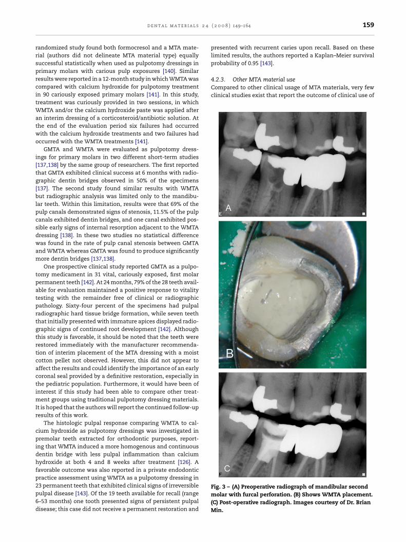
Fig. 3 – (A) Preoperative radiograph of mandibular secondmolar with furcal perforation. (B) Shows WMTA placement.(C) Post-operative radiograph. Images courtesy of Dr. BrianMin.

l s 2
r
160 d e n t a l m a t e r i a
MTA materials as root-end filling materials and root repair, asin the clinical cases noted in Figs. 2 and 3. A prospective 24-month clinical study compared GMTA to a ZOE preparationas root-end filling materials in 122 adult patients referred forendodontic surgery [144]. At both 12- and 24-month recalls,acceptable results were noted with both materials; while theauthors implied a higher success rate with GMTA treatment,no statistical difference was noted between the two materials[144]. It is hoped that this 24-month report will continue tobe followed. A retrospective study concerning GMTA use forroot perforation repair in 16 cases within an endodontic resi-dency caseload has been reported [145]. This report involvedfive lateral root perforations, five strip perforations, three fur-cation perforations, and three apical perforations that weretreated with a follow-up range of 12–45 months. Results werethat all treatments demonstrated clinical and radiographicsigns of healing with return of normal radiographic architec-ture to the repair sites [145]. Although these initial results arepromising, this study represents a limited number of clinicalcases and no comparison group was included. Clinical casereports in which GMTA has been used to repair horizontal rootfractures, root resorption, internal resorption, and furcationperforations with both clinical and radiographic success havealso been reported [146–152].
At the time of this review, no prospective studies usingMTA materials for apexification and/or apexogenesis pro-cedures have been reported. However, successful individualcase reports of using GMTA and/or WMTA for apexifica-tion/apexogenesis treatments do exist [147,148,153–156].
5. Conclusion
The physical properties, sealing ability, biocompatibility, andclinical performance of MTA materials have been discussed.MTA materials appear not only to demonstrate acceptablebiocompatible behavior but also exhibits acceptable in vivobiologic performance when used for root-end fillings, perfora-tion repairs, pulp-capping and pulpotomy, and apexificationtreatment. However, it should be noted that the supportingdata have been overwhelmingly from either in vitro or animalstudies. Reports have strongly suggested that the favorablebiologic performance exhibited by MTA materials is due tohydroxyapatite formation when these materials are exposedto physiologic solutions. Although some studies suggest thatthe less-expensive Portland cement parent compound couldpossibly be used in the place of MTA, characterization studieshave shown that MTA materials are compositionally differ-ent than Portland cement and it is not recommended atthis time that Portland cement can serve as a suitable MTAsubstitute. GMTA has been investigated more than the more-esthetic WMTA, and while some reports suggest that GMTAmay invoke a more desirable biologic response than WMTA,existing reports as a whole are equivocal and more stud-ies are encouraged. Although the overall results in human
studies involving MTA materials are very positive, further lon-gitudinal studies are encouraged, as at present insufficientwell-designed and controlled clinical studies exists that allowsystematic and meta-analysis review of MTA materials in allof its suggested clinical indications.4 ( 2 0 0 8 ) 149–164
Acknowledgements
The authors wish to express appreciation to Col. Richard Rut-ledge, Majors Dennis Holt, James Watts, Kim Wilkinson; andCaptain Brian Min at the Endodontic Residency, 59 DentalGroup, Lackland Air Force Base, Texas, for corroboration withthe clinical photographs.
e f e r e n c e s
[1] Nash KD, Brown J, Hicks ML. Private practicingendodontists: production of endodontic services andimplications for workforce policy. J Endod 2002;28:699–705.
[2] Chong BS. Managing endodontic failure in practice.Chicago: Quintessence Publishing Co., Ltd.; 2004. p. 123–47.
[3] Lee YL, Lee BS, Lin FH, Lin AY, Lan WH, Lin CP. Effects ofphysiological environments on the hydration behavior ofmineral trioxide aggregate. Biomaterials 2004;25:787–93.
[4] Johnson BR. Considerations in the selection of a root-endfilling material. Oral Surg Oral Med Oral Pathol Oral RadiolEndod 1999;87:398–404.
[5] Kratchman SI. Perforation repair and one-stepapexification procedures. Dent Clin N Am 2004;48:291–307.
[6] Bryan EB, Wollard G, Mitchell WC. Nonsurgical repair offurcal perforations: a literature review. Gen Dent1999;47:274–80.
[7] Lee SJ, Monsef M, Torabinejad M. Sealing ability of amineral trioxide aggregate for repair of lateral rootperforations. J Endod 1993;19:541–4.
[8] Schmitt D, Bogen G. Multifaceted use of ProRoot MTA rootcanal repair material. Pediatr Dent 2001;23:326–30.
[9] Acute pain management: operative or medical proceduresand trauma. Rockville, MD: US Department of Health andHuman Services, Agency for Health Care Policy andResearch; 1992.
[10] Sarkar NK, Caidedo R, Tirwik P, Moiseyeva R, Kawashima I.Physicochemical basis of the biologic properties of mineraltrioxide aggregate. J Endod 2005;31:97–100.
[11] Camilleri J, Montesin FE, Brady K, Sweeney R, Curtis RV, PittFord TR. The constitution of mineral trioxide aggregate.Dent Mater 2005;21:297–303.
[12] Dammaschke T, Gerth HUV, Zuchner H, Schafer E.Chemical and physical surface and bulk materialcharacterization of white ProRoot MTA and two Portlandcements. Dent Mater 2005;21:731–8.
[13] Abdullah D, Pitt Ford TR, Papaioannou S, Nicholson J,McDonald F. An evaluation of accelerated Portland cementas a restorative material. Biomaterials 2002;23:4001–10.
[14] Islam I, Chng HK, Yap AUJ. Comparison of the physical andmechanical properties of MTA and Portland cement. JEndod 2006;32:193–7.
[15] Torabinejad M, Hong CU, McDonald F, Pitt Ford TR. Physicaland chemical properties of a new root-end filling material. JEndod 1995;21:349–53.
[16] Asgary S, Parirokh M, Egbbal MJ, Brink F. Chemicaldifferences between white and gray mineral trioxideaggregate. J Endod 2005;31:101–3.
[17] ProRoot MTA (Mineral Trioxide Aggregate) Root CanalRepair Material Material Safety Data Sheet, DENTSPLY
Tulsa Dental, accessed at www.tulsadental.dentsply.com;accessed 10 March 2005.[18] Bentz DP, Hansen KK. Preliminary observations of watermovement in cement pastes during using X-ray absorption.Cement Concrete Res 2000;30:1157–68.

2 4
d e n t a l m a t e r i a l s[19] Bentz DP, Geiker MR, Hansen KK. Shrinkage-reducingadmixtures and early-age dessication in cement pastesand mortars. Cement Concrete Res 2001;31:1075–85.
[20] Kogan P, He J, Glickman GN, Watanabe I. The effects ofvarious additives on setting properties of MTA. J Endod2006;32:569–72.
[21] Gancedo L, Garcia-Barbero E. Influence of humidity andsetting time on the push-out strength of mineral trioxideaggregate obturations. J Endod 2006;32:894–6.
[22] Duarte MAH, de Oliveria Demarchi ACC, Yamashita JC,Kuga MC, de Campos Fraga S. pH and calcium ion release oftwo root-end filling materials. Oral Surg Oral Med OralPathol Oral Radiol Endod 2003;95:345–7.
[23] Walker MP, Diliberto A, Lee C. Effect of setting conditionson mineral trioxide aggregate flexural strength. J Endod2006;32:334–6.
[24] Fridland M, Rosado R. Mineral trioxide aggregate (MTA)solubility and porosity with different water-to-powderrations. J Endod 2003;29:814–7.
[25] Fridland M, Rosado R. MTA solubility: a long term study. JEndod 2005;31:376–9.
[26] Danesh G, Dammaschke T, Gerth HUV, Zandbiglari T,Schafer E. A comparitive study of selected properties ofProRoot mineral trioxide aggregate and two Portlandcements. Int Endod J 2006;39:213–9.
[27] Sluyk SR, Moon PC, Hartwell GR. Evaluation of settingproperties and retention characteristics of mineral trioxideaggregate when used as a furcation perforation material. JEndod 1998;24:768–71.
[28] VandeWeele RA, Schwartz SA, Beeson TJ. Effect of bloodcontamination on retention characteristics of MTA whenmixed with different liquids. J Endod 2006;32:421–4.
[29] Loxley EC, Liewehr FR, Buxton TB, McPherson III JC. Theeffect of various intracanal oxidizing agents on thepush-out strength of various perforation repair materials.Oral Surg Oral Med Oral Pathol Oral Radiol Endod2003;95:490–4.
[30] Yan P, Peng B, Fan B, Fan M, Bian Z. The effects of sodiumhypochlorite (5.25%), chlorhexidine (2%), and Glyde FilePrep on the bond strength of MTA-dentin. J Endod2006;32:58–60.
[31] Hachmeister DR, Schindler WG, Walker WA, Thomas DD.The sealing ability and retention characteristics of mineraltrioxide aggregate in a model of apexification. J Endod2002;28:386–90.
[32] Aminoshariae A, Hartwell GR, Moon PC. Placement ofmineral trioxide aggregate using two different techniques. JEndod 2003;29:679–82.
[33] Yeung P, Liewehr FR, Moon PC. A quantitative comparisonof the fill density of MTA produced by two placementtechniques. J Endod 2006;32:456–9.
[34] Peters CI, Peters OA. Occlusal loading of EBA and MTAroot-end fillings in a computer-controlled masticator: ascanning electron microscope study. Int Endod J2002;35:22–9.
[35] Vargas JW, Liewehr FR, Joyce AP, Runner RR. A comparisonof in the in vitro retentive strength of glass-ionomercement, zinc-phosphate cement, and mineral trioxideaggregate for the retention of prefabricated posts in bovineincisors. J Endod 2004;30:775–7.
[36] Andreasen JO, Munksgaard EC, Bakland LK. Comparison offracture resistance in root canals of immature sheep teethafter filling with calcium hydroxide or MTA. Dent Trauma2006;22:154–6.
[37] Eldeniz AU, Hadimli HH, Ataoglu H, Ørstavik D.
Antibacterial effect of selected root-end filling materials. JEndod 2006;32:345–9.[38] Stowe TJ, Sedgley CM, Stowe B, Fenno JC. The effects ofchlorhexidine gluconate (0.12%) on the antimicrobial
( 2 0 0 8 ) 149–164 161
properties of tooth-colored ProRoot mineral trioxideaggregate. J Endod 2004;30:429–31.
[39] Al-Nazhan S, Al-Judai A. Evaluation of antifungal activity ofmineral trioxided aggregate. J Endod 2003;29:826–7.
[40] Al-Hezaimi K, Naghshbandi J, Oglesby S, Simon JHS,Rotstein I. Comparison of antifungal activity ofwhite-colored and gray-colored mineral trioxide aggregate(MTA) at similar concentrations against Candida albicans. JEndod 2006;32:365–7.
[41] Torabinejad M, Rastegar A, Kettering JD, Pitt Ford TR.Bacterial leakage of mineral trioxide aggregate as aroot-end filling material. J Endod 1995;21:109–12.
[42] Torabinejad M, Watson TF, Pitt Ford TR. Sealing ability of amineral trioxide aggregate when used as a root-end fillingmaterial. J Endod 1993;19:591–5.
[43] Torabinejad M, Higa RK, McKendry DJ, Pitt Ford TR. Dyeleakage of four root end filling materials: effect of bloodcontamination. J Endod 1993;20:159–63.
[44] Aqrabawi J. Sealing ability of amalgam, Super EBA cement,and MTA when used as retrograde filling materials. Br DentJ 2000;188:266–8.
[45] Bates CF, Carnes DL, del Rio CE. Longitudinal sealing abilityof mineral trioxide aggregate as a root-end filling material.J Endod 1996;22:575–8.
[46] Fogel HM, Peikoff MD. Microleakage of root-end fillingmaterials. J Endod 2001;27:456–8.
[47] Yatsushiro JD, Baumgartner JC, Tinkle JS. Longitudinalstudy of the microleakage of two root-end filling materialsusing a fluid conductive system. J Endod 1998;24:716–9.
[48] Wu MK, Kontakiotis EG, Wesselink PR. Long-term sealprovided by some root-end filling materials. J Endod1998;24:557–60.
[49] Lamb EL, Loushine RJ, Weller N, Kimborugh WF, PashleyDH. Effect of root resection on the apical sealing ability ofmineral trioxide aggregate. Oral Surg Oral Med Oral PatholOral Radiol Endod 2003;95:732–5.
[50] Valois CR, Costa ED. Influence of the thickness of mineraltrioxide aggregate on the sealing ability of root-end fillingsin vitro. Oral Surg Oral Med Oral Pathol Oral Radiol Endod2004;97:108–11.
[51] Roy CO, Heansonne BG, Gerrets TF. Effect of an acidenvironment on leakage of root-end filling materials. JEndod 2001;27:7–8.
[52] Davis JL, Jeansonne BG, Davenport WD, Gardiner D. Theeffect of irrigation with doxycycline or citric acid onleakage and osseous would healing. J Endod 2003;29:31–5.
[53] Matt GD, Thorpe JR, Strother JM, McClanahan SB.Comparative study of white and gray mineral trioxideaggregate (MTA) simulating a one- or two-step apicalbarrier technique. J Endod 2004;30:876–9.
[54] Vizgirda PJ, Liewehr FR, Patton WR, McPherson JC, BuxtonTB. A comparison of laterally condensed gutta-percha,thermoplasticized gutta-percha, and mineral trioxideaggregate as root canal filling materials. J Endod2004;30:103–6.
[55] Jenkins S, Kulid J, Williams K, Lyons W, Lee C. Sealing abilityof three materials in the orifice of root canal systemsobturated with gutta-percha. J Endod 2006;32:225–7.
[56] Hamad HA, Tordik PA, McClanahan SB. Furcationperforation repair comparing gray and white MTA: a dyeextraction study. J Endod 2006;32:337–40.
[57] Bortoluzzi EA, Broon NJ, Bramante CM, Garcia RB, de MoresIG, Bernardineli N. Sealing ability of MTA and radiopaquePortland cement with or without calcium chloride forroot-end filling. J Endod 2006;32:897–900.
[58] Hardy I, Liewehr FR, Joyce AP, Agee K, Pashley DH. Sealingability of One-Up Bond and MTA and without a secondaryseal as furcation perforation repair materials. J Endod2004;30:658–61.

l s 2
162 d e n t a l m a t e r i a[59] De Bruyne MAA, De Bruyne RJE, Rosiers L, De Moor RJG.Longitudinal study on microleakage of three root-endfilling materials by the fluid transport method and bycapillary flow porometry. Int Endod J 2005;38:129–36.
[60] Weldon JK, Pashley DH, Loushine RJ, Weller RN, KimbroughWF. Sealing ability of mineral trioxide aggregate when useda furcation repair materials: a longitudinal study. J Endod2002;28:467–70.
[61] Al-Hezaimi K, Naghshbandi J, Oglesby S, Simon JHS,Rotstein I. Human saliva penetration of root canalsobturated with two types of mineral trioxide aggregatecements. J Endod 2005;31:453–6.
[62] Andelin WE, Browning DF, Hsu GH, Roland DD, TorabinejadM. Microleakage of resected MTA. J Endod 2002;28:573–4.
[63] Gondim Jr E, Zaia AA, Gomes BFBA, Ferraz CCR, Teixeira FB,Souza-Filho FJ. Investigation of the marginal adaptation ofroot-end cavities prepared with ultrasonic tips. Int Endod J2003;36:491–9.
[64] Shipper G, Grossman ES, Botha AJ, Cleaton-Jones PE.Marginal adaptation of mineral trioxide aggregate (MTA)compared with amalgam as a root-end filling material: alow-vacuum (LV) versus high-vacuum (HV) SEM study. IntEndod J 2004;37:325–36.
[65] Fischer EJ, Arens DE, Miller CH. Bacterial leakage of mineraltrioxide aggregate as compared with zinc-free amalgam,intermediate restorative material, and Super EBA as aroot-end filling material. J Endod 1998;24:176–9.
[66] Nakata TT, Bae KS, Baumgartner JC. Perforation repaircomparing mineral trioxide aggregate and amalgam usingan anaerobic bacterial leakage model. J Endod1998;24:184–6.
[67] Ferris DM, Baumgartner JC. Perforation repair comparingtwo types of mineral trioxide aggregate. J Endod2004;30:422–4.
[68] Adamo HL, Buruiana R, Schertzer L, Boylan RJ. Acomparison of MTA, Super-EBA, composite, and amalgamas root-end filling materials using a bacterial microleakagemodel. Int Endod J 1999;32:197–203.
[69] Tang HM, Torabinejad M, Kettering JD. Leakage evaluationof root end filling materials using endotoxin. J Endod2002;28:5–7.
[70] Scheerer SQ, Steiman HR, Cohen J. A comparativeevaluation of three root-end filling materials: an in vitroleakage study using Prevotella nigrescens. J Endod2001;27:40–2.
[71] Maltezos CM, Glickman GN, Ezzo P, He J. Comparison of thesealing of Resilon, Pro Root MTA, and Super-EBA asroot-end filling materials: a bacterial leakage study. J Endod2006;32:324–7.
[72] Montellano AM, Schwartz SA, Beeson TJ. Contamination oftooth-colored mineral trioxide aggregte used as a root-endfilling material: a bacterial leakage study. J Endod2006;32:452–5.
[73] de Leimburg ML, Angeretti A, Ceruti P, Lendini M,Pasqualini D, Berutti E. MTA obturation of pulpless teethwith open apices: bacterial leakage as detected bypolymerase chain reaction assay. J Endod 2004;30:883–6.
[74] Al-Kahtani A, Shostad S, Schifferle R, Bhambbani S. In-vitroevaluation of microleakage of an orthograde apical plug ofmineral trioxide aggregate in permanent teeth withsimulated immature apices. J Endod 2005;31:117–9.
[75] Tselnik M, Baumgartner JC, Marshall JG. Bacterial leakagewith mineral trioxide aggregate or a resin-modified
glass-ionomer used as a coronal barrier. J Endod2004;30:782–4.[76] Mah T, Basrani B, Santos JM, Pascon EA, Tjaderhane L,Yared G, et al. Periapical inflammation affecting
4 ( 2 0 0 8 ) 149–164
coronally-inoculated dog teeth with root fillings augmentedwith white MTA orifice plugs. J Endod 2003;29:442–6.
[77] Kettering JD, Torabinejad M. Investigation of mutagenicityof mineral trioxide aggregate and other commonly usedroot-end filling materials. J Endod 1995;21:537–9.
[78] Braz MG, Camargo EA, Salvadori DMF, Marques MEA,Ribeiro DA. Evaluation of genetic damage in humanperipheral lymphocytes exposed to mineral trioxideaggregate and Portland cement. J Oral Rehab 2006;33:234–9.
[79] Riberio DA, Duarte MAH, Matsumoto MA, Marques MEA,Salvidori DMF. Biocompatibility in vitro tests of mineraltrioxide aggregate and regular and white Portland cements.J Endod 2005;31:605–7.
[80] Riberio DA, Sugui MM, Matsumoto MA, Duarte MAH,Marques MEA, Salvidori DMF. Genotoxicity and cytotoxicityof mineral trioxide aggregate and regular and whitePortland cements on Chinese hamster ovary (CHO) cells invitro. Oral Surg Oral Med Oral Pathol Oral Radiol Endod2006;101:258–61.
[81] Torabinejad M, Hong CU, Pitt Ford TR, Kettering JD.Cytotoxicity of four root end filling materials. J Endod1995;21:489–92.
[82] Keiser K, Johnson CC, Tipton DA. Cytotoxicity of mineraltrioxide aggregate using human periodontal ligamentfibroblasts. J Endod 2000;26:288–91.
[83] Balto HA. Attachment and morphological behavior ofhuman periodontal ligament fibroblasts to mineral trioxideaggregate: a scanning electron microscope study. J Endod2003;30:25–8.
[84] Pistorius A, Willershausen B, Marroquin BB. Effect of apicalroot-end filling materials on gingival fibroblasts. Int EndodJ 2003;36:610–5.
[85] Osorio RM, Hefti A, Vertucci FJ, Shawley AL. Cytotoxicity ofendodontic materials. J Endod 1998;24:91–6.
[86] Lin CP, Chen YJ, Lee YL, Want JS, Chang MC, Lan WH, et al.Effects of root-end filling materials and eugenol onmitochondrial dehydrogenase activity and cytotoxicity tohuman periodontal ligament fibroblasts. J Biomed MaterRes Part B Appl Biomater 2004;71B:429–40.
[87] Fayad MI, Hawkinson R, Daniel J, Hao J. The effect of CO2
laser irradiation on PDL cell attachment to resected rootsurfaces. Oral Surg Oral Med Oral Pathol Oral Radiol Endod2004;97:518–23.
[88] Bonson S, Jeansonne BG, Lallier TE. Root-end fillingmaterials alter fibroblast differentiation. J Dent Res2004;83:408–13.
[89] Haglund R, He J, Safavi KE, Spangberg LSW, Zhu Q. Effectsof root-end filling materials on fibroblasts andmacrophages in vitro. Oral Surg Oral Med Oral Pathol OralRadiol Endod 2003;95:739–45.
[90] Hernandez EP, Botero TM, Mantellini MG, McDonald NJ, NorJE. Effect of ProRoot MTA mixed with chlorihexidine onapoptosis and cell cycle of fibroblasts and macrophages invitro. Int Endod J 2005;38:137–43.
[91] Melegari KK, Botero TM, Holland GR. Prostaglandin E2production and viability of cells cultured in contact withfreshly mixed endodontic materials. Int Endod J2006;39:357–62.
[92] Koh ET, Torabinejad M, Pitt Ford TR, Brady K, McDonald F.Mineral trioxide aggregate stimulates a biological responsein human osteoblasts. J Biomed Res 1997;37:432–9.
[93] Mitchell PJC, Pitt Ford TR, Torabinejad M, McDonald F.Osteoblast biocompatibility of mineral trioxide aggregate.
Biomaterials 1999;20:167–73.[94] Zhu Q, Haglund R, Safavi KE, Spangberg LS. Adhesion ofhuman osteoblasts on root-end filling materials. J Endod2000;26:404–6.
2 4
d e n t a l m a t e r i a l s[95] Huang TH, Ding SJ, Hsu TC, Kao CT. Effects of mineraltrioxide aggregate (MTA) extracts on mitogen-activatedprotein kinase activity in human osteosarcoma cell line(U2OS). Biomaterials 2003;24:3909–13.
[96] Pellicioni GA, Ciapeti G, Cenni E, Granchi D, Nanni M,Pagani S, et al. Evaluation of osteoblast-like cell response toProRoot MTA (mineral trioxide aggregate). J Mater Sci MaterMed 2004;15:167–73.
[97] Camilleri J, Monstein FE, Papaioannou S, McDonald F, PittFord TR. Biocompatibility of two commercial forms ofmineral trioxide aggregate. Int Endod J 2004;37:699–704.
[98] Huang TH, Yang CC, Ding SJ, Meng Y, Kao CT, Chou MY.Inflammatory cytokines reaction elicited by root-end fillingmaterials. J Biomed Mater Res Part B Appl Biomater2005;73B:123–8.
[99] Asrari M, Lobner D. In vitro neurotoxic evaluation ofroot-end filing materials. J Endod 2003;29:743–6.
[100] Thomson TS, Berry JE, Somerman MJ, Kirkwood KL.Cementoblasts maintain expression of osteocalcin in thepresence of mineral trioxide aggregate. J Endod2003;29:407–12.
[101] Moghaddame-Jafari S, Mantellini MG, Botero TM, McDonaldNJ, Nor JE. Effect of ProRoot MTA on pulp cell apoptosis andproliferation in vitro. J Endod 2005;31:387–91.
[102] Takita T, Hayashi M, Takeichi O, Ogiso B, Suzuki N, OtsukaK, et al. Effect of mineral trioxide aggregate on proliferationof cultured human dental pulp cells. Int Endod J2006;39:415–22.
[103] Oviir T, Pagoria D, Ibarra G, Geurtsen W. Effects of gray andwhite mineral trioxide aggregate on the proliferation of oralkeratinocytes and cementoblasts. J Endod 2006;32:21–13.
[104] Torabinejad M, Hong CU, Pitt Ford TR, Kariyawasam SP.Tissue reaction to implanted Super-EBA and mineraltrioxide aggregate in the mandible of guinea pigs: apreliminary report. J Endod 1995;21:569–71.
[105] Saidon J, He J, Zhu Q, Safavi K, Spangberg LSW. Cell andtissue reactions to mineral trioxide aggregate and Portlandcement. Oral Surg Oral Med Oral Pathol Oral Radiol Endod2003;95:483–9.
[106] de Morais CAH, Bernardineli N, Garcia RB, Duarte AH,Guerisoli DMZ. Evaluation of tissue response to MTA andPortland cement with iodoform. Oral Surg Oral Med OralPathol Oral Radiol Endod 2006;102:417–21.
[107] Torabinejad M, Pitt Ford TR, Abedi HR, Kariyawasam SP,Tang HM. Tissue reaction to implanted root-end fillingmaterials in the tibia and mandible of guinea pigs. J Endod1998;24:468–71.
[108] Cintra LTA, de Moraes IG, Estrada BPF, Gomes-Filho JE,Bramante CM, Garcia RB, et al. Evaluation of the tissueresponse to MTA and MBPC: microscopic analysis ofimplants in alveolar bone of rats. J Endod 2006;32:556–9.
[109] Yaltirik M, Ozbas H, Bilgic B, Issever H. Reactions ofconnective tissue to mineral trioxide aggregate andamalgam. J Endod 2004;30:95–9.
[110] Shahi S, Rahini S, Lotfi M, Yavari HR, Gaderian AR. Acomparative study of the biocompatibility of three root-endfilling materials in rat connective tissue. J Endod2006;32:776–80.
[111] Masuda EL, Wang X, Hossain M, Unno A, Jayawardena JA,Saito K, et al. Evaluation of biocompatibility of mineraltrioxide aggregate with an improved rabbit ear chamber. JOral Rehab 2005;32:145–50.
[112] Torabinejad M, Hong CU, Lee SJ, Monsef M, Pitt Ford TR.Investigation of mineral trioxide aggregate for root-endfilling in dogs. J Endod 1995;21:603–8.
[113] Regan JD, Gutmann JL, Witherspoon DE. Comparison ofDiaket and MTA when used as root-end filling materials tosupport regeneration of the periradicular tissues. Int EndodJ 2002;35:840–7.
( 2 0 0 8 ) 149–164 163
[114] Economides N, Pantelidou O, Tziafas K, Tziafas D.Short-term periradicular tissue response to mineraltrioxide (MTA) as root-end filling material. Int Endod J2003;36:44–8.
[115] Baek S-H, Plenk Jr H, Kim S. Periapical tissue responses andcementum regeneration with amalgam, SuperEBA, andMTA as root-end filling materials. J Endod 2005;31:444–9.
[116] Apaydin ES, Shabahang S, Torabenijad M. Hard-tissuehealing after application of fresh or set MTA asroot-end-filling material. J Endod 2003;30:21–4.
[117] Apaydin ES, Torabinejad M. The effect of calcium sulfate onhard-tissue healing after periradicular surgery. J Endod2004;30:17–20.
[118] Torabinejad M, Pitt Ford TR, McKendry DJ, Abedi HR, MillerDA, Kariyawasam SP. Histologic assessment of mineraltrioxide aggregate as a root-end filling in monkeys. J Endod1997;23:225–8.
[119] Pitt Ford TR, Torabinejad M, McKendry DJ, Hong CU,Kariyawasam SP. Use of mineral trioxide aggregate forrepair of furcal perforations. Oral Surg Oral Med Oral PatholOral Radiol Endod 1995;79:756–63.
[120] Holland R, Filho JAO, de Souza V, Nery MJ, Bernabe PFE.Mineral trioxide aggregate repair of lateral rootperforations. J Endod 2001;27:281–4.
[121] Holland R, de Souza V, Nery MJ, Filho JAO, Bernabe PFE,Dezan E. Reaction of dog’s teeth to root canal filling withmineral trioxide aggregate or a glass-ionomer sealer. JEndod 1999;25:728–30.
[122] Bozeman TB, Lemon RR, Eleazer PD. Elemental analysis ofcrystal precipitate from gray and white MTA. J Endod2006;32:425–8.
[123] Pitt Ford TR, Torabinejad M, Abredi HR, Bakland LK,Kariyawasam SP. Using mineral trioxide aggregate as apulp-capping material. J Am Dent Assoc 1996;127:1491–4.
[124] Faraco IM, Holland R. Response of the pulp of dogs tocapping with mineral trioxide aggregate of a calciumhydroxide cement. Dent Traumatol 2001;17:163–6.
[125] Tzias D, Pantelidou O, Alvanou A, Belibasakis G,Papadimitriou S. The dentinogenic effect of mineraltrioxide aggregate (MTA) in short-term cappingexperiments. Int Endod J 2002;35:245–54.
[126] Chacko V, Kurikose S. Human pulpal response to mineraltrioxide aggregate (MTA): a histologic study. J Clin PediatrDent 2006;30:203–9.
[127] Parirokh M, Asgary S, Eghbal MJ, Stowe S, Eslami B,Eskandarizade A, et al. A comparative study of white andgrey mineral trioxide aggregate as pulp capping in dog’steeth. Dent Traumatol 2005;21:150–4.
[128] Andelin WE, Shabahang S, Wirght K, Torabinejad M.Identification of hard tissue after experimental pulpcapping using dentin sialoprotein (DSP) as a marker. JEndod 2003;29:646–50.
[129] Menezes R, Bramante CM, Letra A, Carvalho VGG, GarciaRB. Histologic evaluation of pulpotomies in dog using twotypes of mineral trioxide aggregate and regular and whitePortland cements as wound dressings. Oral Surg Oral MedOral Pathol Oral Radiol Endod 2004;98:376–9.
[130] Felippe WT, Felippe MCS, Rocha MJC. The effect of mineraltrioxide aggregate on the apexification and periapicalhealing of teeth with incomplete root formation. Int EndodJ 2006;39:2–9.
[131] Aeinehchi M, Eslami B, Ghanbariha M, Saffar AS. Mineraltrioxide aggregate (MTA) and calcium hydroxide as
pulp-capping agents in human teeth: a preliminary report.Int Endod J 2002;36:225–31.[132] Iwamoto CE, Adachi E, Pameijer CH, Barnes D, Romberg EE,Jeffries S. Clinical and histological evaluation of white

l s 2
non-vital immature incisor with mineral trioxide aggregate
164 d e n t a l m a t e r i a
ProRoot MTA in direct pulp capping. Am J Dent2006;19:85–90.
[133] Bodem O, Blumenshine S, Zeh D, Koch MJ. Direct pulpcapping with mineral trioxide aggregate in a primarymolar: a case report. Int J Pediatr Dent 2004;14:376–9.
[134] Koh ET, Pitt Ford TR, Kariyawasam SP, Chen NN,Torabenijad M. Prophylactic treatment of dens evaginatususing mineral trioxide aggregate. J Endod 2001;27:540–2.
[135] Eidelman E, Holan G, Fuks AB. Mineral trioxide aggregatevs. formocreosol in pulpotomized primary molars: apreliminary report. Pediatr Dent 2001;23:15–8.
[136] Agamy HA, Bakry NS, Mounir MMF, Avery DR. Comparisonof mineral trioxide aggregate and formocresol aspulp-capping agents in pulpotomized primary teeth.Pediatr Dent 2004;26:302–9.
[137] Maroto M, Barberia E, Planells P, Garcia-Godoy F. Dentinbridge formation after mineral trioxide aggregate (MTA)pulpotomies in primary teeth. Am J Dent 2005;18:151–4.
[138] Maroto M, Barberia E, Vera V, Garcia-Godoy F. Dentin bridgeformation after white mineral trioxide aggregate (whiteMTA) pulpotomies in primary molars. Am J Dent2006;19:75–9.
[139] Farsi N, Alamoudi N, Balto K, Mushayt A. Success ofmineral trioxide aggregate in pulpotomized primarymolars. J Clin Pediatr Dent 2005;29:307–11.
[140] Holan G, Eidelman E, Fuks AB. Long-term evaluation ofpulpotomy in primary molars using mineral trioxideaggregate or formocreosol. Pediatr Dent 2005;27:129–36.
[141] Percinoto C, Castro AM, Pinto LM. Clinical and radiographicevaluation of pulpotomies employing calcium hydroxideand trioxide mineral aggregate. Gen Dent 2006;54:258–61.
[142] Barrieshi-Nusair K, Qudeimat MA. A prospective clinicalstudy of mineral trioxide aggregate for partial pulpotomy incariously exposed permanent teeth. J Endod 2006;32:731–5.
[143] Witherspoon DE, Small JC, Harris GZ. Mineral trioxideaggregate pulpotomies: a case series outcome assessment.J Am Dent Assoc 2006;137:610–8.
[144] Chong BS, Pitt Ford TR, Hudson MB. A prospective clinicalstudy of mineral trioxide aggregate and IRM when used as
4 ( 2 0 0 8 ) 149–164
root-end filling materials in endodontic surgery. Int Endod J2003;36:520–6.
[145] Main C, Mirazayan N, Shabahang S, Torabinejad M. Repairof root perforations using mineral trioxide aggregate: along-term study. J Endod 2004;30:80–3.
[146] Bargholz C. Perforation repair with mineral trioxideaggregate: a modified matrix concept. Int Endod J2005;38:59–69.
[147] Torabenejad M, Chivian N. Clinical applications of mineraltrioxide aggregate. J Endod 1999;25:197–205.
[148] Schwartz RS, Mauger M, Clement DJ, Walker WA. Mineraltrioxide aggregate: a new material for endodontics. J AmDent Assoc 1999;130:967–75.
[149] Menezes R, da Silva Neto UX, Carneiro E, Letra A, BramanteCM, Bernadinelli N. MTA repair of a supracrestalperforation: a case report. J Endod 2005;31:212–4.
[150] White Jr C, Bryant N. Combined therapy of mineral trioxideaggregate and guided tissue regeneration in the treatmentof external root resorption and an associated osseousdefect. J Periodontol 2002;73:1517–21.
[151] Sari S, Sonmez D. Internal resorption treated with mineraltrioxide aggregate in a primary molar tooth: 18-monthfollow-up. J Endod 2006;32:69–71.
[152] Kim S. Endodontic microsurgery. In: Cohen S, Burns RC,editors. Pathways of the pulp. 8th ed. St. Louis: Mosby;2002. p. 718–21.
[153] Karabucak B, Li D, Lim J, Iqbal M. Vital pulp therapy withmineral trioxide aggregate. Dent Traumatol 2005;21:240–3.
[154] Giuliani V, Baccetti T, Pace R, Pagavino G. The use of MTA inteeth with necrotic pulps and open apices. Dent Traumatol2002;18:217–21.
[155] Maroto M, Barberia E, Planells P, Vera V. Treatment of a
(MTA). Dent Traumatol 2003;19:165–9.[156] Hayashi M, Shimizu A, Ebisu S. MTA for obturation of
mandibular central incisors with open apices: case report. JEndod 2004;30:120–2.