middle interhemispheric variant of holoprosencephaly: a distinct cliniconeuroradiologic subtype
TRANSCRIPT
7/29/2019 Middle interhemispheric variant of holoprosencephaly: A distinct cliniconeuroradiologic subtype
http://slidepdf.com/reader/full/middle-interhemispheric-variant-of-holoprosencephaly-a-distinct-cliniconeuroradiologic 1/8
2002;59;1860-1865 NeurologyHahn
A.J. Lewis, E.M. Simon, A.J. Barkovich, N.J. Clegg, M.R. Delgado, E. Levey and J.S. cliniconeuroradiologic subtype
Middle interhemispheric variant of holoprosencephaly: A distinct
This information is current as of April 21, 2006
http://www.neurology.org/cgi/content/full/59/12/1860located on the World Wide Web at:
The online version of this article, along with updated information and services, is
Print ISSN: 0028-3878. Online ISSN: 1526-632X.published continuously since 1951. Copyright © 2002 by AAN Enterprises, Inc. All rights reserved.Neurology is the official journal of AAN Enterprises, Inc. A bi-monthly publication, it has been
at Johns Hopkins University on April 21, 2006www.neurology.orgDownloaded from
7/29/2019 Middle interhemispheric variant of holoprosencephaly: A distinct cliniconeuroradiologic subtype
http://slidepdf.com/reader/full/middle-interhemispheric-variant-of-holoprosencephaly-a-distinct-cliniconeuroradiologic 2/8
Articles
CME Middle interhemispheric variant
of holoprosencephaly
A distinct cliniconeuroradiologic subtype
A.J. Lewis, MD, PhD; E.M. Simon, MD; A.J. Barkovich, MD; N.J. Clegg, PhD; M.R. Delgado, MD;E. Levey, MD; and J.S. Hahn, MD
Abstract— Background: The middle interhemispheric variant (MIH) is a subtype of holoprosencephaly (HPE) in which
the posterior frontal and parietal areas lack midline separation, whereas more polar areas of the cerebrum are fully
cleaved. While the neuroradiologic features of this subtype have been recently detailed, the clinical features are largely
unknown. Objective: To present the clinical manifestations of MIH and to compare them with classic subtypes (alobar,
semilobar, and lobar) of HPE. Methods: The authors evaluated 15 patients with MIH in a multicenter study. Neuroimag-
ing and clinical data were collected and correlated. They compared the data with those of 68 patients who had classic
HPE. Results: The frequency of endocrinopathy in MIH (0%) was lower compared with the classic subtypes (72%) ( p Ͻ
0.0001). This correlated with the lack of hypothalamic abnormalities. The percentage of patients with seizures (40%) did
not significantly differ from classic HPE. Spasticity was the most common motor abnormality, seen in 86% of MIH
patients, similar to other subtypes. The frequency of choreoathetosis in MIH (0%) was lower than that for semilobar HPE
(41%) ( p Ͻ 0.0039). This correlated with the lack of caudate and lentiform nuclei abnormalities. Developmental functions,
including mobility, upper-extremity function, and language, of the MIH group were similar to the least severe classic type,
lobar HPE. Conclusion: MIH is a recognizable variant of HPE with differing clinical prognosis. Similar to the lobar
subtype by functional measures, MIH differs from classic HPE by the absence of endocrine dysfunction and
choreoathetosis.
NEUROLOGY 2002;59:1860–1865
Classic holoprosencephaly (HPE) is a brain malfor-mation that results from a primary defect in basalforebrain patterning during the first 4 weeks of em-bryogenesis.1 This defect results in incomplete sepa-ration of the cerebral hemispheres. Based on thedegree of hemispheric nonseparation, HPE has tradi-tionally been classified into three “classic” types: alo-bar, semilobar, and lobar.2 A fourth subtype, called
the middle interhemispheric variant (MIH) of holo-prosencephaly or syntelencephaly, was first identi-fied in 1993.3 MIH consists of an abnormal midlinecontinuity of the posterior frontal and parietal re-
gions of the cerebral hemispheres, with separation of the basal forebrain, anterior frontal lobes, and occip-ital regions (figure 1).4
Detailed neuroimaging analysis in 21 patients
with MIH compared with those observed in classicHPE has been recently reported.5 In addition to thetopographic distribution difference of hemisphericnonseparation, deep gray nuclei differences werealso noted. All MIH patients had normal separationof the lentiform nuclei and hypothalamus, unlike theubiquitous nonseparation of various severities seenin classic HPE. The most commonly affected deep
gray nucleus in MIH was the thalamus (nonsepa-rated in 33% of the cases). The topographic distribu-tion of the structures most commonly involvedsuggested that MIH was caused by a defect in dorsal
patterning early in embryogenesis.5,6
In the current study, we detail the clinical charac-teristics of a cohort of MIH patients evaluatedthrough a nationwide clinical research consortium.
See also pages 1833 and 1968
From Stanford University School of Medicine and Lucile Packard Children’s Hospital (Drs. Lewis and Hahn), CA; Children’s Hospital of Philadelphia (Dr.
Simon), PA; University of California at San Francisco (Dr. Barkovich); Texas Scottish Rite Hospital (Drs. Clegg and Delgado), Dallas; and Kennedy KriegerInstitute (Dr. Levey), Baltimore, MD.
Supported by the Carter Centers for Brain Research in Holoprosencephaly and Related Malformations and the Don and Linda Carter Foundation.
Received July 10, 2002. Accepted in final form August 19, 2002.
Address correspondence and reprint requests to Dr. Jin S. Hahn, Department of Neurology, A343, 300 Pasteur Drive, Stanford, CA 94305-5235; e-mail:
1860 Copyright © 2002 by AAN Enterprises, Inc. at Johns Hopkins University on April 21, 2006www.neurology.orgDownloaded from

7/29/2019 Middle interhemispheric variant of holoprosencephaly: A distinct cliniconeuroradiologic subtype
http://slidepdf.com/reader/full/middle-interhemispheric-variant-of-holoprosencephaly-a-distinct-cliniconeuroradiologic 3/8
The goals of this study were to compare the clinicalproblems and neurodevelopmental function of MIHwith the classic forms of HPE.
Methods. Patient selection. Patients evaluated at one
of three Carter Centers (a national consortium funded by a
nonprofit private foundation) were prospectively enrolled be-
tween 1998 and 2001. Confirmation of diagnosis by review of
imaging studies and formal evaluation at the Carter Centers
were required for inclusion. Each Institution’s Review Board
approved the study before initiation. Informed consent was
obtained from the parents before enrollment.
Neuroimaging assessment. Two pediatric neuroradi-
ologists (E.M.S. and A.J.B.), who were unaware of the pa-
tients’ clinical status, evaluated the neuroimaging studies.
Available imaging included MRI or high-quality CT. To be
included in the study, the CT had to have slice thickness of
Յ5 mm and adequate image quality to allow for the assess-ment of key structures (basal ganglia, thalami, and inter-
hemispheric fissure). The type of HPE (alobar, semilobar,
lobar, or MIH) was determined by previously published
criteria.2,5,7 The neuroimaging features of subsets of these
patients have been previously reported.5,8,9
Neuroradiologists graded the degree of caudate, lenti-
form, and thalamic nuclei nonseparation according to pre-
viously published criteria.8 The portions of the corpus
callosum present (determined by assuring that the com-
missure connected neocortical white matter of each hemi-
sphere) were documented. When imaging allowed, the
pituitary gland was subjectively graded as normal or ab-
normal in size and signal intensity for age. The interocular
distance (IOD) was determined by correlating the interpu-
pillary distance and bony interorbital distance with pa-
tient age, according to standard techniques.
Clinical assessment and scoring. All 15 patients whose
scans were scored as described above received completeevaluations at one of the participating Carter Centers. The
evaluations included obtaining medical history from direct
questions and review of medical records, physical examina-
tion, and assessment of developmental achievements. For
comparison, we used the 68 patients with classic HPE types
(alobar, semilobar, and lobar) that were evaluated in a simi-
lar manner. The clinical characteristics of classic HPE pa-
tients have been described in a recent publication.10
Results. There were 15 patients (6 boys and 9 girls)
identified with MIH who were evaluated at the Carter
Centers. The detailed neuroimaging analyses of these pa-
tients were derived from MRI (12 cases) and CT (3 cases).The mean age at time of evaluation was 3.8 years (range
0.5 to 14 years). One patient was less than 1 year of age
and was excluded from the analysis of motor and develop-
mental functions. All patients had normal chromosomes.
All patients had mutational analysis for Sonic Hedgehog
and ZIC2 gene. Only one patient had a documented muta-
tion located in the ZIC2.
The clinical and neurologic problems in MIH patients
are detailed in the following sections. The comparison of
MIH patients with the classic HPE cohort is provided in
the Discussion section.
Endocrinopathy and temperature dysregulation. None
of the 15 patients with MIH had any type of endocrinopa-
Figure 1. MIH patients’ MRIs. (A) Sagit-
tal T1-weighted image through the mid-
line shows the presence of genu and
splenium of the corpus callosum (black
arrows). The body of the corpus callosum
is absent in the region of nonseparated
hemispheres (white arrowhead). (B) Coro-nal T1-weighted image through incom-
pletely separated hemispheres at the level
of the body of the lateral ventricle shows
continuity of the gray matter (black ar-
rows). The septum pellucidum is absent.
The deep gray nuclei are well separated.
(C) Axial T2-weighted image shows pres-
ence of anterior and posterior interhemi-
spheric fissures (white arrows), as well
as, the presence of frontal and occipital
horns of the lateral ventricles. (D) Poste-
rior frontal and parietal region axial T2-
weighted image shows the abnormallyappearing sylvian fissures communicat-
ing across the midline over the vertex
(black arrows).
December (2 of 2) 2002 NEUROLOGY 59 1861 at Johns Hopkins University on April 21, 2006www.neurology.orgDownloaded from
7/29/2019 Middle interhemispheric variant of holoprosencephaly: A distinct cliniconeuroradiologic subtype
http://slidepdf.com/reader/full/middle-interhemispheric-variant-of-holoprosencephaly-a-distinct-cliniconeuroradiologic 4/8
thy (including diabetes insipidus). None required treatment
with replacement hormones. No patient had temperature
dysregulation. Seizures and epilepsy. Six of 15 patients (40%) had a
history of at least one seizure. Of these six, only one (17%)
had difficult-to-control seizures. Despite being treated with
two antiseizure medications, the patient continues to have
several seizures each year. Dorsal cyst, hydrocephalus, and CSF shunting. Four of
the six patients (67%) with a dorsal cyst on neuroimaging
studies required CSF shunting because of the accompany-
ing hydrocephalus. No patient without a dorsal cyst re-
quired shunting. The degree of thalamic nonseparation
(graded on a 0 to 3 scale8) correlated with the presence or
absence of a dorsal cyst; patients with high degree of non-
separation were more likely to have a dorsal cyst ( p ϭ
0.029, Mann–Whitney U test).
Seven of 15 (47%) patients were microcephalic at the time
of the evaluation. Of these seven, only one patient had a
dorsal cyst and did not require a shunting procedure. Midline craniofacial anomalies. None of the 15 pa-
tients had severe midline craniofacial anomalies, such as
cyclopia, ethmocephaly, cebocephaly, or premaxillary agen-
esis. Three patients (20%) had moderate dysmorphic fea-
tures (two with nonmedian cleft lip and palate and one
with a median cleft palate). Nine other patients had only
mild facial dysmorphisms, such as a single central maxil-
lary incisor or hypertelorism. Our previous neuroimaging
study showed that none of these patients had hypotelor-
ism, and four had hypertelorism.5
Motor dysfunctions. Spasticity was the most common
motor dysfunction seen in seven of 14 patients (50%) to a
mild degree and five patients (36%) to a moderate degree.
No patient had severe spasticity (table). Hypotonia of var-
ious degrees was the second most common motor problemseen in eight of 14 patients (57%). Dystonia was identified
in half of the patients (see the table). None of the patients
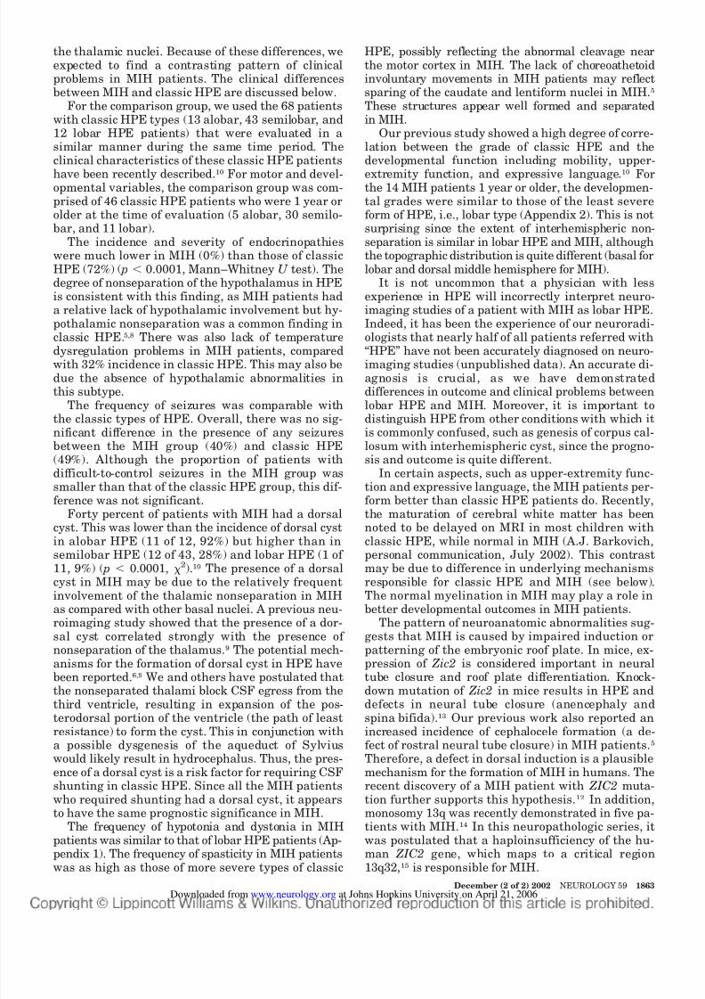
had choreoathetosis (involuntary movement disorder). Developmental abnormalities. One of 14 patients (7%)
greater than 1 year of age was able to ambulate indepen-
dently, whereas five (36%) were able to ambulate with
support (figure 2). One of 14 patients (7%) had normal
upper-extremity function, whereas nine (64%) were able to
use their upper-extremity with mild dysfunction.
Speech and oromotor development in MIH patients
were delayed. None of 14 patients over 1 year of age had
normal speech, whereas three (21%) were able to speak in
short sentences and eight (57%) uttered single words (see
figure 2). Oromotor feeding difficulties were noted in six
(43%) patients (see the table).
Discussion. The classic classification of HPE pro-posed in 1964 by DeMyer et al.2 was based on grossneuroanatomic abnormality and provided a convenientsubdivision into alobar, semilobar, and lobar types. Ad-
vances in neuroimaging over the past decade have led
to a better understanding of the pathogenesis of HPEand the variability of this condition.4,5,8,9,11 In MIH, theposterior frontal and parietal lobes fail to separate inthe midline despite a separation of anterior and poste-rior portions of the cerebral hemispheres. Basal fore-brain is nearly normal with a greater formed anteriorinterhemispheric fissure. Despite the differences be-tween classic HPE and MIH, they share a fundamentalsimilarity, nonseparation of a significant portion of thecerebrum in two separate hemispheres. Mutations in
ZIC2 gene have been recently reported in 16 patientswith HPE.12 This case series was comprised mostly of alobar HPE patients, but also included one patient
with MIH (also one of the current study cases). Thefact that mutations in ZIC2 cause classic HPE as wellas MIH provides further evidence that MIH is a vari-ant of HPE.
A recent neuroimaging study highlighted the fre-quent involvement of deep brain structures in classicHPE, such as basal ganglia, thalamic nuclei, hypo-thalamic nuclei, and mesencephalon.8 There is a dif-ferent nonseparation pattern of the deep gray nucleiin MIH. The most commonly affected basal nucleus isthe thalamus, whereas the caudate, lentiform, and hy-pothalamus nuclei are known to be well separated.5 Inthe current study, 8 of 15 (53%) had nonseparation of
Figure 2. Distribution of developmental function of MIH
patients for mobility, upper-extremity function, and expres-
sive language. Sits ϭ patients able to sit independently;
attains ϭ patients able to reach and attain objects; grasp ϭ
patients able to hold objects when placed in hands; sen-
tences ϭ patients able to speak short multiword sentences;single words ϭ patients able to utter only single words;
consonants ϭ patients able to utter consonants sounds
only; vowels ϭ patients able to utter vowel sounds only.
Table Motor dysfunction
No. of patients (%)
None Mild Moderate* Severe
Hypotonia 6 (43) 6 (43) 2 (14)
Dystonia 7 (50) 6 (43) 1 (7)
Spasticity 2 (14) 7 (50) 5 (36) 0 (0)
Choreoathetosis 14 (100) 0 (0) 0 (0)
Feeding difficulty 8 (57) 4 (29) 2 (14)
* Only spasticity included a moderate category in the grading
scheme.
1862 NEUROLOGY 59 December (2 of 2) 2002 at Johns Hopkins University on April 21, 2006www.neurology.orgDownloaded from
7/29/2019 Middle interhemispheric variant of holoprosencephaly: A distinct cliniconeuroradiologic subtype
http://slidepdf.com/reader/full/middle-interhemispheric-variant-of-holoprosencephaly-a-distinct-cliniconeuroradiologic 5/8
the thalamic nuclei. Because of these differences, weexpected to find a contrasting pattern of clinicalproblems in MIH patients. The clinical differencesbetween MIH and classic HPE are discussed below.
For the comparison group, we used the 68 patientswith classic HPE types (13 alobar, 43 semilobar, and12 lobar HPE patients) that were evaluated in asimilar manner during the same time period. Theclinical characteristics of these classic HPE patients
have been recently described.10 For motor and devel-opmental variables, the comparison group was com-prised of 46 classic HPE patients who were 1 year orolder at the time of evaluation (5 alobar, 30 semilo-bar, and 11 lobar).
The incidence and severity of endocrinopathieswere much lower in MIH (0%) than those of classicHPE (72%) ( p Ͻ 0.0001, Mann–Whitney U test). Thedegree of nonseparation of the hypothalamus in HPEis consistent with this finding, as MIH patients hada relative lack of hypothalamic involvement but hy-pothalamic nonseparation was a common finding inclassic HPE.5,8 There was also lack of temperature
dysregulation problems in MIH patients, comparedwith 32% incidence in classic HPE. This may also bedue the absence of hypothalamic abnormalities inthis subtype.
The frequency of seizures was comparable withthe classic types of HPE. Overall, there was no sig-nificant difference in the presence of any seizuresbetween the MIH group (40%) and classic HPE(49%). Although the proportion of patients withdifficult-to-control seizures in the MIH group wassmaller than that of the classic HPE group, this dif-ference was not significant.
Forty percent of patients with MIH had a dorsal
cyst. This was lower than the incidence of dorsal cystin alobar HPE (11 of 12, 92%) but higher than insemilobar HPE (12 of 43, 28%) and lobar HPE (1 of 11, 9%) ( p Ͻ 0.0001, 2).10 The presence of a dorsalcyst in MIH may be due to the relatively frequentinvolvement of the thalamic nonseparation in MIHas compared with other basal nuclei. A previous neu-roimaging study showed that the presence of a dor-sal cyst correlated strongly with the presence of nonseparation of the thalamus.9 The potential mech-anisms for the formation of dorsal cyst in HPE havebeen reported.6,9 We and others have postulated thatthe nonseparated thalami block CSF egress from the
third ventricle, resulting in expansion of the pos-terodorsal portion of the ventricle (the path of leastresistance) to form the cyst. This in conjunction witha possible dysgenesis of the aqueduct of Sylviuswould likely result in hydrocephalus. Thus, the pres-ence of a dorsal cyst is a risk factor for requiring CSFshunting in classic HPE. Since all the MIH patientswho required shunting had a dorsal cyst, it appearsto have the same prognostic significance in MIH.
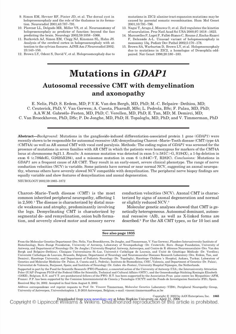
The frequency of hypotonia and dystonia in MIHpatients was similar to that of lobar HPE patients (Ap-pendix 1). The frequency of spasticity in MIH patientswas as high as those of more severe types of classic
HPE, possibly reflecting the abnormal cleavage nearthe motor cortex in MIH. The lack of choreoathetoidinvoluntary movements in MIH patients may reflectsparing of the caudate and lentiform nuclei in MIH.5
These structures appear well formed and separatedin MIH.
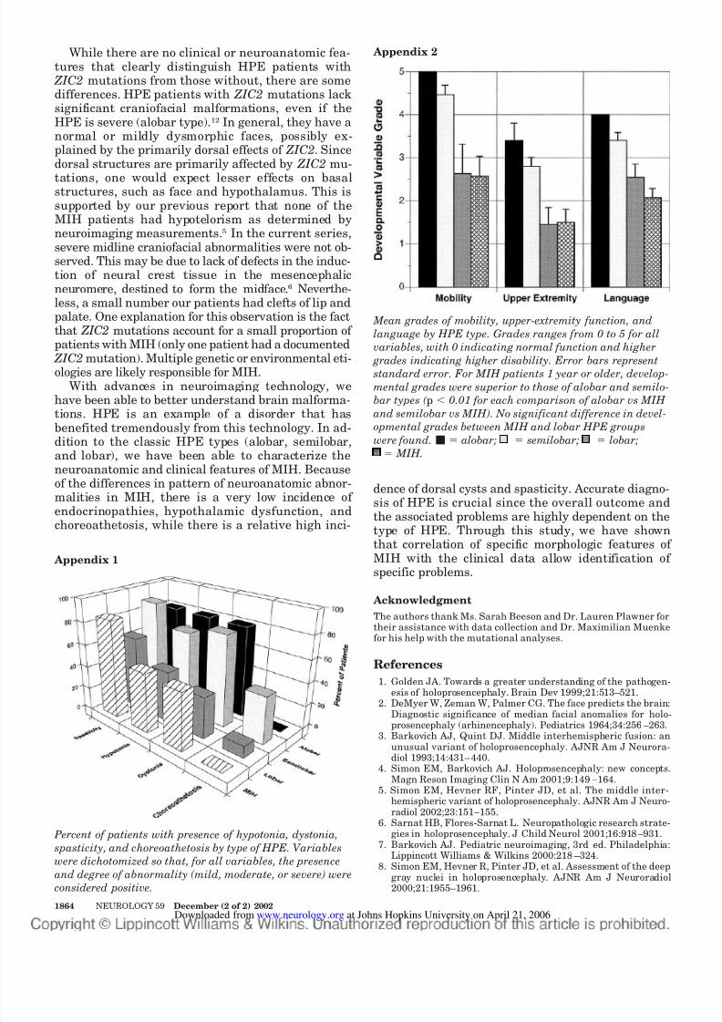
Our previous study showed a high degree of corre-lation between the grade of classic HPE and thedevelopmental function including mobility, upper-
extremity function, and expressive language.10 Forthe 14 MIH patients 1 year or older, the developmen-tal grades were similar to those of the least severeform of HPE, i.e., lobar type (Appendix 2). This is notsurprising since the extent of interhemispheric non-separation is similar in lobar HPE and MIH, althoughthe topographic distribution is quite different (basal forlobar and dorsal middle hemisphere for MIH).
It is not uncommon that a physician with lessexperience in HPE will incorrectly interpret neuro-imaging studies of a patient with MIH as lobar HPE.Indeed, it has been the experience of our neuroradi-ologists that nearly half of all patients referred with
“HPE” have not been accurately diagnosed on neuro-imaging studies (unpublished data). An accurate di-agnosis is crucial, as we have demonstrateddifferences in outcome and clinical problems betweenlobar HPE and MIH. Moreover, it is important todistinguish HPE from other conditions with which itis commonly confused, such as genesis of corpus cal-losum with interhemispheric cyst, since the progno-sis and outcome is quite different.
In certain aspects, such as upper-extremity func-tion and expressive language, the MIH patients per-form better than classic HPE patients do. Recently,the maturation of cerebral white matter has been
noted to be delayed on MRI in most children withclassic HPE, while normal in MIH (A.J. Barkovich,personal communication, July 2002). This contrastmay be due to difference in underlying mechanismsresponsible for classic HPE and MIH (see below).The normal myelination in MIH may play a role inbetter developmental outcomes in MIH patients.
The pattern of neuroanatomic abnormalities sug-gests that MIH is caused by impaired induction orpatterning of the embryonic roof plate. In mice, ex-pression of Zic2 is considered important in neuraltube closure and roof plate differentiation. Knock-down mutation of Zic2 in mice results in HPE and
defects in neural tube closure (anencephaly andspina bifida).13 Our previous work also reported anincreased incidence of cephalocele formation (a de-fect of rostral neural tube closure) in MIH patients.5
Therefore, a defect in dorsal induction is a plausiblemechanism for the formation of MIH in humans. Therecent discovery of a MIH patient with ZIC2 muta-tion further supports this hypothesis.12 In addition,monosomy 13q was recently demonstrated in five pa-tients with MIH.14 In this neuropathologic series, itwas postulated that a haploinsufficiency of the hu-man ZIC2 gene, which maps to a critical region13q32,15 is responsible for MIH.
December (2 of 2) 2002 NEUROLOGY 59 1863 at Johns Hopkins University on April 21, 2006www.neurology.orgDownloaded from
7/29/2019 Middle interhemispheric variant of holoprosencephaly: A distinct cliniconeuroradiologic subtype
http://slidepdf.com/reader/full/middle-interhemispheric-variant-of-holoprosencephaly-a-distinct-cliniconeuroradiologic 6/8
While there are no clinical or neuroanatomic fea-tures that clearly distinguish HPE patients with
ZIC2 mutations from those without, there are somedifferences. HPE patients with ZIC2 mutations lacksignificant craniofacial malformations, even if theHPE is severe (alobar type).12 In general, they have anormal or mildly dysmorphic faces, possibly ex-plained by the primarily dorsal effects of ZIC2. Sincedorsal structures are primarily affected by ZIC2 mu-
tations, one would expect lesser effects on basalstructures, such as face and hypothalamus. This issupported by our previous report that none of theMIH patients had hypotelorism as determined byneuroimaging measurements.5 In the current series,severe midline craniofacial abnormalities were not ob-served. This may be due to lack of defects in the induc-tion of neural crest tissue in the mesencephalicneuromere, destined to form the midface.6 Neverthe-less, a small number our patients had clefts of lip andpalate. One explanation for this observation is the factthat ZIC2 mutations account for a small proportion of patients with MIH (only one patient had a documented
ZIC2 mutation). Multiple genetic or environmental eti-ologies are likely responsible for MIH.
With advances in neuroimaging technology, wehave been able to better understand brain malforma-tions. HPE is an example of a disorder that hasbenefited tremendously from this technology. In ad-dition to the classic HPE types (alobar, semilobar,and lobar), we have been able to characterize theneuroanatomic and clinical features of MIH. Becauseof the differences in pattern of neuroanatomic abnor-malities in MIH, there is a very low incidence of endocrinopathies, hypothalamic dysfunction, andchoreoathetosis, while there is a relative high inci-
dence of dorsal cysts and spasticity. Accurate diagno-sis of HPE is crucial since the overall outcome andthe associated problems are highly dependent on thetype of HPE. Through this study, we have shownthat correlation of specific morphologic features of MIH with the clinical data allow identification of specific problems.
Acknowledgment
The authors thank Ms. Sarah Beeson and Dr. Lauren Plawner fortheir assistance with data collection and Dr. Maximilian Muenkefor his help with the mutational analyses.
References
1. Golden JA. Towards a greater understanding of the pathogen-esis of holoprosencephaly. Brain Dev 1999;21:513–521.
2. DeMyer W, Zeman W, Palmer CG. The face predicts the brain:
Diagnostic significance of median facial anomalies for holo-prosencephaly (arhinencephaly). Pediatrics 1964;34:256 –263.
3. Barkovich AJ, Quint DJ. Middle interhemispheric fusion: anunusual variant of holoprosencephaly. AJNR Am J Neurora-diol 1993;14:431– 440.
4. Simon EM, Barkovich AJ. Holoprosencephaly: new concepts.Magn Reson Imaging Clin N Am 2001;9:149 –164.
5. Simon EM, Hevner RF, Pinter JD, et al. The middle inter-hemispheric variant of holoprosencephaly. AJNR Am J Neuro-radiol 2002;23:151–155.
6. Sarnat HB, Flores-Sarnat L. Neuropathologic research strate-gies in holoprosencephaly. J Child Neurol 2001;16:918 –931.
7. Barkovich AJ. Pediatric neuroimaging, 3rd ed. Philadelphia:Lippincott Williams & Wilkins 2000:218 –324.
8. Simon EM, Hevner R, Pinter JD, et al. Assessment of the deepgray nuclei in holoprosencephaly. AJNR Am J Neuroradiol2000;21:1955–1961.
Percent of patients with presence of hypotonia, dystonia,
spasticity, and choreoathetosis by type of HPE. Variables
were dichotomized so that, for all variables, the presence
and degree of abnormality (mild, moderate, or severe) were
considered positive.
Appendix 1
Mean grades of mobility, upper-extremity function, and
language by HPE type. Grades ranges from 0 to 5 for all
variables, with 0 indicating normal function and higher
grades indicating higher disability. Error bars representstandard error. For MIH patients 1 year or older, develop-
mental grades were superior to those of alobar and semilo-
bar types ( p Ͻ 0.01 for each comparison of alobar vs MIH
and semilobar vs MIH). No significant difference in devel-
opmental grades between MIH and lobar HPE groups
were found. Ⅵ ϭ alobar; ϭ semilobar; ϭ lobar;
; ; ;
; ; ;
; ; ;
ϭ MIH.
Appendix 2
1864 NEUROLOGY 59 December (2 of 2) 2002 at Johns Hopkins University on April 21, 2006www.neurology.orgDownloaded from
7/29/2019 Middle interhemispheric variant of holoprosencephaly: A distinct cliniconeuroradiologic subtype
http://slidepdf.com/reader/full/middle-interhemispheric-variant-of-holoprosencephaly-a-distinct-cliniconeuroradiologic 7/8
9. Simon EM, Hevner RF, Pinter JD, et al. The dorsal cyst inholoprosencephaly and the role of the thalamus in its forma-tion. Neuroradiol 2001;43:787–791.
10. Plawner LL, Delgado MR, Miller VS, et al. Neuroanatomy of holoprosencephaly as predictor of function: beyond the facepredicting the brain. Neurology 2002;59:1058 –1066.
11. Barkovich AJ, Simon EM, Clegg NJ, Kinsman SL, Hahn JS. Analysis of the cerebral cortex in holoprosencephaly with at-tention to the sylvian fissures. AJNR Am J Neuroradiol 2002;23:143–150.
12. Brown LY, Odent S, David V, et al. Holoprosencephaly due to
mutations in ZIC2: alanine tract expansion mutations may be
caused by parental somatic recombination. Hum Mol Genet2001;10:791–796.
13. Nagai T, Aruga J, Minowa O, et al. Zic2 regulates the kineticsof neurulation. Proc Natl Acad Sci USA 2000;97:1618 –1623.
14. Marcorelles P, Loget P, Fallet-Bianco C, Roume J, Encha-RazaviF, Delezoide A-L. Unusual variant of holoprosencephaly inmonosomy 13q. Pediatr Dev Pathol 2002;5:170 –178.
15. Brown SA, Warburton D, Brown LY, et al. Holoprosencephalydue to mutations in ZIC2, a homologue of Drosophila odd-paired. Nat Genet 1998;20:180 –183.
Mutations in GDAP1
Autosomal recessive CMT with demyelination
and axonopathy
E. Nelis, PhD; S. Erdem, MD; P.Y.K. Van den Bergh, MD, PhD; M.-C. Belpaire–Dethiou, MD;C. Ceuterick, PhD; V. Van Gerwen; A. Cuesta, PharmB, MSc; L. Pedrola, BSc; F. Palau, MD, PhD;
A.A.W.M. Gabreëls–Festen, MD, PhD; C. Verellen, MD, PhD; E. Tan, MD; M. Demirci, MD;
C. Van Broeckhoven, PhD, DSc; P. De Jonghe, MD, PhD; H. Topaloglu, MD, PhD; and V. Timmerman, PhD
Abstract— Background: Mutations in the ganglioside-induced differentiation-associated protein 1 gene (GDAP1) were
recently shown to be responsible for autosomal recessive (AR) demyelinating Charcot–Marie–Tooth disease (CMT) type 4A
(CMT4A) as well as AR axonal CMT with vocal cord paralysis. Methods : The coding region of GDAP1 was screened for the
presence of mutations in seven families with AR CMT in which the patients were homozygous for markers of the CMT4A
locus at chromosome 8q21.1. Results: A nonsense mutation was detected in exon 5 (c.581CϾG, S194X), a 1-bp deletion in
exon 6 (c.786delG, G262fsX284), and a missense mutation in exon 6 (c.844CϾT, R282C). Conclusions: Mutations inGDAP1 are a frequent cause of AR CMT. They result in an early-onset, severe clinical phenotype. The range of nerve
conduction velocities (NCV) is variable. Some patients have normal or near normal NCV, suggesting an axonal neuropa-
thy, whereas others have severely slowed NCV compatible with demyelination. The peripheral nerve biopsy findings are
equally variable and show features of demyelination and axonal degeneration.NEUROLOGY 2002;59:1865–1872
Charcot–Marie–Tooth disease (CMT) is the mostcommon inherited peripheral neuropathy, affecting 1in 2,500.1 The disease is characterized by distal mus-cle weakness and atrophy, predominantly involving the legs. Demyelinating CMT is characterized bysegmental de- and remyelination, onion bulb forma-tion, and severely slowed motor and sensory nerve
conduction velocities (NCV). Axonal CMT is charac-terized by signs of axonal degeneration and normalor slightly reduced NCV.2
Molecular genetic analyses showed that CMT is ge-netically heterogeneous. Autosomal dominant, autoso-mal recessive (AR), as well as X-linked forms aredescribed.2 For the AR CMT types, so far 10 loci and
See also page 1835
From the Molecular Genetics Department (Drs. Nelis, Van Broeckhoven, De Jonghe, and Timmerman, V. Van Gerwen), Flanders Interuniversity Institute of
Biotechnology, Born–Bunge Foundation, University of Antwerp, Laboratory of Neuropathology (Dr. Ceuterick), Born–Bunge Foundation, University of
Antwerp, and Department of Neurology (Dr. De Jonghe), University Hospital Antwerp, Antwerpen, and Centre de Réf érence Neuromusculaire (Drs. Van den
Bergh and Belpaire–Dethiou), Cliniques Universitaires St.-Luc, Université Catholique de Louvain, and Unité de Génétique Médicale (Dr. Verellen),
Université Catholique de Louvain, Brussels, Belgium; Department of Neurology and Neuromuscular Diseases Research Laboratory (Drs. Erdem, Tan, and
Demirci), Hacettepe University, and Department of Pediatric Neurology (Dr. Topaloglu), Hacettepe Children’s Hospital, Ankara, Turkey; Laboratory of
Genetics and Molecular Medicine (Dr. Palau, A. Cuesta and L. Pedrola), Instituto de Biomedicina, CSIC, Valencia, and Department of Genetics (Dr. Palau),
Universitat de Valencia, Burjassot, Spain; and Institute of Neurology (Dr. Gabre ëls–Festen), University Hospital Nijmegen, the Netherlands.
Supported in part by the Fund for Scientific Research (FWO-Flanders), a concerted action of the University of Antwerp (UIA), the Interuniversity Attraction
Poles (IUAP) Program P5/19 of the Federal Office for Scientific, Technical and Cultural Affairs (OSTC), and the Geneeskundige Stichting Koningin Elisabeth
(GSKE), Belgium. E.N. and V.T. are postdoctoral fellows of the FWO. H.T. has been supported by the Association Fran çaise contre les Myopathies (AFM,
France). F.P. has been supported by the Comision Interministerial de Ciencia y Tecnologia (CICYT) and the Fondo de Investigacion Sanitaria (FIS), Spain.
Received May 24, 2002. Accepted in final form August 9, 2002.
Address correspondence and reprint requests to Prof. Dr. Vincent Timmerman, Molecular Genetics Laboratory (VIB8), Peripheral Neuropathy Group,
University of Antwerpen, Universiteitsplein 1, B-2610 Antwerpen, Belgium; e-mail: [email protected]
Copyright © 2002 by AAN Enterprises, Inc. 1865 at Johns Hopkins University on April 21, 2006www.neurology.orgDownloaded from
7/29/2019 Middle interhemispheric variant of holoprosencephaly: A distinct cliniconeuroradiologic subtype
http://slidepdf.com/reader/full/middle-interhemispheric-variant-of-holoprosencephaly-a-distinct-cliniconeuroradiologic 8/8
2002;59;1860-1865 NeurologyHahn
A.J. Lewis, E.M. Simon, A.J. Barkovich, N.J. Clegg, M.R. Delgado, E. Levey and J.S. cliniconeuroradiologic subtype
Middle interhemispheric variant of holoprosencephaly: A distinct
This information is current as of April 21, 2006
& ServicesUpdated Information
http://www.neurology.org/cgi/content/full/59/12/1860including high-resolution figures, can be found at:
Subspecialty Collections
http://www.neurology.org/cgi/collection/all_epilepsy_seizures
Epilepsy/SeizuresAllhttp://www.neurology.org/cgi/collection/all_clinical_neurology
All Clinical Neurology http://www.neurology.org/cgi/collection/developmental_disorders
disordersDevelopmentalhttp://www.neurology.org/cgi/collection/mri
MRIfollowing collection(s):This article, along with others on similar topics, appears in the
Permissions & Licensing
http://www.neurology.org/misc/Permissions.shtmlor in its entirety can be found online at:Information about reproducing this article in parts (figures, tables)
Reprints http://www.neurology.org/misc/reprints.shtml
Information about ordering reprints can be found online:
at Johns Hopkins University on April 21, 2006www.neurology.orgDownloaded from