microwave assisted synthesis of 3-(2-benzoyl-6...
TRANSCRIPT
MICROWAVE ASSISTED SYNTHESIS OF 3-(2-BENZOYL-6-HYDROXY-3-METHYL BENZO[b] FURAN-5-YL)-5-(ARYL)-4,5-DIHYDRO-1H-PYRAZOLE
CARBOTHIOAMIDES AND THEIR ANTIBACTERIAL ACTIVITY
Ashok D *, Khalilullaha M, Sudershan K b
* Department of Chemistry, Osmania University, Hyderabad-500 007, India. aDepartment Of Chemistry, JNTU, Kukatpally, Hyderabad-500 085, India.
bSven Genetech Ltd, I.D.A, Phase-II, Cherlapally, Hyderabad-500 051,India. Email: [email protected]
Abstract: A series of 3-(2-Benzoyl-6-hydroxy-3-methyl benzo[b] furan-5-yl)-5-(aryl)-4, 5-dihydro-1H-pyrazole carbothioamides have been prepared by the reaction of (E)-1-(2-Benzoyl-6-hydroxy-3-methylbenzo[b]furan-5-yl)-3-aryl-2-propen-1-ones with thiosemicarbazide in the presence of sodium hydroxide under microwave irradiation. The structures of newly synthesized compounds have been confirmed on the basis of elemental analysis, IR, 1H-NMR, 13C-NMR and mass spectral data. All the compounds were screened for their antibacterial activity. Keywords: Microwave irradiation, chalcone, pyrazoline, benzofuran, carbothioamides. Introduction A number of pyrazoline derivatives have been shown to exhibit a broad spectrum of biological and pharmaceutical activities which include anti-inflammatory1-2, antibacterial3, analgesic4, antifungal5, anticancer6 and anticonvulsant7 activities. Many 1-Thiocarbamoyl-3,5-diphenyl-2-pyrazolines are reported to have antimycobacterial8, antidepressant9 and monoamine oxidase inhibitory activities 10-11. Benzofuran derivatives are also associated with vide variety of physiological activities such as antihistaminic 12, anti-inflammatory 13, estrogenic and anti-implantation14activities. In order to know the combined effect of both pyrazoline and benzofuran moieties on physiological activity, we have taken up the synthesis of 3-(2-Benzoyl-6-hydroxy-3-methylbenzo[b]furan-5-yl)-5-(aryl)-4,5-dihydro-1H-pyrazole carbothioamides(3a-g). Recently microwave assisted organic synthesis (MAOS) has gained popularity as a non-conventional technique for rapid organic synthesis15. It is eco-friendly, economical and is believed to be a step towards green chemistry. Synthesis and biological evaluation of
Communicated to Heterocyclic communications
Antibacterial Activity of Alpinia galanga (L) WilldCrude Extracts
Kiranmayee Rao & Bhuvaneswari Ch &
Lakshmi M. Narasu & Archana Giri
Received: 21 July 2009 /Accepted: 28 December 2009 /Published online: 13 April 2010# Springer Science+Business Media, LLC 2010
Abstract Methanol, acetone and diethyl ether extracts of Alpinia galanga have beenevaluated against pathogens viz. Bacillus subtilis MTCC 2391, Enterobacter aerogene,Enterobacter cloacae, Enterococcus faecalis, Escherichia coli MTCC 1563, Klebsiellapneumoniae, Pseudomonas aeruginosa MTCC 6642, Salmonella typhimurium, Staphylo-coccus aureus and Streptococcus epidermis using Agar well diffusion method. Minimuminhibitory concentration (MIC) and minimum bactericidal concentration (MBC) of all theextracts were determined using the macrodilution method. Methanol extracts have shownexcellent activity towards all the pathogens with MIC and MBC values ranging from 0.04–1.28 mg/ml and 0.08–2.56 mg/ml, respectively. The GC–MS analysis of methanol extractshave yielded compounds like 5-hydroxymethyl furfural (59.9%), benzyl alcohol (57.6%),1,8 cineole (15.65%), methylcinnamate (9.4%), 3-phenyl-2-butanone (8.5%) and 1,2benzenedicarboxylic acid (8.9%), which could be responsible for its broad spectrumactivity. So, A. galanga can be quite resourceful for the development of new generationdrugs.
Keywords Alpinia galanga . Antimicrobial activity . MIC . MBC . GC–MS analysis .
Pathogens . Plant extracts
Introduction
Plants have been used throughout the world as drugs and remedies for various diseasessince time immemorial. According to pharmacologists, developed countries are turning tothe use of traditional medicinal systems, with about 1,400 herbal preparations being in use[1]. Plants are invaluable sources of pharmaceutical products that have drawn the attentionof ethno-pharmacologists from around the world [2]. Many plants have been used becauseof their antimicrobial constituents, which are due to compounds synthesized in the
Appl Biochem Biotechnol (2010) 162:871–884DOI 10.1007/s12010-009-8900-9
K. Rao : B. Ch : L. M. Narasu : A. Giri (*)Centre for Biotechnology, Institute of Science and Technology,Jawaharlal Nehru Technological University Hyderabad, Kukatpally, Hyderabad 500 085, Indiae-mail: [email protected]


secondary metabolism of the plant. These products are known by their active substances,e.g. the phenolic components which are part of the essential oils [3], as well as in tannins[4]. Essential oils from aromatic and medicinal plants receive particular attention aspotential natural agents for food preservation due to their effectiveness against a wide rangeof microorganisms [5–8]. In recent years, several reports have been published concerningthe composition and biological properties (antimicrobial, antioxidant, anticancer and thestimulated effect on the immune system) of Zingiberaceae extracts [9–15]. Even though anumber of antimicrobials have been isolated and studied, there is a growing need for thediscovery of new antimicrobials as there is an ever increasing crisis of bacterial resistancetowards the existing drugs.
Alpinia galanga (L) Willd syn. Languas galanga commonly called greater galangal,belonging to the family Zingiberaceae is a rhizomatous herb distributed in various parts ofIndia and throughout Southeast Asia. A. galanga has been used as food additive in Thailandand other countries in Asia for a long time. The rhizome is used against rheumatism,bronchial catarrh, bad breath and ulcers, whooping colds in children, throat infections andfever. 1′-Acetoxychavicol acetate, a component of A. galanga, was found to have very goodantimicrobial activity [16, 17]. The essential oil of A. galanga rhizome has been found tohave inhibitory activity against certain dermatophytes, filamentous fungi and yeast [18].Suganya and Sombat have reported that higher potential in antioxidant and antimicrobialactivities of A. galanga oil was supposed to be due to the composition of certain constituentsviz. 1, 8-cineole, 4-allylphenyl acetate and β-bisabolene within the essential oil [19].
The objective of the present study is to evaluate the active components responsible forantimicrobial activity with different solvent systems like methanol, acetone and diethylether in different parts (root, rhizome and leaf) of A. galanga and to compare their activitytowards the human pathogens. A. galanga constituents when extracted in acidic pH hadpronounced activity towards the pathogens. The extracts were shown to have broad-spectrum activity towards microorganisms. The activities of all the extracts were comparedand the best solvent, methanol was subjected to gas chromatography–mass spectroscopy(GC–MS) analysis.
Materials and Methods
Chemicals and Reagents
Amikacin, Mueller–Hinton agar (MHA) and Mueller–Hinton broth were supplied byHimedia, India. Methanol, acetone and diethyl ether were of analytical/HPLC gradesupplied by Merck.
Plant Extracts
The plants of A. galanga (L) Willd were collected from AG biotek, Hyderabad. The plantswere thoroughly washed and separated into three different parts, i.e. rhizome, root andleaves. They were oven dried at 60 °C for 24 h to remove the moisture and finely groundinto a powder using an electric blender. Eighteen extracts were obtained with methanol,acetone and diethyl ether as the solvent systems using a soxhlet apparatus. Thephytoconstituents were extracted in acidic (5.5) and neutral range. The pH was adjustedusing 0.1 N HCl. The extracts were concentrated under reduced pressure using rotavapour(Heidolph Rotacool, Germany).
872 Appl Biochem Biotechnol (2010) 162:871–884
Bacterial Cultures
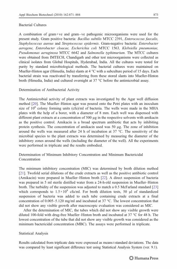
A combination of gram+ve and gram–ve pathogenic microorganisms were used for thepresent study. Gram positive bacteria: Bacillus subtilis MTCC 2391, Enterococcus faecalis,Staphylococcus aureus and Streptococcus epidermis. Gram-negative bacteria: Enterobacteraerogene, Enterobacter cloacae, Escherichia coli MTCC 1563, Klebsiella pneumoniae,Pseudomonas aeruginosa MTCC 6642 and Salmonella typhimurium. The MTCC cultureswere obtained from IMTECH, Chandigarh and other test microorganisms were collected asclinical isolates from Global Hospitals, Hyderabad, India. All the cultures were tested forpurity by standard microbiological methods. The bacterial cultures were maintained onMueller-Hinton agar (Himedia, India) slants at 4 °C with a subculture period of 15 days. Eachbacterial strain was reactivated by transferring from these stored slants into Mueller-Hintonbroth (Himedia, India) and cultured overnight at 37 °C before the antimicrobial assay.
Determination of Antibacterial Activity
The Antimicrobial activity of plant extracts was investigated by the Agar well diffusionmethod [20]. The Mueller–Hinton agar was poured onto the Petri plates with an inoculumsize of 106 colony forming units (cfu)/ml of bacteria. The wells were made in the MHAplates with the help of a borer, with a diameter of 8 mm. Each well was dispensed withdifferent plant extracts at a concentration of 500 µg in the respective solvents with amikacinas the positive control. Amikacin is a broad spectrum antibiotic that acts by inhibitingprotein synthesis. The concentration of amikacin used was 50 µg. The zone of inhibitionaround the wells was measured after 24 h of incubation at 37 °C. The sensitivity of themicrobial species to the plant extracts was determined by measuring the diameter of theinhibitory zones around the wells (including the diameter of the well). All the experimentswere performed in triplicate and the results embodied.
Determination of Minimum Inhibitory Concentration and Minimum BactericidalConcentration
The minimum inhibitory concentration (MIC) was determined by broth dilution method[21]. Twofold serial dilutions of the crude extracts as well as the positive antibiotic control(Amikacin) were prepared in Mueller–Hinton broth [22]. A direct suspension of bacteriawas prepared in 5 ml sterile distilled water from a 24-h-old suspension in Mueller–Hintonbroth. The turbidity of the suspension was adjusted to match a 0.5 McFarland standard [23]which corresponds to 1.5×10⁸ cfu/ml. For broth dilution tests, 50 µl of standardizedsuspension of bacteria was added to each tube containing crude extracts at a finalconcentration of 0.005–5.120 mg/ml and incubated at 37 °C. The lowest concentration thatdid not show any visible growth after macroscopic evaluation was considered as MIC.
After the determination of MIC, the tubes which did not show any visible growth werediluted 100-fold with drug-free Mueller–Hinton broth and incubated at 37 °C for 48 h. Thelowest concentration of the tube that did not show any visible growth was considered as theminimum bactericidal concentration (MBC). The assays were performed in triplicate.
Statistical Analysis
Results calculated from triplicate data were expressed as means±standard deviations. The datawas compared by least significant difference test using Statistical Analysis System (ver. 9.1).
Appl Biochem Biotechnol (2010) 162:871–884 873

Analysis by GC–MS
For GC–MS analysis, the samples were injected into a HP-5MS capillary column (30 mlength×0.25 m i.d×0.25 µm film thickness), Agilent Technologies, USA GC–MS model,consisting of 6,890 N gas chromatograph coupled with 5973 insert Mass Selective Detector.The injector was set at 250 °C and the detector at 280 °C. The stepped temperature programwas as follows: held at 50 °C for 2 min, then, from 50–280 °C at the rate of 10 °C/min, heldfor 5 min. The total run time was of 30 min. The GC–MS interface temperature at 280 °C.The injection volume was 1 µl. The solvent delay was 2 min and injected in a split ratio of1:10. The MS scan range was from 35–6,000 Da. Compound identification was obtained bycomparing the retention times with those of authentic compounds and the spectral dataobtained from library data of corresponding compounds.
Results and Discussion
Results
The antimicrobial principles were extracted using three different solvent systems viz.methanol, acetone and diethyl ether. The phytoconstituents of leaf, rhizome and root wereextracted separately. Ten different bacterial species were used to evaluate antimicrobialactivity of A. galanga (L) Willd. The microorganisms used for the study include food borne,intestinal and respiratory pathogens viz. S. aureus, which can cause a host of infections fromsimple skin infections to the persistent types like pneumonia and endocarditis.
The extracts of all the plant parts of A. galanga viz. leaves, rhizomes and roots haveshown good activity towards all the pathogens, the results of which are given in Table 1.The activity of plant extracts diminished with decrease in solvent polarity, i.e methanol>acetone>diethyl ether. The results clearly indicate that among the three solvents used forthe study, the activity of methanol extracts at acidic pH (5.5) were excellent. The activity ofacetone and diethyl ether were almost similar with acetone taking a slightly higher edge.The antimicrobial principles were extracted at acidic as well as neutral range. The activityof the plant samples extracted at acidic pH (5.5) was superior when compared to thesamples at neutral pH (7.0). On further lowering the pH, the activity slowly diminished.Amikacin, a broad-spectrum aminoglycoside antibiotic was used as positive standardreference that displayed good activity towards the entire set of pathogenic microorganisms,irrespective of the gram’s reaction. The respective solvents were checked for antimicrobialactivity that served as the negative controls.
The inhibition zones for methanol extracts ranged from 30.72±0.30 mm (B. subtilis) to14.16±0.29 mm (P. aeruginosa). In the case of S. epidermis (27.50±0.36 mm) and S .aureus (28.19±0.37 mm), the diameter of inhibition zone for methanol extract wasequivalent to the positive antibiotic standard (28.45±0.38 mm and 29.03±0.17 mm,respectively). The inhibition zone of methanol root extract towards S. typhimurium (29.08±0.13 mm) was more, compared to the inhibitory zone of the positive standard (27.74±0.15 mm) displaying the potent broad spectrum activity of the extracts. K. pneumoniae andP. aeruginosa were less susceptible to methanol extracts but were inhibited by acetone anddiethyl ether extracts. The leaf extracts of A. galanga in acetone were more efficient ininhibiting K. pneumoniae and P. aeruginosa with the inhibition zones of 24.59±0.06 mmand 21.89±0.32 mm, respectively. The response for plant parts was different towards eachof the pathogens. In all the cases of gram+ve bacteria, rhizome was showing the best result.
874 Appl Biochem Biotechnol (2010) 162:871–884

In the present study, B. subtilis was found to be the most sensitive and P. aeruginosa themost resistant.
MIC and MBC Values of A. galanga Towards the Pathogens Different extracts were studiedunder a wide range of concentrations ranging from 0.005–5.120 mg/ml. The methanolextract was excellent in showing very low values of MIC, in the range of 0.040–0.640 mg/mlwith an exception of P. aeruginosa showing a value of 1.28 mg/ml. The MBC values were inthe range of 0.080–2.56 mg/ml. The values for methanol extracts were comparable to thepositive standard reference used in the study. The acetone and ether extracts also hadsignificant MIC and MBC values in the range of 0.16–2.56 and 0.32–>5.12 mg/ml and 0.32–2.56 and 0.32–>5.12 mg/ml respectively. The MIC and MBC results of A. galanga leaf,rhizome and root are given in Table 2.
GC–MS Results of A. galanga in Methanol In rhizome and leaf, 15 compounds each havebeen detected where as in root 21 compounds have been detected. In case of rhizome androot, 5-hydroxymethyl furfural and benzyl alcohol were present in higher quantities, i.e.59.975% and 57.665%, respectively. Both these compounds were not detected in the leafextract which was replaced by 1, 8-cineole in considerable amount (15.65%). Compoundslike 3-phenyl-2-butanone, methyl cinnamate, phenylpropionaldehyde and 1, 2-benzenedicarboxylic acid were also present in noticeable quantities in the leaf. The difference inactivities of the extracts was justified by the detection of varied phytoconstituents indifferent parts of the plant. The results of methanolic extract of leaf, rhizome and root(Figs. 1, 2 and 3) have been analyzed using GC–MS and tabulated (Table 3).
Discussion
The emergence of bacterial super resistant strains is a result of the currently used antibioticagents, failing to end many bacterial infections [24]. Plants readily synthesize substancesfor defense against attack by insects, herbivores and microorganisms [25]. The mainadvantage of natural agents is that, the crude extracts contain a mixture of compounds likephenols, acids, esters, aldehydes etc., for which it is difficult to develop resistance bybacteria unlike the synthetic antibiotics that contain a single compound. Farnsworth andBunyapraphatsara in 1992 reported that essential oils from both fresh and dried rhizomes ofgalangal have antimicrobial activities against bacteria, fungi, yeast and parasites [26]. Inaddition to the sesquiterpenes, essential oils and phenylpropanoids; A. galanga alsocontains phenols, esters, aldehydes etc. which makes it an alternative choice for developingnew drug candidates.
A. galanga plant parts like the root, rhizome and leaf in methanol, acetone and diethylether have shown good activity towards the pathogens. Among all these methanol extracthas proven to be the best in acidic pH (5.5). The activity of the extract at acidic pH has beenproved by Syed et al., in a study on the activity of Raphanus sativus against pathogenicbacteria in varied pH range [27]. However, Soma Roy et al. (2009) have reported thatduring the antimicrobial evaluation of Andrographis paniculata extract against pathogens,the activity of the extract at neutral pH was much better when compared to that at acidic pH[28]. The root and rhizome extracts were almost similar in their activity with all thesolvents, but the leaf extracts in both methanol and acetone were showing slightly higheractivity against K. pneumoniae and P. aeruginosa. Whereas the ether extract of the leaf wasgood against P. aeruginosa only. In both the cases, acetone leaf extracts was much betterwhen compared to the methanol extracts. The reduced activity of methanol extract against
Appl Biochem Biotechnol (2010) 162:871–884 875

Tab
le1
Inhibitio
nzonesof
plantextractin
differentsolventssystem
s.
Nam
eof
theorganism
Plant
parts
Diameter
ofinhibitio
nzone
(mm)
Methanol
Acetone
Diethyl
ether
Antibiotic
Acidic(5.5)
Neutral
(7.0)
Acidic(5.5)
Neutral
(7.0)
Acidic(5.5)
Neutral
(7.0)
Gram
positiv
e
B.subtilis
Leaf
27.79±0.44
26.32±0.60
22.08±0.15
16.16±0.32
20.06±0.19
14.86±0.26
34.12±0.23
Rhizome
30.72±0.30
27.10±0.20
20.93±0.25
17.14±0.32
21.06±0.18
17.44±0.45
Root
27.94±0.20
26.05±0.07
21.19±0.20
18.98±0.10
17.02±0.25
13.94±0.34
E.faecalis
Leaf
24.56±0.39
21.46±0.10
20.46±0.49
18.26±0.28
17.27±0.39
14.37±0.76
32.09±0.24
Rhizome
27.83±0.22
22.53±0.80
18.14±0.44
15.27±0.04
19.27±0.27
15.54±0.14
Root
25.93±0.13
22.48±0.14
19.67±0.13
14.19±0.19
20.72±0.28
18.22±0.28
S.au
reus
Leaf
22.08±0.24
18.28±0.36
16.32±0.65
15.11±0.36
14.07±0.18
10.65±0.39
29.03±0.17
Rhizome
27.21±0.21
21.88±0.21
17.70±0.19
14.32±0.32
13.34±0.49
9.34
±0.28
Root
28.19±0.37
20.52±0.01
14.90±0.23
12.14±0.23
11.49±0.54
9.72
±0.36
S.epidermis
Leaf
24.31±0.35
20.88±0.21
15.61±0.46
12.65±0.29
14.58±0.36
11.22±0.67
28.45±0.38
Rhizome
27.50±0.36
22.30±0.55
19.60±0.42
15.27±0.16
16.79±0.18
12.17±0.20
Root
21.41±0.12
17.47±0.23
21.97±0.30
13.99±0.03
19.25±0.29
15.15±0.39
876 Appl Biochem Biotechnol (2010) 162:871–884

Gram
negativ
e
E.aerogene
Leaf
16.46±0.11
14.18±0.54
19.25±0.60
16.72±0.43
14.85±0.27
11.93±0.58
30.63±0.51
Rhizome
20.81±0.26
18.89±0.29
15.97±0.32
11.37±0.75
16.04±0.50
13.92±0.27
Root
19.69±0.43
15.05±0.52
15.55±0.25
11.99±0.06
19.64±0.35
15.86±0.34
E.cloacae
Leaf
21.01±0.28
14.73±0.47
15.89±0.38
10.93±0.53
11.84±0.38
8.91
±0.32
29.86±0.47
Rhizome
19.73±0.78
15.27±0.39
14.30±0.24
10.05±0.34
13.09±0.42
11.19±0.23
Root
24.16±0.34
17.22±0.34
15.19±0.21
12.24±0.29
16.14±0.25
13.21±0.19
E.coli
Leaf
24.29±0.20
20.94±0.12
20.43±0.63
18.84±0.49
23.43±0.54
21.14±0.19
31.72±0.89
Rhizome
29.93±0.25
27.26±0.10
25.74±0.56
19.99±0.32
20.43±0.51
19.26±0.59
Root
27.15±0.17
24.43±0.32
26.78±0.22
24.90±0.25
23.49±0.46
20.56±0.58
K.pn
eumon
iaLeaf
22.23±0.30
19.58±0.30
24.59±0.06
18.34±0.10
21.24±0.12
12.45±0.12
26.83±0.42
Rhizome
17.19±0.32
14.48±0.21
20.23±0.09
15.40±0.21
16.46±0.13
14.51±0.17
Root
18.25±0.28
14.27±0.13
21.32±0.67
16.48±0.12
17.45±0.58
16.50±0.47
P.aeruginosa
Leaf
16.75±0.43
13.84±0.25
21.89±0.32
16.02±0.30
20.66±0.19
13.14±0.28
25.92±0.64
Rhizome
18.09±0.24
14.86±0.29
18.56±0.45
14.83±0.24
15.00±0.38
11.19±0.25
Root
14.16±0.29
10.36±0.41
18.92±0.39
14.02±0.21
13.03±0.17
10.24±0.42
S.typh
imurium
Leaf
25.56±0.10
21.47±0.16
17.17±0.21
15.16±0.42
14.60±0.25
10.74±0.28
27.74±0.15
Rhizome
28.65±0.08
23.71±0.28
18.97±0.34
16.52±0.40
13.49±0.06
11.14±0.13
Root
29.08±0.13
25.44±0.23
21.36±0.32
18.48±0.07
16.32±0.12
14.48±0.23
Borer
size
is8mm
Appl Biochem Biotechnol (2010) 162:871–884 877

the above microbes could be due to the permeability of the compounds and resistancemechanisms displayed by the organisms towards the extracts. The outer membrane of thegram-negative bacterial cell wall appears to act as a barrier to many substances includingantibiotics [29].
The MIC and MBC values of the extract even though in a crude form and at higherconcentration (with reference to the standard), were comparable to that of the standardantibiotic. In case of E. cloacae, E. aerogene and S. epidermis the MIC and MBC values
Table 2 The MIC AND MBC values of plant extracts towards the pathogens.
Name of the organism Plant parts Concentration (mg/ml)
Methanol Acetone Diethyl ether Antibiotic
MIC MBC MIC MBC MIC MBC MIC MBC
Gram positive
B. subtilis Leaf 0.08 0.32 0.16 0.32 0.64 1.28 0.08 0.16
Rhizome 0.04 0.16 0.16 0.64 0.32 0.64
Root 0.08 0.16 0.32 1.28 0.64 1.28
E. faecalis Leaf 0.08 0.64 0.16 0.64 0.64 2.56 0.02 0.04
Rhizome 0.04 0.32 0.32 1.28 0.64 1.28
Root 0.08 0.32 0.32 1.28 0.64 1.28
S. aureus Leaf 0.16 0.64 0.64 1.28 0.64 2.56 0.08 0.16
Rhizome 0.04 0.32 0.64 1.28 1.28 2.56
Root 0.04 0.16 2.65 >5.12 1.28 >5.12
S. epidermis Leaf 0.16 0.64 0.64 1.28 1.28 2.56 0.16 0.32
Rhizome 0.04 0.16 0.32 0.64 1.28 2.56
Root 0.16 0.64 0.16 0.64 0.64 2.56
Gram negative
E. aerogene Leaf 0.64 1.28 0.16 0.32 0.64 2.56 0.08 0.16
Rhizome 0.16 0.32 0.16 0.64 0.64 1.28
Root 0.16 0.64 0.16 0.64 0.32 1.28
E. cloacae Leaf 0.16 0.32 1.28 2.56 2.56 >5.12 0.08 0.16
Rhizome 0.16 0.64 2.56 5.12 1.28 5.12
Root 0.08 0.32 1.28 2.56 1.28 2.56
E. coli Leaf 0.08 0.32 0.16 0.32 0.32 0.64 0.005 0.01
Rhizome 0.04 0.08 0.08 0.16 0.32 1.28
Root 0.08 0.16 0.04 0.16 0.16 0.32
K. pneumonia Leaf 0.16 0.64 0.16 0.32 0.64 1.28 0.08 0.32
Rhizome 0.32 1.28 0.32 1.28 0.64 1.28
Root 0.32 0.64 0.32 1.28 0.32 0.64
P. aeruginosa Leaf 0.64 2.56 0.16 0.64 0.32 1.28 0.04 0.08
Rhizome 0.64 2.56 0.32 1.28 1.28 2.56
Root 1.28 2.56 0.32 1.28 2.56 >5.12
S. typhimurium Leaf 0.08 0.64 0.64 2.56 1.28 >5.12 0.08 1.28
Rhizome 0.04 0.16 0.64 1.28 1.28 >5.12
Root 0.04 0.08 0.32 1.28 0.64 2.56
878 Appl Biochem Biotechnol (2010) 162:871–884
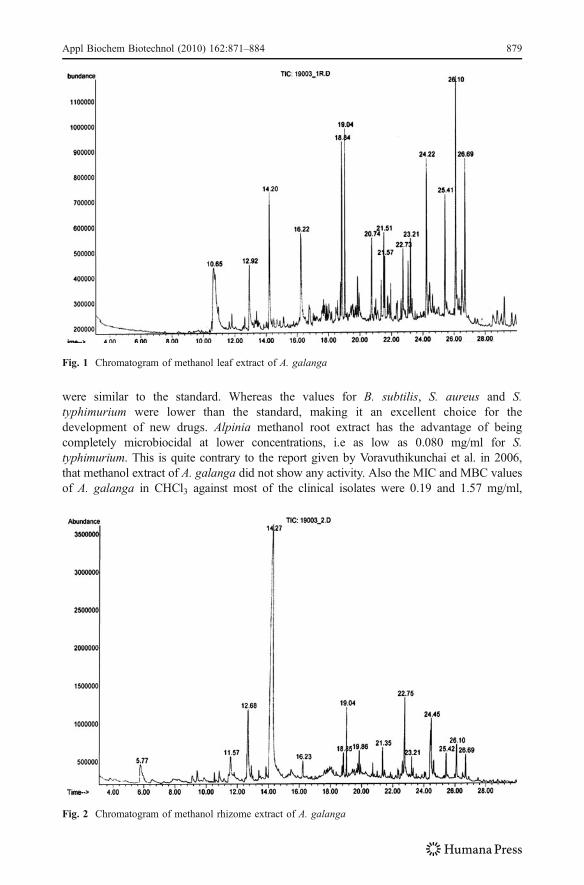
were similar to the standard. Whereas the values for B. subtilis, S. aureus and S.typhimurium were lower than the standard, making it an excellent choice for thedevelopment of new drugs. Alpinia methanol root extract has the advantage of beingcompletely microbiocidal at lower concentrations, i.e as low as 0.080 mg/ml for S.typhimurium. This is quite contrary to the report given by Voravuthikunchai et al. in 2006,that methanol extract of A. galanga did not show any activity. Also the MIC and MBC valuesof A. galanga in CHCl3 against most of the clinical isolates were 0.19 and 1.57 mg/ml,
Fig. 2 Chromatogram of methanol rhizome extract of A. galanga
Fig. 1 Chromatogram of methanol leaf extract of A. galanga
Appl Biochem Biotechnol (2010) 162:871–884 879

respectively; which is less when compared to the present value of 0.08 and 0.64 mg/ml [30].Even though the acetone and diethyl ether extracts have shown considerable activity, in fewcases the MBC values were >5.12 mg/ml. In an earlier report, galangal rhizome oil could notinhibit the growth of P. aeruginosa due to a failure of outer membrane penetration [31]. Butin the present study, the leaf extracts were successful in inhibiting K. pneumoniae and P.aeruginosa. A related possibility is that terpenes, synergise the effects of other compounds byacting as solvents to facilitate their passage through membranes. The terpenoids and phenoliccompounds in pure form demonstrate high antibacterial activity [32]. The lipophilic nature ofterpenes, suggests that their principle targets are the cell membranes and their toxicity iscaused by loss of chemiosmotic control [33, 34]. According to the earlier study by JirawanOonmetta-aree in 2006, the MBC value of Alpinia ethanol extract against S. aureus was1.3 mg/ml and chloroform extract was 0.256 mg/ml as reported by Voravuthikunchai et al in2005, in contrast to the present report of 0.16 mg/ml for methanol root extract, which is 12.5folds and 1.5 folds higher respectively [16, 35]. This clearly indicates the potential of A.galanga extract as a good source of new age antimicrobials.
The GC–MS results of the methanol extract have furnished an array of compounds inleaf, rhizome and root. The major constituents identified in Alpinia leaf are 1,8-cineole,phenylpropionaldehyde and methylcinnamate among many others which could beresponsible for the antimicrobial activity. The activities of 1,8-cineole, phenylpropion-aldehyde and methylcinnamate have already been analyzed in other plant species [36–38].In case of rhizome, 5-hydroxymethyl furfural is the major constituent (59.9%), which couldbe responsible for its activity. The activity was confirmed by Fausto Rivero et al. in Vitisvinifera against selected oral pathogens [39]. Whereas in root, the major phytochemicalidentified was benzyl alcohol (57.6), whose antimicrobial activity has been confirmed byZheng-Zhu. et al. in 2006 [40].The Directorate General of the European commission forhealth and consumer protection has recommended the use of benzyl alcohol in foodpreservation and also as flavouring agent.
Fig. 3 Chromatogram of methanol root extract of A. galanga
880 Appl Biochem Biotechnol (2010) 162:871–884

Table 3 GC-MS results of A. galanga in methanol.
S.No Rt (min) Name of the Compound Percent of the Total
Leaf Rhizome Root
1 5.77 Furfuraldehyde – 4.595 –
2 10.65 Benzenemethanol 4.674 – –
3 10.69 BenzylAlcohol – – 57.665
4 11.57 Unknown – 2.982 –
5 12.68 2,3-Dihydro-3, 5-dihydroxy-6-methyl-4H-pyran-4-one – 7.441 –
6 12.92 3-Phenylpropionaldehyde 5.237 – –
7 13.85 β-Fenchyl Acetate – – 1.704
8 14.20 3-Phenyl-2-Butanone 8.517 – –
9 14.27 5-Hydroxymethyl furfural – 59.975 –
10 16.22 Methyl Cinnamate 9.423 1.235 –
11 16.24 5-Tetradecene – – 1.154
12 18.84 1,2-Benzene dicarboxylic acid 8.932 1.443 2.361
13 19.04 γ-Cadinene 6.604 – –
14 19.04 Carotol – 2.503 2.825
15 19.77 Unknown – – 1.830
16 19.86 Unknown – 1.213 1.258
17 19.95 Unknown – – 1.168
18 20.74 Unknown 4.609 – –
19 21.35 Unknown – 1.169 2.362
20 21.45 Unknown – – 1.595
21 21.51 Neophytadiene 3.753 – –
22 21.57 Hexahydrofarnesyl acetone 2.235 – –
23 22.33 Unknown – – 1.831
24 22.63 Unknown – – 2.230
25 22.74 Hexadecanoic acid 3.693 5.500 4.785
26 23.21 Unknown 3.252 0.849 1.576
27 23.34 Unknown – – 1.474
28 24.11 Unknown – – 1.926
29 24.22 2,6,10-Trimethyl, 14-Ethylene-14-Pentadecene 8.637 – –
30 24.44 9-Octadecenoic acid – 6.454 3.766
31 25.41 Unknown 6.234 1.301 –
32 25.43 Unknown – – 1.537
33 26.10 1,8-Cineole 15.650 1.963 –
34 26.69 Unknown 8.550 1.376 –
35 26.90 Unknown – – 1.383
36 27.87 Unknown – – 2.042
37 28.50 Unknown – – 3.538
Rt Retention time in minutes
Appl Biochem Biotechnol (2010) 162:871–884 881

Developing countries are paying increased attention to food safety, because of growingrecognition of its potential impact on public health, food security, and trade competitive-ness. Increasing scientific understanding of the public health consequences of unsafe food,amplified by the rapid global transmission of information regarding the public health threatsassociated with food-borne and zoonotic diseases (e.g. E. coli and Salmonella) [41, 42]. InIndia, it is estimated that 20% of deaths among children under five are caused by diarrhealdisease [43]. The presence of high amount of benzyl alcohol has been reported for the firsttime in this particular Indian variety, which can be used as food preservative in view ofincreasing the shelf life of foods as well as in the control of food borne diseases. Previously,the rhizome and root parts were studied, as they were used since long in ayurveda and unanimedicines. In the present study, in addition to the root and rhizome, antimicrobial activity ofthe leaf towards various pathogens has been reported for the first time which could be asource for alternative antimicrobials.
Conclusion
Even though, pharmaceutical industries have produced a number of new antimicrobialdrugs in the last few years, resistance to these drugs by microorganisms has increased. Ingeneral, bacteria have the genetic ability to transmit and acquire resistance to drugs used astherapeutic agents [44]. The use of phytochemicals as natural antimicrobial agentscommonly called ‘biocides’ is gaining popularity. Aromatic and medicinal plants areknown to produce certain bioactive molecules which react with other organisms in theenvironment, inhibiting bacterial or fungal growth [45, 46]. The substances that can inhibitpathogens and have little toxicity to host cells are considered candidates for developing newantimicrobial drugs. Many plant extracts owe their potency to the presence of substanceslike tannins, phenolic compounds and so on. A. galanga represents a potent antimicrobialsystem for the development of natural drugs, as the whole plant can be used. Though in acrude form, the plant extracts have displayed broad spectrum activity towards themicroorganisms. It contains a mixture of compounds like phenylpropanoids, monoterpenes,sesquiterpenes and essential oils that help in checking the antibiotic resistance. A. galangacan be used for preservation of foods, as it possesses characteristic flavour as well asantimicrobial activity.
References
1. Hoareau, L., & Da Silva, E. J. (1999). Electronic Journal of Biotechnology, V, 2.2. Olalde Rangel, J. A. (2005). The systematic theory of living systems and relevance to CAM. Part I: The
theory. Evidence-Based Complementary Alternative Medicine, 2, 13–18.3. Jansen, A. M., Cheffer, J. J. C., & Svendsen, A. B. (1987). Planta Medica, 40, 395–398.4. Saxena, G., Mc Cutcheon, A. R., Fasmer, S., Towers, G. H. N., & Hancock, R. E. W. (1994). Journal of
Ethanopharmacology, 42, 95–99.5. Deans, S. G. (1991). Evaluation of antimicrobial activity of essential (volatile) oils. In H. F. Linskens &
J. F. Jackson (Eds.), Modern method of plant analysis, New Series, vol. 12. Essential oils and Waxes(pp. 309–320). Berlin: Springer.
6. Baratta, M. T., Dorman, H. J. D., Deans, S. G., Biondi, D. M., & Roberto, G. (1998). Journal ofEssential oil Research, 10, 618–627.
7. Marino, M., Bersani, C., & Comi, G. (2001). International Journal of Food Microbiology, 67(3), 187–195.
882 Appl Biochem Biotechnol (2010) 162:871–884

8. Mimica-Dukie, N., Bozin, B., Sokovic, M., Mihajlovie, B., & Matavulj, M. (2003). Planta Medica, 69,413–419.
9. Bendjeddou, D., Lalaoui, K., & Satta, D. (2003). Journal of Ethnopharmacology, 88, 155–160.10. Jirovetz, L., Buchbauer, G., Pottachola, M., & Kalathil, N. (2003). Acta Pharmaceutica, 53, 73–81.11. Leal, P. F., Braga, M. E. M., Sato, D. N., Carvalho, J. E., Marques, M. O. M., & Meireles, M. A. A.
(2003). Journal of Agricultural and Food Chemistry, 51, 2520–2525.12. Negi, P. S., Jayaprakasha, G. K., & Jena, B. S. (1999). LWT-Food Science and Technology, 41, 1857–
1861.13. Nguefack, J., Leth, V., Amvam, P. H., & Mathur, S. B. (2004). International Journal of Food
Microbiology, 94, 329–334.14. Scartezzini, P., & Speroni, E. (2000). Journal of Ethnopharmacology, 71, 23–43.15. Sekiwa, Y., Kubota, K., & Kobayashi, A. (2000). Journal of Agricultural and Food Chemistry, 48, 373–
377.16. Oonmetta-aree, J., Suzuki, T., Gasaluck, P., & Eumkeb, G. (2006). LWT— Food Science and Technology,
39(10), 1214–1220.17. Vuddhakul, V., Bhoopong, P., Hayeebilan, F., & Subhadhirasakul, S. (2007). Food Microbiology, 24(4),
413–418.18. Mohd, M. S., Chin, C. B., Chen, L. L., & Sim, N. L. (2003). Pharmaceutical Biology, 41(5), 302–307.19. Tachakittirungrod, S., & Chowwanapoonpohn, S. (2007). Chang Mai University Journal of Natural
Sciences, 6(1), 31.20. Perez, C., Pauli, M., & Bazerque, P. (1990). Acta Biologiae et Medicine Experimentalis, 15, 113–115.21. Chattopadhyay, D., Mukherjee, T., Pal, P., Saha, B., & Bhadra, R. (1998). Journal of Antimicrobial
Chemotherapy, 42, 83–86.22. Chattopadhyay, D., Sinha, B., & Vaid, L. K. (1998). Fitoterapia, 69(4), 365–367.23. McFarland, J. (1907). Journal of the American Medical Association, 49, 1176–1178.24. Bhavani, S. M., & Ballow, C. H. (2000). Current Opinion in Microbiology, 3, 528–534.25. Marjorie, M. C. (1999). Clinical Microbiology Reviews, 12, 564–582.26. Farnsworth, N. R., & Bunyapraphatsara, N. (1992). Thai medicinal plants recommended for primary
health care system. Bangkok: Prachachon.27. Beevi, S. S., Mangamoori, L. N., & Anabrolu, N. (2009). World Journal of Microbiology and
Biotechnology, 25, 465–473. doi:10.1007/s11274-008-9911-3.28. Roy, S., Rao, K., Bhuvaneswari, C., Giri, A., & Mangamoori, L. N. (2010). World Journal of
Microbiology and Biotechnology, 26, 85–91. doi:10.1007/s11274-009-0146-8.29. Tortora, G. J., Funk, B. R., & Case, C. (2001). Microbiology: an introduction. San Francisco: Benjamin
Cummings.30. Voravuthikunchai, S. P., Limsuwan, S., Supapol, O., & Subhadhirasakul, S. (2006). Journal of Food
Safety, 26, 325–334.31. Gao, Y., Van Belkum, M. J., & Stiles, M. E. (1999). Applied and Environmental Microbiology, 65, 4329–
4333.32. Conner, D. E. (1993). Naturally occurring compounds. In P. Davidson & A. L. Branen (Eds.),
Antimicrobials in foods (pp. 441–468). New York: Marcel Dekker.33. Cox, S. D., Mann, C. M., Markham, J. L., Bell, H. C., Gustafson, J. E., Warmington, J. R., et al. (2000).
Journal of Applied Microbiology, 88, 170–175.34. Inouea, Y., Shiraishia, A., Hadaa, T., Hirosea, K., Hamashimaa, H., & Shimadaa, J. (2004). FEMS
Microbiology Letters, 237, 325–331.35. Voravuthikunchai, S. P., Phongpaichit, S., & Subhadhirasakul, S. (2005). Pharmaceutical Biology, 43(8),
701–706.36. Satrani, B., Farah, A., & Talbi, M. (2006). Acta Botanica Gallica, 153, 235–242.37. Chang, S.-T., Chen, P.-F., & Chang, S.-C. (2001). Journal of Ethnopharmacology, 77, 123–127.38. Bruni, R., Medici, A., Andreotti, E., Fantin, C., Muzzoli, M., Dehesa, M., et al. (2004). Food Chemistry,
85(3), 415–421.39. Rivero-Cruz, F., Fausto Rivero-Cruz, J., Min Zhu, A., Kinghorn, D., & Wu, C. D. (2008).
Phytochemistry Letters, 1(3), 151–154.40. Zhang, Z.-Z., Li, Y.-B., Qi, L., & Wan, X.-C. (2006). Journal of Agricultural and Food Chemistry, 54,
3936–3940.41. Lindsay, J.A.(1997). Emerging Infectious Diseases 3(4). Oct-Dec 1997.42. Kafferstein, Fritz, K. (2003). Food Safety as a Public Health Issue for Developing Countries. In: L.
Unnevehr (Ed), Food Safety in Food Security and Trade, 2020 Vision for Food Agriculture and theEnvironment, Focus 20, Brief 2, International Food Policy Research Institute, Washington
Appl Biochem Biotechnol (2010) 162:871–884 883

43. World Health Organization, 2006b, Core Health Indicators, www3/who/int/whois/core/core_select_process.cfm
44. Nascimento, G. G. F., Locatelli, J., Frcitas, P. C., & Silva, G. L. (2000). Brazilian Journal ofMicrobiology, 31, 247–256.
45. Chopra, R. N., Nayer, S. L., & Chopra, I. C. (1992). Glossary of Indian Medicinal plants (3rd ed., pp. 7–246). New Delhi: Council of Scientific and Industrial Research.
46. Bruneton, J. (1995). Pharmacognosy, Phytochemistry, Medicinal plants (pp. 265–380). France:Lavoisiler Publishing Co.
884 Appl Biochem Biotechnol (2010) 162:871–884

benzofurans and dihydropyrazole thiocarbothioamides have been a topic of special interest to organic and medicinal chemists. In continuation of our earlier work on microwave assisted synthesis16-18 of biodynamic heterocycles and to explore their biological activity, herein we report the microwave assisted synthesis of novel benzofuranyl pyrazolines with possible antibacterial activity. Results and discussion The required starting material 5-Acetyl-2-benzoyl-6-hydroxy-3-methyl benzofuran19,
20(1) was synthesized by the reaction of 4, 6-diacetylresorcinol21 with ώ- bromoacetophone22 (1:1) in the presence of anhydrous K2CO3 under microwave irradiation. The desired (E)-1-(2-Benzoyl-6-hydroxy-3-methylbenzo[b] furan-5-yl)-3-aryl-2-propen-1-ones (2a-g) were synthesized by the condensation of 5-Acetyl-2-benzoyl-6-hydroxy-3-methyl benzofuran(1) with aromatic/hetero aromatic aldehydes in the presence of sodium methoxide under microwave irradiation. Several 3-(2-Benzoyl-6-hydroxy-3-methylbenzo[b]furan-5-yl)-5-(aryl)-4,5-dihydro-1H-pyrazolecarbo thioamides (3a-g) were synthesized by the reaction of (E)-1-(2-Benzoyl-6-hydroxy-3-methyl benzo[b]furan-5-yl)-3-aryl-2-propen-1-ones with thiosemicarbazide in the presence of sodium hydroxide under microwave irradiation in excellent yields. The synthesis of 3a-g was also carried out under conventional heating. Yields obtained under conventional heating (62-76%) as compared to microwave irradiation (80-90%), demonstrating that the effect of microwave irradiation. Experimental Melting points were determined on Polmon MT 96 melting point apparatus and are uncorrected. IR Spectra were measured on Shimadzu FTIR-8400S. 1H-NMR Spectra, 13C-NMR spectra were recorded in DMSO-d6 on Avance 300, spectrometer. Elemental analysis was recorded on Thermo Finnigan CHNS analyzer. Mass spectra were recorded on Thermo Finnigan. The purity of the compounds was checked by TLC using precoated silica gel plates (F-254), Merck. Microwave irradiation was carried out in Multisynth series microwave system (Milestone). Scheme:
OH
O
OOOH
O
OO
Ar
OHOO
ArCHO
aq.NaOH
N N
Ar
NH2S
NH2NHCSNH2
NaOMe
3a-g
Ar = a) phenyl e) 4-methoxyphenyl b) 2-chlorophenyl f) 1-naphthyl c) 4-chlorophenyl g) 2-furyl d) 3-nitrophenyl
2a-g1
HA
HB
HX
or MWI
or MWI
12
3
45

Table1: Analytical data of 3-(2-Benzoyl-6-hydroxy-3-methylbenzo[b]furan-5-yl)-5-(aryl)-4,5-dihydro-1H-pyrazole carbothioamides (3a-g)
Compound M.P. (°C)
Conventional heating
Microwave irradiation
Time (hr)
Yield (%)
Time (min)
Yield (%)
3a 250 8 76 5 82 3b 255 8 65 5 86 3c 185 8 65 5 85 3d 242 8 62 5 80 3e 240 10 65 5 82 3f 205 8 62 6 83 3g 245 8 66 5 90
Synthesis of (E)-1-(2-Benzoyl-6-hydroxy-3-methylbenzo[b]furan-5-yl)-3-aryl-2-propen-1-ones (2a-g) a) Conventional method A mixture of 5-Acetyl-2-benzoyl-6-hydroxy-3-methyl benzofuran (1) (0.01 mol), appropriate aromatic / hetero aromatic aldehydes (0.01mol), sodium methoxide (0.04 mol) and ethanol (20ml) was stirred for 16 hr at room temperature. After completion of reaction as indicated by TLC, the reaction mixture was poured on to crushed ice and neutralized with dil.HCl. The solid separated was filtered and recrystallized from methanol as yellow powder. b) Microwave irradiation method A mixture of 5-Acetyl-2-benzoyl-6-hydroxy-3-methyl benzofuran (1) (0.001 mol), appropriate aromatic / hetero aromatic aldehydes (0.001mol), sodium methoxide (0.004 mol) and ethanol (5 ml) was taken in a quartz tube and inserted into teflon vial with screw capped and subjected to microwave irradiation at the constant temperature 70°C for 5 min. After completion of reaction as indicated by TLC, the reaction mixture was poured on to crushed ice and neutralized with dil.HCl. The solid separated was filtered and recrystallized from methanol as yellow powder. Synthesis of 3-(2-Benzoyl-6-hydroxy-3-methylbenzo[b]furan-5-yl)-5-(aryl)-4,5- dihydro-1H-pyrazole carbothioamides (3a-g): a) Conventional heating method A mixture of (E)-1-(2-Benzoyl-6-hydroxy-3-methylbenzo[b]furan-5-yl)-3-aryl-2-propen-1-ones (0.001mol), thiosemicarbazide (0.001mol), aq. NaOH (0.08g) in 1 ml of water and ethanol (10 ml), was taken in a 25ml round bottomed flask and the reaction mixture was refluxed for 8-10hrs. The progress of the reaction was monitored by TLC. After completion of the reaction, the reaction mixture was diluted with cold water and

neutralized with dil. HCl. The precipitate thus formed was filtered and recrystallized from methanol as yellow powder. b) Microwave irradiation method A mixture of (E)-1-(2-Benzoyl-6-hydroxy-3-methylbenzo[b]furan-5-yl)-3-aryl-2-propen-1-ones (0.001mol), thiosemicarbazide (0.001mol), aq. NaOH (0.08g) in 1 ml of water and DMF (5 ml) was taken in a quartz tube and inserted into teflon vial with screw capped and subjected to microwave irradiation at the constant temperature 120°C for 5-6 min. The progress of the reaction was monitored by TLC. After completion of the reaction, the reaction mixture was diluted with cold water and neutralized with dil. HCl. The precipitate thus formed was filtered and recrystallized from methanol as yellow powder. IR, 1H-NMR, 13C-NMR, mass spectral data and elemental analysis 3-(2-Benzoyl-6-hydroxy-3-methylbenzo[b]furan-5-yl)-5-(phenyl)-4,5-dihydro-1H-pyrazole carbothioamide (3a). IR (KBr): 3434 (OH), 3276 (NH2); 1637(C=O), 1596(C=N), 1564, 1448, 1373, 1344 (C=S) 1294, 1247, 1149, 1097(C-O-C) cm-1. 1H-NMR(300 MHz, CDCl3) : δ 2.57 (s, 3H, CH3), 3.47(dd,1H,HA), 4.09(dd,1H,HB), 6.0(dd, 1H, HX ) 6.37 (s, 2H, NH2) 7.01 (s, 1H, C7-H), 7.26-7.77 (m, 8H, Ar-H) 8.03-8.05 (m,2H, Ar-H), 8.27 (s, 1H, C4-H), MS: [M+H]+, m/z=456 (100%),447(5%),397(5%), 383(20%), 295(10%),180(5%), 106(10%). Anal. Calcd. for C26H21N3O3S: C, 68.57; H, 4.61; N, 9.23%, Found: C, 68.43; H, 4.72; N, 9.31%.
3-(2-Benzoyl-6-hydroxy-3-methylbenzo[b]furan-5-yl)-5-(o-chlorophenyl)-4,5-dihydro-1H-pyrazole carbothioamide (3b). IR (KBr):3444(OH), 3265(NH2); 1637 (C=O), 1596(C=N), 1562, 1473, 1446, 1377, 1344 (C=S), 1294, 1247, 1184, 1149, 1097 cm-1 (C-O-C). 1H-NMR (300 MHz, CDCl3): δ 2.58 (s, 3H, CH3), 3.36(dd, 1H, HA) 4.18 (dd, 1H, HB) 6.40 (dd, 1H, HX) 7.12-7.61 (m, 11H, Ar-H, NH2), 10.06 (s, 1H, OH ).13C-NMR (75 MHz, DMSO-d6): δ 10.04, 60.22, 98.79, 115.1, 122.08, 123.62, 126.05, 127.66, 128.69, 128.93, 129.26, 129.87, 130.63, 132.81, 137.77, 139.88, 147.94, 156.31, 158.43, 176.05, 184.72, 190.42. MS: [M]+, m/z=489 (60%),292(100%), 214(40%). Anal. Calcd. for C26H20N3O3ClS: C, 63.73; H, 4.08; N, 8.58%, Found: C, 63.70; H, 4.11; N, 8.67%, 3-(2-Benzoyl-6-hydroxy-3-methylbenzo[b]furan-5-yl)-5-(p-chlorophenyl)-4,5-dihydro-1H-pyrazole carbothioamide (3c) IR (KBr): 3467(OH), 3251(NH2); 3145, 1629 (C=O), 1598(C=N), 1560, 1488, 1473, 1460, 1342 (C=S), 1292, 1247, 1182, 1149, 1091 cm-1 (C-O-C). 1H-NMR (300 MHz, DMSO-d6): δ 2.57 (s, 3H, CH3), 3.39-3.45(dd, 1H, HA) 4.04-4.14 (dd, 1H, HB ) 6.06-6.10
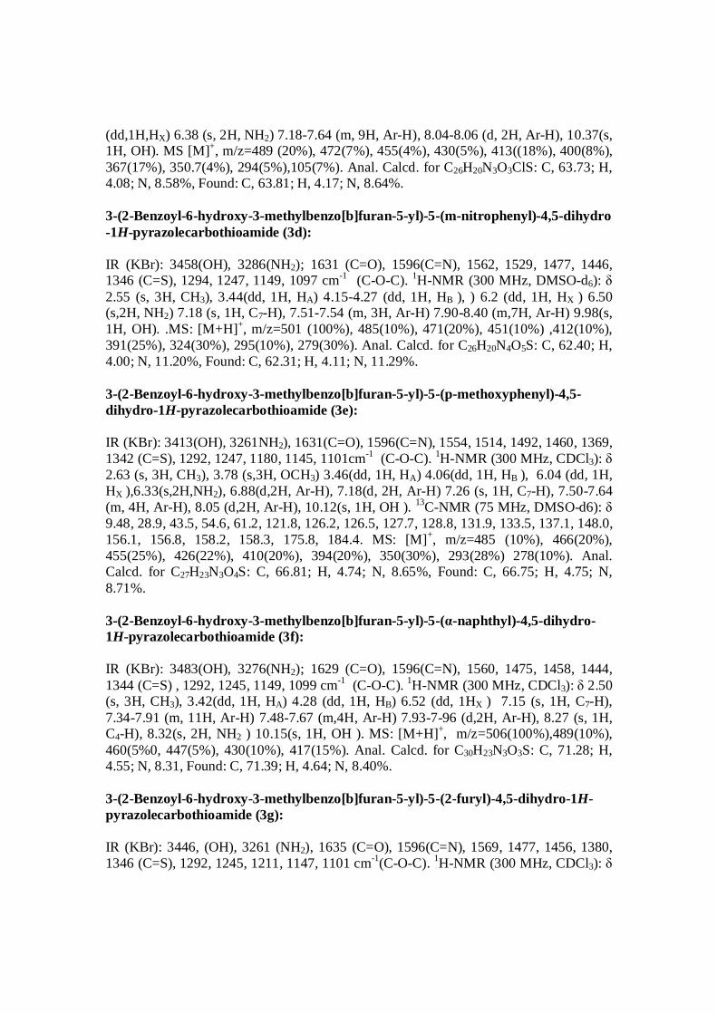
(dd,1H,HX) 6.38 (s, 2H, NH2) 7.18-7.64 (m, 9H, Ar-H), 8.04-8.06 (d, 2H, Ar-H), 10.37(s, 1H, OH). MS [M]+, m/z=489 (20%), 472(7%), 455(4%), 430(5%), 413((18%), 400(8%), 367(17%), 350.7(4%), 294(5%),105(7%). Anal. Calcd. for C26H20N3O3ClS: C, 63.73; H, 4.08; N, 8.58%, Found: C, 63.81; H, 4.17; N, 8.64%. 3-(2-Benzoyl-6-hydroxy-3-methylbenzo[b]furan-5-yl)-5-(m-nitrophenyl)-4,5-dihydro -1H-pyrazolecarbothioamide (3d): IR (KBr): 3458(OH), 3286(NH2); 1631 (C=O), 1596(C=N), 1562, 1529, 1477, 1446, 1346 (C=S), 1294, 1247, 1149, 1097 cm-1 (C-O-C). 1H-NMR (300 MHz, DMSO-d6): δ 2.55 (s, 3H, CH3), 3.44(dd, 1H, HA) 4.15-4.27 (dd, 1H, HB ), ) 6.2 (dd, 1H, HX ) 6.50 (s,2H, NH2) 7.18 (s, 1H, C7-H), 7.51-7.54 (m, 3H, Ar-H) 7.90-8.40 (m,7H, Ar-H) 9.98(s, 1H, OH). .MS: [M+H]+, m/z=501 (100%), 485(10%), 471(20%), 451(10%) ,412(10%), 391(25%), 324(30%), 295(10%), 279(30%). Anal. Calcd. for C26H20N4O5S: C, 62.40; H, 4.00; N, 11.20%, Found: C, 62.31; H, 4.11; N, 11.29%. 3-(2-Benzoyl-6-hydroxy-3-methylbenzo[b]furan-5-yl)-5-(p-methoxyphenyl)-4,5-dihydro-1H-pyrazolecarbothioamide (3e): IR (KBr): 3413(OH), 3261NH2), 1631(C=O), 1596(C=N), 1554, 1514, 1492, 1460, 1369, 1342 (C=S), 1292, 1247, 1180, 1145, 1101cm-1 (C-O-C). 1H-NMR (300 MHz, CDCl3): δ 2.63 (s, 3H, CH3), 3.78 (s,3H, OCH3) 3.46(dd, 1H, HA) 4.06(dd, 1H, HB ), 6.04 (dd, 1H, HX ),6.33(s,2H,NH2), 6.88(d,2H, Ar-H), 7.18(d, 2H, Ar-H) 7.26 (s, 1H, C7-H), 7.50-7.64 (m, 4H, Ar-H), 8.05 (d,2H, Ar-H), 10.12(s, 1H, OH ). 13C-NMR (75 MHz, DMSO-d6): δ 9.48, 28.9, 43.5, 54.6, 61.2, 121.8, 126.2, 126.5, 127.7, 128.8, 131.9, 133.5, 137.1, 148.0, 156.1, 156.8, 158.2, 158.3, 175.8, 184.4. MS: [M]+, m/z=485 (10%), 466(20%), 455(25%), 426(22%), 410(20%), 394(20%), 350(30%), 293(28%) 278(10%). Anal. Calcd. for C27H23N3O4S: C, 66.81; H, 4.74; N, 8.65%, Found: C, 66.75; H, 4.75; N, 8.71%. 3-(2-Benzoyl-6-hydroxy-3-methylbenzo[b]furan-5-yl)-5-(α-naphthyl)-4,5-dihydro-1H-pyrazolecarbothioamide (3f): IR (KBr): 3483(OH), 3276(NH2); 1629 (C=O), 1596(C=N), 1560, 1475, 1458, 1444, 1344 (C=S) , 1292, 1245, 1149, 1099 cm-1 (C-O-C). 1H-NMR (300 MHz, CDCl3): δ 2.50 (s, 3H, CH3), 3.42(dd, 1H, HA) 4.28 (dd, 1H, HB) 6.52 (dd, 1HX ) 7.15 (s, 1H, C7-H), 7.34-7.91 (m, 11H, Ar-H) 7.48-7.67 (m,4H, Ar-H) 7.93-7-96 (d,2H, Ar-H), 8.27 (s, 1H, C4-H), 8.32(s, 2H, NH2 ) 10.15(s, 1H, OH ). MS: [M+H]+, m/z=506(100%),489(10%), 460(5%0, 447(5%), 430(10%), 417(15%). Anal. Calcd. for C30H23N3O3S: C, 71.28; H, 4.55; N, 8.31, Found: C, 71.39; H, 4.64; N, 8.40%. 3-(2-Benzoyl-6-hydroxy-3-methylbenzo[b]furan-5-yl)-5-(2-furyl)-4,5-dihydro-1H-pyrazolecarbothioamide (3g): IR (KBr): 3446, (OH), 3261 (NH2), 1635 (C=O), 1596(C=N), 1569, 1477, 1456, 1380, 1346 (C=S), 1292, 1245, 1211, 1147, 1101 cm-1(C-O-C). 1H-NMR (300 MHz, CDCl3): δ

2.60 (s, 3H, CH3), 3.70(dd, 1H, HA) 3.90 (dd, 1H, HB) 6.24 (dd, 1HX ), 6.35 (d,1H,H Hα -furyl), 6.52 (d,1H, Hβ-furyl), 7.16 (s,1H,C7-H), 7.26-7.61 (m, 7H, Ar-H, NH2), 8.04-8.06(d, 2H, Ar-H) 10.02 (s, 1H, OH). MS:[M]+,m/z =445 (20%), 428(8%), 411(10%), 393(35%), 386(50%), 369(30%), 358(12%), 293(12%), 279(10%), 264(6%), 249(5%), 169(10%), 165(5%), 153(5%), 140(5%), 115(5%), 109(10%), 105(100%),94(6%), 81(16%), 77(75%), 73(10%), 65(5%), 59(18%), 51(10%), 43(10%). Anal. Calcd. for C24H19N3O4S: C, 64.71; H, 4.26; N, 9.43%. Found: C, 64.80; H, 4.34, N, 9.52%, Antibacterial Activity The activity was determined using cup-plate agar diffusion method23 by measuring the inhibition zone in mm. All the compounds were screened for their antibacterial activity against a variety of bacterial strains such as Bacillus subtilis (ATCC-6633), staphylococcus aureus (ATCC-29737), Escherichia coli (ATCC-10536) Pseudomonas auregones (ATCC-27853) using streptomycin, Tetracycline, Chloramphenicol, Carbenicillin respectively as standard drug. Nutrient Agar was used as a culture medium. A 1mg/ml solution in dimethylformamide was used. DMF showed no inhibition zones. The agar medium was inoculated with bacterial cultures tested. After 24 hours of incubation at 37oC, the diameter of inhibition zone (mm) was measured. Streptomycin (25 μg/well) used against Bacillus subtilis, Tetracycline (30 μg/well) used against S.aureus, Chloramphenicol (25 μg/well) used against E. coli, Carbenicillin (100 μg/well) used against pseudomonas aeruginosa as a references (as standard) for antibacterial activity. The results of the antibacterial activity are given in Table2. 3-(2-Benzoyl-6-hydroxy-3-methylbenzo[b]furan-5-yl)-5-(aryl)-4,5-dihydro-1H-pyrazole carbothioamides 3b, 3e and 3g exhibited good antibacterial activity. Table2. Antibacterial activity of some synthesized compounds and inhibition zones
Compound
No.
Gram Positive Bacteria Gram Negative Bacteria Bacillus subtilis (ATCC-6633)
Staphylococcus aureus (ATCC-29737)
Escherichia coli (ATCC-10536)
Pseudomonas aeruginosa (ATCC-27853)
3a 10.0 mm 10 mm 8 mm - ve 3b 11.5 mm 11 mm 8 mm 8 mm 3c 8 mm 9mm - ve - ve 3d 8 mm 10 mm - ve 8mm 3e 9 mm 8 mm 9 mm 9 mm
3f 10.5mm 10 mm 8 mm - ve 3g 9 mm 10 mm 9 mm 8 mm
Standard 22 mm Streptomycin
15 mm Tetracycline
13 mm Chloramphenicol
13 mm (Carbenicillin)

Conclusions: In conclusion, we have successfully synthesized new 3-(2-Benzoyl-6-hydroxy-3-methylbenzo[b]furan-5-yl)-5-(phenyl)-4,5-dihydro-1H-pyrazole carbothioamides under microwave irradiation conditions. This methodology provides an easier facile and environmentally benign synthesis in which the reaction time is reduced with better yields. The compounds 3b, 3e and 3g exhibited good antibacterial activity. Acknowledgements: Authors are thankful to the Prof. K. Veera Reddy Head, Department of Chemistry, Osmania University, Hyderabad and K. V. Ramana, Chairman and Managing Director Sven Genetech Limited for providing laboratory facilities to carry out the research work.
References: (1) H. Singh, and M. N. Ghosh, J.Phar. Pharmacol., 20, 312, (1958). (2) A. Kerntzberger and K. Burgwitz, Arch. Pharm., 312, 873 (1979). (3) P. Descacq, A. Nuhrich, M. Capdepuy and G. Devaux, Eur. J. Med. Chem. 25, 285, (1990). (4) Z. Brozozowsk and E. Pomarnacka, Acta Pol. Pharm, 37, 1378 (1980). (5) Safak Cihat, Tayhan A yla, Sarac Selma and Yulug Nuran, J. Ind. Chem. Soc, 67,
571 (1990). (6) V. S. Jolly, G. D. Arora and Talwar Preeti, J. Ind. Chem. Soc, 67, 1001 (1990). (7) S. S. Parmar, B. R. Pandey and C. Diwedi, J. Pharm. Sci., 63, 1152, (1974). Chem.
Abstr., 82, 1975, 259660u. (8) A. Mohammad Ali, Mohammaad Shaharyar, A. Anees Siddiqui, Asif Husain and
Mustaqeem Abdulliah Acta poloniae Pharmaceutica – Drug research, 63, 5, 435 (2007).
(9) Ozan Ruhoglu, Zuhal özdemir, ünsal Calis, Bülent Gümüsel and Abdullah Altan Bilgin, Arzneim.-Forsch./Drug. Res. 55, No.8, 431, (2005).
(10) Venkatesan Jayaprakash, N. Barij Sinha, Gulberk Ucarb, and Ayse Ercan, Bioorg.& Med. Chem .lett., 18, 6362, (2008).
(11) Franco Chimenti, Elias Maccionic, Daniela Secci, Adriana Bolasco, Paola Chimenti, Arianna Graness, Olivia Befani, Paola Turini, Stefano Alcaro, Francesco Ortuso, Roberto Cirilli, Franceson La Torr, Maria C. Cardia and Simona Distinto, J. Med. Chem. 48, 7113, (2005).
(12) G. Doria, C. Romeo, M. L. Carno, and G. Cadelli, Farmaco Ed. Sci., 35, 674 (1980).
(13) B. Kadin and Saul, J. Med. Chem., 15, 551, (1972). (14) K .V, B. Rao and R. N. Iyer, Ind. J. Chem., 19B, 992, (1980). (15) R. S. Varma, Green chemistry 1, 43, (1999). (16) D. Ashok, K. Pallavi, G. Jagath Reddy, and K. Srinivas Rao, Heterocyclic
Communications, 14, 55, (2008). (17) D. Ashok, K. Pallavi, G. Jagath Reddy, and K. Srinivas Rao, Heterocyclic
Communications, 14, 33, (2008).

(18) D. Ashok, M. Khalilullah, K. Sudershan, Ind. J. Heterocyclic Chem., 18, 109, (2008).
(19) J. Sharada, Y. Ratna Kumari, and M. K. Lingeswara Rao, Ind. J. Chem., 25B, 334, (1986).
(20) K. Vishnu Vardhan Reddy, P. Sampath Rao, and D. Ashok, Synth. Commun., 27, 3871, (1997).
(21) A. S. R. Anjaneyulu, A. V. R. Prasad and D. S. K. Reddy, Curr. Sci., 48, 300, (1979).
(22) a) J. R. Rather, and E. M. Reid, J. Am. Chem. Soc 41, 75, 77, (1919). b) W. D. Longley, “Org, Synth. Coll. Vol. 2”, John Wiley and Sons. NewYork, 127,
(1947). (23) Biological tests and Assay-An official monograph of USP 25, 1883-1889.