microscopy of abrasive-planed and knife-planed surfaces in
TRANSCRIPT
MICROSCOPY OF ABRASIVE-PLANED AND KNIFE-PLANED SURFACES IN
WOOD-ADHESIVE BONDS
Lidija Murmanis and Bryan H. River Research Forest Products Technologists
Forest Products Laboratory, Madison, WI 53705
and
Harold Stewart Forest Products Technologist
Forestry Sciences Laboratory, Carbondale, IL1
(Received 2 March 1981)
ABSTRACT
Fluorescence microscopy (FM) disclosed no differences in wood cell structure between abrasive-and knife-planed Douglas-fir joints under constant conditions. However, after a one-cycle soak-dry exposure, formation of checks along the rays were visible in both abrasive- and knife-planed samples by fluorescence microscopy. For this same exposure, scanning electron microscopy revealed many radial cracks in the S2 layer and ruptures between the S1 and S2 layers in abrasive-planed samples. Knife-planed samples had few ruptures between the S1 and S2 layers and very few cracks in the S2
layer.Previous work showed that, although knife planing gave much smoother surfaces at the cellular
level than did abrasive planing, both surfaces resulted in high strength bonds. When those bonded samples were subjected to a soak-dry treatment, however, strength of abrasive-planed samples was much lower than that of knife-planed samples.
The substantially intact S2 layers in knife-planed samples, as revealed here, apparently retain con-siderable strength, while rupturing and cracking in the abrasive-planed samples explain the loss of bond quality reported in earlier work.
Keywords: Planed surfaces, wood-adhesive bonds, microscopy, yellow-poplar, Douglas-fir.
INTRODUCTION
The visible macroscopic characteristics of a wood surface must be augmented with microscopic views to clarify certain behavior in wood-adhesive bonds.
Recent studies of abrasive-planed and knife-planed adhesive joints showed comparable strength values under constant dry conditions. When the same joints were exposed to soak-dry cycling, however, the bond quality of the abrasive-planed samples fell off drastically (Jokerst and Stewart 1976; River et al. 1981).
The study reported here was designed to investigate microscopically the mor-phology of bondlines in those specimens from an earlier work (River et al. 1981). The objective was to relate differences in the physical nature of wood after the
1Both laboratories are part of the U.S. Department of Agriculture, Forest Service; the Forest Products Laboratory is maintained in cooperation with the University of Wisconsin. This article was written and prepared by U.S. Government employees on official time and is therefore in the public domain.
Wood and Fiber Science, 15(2), 1983, pp. 102-115

Murmanis et al.—MICROSCOPY OF PLANED SURFACES 103
various surfacing and exposure treatments to differences in the attendant joint strengths, as determined in that study.
This report is a pictorial comparison by fluorescence light microscopy and scanning electron microscopy of the effect of abrasive planing, knife planing, and soaking and drying stresses on the bondline.
MATERIALS AND METHODS
Specimens were obtained from a set prepared earlier for bond strength tests (River et al. 1981). Yellow-poplar (Liriodendron tulipifera L.) specimens were bonded with urea-formaldehyde and Douglas-fir (Pseudotsuga menziesii (Mirb.)Franco) with phenol-resorcinol formaldehyde. The six combinations in abrasive planing involved two feed speeds—45 and 90 ft/min–and three depths of cut– 0.010, 0.040, and 0.080 inch–all with No. 36 grit. The bonding procedure, ex-posure before test, and strength tests have been described (River et al. 1981).
Before testing the strength properties (River et al. 1981), the specimens were divided into two groups. The control groups consisted of Douglas-fir conditioned to 12% moisture content (MC) oven-dry basis and yellow-poplar conditioned to 6% MC. The other group was subjected to one cycle of vacuum-pressure soak-dry treatment and then conditioned to 12 and 6% MC before examination. The yellow-poplar samples of the exposed group could not be studied because the urea-formaldehyde adhesive deteriorated during exposure.
For fluorescence microscopy, small blocks with about 1 cm2 of transverse surface area, including a bondline, were smoothed with a sliding microtome using a freezing (CO,) attachment and examined in reflected near-UV light (peak trans-mission at 365 mµ). Our experience, supported by those of other scientists (Nearn 1974; Quirk 1968) showed that FM near-UV radiation gives best representation of wood and adhesives because of the autofluorescence of wood and of some adhesives.
For scanning electron microscopy, blocks with a smoothed surface, approxi-mately 8 mm on a side, were mounted onto standard aluminum stubs with silver paint and coated with gold in a sputter-coater. The microscope was the Cambridge Stereoscan, Type 2A.
RESULTS
Fluorescence microscopy
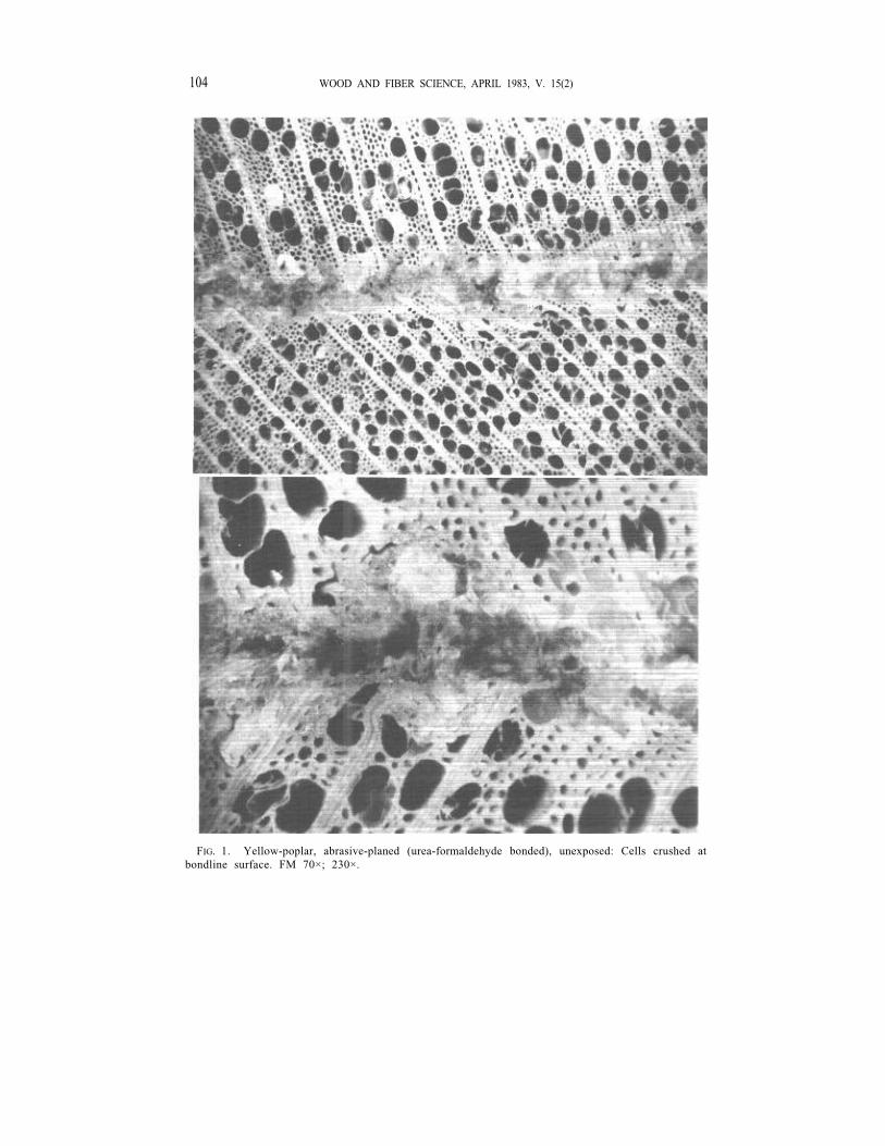
Fluorescence microscopy indicated that abrasive planing gave strikingly dif-ferent bonding surfaces than did knife planing. In abrasive planing, all six com-binations of speed and depth of cut produced similar morphological features: thick and extremely irregular bondlines but with practically no voids within them. A few cells adjoining the bondline were penetrated by the adhesive, but none at a greater distance were. Rays showed little penetration. Beneath the surface, the cells of the axial system were crushed, locally quite severely, and rays were often bent or broken (Figs. 1, 2). Where severe crushing of cells occurred, the adhesive was not able to penetrate through the crushed layer to the sound wood. In the dark-colored bondline of phenol-resorcinol in Douglas-fir, the filler (walnut-shell flour) particles are visible as white flecks due to fluorescing cells walls. In Doug-las-fir, earlywood and latewood were affected differently by abrasive planing: the
104 WOOD AND FIBER SCIENCE, APRIL 1983, V. 15(2)
FIG. 1. Yellow-poplar, abrasive-planed (urea-formaldehyde bonded), unexposed: Cells crushed at bondline surface. FM 70×; 230×.

Murmanis et al.—MICROSCOPY OF PLANED SURFACES 105
FIG. 2. Douglas-fir, abrasive-planed (phenol-resorcinol bonded), unexposed: Tracheids crushed in earlywood, but not in latewood. FM 75×; 120×.

106 WOOD AND FIBER SCIENCE, APRIL 1983, V. 15(2)
FIG. 3. Yellow-poplar, knife-planed, unexposed: Cells not crushed at bondline surface. FM 75×; 120×.

Murmanis et al.—MICROSCOPY OF PLANED SURFACES 107
FIG. 4. Douglas-fir, knife-planed, unexposed: Tracheids not crushed in earlywood or latewood. FM 80×; 120×.

108 WOOD AND FIBER SCIENCE, APRIL 1983, V. 15(2)
FIG. 5. Douglas-fir, abrasive-planed, exposed to soak-dry cycle: Tracheids crushed in earlywood; checks extend along rays (arrows). FM 80×; 130×.
Murmanis et al.—MICROSCOPY OF PLANED SURFACES 109
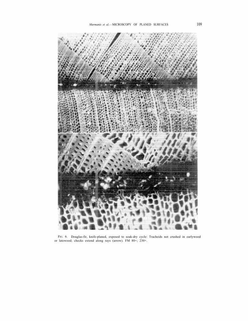
FIG. 6. Douglas-fir, knife-planed, exposed to soak-dry cycle: Tracheids not crushed in earlywood or latewood; checks extend along rays (arrow). FM 80×; 230×.

110 WOOD AND FIBER SCIENCE, APRIL 1983, V. 15(2)
FIG. 7. Douglas-fir, abrasive-planed. Bondline at right. Upper, unexposed: Earlywood tracheids crushed next to bondline; ray (R) penetrated by adhesive. SEM 1,500× Lower, exposed to soak-dry cycle: Earlywood tracheids crushed and their walls broken (arrows). SEM 1,400×.

Murmanis et al.—MICROSCOPY OF PLANED SURFACES
FIG. 8. Douglas-fir, knife-planed. Bondline at right. Upper, unexposed: Earlywood tracheids not crushed. SEM 1,200×. Lower, exposed to soak-dry cycle: Earlywood tracheids not crushed. SEM 1,200×.

112 WOOD AND FIBER SCIENCE, APRIL 1983, V. 15(2)
FIG. 9. Douglas-fir, abrasive-planed. Bondline at right. Upper, unexposed: A few radial cracks in S2 layer of latewood tracheids (arrow) and a few separations between S1 and S2 layers (arrow). SEM 1,200×. Lower, exposed to soak-dry cycle: Many radial cracks in S2 layer (arrow) and separations between S1 and S2 layers of latewood tracheids (arrow). SEM 1,400×.

Murmanis et al.—MICROSCOPY OF PLANED SURFACES
FIG. 10. Douglas-fir, knife-planed. Bondline at left. Upper, unexposed: A few separations between S1 and S2 layers of latewood tracheids (arrow). SEM 1,200×. Lower, exposed to soak-dry cycle: A few separations between S1 and S2 layers. SEM 1,400×.
114 WOOD AND FIBER SCIENCE, APRIL 1983, V. 15(2)
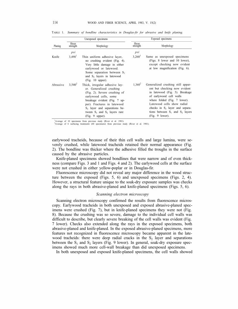
TABLE 1. Summary of bondline characteristics in Douglas-fir for abrasive and knife planing.
Unexposed specimens Exposed specimens
Shear ShearPlaning strength Morphology strength Morphology
psi
Knife 3,4901 Thin uniform adhesive layer, no crushing evident (Fig. 4). Very little damage in either earlywood or latewood. Some separation between S1
and S2 layers in latewood (Fig. 10 upper).
Abrasive 3,5402 Thick, irregular adhesive lay-er. Generalized crushing (Fig. 2). Severe crushing of earlywood cells, some breakage evident (Fig. 7 up-per). Fractures in latewood S2 layer and separations be-tween S1 and S2 layers rare (Fig. 9 upper).
psi
3,2601 Same as unexposed specimens (Figs. 8 lower and 10 lower), except checking now evident at low magnification (Fig. 6).
1,3602 Generalized crushing still appar-ent but checking now evident in latewood (Fig. 5). Breakage of earlywood cell walls where folded (Fig. 7 lower). Latewood cells show radial checks in S2 layer and separa-tions between S1 and S2 layers (Fig. 9 lower).
1Average of 10 specimens from previous study (River et al. 1981). 2Average of 6 surfacing treatments (60 specimens) from previous study (River et al. 1981).
earlywood tracheids, because of their thin cell walls and large lumina, were se-verely crushed, while latewood tracheids retained their normal appearance (Fig. 2). The bondline was thicker where the adhesive filled the troughs in the surface caused by the abrasive particles.
Knife-planed specimens showed bondlines that were narrow and of even thick-ness (compare Figs. 3 and 1 and Figs. 4 and 2). The earlywood cells at the surface were not crushed in either yellow-poplar or in Douglas-fir.
Fluorescence microscopy did not reveal any major difference in the wood struc-ture between the exposed (Figs. 5, 6) and unexposed specimens (Figs. 2, 4). However, a structural feature unique to the soak-dry exposure samples was checks along the rays in both abrasive-planed and knife-planed specimens (Figs. 5, 6).
Scanning electron microscopy
Scanning electron microscopy confirmed the results from fluorescence micros-copy. Earlywood tracheids in both unexposed and exposed abrasive-planed spec-imens were crushed (Fig. 7), but in knife-planed specimens they were not (Fig. 8). Because the crushing was so severe, damage to the individual cell walls was difficult to describe, but clearly severe breaking of the cell walls was evident (Fig. 7 lower). Checks also extended along the rays in the exposed specimens, both abrasive-planed and knife-planed. In the exposed abrasive-planed specimens, more features not recognized in fluorescence microscopy became apparent in the late-wood tracheids: there were deep radial cracks in the S2 layer and separations between the S1 and S2 layers (Fig. 9 lower). In general, soak-dry exposure spec-imens showed much more cell-wall breakage than did unexposed specimens.
In both unexposed and exposed knife-planed specimens, the cell walls showed

Murmanis et al.—MICROSCOPY OF PLANED SURFACES 115
only occasional separations between S1 and S2 layers and very few radial cracks in the latewood S2 layer (Fig. 10). Also, in these specimens, the only perceptible difference was the presence of checks extending along the rays in the exposed specimens.
CONCLUSIONS
Fluorescence microscopy clearly showed morphological differences in bonded wood specimens with respect to their surface machining. Knife-planing gave much smoother surfaces as seen at the cellular level than did abrasive planing. On the other hand, this morphological difference did not seem to affect the strength of the bonds, as the shear strength of dry, unexposed abrasive-planed specimens was comparable to that of dry, unexposed knife-planed specimens (River et al. 1981).
Under adverse conditions, however, such as the one-cycle, soak-dry treatment, the condition of cells-intact or crushed-was more critical. Generally, the strength of exposed specimens was markedly lower, and strength of exposed abrasive-planed specimens was considerably lower than that of exposed knife-planed sam-ples.
Scanning electron microscopy of abrasive-planed specimens after the soak-dry treatment revealed that the walls of tracheids had been greatly damaged-i.e., radial cracks in the S2 layer and separations between the S1 and S2 layers. In exposed knife-planed specimens, the cell walls in the latewood tracheids showed only occasional separations between S1 and S2 layers (the weakest region in the cell wall [Wardrop and Addo-Ashong 1963]), and because of the relatively intact S2 layer, the wood retained considerable strength. In contrast, damaged wall layers in the exposed abrasive-planed specimens likely explain the poor bond quality shown in previous studies (Jokerst and Stewart 1976; River et al. 1981).
REFERENCES
JOKERST, RONALD W., AND HAROLD A. STEWART. 1976. Knife- versus abrasive-planed wood: Quality of adhesive bonds. Wood Fiber 8(2):107-113.
NEARN, WILLIAM T. 1974. Application of the ultrastructure concept in industrial wood products research. Wood Sci. 6(3):285-293.
QUIRK, JOHN T. 1968. Fluorescence microscopy for detecting adhesives on fracture surfaces. U.S. For. Serv. Res. Note FPL-0191. For. Prod. Lab., Madison, WI.
RIVER, B. H., L. MURMANIS, AND H. A. STEWART. 1981. Effect of abrasive-planing stock removal rate on adhesive-bonded joint performance. In Wood adhesives-Research, application, and needs, Proc. 1980 Adhesive Symp., Sept. 23-25, 1980, Madison, Wis. USDA For. Serv., For. Prod. Lab., Madison, WI.
WARDROP, A. B., AND F. W. ADDO-ASHONG. 1963. The anatomy and fine structure of wood in relation to mechanical failure. Pages 169-200 in Tewksbury symp. on fracture. D.F.P. Reprint No. 560. C.S.I.R.O., Div. For. Prod., South Melbourne, Australia.