microrna-34a dependent regulation of axl controls the...
TRANSCRIPT

ARTICLE
Received 30 Aug 2016 | Accepted 5 May 2017 | Published 22 Jun 2017
MicroRNA-34a dependent regulation of AXLcontrols the activation of dendritic cells ininflammatory arthritisMariola Kurowska-Stolarska1, Stefano Alivernini2, Emma Garcia Melchor1, Aziza Elmesmari1, Barbara Tolusso2,
Clare Tange1, Luca Petricca2, Derek S. Gilchrist1, Gabriele Di Sante2, Chantal Keijzer1, Lynn Stewart1,
Clara Di Mario2, Vicky Morrison1, James M. Brewer1, Duncan Porter1, Simon Milling1, Ronald D. Baxter1,3,
David McCarey3, Elisa Gremese2, Greg Lemke4, Gianfranco Ferraccioli2, Charles McSharry1 & Iain B. McInnes1
Current treatments for rheumatoid arthritis (RA) do not reverse underlying aberrant immune
function. A genetic predisposition to RA, such as HLA-DR4 positivity, indicates that dendritic
cells (DC) are of crucial importance to pathogenesis by activating auto-reactive lymphocytes.
Here we show that microRNA-34a provides homoeostatic control of CD1cþ DC activation via
regulation of tyrosine kinase receptor AXL, an important inhibitory DC auto-regulator. This
pathway is aberrant in CD1cþ DCs from patients with RA, with upregulation of miR-34a and
lower levels of AXL compared to DC from healthy donors. Production of pro-inflammatory
cytokines is reduced by ex vivo gene-silencing of miR-34a. miR-34a-deficient mice are
resistant to collagen-induced arthritis and interaction of DCs and T cells from these mice are
reduced and do not support the development of Th17 cells in vivo. Our findings therefore
show that miR-34a is an epigenetic regulator of DC function that may contribute to RA.
DOI: 10.1038/ncomms15877 OPEN
1 Institute of Infection, Immunity and Inflammation, University of Glasgow, 120 University Place, Glasgow G12 8TA, UK. 2 Division of Rheumatology,Fondazione Policlinico Universitario A. Gemelli, Catholic University of the Sacred Heart, Rome 00168, Italy. 3 NHS Greater Glasgow and Clyde, 1055 GreatWestern Road, Glasgow G12 0XH, UK. 4 Molecular Neurobiology Laboratory, The Salk Institute for Biological Studies, 10010 North Torrey Pines Road, La Jolla,California 92037, USA. Correspondence and requests for materials should be addressed to M.K.S. (email [email protected]) or toI.B.M. (email: [email protected]).
NATURE COMMUNICATIONS | 8:15877 | DOI: 10.1038/ncomms15877 | www.nature.com/naturecommunications 1

Dendritic cells (DC) are sentinels of the immune systemthat initiate and direct the immune responsetowards foreign antigens while inducing tolerance to
self-antigens1. Numerous studies have shown that altered DCfunction can promote breach of tolerance to self-antigens anddevelopment of autoimmunity and inflammation2–4.
Rheumatoid arthritis (RA) is the most common autoimmunepolyarthritis, with a lifetime risk of 3.6% in women and 1.7% inmen5,6, causing pain, disability, impaired quality of life andreduced life expectancy. Genetic, epigenetic and epidemiologicalstudies suggest that autoimmunity lies at the core of RApathogenesis. RA is particularly associated with breach oftolerance to post-translationally modified self-proteins, usuallyvia citrullination or carbamylation, often predating clinicaldisease by years7–9. These early events are initiated by DCs thatstimulate auto-reactive T and B cells8,10,11, which thereafterorchestrate progression to chronic joint inflammation,destruction and comorbidity12,13. The majority of current RAtreatments target inflammation; however, partial or non-responseis common and sustained remission occurs in a minority ofpatients, representing a considerable unmet clinical need.For example, tumour necrosis factor (TNF), interleukin-6 (IL-6)receptor (IL-6R) and JAK inhibitors reduce inflammation, andslow down radiographic progression, but do not generallymediate restoration of immune homeostasis14,15. Moreover,current approaches targeting the adaptive immune response viaB cell depletion and co-stimulatory molecule blockade do notseem to promote tolerance16. Therefore, restoring homoeostaticDC function to achieve tolerance would be a compelling strategyfor treatment of RA.
Accordingly, we sought to dissect molecular pathways thatdrive the activation of RA DCs. DCs can be broadly categorizedinto plasmacytoid and myeloid (conventional) lineages. Humanmyeloid DCs comprise CD141þ , CD16þSLANþ , CD16þ
SLAN� and CD1cþ cells17,18. In addition, ‘inflammatory DCs’can differentiate from monocytes infiltrating inflammatory tissuesand show phenotypic overlap (for example, CD1c expression)and function with conventional CD1cþ 18 DCs, althoughthe latter cells differentiate from a distinct precursor19. Humanconventional CD1cþ DCs and inflammatory DC populations arecritical for T helper type 17 (Th17) responses (for example, viaIL-23, IL-6, IL-1b) and pro-inflammatory TNF production,particularly upon TLR7/8 stimulation20–24. The activation ofDCs is controlled by multiple negative-feedback loops25–27. TAMreceptor family containing tyrosine kinase receptors Tyro3, AXLand MERTK are critical in switching off TLR ligand-induced DCactivation26. Recently, a constitutive AXL expression that wasassociated with a lower level of major histocompatibility class(MHC) class II was identified as a feature of a distinctsubpopulation of human conventional CD1cþ DCs28.
CD1cþ DC from RA patients show activated phenotype. RAsynovial fluid contains both conventional CD1cþ andinflammatory CD1cþ cells that can prime naive T cells29–31.In addition, these cells support synovial inflammationby producing TNF upon stimulation with TLR7/8 ligands23,32,33,and human transgenic TLR8 mice develop spontaneous arthritisdue to constitutive activation of DCs that drive autoimmunity34.
Environmental factors such as smoking strongly influence riskof developing RA, suggesting that epigenetic mechanisms areinvolved in RA pathogenesis35. MicroRNAs are epigeneticregulators that fine-tune complex molecular pathways bymediating mRNA degradation or translation inhibition basedon 6–8 nucleotide (nt) complementarity sequences betweenmicroRNA and the 30-untranslated region (UTR) of targetmRNAs36. Aberrations in miRNA control mechanismscontribute to the development of autoimmune diseases,
including arthritis37–39. Herein we describe a role for miR-34ain driving deregulation of DC function in inflammatory arthritis.
ResultsmiR-34a is upregulated in CD1cþ DCs from RA patients.To identify molecular pathways that are deregulated inRA myeloid cells at the site of pathology, we previously generateda transcriptomic signature of RA synovial fluid (SF) myeloid cellsand found key upregulation of miR-34a compared to matchedcells from blood40. As miR-34a is enriched in DCs41,42, wehypothesised that miR-34a has a regulatory role in DC functionand thereby in the pathogenesis of RA. MiR-34a (chromosome1p36) and family-members miR-34b/c (co-transcribed from11q23) have similar functions but have distinct expressionpatterns43. To investigate the expression of miR-34a and c inRA CD1cþ cells, we purified CD1cþ DCs (sorting strategy inSupplementary Fig.1) from peripheral blood (PB) of healthydonors and from PB and SF from RA patients with establishedRA (42 years from diagnosis). Blood CD1cþ DCs from RApatients had greater miR-34a expression than healthy donors, andthis was further increased in SF DCs (Fig. 1a). Elevated miR-34aexpression was also observed in blood DCs from patients withearly RA (o6 months from diagnosis) (Fig. 1b). MiR-34c wasexpressed only at low levels and was not significantlydifferentially expressed across disease or tissue and was notfurther examined (Fig. 1a).
DCs primarily function within lymph node structures orthe tissue. We therefore investigated the expression of miR-34a inCD1cþ cells from synovial tissue of patients with elevated diseaseactivity (mean±s.d. DAS28: 5.07±0.68) and compared it tothe cells from their PB and to PB cells from age-matched healthydonors. MiR-34a was strongly upregulated in CD1cþ sorted fromRA synovial tissues compared to RA PB and healthy PB cells(Fig. 1c). This analysis of an independent patient cohort confirmedan increase expression of miR-34a in RA PB CD1cþ compared toage-matched healthy donors PB cells, and revealed that the highestfold change in PB and synovial tissue was among patientsresistant-to-treatment and positively correlated with diseaseduration (r¼ 0.543; P¼ 0.016; Pearson Correlation Coefficient).An evaluation of miR-34a expression in blood and synovial tissueCD1cþ cells from patients with comparator inflammatory jointdisease, psoriatic arthritis (PsA) showed no significant changes inthe blood but an increase in expression in synovial tissue DCsalthough to a lesser degree than in RA (P¼ 0.02; Kruskal–Walliswith Dunn’s multiple comparison test; Supplementary Fig. 2).Demographic and clinical information for both sets of RA patients;and PsA patients is presented in Supplementary Tables 1 and 2.Collectively, these data suggest a potential role for miR-34a in theregulation of DCs function in RA.
We next investigated the regulation of miR-34a expression inCD1cþ DCs. MiR-34a was highest in CD1cþ cells from SF andsynovial tissues, which contains both conventional CD1cþ andmonocyte-derived CD1cþ cells; therefore, we compared theexpression of miR-34a in monocytes and monocyte-derived DCs.The copy number of miR-34a in blood monocyte subsets(CD14þCD16� , CD14dimCD16þ and CD14þCD16þ ) fromhealthy donors was low (Fig. 2a,b), but increased significantlywhen they were differentiated to inflammatory CD1cþ DCs bygranulocyte macrophage colony-stimulating factor (GM-CSF),which was also reflected by a fold-change as compared tomonocytes at day 0 (Fig. 2c). The miR-34a levels in these day 7immature CD1cþDCs were rapidly downregulated uponmaturation with ligands CL097 for intracellular TLR7/8and lipopolysaccharide (LPS) for surface TLR4 (Fig. 2d). Thus,miR-34a can be induced during GM-CSF-triggered DC
ARTICLE NATURE COMMUNICATIONS | DOI: 10.1038/ncomms15877
2 NATURE COMMUNICATIONS | 8:15877 | DOI: 10.1038/ncomms15877 | www.nature.com/naturecommunications

differentiation and then undergoes dynamic regulation upon DCmaturation suggesting a potential role in this process. We next setout to explore the role of miR-34a in DC activation and its impacton arthritis.
miR-34a� /� mice have reduced antigen-induced arthritis.We explored a functional role for miR-34a in experimentalarthritis using a collagen-induced model in which DCs play a keypathogenic role. Compared to wild-type (WT), miR-34a� /�
mice had reduced incidence and severity of arthritis (Fig. 3a,b)accompanied by reduced histopathological articularinflammation and damage (Fig. 3c,d). Serum cytokine profilingrevealed selectively reduced IL-17 concentrations (Fig. 3e)suggesting a role for miR-34a in the generation ofIL-17-producing, auto-reactive T lymphocytes that drive experi-mental arthritis44,45. There was no difference in concentration ofthe prototypical Th1 or Th2 cytokines, IFNg (P¼ 0.18;Mann–Whitney u-test) and IL-5 (P¼ 0.08; Mann–Whitneyu-test), respectively, nor in the production of collagen-specificantibodies (Supplementary Fig. 3).
To address the specific role of miR-34a in DC-drivenantigen-specific T lymphocyte activation, we adoptively trans-ferred naive OVA-specific OT-II T lymphocytes (congenicCD45.1 background) to WT and miR-34a� /� recipients andchallenged them with OVA antigen as outlined on Fig. 3f.MiR-34a� /� and WT recipients had similar numbers of totalantigen-specific CD4þ and CD4þ INFgþ cells but miR-34a� /�
mice showed a selective reduction in antigen-specific Th17 cellnumbers (Fig. 3g,h and gating strategy in Supplementary Fig. 4).Collectively, these data suggest that miR-34a expression isimportant in the development of experimental arthritis, in partvia DC-driven expansion of Th17 cells.
miR-34a regulates DC activation by targeting AXL. To dissectthe specific contribution of miR-34a to DC function in thecontext of T cell priming and joint inflammation, we evaluatedMHC class II, co-stimulatory and inhibitory molecules and abroad range of soluble mediators in miR-34a� /� and controlWT bone marrow derived DCs (gating in Supplementary Fig. 5A)stimulated with various TLR ligands. MiR-34a� /� DCs
produced less pro-inflammatory TNF (Fig. 4a), and cytokinesimplicated in the differentiation (IL-6 and IL-1b) and expansion(p40 subunit of IL-12/IL23 and p19 subunit of IL-23) ofTh17 cells (Fig. 4a and Supplementary Fig. 5B). In addition,miR-34a� /� DCs had reduced upregulation of MHC class IIsurface expression compared to WT DCs upon maturation withTLR ligands. There was no significant difference in the surfaceexpression of co-stimulatory molecules CD86, CD40 andinhibitory PD-L1 molecule (Fig. 4b,c), as well as in expression ofcandidate adhesion molecules ICAM-1, CD11c, CD11b andCD18a (Supplementary Fig. 6). To test the impact of reducedexpression of MHC class II on DC–T-cell interaction, weanalysed the contact area between fluorescently labelled OTII Tcells (carboxyfluorescein succinimidyl ester (CFSE)-green) andWT or miR-34a� /� DCs (CMTPX-red) in the presence ofantigen using a high content image analyser (IN Cell Analyzer2000) (Fig. 4d). This showed that the increased interactionbetween DCs and T cells associated with antigen and enhancedwith TLR ligand was significantly reduced in miR-34a� /�
compared to WT DC co-cultures (Fig. 4d,e).To extend the studies to the clinical setting, we first
over-expressed miR-34a in blood monocyte-derived inflammatoryDCs from healthy donors to mimic the constitutively raisedmiR-34a expression in RA CD1cþ DCs (Fig. 1), and stimulatedthem with LPS or CL097. MiR-34a over-expression increased DCactivation as demonstrated by an increase in their TNF production(Fig. 4f). Consistent with this, inhibition of endogenous miR-34a bygene-silencing during TLR stimulation of monocyte-derived DCsdecreased TNF, p40 (IL-12/IL23) and IL-6 production (Fig. 4g).This pro-inflammatory role for miR-34a was confirmed inconventional PB CD1cþ DCs sorted from RA patients inwhich gene-silencing of miR-34a-inhibited TLR-mediated TNFproduction (Fig. 4h).
Together these data indicate that miR-34a can regulate DCfunction, firstly for antigen presentation via modulating class IIexpression and secondly by promoting pro-inflammatory and Th17responses. These data were consistent with the reduced develop-ment of antigen-specific Th17 response and limited onset and jointpathology of antigen-induced arthritis in miR-34a� /� mice.
To explore mechanisms whereby miR-34a regulated DCactivation, we firstly performed in silico analysis of predicted
a
0.01
0.02
0.03
0.04
b
*
HD PB eRA PB
miR-34a
0.020.030.04
miR
-34a
/c (
rela
tive
expr
essi
on) miR-34c
*
**
0.01
0.1
0.2
0.3
0.4miR-34a
c
0.001
0.01
0.1
1
10
100
1,000
*
**miR-34a
HD PB
RA PB
RA SM
miR
-34a
(re
lativ
e ex
pres
sion
)
NaiveMTX resistantTNFi resistant
ns
RA SF
RA PB
HD PB
RA SF
RA PB
HD PB
Figure 1 | MiR-34a expression is upregulated in DCs from patients with RA. (a) CD1cþ DCs were FACS-sorted from PB of healthy donors (HD, n¼6),
and from PB (n¼6) and SF (n¼6) of RA patients with established RA of 42 years duration. Compared to HD, miR-34a expression was upregulated in PB
CD1cþ in RA patients (*Po0.05, Mann–Whitney U-test); and further upregulated in matched SF (**Po0.01, paired t-test). There was no significant
difference (ns) in miR-34c expression between these groups. (b) miR-34a expression was upregulated in PB CD1cþ cells in patients (n¼ 14) with early RA
o6 months from diagnosis (eRA), as compared to age-matched HD (n¼6) (*Po0.05, Kolmogorov–Smirnov test). (c) MiR-34a expression was
upregulated in CD1cþ DCs FACS sorted from dispersed RA synovial membrane (SM, n¼ 9) as compared to RA PB (n¼ 19) and HD PB (n¼ 10) cells
(**Po0.01 Kruskal–Wallis test and Dunn’s multiple comparison test). Data presented as median and inter-quartile range boxplots; and for c, each dot
represents single sample from naive (naive-to-treatment); MTX resistant (resistant to methotrexate treatment); TNFi resistant (resistant to combined
therapy of methotrexate and TNF inhibitors). Demographic and clinical information for samples in a–b is presented in Supplementary Table 1 (Cohort 1);
and for samples in c is presented in Supplementary Table 2 (Cohort 2). ns, nonsignificant.
NATURE COMMUNICATIONS | DOI: 10.1038/ncomms15877 ARTICLE
NATURE COMMUNICATIONS | 8:15877 | DOI: 10.1038/ncomms15877 | www.nature.com/naturecommunications 3

and validated conserved mouse and human miR-34a mRNAtargets (TargetScan, n¼ 416). To map targets to those expressedin cells of myeloid origin we overlapped this with the transcriptspresent in RA synovial myeloid cells40. This strategy identified 43putative myeloid-cell relevant miR-34a targets (Fig. 5a). Thisincluded AXL, a tyrosine kinase receptor and member of theTAM receptor family (TYRO3, AXL, MERTK). We chose toinvestigate AXL because it has been shown to function as a keyinhibitory DC auto-regulator by induction of SOCS1/3 andTWIST1 upon binding its ligand Gas6 (refs 26,46–49).We confirmed that Axl-deficient DCs show enhanced activationupon stimulation by TLR ligands manifested by an increasedexpression of MHC class II and production of IL-6 and TNFcompared to WT cells (Supplementary Fig. 7).
We validated whether miR-34a directly targets human AXLmRNA using a reporter assay. A luciferase plasmid containingAXL 30-UTR in ‘sense’ but not in ’anti-sense’ orientation showedreduced luciferase activity after transfection with miR-34a mimic(Fig. 5b). Similarly, transfection of human monocyte-derived DCswith miR-34a mimic or inhibitor, repressed or de-repressed AXLmRNA expression, respectively (Fig. 5c). To investigate whetherthe relationship between miR-34a and its target Axl is reciprocal,murine WT bone marrow derived DCs were stimulated with LPS,
and expression levels of both were evaluated at differenttime points. miR-34a was downregulated by LPS at 24 h, whereasAxl mRNA was upregulated but then decreased at later timepoints, corresponding with the recovery of miR-34a expression(Fig. 5d). Moreover, immature bone marrow derived DCs ofmiR-34a� /� mice, and human monocyte-derived DCstransfected with miR-34a inhibitor expressed higher levels ofAXL protein compared to WT cells, or to cells transfected withcontrol inhibitor, as shown by Western blot (Fig. 5e,f andSupplementary Fig. 8). This was associated with a higher surfaceexpression of Axl in miR-34a� /� DCs in the immature state andduring maturation with TLR ligands. Maturation of WT DCs ledto an increased surface expression of Axl at 24 h, followed by adecline at 48 h. Expression of Axl in miR-34a� /� DCs washigher than WT cells at all time points (Fig. 5g). This wasassociated with a decrease in soluble GAS6 levels in miR-34a� /�
cultures as compared to controls upon LPS stimulation suggestingthat there might be an enhanced binding of GAS6 to increasedlevels of Axl on miR-34a� /� DCs50 (Supplementary Fig. 9A).Consistent with these findings, Axl and its downstream mediatorSocs-3 (ref. 26) were upregulated while inflammatory mediatorsIL-23 (p19) downregulated in the joint tissue of miR-34a� /�
compared to WT mice with experimental arthritis (Fig. 5h,i and
0.5
1.0
TLR7/8 stimulation
*
d0 d3 d7
5,000
10,000
15,000
miR
-34a
cop
y nu
mbe
r(p
er 1
0,00
0 le
t-7a
)
GM-CSF stimulation
0.5
1.0
TLR4 stimulation
C 2 h 6 h 24 h C 2 h 6 h 24 h
*
miR
-34a
(fo
ld c
hang
e c
ompa
red
to c
ontr
ol)
5
10
15
20
miR
-34a
(fo
ld c
hang
e)
GM-CSF stimulation
d0 d3 d7
0
500
1,000
1,500
Cop
y nu
mbe
r(p
er 1
0,00
0 le
t-7a
)
CD
2,C
D15
,CD
19,
CD
56, N
Kp4
6MHC II
CD
14
CD16
CD
1
**
miR-34a in monocytesubpopulations
CD14dim
CD16
+
CD14hi
CD16+
CD14hi
CD162–
105
104
103
103 104 105–103
105
104
103
–103
–103 0 103 104 105–103 0
a b
c
d
Figure 2 | MiR-34a expression is regulated by GM-CSF and TLR ligands. (a) A representative FACS-gating strategy for sorting healthy donor human
blood monocyte subsets. (b) Each monocyte subset expressed low copy numbers of miR-34a relative to let-7a housekeeping control (c), which increased
following differentiation with GM-CSF (50 ng ml� 1) for 3 and 7 days. Data in b are presented as a dot plot with dotted lines joining the cells from the
same donor for clarity. (d) Monocyte-derived DCs (n¼ 3) were stimulated with TLR agonists CL097 (TLR7/8; 1 mg ml� 1) or LPS (TLR4; 1 ng ml� 1) or left
un-stimulated (control, C), which decreased miR-34a expression between 2 and 24 h. *Po0.05; one-way ANOVA with Tukey’s multiple comparison test.
Data are presented as mean±s.e.m. copy number normalized to let7a or fold-change compared to the respective time point control (n¼ 3-4 biological
replicates from three independent experiments).
ARTICLE NATURE COMMUNICATIONS | DOI: 10.1038/ncomms15877
4 NATURE COMMUNICATIONS | 8:15877 | DOI: 10.1038/ncomms15877 | www.nature.com/naturecommunications

Supplementary Fig. 9B). Together, these data demonstrate thatAXL is under the epigenetic control of miR-34a in DCs.
To test whether AXL is responsible for the miR-34a-mediatedphenotype of human DC, we transfected human monocyte-derived
DCs with miR-34a inhibitor or control inhibitor in the presence ofAXL siRNA or control siRNA. Inhibition of miR-34a led to asignificant reduction of DC activation, as shown by decreased TNFproduction, which was restored by downregulation of AXL by
Incidence of arthritis
19 21 24 26 28 31 33
51015202530354045
WTmiR34a–/–
WTmiR34a–/–
% o
f mic
e
Severity of arthritis
24 26 28 31 33
1
2
3
4
5
6
Mea
n se
verit
y sc
ore
WT (CIA)
Days
WT
IL-1
7 (p
g m
l–1)
WT WT
IL-5
(pg
ml–1
)
miR34a–/– miR34a–/– miR34a–/–
WT
OVAn=10
PBSn=3
CD45.1 T cells: IL-17,IL-4, IFNγ
24 h latermiR34a–/–
OVAn=10
5 days later
OT-II IFN-γ+
OT
-II I
L-17
+
WT
miR34a–/–
WT
miR34a–/–
WT miR34a–/–
1
2
3
Infla
mm
atio
n sc
ore
*
WT miR34a–/–
1
2
3
Car
tilag
ede
grad
atio
n sc
ore
*
WT miR34a–/–
1
2
3
Bon
ede
grad
atio
n sc
ore
*
*
*
IL-17
10
20
30
40 *
25
50
75
100
125
150
175IL-5
50
100125150175
IFN-γ
Isotype
Isot
ype
5
10
15
20
PBS OVA
WT
miR34a–/–
2
4
6
8
10*
*
0
2
4
6
8
% o
f IL-
17+
OT
-II
% o
f IF
Nγ+
OT
-II
% o
f CD
4+C
D45
.1+
(O
T-I
I)
ns
200 μm 200 μm
IFN
-γ (
pg m
l–1)
OT-II T cells (CD45.1)0.5×10⁶ or 1.5×10⁶
PBSn=3
miR34a–/– (CIA)
1.5×10⁶ OT-II0.5×106 OT-II
Q13.42%
Q20.417%
Q34.15%
Q492.0%
Q11.30%
Q20.183%
Q34.98%
Q493.5%
a b
dc
e f
g h
NATURE COMMUNICATIONS | DOI: 10.1038/ncomms15877 ARTICLE
NATURE COMMUNICATIONS | 8:15877 | DOI: 10.1038/ncomms15877 | www.nature.com/naturecommunications 5

siRNA (Fig. 6a,b). This demonstrates that at least some facets of DCactivation regulated by miR-34a are AXL-dependent.
AXL expression is reduced in RA CD1cþ DCs. Since CD1cþ
DCs of RA patients had high expression levels of miR-34a, wenext investigated the mRNA expression of AXL in these CD1cþ
cells and found reduced AXL expression in two RA cohorts(Fig. 6c,d) that correlated negatively with the expression ofmiR-34a (r¼ � 0.463; P¼ 0.04; Pearson correlation coefficient).The reduced AXL expression was associated with an increasedactivation of RA DCs as shown by an elevated expression level ofIL-6 (r¼ � 0.560; P¼ 0.04; Pearson correlation coefficient)(Fig. 6e). This suggests that RA DCs have an epigeneticallyderegulated homoeostatic mechanism that would normally limitDCs activation preventing inflammation and autoimmunity26.
We also sought to evaluate other components of the AXLpathway. Serum concentrations of the endogenous ligand ofAXL; GAS6, and its decoy receptor soluble AXL (sAXL) whichis an inhibitor of the cellular AXL/SOCSs pathway, weremeasured in RA patient groups: naive-to-treatment (n¼ 20),resistant-to-treatment (n¼ 20), in remission (n¼ 10), and inpatients with osteoarthritis (n¼ 10) and age-matched healthydonors (n¼ 10). Demographic and clinical information for thesecohorts is presented in Supplementary Table 3. We found nodifference in serum GAS6 levels in any group of patientscompared to healthy donors. The serum concentrations of sAXLin OA were similar to those of healthy donors. In contrast, solubleAXL was upregulated in the naive-to-treatment and resistant-to-treatment RA groups compared to healthy donors (P¼ 0007 and0.0042, respectively; Kruskal–Wallis with Dunn’s multiplecomparison test), with the highest concentration of sAXLdetected in the naive-to-treatment group. RA in remissionshowed a nonsignificant increase in serum concentration ofsAXL compared to healthy donors (P¼ 0.156; Kruskal–Walliswith Dunn’s multiple comparison test) (Supplementary Fig. 10).Thus, elevated levels of serum sAXL in RA could be associatedwith underlying autoimmunity and to a lesser extentinflammation.
In summary, the reduced levels of cell-associated AXL receptorin patients’ DCs and higher serum concentrations of GAS6 decoyreceptor (sAXL) may prevent engagement of GAS6-AXLinhibitory pathway in RA DCs, particularly in naive-to-treatmentearly RA.
DiscussionCurrent treatments for RA cannot cure damaged joints orre-establish immunological homeostasis8,15,16. The genetic locilinked to RA, for example, HLA-DR4 (DRB1*0401) (refs 10,13)suggest that DCs are of crucial importance to pathogenesis byinitiating the activation, migration and differentiation ofcitrullinated protein-specific, auto-reactive Th1, Th17 and
follicular helper T cells. These subsequently support theproduction of auto-antibodies by B cells. Autoimmunity is thenfollowed by localization of the inflammatory response to thejoints8. The intrinsic biology of DCs encompasses the potential tore-instate immune homeostasis1. In support of this concept, aphase I clinical trial demonstrated improvement in DAS28in RA patients transfused with autologous tolerogenic DCsmodified in vitro with an NF-kB inhibitor51, and this wasassociated with a decrease in antigen-specific effector T cells andserum pro-inflammatory cytokines, and an increase in regulatoryT cells. A similar study with intra-articular delivery of autologoustolerogenic DCs showed stabilization of knee symptoms52. Thus,a better understanding of the regulatory mechanisms operating inDCs of RA could help development next generation DC-basedtherapies. We found a negative-feedback control of DC activation,mediated by miR-34a governing immunosuppressive AXLpathway activation, that is aberrant in circulating, SF andsynovial tissue CD1cþ DCs of RA patients; and that alsocontributes to experimental arthritis.
We demonstrated that miR-34a is expressed in conventionalhuman CD1cþ cells, and in monocyte and bone marrow derivedinflammatory DCs. Mildner et al.42 also found its expression inmouse conventional DCs. MiR-34a expression was induced byGM-CSF during differentiation from precursor to inflammatoryDCs. Hashimi et al. showed that in these immature humaninflammatory DCs, miR-34a supports endocytosis by regulatingthe JAG1/Wnt1 pathway enhancing antigen uptake41. Weshowed here that during maturation of inflammatory DCs withTLR ligands, that leads to their activation, miR-34a is rapidlydownregulated allowing the re-emergence of its epigenetic targetAXL which then terminates the activation of DCs. AXL, alongwith two other members of the TAM receptor family (TYRO3and MERTK) are key receptors that in response to bindingtheir ligands GAS6 or Protein S produced by innate cells26 orexpressed on the surface of activated T cells (Protein S)48
terminate both the inflammatory and the adaptive immuneresponse by inducing SOCS1/3 and TWIST1 in order to reinforcehomoeostasis. When this is aberrant, for example, inTAM-deficient mice, this leads to spontaneous chronicinflammation and autoimmunity, including production ofanti-collagen antibodies and inflammatory histopathology injoints, gut, skin and central nervous system46–48,53. Thefundamental importance of this regulatory pathway has alsobeen demonstrated in cancer54 in which reduced expression ofmiR-34a due to promoter methylation leads to over-expression ofAXL thereby promoting an immunosuppressive environmentcontributing to poor prognosis55,56. We found the opposite in RApatients that contributed to RA DC activation thus potentiallycreating an autoimmune-hypersensitive environment.
In RA patients circulating CD1cþ DCs have constitutivelyupregulated miR-34a associated with decreased AXL expression.In addition, RA patients have increased serum concentrations of
Figure 3 | MiR-34a� /� mice are resistant to arthritis. (a–e) WT (n¼ 15) and miR-34a� /� (n¼ 14) mice were sensitized according to the protocol in
Methods. Mice were monitored for disease onset and paw swelling from day 10. Mice were killed on day 33 and tissue samples harvested.
(a,b) miR-34a� /� mice had a reduced incidence (a) and severity (b) of arthritis; Po 0.05 two-way ANOVA; severity data are presented as mean±s.d.
(c,d) miR-34a� /� mice had reduced joint pathology. (c) Representative histology of typical WT and miR-34a� /� joints. Tissue sections (WT, n¼ 15;
miR-34a� /� , n¼ 14) were stained with trichrome (cartilage and bone collagen stained turquoise) and H&E (red; inflammatory synovium).
(d) Quantitative evaluation of synovial inflammation, and cartilage and bone degradation showed reduced scores for miR-34a� /� mice.
(e) MiR-34a� /� CIA mice showed reduced concentrations of IL-17 in the serum compared to WT, but no difference in interferon-gamma (IFNg) or
interleukin (IL)-5. Data are presented as dot-plots with median; (d,e) *Po0.05, Mann–Whitney u-test. (f) Schematic description of the adoptive transfer of
OT-II OVA-specific T cells into WT (n¼ 13) and miR-34a� /� (n¼ 13) mice. (g,h) After adoptive transfer of OVA-specific T cells, miR-34a� /� mice
showed reduced development of antigen-specific IL-17-producing cells compared to WT recipients. Representative dot-plot representation (g) and
quantitative evaluation (h) of total antigen-specific cells and IL-17/IFNg producing cells in WT and miR-34a� /� recipients are shown. *Po0.05,
Mann–Whitney U-test. Data are presented as median±IQR obtained in two independent experiments. ns, not significant.
ARTICLE NATURE COMMUNICATIONS | DOI: 10.1038/ncomms15877
6 NATURE COMMUNICATIONS | 8:15877 | DOI: 10.1038/ncomms15877 | www.nature.com/naturecommunications
1,000
2,000
3,000
4,000
*
*
LPS CL097C
IL-6
500
1,000
1,500
2,000
2,500(p
g m
l–1)
TNF
LPS CL097C
*
*WTmiR34a–/–
1,000
2,000
3,000
4,000
5,000
p40
LPS CL097C
*
*
50
100
150
LPS CL097C
IL-1β
WTmiR-34a–/–
1,000
2,000
3,000
4,000
CD
86 (
MF
I)
20,000
40,000
60,000
80,000
PD
-L1
(M
FI)
Ci 34i
CL097
1,000
2,000
3,000
LPS CL097C
Cm 34am Cm 34am Cm 34am
**
(pg
ml–1
)
2,000
4,000
6,000
CL097C
Ci 34i Ci 34ai
*
2,000
4,000
6,000
CD
40 (
MF
I)
(–) LPS CL097 (–) LPS CL097 (–) LPS CL0970 104 105
MHC cl II (LPS)
20,000
40,000
60,000M
HC
cl I
I (M
FI)
*
*
(–) LPS CL097
Isotype
100
300
500
700
900
1,100 *TNF TNF
CL097
Ci 34ai
CL097
Ci 34ai
IL-6 TNF (RA CD1c+)
p40
*
WT DCs+ OTII T cells (OVA+LPS) miR34a–/– DCs+ OTII T cells (OVA+LPS)
0
10
20
30
40
DC
/T c
ell i
nter
actio
n(%
of o
verla
p ar
ea)
WT DCs
miR34a–/– DCs
*
* &
&
*
&
&
(–) OVA OVA+LPS
OVA+LPS
(–) OVA
1:1 1:3
*
*
200
400
600
800 *
100200300400500600700 *
a
b c
d e
f g h
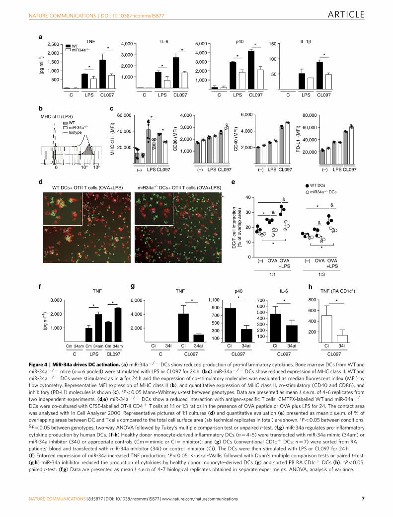
Figure 4 | MiR-34a drives DC activation. (a) miR-34a� /� DCs show reduced production of pro-inflammatory cytokines. Bone marrow DCs from WT and
miR-34a� /� mice (n¼6 pooled) were stimulated with LPS or CL097 for 24 h. (b,c) miR-34a� /� DCs show reduced expression of MHC class II. WT and
miR-34a� /� DCs were stimulated as in a for 24 h and the expression of co-stimulatory molecules was evaluated as median fluorescent index (MFI) by
flow cytometry. Representative MFI expression of MHC class II (b), and quantitative expression of MHC class II, co-stimulatory (CD40 and CD86), and
inhibitory (PD-L1) molecules is shown (c). *Po0.05 Mann–Whitney u-test between genotypes. Data are presented as mean±s.e.m. of 4–6 replicates from
two independent experiments. (d,e) miR-34a� /� DCs show a reduced interaction with antigen-specific T cells. CMTPX-labelled WT and miR-34a� /�
DCs were co-cultured with CFSE-labelled OT-II CD4þ T cells at 1:1 or 1:3 ratios in the presence of OVA peptide or OVA plus LPS for 24. The contact area
was analysed with In Cell Analyzer 2000. Representative pictures of 1:1 cultures (d) and quantitative evaluation (e) presented as mean±s.e.m. of % of
overlapping areas between DC and T cells compared to the total cell surface area (six technical replicates in total) are shown. *Po0.05 between conditions,&Po0.05 between genotypes, two-way ANOVA followed by Tukey’s multiple comparison test or unpaired t-test. (f,g) miR-34a regulates pro-inflammatory
cytokine production by human DCs. (f–h) Healthy donor monocyte-derived inflammatory DCs (n¼4–5) were transfected with miR-34a mimic (34am) or
miR-34a inhibitor (34i) or appropriate controls (Cm¼mimic or Ci¼ inhibitor); and (g) DCs (conventional CD1cþ DCs; n¼ 7) were sorted from RA
patients’ blood and transfected with miR-34a inhibitor (34i) or control inhibitor (Ci). The DCs were then stimulated with LPS or CL097 for 24 h.
(f) Enforced expression of miR-34a increased TNF production; *Po0.05, Kruskal–Wallis followed with Dunn’s multiple comparison tests or paired t-test.
(g,h) miR-34a inhibitor reduced the production of cytokines by healthy donor monocyte-derived DCs (g) and sorted PB RA CD1cþ DCs (h). *Po0.05
paired t-test. (f,g) Data are presented as mean±s.e.m of 4–7 biological replicates obtained in separate experiments. ANOVA, analysis of variance.
NATURE COMMUNICATIONS | DOI: 10.1038/ncomms15877 ARTICLE
NATURE COMMUNICATIONS | 8:15877 | DOI: 10.1038/ncomms15877 | www.nature.com/naturecommunications 7

soluble AXL that could function as a decoy receptor for the AXLligand, GAS6. Ex vivo production of pathogenic cytokines, forexample, TNF by circulating RA CD1cþ was reduced byinhibition of endogenous miR-34a expression, whereasmimicking the phenotype of RA DCs by increasing miR-34a inDC from healthy donors enhanced TLRs induced TNFproduction. In vivo miR-34a deficiency led to attenuation ofantigen-induced experimental arthritis that was associated withan increased expression of the miR-34a target pathway Axl/Socs-3in joint tissue. Consistent with this observation, recent pre--clinical therapeutic studies showed that local and systemicoverexpression of GAS6 and Protein S could ameliorate
experimental arthritis57. In vitro miR-34a� /� DCs hadconstitutively increased levels of AXL and decreased productionof pro-inflammatory cytokines (for example, TNF), and Th17inducing cytokines (for example, IL-6, IL-1b and IL-23) that drivejoint pathology. A similar DC phenotype was observed uponactive inhibition of endogenous miR-34a in human activated DCsin vitro. In addition to the role in regulation of pro-inflammatorymediators, miR-34a supported MHC class II expression, andmiR-34a� /� DCs had reduced surface MHC class II inimmature state and limited upregulation upon maturation. Thissuggests that miR-34a levels in mature DCs may regulate thesignal strength of the MHC/T-cell receptor synapse that controls
n=2,312 43 n=416
RA synovial fluidmyeloid cells
transcriptomic signature
AADACL1 NAV1ALS2CR13 NMT2AXL NRIP3BCL2 PDCD4C12orf59 PEA15CAMSAP1 PTPRMCAMTA1 RAB11FIP4CASP2 RALGDSCDK6 RNF44CDKN1C SATB2CREB5 SERPINE1
CTNND1 SLC44A2EHD4 SLC4A7EHD4 SOX4FGD6 STK38LFLOT2 TANC2FNDC3B TMEM200BLIMD2 TMEM97MCFD2 TNRC6BMEX3C TPD52MGAT4A VAT1MYO1C
Axl
20
40
60
80
100
120
Axl
luci
fera
se a
ctiv
ity
*
3'UTR Axl sense3'UTR Axl antisenseControl mimicmiR34a mimic
+–+–
+–
+
+–
+––
0.5
1.0
Fol
d ch
ange
Cm 34am
*
miR-34a–/– WTAxl
(140 kDa)
β-actin(43 kDa)
1
2
3
Fol
d ch
ange
Ci 34ai
Axl
500
1,000
1,500
2,000
2,500
Axl
(M
FI)
* *
500
1,000
1,500
2,000
(–) LPS
*
*
(–) LPS
WTmiR-34a–/–
Ci
AXL(140 kDa)
β-actin(43 kDa)
34aiCi
34ai
0.02
0.08
0.06
0.04AX
Lre
lativ
e to
β-a
ctin
Axl
(re
lativ
e ex
pres
ion
to 1
8S)
WTmiR-34a–/–
10–4
10–3
10–2 *
0.02
0.04
0.06
0.08*
0.020.0150.0100.005
SO
CS
-3 (
rela
tive
expr
esio
n to
18S
)
0
1
2
3
Fol
d ch
ange
LPS
24 h 48 h 72 hCtrs
AxlmiR-34a
24 h 48 h
*miR-34a predictedtargets (TargetScan)
a b c
d e f
g h i
Figure 5 | MiR-34a controls DC activation by regulating AXL. (a) Epigenetic miR-34a mRNA targets relevant to myeloid cell biology were identified by
integrating the conserved miR-34a targets generated by the prediction algorithm TargetScan (Probability of conserved targeting value 40.4) in a Venn
diagram along with the mRNA transcriptomic signature of RA SF myeloid cells68. (b,c) AXL is targeted by miR-34a. (b) Luciferase reporter assay for AXL
shows a reduction in luciferase activity when cells were transfected with miR-34a mimic (details in Methods). Data are presented as mean±s.e.m of three
technical replicates and is representative of two independent experiments. *Po0.05, paired t-test between AXL sense and anti-sense. (c) AXL mRNA
expression is regulated by miR-34a in monocyte-derived DCs in vitro. DCs transfected with either miR-34a mimic (34am) or miR-34a inhibitor (34ai) or
appropriate controls (Cm¼mimic; Ci¼ inhibitor), had decreased or increased AXL expression, respectively, after 24 h. Data are presented as mean±s.e.m.
of three biological replicates; *Po0.05, Mann–Whitney U-test. (d) miR-34a downregulation during LPS-induced mouse bone marrow DC maturation is
associated with an increase in Axl mRNA at 24hr. Data presented as mean±s.e.m of three technical replicates. (e) GM-CSF differentiated mouse
miR-34a� /� DCs contain higher concentrations of total AXL protein compared to WT as demonstrated by western blot. (f) Healthy donor human PB
monocyte-derived DCs transfected with miR-34a inhibitor (34ai) showed higher expression levels of AXL compared with cells transfected with control
inhibitor (Ci). (g) miR-34a� /� DCs express higher levels of membrane AXL compared to WTcontrol DCs measured by FACS. WT and miR-34a� /� bone
marrow DC were cultured in medium (control) or stimulated with LPS (1 ng ml� 1) for 24 and 48 h and AXL expression evaluated as MFI by cytometry. Data
presented as mean±s.e.m of four biological replicates from two independent experiments. (h–i) miR-34a� /� had higher levels of Axl (h) and Socs-3
(i) mRNA in joint tissue compared with WT at day 33 of CIA. Data presented as dot-plots with median lines. *Po0.05, Mann–Whitney U-test.
ARTICLE NATURE COMMUNICATIONS | DOI: 10.1038/ncomms15877
8 NATURE COMMUNICATIONS | 8:15877 | DOI: 10.1038/ncomms15877 | www.nature.com/naturecommunications

the activation of T cells; in particular Th17 and Th1 whichrequire a strong T-cell receptor signal58. This was confirmed byobservation of decreased interaction between miR-34a� /� DCsand antigen-specific T cells. Commensurate with ex vivo andin vitro phenotypes upon miR-34a manipulation, deficiency ofmiR-34a in antigen-presenting cells resulted in the lack ofdevelopment of antigen-specific Th17 cells, confirmed by lowserum concentrations of IL-17; and an attenuated joint pathologyin miR-34a� /� mice during experimental arthritis.
We observed that miR-34a expression in circulating CD1cþ
DCs is already increased in some patients with early RA, (r6months since diagnosis) as compared to healthy donors,suggesting a role in the onset of disease. The question remainswhat triggers this increase. The miR-34a promoter is abundant inCpG islands suggesting that environmental factors drivingdemethylation of the promoter59,60 is a plausible epigeneticmechanism of regulation as first reported in cancer59. In addition,the miR-34a promoter has STAT3-binding sites54 and aIL-6/STAT3 gene signature was found to be enriched incirculating T cells of early RA patients61.
In summary, we describe herein that miR-34a controls AXL, atyrosine kinase receptor that governs the termination of DCactivation and contributes to the development of experimentalarthritis. In RA patients, sustained expression of high levels ofmicroRNA-34a in CD1cþ DCs inhibits cellular AXL expressionand this, together with an increased level of decoy receptorsoluble AXL in serum, may render DCs more sensitive tomaturation signals, which can cause DCs to support auto-reactiveT cells (Fig. 7).
Thus, modulating the miR-34a/AXL pathway in RA DCs byusing miR-34a inhibitors or AXL agonist could be a feasibletherapeutic strategy to restore homoeostasis and promoteresolution of RA.
MethodsPatients and healthy donors. For the evaluation of miR-34 and AXL mRNAexpression in PB, SF and synovial tissue, samples were obtained from healthydonors, RA patients and PsA patients at Rheumatology clinics (Glasgow, UK) andfrom the Division of Rheumatology, Fondazione Policlinico Universitario A.Gemelli, Catholic University of the Sacred Heart (Rome, Italy). RA patients met the2010 ACR/EULAR diagnostic criteria62. Demographic and clinical information forthree cohorts used in the study are detailed in Supplementary Tables 1 and 2. Incohort 1, healthy donors were younger than RA patients, however age wasunrelated to miR-34a and AXL expression (r¼ � 0.080, P¼ 0.736 and r¼ 0.434,P¼ 0.056, respectively; Pearson correlation coefficient). For the evaluation of GAS6and sAXL, the concentrations of GAS6 and sAXL were quantified in sera of healthydonors, RA patients and OA patients by commercial enzyme-immunoassay (R&DSystem, DuoSets: Dy154 and Dy885). Demographic and clinical information aredetailed in Supplementary Table 3. The study protocol was approved by the localEthics Committee of the Catholic University of the Sacred Heart and by the Westof Scotland Research Ethical Committee (11/S0704/7). All the donors providedsigned informed consent.
Human DC cultures. CD14þ monocytes from PB were isolated using CD14þ
micro-beads (#130050201/Miltenyi Biotec) and Auto-MACS separator accordingto the manufacturer’s protocol. Monocyte purity evaluated by flow cytometry wastypically 496%. For their differentiation to inflammatory DCs, monocytes werecultured in complete medium (RPMI 1640, 10% fetal calf serum, 2 mM glutamine,100 U ml� 1 penicillin, 100 mg ml� 1 streptomycin; Life Technologies) withGM-CSF (100 ng ml� 1) and IL-4 (20 ng ml� 1; both from Peprotech) for 6-7days and the DC phenotype was confirmed by their expression of CD11c(#337216/PE/Cy7 anti-human CD11c), CD1c (#331524/APC anti-human CD1c)
++
+
+
–
–
–
–
200
400
600
TN
F (
pg m
l–1)
TLR stimulation
miR34aiCsiRNA
AxlsiRNA
TN
F (f
old
chan
ge c
ompa
red
to C
i plu
s C
siR
NA
)
TLR stimulation
+
AXL
(rel
ativ
e ex
pres
sion
)
CD1c+ DCs (first cohort)
Control i
0.5
1.0
1.5
2.0 *
+–
–+–––
++
+
–
––
––
–
–––
+
+–
–
+
+
––
++
–
–
+
+
–
–
+–
–+
+–
+–
+
+–
–+
+
––
10–5
10–4
10–3
10–2
10–6
10–5
10–4
10–3
CD1c+ DCs (second cohort)
*
IL-6
(re
lativ
e ex
pres
sion
)
IL-6
*
RA RAHD HDRAHD
* 10–4
10–5
10–6
10–7
10–8
10–9
a b
c d e
Figure 6 | AXL expression is constitutively reduced in RA CD1cþ DCs. (a,b) The miR-34a-dependent production of TNF by DCs is mediated by AXL.
Healthy donor (n¼ 7) monocyte-derived DCs were transfected with a combination of miR-34a inhibitor or AXL siRNA or control inhibitor or siRNA for 16 h,
and then stimulated with CL097 (1 mg ml� 1) for 24 h. (a) Representative experimental data for TNF production; mean±s.d. of 3 biological replicates from
one donor. (b) Summary of the relative production of TNF from all 7 donors tested; each normalised to the TNF production by DCs transfected with Control
inhibitor (Ci) plus Control siRNA (C siRNA). Po0.05 Kruskal–Wallis with Dunn’s multiple comparison test. (c–e) CD1cþ DCs in RA patients show
decreased expression of AXL mRNA and increase expression of IL-6. (c) CD1cþ DCs were FACS-sorted from blood of healthy donors (HD, n¼ 17) and RA
patients with established disease (RA, n¼ 15; cohort 1 described in Supplementary Table 1); and d,e CD1cþ DCs were FACS-sorted from age-matched
healthy donors (n¼ 11) and RA patients with early and established disease (n¼ 19, cohort 2 described in Supplementary Table 2). AXL (c–d) and IL-6 (e)
mRNAs were evaluated by qPCR. Data are presented as dot-plots with median lines. *Po0.05, Mann–Whitney U-test.
NATURE COMMUNICATIONS | DOI: 10.1038/ncomms15877 ARTICLE
NATURE COMMUNICATIONS | 8:15877 | DOI: 10.1038/ncomms15877 | www.nature.com/naturecommunications 9

and MHC class II (#307642/Brilliant Violet 785 anti-human HLA-DR; allantibodies from BioLegend; dilution 1: 100) by cytometry. The conventionalCD1cþ population from PB of RA and healthy donors, and from RA and PsA SFand synovial tissue were sorted by FACSAria III according to the sorting strategypresented in Supplementary Figure 1. In preparation for flourescence-activated cellsorting (FACS), SF was overlaid on Histopague-1077 (Sigma) and centrifuged(2,100 r.p.m., 25 min) to obtain mononuclear cells. Synovial tissues wereobtained using ultrasound-guided synovial tissue biopsy of the knee following thepublished protocol63. Briefly, all patients underwent ultrasound evaluation of theknee using an ultrasound machine with a multifrequency linear transducer (MyLabTwice, Esaote). Using the ultrasound view, the best point of entrance for the needlewas identified on the lateral margin of the suprapatellar recess. Each patient wasprovided with a face-mask and cap and the whole procedure was undertaken understerile conditions. Skin disinfection was done with iodine solution (performedtwice, starting from the point of needle entrance up to 25 cm proximally anddistally). Arthrocentesis of the knee joint was performed using the lateralsuprapatellar access if joint effusion was present. The skin, subcutaneous tissue andjoint capsule was anaesthetised with 10 ml lidocaine 2%. Next, a 14G needle(Precisa 1410-HS Hospital Service Spa, Italy) was inserted into the joint. Regionsof synovial hypertrophy were identified under grey-scale guidance to ensuresampling of representative synovial tissue. All the synovial tissue specimensobtained (at least eight pieces for each analysis) were placed on a nonwovenwet gauze for collection63.
Synovial biopsies were cut into small pieces and digested with LiberaseResearch Grade, in RPMI (150 mg ml� 1 Roche) with rotation in 37 �C for 1.5 h.Digested tissue was passed through a 100mm tissue-strainer and washed oncewith PBS/2%FCS/2 mM EDTA. FACS-analysis of single cell suspensions wasperformed after staining with Fixable Viability Dye eFluor 780 (#65086514/ThermoFisher Scientific; dilution 1:1,000) to exclude dead cells. Cells were thenincubated (4 �C 30 min) with antibody-conjugates, which were from BioLegendand used in 1:100 dilution. These included #307642/Brilliant Violet 785anti-human HLA-DR, #337216/PE/Cy7 anti-human CD11c, #344104/PEanti-human CD141, #331524/APC anti-human CD1c, #302040/Brilliant Violet605 anti-human CD16, #304050/Brilliant Violet 711 anti-human CD45, #323004/fluorescein isothiocyanate (FITC) anti-human CD15 (SSEA-1), #302206/FITCanti-human CD19, #304604/FITC anti-human CD56, #313232/FITC anti-humanCD117, #300440/FITC anti-human CD3 and #301822/Alexa Fluor 700 anti-humanCD14. The gating strategy was based on fluorescence minus one and unstainedcontrols (Supplementary Fig 1). Briefly, dead and CD56, CD3, CD19, CD15,
CD117 and CD14 lineage-positive cells were excluded, and MHC class IIþ
CD11cþ cells gated. The CD1cþ population was gated based on the lack of CD141and CD16 and the presence of CD1cþ expression. CD1cþ cells were then FACS-sorted into tubes with complete media. Blood monocytes were sorted based ontheir lack of CD2 (#309208/BioLegend; dilution 1:10), CD15 (#323006/BioLegend;dilution 1:10), CD19 (#302208/BioLegend; dilution 1:10), CD56 (#130090755/Miltenyi Biotec; dilution 1:100) and NKp46 (#331908/BioLegend; dilution 1:20)and the presence of MHC class II (#307633/BioLegend; dilution 1:50). The MHCclass IIþ cells were then gated based on the expression of CD14 (#325608/BioLegend; dilution 1:20) and CD16 (#302006/BioLegend; dilution 1:20); resultingin three monocyte populations CD14þCD16� ; CD14þCD16þ ,CD14dimCD16þ . Sorted DCs and monocytes were used for cell cultureexperiments or were immediately lysed with Qiagen buffer and RNA isolated withmicro-miRNAeasy kit (#217084) or miRNeasy mini Kit (#217004) according to themanufacturer’s protocol (Qiagen). Cultured DCs in complete medium weretransfected either with miR-34a inhibitor or control inhibitor (both at 20 nM;Qiagen) or with miR-34a mimic or control mimic (C. elegans miR-67), both at20 nM (Dharmacon) using DharmaFECT 3 transfection reagent (ThermoFisherScientific). For some experiments, DCs were transfected with AXL-specific siRNAor All Stars Negative Control siRNA (40 nM, both from Qiagen). To monitortransfection efficiency DCs were transfected with Dy547-labelled mimic or All StarsNegative Control siRNA labelled with Alexa Fluor 488. After 16 h, cells werestimulated with LPS (1 ng ml� 1) or CL097 (1mg ml� 1, both from InvivoGen) andcells and supernatant were collected after 24 h. In all experiments, the transfectionefficiency of siRNA and microRNA mimic was checked by flow cytometry and onlywhen 460% of cells were positive were they used for further analysis.
Mice. MicroRNA-34a gene-deficient (miR-34a� /� ) mice on a C57BL/6background were purchased from Jackson Laboratories64. WT littermates wereobtained by backcrossing miR-34a� /� mice with C67BL/6 mice (Jackson Labs).Axl� /� mice and WT littermates C57BL/6 (B6Nq; Johan Backlund; KarolinskaInstitute, Sweden) were bred and maintained in a pathogen-free facility atUniversity of Glasgow. Procedures were approved by University of Glasgow EthicalCommittee and the UK Home Office (project licence 60/4430). In vivo experimentswere conducted on males age 8–12 weeks. In vitro experiments were conducted oncells from males and females age 8–12 weeks.
Collagen-induced arthritis. To induce collagen-induced arthritis, mice wereinjected intradermally with type II chicken collagen emulsified in Freund’scomplete adjuvant (200 mg in 0.2 ml. MD BioSciences) on day 0. On day 21, type IIchicken collagen/PBS (200 mg in 0.2 ml) was injected i.p65. Mice were monitoredfor development of arthritis by microcaliper measurements of paw swelling. Inaddition, a clinical severity score was used in which 0¼ no reaction; 1¼mild butdefinite redness and swelling of the ankle/wrist/digits; 2¼moderate-to-severeredness and swelling of the ankle/wrist; 3¼ redness and swelling of the entire pawincluding digits; and 4¼maximally inflamed limb with involvement of multiplejoints66. Mice were killed on day 33 and cells and tissues harvested. Tworesearchers measured paw swelling (one of them blind); and both evaluatedhistology sections blind to experimental condition.
Adoptive transfer of OT-II T cells. OT-II T cells on a CD45.1 background (at 0.5and 1.5� 106) were injected into the tail vain of WT and miR-34� /� mice on day0. On day 1, mice were injected with 100mg of OVA peptide or PBS per mouse(Cambridge Reserved Biochemical) in complete Freunds adjuvant (0.5 mg ml� 1;Sigma) in 0.2 ml into the right leg muscle. On day 6, mice were culled, the draininglymph nodes harvested and dispersed cells re-stimulated with PMA/ionomycin(50/500ng ml� 1; Sigma) overnight in the presence of Golgi Stop (LifeTechnologies), and analysed for the intracellular production of IL-17, IFN-g andIL-5 using fluorescently labelled antibodies (#559502/PE anti-mouse IL-17A;BD Pharmingen; #505810/APC anti-mouse IFNg; BioLegend; #504311/BV421anti-mouse IL-5; BioLegend; all in 1:50 dilution and Fix/Perm kit according toprotocols (#420801 and # 421002, BioLegend).
Mouse DC culture. Bone marrow cells from WT and miR-34a� /� mice werecultured with GM-CSF (20 ng ml� 1, PeproTech) at a concentration of 0.2�106 ml� 1 in 10 ml of complete medium in petri dishes. After 3 and 6 days themedium was replaced with fresh medium supplemented with GM-CSF. On day 8,cells were seeded into 24-well culture plates at a concentration of 0.25� 106 ml� 1
and stimulated with LPS (1 ng ml� 1) or CL097 (1 mg ml� 1). Cells and culturesupernatant were collected at various time points for analysis of cytokineproduction, cell surface markers and AXL protein. For some in vitro experimentsCD11bþCD4þ spleen cells were isolated with CD4þ DC isolation kit(#130091262/Miltenyi Biotec).
Evaluation of DC/T cell interaction with IN Cell Analyzer 2000. Spleens wereisolated from OTII transgenic mice and prepared into single cell suspensions andtreated with 5 ml of 1� red blood cell lysis buffer (eBioscience) for 5 min at RT.CD4þ T cells were isolated from splenocytes by negative selection (#130095248/Miltenyi Biotec). Bone marrow derived WT and miR-34a� /� DCs and CD4þ Tcells were re-suspended at a density of 107 cells per ml in serum-free RPMI-1640
GM-CSFR
TLRs
AXL
GAS6Activation
SOCSs
4
2
1
3
3
MIR-34a
Figure 7 | A model describing miR-34/AXL function in DCs. The
expression of miR-34a increases during CD1cþ DC differentiation (1) and
epigenetically down-regulates AXL expression (2). DC maturation, by TLR
ligation, induces DC activation, for example, MHC class II upregulation and
cytokine production. This maturation signal also down-regulates miR-34a
(3), which then de-represses AXL, which in turn induces SOCSs and
terminates DC activation (4), thus providing homeostatic feedback control
of DC activation. In RA patients, sustained expression of high levels of
miR-34a in CD1cþ DCs inhibits AXL expression and may render DCs more
sensitive to maturation signals, which can cause DCs to support sustained
auto-reactive T-cell activation.
ARTICLE NATURE COMMUNICATIONS | DOI: 10.1038/ncomms15877
10 NATURE COMMUNICATIONS | 8:15877 | DOI: 10.1038/ncomms15877 | www.nature.com/naturecommunications

medium and labelled with Cell Tracker Red CMTPX or CFSE cell tracer kits(#C34552 and #C34554 respectively; Molecular probes) at 7.5 mM, respectively,for 10 min in a 37 �C incubator. After incubation, cells were washed by cen-trifugation at 300� g for 5 min at 4 �C three times in RPMI medium containing10% FCS to deactivate the dye. DCs and CD4þ T cells were co-cultured in384-well m-clear tissue culture treated micro-plates (Greiner) at ratios of 1:1 (8.000of each cell type) or 1:3 with ovalbumin peptide (OVA) at 1 mg ml� 1; with orwithout lipopolysaccharide (LPS) at 100 ng ml� 1 for 24 h.
Image acquisition. IN Cell Analyzer 2000 is a lamp-based high content analysissystem equipped with a large chip CCD camera (Photometrics CoolSNAP K4)allowing for whole-well imaging. The Nikon � 10 magnifying objective with flatfield and apochromatic corrections (plan Apo), chromatic aberration-free infinity(CF160) and 0.45 numerical aperture (NA) was used. Cells were imaged at 37 �Cand 2D images of CFSE-labelled CD4þ T cells were acquired in the FITC channelusing 490/� 20 excitation, 525/36 m emission. CMTPX-labelled DCs in theTexasRed (TR) channel were acquired using 555/� 25 excitation and 605/52 memission. One tile was imaged per well covering B20% of the well area. Imagesacquired by the IN Cell Analyzer 2000 were analysed using the Developer Toolboxsoftware V1.9.2 (GE Healthcare). Data are shown as representative screenshots of a384-well or the percentage of OTII T cells that overlap with DCs.
Western blot for AXL. Bone marrow derived DCs were seeded in complete mediainto six-well plates (1� 106 per well). Human PB monocyte-derived DCs (1� 106
per well) were transfected with control or miR-34a inhibitor (both 50 nM, Qiagen).After 24 h (mouse DCs) or 48 h (human DCs) whole-cell lysates were preparedusing M-PER (ThermoFisher Scientific) lysis buffer. Lysate samples (10 mg protein)were run on a 4–12% SDS–polyacrylamide gel electrophoresis gel, followed bytransfer to polyvinylidene difluoride membrane and incubation with either goatanti-mouse AXL (1:200 dilution, #/AF854/R&D Systems), rabbit anti-human AXL(1:500 dilution, #8661S/Cell Signaling Technology), or anti-mouse/human b-actincontrol (1 mg ml� 1, #3c-81178/Santa Cruz Biotechnology).
Flow cytometry of DC surface markers. Expression of DC surface markers wasevaluated by LSR-II cytometer (BD Bioscience) using the following antibodies:anti-MHC class II-PE-Cy7 (#107629/BioLegend) or APC-Cy7 (#107628/BioLe-gend) anti-CD11c-FITC (#117306/BioLegend) or PerCP-Cy5.5 (#560584/BDPharmingen), anti-CD86-PerCP (#105025/BioLegend), anti-CD40-Pacific Blue(#124626/BioLegend), anti-PD-DL-1-Qdot-605 (#124321/BioLegend), ICAM-1-FITC (#11054182/eBioscience), CD18-PE (#101408/BioLegend), CD69-PE-Cy7(#25069182/eBioscience), CD11a-APC (#101120/BioLegend), CD44-e450(#48044182/eBioscience), CD11b BV605 (#83011242/eBioscience; all used in 1:100dilution), and anti-AXL-APC (#FAB8541A/R&D Systems; dilution 1:20). The deadcells were excluded with Fixable Viability Dye eFluor 780 or e506 (#650866/ThermoFisher Scientfic). There was no difference in viable cell proportion of BM-derived DCs between WT and miR-34a� /� genotypes (mean±s.d: 75% ±12 and71% ±10, respectively).
AXL 30-UTR Luciferase assay. Human AXL 30-UTR in sense or anti-senseorientation were cloned downstream of the Renilla Luciferase gene in pmiRGLOvector (Promega). HEK293 cells were co-transfected with 0.2 mg pmiRGLOcontaining sense or anti-sense AXL 30-UTR and 40 nM miR-34a mimic orscrambled mimic control, using Attractene (Qiagen). Luciferase activity wasmeasured 24 h later using the Dual-GLO Luciferase assay system (#E2920/Promega)67.
Quantitative PCR. Total DC RNA was extracted using a miRNeasy mini Kit(#217004) or micro-miRNAeasy kit (#217084) (both from Qiagen). For microRNAanalysis, cDNA was transcribed from RNA using either a miScript ReverseTranscription Kit II (#218160 Qiagen) or TaqMan microRNA ReverseTranscription Kit (#4366596/Life Technologies). For mRNA analysis, a HighCapacity cDNA Reverse Transcription Kit was used (#4368814/ThermoFisherScientific). TaqMan mRNA or miRNA assays (Life technologies) or miScriptprimer assays (Qiagen) were used for semi-quantitative determination of theexpression of miR-34a (Hs000426 Life Technologies; MS00001428 Qiagen),miR-34c (Hs000428), Let7a (Hs000377, U6B snRNA (Hs001973; MS00033740),TNF Mm00443258_m1), AXL (Hs01064444_m1, Mm00437221_m1), Socs3(Mm00545913_s1), p19 (Mm00518984_m1), IL-6 (Hs00174131_m1) and 18S(Hs03003631_g1) with miRScript SybR Green PCR kit (#218073/Qiagen) orTaQman Gene Expression master mixes (Life Technologies). In some experimentsmiR-34a copy number was evaluated. Known copy numbers of synthetic miR-34amimic and housekeeping let7a (both from Dharmacon) were used to prepare astandard curve of serial 10-fold dilutions in nuclease-free water (range from1� 109 to 1� 103 copies). Each of these standards was used alongside cDNA fromthe samples in qPCR (miScript Sybr Green PCR kit) with specific human miR-34aand let7a primers (Qiagen) on a 7900HT TaqMan reader. Based on the standardcurves, the copy number of miR-34a and let7a the samples were calculated and datawere presented as the copy number of miR-34a per 10,000 copies let7a. In some
experiments, the expression of miR-34a is presented as a relative value (i) 2�DCT,where DCt¼Cycle threshold for RNU6 (housekeeping) minus Ct for miR-34a or(ii) fold change, where DCt for selected control condition¼ 1 or 100%.
To compare miR-34a and AXL expression in synovial CD1cþ DCs with bloodCD1cþ DCs, amplification of cDNA was necessary due to the small number ofCD1cþ DCs obtained from synovial biopsies (mean cell number: 2,000 cells) byFACS-sorting. Equal numbers of blood, SF and synovial tissue CD1cþ DCs wereused and RNA isolated (cohort 2; Supplementary Table 2). RNA was divided intotwo aliquots for separate transcription into cDNA with either High CapacityReverse Transcription kit (#4368814/ThermoFisher Scientific) to evaluate AXLexpression or with miScript Reverse Transcription Kit II (#218160/Qiagen) toevaluate miR-34a expression. In the next step, cDNA for AXL, IL-6 andhousekeeping 18S were amplified with PreAmp Master kit (#4391128/ThermoFisher Scientific) and cDNA for RNU6, miR-34a were amplified withmiScript PreAMP PCR Kit (#331452/Qiagen) followed by standard qPCR asdescribed above.
Luminex and enzyme immuno-assays. The concentration of cytokines andchemokines in mouse and human DC culture supernatants was quantified byimmuno-fluorescence assay using a 20-plex cytokine assay (LMC0006; Invitrogen)on a Bio-Plex platform (Bio-Rad) or using paired antibodies ELISAs (mouse andhuman TNF, #CHC3013 and #CHC1753/Life Technologies; mouse IL-23(#88723088/ ThermoFisher Scientific) and mouse GAS6 (#DY986/R&DSystems)66.
Statistical analysis. The data were analysed by Graph Pad Prism version 5.0 andMinitab 17 software. Data was summarized as mean±s.d./s.e.m. or median±IQR,and between-group differences were tested by t-test, by paired t-test or byMann–Whitney u-test, or Kolmogorov–Smirnov test, depending on datadistribution, or by ANOVA or Kruskal–Wallis tests with correction for multiplecomparisons. Correlation between normally distributed continuous variables wastested using the two-tailed Pearson Correlation Coefficient. A P-value o0.05 wasconsidered significant.
Data availability. The data that support the findings of this study are availablefrom the corresponding author upon request.
References1. Banchereau, J. & Steinman, R. M. Dendritic cells and the control of immunity.
Nature 392, 245–252 (1998).2. Kim, S. J., Zou, Y. R., Goldstein, J., Reizis, B. & Diamond, B. Tolerogenic
function of Blimp-1 in dendritic cells. J. Exp. Med. 208, 2193–2199 (2011).3. Huang, Q. Q. et al. CD11c-mediated deletion of Flip promotes autoreactivity
and inflammatory arthritis. Nat. Commun. 6, 7086 (2015).4. Jongbloed, S. L. et al. Plasmacytoid dendritic cells regulate breach of
self-tolerance in autoimmune arthritis. J. Immunol. 182, 963–968 (2009).5. Crowson, C. S. et al. The lifetime risk of adult-onset rheumatoid arthritis and
other inflammatory autoimmune rheumatic diseases. Arthritis Rheum. 63,633–639 (2011).
6. Cross, M. et al. The global burden of rheumatoid arthritis: estimates from theglobal burden of disease 2010 study. Ann. Rheum. Dis. 73, 1316–1322 (2014).
7. Frisell, T. et al. Familial risks and heritability of rheumatoid arthritis: role ofrheumatoid factor/anti-citrullinated protein antibody status, number and typeof affected relatives, sex, and age. Arthritis Rheum. 65, 2773–2782 (2013).
8. Smolen, J. S., Aletaha, D. & McInnes, I. B. Rheumatoid arthritis. Lancet 388,2023–2038 (2016).
9. Nielen, M. M. et al. Specific autoantibodies precede the symptoms ofrheumatoid arthritis: a study of serial measurements in blood donors. ArthritisRheum. 50, 380–386 (2004).
10. Klareskog, L. et al. A new model for an etiology of rheumatoid arthritis:smoking may trigger HLA-DR (shared epitope)-restricted immune reactions toautoantigens modified by citrullination. Arthritis Rheum. 54, 38–46 (2006).
11. James, E. A. et al. Citrulline-specific Th1 cells are increased in rheumatoidarthritis and their frequency is influenced by disease duration and therapy.Arthritis Rheumatol. 66, 1712–1722 (2014).
12. McInnes, I. B. & Schett, G. The pathogenesis of rheumatoid arthritis. N. Engl. J.Med. 365, 2205–2219 (2011).
13. Okada, Y. et al. Genetics of rheumatoid arthritis contributes to biology anddrug discovery. Nature 506, 376–381 (2014).
14. Hyrich, K. L., Lunt, M., Dixon, W. G., Watson, K. D. & Symmons, D. P.Register BSRB. Effects of switching between anti-TNF therapies on HAQresponse in patients who do not respond to their first anti-TNF drug.Rheumatology (Oxford) 47, 1000–1005 (2008).
15. Smolen, J. S. et al. EULAR recommendations for the management ofrheumatoid arthritis with synthetic and biological disease-modifyingantirheumatic drugs: 2013 update. Ann. Rheum. Dis. 73, 492–509 (2014).
NATURE COMMUNICATIONS | DOI: 10.1038/ncomms15877 ARTICLE
NATURE COMMUNICATIONS | 8:15877 | DOI: 10.1038/ncomms15877 | www.nature.com/naturecommunications 11
16. Meier, F. M. & McInnes, I. B. Small-molecule therapeutics in rheumatoidarthritis: scientific rationale, efficacy and safety. Best Pract. Res. Clin.Rheumatol. 28, 605–624 (2014).
17. Ziegler-Heitbrock, L. et al. Nomenclature of monocytes and dendritic cells inblood. Blood 116, e74–e80 (2010).
18. O’Keeffe, M., Mok, W. H. & Radford, K. J. Human dendritic cell subsets andfunction in health and disease. Cell Mol. Life Sci. 72, 4309–4325 (2015).
19. Breton, G. et al. Circulating precursors of human CD1cþ and CD141þdendritic cells. J. Exp. Med. 212, 401–413 (2015).
20. Dillon, S. M. et al. Human intestinal lamina propria CD1cþ dendritic cellsdisplay an activated phenotype at steady state and produce IL-23 in response toTLR7/8 stimulation. J. Immunol. 184, 6612–6621 (2010).
21. Ganguly, D. et al. Self-RNA-antimicrobial peptide complexes activate humandendritic cells through TLR7 and TLR8. J. Exp. Med. 206, 1983–1994 (2009).
22. Gerosa, F. et al. Differential regulation of interleukin 12 and interleukin 23production in human dendritic cells. J. Exp. Med. 205, 1447–1461 (2008).
23. Segura, E. et al. Human inflammatory dendritic cells induce Th17 celldifferentiation. Immunity 38, 336–348 (2013).
24. Hambleton, S. et al. IRF8 mutations and human dendritic-cellimmunodeficiency. N. Engl. J. Med. 365, 127–138 (2011).
25. Kim, S. J., Gregersen, P. K. & Diamond, B. Regulation of dendritic cellactivation by microRNA let-7c and BLIMP1. J. Clin. Invest. 123, 823–833(2013).
26. Rothlin, C. V., Ghosh, S., Zuniga, E. I., Oldstone, M. B. & Lemke, G. TAMreceptors are pleiotropic inhibitors of the innate immune response. Cell 131,1124–1136 (2007).
27. Brain, O. et al. The intracellular sensor NOD2 induces microRNA-29expression in human dendritic cells to limit IL-23 release. Immunity 39,521–536 (2013).
28. Villani, A. C. et al. Single-cell RNA-seq reveals new types of human blooddendritic cells, monocytes, and progenitors. Science 356, eaah4573 (2017).
29. Moret, F. M. et al. Intra-articular CD1c-expressing myeloid dendritic cells fromrheumatoid arthritis patients express a unique set of T cell-attractingchemokines and spontaneously induce Th1, Th17 and Th2 cell activity.Arthritis Res. Ther. 15, R155 (2013).
30. Jongbloed, S. L. et al. Enumeration and phenotypical analysis of distinctdendritic cell subsets in psoriatic arthritis and rheumatoid arthritis. ArthritisRes. Ther. 8, R15 (2006).
31. Moret, F. M., Bijlsma, J. W., Lafeber, F. P. & van Roon, J. A. The efficacy ofabatacept in reducing synovial T cell activation by CD1c myeloid dendritic cellsis overruled by the stimulatory effects of T cell-activating cytokines. ArthritisRheumatol. 67, 637–644 (2015).
32. Sacre, S. M. et al. Inhibitors of TLR8 reduce TNF production from humanrheumatoid synovial membrane cultures. J. Immunol. 181, 8002–8009 (2008).
33. Napolitani, G., Rinaldi, A., Bertoni, F., Sallusto, F. & Lanzavecchia, A. SelectedToll-like receptor agonist combinations synergistically trigger a T helper type1-polarizing program in dendritic cells. Nat. Immunol. 6, 769–776 (2005).
34. Guiducci, C. et al. RNA recognition by human TLR8 can lead to autoimmuneinflammation. J. Exp. Med. 210, 2903–2919 (2013).
35. McInnes, I. B., Buckley, C. D. & Isaacs, J. D. Cytokines in rheumatoid arthritis -shaping the immunological landscape. Nat. Rev. Rheumatol. 12, 63–68 (2016).
36. Pasquinelli, A. E. MicroRNAs and their targets: recognition, regulation and anemerging reciprocal relationship. Nat. Rev. Genet. 13, 271–282 (2012).
37. Kurowska-Stolarska, M. et al. MicroRNA-155 as a proinflammatory regulatorin clinical and experimental arthritis. Proc. Natl Acad. Sci. USA 108,11193–11198 (2011).
38. O’Connell, R. M. et al. Sustained expression of microRNA-155 inhematopoietic stem cells causes a myeloproliferative disorder. J. Exp. Med. 205,585–594 (2008).
39. Calin, G. A. & Croce, C. M. MicroRNA signatures in human cancers. Nat. Rev.Cancer 6, 857–866 (2006).
40. Asquith, D. L. et al. The liver X receptor pathway is highly upregulated inrheumatoid arthritis synovial macrophages and potentiates TLR-drivencytokine release. Ann. Rheum. Dis. 72, 2024–2031 (2013).
41. Hashimi, S. T. et al. MicroRNA profiling identifies miR-34a and miR-21 andtheir target genes JAG1 and WNT1 in the coordinate regulation of dendriticcell differentiation. Blood 114, 404–414 (2009).
42. Mildner, A. et al. Mononuclear phagocyte miRNome analysis identifies miR-142 as critical regulator of murine dendritic cell homeostasis. Blood 121,1016–1027 (2013).
43. Hermeking, H. The miR-34 family in cancer and apoptosis. Cell Death Differ.17, 193–199 (2010).
44. Sato, K. et al. Th17 functions as an osteoclastogenic helper T cell subset thatlinks T cell activation and bone destruction. J. Exp. Med. 203, 2673–2682(2006).
45. Nakae, S., Nambu, A., Sudo, K. & Iwakura, Y. Suppression of immuneinduction of collagen-induced arthritis in IL-17-deficient mice. J. Immunol.171, 6173–6177 (2003).
46. Lu, Q. & Lemke, G. Homeostatic regulation of the immune system by receptortyrosine kinases of the Tyro 3 family. Science 293, 306–311 (2001).
47. Rothlin, C. V., Carrera-Silva, E. A., Bosurgi, L. & Ghosh, S. TAM receptorsignaling in immune homeostasis. Annu. Rev. Immunol. 33, 355–391 (2015).
48. Carrera Silva, E. A. et al. T cell-derived protein S engages TAM receptorsignaling in dendritic cells to control the magnitude of the immune response.Immunity 39, 160–170 (2013).
49. Nassar, M. et al. GAS6 is a key homeostatic immunological regulator ofhost-commensal interactions in the oral mucosa. Proc. Natl Acad. Sci. USA 114,E337–E346 (2017).
50. Zagorska, A., Traves, P. G., Lew, E. D., Dransfield, I. & Lemke, G.Diversification of TAM receptor tyrosine kinase function. Nat. Immunol. 15,920–928 (2014).
51. Benham, H. et al. Citrullinated peptide dendritic cell immunotherapy in HLArisk genotype-positive rheumatoid arthritis patients. Sci. Transl. Med. 7,290ra287 (2015).
52. Bell, G. M. et al. Autologous tolerogenic dendritic cells for rheumatoid andinflammatory arthritis. Ann. Rheum. Dis. 76, 224–227 (2017).
53. Weinger, J. G. et al. Loss of the receptor tyrosine kinase Axl leads to enhancedinflammation in the CNS and delayed removal of myelin debris duringexperimental autoimmune encephalomyelitis. J. Neuroinflammation 8, 49 (2011).
54. Hermeking, H. The miR-34 family in cancer and apoptosis. Cell Death Differ.17, 193–199.
55. Paolino, M. et al. The E3 ligase Cbl-b and TAM receptors regulate cancermetastasis via natural killer cells. Nature 507, 508–512 (2014).
56. Li, X. J., Ren, Z. J. & Tang, J. H. MicroRNA-34a: a potential therapeutic targetin human cancer. Cell Death Dis. 5, e1327 (2014).
57. van den Brand, B. T. et al. Therapeutic efficacy of Tyro3, Axl, and Mer tyrosinekinase agonists in collagen-induced arthritis. Arthritis Rheum. 65, 671–680(2013).
58. van Panhuys, N. TCR signal strength alters T-DC activation and interactiontimes and directs the outcome of differentiation. Front. Immunol. 7, 6 (2016).
59. Lodygin, D. et al. Inactivation of miR-34a by aberrant CpG methylation inmultiple types of cancer. Cell Cycle 7, 2591–2600 (2008).
60. Niederer, F. et al. Downregulation of microRNA-34a* in rheumatoid arthritissynovial fibroblasts promotes apoptosis resistance. Arthritis Rheum. 64,1771–1779 (2012).
61. Anderson, A. E. et al. IL-6-driven STAT signalling in circulating CD4þlymphocytes is a marker for early anticitrullinated peptide antibody-negativerheumatoid arthritis. Ann. Rheum. Dis. 75, 466–473 (2016).
62. Aletaha, D. et al. 2010 rheumatoid arthritis classification criteria: an AmericanCollege of Rheumatology/European League Against Rheumatism collaborativeinitiative. Ann. Rheum. Dis. 69, 1580–1588 (2010).
63. van de Sande, M. G. et al. Evaluating antirheumatic treatments using synovialbiopsy: a recommendation for standardisation to be used in clinical trials. Ann.Rheum. Dis. 70, 423–427 (2011).
64. Choi, Y. J. et al. miR-34 miRNAs provide a barrier for somatic cellreprogramming. Nat. Cell Biol. 13, 1353–1360 (2011).
65. Kurowska-Stolarska, M. et al. MicroRNA-155 as a proinflammatory regulator inclinical and experimental arthritis. Proc. Natl Acad. Sci. USA 108, 11193 (2011).
66. Brand, D. D., Latham, K. A. & Rosloniec, E. F. Collagen-induced arthritis. Nat.Protoc. 2, 1269–1275 (2007).
67. Miller, A. M. et al. MiR-155 has a protective role in the development ofnon-alcoholic hepatosteatosis in mice. PLoS ONE 8, e72324 (2013).
68. Asquith, D. L. et al. The liver X receptor pathway is highly upregulated inrheumatoid arthritis synovial macrophages and potentiates TLR-drivencytokine release. Ann. Rheum. Dis. 72, 2024–2031 (2013).
AcknowledgementsWe thank the RA patients and healthy donors who participated in this study. We wouldthank Mrs Diane Vaughan for her outstanding support in cell sorting and phenotyping;Dr Eric Kalkman for his help with IN Cell Analyzer 2000; Dr Tom Barr for his help withadoptive transfer model; and Drs Siebert and Zhao for some of the PsA samples. Wethank Mr David Riggans from University of Glasgow Mail Room who facilitated smoothdeliveries of clinical samples from Rome to Glasgow. MKS was supported by ArthritisResearch UK (ARUK) Career Development Fellowship (x19213). SA was supported by afellowship from Novo Nordisk UK. CT was supported by the Oliver Bird RheumatismProgramme/ Nuffield Foundation. EGM was supported by European League AgainstRheumatism (EULAR) and the Catalonian Society of Rheumatology. VM was supportedby ARUK Fellowship (x20848). This work was performed in, and support by, the ARUKCentre of Excellence for the Pathogenesis of Rheumatoid Arthritis (RACE/x20298).
Author contributionsM.K.-S. designed and performed experiments, analysed data and wrote the manuscript.S.A. contributed to designing the study, performing experiments, analysing the data, andcontributed with clinical samples; E.G.M., C.T., Ch.K. and L.S., G.D.S. and C.D.M.performed experiments. D.S.G. and V.M., provided the technology and performed
ARTICLE NATURE COMMUNICATIONS | DOI: 10.1038/ncomms15877
12 NATURE COMMUNICATIONS | 8:15877 | DOI: 10.1038/ncomms15877 | www.nature.com/naturecommunications
experiments. A.Z. performed experiments and analysed clinical data. B.T. and L.P.provided clinical samples and performed experiments. D.P., R.D.B., D.M. providedclinical samples and analysed clinical data. J.M.B., G.L., S.M. and E.G. provided tech-nologies. C.M. and G.F. contributed to designing the study and writing the manuscript.I.B.M. contributed to designing the study, analysing the data and writing the manuscript.
Additional informationSupplementary Information accompanies this paper at http://www.nature.com/naturecommunications
Competing interests: The authors declare no competing financial interests.
Reprints and permission information is available online at http://npg.nature.com/reprintsandpermissions/
How to cite this article: Kurowska-Stolarska, M. et al. MicroRNA-34a dependentregulation of AXL controls the activation of dendritic cells in inflammatory arthritis.Nat. Commun. 8, 15877 doi: 10.1038/ncomms15877 (2017).
Publisher’s note: Springer Nature remains neutral with regard to jurisdictional claims inpublished maps and institutional affiliations.
Open Access This article is licensed under a Creative CommonsAttribution 4.0 International License, which permits use, sharing,
adaptation, distribution and reproduction in any medium or format, as long as you giveappropriate credit to the original author(s) and the source, provide a link to the CreativeCommons license, and indicate if changes were made. The images or other third partymaterial in this article are included in the article’s Creative Commons license, unlessindicated otherwise in a credit line to the material. If material is not included in thearticle’s Creative Commons license and your intended use is not permitted by statutoryregulation or exceeds the permitted use, you will need to obtain permission directly fromthe copyright holder. To view a copy of this license, visit http://creativecommons.org/licenses/by/4.0/
r The Author(s) 2017
NATURE COMMUNICATIONS | DOI: 10.1038/ncomms15877 ARTICLE
NATURE COMMUNICATIONS | 8:15877 | DOI: 10.1038/ncomms15877 | www.nature.com/naturecommunications 13