microbiology, clinical
DESCRIPTION
Gram negative rodsTRANSCRIPT
Enterobacteriaceae 21
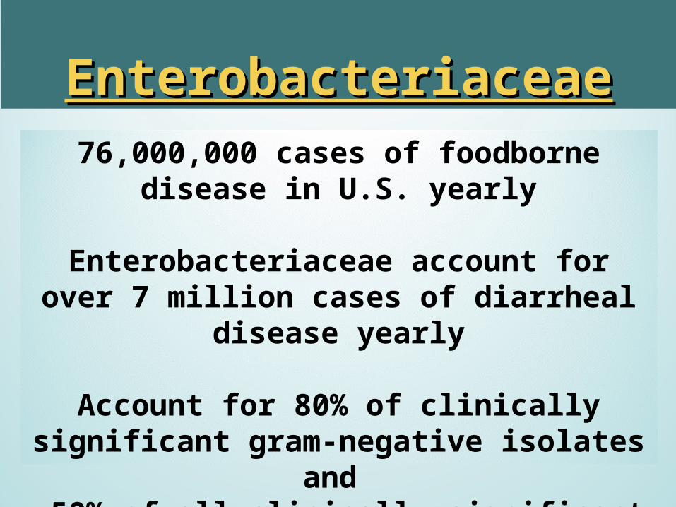
EnterobacteriaceaeEnterobacteriaceae76,000,000 cases of foodborne disease in U.S.
yearly
Enterobacteriaceae account for over 7 million cases of diarrheal disease yearly
Account for 80% of clinically significant gram-negative isolates and
~50% of all clinically significant isolates.
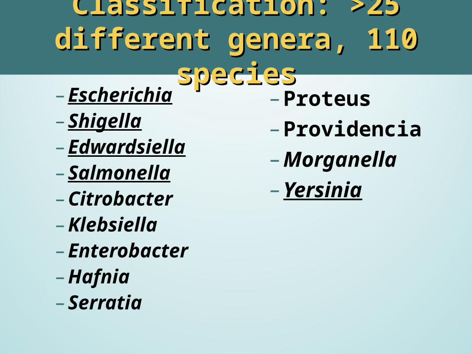
Classification: >25 different genera, Classification: >25 different genera, 110 species110 species
– Escherichia– Shigella– Edwardsiella– Salmonella– Citrobacter– Klebsiella– Enterobacter– Hafnia– Serratia
– Proteus– Providencia– Morganella– Yersinia
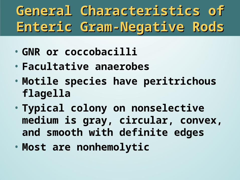
General Characteristics of Enteric Gram-General Characteristics of Enteric Gram-Negative RodsNegative Rods
• GNR or coccobacilli• Facultative anaerobes• Motile species have peritrichous flagella• Typical colony on nonselective medium is
gray, circular, convex, and smooth with definite edges
• Most are nonhemolytic

Enterobacteriaceae: Motility (peritrichous flagella)
Non-motile species: Klebsiella, Shigella, Yersinia
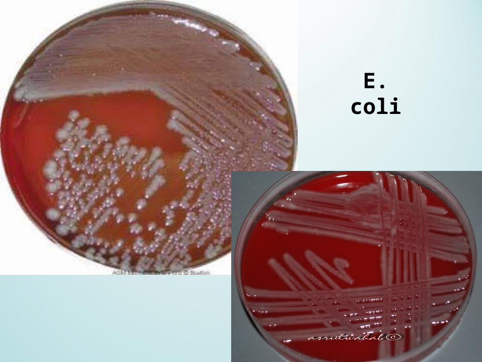
E. coli
General Characteristics of Enteric Gram-General Characteristics of Enteric Gram-Negative RodsNegative Rods
•Transduction and conjugation readily occurs
•High antigenic variability•Can be resistant to multiple antibiotics
(unpredictable antibiogram)
General Characteristics of Enteric Gram-General Characteristics of Enteric Gram-Negative RodsNegative Rods
•Three characteristics in common– Ferment glucose with production of acid– Reduce nitrates to nitrites– Oxidase negative (except for Plesiomonas)
Selective and Differential MediaSelective and Differential Media
•Selective:– Incorporation of dyes and bile salts to
inhibit G+ organisms – May suppress growth of non-pathogenic
species.•Differential
– Based on lactose fermentation and/or H2S production.

Selective and Differential MediaSelective and Differential Media
• EMB• MAC• XLD• HE• SS• BIS
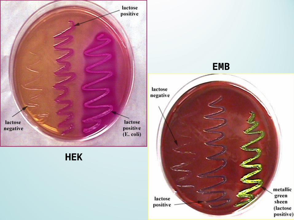
EMB
HEK
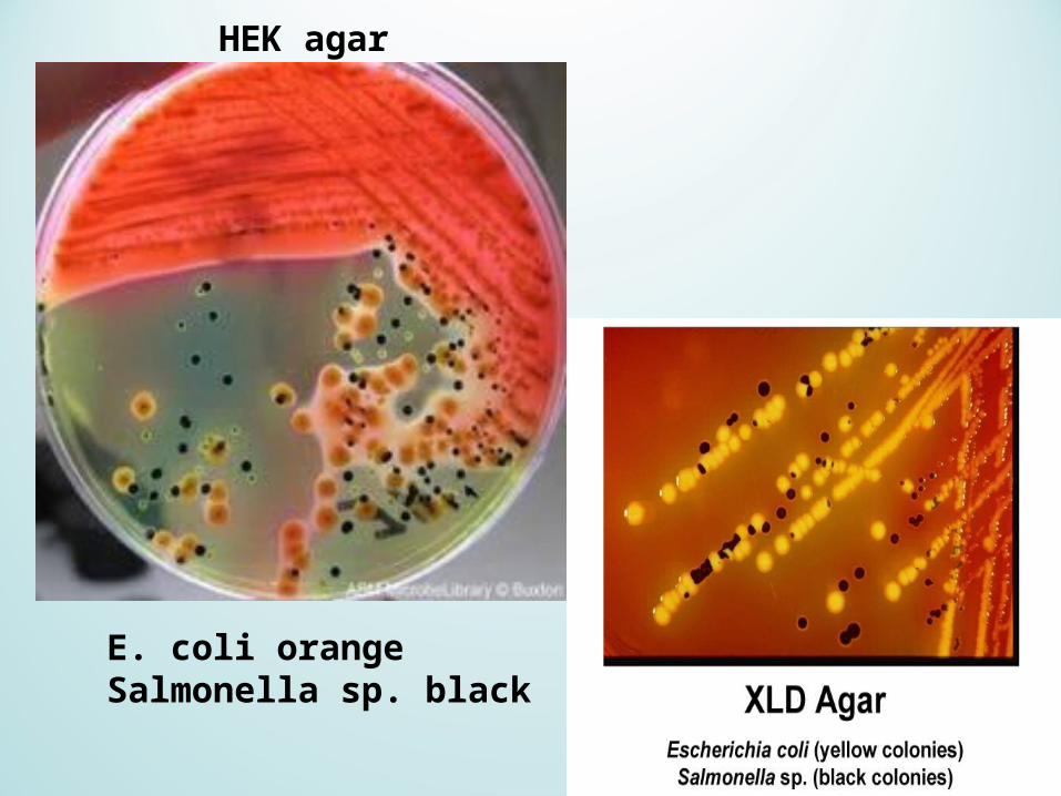
HEK agar
E. coli orangeSalmonella sp. black
Biochemical IdentificationBiochemical Identification
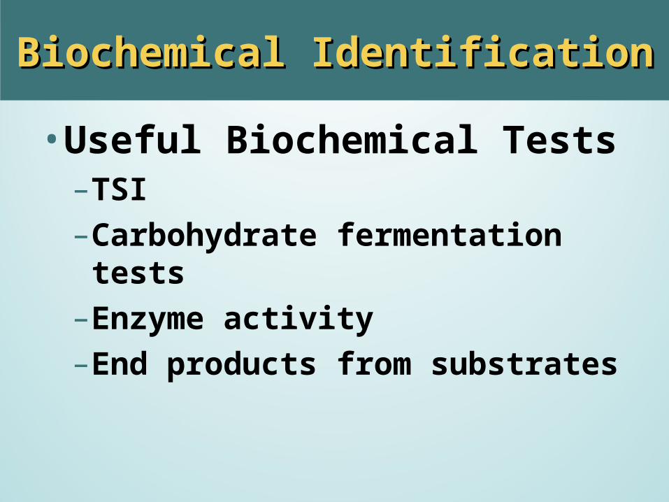
•Useful Biochemical Tests– TSI– Carbohydrate fermentation tests– Enzyme activity– End products from substrates
Biochemical IdentificationBiochemical Identification
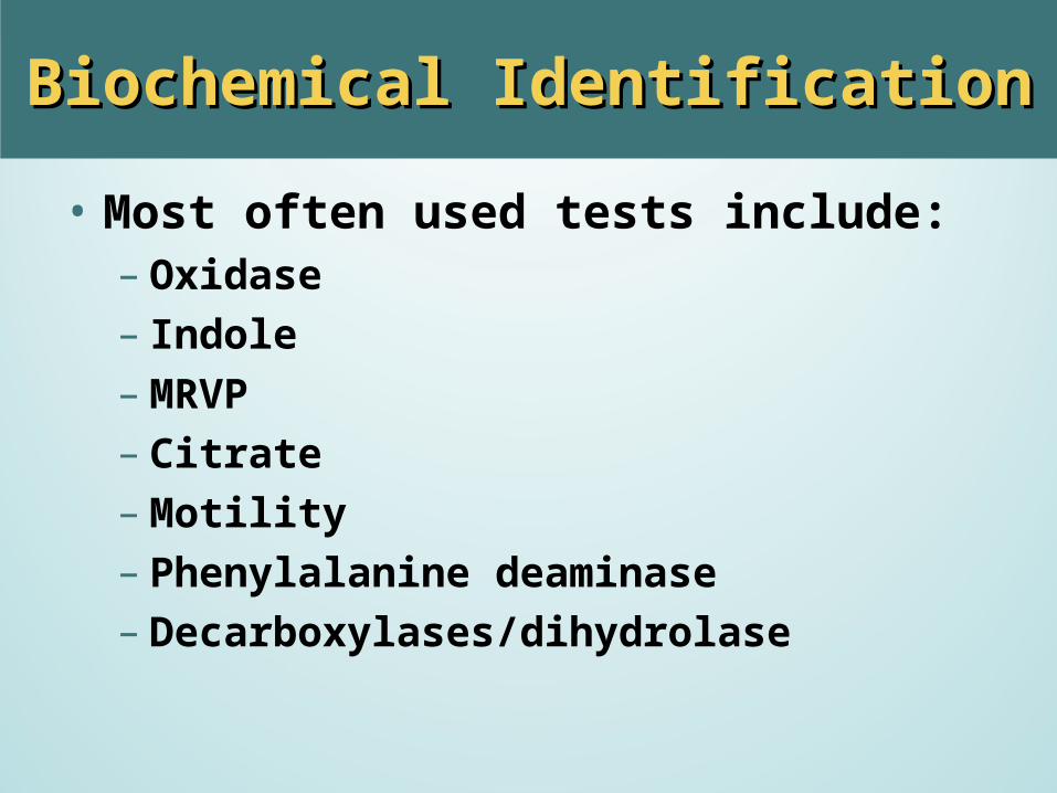
• Most often used tests include:– Oxidase– Indole– MRVP– Citrate– Motility– Phenylalanine deaminase– Decarboxylases/dihydrolase
Biochemical IdentificationBiochemical Identification
• Most often used tests include:– Gelatin hydrolysis– Urease– H2S– Fermentation of glucose, lactose, and other
carbohydrates
Serological IdentificationSerological Identification
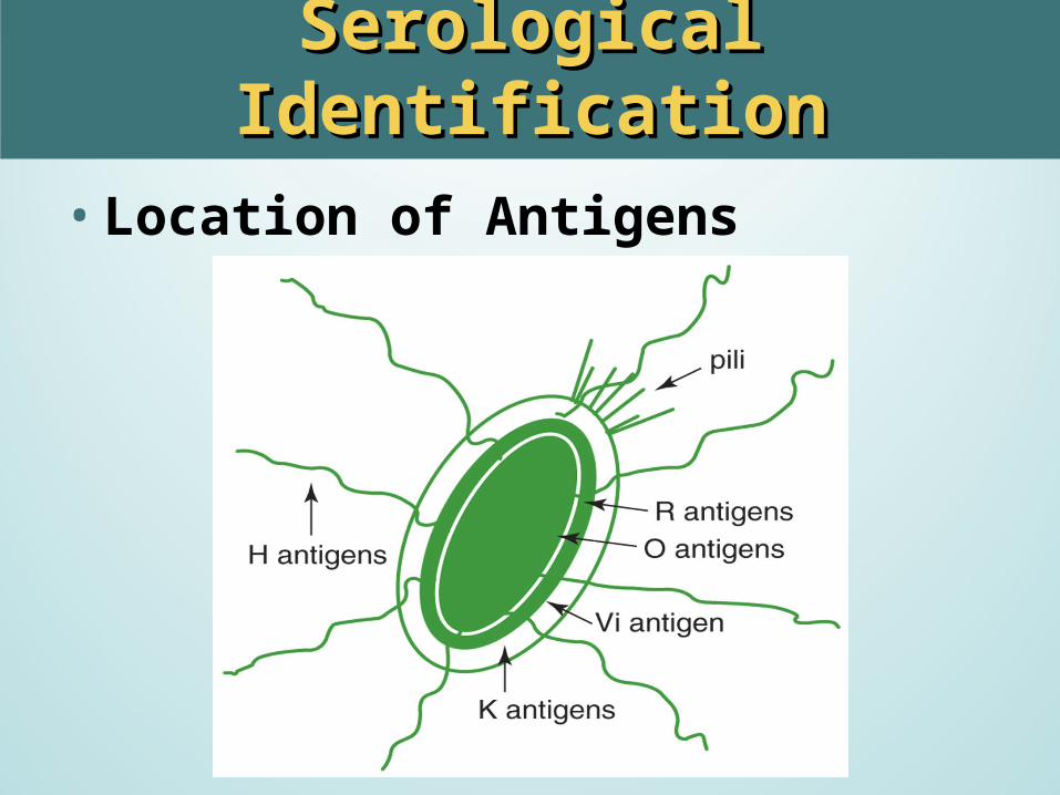
• Location of Antigens
Enterobacteriaceae: AntigensEnterobacteriaceae: Antigens
• Antigenic structure: differences genus/species.1. O antigens –heat stable polysaccharide (galactose-
mannose-rhamnose)2. Flagellar H antigens –heat labile (flagellin)3. Envelope or capsule K antigens – over surface of O
antigen and may block agglutination by O specific antisera. – Boiling for 15 min will destroy the K antigen– The K antigen is called the Vi (virulence) antigen
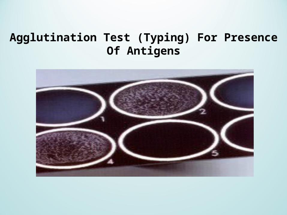
Agglutination Test (Typing) For Presence Of Antigens

Serological IdentificationSerological Identification
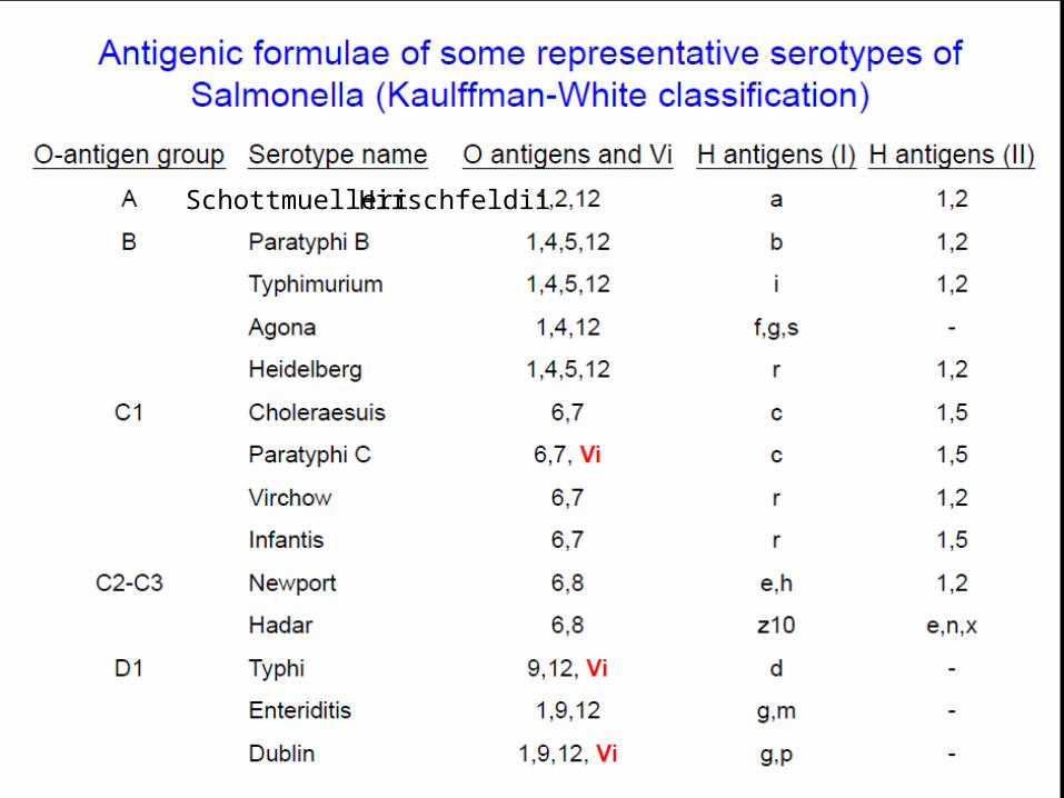
•Antigenic formula– Always listed in the same sequence– Sequence is O, K, H– Example: Salmonella typhi
Has O antigens 9 and 12; Vi antigen; phase 1 flagellar antigen d; and no phase 2 flagellar antigen
Antigenic formula is 9, 12 (Vi): d:-
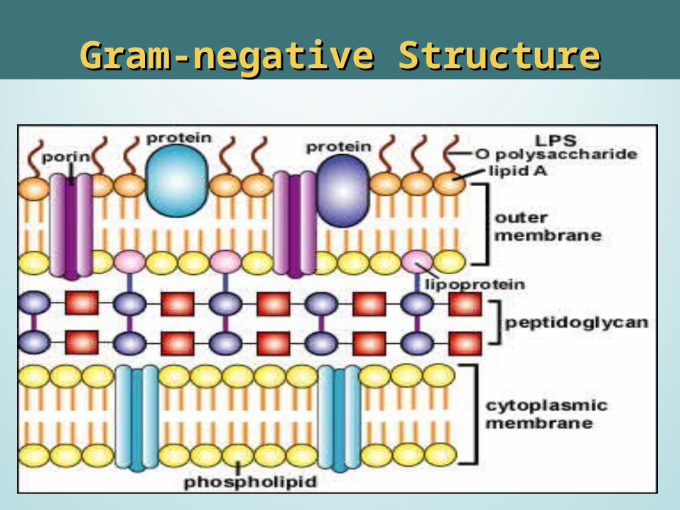
Gram-negative StructureGram-negative Structure
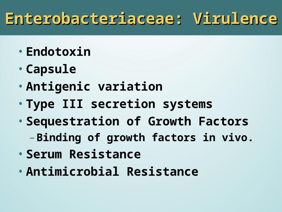
Enterobacteriaceae: VirulenceEnterobacteriaceae: Virulence
• Endotoxin•Capsule•Antigenic variation• Type III secretion systems• Sequestration of Growth Factors
– Binding of growth factors in vivo.
• Serum Resistance•Antimicrobial Resistance
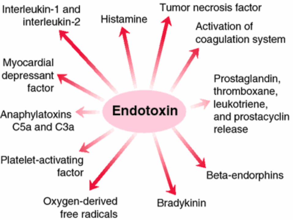
EndotoxinEndotoxin
• Lysis of organism releases endotoxin.• Lipid A released.• Fever,
leukopenia,hypotension/shock, tissue necrosis
• Activation of alternative complement cascade
• Disseminate Intravascular Coagulation
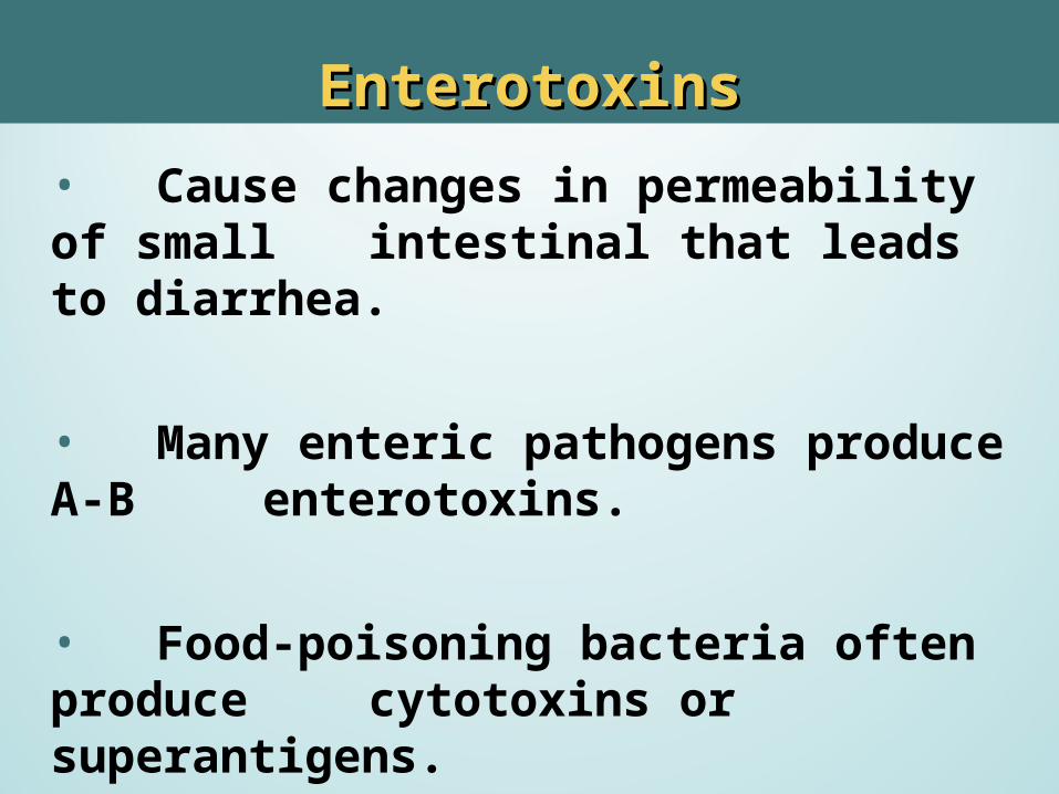
EnterotoxinsEnterotoxins• Cause changes in permeability of small intestinal that leads to diarrhea.
• Many enteric pathogens produce A-B enterotoxins.
• Food-poisoning bacteria often produce cytotoxins or superantigens.
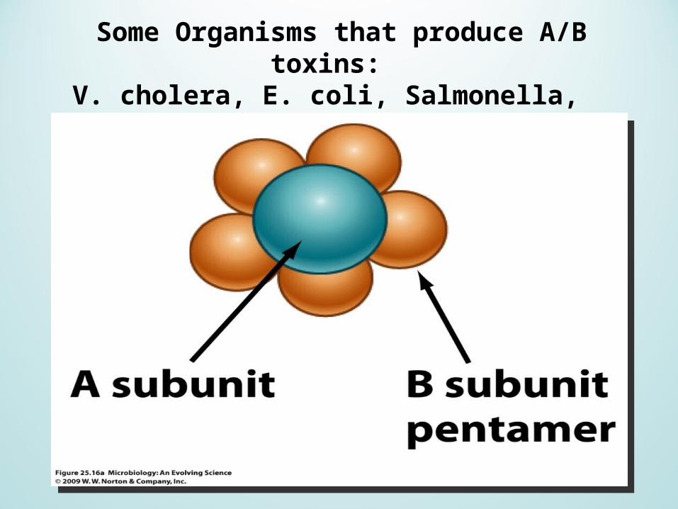
Some Organisms that produce A/B toxins: V. cholera, E. coli, Salmonella,
C. tetani, C. botulinum, C. diphtheriae
Enteric PathogensEnteric Pathogens
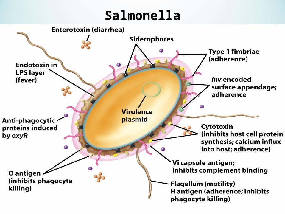
Salmonella
SalmonellaSalmonella
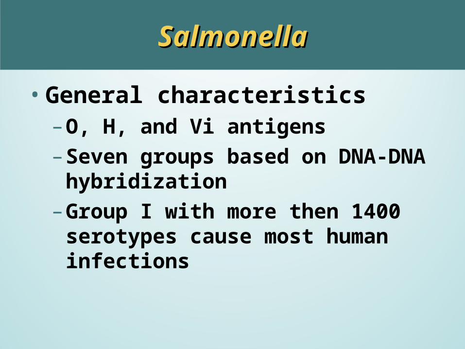
•General characteristics– O, H, and Vi antigens– Seven groups based on DNA-DNA
hybridization– Group I with more then 1400 serotypes
cause most human infections
SalmonellaSalmonella
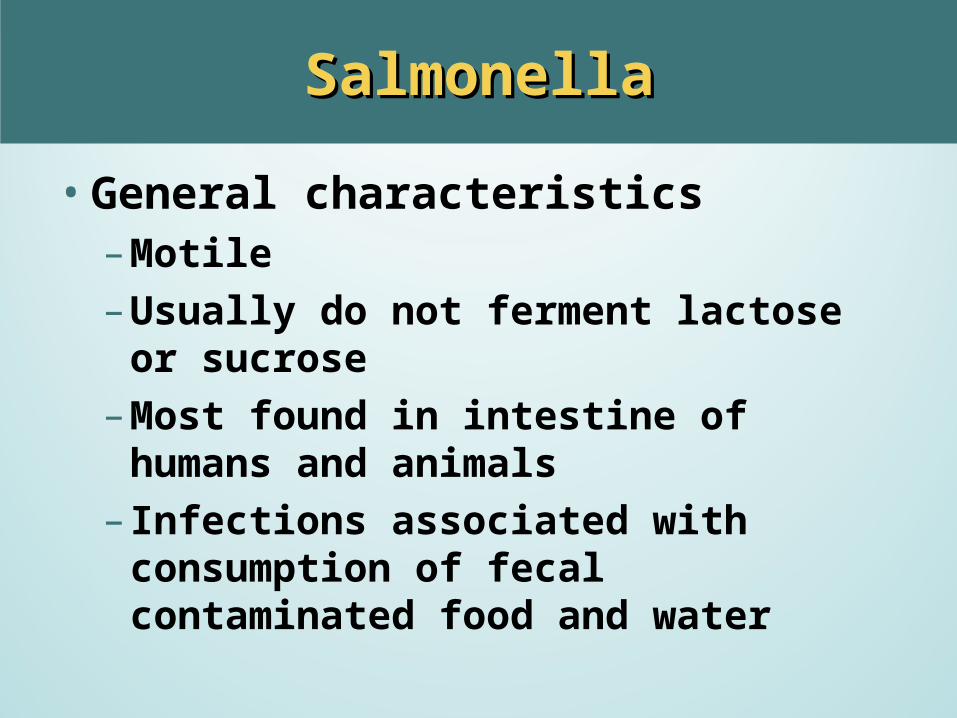
•General characteristics– Motile– Usually do not ferment lactose or sucrose– Most found in intestine of humans and
animals– Infections associated with consumption of
fecal contaminated food and water
SalmonellaSalmonella
•Usual specimens isolated from– Stool– Blood– Urine– Bone marrow

Salmonella: culture mediaSalmonella: culture media
•To detect nonlactose fermenters EMB, MAC
–To detect H2S production SS, HE, XLD
–Enrichment medium-fecal pathogens GN broth, Selenite F, Tetrathionate broth
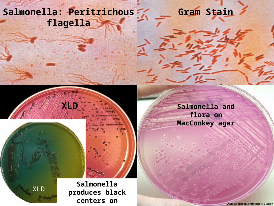
XLD Salmonella and flora onMacConkey agar
Salmonella: Peritrichous flagella Gram Stain
XLDSalmonella
produces black centers on
XLD and HEK
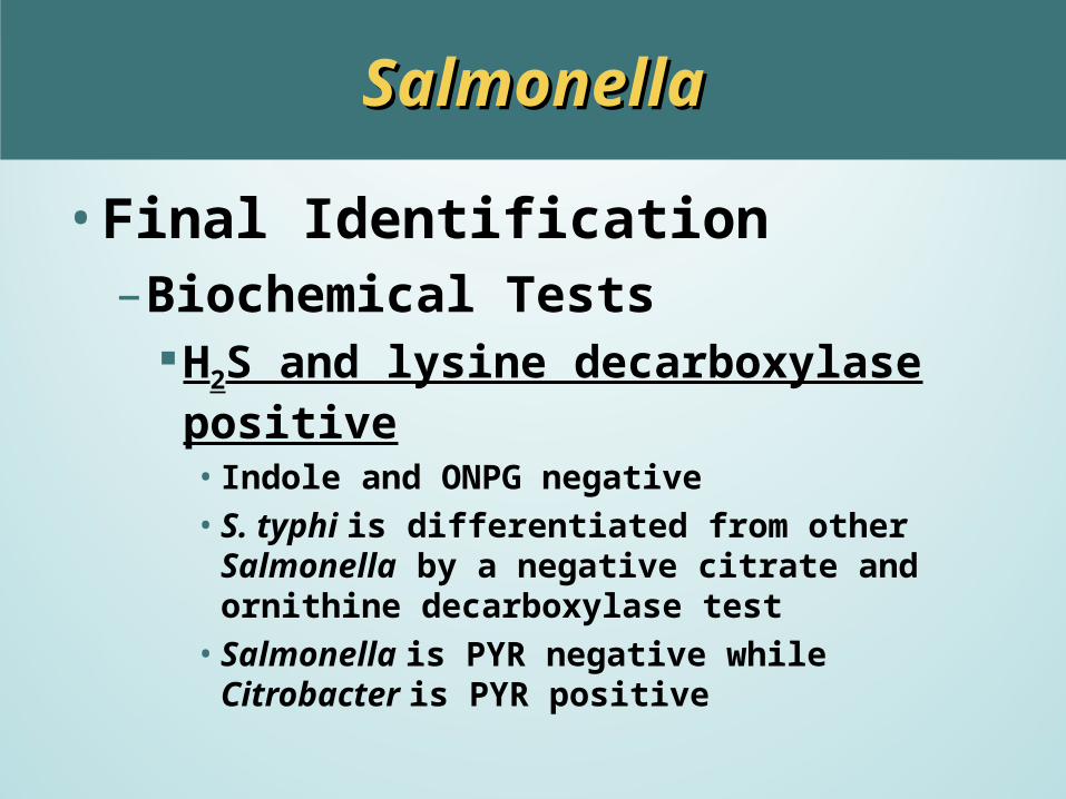
SalmonellaSalmonella
•Final Identification– Biochemical Tests
H2S and lysine decarboxylase positive• Indole and ONPG negative• S. typhi is differentiated from other Salmonella by a
negative citrate and ornithine decarboxylase test • Salmonella is PYR negative while Citrobacter is PYR
positive
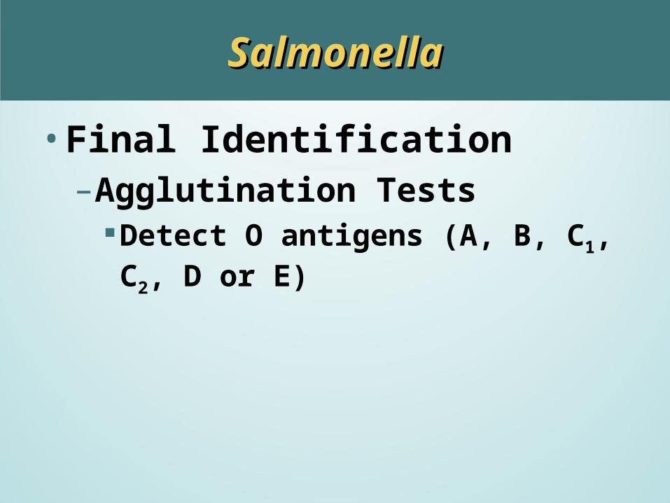
SalmonellaSalmonella
•Final Identification– Agglutination Tests
Detect O antigens (A, B, C1, C2, D or E)
SchottmuelleriHirschfeldii
SalmonellaSalmonella
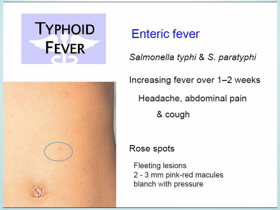
•Pathogenesis and Infectious Diseases Typhoid fever (S. typhi) Bacteremia Enterocolitis

ShigellaShigella
• General characteristics– 4 serogroups
A (S. dysenteriae, 12 serotypes) B (S. flexneri, 6 serotypes) C (S. boydii, 23 serotypes) D (S. sonnei, 1 serotype)
– Fecal-Oral Transmission and contaminated water– Found in intestines of humans and some
nonhuman primates
ShigellaShigella
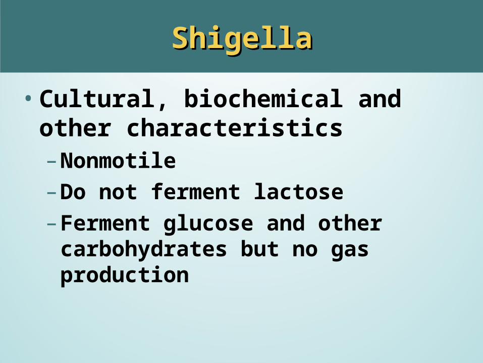
•Cultural, biochemical and other characteristics– Nonmotile– Do not ferment lactose– Ferment glucose and other carbohydrates
but no gas production
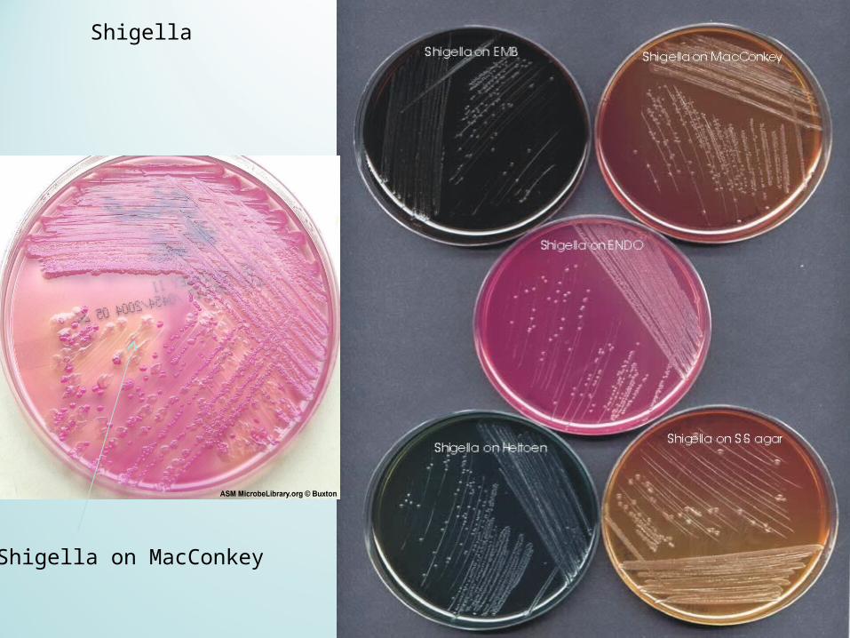
Shigella
Shigella on MacConkey
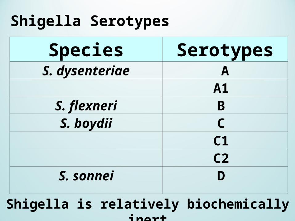
Species SerotypesS. dysenteriae A
A1 S. flexneri B S. boydii C
C1 C2
S. sonnei D
Shigella Serotypes
Shigella is relatively biochemically inert
ShigellaShigella
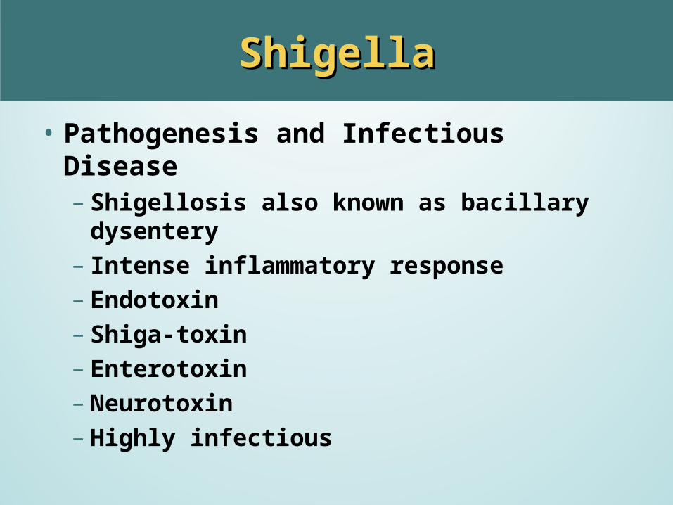
• Pathogenesis and Infectious Disease– Shigellosis also known as bacillary dysentery– Intense inflammatory response– Endotoxin– Shiga-toxin– Enterotoxin– Neurotoxin– Highly infectious
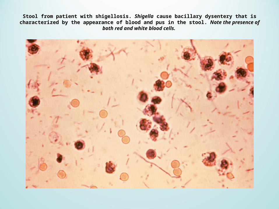
Stool from patient with shigellosis. Shigella cause bacillary dysentery that is characterized by the appearance of blood and pus in the stool. Note the presence of both red and white blood cells.
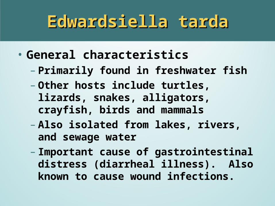
Edwardsiella tardaEdwardsiella tarda
• General characteristics– Primarily found in freshwater fish– Other hosts include turtles, lizards, snakes,
alligators, crayfish, birds and mammals– Also isolated from lakes, rivers, and sewage
water– Important cause of gastrointestinal distress
(diarrheal illness). Also known to cause wound infections.

Edwardsiella tardaEdwardsiella tarda
• Cultural, biochemical and other characteristics– Motile GNR– H2S positive– Ferments Glucose with gas production– Does not ferment lactose, sucrose or most other
carbohydrates
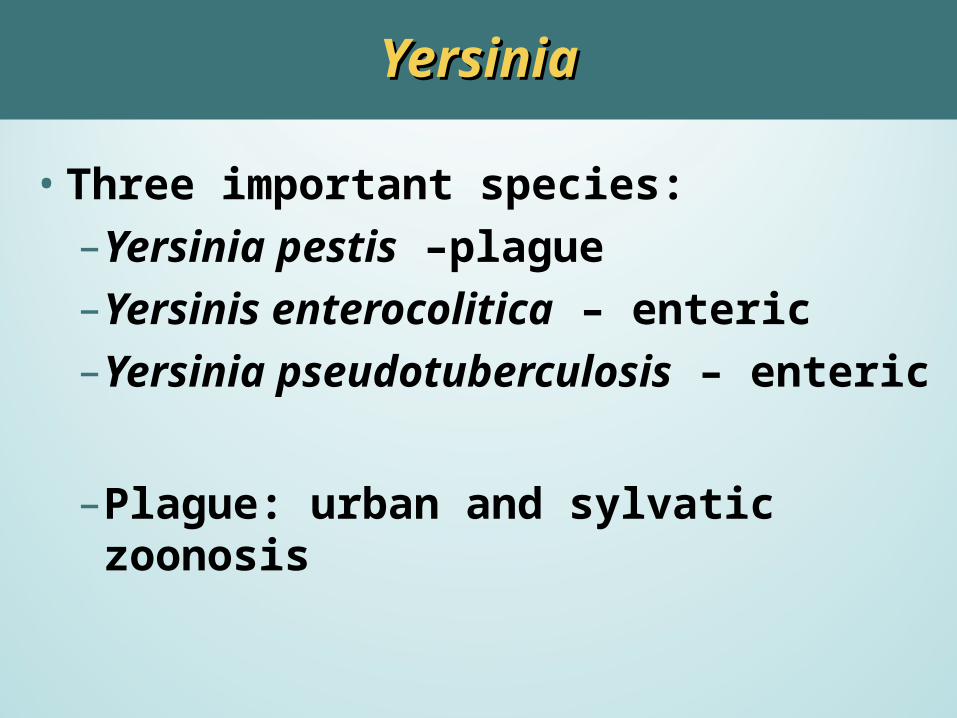
YersiniaYersinia
•Three important species:– Yersinia pestis –plague– Yersinis enterocolitica – enteric– Yersinia pseudotuberculosis – enteric
– Plague: urban and sylvatic zoonosis
Yersinia pestisYersinia pestis
•Non-motile•Bipolar staining• Slow growth of small colonies on ordinary
culture media – it grows better at lower temperature (25-300 C)
Y. pestisY. pestis
• Bubonic Plague:– Transmitted by fleas from rodent
1. Engulfed by fixed macrophages in lymph node2. Enlarged lymph nodes in the groin and armpit
(buboes). 3. Bacteria go from lymph nodes to blood. 4. Lysis of the bacteria releases LPS, septic shock. 5. Black death- Subcutaneous hemorrhages, LPS
causing DIC.
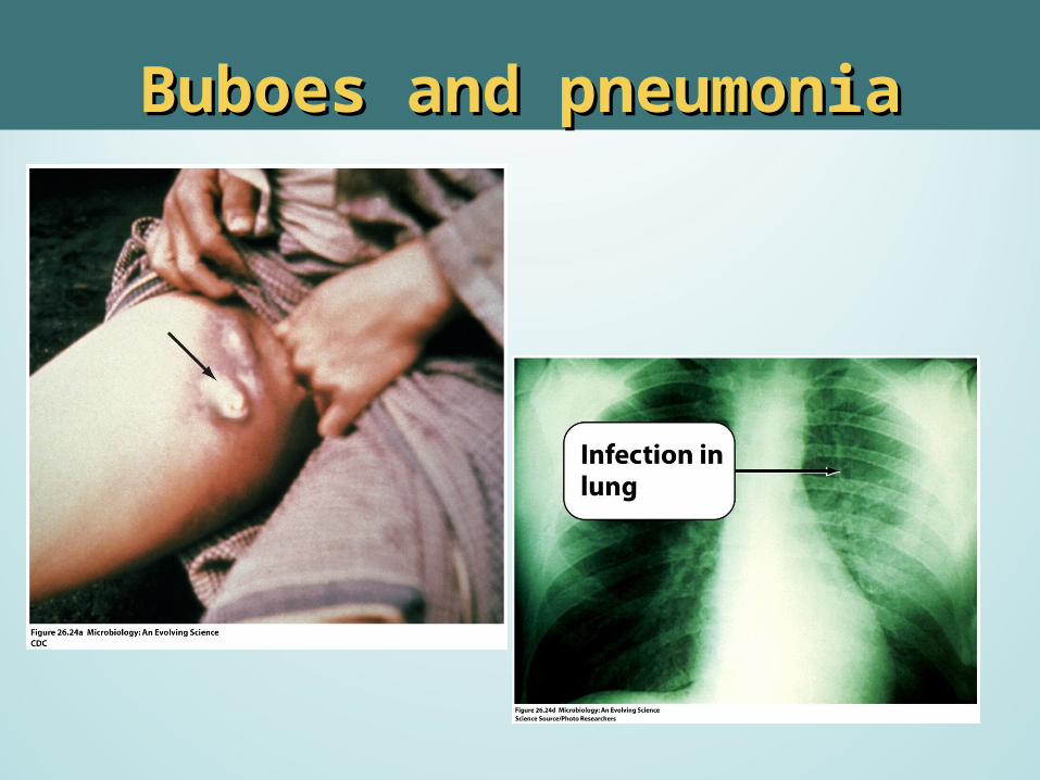
Buboes and pneumoniaBuboes and pneumonia
Y. pestisY. pestis
•Pneumonic Plague:– Bacteria reach the lungs – Ingested by lung macrophages
transmitted directly via aerosol.
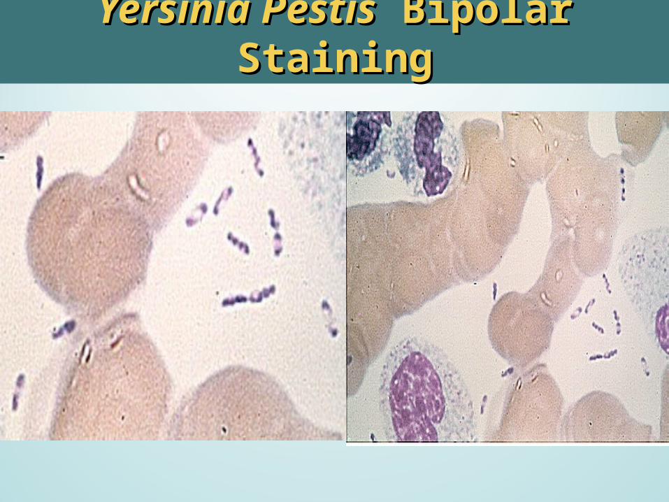
Yersinia PestisYersinia Pestis Bipolar Staining Bipolar Staining
Yersinia pestisYersinia pestis
•TSI- K/A no gas•Urea (–)•Direct fluorescent antibody test•New DNA probe test
Other pathogenic Yersinia speciesOther pathogenic Yersinia species::
• Ingestion contaminated food/H2O.
– Y. enterocolitica-watery diarrhea – Y. pseudotuberculosis
Fever and abdominal pain.
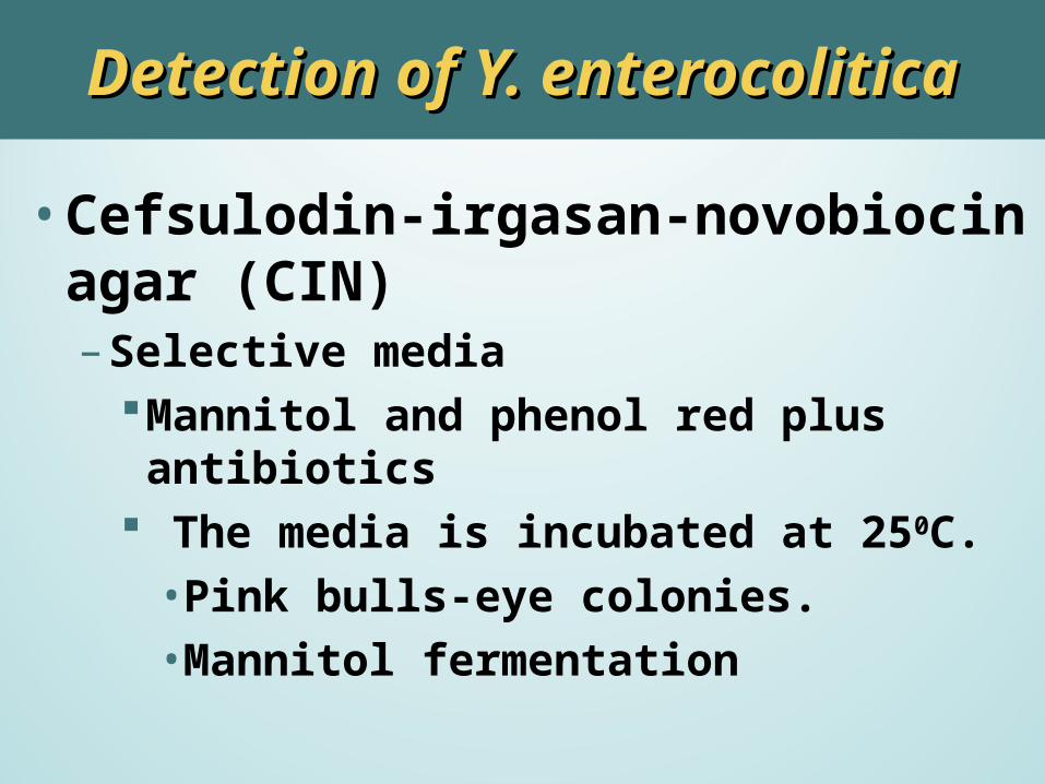
Detection of Y. enterocoliticaDetection of Y. enterocolitica
•Cefsulodin-irgasan-novobiocin agar (CIN) – Selective media
Mannitol and phenol red plus antibiotics The media is incubated at 250C.•Pink bulls-eye colonies.•Mannitol fermentation
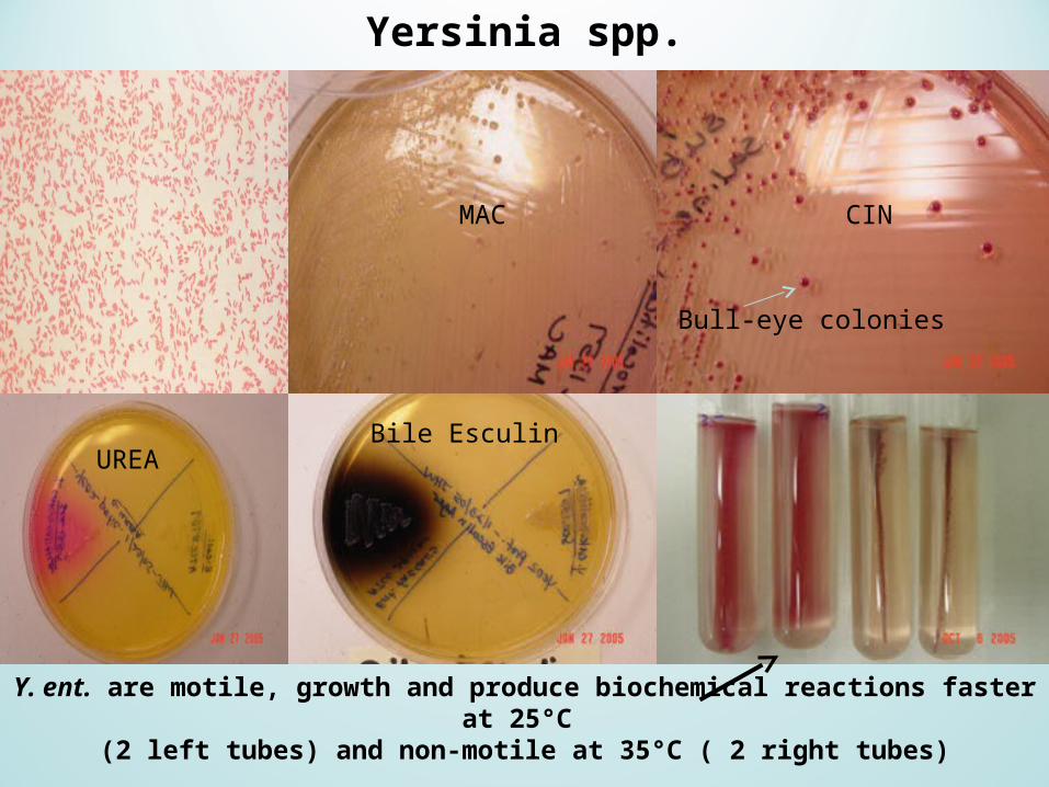
Yersinia spp.
Y. ent. are motile, growth and produce biochemical reactions faster at 25°C (2 left tubes) and non-motile at 35°C ( 2 right tubes)
CINMAC
UREABile Esculin
Bull-eye colonies

Enteric OpportunistsEnteric Opportunists
EscherichiaEscherichia coli coli
– Normal inhabitant of G.I. tract. – Various forms of gastroenteritis.– Cause of UTI, septicemia,and neonatal
meningitis.– May have capsule.– ID: Biochemistry reactions
Most are motile.Indole positive
Escherichia coli
•Epidemiology– Epidemics occur from:
Person-to-person spreadContaminated food and waterUnpasteurized milk and juices
– Humans, domestic and wild animals-sources of pathogens
E. coliE. coli
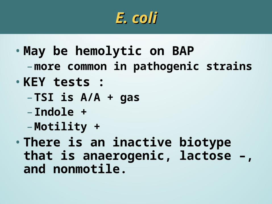
•May be hemolytic on BAP – more common in pathogenic strains
•KEY tests :– TSI is A/A + gas– Indole +– Motility +
•There is an inactive biotype that is anaerogenic, lactose –, and nonmotile.
E. coli: E. coli: Clinical SignificanceClinical Significance
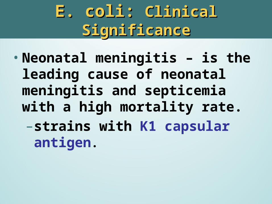
•Neonatal meningitis – is the leading cause of neonatal meningitis and septicemia with a high mortality rate.– strains with K1 capsular antigen.
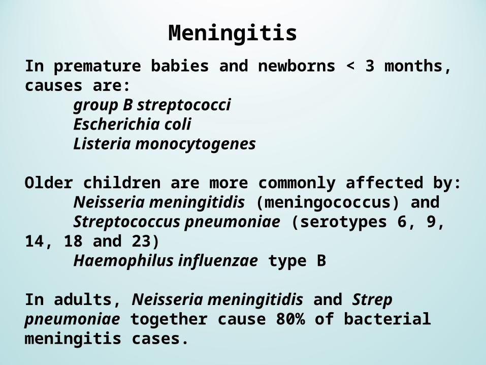
In premature babies and newborns < 3 months, causes are: group B streptococciEscherichia coliListeria monocytogenes
Older children are more commonly affected by: Neisseria meningitidis (meningococcus) and Streptococcus pneumoniae (serotypes 6, 9, 14, 18 and 23)Haemophilus influenzae type B
In adults, Neisseria meningitidis and Strep pneumoniae together cause 80% of bacterial meningitis cases.
Risk of infection with Listeria monocytogenes is increased in persons over 50 years old.
Meningitis
E. coliE. coli: : Clinical SignificanceClinical Significance
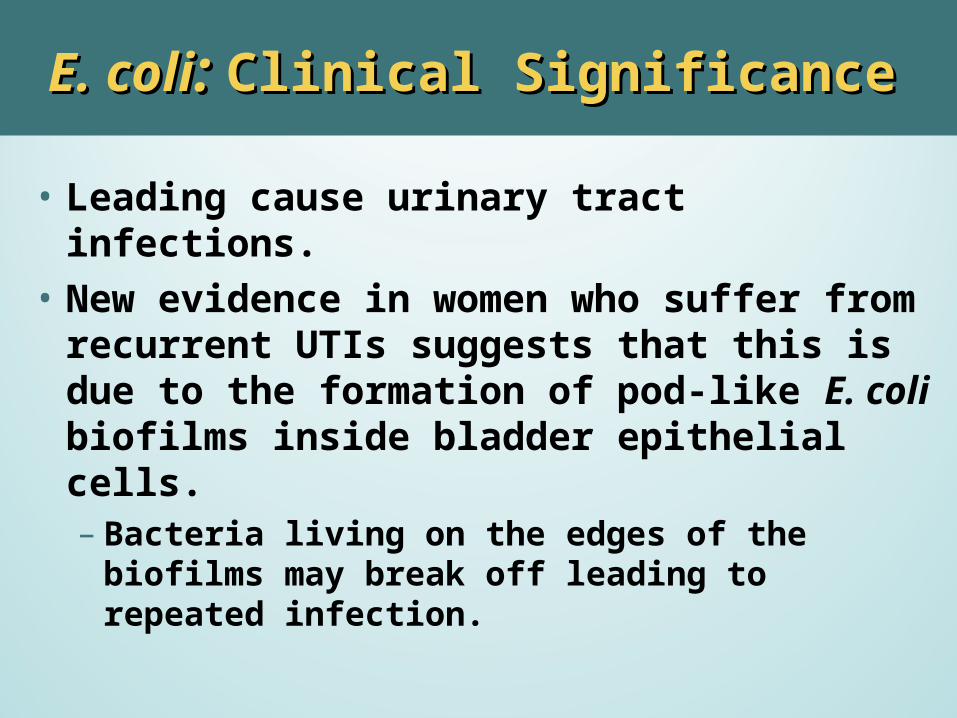
• Leading cause urinary tract infections.• New evidence in women who suffer from
recurrent UTIs suggests that this is due to the formation of pod-like E. coli biofilms inside bladder epithelial cells.– Bacteria living on the edges of the biofilms may
break off leading to repeated infection.
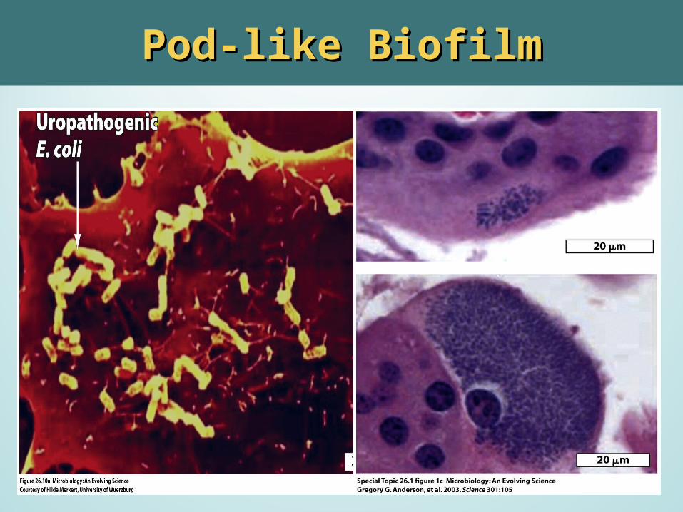
Pod-like BiofilmPod-like Biofilm
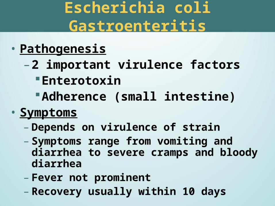
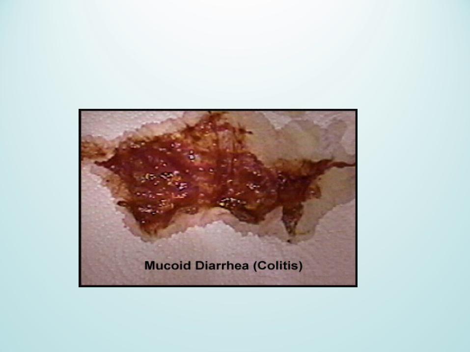
Escherichia coli Gastroenteritis
• Pathogenesis– 2 important virulence factors
EnterotoxinAdherence (small intestine)
• Symptoms– Depends on virulence of strain– Symptoms range from vomiting and diarrhea to
severe cramps and bloody diarrhea– Fever not prominent– Recovery usually within 10 days
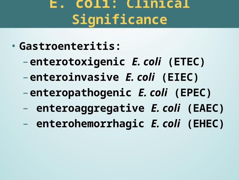
• Gastroenteritis:– enterotoxigenic E. coli (ETEC) – enteroinvasive E. coli (EIEC) – enteropathogenic E. coli (EPEC) – enteroaggregative E. coli (EAEC)– enterohemorrhagic E. coli (EHEC)
E. coli: Clinical Significance
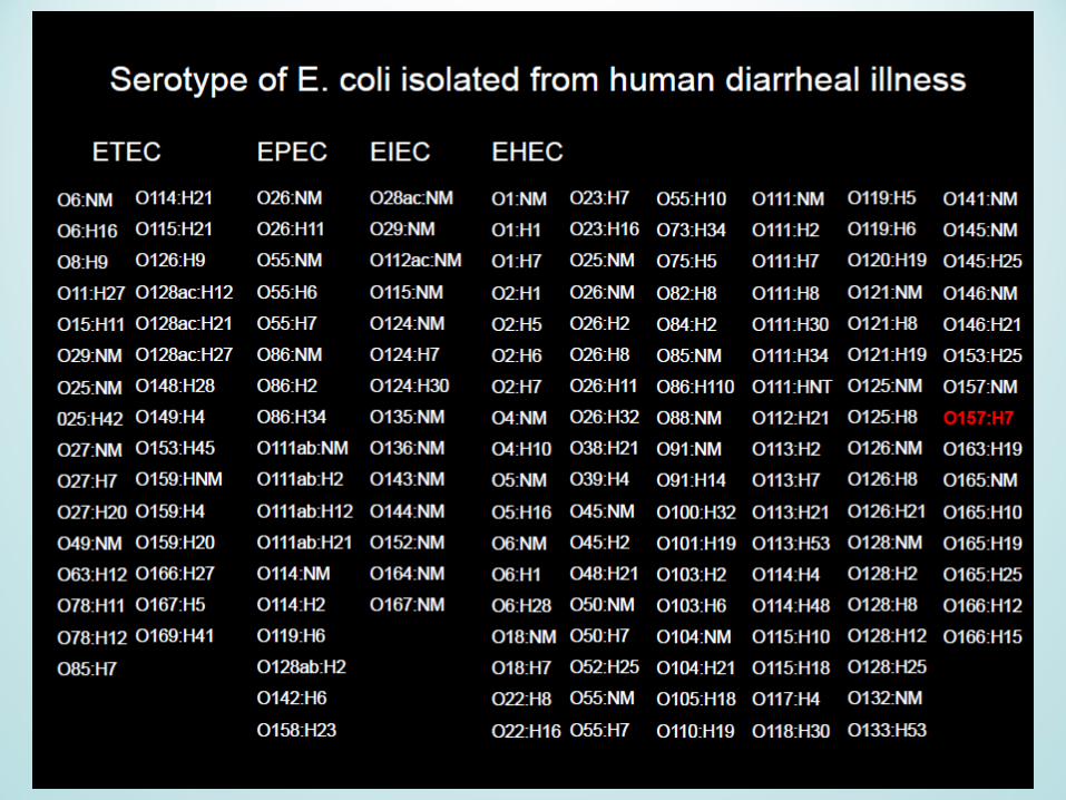
E coli 0157: H7E coli 0157: H7
• Enterohemorrhagic: – Hemorrhagic diarrhea
(young children and elderly) – Hemolytic Uremic Syndrome
•Cause: – undercooked, contaminated ground beef,
unpasteurized milk, contaminated water and vegetables.
Enterohemorrhagic E. coliEnterohemorrhagic E. coli
• Shiga-type toxin –(verotoxin) •Produced by EHEC •Cytotoxic, enterotoxic, neurotoxic, and
diarrhea,ulceration of G.I. tract. – Two types
Shiga-like toxin 1 /shiga-like toxin 2. Inhibit protein synthesis by cleaving a 28S
rRNA that’s part of the 60S subunit
•Diagnosis: – Slow fermentation of sorbitol – Chromagar– MUG negative
(methylumbelliferyl-beta-D-glucuronide)
– Toxin production– Latex agglutination tests (typing) – Characteristic biochemical codes.
E coli 0157: H7E coli 0157: H7
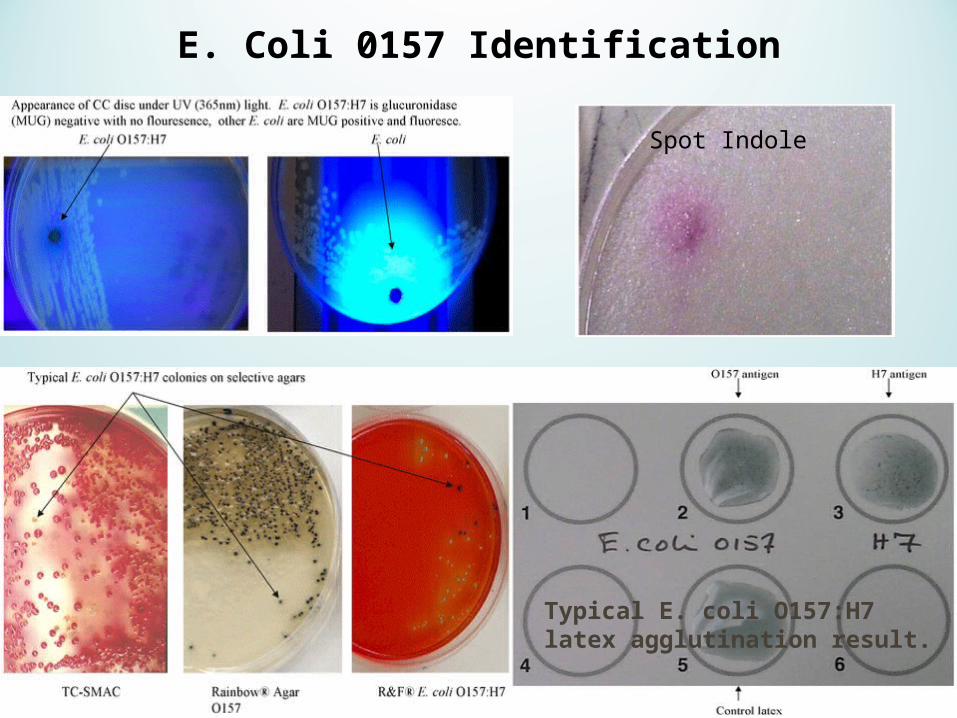
Spot Indole
Typical E. coli O157:H7 latex agglutination result.
E. Coli 0157 Identification
KlebsiellaKlebsiella
• General characteristics– Produce a large capsule– Nonmotile
• K. pneumoniae:– Colonies are glistening, large, and mucoid– Ferment lactose and nonmotile– Ferment glucose with gas– Positive citrate and lysine decarboxylase tests
KlebsiellaKlebsiella
•Pathogenesis and Infectious Disease– K. pneumoniae is a significant cause of
respiratory (especially pneumonia) and urinary tract diseases (opportunist).
– K. ozaenae and K. rhinoscleromatis are associated with chronic inflammatory diseases of the upper respiratory tract
– K. granulomatis is the causative agent of granuloma inguinale.
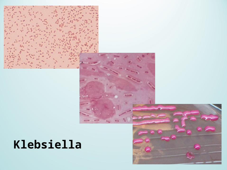
Klebsiella
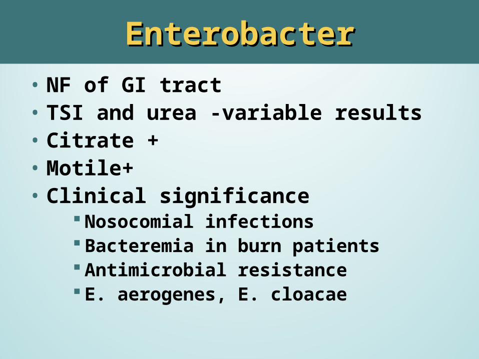
EnterobacterEnterobacter• NF of GI tract• TSI and urea -variable results• Citrate +• Motile+• Clinical significance
Nosocomial infections Bacteremia in burn patients Antimicrobial resistance E. aerogenes, E. cloacae
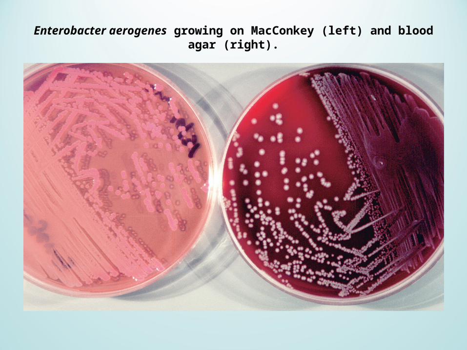
Enterobacter aerogenes growing on MacConkey (left) and blood agar (right).
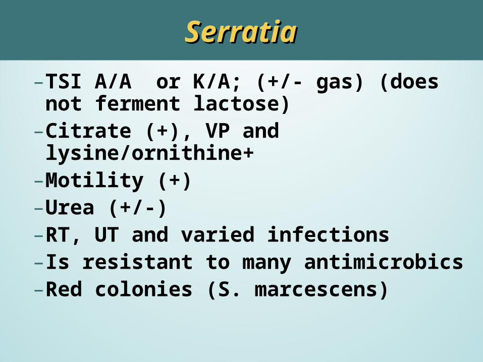
– TSI A/A or K/A; (+/- gas) (does not ferment lactose)
– Citrate (+), VP and lysine/ornithine+– Motility (+)– Urea (+/-)– RT, UT and varied infections– Is resistant to many antimicrobics– Red colonies (S. marcescens)
SerratiaSerratia
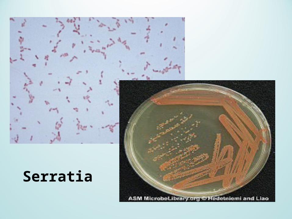
Serratia
Proteus-Providencia-MorganellaProteus-Providencia-Morganella
•General characteristics– Major species:
Proteus (P. mirabilis and P. vulgaris) Providencia (P. rettgeri – most common;
P.alcalifaciens, and P. stuartii) Morganella morganii
– In this group, P. mirabilis is most frequent isolate from clinical specimens
Proteus, Providencia, and MorganellaProteus, Providencia, and Morganella
• NF of the GI tract (except Providencia).
• All motile, with Proteus swarming
• PDA (+)
• Lysine deamination (+)
• Urea (+) Proteus and P. rettgeri
• TSI variable, H2S (+)
• UTI, kidney stones (Proteus), RTI, septicemia, wounds
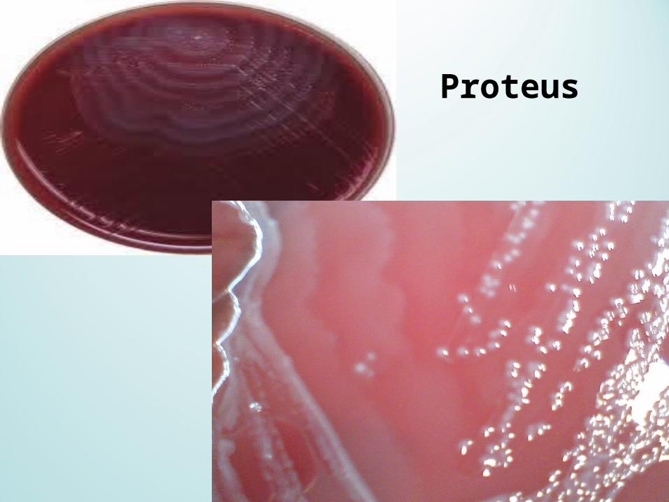
Proteus
Proteus-Providencia-MorganellaProteus-Providencia-Morganella
•P. mirabilis is indole negative and ampicillin sensitive
•P. vulgaris is indole positive
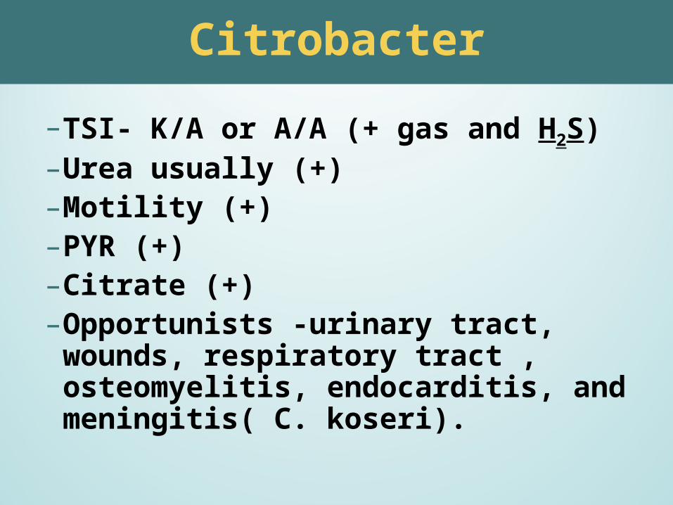
– TSI- K/A or A/A (+ gas and H2S)– Urea usually (+)– Motility (+)– PYR (+)– Citrate (+)– Opportunists -urinary tract, wounds,
respiratory tract , osteomyelitis, endocarditis, and meningitis( C. koseri).
Citrobacter

HafniaHafnia
• Cultural, biochemical and other characteristics– Motile– Usually indole and urease negative– VP, lysine and ONPG positive– Ferment glucose with gas production– Often seen as a cause of pediatric gastroenteritis– Rare respiratory tract infections, urinary tract infections,
liver abscesses, appendicitis, and postoperative endophthalmitis
– Biliary abscesses
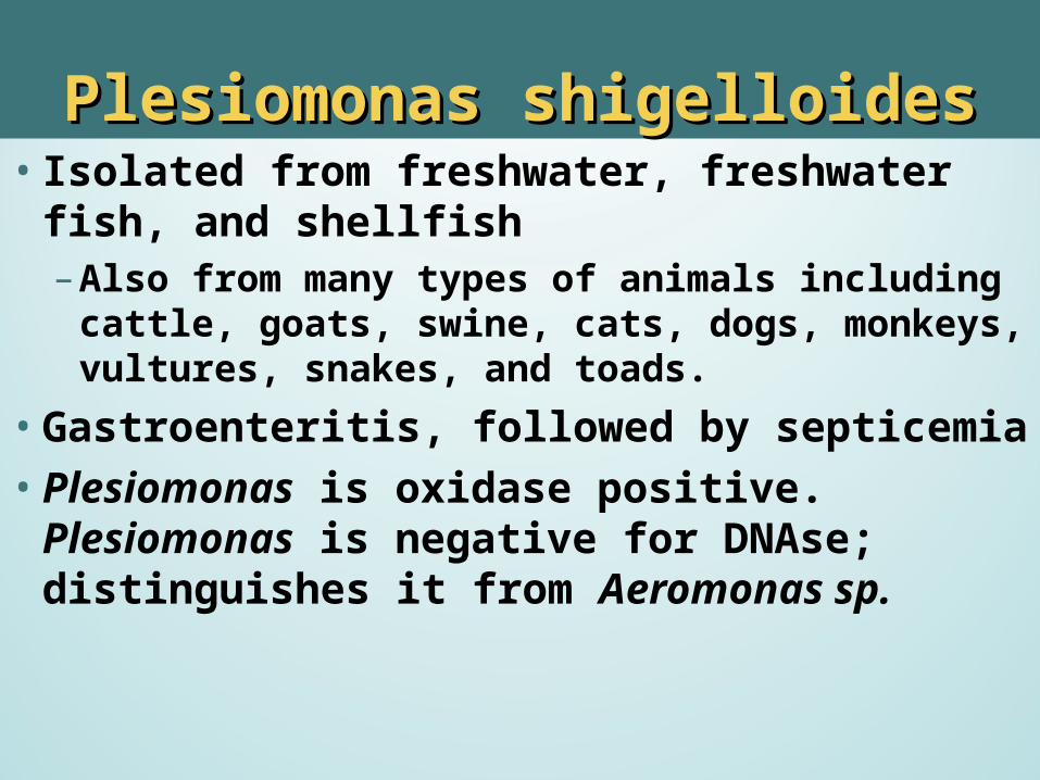
• Isolated from freshwater, freshwater fish, and shellfish– Also from many types of animals including cattle,
goats, swine, cats, dogs, monkeys, vultures, snakes, and toads.
•Gastroenteritis, followed by septicemia•Plesiomonas is oxidase positive. Plesiomonas is
negative for DNAse; distinguishes it from Aeromonas sp.
Plesiomonas shigelloidesPlesiomonas shigelloides