microbiological and toxicological assessment of pharmaceutical
TRANSCRIPT
Research ArticleMicrobiological and Toxicological Assessment of PharmaceuticalWastewater from the Lagos Megacity, Nigeria
Avemaria Ifeoma Obasi,1 Nnamdi Henry Amaeze,2 and Damilola Dorcas Osoko2
1 Department of Microbiology, University of Lagos, Akoka-Yaba, Lagos, Nigeria2 Ecotoxicology Laboratory, Department of Zoology, University of Lagos, Akoka-Yaba, Lagos, Nigeria
Correspondence should be addressed to Nnamdi Henry Amaeze; [email protected]
Received 23 May 2014; Revised 31 August 2014; Accepted 31 August 2014; Published 28 September 2014
Academic Editor: Qiusheng Zheng
Copyright © 2014 Avemaria Ifeoma Obasi et al.This is an open access article distributed under the Creative Commons AttributionLicense, which permits unrestricted use, distribution, and reproduction in any medium, provided the original work is properlycited.
We conducted a microbiological and toxicological profiling of a pharmaceutical wastewater, one of the major wastes entering theLagos lagoon. The morphological characterization of seven bacterial isolates from the wastewater indicated that 4 of them weregram positive bacilli while 3 were cocci of both gram reactions.The bacterial isolates exhibited varying degrees of enzyme activitiesbut most were able to hydrolyze starch to yield amylase. Only 3 of the isolates showed prospects as antibacterial agents, given theirmoderate inhibition to Staphylococcus xylosus relative to 8 other species tested. Overall, 81.3% of the isolates were resistant, and3.3% were susceptible while 15.4% of the isolates showed intermediate sensitivity to the antibiotics. The assessment of antioxidantactivities in liver samples of Nile tilapia, Oreochromis niloticus, exposed to sublethal concentrations of the effluents indicated someform of oxidative stress given the higher levels of lipid peroxidation product, malondialdehyde, in the exposed fishes relative tothe control kept in dechlorinate tap water. But for reduced glutathione, activities of the antioxidative stress enzymes, superoxidedismutase (SOD), catalase (CAT), and glutathione-s-transferase (GST), were higher in the effluent exposed tilapia. Responses werenot dose dependent and enzyme activities were often higher at day 14 compared to day 28. This relevance of the findings to waterquality was discussed.
1. Introduction
The fate of pharmaceutical effluents in aquatic environmentsis increasingly eliciting scientific inquiry owing to recentfindings of their effects in biological systems [1]. Nigeria hasa few pharmaceutical companies most of which are situatedin Ogun and Lagos States in the country’s south westernregion, discharging effluents into neighbouring rivers, creeks,and lagoons. Pharmaceutical effluents are wastes generatedby pharmaceutical industries during the process of drugmanufacturing [2]. These wastes by virtue of their sourceare composed of a variety of biologically active chemi-cal components including antibiotics, lipid regulators, anti-inflammatories, antiepileptics, tranquilizers, oil and grease[3], heavy metals [4], and myriads of other compoundsdepending on the drug or personal care products beingmanufactured. Pharmaceutical effluents particularly thoserich in contraceptives have also been linked with endocrine
disrupting effects [5]. Microbial actions on effluents oftenresult in products which may also interfere with endocrinefunctions.Mycobacterium smegmatis is known to metabolizestigmasterol in effluents into a potent androgen, androstene-dione [6]. Exposure of aquatic organisms to androgenic orestrogenic compounds in pharmaceutical wastes and sewageis a known cause of sex reversal in fishes which could impairreproductive success [7].
Industrial wastewater management systems in Nigeriaare either nonexistent, inappropriate, or at best inefficient.When efforts are made towards effluent management, basicdilution, filtration, sedimentation, and aeration techniquesare usually put in place and thesemay not have the capacity tobreak down the chemical components of the effluents. Theseeffluents eventually end up in natural water bodies wherethey cause contamination of water and toxic effects on thebiotic communities [8]. Effluents typically contain variouscomponents, some of which could have the tendency to
Hindawi Publishing CorporationChinese Journal of BiologyVolume 2014, Article ID 638142, 9 pageshttp://dx.doi.org/10.1155/2014/638142
2 Chinese Journal of Biology
combinewith the unsaturated fatty acids of the phospholipidsof cell membranes leading to oxidative damage and releaseof byproducts such as malondialdehyde [9]. The resultingoxidative damage and the reactive oxygen species are contin-ually countered by the action of antioxidative stress enzymessuch as superoxide dismutase (SOD), catalase (CAT), andglutathione-s-transferase (GST) [9].
In view of the fate of pharmaceuticals in our environmentand the growing concerns over their biological effects, wetherefore conducted an extensive microbiological profilingand toxicological evaluations using the Nile tilapia, Ore-ochromis niloticus (a commonly bred fish for laboratorystudies in Nigeria), of pharmaceutical wastewater from anindustrial estate in the South-West of Nigeria. The termwastewater is used in this study to represent effluents whichhave not yet been discharged into the environment.
2. Materials and Methods
2.1. Sample Collection. Wastewater was sampled from thereservoir of a pharmaceutical company in industrial estate,at Agbara, the South-West of Nigeria. Untreated wastewatersample from the production of ciprofloxacin was collecteddirectly into a 4-liter sterile plastic container and put on iceand immediately taken to the laboratory for routine microbi-ological, toxicological, and biochemical analyses. Storage wasat 4∘C prior to analysis and assays.
2.2. Assessment of the Physicochemical Characteristics of theEffluents. The wastewater sample was analyzed for basicphysicochemical characteristics using standard methods asdescribed by APHA-AWWA-WEF [10].The parameters anal-ysed in the effluent sample included pH, salinity, conduc-tivity, total dissolved solids (TDS), dissolved oxygen (DO),biological oxygen demand (BOD), chemical oxygen demand(COD), iron (Fe), zinc (Zn), copper (Cu), nickel (Ni), andcadmium (Cd). Specifically, the heavy metal concentrationswere measured using atomic absorption spectrophotometer,after the samples have been subjected to acid digestion.Determination of DO level was not possible in situ; thus,samples were stored cool (4∘C) at the point of collection andtransported to the lab for immediate analysis, while otheranalyses were carried out within 1 week of sampling. Also allglassware was subjected to heat and acid wash before use toprevent contamination and false readings.
2.3. Determination of THC. The analysis for total hydrocar-bon content (THC) of the sample was done using ASTMD 2887-93 method [11] on an Agilent 4890D gas chromato-graph/flame ionization detector subjected to prior calibrationusing a known standard (Sigma Aldrich, USA).
2.4. Microbiological Analysis
2.4.1. Isolation of Microorganism. One mL of water samplewas added to 9mLof sterile distilledwater and a tenfold serialdilution was done, and the lower, middle, and high dilutionswere plated in duplicate into nutrient agar (Biotec; UK),
MacConkey agar, and potato dextrose agar plates alreadyprepared.These were incubated at 37∘C for 18–24 hrs for totalbacteria and coliforms. Also, incubation for 3 to 5 days at 27∘C(Rm temp.) was achieved for fungi isolates. Colonies on plateswere observed and counted and the population density wasestimated; bacterial colonies were picked according to theircultural morphology on the plates and these were streakedon new nutrient agar plates for pure colonies [12].
2.4.2.Morphological and Enzymatic Assay of Bacterial Isolates.The morphological characteristics of isolates were observedand recorded and this was the basis for the isolation ofcolonies. The cell shape and arrangements of isolates weredetermined following the standard procedures of basic stain,gram stain [12]. The isolates were screened for the presenceof protease, amylase, and phospholipase using methods asdescribed by Talaro [13].
2.4.3. Screening for Antibacterial Agent. All isolates werescreened for antibacterial activity against 8 types of clinicalbacterial species, obtained from Lagos University TeachingHospital (LUTH), using the agar well diffusion method asdescribed by John et al. [14]. Overnight bacterial cultures innutrient broth were standardized and adjusted to a McFar-land standard of 0.5 which is equivalent to approximately108 cfu/mL. The already prepared Mueller Hinton agar plateswere swabbed with the different clinical bacterial isolateswith a sterile swab stick; this was done by streaking theswab over the entire surface of the Mueller Hinton agarplates and the plates were rotated 60∘ and the streakingwas repeated until the entire plate was swabbed. Completeinoculation was done by running the swab around the rimof the agar plates. The lid of the plates was left open for about5 minutes to allow any excess moisture to be absorbed beforeboring the agar with a sterile cork borer. Few drops of about0.1mL of the test bacterial isolate were introduced usinga 100 𝜇L micropipette into the wells and this was repeatedfor all the swabbed clinical bacterial isolates. The clinicalbacteria used are four gram positive bacteria (Staphylococcusxylosus, Staphylococcus warneri, Staphylococcus epidermidis,and Enterococcus faecalis) and four gram-negative bacteria(Pseudomonas aeruginosa, Proteus mirabilis, Shigella sp., andSalmonella sp.). The plates were incubated at 37∘C for 24 hrs.The assay for antimicrobial properties was determined bymeasuring the diameter of the inhibition zone around thewells and interpretation of results was expressed as moderate(6 to 9mm), strong (10 to 14mm), or very strong (15 to 18mm)according to Liasi et al. [15].
2.5. Determination of the Acute Toxicity of the Effluent
2.5.1. Selection and Acclimatization of Test Fishes. The Niletilapia Oreochromis niloticus fingerlings, measuring an aver-age of 1.55 ± 0.15 cm, were obtained from a commercial fishfarm in Badagry on the outskirts of Lagos. The fishes weretransported in aerated plastic tanks and acclimatized for 1week in dark plastic tanks (stocking density: 10 fishes per litre,photoperiod: 12 hrs dark 12 hrs light). The fish stock selected
Chinese Journal of Biology 3
for the assay recorded mortality of less than 1% after 1 weekof acclimatization in the laboratory.
2.5.2. Bioassay Procedure. Wastewater samples were removedfrom storage in the fridge (4∘C) and allowed to adjust toroom temperature, until the temperature of the samples wasapproximately equal to those of the water in the bioassaytanks (about 26∘C).The bioassay tanks had a stocking densityof 10 fingerlings per litre and concentrations were set induplicate, with each bioassay repeated twice after an initialrange finding experiment.
Quantal response of mortality was employed in assessingeffluent toxicity based on established techniques [16] andthe 96-hour LC
50of the effluent samples acting against the
tilapia fingerlings was used in determining the sublethalconcentrations for the biochemical assays.
2.6. Assessment of Biochemical Responses of O. niloticusExposed to the Pharmaceutical Effluent. After 14 and 28days of exposure of the fingerlings to sublethal concentra-tions of the pharmaceutical effluent (1/10th 96-hour LC
50=
5.120mL/L; 1/100th 96-hour LC50= 0.512mL/L), their liver
sampleswere excised using a sterile dissecting set after a sharpblow to their head to immobilize them. (Ethical permit wasin line with the Zoology Department, University of Lagosstandard for fair treatment of lab animals.)
The liver samples were collected in unused universalbottles prior to analysis. The samples were assessed for thelipid peroxidation product, malondialdehyde (MDA), usingthe thiobarbituric acid reaction (TBARS) assay as describedby Yagi [17]. Also antioxidative stress enzyme activities wereassessed using established techniques as follows: superoxidedismutase [18], catalase [19], reduced glutathione as nonpro-tein sulfhydryls [20], and glutathione-s-transferase based ontheir relatively high activity with 1-chloro-2,4-dinitrobenzeneas a second substrate [21].
2.7. Statistical Analysis. The Probit assay was used to deter-mine the dose-response of mortality using independentsample 𝑡-test and two-way ANOVA (SPSS Version 16). Thesoftware used was SPSS Version 16 to determine the 96-hourLC50
(i.e., the median lethal concentration that causes 50%mortality of the exposed fingerlings).
3. Results
3.1. Physicochemical Characteristics of the PharmaceuticalEffluent. Thephysicochemical characteristics of the pharma-ceutical effluents showed that it was slightly acidic, with someconsiderable levels of electrical conductivity. There was alsolow level of dissolved oxygen and evidence of degradationaction of microorganisms with BOD and COD levels of 36.0and 72.0mg/L, respectively. The effluent was low on heavymetals except for iron and manganese which were high inconcentrations (Table 1).
3.2. Microbiological Assessments of the Pharmaceutical Efflu-ent. The initial characterization of the microbial population
Table 1: Physicochemical characteristics of the pharmaceuticaleffluents.
Parameter EffluentColor PtCo 30Turbidity FAU 16pH 5.0Dissolved oxygen (mg/L) 4.70Biological oxygen demand (mg/L) 36.0Chemical oxygen demand (mg/L) 72.0Conductivity (𝜇S/cm) 320Total dissolved solids (mg/L) 160Salinity (‰) 0Temperature (∘C) 26.65Chloride (mg/L) 66Sulfate (mg/L) 5.0Total hardness (mg/L) 180Calcium (mg/L) 120Magnesium (mg/L) 60Sodium (mg/L) 11.50Oil and grease (mg/L) 0.10Total carbonate alkalinity (mg/L) 0Nitrate (mg/L) 0.18Nitrite (mg/L) 0.01Phosphate (mg/L) 1.96Hydrogen sulfide (mg/L) 0TOC (%) 4.40Iron (mg/L) 36.45Manganese (mg/L) 20.0Copper (mg/L) 0.82Zinc (mg/L) 0.88Lead (mg/L) 0.22Nickel (mg/L) 0.26Chromium (mg/L) 0.31Mercury (mg/L) NDArsenic (mg/L) 0.04Barium (mg/L) 0.88Cobalt (mg/L) 0.04ND: not detected.
densities from the effluent indicated a bacterial and totalfungal count of 2.7 × 10¹² cfu/mL and 4.8 × 10⁴ cfu/mL,respectively. There was no evidence of coliform growth whensamples were cultured on MacConkey agar, thus implyingthat there was no faecal contamination in the effluent.
3.2.1. Morphological Characteristics of Isolates. An assess-ment of the cultural characteristics of the isolates observedon plates indicated that the seven isolatesweremostly circularin form except for isolate ME6 (Table 2). The isolates were allopaque and smooth, with two of them being gram negativewhile the other four were gram positive. Also, cellularmorphology showed that four of the isolates are rods whilethree are cocci. All but one of the cocci were gram negative,
4 Chinese Journal of Biology
Table 2: Morphology of bacterial isolates from pharmaceutical wastewater.
Isolates ME1 ME2 ME3 ME4 ME5 ME6 ME7Form of colony Circular Circular Circular Circular Circular Punctiform CircularTranslucency/opacity Opaque Opaque Opaque Opaque Opaque Opaque OpaqueElevation of colony Flat Flat Flat Flat Flat Raised FlatMargin of colony Entire Entire Entire Entire Entire Undulate EntireSurface of colony Smooth Smooth Smooth Smooth Smooth Smooth SmoothCell morphology Cocci Rods Cocci Rods Cocci Rods RodsGram stain reaction Negative Positive Positive Positive Negative Positive PositivePigmentation Light yellow Milky white Yellow Milky white Milky white Milky white Yellow
while all the rods were gram positive and the pigmentation ofisolates ranged from milky white to yellow (Table 2).
3.2.2. Assessment of Enzymatic Assay of Bacterial Isolates.Four isolates (ME2, ME4, ME5, and ME7) were observedto hydrolyze starch, producing amylase, of which two ME4and ME5 produced high amylase activity. The only bacterialisolates that possessed moderate protease activity and highphospholipase activity were ME4 and ME7, respectively,(Table 3).
3.2.3. Inhibitory Spectrum of Antibacterial-Producing Bacteriaon Clinical Isolates. The isolates were tested for their abilityto produce inhibitory substances against 8 indicator species(Table 4). Isolates ME2, ME3, and ME7 amongst othersshowedmoderate inhibition zones of 3mm, 6mm, and 8mm,respectively, against gram positive Staphylococcus xylosus.
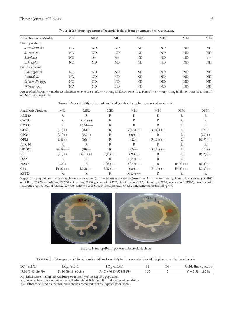
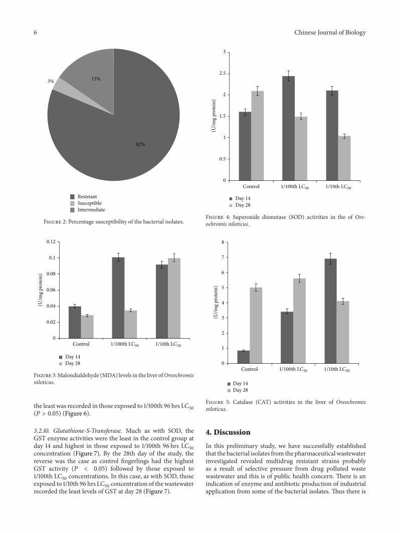
3.2.4. Antibiotic Susceptibility Pattern of Bacterial Isolates.Susceptibility test of the isolates using the Kirby-Bauermethod as shown in Table 5 indicated that bacterial isolatesME3,ME5, andME6were resistant to all 13 antibiotics tested,although other isolates revealed some level of resistance tosome of the antibiotics as well. The pattern of susceptibilityis as shown in Figure 1. Overall, 81.3% of the isolates wereresistant, and 3.3%of the isolates were susceptible while 15.4%showed intermediate sensitivity to the antibiotics (Figure 2).
3.2.5. Acute Toxicity of the Pharmaceutical Effluents on Ore-ochromis niloticus. The results of the acute toxicity studies onO. niloticus indicate a generally low to moderate toxicity. Onthe basis of derived 96-hour LC
50values, the wastewater was
found to have a LC50value of 51.20mL/L (Table 6).
Biochemical responses products in Oreochromis niloticuswere exposed to sublethal concentrations of pharmaceuticalwastewater.
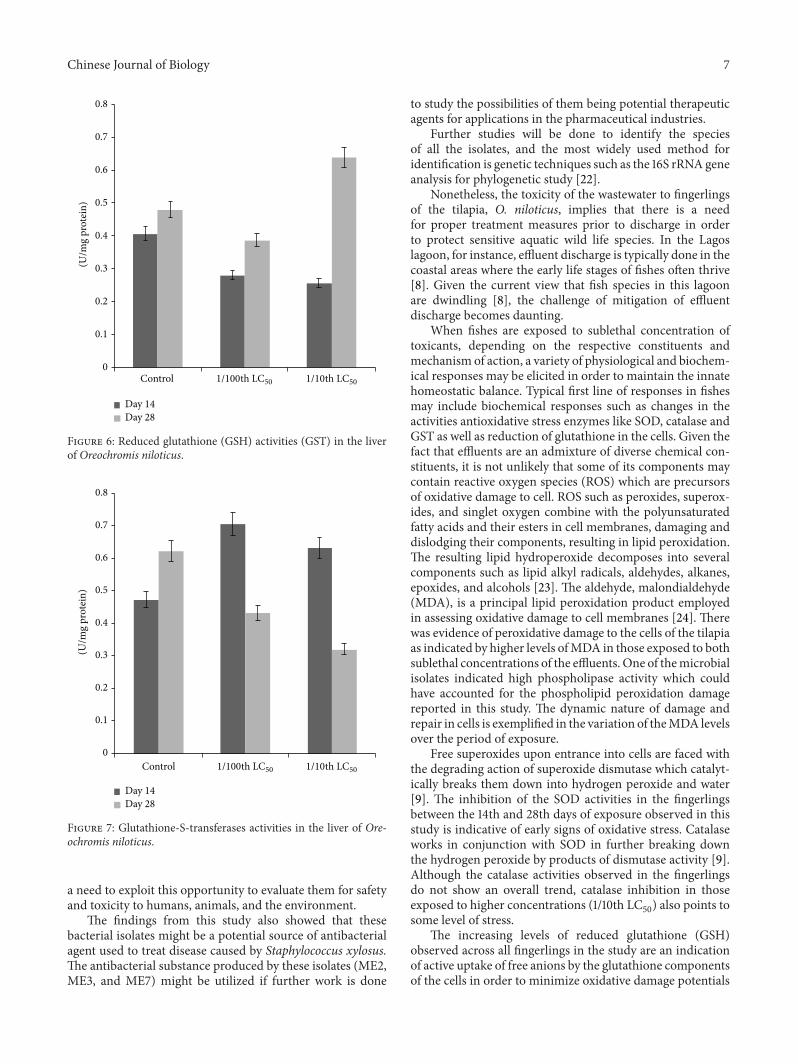
3.2.6. Lipid Peroxidation. The aldehyde byproduct of lipidperoxidation, MDA, was lower in the control at both days14 and 28 relative to the wastewater exposed tilapia groups(Figure 3). Specifically, the MDA levels at day 14 weresignificantly lower (𝑃 < 0.05) in the control group thanthose exposed to the wastewater. However, by day 28 ofthe experiments, the levels, though lower in the controls,
Table 3: Enzymatic assay of bacterial isolates from pharmaceuticalwastewater.
Isolates/enzyme Amylase Protease PhospholipaseME1 ND ND NDME2 7++ ND NDME3 ND ND NDME4 11+++ 8++ NDME5 (14)+++ ND NDME6 ND ND NDME7 6+ ND +++Degree of enzymatic activity: + = low (≤2mm), ++ = moderate (>2–8mm),+++ = high (>8mm), and ND = nondetectable.
did not vary significantly with those in the effluent exposedgroups. Among the exposed groups, MDA was higher inthose exposed to 1/100th 96 hrs LC
50(𝑃 > 0.05) at day 14
but the reverse was the case by day 28, with those exposedto the 1/10th 96 hrs LC
50having a significantly higher level
(𝑃 < 0.05) of MDA (Figure 3).
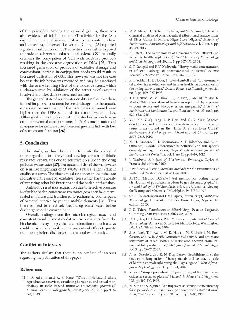
3.2.7. Superoxide Dismutase. The activities of SOD in theliver samples from the exposed O. niloticus were observed toreduce significantly (𝑃 < 0.05), between the 14th and 28thdays of assessments except in the control samples. The leastas well as the highest SOD activities in the tilapia fingerlingswere recorded at days 14 and 28, respectively, in the controlindividuals (Figure 4). Also, SOD activity was lower in thoseexposed to 1/10th 96 hrs LC
50at both days 14 and 28 of
exposure but the difference was not significant (𝑃 > 0.05).
3.2.8. Catalase. Catalase enzyme activity was least at day 14in control tilapia fingerlings and its levels were significantlylower (𝑃 < 0.05) than in those exposed to 1/10th and 1/100thof the LC
50(𝑃 < 0.05). Among thewastewater exposed group,
1/10th 96 hrs LC50
concentration induced higher activity atday 14 but the reverse was the case by day 28 (Figure 5).
3.2.9. Reduced Glutathione. The extent of glutathione reduc-tion (GSH)was highest in the control fingerlings at day 14 andleast in those exposed to 1/10th 96 hrs LC
50concentrations.
The reverse was the case by day 28 with those exposedto 1/10th 96 hrs LC
50having the highest GSH levels while
Chinese Journal of Biology 5
Table 4: Inhibitory spectrum of bacterial isolates from pharmaceutical wastewater.
Indicator species/isolate ME1 ME2 ME3 ME4 ME5 ME6 ME7Gram positive
S. epidermidis ND ND ND ND ND ND NDS. warneri ND ND ND ND ND ND NDS. xylosus ND 3+ 6+ ND ND ND 8+E. faecalis ND ND ND ND ND ND ND
Gram negativeP. aeruginosa ND ND ND ND ND ND NDP. mirabilis ND ND ND ND ND ND NDSalmonella spp. ND ND ND ND ND ND NDShigella spp. ND ND ND ND ND ND ND
Degree of inhibition: + = moderate inhibition zone (6 to 9mm), ++ = strong inhibition zone (10 to 14mm), +++ = very strong inhibition zone (15 to 18mm),and ND = nondetectable.
Table 5: Susceptibility pattern of bacterial isolates from pharmaceutical wastewater.
Antibiotics/isolates ME1 ME2 ME3 ME4 ME5 ME6 ME7AMP10 R R R R R R RCAZ30 R R(8)+++ R R R R RCRX30 R R(15)+++ R R R R RGEN10 (20)++ (16)++ R R(15)+++ R(14)+++ R (17)++CPR5 (20)++ (20)++ R (20)++ R R (20)++OFL5 (18)++ (16)++ R (22)+ R(10)+++ R R(15)+++AUG30 R R R R R R RNIT300 R(11)+++ (18)++ R (24)+ R(12)+++ R (20)++E15 (20)++ R(8)+++ R(12)+++ (20)++ R R R(12)+++DA2 R R R R(15)+++ R R RNA30 (22)+ R R(15)+++ R(14)+++ R R(12)+++ R(15)+++C30 R(15)+++ R(12)+++ R(12)+++ (20)++ R(10)+++ R(15)+++ R(10)+++SXT25 R R R R(12)+++ R R RDegree of susceptibility: + = susceptible/sensitive (>21mm), ++ = intermediate (16 to 20mm), and +++ = resistant (≤15mm). R = resistant; AMP10,ampicillin; CAZ30, ceftazidime; CRX30, cefuroxime; CN10, gentamycin; CPR5, ciprofloxacin; OFL5, ofloxacin; AUG30, augmentin; NIT300, nitrofurantoin;E15, erythromycin; DA2, clindamycin; NA30, nalidixic acid; C30, chloramphenicol; SXT25, sulfamethoxazole/trimethoprim.
Figure 1: Susceptibility pattern of bacterial isolates.
Table 6: Probit response of Oreochromis niloticus to acutely toxic concentrations of the pharmaceutical wastewater.
LC5 (mL/L) LC50 (mL/L) LC95 (mL/L) SE DF Probit-line equation15.14 (0.02–29.59) 51.20 (19.14–90.24) 173.21 (96.19–52485.55) 1.32 2 𝑌 = 2.33 − 2.28𝑥
LC5: lethal concentration that will bring 5% mortality of the exposed population.LC50: median lethal concentration that will bring about 50% mortality to the exposed population.LC95: lethal concentration that will bring about 95% mortality of the exposed population.
6 Chinese Journal of Biology
ResistantSusceptibleIntermediate
3% 15%
82%
Figure 2: Percentage susceptibility of the bacterial isolates.
0
0.02
0.04
0.06
0.08
0.1
0.12
Control
Day 14Day 28
1/100th LC50 1/10th LC50
(U/m
g pr
otei
n)
Figure 3:Malondialdehyde (MDA) levels in the liver ofOreochromisniloticus.
the least was recorded in those exposed to 1/100th 96 hrs LC50
(𝑃 > 0.05) (Figure 6).
3.2.10. Glutathione-S-Transferase. Much as with SOD, theGST enzyme activities were the least in the control group atday 14 and highest in those exposed to 1/100th 96 hrs LC
50
concentration (Figure 7). By the 28th day of the study, thereverse was the case as control fingerlings had the highestGST activity (𝑃 < 0.05) followed by those exposed to1/100th LC
50concentrations. In this case, as with SOD, those
exposed to 1/10th 96 hrs LC50concentration of thewastewater
recorded the least levels of GST at day 28 (Figure 7).
0
0.5
1
1.5
2
2.5
3
Control
(U/m
g pr
otei
n)
Day 14Day 28
1/100th LC50 1/10th LC50
Figure 4: Superoxide dismutase (SOD) activities in the of Ore-ochromis niloticus.
0
1
2
3
4
5
6
7
8
Control
Day 14Day 28
1/100th LC50 1/10th LC50
(U/m
g pr
otei
n)
Figure 5: Catalase (CAT) activities in the liver of Oreochromisniloticus.
4. Discussion
In this preliminary study, we have successfully establishedthat the bacterial isolates from the pharmaceutical wastewaterinvestigated revealed multidrug resistant strains probablyas a result of selective pressure from drug polluted wastewastewater and this is of public health concern. There is anindication of enzyme and antibiotic production of industrialapplication from some of the bacterial isolates. Thus there is
Chinese Journal of Biology 7
Day 14Day 28
0
0.1
0.2
0.3
0.4
0.5
0.6
0.7
0.8
Control 1/100th LC50 1/10th LC50
(U/m
g pr
otei
n)
Figure 6: Reduced glutathione (GSH) activities (GST) in the liverof Oreochromis niloticus.
Day 14Day 28
0
0.1
0.2
0.3
0.4
0.5
0.6
0.7
0.8
Control 1/100th LC50 1/10th LC50
(U/m
g pr
otei
n)
Figure 7: Glutathione-S-transferases activities in the liver of Ore-ochromis niloticus.
a need to exploit this opportunity to evaluate them for safetyand toxicity to humans, animals, and the environment.
The findings from this study also showed that thesebacterial isolates might be a potential source of antibacterialagent used to treat disease caused by Staphylococcus xylosus.The antibacterial substance produced by these isolates (ME2,ME3, and ME7) might be utilized if further work is done
to study the possibilities of them being potential therapeuticagents for applications in the pharmaceutical industries.
Further studies will be done to identify the speciesof all the isolates, and the most widely used method foridentification is genetic techniques such as the 16S rRNAgeneanalysis for phylogenetic study [22].
Nonetheless, the toxicity of the wastewater to fingerlingsof the tilapia, O. niloticus, implies that there is a needfor proper treatment measures prior to discharge in orderto protect sensitive aquatic wild life species. In the Lagoslagoon, for instance, effluent discharge is typically done in thecoastal areas where the early life stages of fishes often thrive[8]. Given the current view that fish species in this lagoonare dwindling [8], the challenge of mitigation of effluentdischarge becomes daunting.
When fishes are exposed to sublethal concentration oftoxicants, depending on the respective constituents andmechanism of action, a variety of physiological and biochem-ical responses may be elicited in order to maintain the innatehomeostatic balance. Typical first line of responses in fishesmay include biochemical responses such as changes in theactivities antioxidative stress enzymes like SOD, catalase andGST as well as reduction of glutathione in the cells. Given thefact that effluents are an admixture of diverse chemical con-stituents, it is not unlikely that some of its components maycontain reactive oxygen species (ROS) which are precursorsof oxidative damage to cell. ROS such as peroxides, superox-ides, and singlet oxygen combine with the polyunsaturatedfatty acids and their esters in cell membranes, damaging anddislodging their components, resulting in lipid peroxidation.The resulting lipid hydroperoxide decomposes into severalcomponents such as lipid alkyl radicals, aldehydes, alkanes,epoxides, and alcohols [23]. The aldehyde, malondialdehyde(MDA), is a principal lipid peroxidation product employedin assessing oxidative damage to cell membranes [24]. Therewas evidence of peroxidative damage to the cells of the tilapiaas indicated by higher levels ofMDA in those exposed to bothsublethal concentrations of the effluents. One of themicrobialisolates indicated high phospholipase activity which couldhave accounted for the phospholipid peroxidation damagereported in this study. The dynamic nature of damage andrepair in cells is exemplified in the variation of theMDA levelsover the period of exposure.
Free superoxides upon entrance into cells are faced withthe degrading action of superoxide dismutase which catalyt-ically breaks them down into hydrogen peroxide and water[9]. The inhibition of the SOD activities in the fingerlingsbetween the 14th and 28th days of exposure observed in thisstudy is indicative of early signs of oxidative stress. Catalaseworks in conjunction with SOD in further breaking downthe hydrogen peroxide by products of dismutase activity [9].Although the catalase activities observed in the fingerlingsdo not show an overall trend, catalase inhibition in thoseexposed to higher concentrations (1/10th LC
50) also points to
some level of stress.The increasing levels of reduced glutathione (GSH)
observed across all fingerlings in the study are an indicationof active uptake of free anions by the glutathione componentsof the cells in order to minimize oxidative damage potentials
8 Chinese Journal of Biology
of the peroxides. Among the exposed groups, there wasalso evidence of inhibition of GST activities by the 28thday of the sublethal assays, relative to the control, wherean increase was observed. Leaver and George [25] reportedsignificant inhibition of GST activities in catfishes exposedto crude oils, benzene, toluene, and xylene. GST naturallycatalyzes the conjugation of GSH with oxidative productsresulting in the oxidative degradation of DNA [25]. Thusincreased generation of products of oxidative damage andconcomitant increase in conjugation needs would result inincreased utilization of GST. This however was not the casebecause the inhibition was recorded and may be associatedwith the overwhelming effect of the oxidative stress, whichis characterized by inhibition of the activities of enzymesinvolved in antioxidative stress mechanisms.
The general state of wastewater quality implies that thereis need for proper treatment before discharge into the aquaticecosystem because many of the parameters examined werehigher than the FEPA standards for natural water bodies.Although dilution factors in natural water bodies would easeout their eventual concentrations, the high concentrations ofmanganese for instance are of concern given its link with lossof neuromotor function [26].
5. Conclusion
In this study, we have been able to relate the ability ofmicroorganisms to survive and develop certain antibioticresistance capabilities due to selective pressure in the drugpollutedwaste water [27].The acute toxicity of thewastewateron sensitive fingerlings of O. niloticus raises salient effluentquality concerns. The biochemical responses in the fishes areindicative of the onset of oxidative stress which has the abilityof impairing other life functions and the health of the fishes.
Antibiotic resistance acquisition due to selective pressureis of public health concerns as resistance genes can be dissem-inated in nature and transferred to pathogenic counterpartsof bacterial species by genetic mobile elements [28]. Thusthere is need to effectively treat drug waste water beforedischarge into the environment.
Overall, findings from the microbiological assays andconsistent trend in most oxidative stress markers from thebiochemical assays imply that a combination of both assayscould be routinely used in pharmaceutical effluent qualitymonitoring before discharges into natural water bodies.
Conflict of Interests
The authors declare that there is no conflict of interestsregarding the publication of this paper.
References
[1] J. D. Salierno and A. S. Kane, “17𝛼-ethinylestradiol altersreproductive behaviors, circulating hormones, and sexual mor-phology in male fathead minnows (Pimephales promelas),”Environmental Toxicology and Chemistry, vol. 28, no. 5, pp. 953–961, 2009.
[2] M. A. Idris, B. G. Kolo, S. T. Garba, and M. A. Ismail, “Physico-chemical analysis of pharmaceutical effluent and surface waterof River Gorax in Minna, Niger State, Nigeria,” Bulletin ofEnvironment, Pharmacology and Life Sciences, vol. 2, no. 3, pp.45–49, 2013.
[3] A. Lateef, “The microbiology of a pharmaceutical effluent andits public health implications,” World Journal of Microbiologyand Biotechnology, vol. 20, no. 2, pp. 167–171, 2004.
[4] S. T. Sankpal and P. V. Naikwade, “Heavy metal concentrationin effluent discharge of pharmaceutical industries,” ScienceResearch Reporter, vol. 2, no. 1, pp. 88–90, 2012.
[5] R. J. Golden, K. L. Noller, L. Titus-Ernstoff et al., “Environmen-tal endocrine modulators and human health: an assessment ofthe biological evidence,” Critical Reviews in Toxicology, vol. 28,no. 2, pp. 109–227, 1998.
[6] T. E. Denton, W. M. Howell, J. J. Allison, J. McCollum, and B.Marks, “Masculinization of female mosquitofish by exposureto plant sterols and Mycobacterium smegmatis,” Bulletin ofEnvironmental Contamination and Toxicology, vol. 35, no. 1, pp.627–632, 1985.
[7] Y.-P. Xie, Z.-Q. Fang, L.-P. Hou, and G.-G. Ying, “Altereddevelopment and reproduction in western mosquitofish (Gam-busia affinis) found in the Hanxi River, southern China,”Environmental Toxicology and Chemistry, vol. 29, no. 11, pp.2607–2615, 2010.
[8] N. H. Amaeze, R. I. Egonmwan, A. F. Jolaosho, and A. A.Otitoloju, “Coastal environmental pollution and fish speciesdiversity in Lagos Lagoon, Nigeria,” International Journal ofEnvironmental Protection, vol. 2, no. 11, pp. 8–16, 2012.
[9] J. Timbrell, Principles of Biochemical Toxicology, Taylor &Francis, 3rd edition, 2000.
[10] APHA-AWWA-WEF, Standard Methods for the Examination ofWater and Wastewater, 21st edition, 2005.
[11] ASTM, “Method D2887-93 test method for boiling rangedistribution of petroleum fractions by gas chromatography,” inAnnual Book of ASTM Standards, vol. 5, p. 27, American Societyfor Testing and Materials, Philadelphia, Pa, USA, 1997.
[12] S. C. U.Nwachukwu andT. V. I. Apata, Principles of QuantitativeMicrobiology, University of Lagos Press, Lagos, Nigeria, 1stedition, 2003.
[13] P. K. Talaro, Foundations in Microbiology, Pearson BenjaminCummings, San Francisco, Calif, USA, 2009.
[14] D. T. John, H. J. James, P. R. Murray et al., Manual of ClinicalMicrobiology, American Society for Microbiology, Washington,DC, USA, 7th edition, 2009.
[15] S. A. Liasi, T. I. Azmi, M. D. Hassan, M. Shuhaimi, M. Ros-farizan, and A. B. Ariff, “Antimicrobial activity and antibioticsensitivity of three isolates of lactic acid bacteria from fer-mented fish product, Bud,” Malaysian Journal of Microbiology,vol. 5, pp. 33–37, 2009.
[16] A. A. Otitoloju and K. N. Don-Pedro, “Establishment of thetoxicity ranking order of heavy metals and sensitivity scaleof benthic animals inhabiting the Lagos lagoon,” West AfricanJournal of Ecology, vol. 3, pp. 31–41, 2002.
[17] K. Yagi, “Simple procedure for specific assay of lipid hydroper-oxides in serum or plasma,” Methods in Molecular Biology, vol.108, pp. 107–110, 1998.
[18] M. Sun and S. Zigman, “An improved spectrophotometric assayfor superoxide dismutase based on epinephrine autoxidationn,”Analytical Biochemistry, vol. 90, no. 1, pp. 81–89, 1978.
Chinese Journal of Biology 9
[19] A. Aksenses and L. Najaa, “Determination of catalase activityin fish,” Comparative Biochemistry and Physiology, vol. 69, pp.893–896, 1981.
[20] J. Sedlak and R. H. Lindsay, “Estimation of total, protein-bound, andnonprotein sulfhydryl groups in tissuewith Ellman’sreagent,” Analytical Biochemistry, vol. 25, pp. 192–205, 1968.
[21] W. H. Habig, M. J. Pabst, and W. B. Jakoby, “GlutathioneS transferases. The first enzymatic step in mercapturic acidformation,”The Journal of Biological Chemistry, vol. 249, no. 22,pp. 7130–7139, 1974.
[22] F. Rasheed, A. Khan, and S. U. Kazmi, “Bacteriological analysis,antimicrobial susceptibility and detection of 16S rRNA geneof Helicobacter pylori by PCR in drinking water samplesof earthquake affected areas and other parts of Pakistan,”Malaysian Journal of Microbiology, vol. 5, pp. 123–127, 2009.
[23] A. Valavanidis, T. Vlahogianni,M.Dassenakis, andM. Scoullos,“Molecular biomarkers of oxidative stress in aquatic organismsin relation to toxic environmental pollutants,”Ecotoxicology andEnvironmental Safety, vol. 64, no. 2, pp. 178–189, 2006.
[24] A. Otitoloju and O. Olagoke, “Lipid peroxidation and antioxi-dant defense enzymes inClarias gariepinus as useful biomarkersfor monitoring exposure to polycyclic aromatic hydrocarbons,”Environmental Monitoring and Assessment, vol. 182, no. 1–4, pp.205–213, 2011.
[25] M. J. Leaver and S. G. George, “A piscine glutathione S-transferase which efficiently conjugates the end-products oflipid peroxidation,”Marine Environmental Research, vol. 46, no.1–5, pp. 71–74, 1998.
[26] D. Mergler, M. Baldwin, S. Belanger et al., “Manganese neuro-toxicity, a continuum of dysfunction: results from a communitybased study,” NeuroToxicology, vol. 20, no. 2-3, pp. 327–342,1999.
[27] H. K. Allen, J. Donato, H. H. Wang, K. A. Cloud-Hansen,J. Davies, and J. Handelsman, “Call of the wild: Antibioticresistance genes in natural environments,” Nature ReviewsMicrobiology, vol. 8, no. 4, pp. 251–259, 2010.
[28] E. M. H. Wellington, A. B. A. Boxall, P. Cross et al., “Therole of the natural environment in the emergence of antibioticresistance in Gram-negative bacteria,” The Lancet InfectiousDiseases Review, vol. 13, no. 2, pp. 155–165, 2013.

Submit your manuscripts athttp://www.hindawi.com
Hindawi Publishing Corporationhttp://www.hindawi.com Volume 2014
Anatomy Research International
PeptidesInternational Journal of
Hindawi Publishing Corporationhttp://www.hindawi.com Volume 2014
Hindawi Publishing Corporation http://www.hindawi.com
International Journal of
Volume 2014
Zoology
Hindawi Publishing Corporationhttp://www.hindawi.com Volume 2014
Molecular Biology International
GenomicsInternational Journal of
Hindawi Publishing Corporationhttp://www.hindawi.com Volume 2014
The Scientific World JournalHindawi Publishing Corporation http://www.hindawi.com Volume 2014
Hindawi Publishing Corporationhttp://www.hindawi.com Volume 2014
BioinformaticsAdvances in
Marine BiologyJournal of
Hindawi Publishing Corporationhttp://www.hindawi.com Volume 2014
Hindawi Publishing Corporationhttp://www.hindawi.com Volume 2014
Signal TransductionJournal of
Hindawi Publishing Corporationhttp://www.hindawi.com Volume 2014
BioMed Research International
Evolutionary BiologyInternational Journal of
Hindawi Publishing Corporationhttp://www.hindawi.com Volume 2014
Hindawi Publishing Corporationhttp://www.hindawi.com Volume 2014
Biochemistry Research International
ArchaeaHindawi Publishing Corporationhttp://www.hindawi.com Volume 2014
Hindawi Publishing Corporationhttp://www.hindawi.com Volume 2014
Genetics Research International
Hindawi Publishing Corporationhttp://www.hindawi.com Volume 2014
Advances in
Virolog y
Hindawi Publishing Corporationhttp://www.hindawi.com
Nucleic AcidsJournal of
Volume 2014
Stem CellsInternational
Hindawi Publishing Corporationhttp://www.hindawi.com Volume 2014
Hindawi Publishing Corporationhttp://www.hindawi.com Volume 2014
Enzyme Research
Hindawi Publishing Corporationhttp://www.hindawi.com Volume 2014
International Journal of
Microbiology