micro-ct evaluation of endosequence bc sealer hiflow
TRANSCRIPT

i
26 June, 2019
-
Micro-CT Evaluation of EndoSequence BC Sealer HiFlow Performance by Technique
Timothy A. Carlson, Maj, USAF, DC 81 DS, KEESLER AFB, UNITED STATES AIR FORCE

Micro CT Evaluation of EndoSequence® BC Sealer Performance by Technique
Major Timothy A. Carlson
APPROVED:
/
Lt Col)oh n A. Brewster
AE<t,9·24 Month Residency Research Director ;/
(teal Jared'1 . Cardon
Endodontic Residency Director
Lt Col Steven W . Black
Endodontic Residency Deputy Director
7 June 2019
Date
Col Jay D. G~ver Dean, Air Force Postgradua te Dental School
UNIFORMED SERVICES UNIVERSITY OF THE HEAL TH SCIENCES AIR FORCE POSTGRADUATE DENTAL SCHOOL
301 Fisher Street Keesler Air Force Base, Mississippi 39534
www .usuhs.mil
The author hereby certifies that the use of any copyrighted mater ial in the thesis/dissertation manuscript entitled :
" Micro -CT Eva luation of EndoSequence BC Sealer Hi Flow Performance by Technique"
is appropriately acknowledged and , beyond br ief excerpts , is with the permissio n of the copyright owner.
~n, Maj, USAF, DC Keesler Endodontics Residency Uniformed Services University 26 June 20 19
Distribution Statement
Distribution A: Public Release. The views presented here are those of the author and are not to be construed as official or reflecting the views of the Uniformed Services University of the Health Sciences, the Department of Defense or the U.S. Government.

ii
Acknowledgements
Special thanks to Lt Col Jared Cardon Lt Col Steve Black
Lt Col John Brewster Col(Ret) Kent Sabey
iii
Abstract Objective: Bioceramic sealers have demonstrated many qualities requisite of an ideal sealer
outlined by Grossman. Biocompatibility, radiopacity, and antimicrobial properties are some of the
qualities that have increased the sealer’s widespread demand and use. EndoSequence® BC Sealer
HiFlowTM is advocated for use in both the single cone (SC) and warm vertical compaction (WVC)
techniques. The aim of this study was to compare the obturation quality of the two techniques
with EndoSequence® BC Sealer HiFlowTM in the apical 6 mm of obturated root canals, using
micro-CT evaluation of the relationship between the sealer, gutta-percha, and canal wall.
Methods: Two groups (n=10) of extracted human teeth with straight, single canals were used. All
teeth were shaped with progressive taper EndoSequence® ESRTM file system to size #45 and
irrigated with 6% NaOCl and 17% EDTA. The samples were randomly assigned to one of two
groups, differentiated by obturation technique: single cone or WVC. each group and obturated
by either SC or WVC. Sealer was applied according to the manufacturer’s instructions, canals
obturated, and accesses sealed. Teeth were stored at 37° C for 72 hours, allowing for the sealer
to set. Micro-CT evaluation was completed to quantify voids within the sealer. Groups were
compared utilizing the Mann-Whitney U test.
Results No statistically significant difference in percentage of open porosity was observed when
comparing the SC (1.47%) and WV (1.63%) groups.
Conclusions: Within the limitations of this study, micro-CT imaging and volumetric analysis shows
both techniques produced statistically similar performance relating to open porosity but neither
were able to produce a void-free obturation.
1
Manuscript Introduction and Background
The success of root canal therapy is significantly impacted by the endodontic sealer’s ability to
provide a hermetic seal 1-2. Microleakage between the gutta-percha (GP) and sealer, between the
sealer and dentin, or through voids within the sealer has been shown to be one of the major
causes of endodontic failure3. The apical segment of the root canal system is highly variable and a
high quality obturation in this area is paramount to a successful outcome4. Obtaining a hermetic
seal is imperative in the apical segment2, as this area is prone to bacterial invasion and
contamination5-6. Premixed calcium silicate endodontic sealers have been introduced into the
market for their biological advantages, mainly their bioactivity as described by Almeida et al and
dimensional stability7.
Calcium silicates are bioceramic sealers that have antimicrobial properties7 and slight expansion
upon setting8. EndoSequence BC Sealer HiFlow (HF) (Brasseler USA, Savannah, GA) is an updated
formulation from the original BC Sealer. The manufacturer states HF has a reduced particle size
for improved flow, higher concentration of zirconium oxide to increase radiopacity and maintains
a lower viscosity when heated to accommodate warm vertical (WV) techniques in addition to the
single-cone (SC) technique. When a matched GP cone is utilized HF and advanced apically, the
manufacturer states hydraulic forces distribute the sealer around the cone with minimal
introduction of porosity. HF, when utilized in a WV technique, may have the ability to provide
additional displacement of sealer into canal fins and isthmuses filled by GP in the traditional warm
vertical technique.
Microcomputed tomography (micro-CT) is a non-destructive 3-dimensional radiographic
evaluation which allows for the assessment of material differences based on changes in
radiodensity. This allows for quantification of areas of dentin, sealer, GP and porosity9. Micro-
leakage demonstrated by Ricucci et al10 shows bacteria can penetrate dentinal tubules from loss
of seal integrity. There are no studies investigating HF performance in different obturation
techniques. This study was designed to compare the sealer porosity produced in root canal
obturation by (micro-CT) analysis utilizing HF in the SC and WV techniques. There are no studies

2
investigating HF performance in different obturation techniques. The aim of this study was to
compare the volume of porosity created in the SC and WV techniques utilizing HF by micro-CT
evaluation. The null-hypothesis is there will be no difference between the groups related to open
porosity.
Materials and Methods
• Sample Selection and Preparation
This vitro model design utilized 20 freshly extracted human canines and premolars with single
systems, with mature apex free from fracture, resorption or root surface caries. Each sample was
stored in 0.1% thymol solution until initiation of protocol, radiographed in a mesio-distal aspect to
verify single-canal system and clean of soft tissue. Hank’s Balanced Salt Solution was utilized as a
72-hour presoak at 37° C to simulate physiologic dentin hydration. Next, the samples were
decononated to a standardized length of 17 mm from anatomical apex and accessed with 330
carbide bur. Samples were randomly assigned a number 1-20, which was etched on the coronal
root surface. Working length was determined as 1 mm short of when a 0.02 taper #10 stainless
steel K-file was visualized at the apex and glidepath secured with a #15 K-file to established WL.
All samples were shaped with the EndoSequence Reciprocating Files System (Brasseler USA,
Savannah, GA) to size large (#45 apical preparation size). Each canal was irrigated with 2 mL 6%
NaOCl (Vista Dental) between each file of instrumentation. Smear layer was completed by
alternating 2 mL 17% EDTA (Vista Dental) and 2 mL 6% NaOCl. Irritants were delivered via a 30-G
side-vented syringe set 1 mm short of the working length. Samples were stored according to
group (SC: 1-10 and WV: 11-20 WV) in HBSS 100% humidity at 37° until the obturation phase. Just
prior to obturation, each sample was rinsed with 1 mL 6% NaOCL and dried with sterile paper
points.
• Sample Obturation
All samples were obturated with size Large ESR Bioceramic gutta-percha points (Brasseler USA).
The heat source was an Elements Downpack unit (Kerr) fitted with a 0.06 Fine System B tip
(Obtura Spartan Endodontics) and backfilling was achieved using a HotShot backfill unit (Discus
Dental) loaded with BC Pellets (Brasseler USA); both units set to 150° C. Prior to obturation, each
3
sample was radiographed to ensure cone placement was consistent with the master apical file
length.
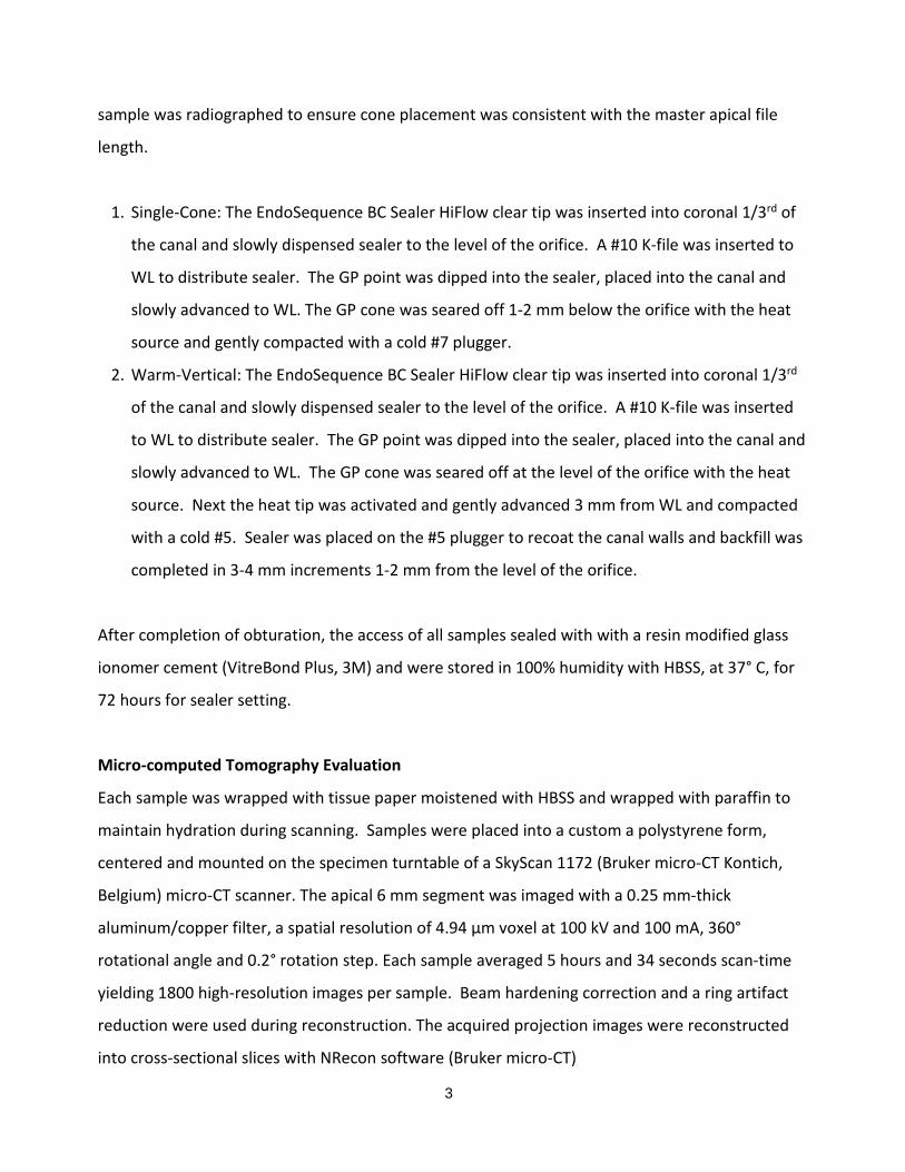
1. Single-Cone: The EndoSequence BC Sealer HiFlow clear tip was inserted into coronal 1/3rd of
the canal and slowly dispensed sealer to the level of the orifice. A #10 K-file was inserted to
WL to distribute sealer. The GP point was dipped into the sealer, placed into the canal and
slowly advanced to WL. The GP cone was seared off 1-2 mm below the orifice with the heat
source and gently compacted with a cold #7 plugger.
2. Warm-Vertical: The EndoSequence BC Sealer HiFlow clear tip was inserted into coronal 1/3rd
of the canal and slowly dispensed sealer to the level of the orifice. A #10 K-file was inserted
to WL to distribute sealer. The GP point was dipped into the sealer, placed into the canal and
slowly advanced to WL. The GP cone was seared off at the level of the orifice with the heat
source. Next the heat tip was activated and gently advanced 3 mm from WL and compacted
with a cold #5. Sealer was placed on the #5 plugger to recoat the canal walls and backfill was
completed in 3-4 mm increments 1-2 mm from the level of the orifice.
After completion of obturation, the access of all samples sealed with with a resin modified glass
ionomer cement (VitreBond Plus, 3M) and were stored in 100% humidity with HBSS, at 37° C, for
72 hours for sealer setting.
Micro-computed Tomography Evaluation
Each sample was wrapped with tissue paper moistened with HBSS and wrapped with paraffin to
maintain hydration during scanning. Samples were placed into a custom a polystyrene form,
centered and mounted on the specimen turntable of a SkyScan 1172 (Bruker micro-CT Kontich,
Belgium) micro-CT scanner. The apical 6 mm segment was imaged with a 0.25 mm-thick
aluminum/copper filter, a spatial resolution of 4.94 µm voxel at 100 kV and 100 mA, 360°
rotational angle and 0.2° rotation step. Each sample averaged 5 hours and 34 seconds scan-time
yielding 1800 high-resolution images per sample. Beam hardening correction and a ring artifact
reduction were used during reconstruction. The acquired projection images were reconstructed
into cross-sectional slices with NRecon software (Bruker micro-CT)
4
The canal, obturation material and porosity was calculated in CTAn software (Bruker micro-CT) by
a custom 15-step algorithm developed collaboration with a Micro Photonics software engineer.
The percentage of porosity in the total obturation material was evaluated from the working
length to the 6-mm level of each root by selecting a volume of interest (VOI, canal volume). The
volume as a percentage of total canal, obturations material (gutta-percha and sealer) and porosity
(internal and external) was calculated. Closed porosity is a void entirely contained within the
obturation material whereas open porosity is a void exposed to the canal wall dentin.
Statistical Analysis
Statistical analysis was performed using SPSS version 22 Software (IBM SPSS Inc, Chicago, IL) at a
95% confidence level (P= .05). The data did not show a normal distribution (% open porosity), and
the Mann-Whitney U test for nonparametric data was used for group segment comparison.
Results
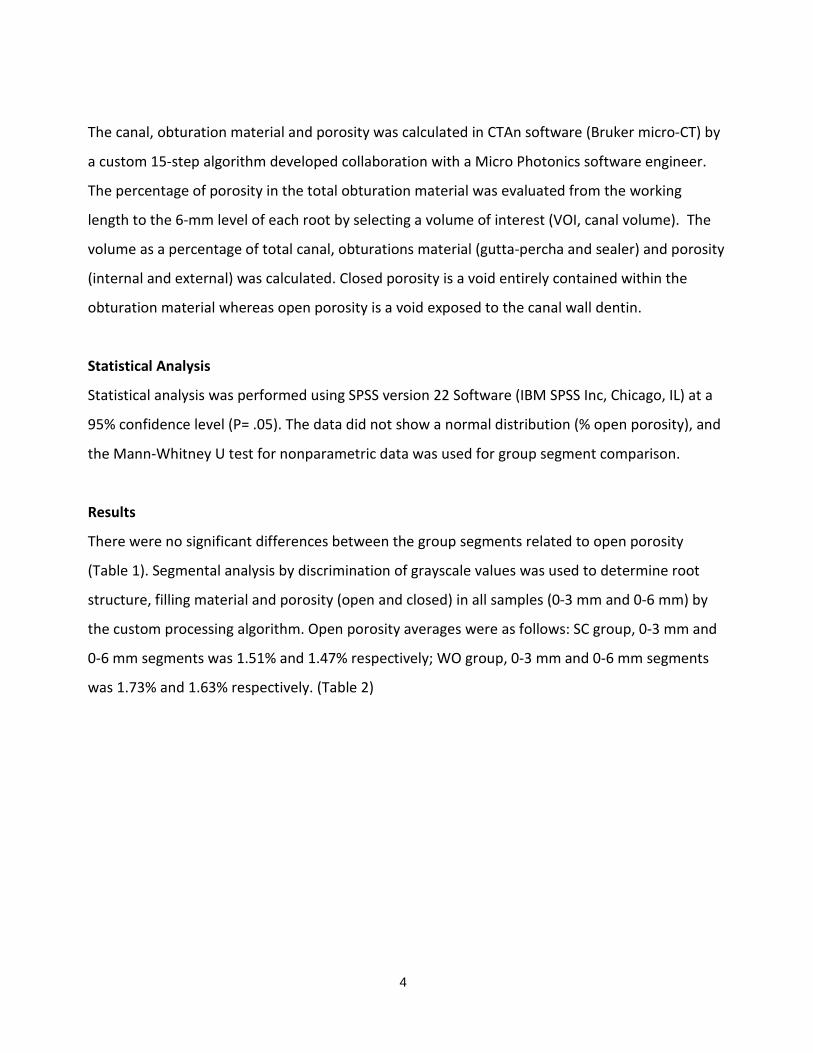
There were no significant differences between the group segments related to open porosity
(Table 1). Segmental analysis by discrimination of grayscale values was used to determine root
structure, filling material and porosity (open and closed) in all samples (0-3 mm and 0-6 mm) by
the custom processing algorithm. Open porosity averages were as follows: SC group, 0-3 mm and
0-6 mm segments was 1.51% and 1.47% respectively; WO group, 0-3 mm and 0-6 mm segments
was 1.73% and 1.63% respectively. (Table 2)
5
Table 1
Table 2
Discussion
There are many ways to evaluate root canal sealers and root filling materials which alter the
physical specimen, and are often conducted by means of dye leakage, pushout bond strength,
fluid filtration, destructive visual evaluation. Evaluation of the sealer and obturation quality by
non-destructive methods is ideal as it does not physically alter the specimen. Micro-CT imaging
has the ability non-destructively yield extremely detailed information.
0
0.5
1
1.5
2
2.5
3
3.5
4
Closed Porosity (%) Open Porosity (%) Total Porosity %
Percent Porosity
Single-Cone Group 0-3 Warm Vertical Group 0-3
Single-Cone Group 0-6 Warm Vertical Group 0-6
0
0.5
1
1.5
2
2.5
3
3.5
0-3mm 0-6mm
Percent Open Porosity
Single-Cone Warm Vertical
6
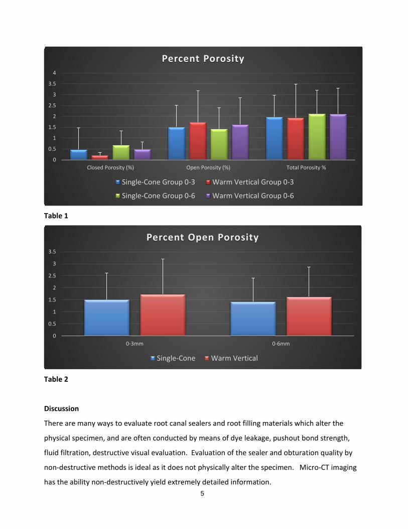
To determine our resolution quality, we tested resolutions similar to historical micro-CT
obturation studies, which ranged from 8.99-17.66 µm voxel (Fig. 1)11-13. To most accurately
threshold of grayscale values, 4.94 µm voxel size demonstrated optimal, well-defined
discrimination between dentin, obturation material and porosity. Then the focus was narrowed to
the apical 6 mm, which allowed for evaluation of the downpack/backfill junction, as well as the
apical 3 mm Kim et al14 describes to contain 98% of apical ramifications. With a clear visualization
of sealer and GP, individual group characteristics were observed.
Figure 1
In our findings, both groups performed almost identically across the groups relating to open
porosity, but our visual observations of the micro-CT images demonstrated very different
behaviors in the SC and WV groups. The SC samples demonstrated GP cones in line with the canal
surrounded by the HF sealer. The WV samples demonstrated a limited 3-dimentional GP fill
(approximately 1 mm) apical to the downpack/backfill junction, then, depending on the canal
morphology, varying degrees of cone deformation surrounded by the HF sealer. Low heat 150° C
is advocated by the manufacturer when the WV technique is utilized. At the sealer to GP
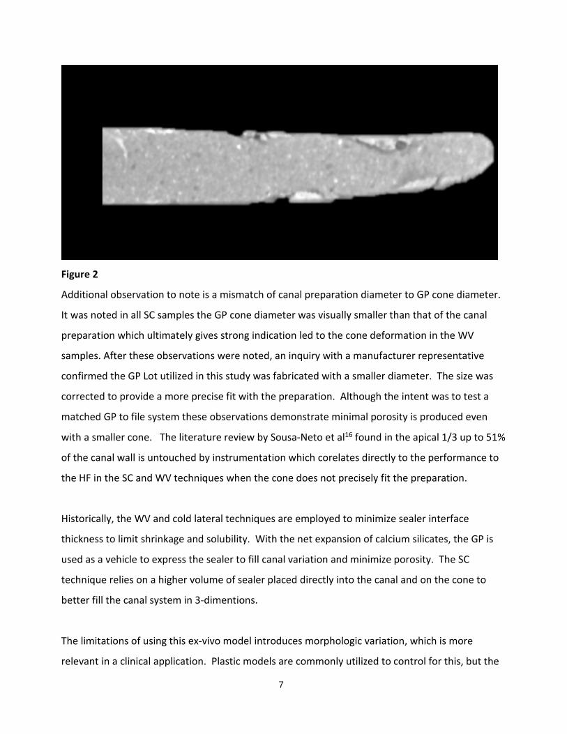
interface voids, gaps and sealer/GP was noted (Fig. 2) primarily at the inside curvature of the GP
cone. These observations show the limited GP flow a WV technique has at the 150° C compared
to the 3-dimensional dense GP fill Schilder describes in 196715.
17.6 µm 360°
13.44 µm 360°
8.99 µm 360°
4.94 µm 360°
7
Figure 2
Additional observation to note is a mismatch of canal preparation diameter to GP cone diameter.
It was noted in all SC samples the GP cone diameter was visually smaller than that of the canal
preparation which ultimately gives strong indication led to the cone deformation in the WV
samples. After these observations were noted, an inquiry with a manufacturer representative
confirmed the GP Lot utilized in this study was fabricated with a smaller diameter. The size was
corrected to provide a more precise fit with the preparation. Although the intent was to test a
matched GP to file system these observations demonstrate minimal porosity is produced even
with a smaller cone. The literature review by Sousa-Neto et al16 found in the apical 1/3 up to 51%
of the canal wall is untouched by instrumentation which corelates directly to the performance to
the HF in the SC and WV techniques when the cone does not precisely fit the preparation.
Historically, the WV and cold lateral techniques are employed to minimize sealer interface
thickness to limit shrinkage and solubility. With the net expansion of calcium silicates, the GP is
used as a vehicle to express the sealer to fill canal variation and minimize porosity. The SC
technique relies on a higher volume of sealer placed directly into the canal and on the cone to
better fill the canal system in 3-dimentions.
The limitations of using this ex-vivo model introduces morphologic variation, which is more
relevant in a clinical application. Plastic models are commonly utilized to control for this, but the
8
water required for setting the sealer is acquired from the dentin and cannot be replicated in a
plastic model.
More studies are needed to analyze the properties changes of the HF sealer when exposed to
heated instruments, how other file systems with matched GP would perform, as well as how
different sealer placement methods may minimize porosity.
Conclusions
The focused of the current study was to examine the performance differences between the SC
and WV techniques related to sealer porosity which leaved dentinal tubules unsealed. There is a
lake of evidence how porosity relates to clinic success and such studies would be challenging and
subject of a high degree of uncontrolled variables. Within the limitations of this study, micro-CT
imaging and volumetric analysis shows both techniques produced statistically similar performance
relating to open porosity but both but were unable to produce a void-free

9
Bibliography:
1. Whitworth J. Methods of filling root canals: Principles and practices. Endodontic Topics. 2005;12(1):2-24
2. Grossman LI. Rationale of endodontic treatment. Dent Clin North Am. 1967:483-490.
3. Hovland EJ, Dumsha TC. Leakage evaluation in vitro of the root canal sealer cement sealapex. In Endod J. 1985;18(3):179-182.
4. Kim S, Kratchman S. Modern endodontic surgery concepts and practice: A review. Journal of Endodontics. 2006;32(7):601-623.
5. Ørstavik D, Nordahl I, Tibballs JE. Dimensional change following setting of root canal sealer materials. Dental Materials. 2001;17(6):512-519.
6. Schilder H. Filling root canals in three dimensions. 1967. Journal of endodontics. 2006;32(4):281.
7. Almeida S, Helena L. Are premixed calcium silicate–based endodontic sealers comparable to conventional materials? A systematic review of In Vitro studies. Journal of Endodontics. 2016;43(4):527-535.
8. Zhou, Hui-min, PhD|Shen, Ya, DDS, PhD|Zheng, Wei, PhD|Li, Li, PhD|Zheng, Yu-feng, PhD|Haapasalo, Markus, DDS, PhD. Physical properties of 5 root canal sealers. Journal of Endodontics. 2013;39(10):1281-1286.
9. Iglecias, E, PhD|Freire, L, PhD|de M, George T, PhD|dos San, M, PhD|Antoniazzi, J, PhD|Gavini, G. Presence of voids after continuous wave of condensation and single-cone obturation in mandibular molars: A micro-computed tomography analysis. Journal of Endodontics. 2016;43(4):638-642.
10. Ricucci D, Bergenholtz G. Bacterial status in root-filled teeth exposed to the oral environment by loss of restoration and fracture or caries – a histobacteriological study of treated cases. Int Endod J. 2003;36(11):787-802.
11. Iglecias, E, PhD|Freire, L, PhD|de M, George T, PhD|dos San, M, PhD|Antoniazzi, J, PhD|Gavini, G. Presence of voids after continuous wave of condensation and single-cone obturation in mandibular molars: A micro-computed tomography analysis. Journal of Endodontics. 2016;43(4):638-642.
12. Celikten B, Uzuntas CF, Orhan AI, et al. Evaluation of root canal sealer filling quality using a single-cone technique in oval shaped canals: An in vitro Micro-CT study. Scanning. 2016;38(2):133-140.
13. Alshehri M, Alamri HM, Alshwaimi E, Kujan O. Micro-computed tomographic assessment of quality of obturation in the apical third with continuous wave vertical compaction and single match taper sized cone obturation techniques. Scanning. 2016;38(4):352-356.

10
14. Kim S, Kratchman S. Modern endodontic surgery concepts and practice: A review. Journal of Endodontics. 2006;32(7):601-623.
15. Schilder H. Filling root canals in three dimensions. 1967. Journal of endodontics. 2006;32(4):281.
16. Sousa-Neto MDd, Silva-Sousa YC, Mazzi-Chaves JF, et al. Root canal preparation using micro-computed tomography analysis: A literature review. Brazilian oral research. 2018;32(suppl 1):e66.