michelle l. ischayek d.o. emergency medicine resident aria ... · extremities: no clubbing,...
TRANSCRIPT
Michelle L. Ischayek D.O.
Emergency Medicine Resident Aria Health
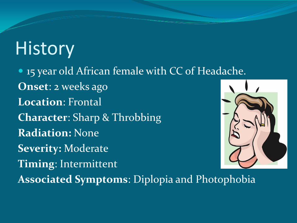
History 15 year old African female with CC of Headache. Onset: 2 weeks ago Location: Frontal Character: Sharp & Throbbing Radiation: None Severity: Moderate Timing: Intermittent Associated Symptoms: Diplopia and Photophobia
History
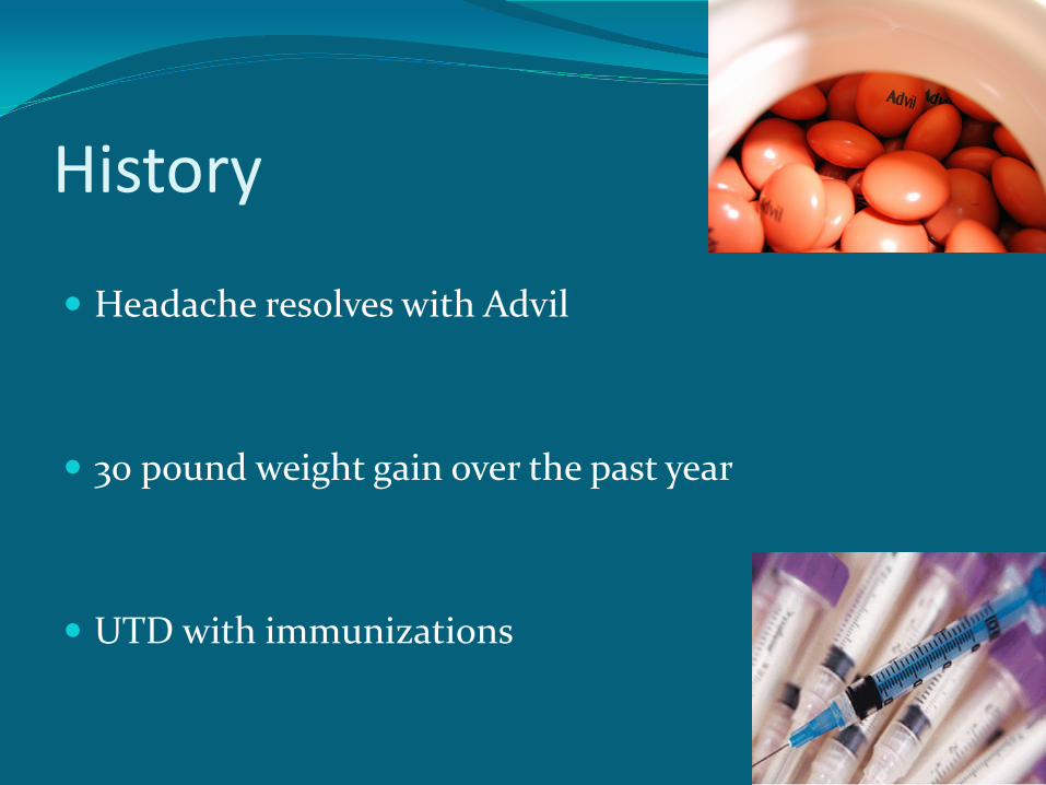
Headache resolves with Advil
30 pound weight gain over the past year UTD with immunizations
History Past Medical Hx: Denies
Family Hx: Denies
Surgical Hx: Denies Social Hx: Denies ETOH or Drug Abuse, Lives with parents
Medications: Denies
Allergies: NKDA
Review of Systems Denies fever, chills, cough, nausea, vomiting, chest
pain, shortness of breath, myalgias or neck stiffness.
Denies previous history of headaches.
Denies recent travel. Complains of frontal headache, diplopia and
photophobia.
Physical Exam VITALS: T: 98.2 F BP: 128/72 P: 72 RR: 18 Oxygen Saturation: 97% on RA GENERAL APPEARANCE: Well-developed, well nourished, alert, cooperative,
no acute distress, generally well appearing. HEENT: mild right-sided esotropia, conjunctiva clear, no nystagmus. No
papilledema. TMs clear, mucous membranes good color.
NECK: - JVD, no neck tenderness. LUNGS: Clear to auscultation bilaterally, no wheezes, rales or rhonchi.
Physical Exam HEART: Regular rate and rhythm, no murmurs, gallups or rubs. ABDOMEN: soft, non-tender, non-distended, bowel sounds x 4, no rebound, no
guarding.
EXTREMITIES: no clubbing, cyanosis or edema. 2+ radial pulses bilaterallly.
SKIN: no rashes noted.
NEURO: right-sided abducens nerve palsy, muscle strength 5/5 in bilateral UE and LE.
Sensation intact in bilateral UE and LE. Finger to nose normal, normal gait, cerebellar function intact.
Labs
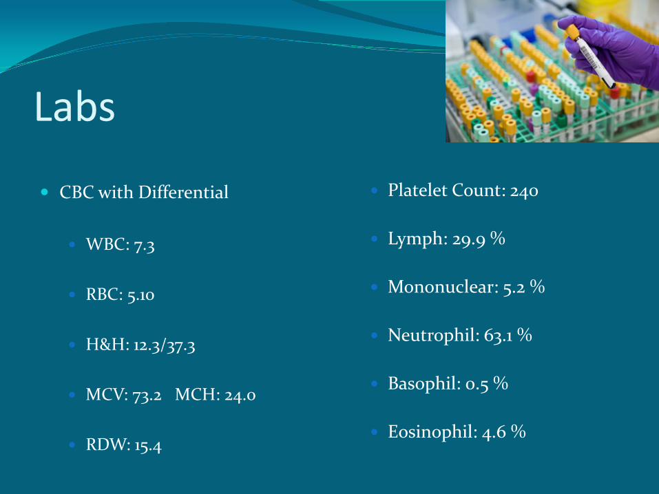
CBC with Differential WBC: 7.3
RBC: 5.10
H&H: 12.3/37.3
MCV: 73.2 MCH: 24.0
RDW: 15.4
Platelet Count: 240
Lymph: 29.9 %
Mononuclear: 5.2 %
Neutrophil: 63.1 %
Basophil: 0.5 %
Eosinophil: 4.6 %
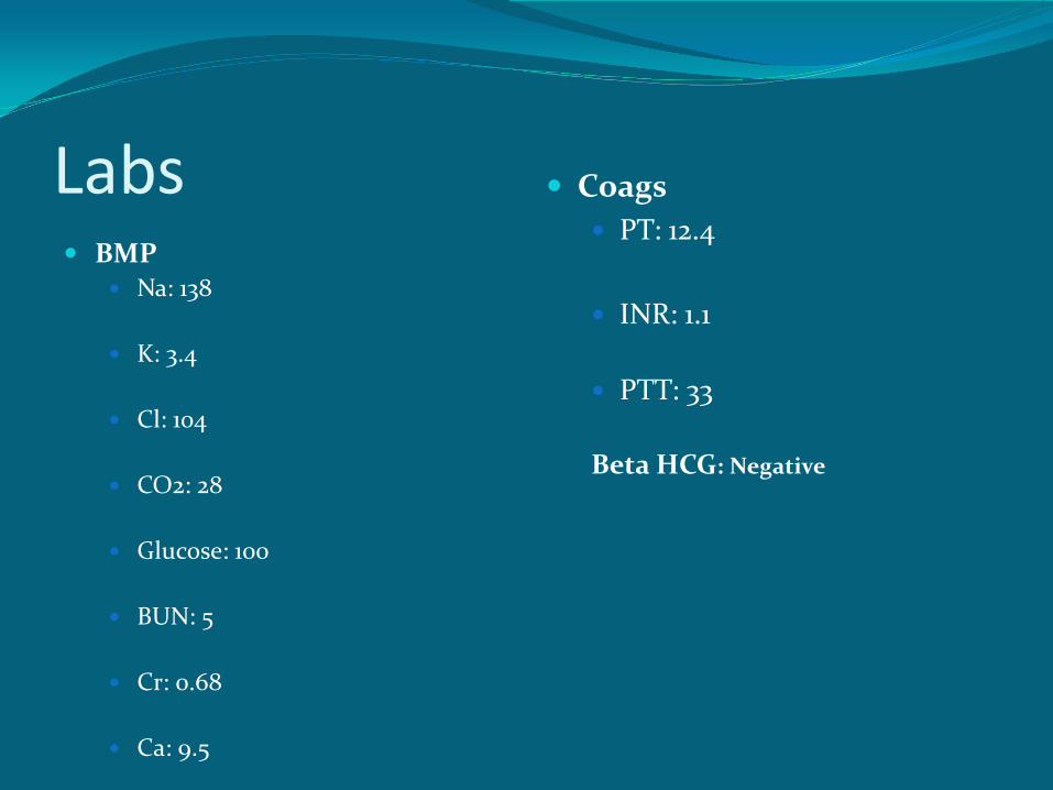
Labs
BMP Na: 138 K: 3.4 Cl: 104 CO2: 28 Glucose: 100 BUN: 5 Cr: 0.68 Ca: 9.5
Coags PT: 12.4 INR: 1.1
PTT: 33
Beta HCG: Negative
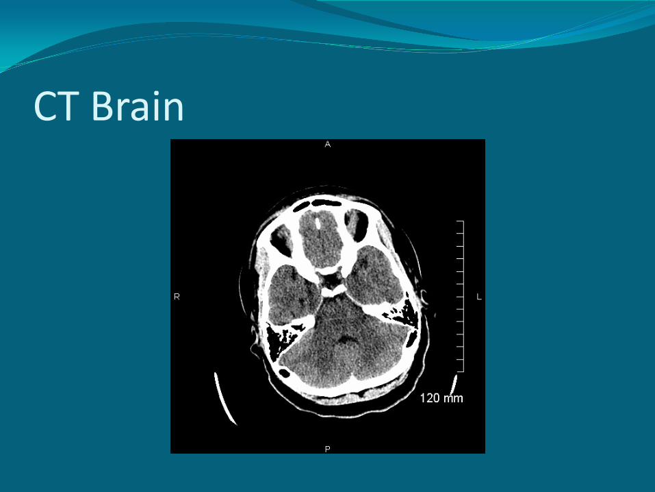
CT Brain
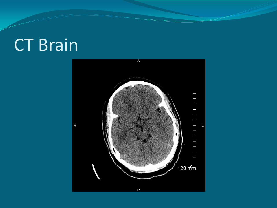
CT Brain
Transfer Patient was transferred to a pediatric emergency
department for further diagnostic workup.
What Is Your Diagnosis???

Introducing
Faculty Discussant:
Dr. Joseph Dougherty, D.O.
Ohio Valley Health System
Michelle L. Ischayek D.O.
Emergency Medicine Resident Aria Health
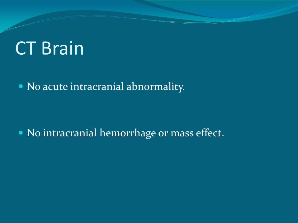
CT Brain No acute intracranial abnormality. No intracranial hemorrhage or mass effect.
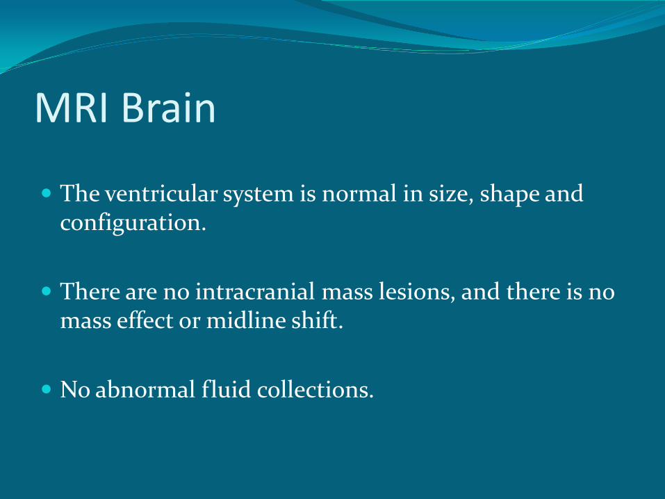
MRI Brain The ventricular system is normal in size, shape and
configuration. There are no intracranial mass lesions, and there is no
mass effect or midline shift. No abnormal fluid collections.
Intracranial MRV The major intracranial venous structures demonstrate
normal flow-related enhancement. There is no MRV evidence for deep venous sinus
thrombosis.
Lumbar Puncture
Elevated Opening Pressure of 35 cm H2O.
Normal CSF.
15 year old African Female CC: Frontal headache, 2 weeks. Sx: Diplopia & Photophobia. PE: Right-sided Esotropia LP: Elevated opening pressure
Brain Imaging: Negative Dispo: Admitted to pediatric facility with…
Diagnosis
Idiopathic Intracranial Hypertension (IIH)
IIH Pseudotumor Cerebri, “false brain tumor,” benign
Intracranial Hypertension. Increased intracranial pressure Normal CSF Absence of tumor Not a benign disorder
Epidemiology Annual incidence is 1-2 per 100,000 population. Higher incidence in obese women between 15 and 44
years. Males and children whom are not overweight affected
too.
Associated Conditions Systemic Diseases
Hereditary Conditions
Vitamin Deficiencies Medications
Pathogenesis Exact Pathogenesis unknown Theories: Abnormalities of cerebral venous outflow tract Increased CSF outflow resistance Increased abdominal and intracranial venous pressure Sodium and water retention Abnormal Vitamin A metabolism
Signs & Symptoms Headache
Transient Visual Obscurations
Pulsatile Tinnitus
Photopsia
Retrobulbar pain
Diplopia
Sustained Visual Loss
Neck Stiffness
Signs & Symptoms Headache is the most common presenting symptom. Nausea and vomiting.
Exacerbated by changes in posture, sneezing or
coughing. Improves with rest or NSAIDs.
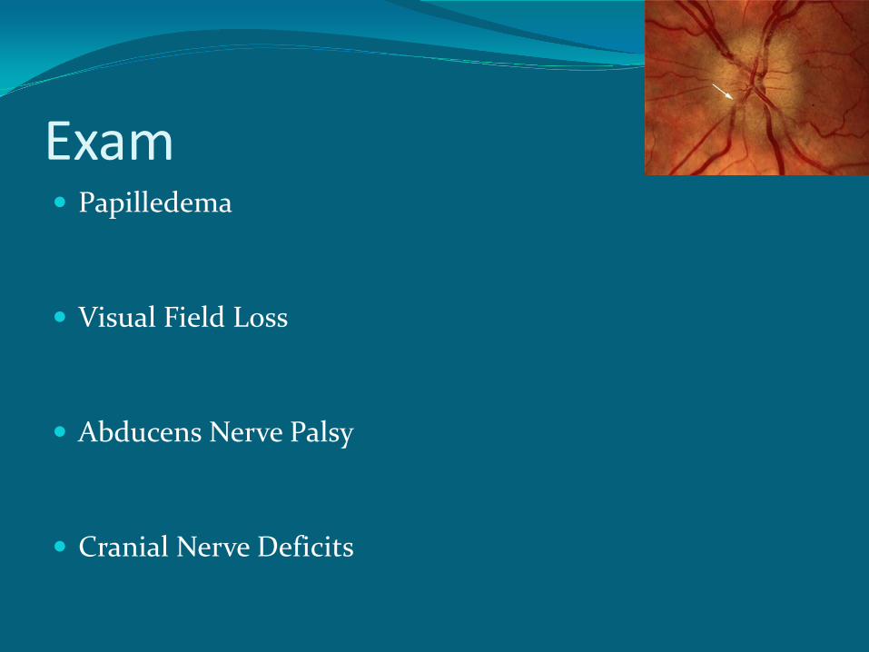
Exam Papilledema Visual Field Loss Abducens Nerve Palsy
Cranial Nerve Deficits
Differential Diagnosis Mass-Tumor, Abscess
Hydrocephalus Cerebral venous thrombosis Choroid plexus papilloma
Diagnosis Modified Dandy Criteria: Increased ICP: headache, transient visual loss,
tinnitus, and papilledema. No other neurologic abnormalities or impaired level of
consciousness. Neuroimaging study that shows no etiology for
intracranial hypertension. No other cause of intracranial hypertension. Elevated intracranial pressure with normal CSF.
Neuroimaging
MRI is the test of choice. If MRI contraindicated, CT Brain.
Lumbar Puncture If neuroimaging negative, perform LP. During LP, measure opening pressure and evaluate CSF cell
count, glucose and protein. Upper limit of normal opening pressure in adults is 20 cm
H20. Pressures can be as high as 28 cm H20 if patient is curled up in lateral decubitus position.
In young children, upper limit of opening pressure is 25 cm
H20.
Prognosis Can last months to years. Slow, gradual onset. With treatment, gradual improvement and stabilization. Permanent vision loss, major morbidity. Fulminant IIH, experience visual loss within a few weeks of
symptom onset.
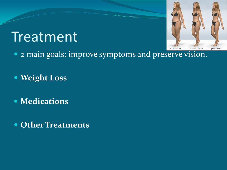
Treatment 2 main goals: improve symptoms and preserve vision.
Weight Loss Medications Other Treatments
Surgical Treatment Indications: Failed medical treatments Visual field defects Visual Acuity loss Intractable headaches Noncompliance
Surgical Procedures: Optic nerve sheath fenestration and
CSF shunting.
Back to Our Patient Hospital Course: Diagnosed with IIH Encouraged to lose weight Discharged on Diamox 250mg BID x 1 week then
500mg BID (total of 274 days) Follow up with Neurology and Ophthalmology
Thank You!
References 1. http://www.uptodate.com/contents/idiopathic-intracranial-
hypertension-pseudotumor-cerebri-clinical-features-and-diagnosis/abstract/8?utdPopup=true
2. Arsava EM, Uluc K, Nurlu G, Kansu T. Electrophysiological evidence of trigeminal neuropathy in pseudotumor cerebri. J Neurol 2002; 249:1601.
3. Beri S, Gosalakkal JA, Hussain N, et al. Idiopathic intracranial hypertension without papilledema. Pediatr Neurol 2010; 42:56.
4. Capobianco DJ, Brazis PW, Cheshire WP. Idiopathic intracranial hypertension and seventh nerve palsy. Headache 1997; 3
5. Krishna R, Kosmorsky GS, Wright KW. Pseudotumor cerebri sine papilledema with unilateral sixth nerve palsy. J Neuroophthalmol 1998; 18:53.
References 6. Malomo AO, Idowu OE, Shokunbi MT, et al. Non-
operative management of benign intracranial hypertension presenting with complete visual loss and deafness. Pediatr Neurosurg 2006; 42:62.
7. Vieira DS, Masruha MR, Gonçalves AL, et al. Idiopathic intracranial hypertension with and without papilloedema in a consecutive series of patients with chronic migraine. Cephalalgia 2008; 28:609.
8. Soler D, Cox T, Bullock P, et al. Diagnosis and management of benign intracranial hypertension. Arch Dis Child 1998; 78:89.
9. Selky AK, Dobyns WB, Yee RD. Idiopathic intracranial hypertension and facial diplegia. Neurology 1994; 44:357.