michael kyba et al- enhanced hematopoietic differentiation of embryonic stem cells conditionally...
TRANSCRIPT
8/3/2019 Michael Kyba et al- Enhanced hematopoietic differentiation of embryonic stem cells conditionally expressing Stat5
http://slidepdf.com/reader/full/michael-kyba-et-al-enhanced-hematopoietic-differentiation-of-embryonic-stem 1/7
Colloquium
Enhanced hematopoietic differentiation of embryonicstem cells conditionally expressing Stat5Michael Kyba†, Rita C. R. Perlingeiro†‡, Russell R. Hoover†§, Chi-Wei Lu†, Jonathan Pierce†, and George Q. Daley†¶
†Whitehead Institute for Biomedical Research, 9 Cambridge Center, Cambridge, MA 02142; and ¶Department of Biological Chemistry and MolecularPharmacology, Harvard Medical School, and Division of Pediatric HematologyOncology, Children’s Hospital and Dana–Farber Cancer Institute,Boston, MA 02115
The signal transducer Stat5 plays a key role in the regulation of
hematopoietic differentiation and hematopoietic stem cell func-tion. To evaluate the effects of Stat5 signaling in the earliest
hematopoietic progenitors, we have generated an embryonic stem
cell line in which Stat5 signaling can be induced with doxycycline.
Ectopic Stat5 activation at the point of origin of the hematopoietic
lineage (from day 4 to day 6 of embryoid body differentiation)
significantly enhances the number of hematopoietic progenitors
with colony-forming potential. It does so without significantly
altering total numbers or apoptosis of hematopoietic cells, sug-gesting a cell-intrinsic effect of Stat5 on either the developmental
potential or clonogenicity of this population. From day-6 embryoid
bodies, under the influence of Stat5 signaling, a population of
semiadherent cells can be expanded on OP9 stromal cells that is
comprised of primitive hematopoietic blast cells with ongoing,
mainly myeloid, differentiation. When these cells are injected into
lethally irradiated mice, they engraft transiently in a doxycycline-
dependent manner. These results demonstrate that the hemato-poietic commitment of embryonicstem cells may be augmented by
a Stat5-mediated signal, and highlight the utility of manipulating
individual components of signaling pathways for engineering
tissue-specific differentiation of stem cells.
hematopoiesis
When differentiated as suspension aggregates (embryoidbodies, EBs), embryonic stem (ES) cells will readily give
rise to differentiated hematopoietic cells (1) as well as colony-forming cells (CFCs) that can be assayed in secondary semisolidhematopoietic cultures (2). However, ES cell differentiationdoes not efficiently generate hematopoietic stem cells (HSCs)capable of repopulating the hematopoietic system of lethallyirradiated adult recipients. In this regard, ES cells recapitulatethe development of the earliest embryonic hematopoietic tissue,the yolk sac. Analysis of knockout mice has implicated severalgenes in theembryonic development of thedefinitive HSC(3–5);however, a detailed understanding of the extracellular signalsthat guidedevelopment fromthe pluripotentstate to thelineage-
restricted HSC state is lacking.In an effort to evaluate signals that promote hematopoietic
differentiation of pluripotent cells, as well as the generation of cells with hematopoietic repopulating potential, we sought totest the effects of Stat5 activation during in vitro differentiationof ES cells. Stats are cytoplasmic signal transducers that arerecruited by ligand-activated receptors via Src homology 2-mediated interactions with receptor-bound Janus kinases. Thisinteraction results in Stat protein phosphorylation, disengage-ment, dimerization, and translocation to the nucleus where theseproteins then function as a transcription factors, binding to andactivating the transcription of target genes (6). Stat5 is encodedby two genes, Stat5a and Stat5b, with 95% sequence identity (7)and is activated by engagement of numerous hematopoietic and
nonhematopoietic receptors (8–11). Stat5 signaling has beenimplicated in cellular proliferation (12, 13), resistance to apo-ptosis (14–16), and differentiation (17, 18).
Mice genetically null for both Stat5a and Stat5b displayobvious defects in response to growth hormone and prolactin(15) and subtler defects in embryonic hematopoietic develop-ment (14). Although definitive HSCs develop in Stat5 knockoutmice, they display a profound defect in competitive repopulation(19–21), suggesting that Stat5 may be interacting cooperatively
and redundantly with other signal transducers in HSC regula-tion. In the classic HSC pathology, chronic myeloid leukemia,regulation of proliferation is disruptedby theoncogene Bcr Abl,
with concomitant activation of Stat5 (22–25). Moreover, dom-inant negative Stat5 mutants can block transformation by Bcr
Abl (26), indicating that inappropriate activation of Stat5 canhave dramatic consequences for HSC regulation.
Because of the pivotal role of Stat5 signaling in hematopoiesisand HSC homeostasis, we selected this pathway for study duringthe earliest stages of hematopoietic specification in an in vitrosystem of ES cell differentiation. For this purpose, we havegenerated an ES cell line with a tetracycline-inducible, domi-nant-active allele of Stat5. We report that induction of Stat5signaling during EB development dramatically enhances hema-topoiesis. Furthermore, on OP9 stromal cell coculture, Stat5promotes the expansion of a blast cell population from day-6EBs. Cultures expanded in this way are rich in primitive,undifferentiated cells, w ith surface marker similarities to HSCs,and have the capacity to engraft lethally irradiated adult mice ina transient, Stat5-dependent manner.
Materials and Methods
Generation of Stat5CA Inducible ES Cells. The cDNA for theconstitutively active mutant of Stat5 (H299RS711F, a gift fromT. Kitamura, University of Tokyo, Tokyo) was subcloned on an EcoRI– NotI fragment from murine stem cell virus (MSCV)-Stat5CA-iresGFP (16) into pLox. This was then co-electropo-rated along with pSalk-CRE (a gift from S. O’Gorman, The SalkInstitute, San Diego) into the targeting cell line Ainv15. The
targeting ES cell line and targeting plasmid, pLox, have beendescribed (27). The resulting cell line was selected and expanded
This paper results from the Arthur M. Sackler Colloquium of the National Academy of
Sciences, ‘‘Regenerative Medicine,’’ held October 18–22, 2002, at the Arnold and Mabel
Beckman Center of the National Academies of Science and Engineering in Irvine, CA.
Abbreviations: ES, embryonicstem; EB, embryoid body;HSC, hematopoietic stemcell; CFC,
colony-forming cell; TRE, tetracycline response element; TPO, thrombopoietin; SCF, stem
cell factor; VEGF, vascular endothelial growth factor; IFS, inactivated fetal serum; MSCV,
murine stem cell virus; IMDM, Iscove’s modified Dulbecco’s medium.
‡Present address: ViaCell, Inc., 26 Landsdowne Street 580, Cambridge, MA 02139-4216.
§Present address:Vertex Pharmaceuticals,130 Waverly Street, Cambridge,MA 02139-4242.
To whom correspondence should be addressed. E-mail: [email protected].
© 2003 by The National Academy of Sciences of the USA
11904–11910 PNAS September 30, 2003 vol. 100 suppl. 1 www.pnas.orgcgidoi10.1073pnas.1734140100
8/3/2019 Michael Kyba et al- Enhanced hematopoietic differentiation of embryonic stem cells conditionally expressing Stat5
http://slidepdf.com/reader/full/michael-kyba-et-al-enhanced-hematopoietic-differentiation-of-embryonic-stem 2/7
in 400 gml G418 (Sigma). ES cells were maintained onirradiated mouse embryonic fibroblasts in DME15% inacti- vated fetal serum (IFS)0.1 mM nonessential amino acids(GIBCO/BRL)2 mM glutamine50 units/ml penicillin50g/ml streptomycin (GIBCO/BRL)0.1 mM 2-mercaptoethanol(Sigma)1,000 units/ml leukemia inhibitory factor (PeproTech,Boston). To induce Stat5CA expression in ES cells, 2 gmldoxycycline (Sigma) was added to the culture medium.
Bandshift Assay. To generate whole cell extracts, cells were washed in ice-cold PBS and lysed in EMSA lysis buffer (150 mMNaCl20 mM TrisHCl, pH 7.41 mM EDTA10 mM Na3VO41mM MgCl21% Nonidet P-401 mM phenylmethyl-sulfonylfluoride10% glycerol) on ice for 10 min. Cells and lysate werescraped with a sterile cell scraper, collected, and spun for 10 min
at max at 4°C on a benchtop centrifuge. Complimentary oligo-nucleotides that contained a Stat5 consensus binding site fromthe -casein promoter (5-AGATTTCTAGGAATTCAATCC-3) were annealed and radiolabeled with [ -32P]ATP by using T4polynucleotide kinase (New England Biolabs). Approximately20,000 cpm (0.2 ng) of probe was incubated with 20 g of wholecell extract in 20 l of 10 mM Hepes, pH 7.950 mM KCl0.2mM DTT10% glycerol0.05% Nonidet P-401 g poly(dIdC)(Sigma) for 20 min at room temperature. Resulting DNAprotein complexes were resolved on a 5% nondenaturing poly-acrylamide gel.
EB Differentiation. ES cells were trypsinized, collected in EBD[Iscove’s modified Dulbecco’s medium (IMDM)15% IFS200g/ml iron-saturated transferrin (Sigma)4.5 mM monothioglyc-
erol (Sigma)50 g/ml ascorbic acid (Sigma)2 mM glutamine]and plated onto fresh T25 flasks (Corning) for 45 min to allowmouse embryonic fibroblasts to adhere. Nonadherent cells werecollected and plated in hanging drops at 100 cells per 10-l dropin an inverted bacterial Petri dish, and cultured for 2 days. They
were then collected from the hanging drops and further culturedin 10 ml of EBD in slowly rotating 10-cm bacterial Petri dishes.
At day 4, EBs were fed by exchanging half of their spent mediumfor fresh EBD. In some cultures, doxycycline was added at day4 at 2 gml to induce expression of Stat5CA.
CFC Assay. Day-6 EBs were dissociated by trypsinization, col-lected, and resuspended in IMDM10% IFS at a concentrationof 5 105 cells per ml. A total of 100 l of this cell suspension
was added to 1.5 ml of complete methylcellulose for murinecolonies (StemCell Technologies, Vancouver, catalog no. 3434).Methylcellulose suspension cultures werenot supplemented withdoxycycline. EryP colonies were counted on day 6 of methyl-cellulose culture, all other colonies were counted at day 10.
OP9 Coculture. Day-6 EBs were trypsinized to a single cellsuspension and plated on a semiconfluent monolayer of OP9cells (a gift of T. Nakano, University of Osaka, Osaka) at adensity of 200,000 cells per well of a six-well dish in IMDM, 10%IFS supplemented with 2 mM glutamine, 50 unitsml penicillin,50 gml streptomycin (GIBCOBRL), 0.1 mM 2-mercapto-ethanol (Sigma), cytokines [40 ngml vascular endothelialgrowth factor (VEGF), 40 ngml thrombopoietin (TPO), 100ngml stem cell factor (SCF), and 100 ngml Flt-3 ligand], and
doxycycline at 1 gml. When cells became confluent, they werepassaged by trypsinization onto fresh OP9.
Fluorescent Staining and FACS Analysis. Staining of day-6 EB cells. EBs were disaggregated by washing once with PBS followed byresuspension in 0.1% trypsinPBS and pipetting for 30 s. Trypsin
was blocked with IMDM10% IFS, the cells were strained toremove clumps and collected by centrifugation. Annexin V–phycoerythrin staining was done at room temperature for 15 minaccording to the manufacturer’s specifications (CLONTECH).
After staining, cells were transferred to 4°C, and 1 l of FITC-conjugated CD41 antibody was added. After 20 min,samples were diluted with annexin V binding buffer containingpropidium iodide and analyzed by FACS.Staining of OP9 cultures. Cells were collected by tr ypsinization and
resuspended in PBS containing 5% IFS. Samples of one millioncells in 100 l were blocked with 1 l of Fc block (PharMingen)and stained with 1 l of phycoerythrin- or FITC-conjugatedantibody for 20 min at 4°C. Samples were washed twice withPBS5% IFS, and resuspended in PBS5% IFS containingpropidium iodide. All antibodies were purchased from Phar-Mingen. FACS analyses were per formed on a Becton DickinsonFACSCalibur. Dead cells were excluded from phycoerythrin-stained cells by gating on FL2 vs. FL3.
RT-PCR. The following primers were used: actin(f) 5-GT-GGGGCGCCCCAGGCACCA-3; actin(r) 5-CTCCTTAATGT-CACGCACGATTTC-3; -H1(f) 5-AGTCCCCATGGAGT-CAAAGA-3; -H1(r) 5-CTCAAGGAGACCTTTGCTCA-3;
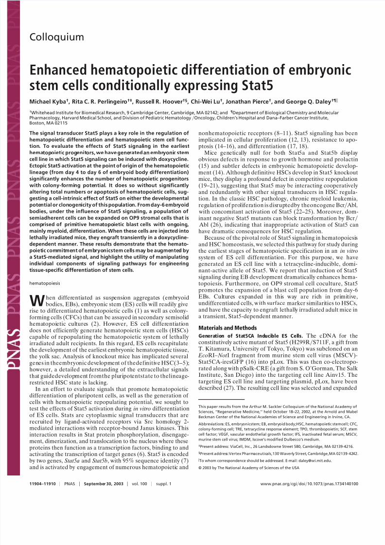
Fig. 1. Generation of the Stat5-inducibleES cellline. (a) Integration of pLoxStat5CA into theLoxPsite on theX chromosome of Ainv15ES cells placesthe cDNA
for Stat5CAunder thecontrolof the tetracycline response element.Recombination between thechromosomal andplasmidLoxP sites, denoted by , ismediated
by transient expression of Cre recombinase. Reconstitution of neo gene function by the promoter-ATG sequence 5 to the loxP site allows for selection of
successfulintegration events. rtTA, reversetetracycline transactivator.(b) Exposureof iStat5CAES cellsto doxycycline, butnot theparental celllineAinv15,results
in the bandshift of a probe containing Stat5 binding sites. Arrowhead denotes the Stat5-specific bandshift.
Kyba et al. PNAS September 30, 2003 vol. 100 suppl. 1 11905
8/3/2019 Michael Kyba et al- Enhanced hematopoietic differentiation of embryonic stem cells conditionally expressing Stat5
http://slidepdf.com/reader/full/michael-kyba-et-al-enhanced-hematopoietic-differentiation-of-embryonic-stem 3/7
-maj(f) 5-CTGACAGATGCTCTCTTGGG-3; -maj(r) 5-CACAACCCCAGAAACAGACA-3. Cycle conditions were asfollows: 2 min at 96°C; 30 cycles of 45 s at 95°C, 1 min at 60°C, and45 s at 72°C; and then 5 min at 72°C.
Retroviral Labeling with GFP. GFP retroviral supernatants wereproduced by FUGENE transfection of 293 cells with pMSCVi-resGFP (28) and pCL-Eco, a packaging-defective helper plasmid(29). 293 cells were grown in DME10% IFS, and medium was
replaced on the day after transfection. Forty-eight hours aftertransfection, supernatants were collected, filtered, plated ontoiStat5CA blast cells growing on OP9 at 3 ml per well of a six-welldish, supplemented with 4 gml polybrene and cytokines (100ngml SCF, 40 ngml VEGF, 40 ngml TPO, 100 ngml Flt-3ligand), and spin-infected at 2,500 rpm for 90 min in a BeckmanGS-6R centrifuge. After several days of growth, GFP-positivecells were separated by FACS and cultured on fresh OP9.Filtered supernatants from GFP-positive cells expanded on OP9
were tested for lateral transfer to 10T12 cells and found to benegative.
Mouse Transplantation. Two- to four-month-old 129OlaHsd(Harlan Breeders, Indianapolis) mice were exposed to 2 500cGy of -irradiation, separated by 4 h, and injected with 1.75
106
cells in 500 l of IMDM10% IFS via lateral tail vein. Micethat received doxycycline were provided drinking water supple-mented with 500 gml doxycycline hydrochloride (Sigma) and5% sucrose.
Results
Stat5 Signaling During EB Development. To generate an ES cell line with inducible Stat5 signaling, we made use of the Tet-Ontargeting cell line, Ainv15 (27). These cells constitutively expressthe reverse tetracycline transactivator from the ROSA26 locus,and have a tetracycline response element (TRE) integrated intothe transcriptionally open chromatin 5 to the HPRT gene on theX chromosome. Downstream of the TRE is a single LoxP site,into which we integrated, by Cre–Lox recombination, the cDNAfor Stat5CA, a constitutively active mutant of Stat5a (a double
mutant of H299R and S711F, also known as 1*6) (12) (Fig. 1 a).The resulting ES cell line, named iStat5CA, as well as itsdifferentiated progeny, express this mutant cDNA when exposedto doxycycline. Expression results in binding of Stat5CA to DNAas measured by bandshifting activity against an oligonucleotideprobe encoding a Stat5 DNA-binding consensus sequence, in thelysate of doxycycline-treated iStat5CA ES cells (Fig. 1 b).
We used this cell line to generate EBs and applied doxycyclineto the cultures for 48 h, from day 4 to day 6 of differentiation.This time window was chosen based on the kinetics of colonyformation in EBs: the bipotent precursor to the hematopoieticand endothelial lineages, the hemangioblast, peaks at day 3.75and is no longer present by day 5 (30), whereas hematopoieticCFC, particularly mixed erythroid–myeloid colonies, first be-come detectable between days 5 and 6. Thus, Stat5 signaling was
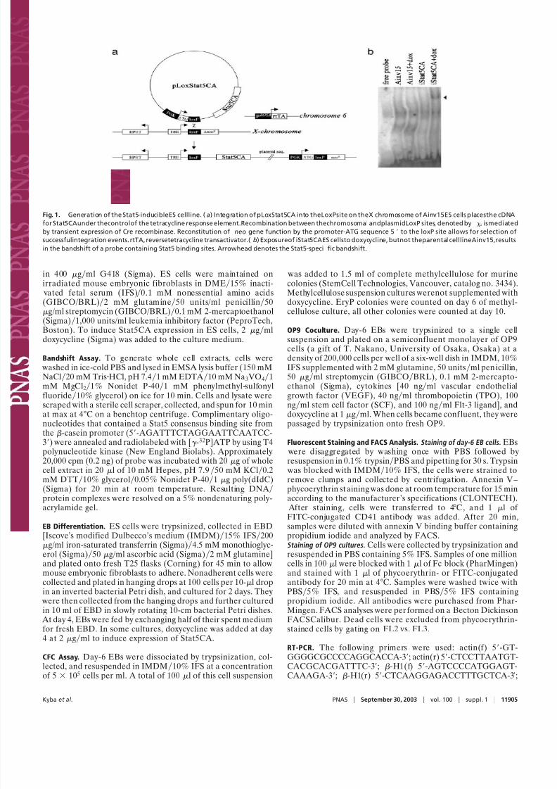
induced at the time of specification of the hematopoietic lineage. At day 6, the EBs were disaggregated into single cells andassayed for hematopoietic CFC content by plating in methylcel-lulose suspension medium with hematopoietic cytokines. Asshown in Fig. 2 a, exposure of EBs to doxycycline increased thenumbers of all types of hematopoietic colonies assayed between2- and 4-fold.
To investigate the mechanism of Stat5-mediated hematopoi-etic enhancement, we analyzed cells from dox ycycline-treated oruntreated day-6 EBs for apoptosis. Preliminary results (notshown) demonstrated that Stat5 activation modestly decreasedthe number of apoptotic (annexin V-positive) cells from day-6EBs; however, a general reduction in apoptosis would increaseCFC frequency only if the hematopoietic lineage were subject to
higher levels of apoptosis than the other nonhematopoieticlineages that arise in a day-6 EB. To assay the levels of apoptosisin hematopoietic vs. nonhematopoietic cells, we stained EB cells
with both annexin V and a pan-hematopoietic antibody. Studiesof adult hematopoiesis commonly use the CD45 antigen as apan-hematopoietic marker; however, this marker is not univer-sally expressed by the earliest embryon ic hematopoietic progen-itors. The recent discovery that the adult megakaryocytic anti-gen CD41 is actually pan-hematopoietic in ver y early embryos as
well as in day-6 EBs (31, 32) prompted us to use this markerrather than CD45 for this purpose. To our surprise, this assayrevealed that apoptosis in day-6 EBs is almost completelyrestricted to the nonhematopoietic population (Fig. 2 b).
Fig. 2. Effects of Stat5 signaling during EB differentiation. EBs were grown
for 6 days, either exposed or not exposed to doxycycline from day 4 to day 6.
(a) CFC assay: day-6 EB cells were disaggregated and plated into methylcellu-
lose suspension culture with hematopoietic cytokines. Filled bars denote
colony number from doxycycline-treated EBs; open bars denote colony num-
ber from untreated EBs. Standard errors for three independent experiments
are shown. (n 3 for each bar; P 0.05 for combined CFCs.) (b) Apoptosis
assay: day-6 EB cellswere disaggregated andstainedwith annexin V ( y axis)to
label apoptotic cells and anti-CD41 ( x axis) to label hematopoietic cells. The
percentage of cells falling into single- and double-positive quadrants is
shown. (c) Hematopoietic compartment quantitation: day-6 EB cells were
disaggregated and stained with antibodies to c-Kit ( y axis) and CD41 ( x axis).
The percentage of cells falling within the double-positive rectangular gate is
shown.
11906 www.pnas.orgcgidoi10.1073pnas.1734140100 Kyba et al.

8/3/2019 Michael Kyba et al- Enhanced hematopoietic differentiation of embryonic stem cells conditionally expressing Stat5
http://slidepdf.com/reader/full/michael-kyba-et-al-enhanced-hematopoietic-differentiation-of-embryonic-stem 4/7
An alternative to a reduction in apoptosis would be overpro-liferation of hematopoietic cells in response to Stat5. Becausehematopoietic CFCs from the day-6 EB are CD41 and c-Kitdouble positive (31, 32), we assayed the frequency of thispopulation in the presence vs. absence of Stat5 induction (Fig.2 c). We observed only a very modest increase with doxycyclinetreatment, which was not sufficient to account for the increasein CFCs. We therefore conclude that Stat5 activation is eitherinfluencing development within the hematopoietic compart-ment, such that it contains a higher ratio of CFCs to moredifferentiated cells, or enhancing the clonogenicity of the CFCs
that are present.
Stat5 Signaling During OP9 Stromal Cell Coculture of Day-6 EB Cells in
Vitro . Cells from day-6 EBs that had been exposed to doxycyclinefor 48 h were also plated on OP9 stromal cells with a cytokinemixture tailored for HSC expansion, consisting of TPO, SCF,Flt-3 ligand, and VEGF. In the absence of doxycycline, there wasminimal growth, whereas in the presence of doxycycline, there
was a dramatic expansion of a semiadherent cell type growingattached to the OP9 feeder layer. These semiadherent cells couldbe passaged by trypsinization and expanded exponentially(Fig. 3 a).
Whole cell protein extracts from cells growing on OP9 in thepresence of doxycycline, but not in its absence, containedStat5-specific DNA-binding activity (Fig. 3 b). The intensity of
the Stat5 bandshift was similar to that observed in the pro-B cellline BaF3 growing in the presence of IL-3, but much less thanthe bandshift observed in BaF3 cells infected with a retrovirusexpressing Stat5CA (Fig. 3 b). This demonstrates that the level of Stat5 activation achieved by exposure to doxycycline approxi-mates the physiological level that hematopoietic cells experience
when growing in the presence of cytokines.The dominant cell type in the OP9 expansion cultures was a
primitive hematopoietic blast; however, other cell types couldalso be detected, particularly differentiated myeloid cells (Fig.3 c). We analyzed these cells for surface antigen expression byflow cytometry (Fig. 4 a and b). Consistent with the blast cellmorphology, the majority of cells were negative for markers of hematopoietic differentiation. Of those cells that were positive
for lineage markers, the majority expressed the myeloid markers,Gr-1 and Mac-1, but a small number expressed markers of lymphoid (B220) and erythroid (Ter-119) differentiation. Weobserved strong positivity for the hematopoietic stem cell mark-ersc-Kitand Sca-1. Thecells were negativefor CD45 butpositivefor CD41, consistent with an early embryonic hematopoieticcharacter, and the majority of both the c-Kit- and Sca-1-positivecells were double positive for CD41. The cells were also stronglypositive for CD31, a marker displayed by hematopoietic stemcells, some differentiated hematopoietic cells, as well as endo-thelial cells. This expression is likely hematopoietic in origin as
opposed to endothelial given the coexpression of CD41. Thisprofile is suggestive of the expansion of an undifferentiatedembryonic hematopoietic progenitor with many characteristicsof the HSC, which undergoes concomitant differentiation mainlyalong the myeloid lineage in vitro.
We assayed globin gene expression in these cells and com-pared it to that seen in a similar cell population obtained byexpression of HoxB4, which we have previously described (27).Whereas embryonic (-H1) globin is almost undetectable in theHoxB4-expanded cells, it is clearly present in those expanded byStat5. The embryonic globin signal is weak compared with adult(-major) globin; however, its presence suggests that Stat5signaling does not efficiently drive primitive to definitive hema-topoietic switching in the same way that HoxB4 appears to.
Stat5-Dependent Engraftment in Vivo . To determinethe capacity of these cells to undergo differentiation in vivo, they were marked
with GFP by retroviral infection with the virus MSCViresGFP(28). Cells were then injected into 10 irradiated isogenic recip-ient adult mice, in two independent experiments, and theperipheral blood was sampled periodically for GFP positivity.We observed no engraftment in mice not treated with doxycy-cline, even at time points as early as 2 weeks. However, whenmice were fed drinking water supplemented with doxycycline, weobserved transient donor cell contribution to the peripheralblood, liver, spleen, and marrow, which was exhausted by 8 weeksafter transplantation. Although we observed contribution to thespleen, we did not observe donor-derived CFU-S. Engraftment
was best when OP9 cocultures were injected as soon as sufficient
Fig. 3. Stat5 signaling promotes blast cell outgrowth. (a) Cumulative cell number from OP9 stromal cell cocultures initiated by 2 105 cells. (b) Stat5
DNA-binding activity was seen in OP9 cocultures of iStat5CA day-6 EB cells grown in the presence of doxycycline, but not in the residual growth that appeared
in the absence of doxycycline. For comparison, the Stat5 bandshift from BaF3 cells growing exponentially in the presence of IL-3, or BaF3 cells infected with
a MSCV retrovirusexpressing Stat5CA are shown. Arrowheaddenotes Stat5-specific bandshift product. (c) Cytospin of iStat5CA day-6 EB cells expanded on OP9.Cells were spun onto glass slides and stained with Wright–Giemsa.
Kyba et al. PNAS September 30, 2003 vol. 100 suppl. 1 11907

8/3/2019 Michael Kyba et al- Enhanced hematopoietic differentiation of embryonic stem cells conditionally expressing Stat5
http://slidepdf.com/reader/full/michael-kyba-et-al-enhanced-hematopoietic-differentiation-of-embryonic-stem 5/7
cells were available, and was gradually lost with extensivepassage in vitro.
FACS analysis of the bone marrow of a typical recipient 1month after transplantation is shown in Fig. 5. Although weobserved low levels of GFP positive cells overall, they counter-stained with markers representing differentiation into all threehematopoietic lineages: lymphoid (B220 and CD4), myeloid(Gr-1 and Mac-1), and erythroid (Ter-119), demonstrating thatthese cells have a broad differentiation potential in vivo. In one
case, we observed an animal succumb to a GFP-positive myeloidleukemia 2 months after transplant, suggesting that at somefrequency, continual activation of Stat5 signaling can result inthe eventual outgrowth of a malignant population, as has beenseen after retroviral transduction of Stat5CA in bone marrowtransplant models (33). However, because donor cells wereeventually lost by the majority of recipients, our results demon-strate that this cell population has limited self-renewal potentialin vivo, even with maintenance of induction of Stat5 signaling.
Discussion
ES cells are competent to differentiate into cells of all embryonicand adult lineages, as evidenced by the derivation of chimericanimals fromblastocystsinjectedwith ES cells (34). This potency
makes ES cells promising source material for regenerativemedicine. However, putting this potential into practice in adults,as opposed to embryos, will require the derivation of adult-repopulating stem cells from ES cells in vitro. In the case of thehematopoietic system, this has proven to be more difficult thanexpected, given that ES cells will readily generate blood in vitro
when differentiated as EBs (1). ES cells seem predisposed to anembryonic mode of blood differentiation akin to that of the earlyextraembryonic yolk sac, producing mainly primitive erythro-
cytes and myeloid progenitors (2, 35), but lacking adult-repopulating cells that are thought to arise in the embr yo proper(36, 37).
The HSC for this primitive (embryonic) mode of hematopoi-esis seems to have the latent potential to generate definitive(adult) lineages. When adult engraftment is enabled by trans-formation with Bcr Abl, an oncogene with specific growth-promoting effects on the HSC, contribution to these lineages canbe observed (38). Adult engraftment and multilineage hemato-poiesis has also been observed when EB-derived cells are madeto overexpress the transcription factor HoxB4 (27). This tran-scription factor has growth-promoting effects on the HSC similarto but less transforming than Bcr Abl. HoxB4 also induces aswitch in the expression pattern of several markers that distin-
Fig. 4. Characterization of Stat5-induced blast cells growing on OP9. (a) Surface antigen expression. Day-6 iStat5CA EB cells expanded on OP9 were labeled
with the antibodies indicated and analyzed by flow cytometry. Cell number is plotted on the y axis; fluorescence intensity is plotted on the x axis. The first plot,
labeled ‘‘iso,’’ is staining by an isotype control, nonspecific antibody. Percentages of cells positive for each marker are indicated. (b) Double staining. The same
cells were costained with antibodies against CD41 ( x axis) vs. c-Kit, Sca-1, or CD31 ( y axis). The percentage of cells falling into each single- and double-positive
quadrant is shown.(c) Globingene expression. RNAwas derived from cells expanded on OP9withStat5CA inductionor with HoxB4-induction. RT-PCRfor actin,
-H1 globin, and -major globin was performed. M represents the molecular mass marker lane.
11908 www.pnas.orgcgidoi10.1073pnas.1734140100 Kyba et al.

8/3/2019 Michael Kyba et al- Enhanced hematopoietic differentiation of embryonic stem cells conditionally expressing Stat5
http://slidepdf.com/reader/full/michael-kyba-et-al-enhanced-hematopoietic-differentiation-of-embryonic-stem 6/7
guish primitive from definitive hematopoiesis, suggesting thepossibility that it may promote a primitive-definitive switch atthe level of the embryonic HSC.
Under conditions that favor expansion of undifferentiatedhematopoietic progenitors, namely coculture with OP9 stromalcells, and the cytokines SCF, TPO, Flt-3 ligand, and VEGF,maintenance of Stat5 signaling in day-6 EB cells enables theoutgrowth of a very primitive cell population expressing many of the markers of the HSC. The levels of Stat5 activation in thesecells are typical of those seen in hematopoietic cells activatingStat5 for physiological reasons, such as growing in the presenceof IL-3.This finding implies that theeffects we have observed arenot caused by superphysiological levels of Stat5 activating spu-rious target genes. Cells expanded by Stat5 activation havesimilarities to both the Bcr Abl-expanded cells and the HoxB4-expanded cells that we have previously described. Morphologi-cally, all three populations consist mainly of undifferentiatedblast cells. By analysis of surface marker expression, the Stat5-expanded cells described here are closer in type to thoseexpanded by HoxB4, in particular in their expression of CD31,CD41, and c-Kit, and in the presence of differentiating cells
expressing myeloid lineage markers. The Bcr Abl-expandedcells were negative for all lineage markers tested, although wedid observe lineage marker expression on some cells in theearliest phase of expansion. It may be the case that Bcr Abldrives expansion of the most primitive cells more efficiently, andis better able to suppress their differentiation in vitro. In contrastto the HoxB4-expanded cells, the in vivo engraftment potentialof the Stat5-expanded cells is quite distinct. Their engraftmentis strictly doxycycline-dependent, and temporally limited,
whereas HoxB4-expanded cells engraft without the need formaintenance of HoxB4 expression in vivo, and readily contributeto long-term hematopoiesis. It may be the case that Stat5 isdriving self-renewal of lineage-committed progenitors or possi-bly short-term-repopulating HSCs in vitro, as opposed to long-
term-repopulating HSCs. Given their maintenance of embryonicglobin expression, it is also likely that the Stat5-expanded cellsare not undergoing a primitive-definitive hematopoietic switch,and that their inability to reconstitute adult hematopoiesis isreflective of their similarity to the earliest hematopoietic pro-genitors of the yolk sac, which suffer a similar defect in adultrepopulation.
Although genetic modification can enable engraftment, itshould in principle be possible to guide the differentiation of unmodified ES cells into definitive HSCs. To achieve this, it willbe necessary to recapitulate, in a temporally appropriate man-ner, all of the extracellular signals that an ES cell and itslineage-restricted progeny experience from the point of blasto-cyst injection to the point of differentiation to fetal liver-stageHSC. This is a daunting task, not only because of the largenumber of known extracellular signaling molecules and combi-natorial possibilities, but also because the relevant moleculesmay not be known at present. Given that many extracellularsignaling molecules share common intracellular mediators, theendeavor may be simplified by focusing on these signal trans-ducers. Focusing on individual mediators has the additional
advantage of bypassing or deconvoluting the complexity of cellsurface receptor-mediated signaling, in which multiple pathwaysare commonly activated by binding of a single ligand. Forexample, signaling by the leukemia inhibitory factor receptorpromotes ES cell self-renewal through activation of Stat3 (39);however, it also activates the mitogen-activated protein kinases,ERK1 and ERK2, resulting a counteractive signal that attenu-ates self-renewal (40). By interfering with or activating individualpathways, unique outcomes can be selected from the manifold of effects initiated by receptor activation.
We chose to study the effects of Stat5 activation on the processof hematopoietic differentiation of ES cells, in part becauseStat5 is a major downstream mediator of Bcr Abl signaling,
which we have previously shown induces the expansion of an
Fig. 5. FACSanalysisof bonemarrow of a doxycycline-treated recipient mouse. Bonemarrow cellswere harvested1 month after transplantation, stained with
the indicated antibodies, and subjected to flow cytometry. GFP fluorescence is plotted on the x axis; antibody staining is plotted on the y axis. ‘‘iso’’ represents
an isotype control nonspecific antibody. The upper left plot is from a control, uninjected mouse; all others are from the experimental mouse. In each plot, the
percentage of double-positive cells is shown in the upper right quadrant. Double positives represent donor cells expressing a given antigen.
Kyba et al. PNAS September 30, 2003 vol. 100 suppl. 1 11909

8/3/2019 Michael Kyba et al- Enhanced hematopoietic differentiation of embryonic stem cells conditionally expressing Stat5
http://slidepdf.com/reader/full/michael-kyba-et-al-enhanced-hematopoietic-differentiation-of-embryonic-stem 7/7
embryonic hematopoietic stem cell population from EBs (38),and in part because of the critical role that Stat5 plays in normalhematopoiesis by acting to transduce signals from a variety of cytokine receptors. Our results demonstrate that at the point of origin of the hematopoietic lineage, day 4 to day 6 of EBdifferentiation, the precursors of this lineage are competent torespond to Stat5 activation. We have determined that theenhanced hematopoiesis driven by Stat5 is not the result of therescue of hematopoietic precursors that were destined for apo-
ptosis, nor is it the result of over proliferation of the hemato-poietic compartment. An attractive possibility is that Stat5modulates the rate of progression from stem cell to committedprogenitor (CFC) to differentiated progeny within the earlyembryonic hematopoietic compartment such that stem cells andprogenitors accumulate to greater numbers in the presence of signaling.
The fact that Stat5 null embryos generate hematopoietic tissuemeans that Stat5 is not essential for hematopoietic development(15). However,the fetal anemia seen in these embryos,whichhasbeen attributed to a defect in er ythropoietin signaling (14) mightalso be due in part to reduction of the stem cell pool in theabsence of Stat5. It is noteworthy that Stat5 functions in apartially redundant manner in the regulation of the definitiveHSC: Stat5 null HSCs are capable of repopulating irradiated
recipients, but they are at a tremendous disadvantage whenplaced into competition with wild-type HSCs. If Stat5 has a
physiological function in the primitive HSC it is must be similarlyredundant. Physiologically relevant or not, the sensitivity of embryonic hematopoietic precursors to Stat5 signaling enablesenhanced hematopoietic development from ES cells exposed toactivation of Stat5 during differentiation. In our hands this wasachieved through inducible expression of a constitutively activemutant; however, one could envision a small molecule agonist ortransducible protein having the same effect.
We have demonstrated that expression of several transgenes
(BcrAbl, HoxB4, and now Stat5CA) can lead to the outgrowthof primitive hematopoietic blast cell populations from differen-tiating ES cells. Each of these populations has its own distinctcharacteristics, and none yet equals the definitive HSC in termsof its efficiency at engrafting adult recipients and contributing tolong-term multilineage hematopoiesis. Realizing that obtainingsuch efficiency from ES cells will likely involve more than simplegenetic modifications, we would benefit enormously from abetter understanding of the factors that govern the origin of thedefinitive HSC in embryogenesis.
We thank M. William Lensch for comments on the manuscript. This work was supported by National Institutes of Health Grants CA86991and DK59279, the A lberta Heritage Foundation for Medical Research,the Canadian Institutes of Health Research, the State of Sao PauloResearch Foundation (FAPESP), the MIT Biotechnology Process En-gineering Center, and the Burroughs Wellcome Fund. G.Q.D. is theBirnbaum Scholar of the Leukemia and Lymphoma Society of America.
1. Doetschman, T. C., Eistetter, H., Katz, M., Schmidt, W. & Kemler, R. (1985) J. Embryol. Exp. Morphol. 87, 27– 45.
2. Keller, G., Kennedy, M., Papayannopoulou, T. & Wiles, M. V. (1993) Mol. Cell.
Biol. 13, 473–486.3. Porcher, C., Swat, W., Rockwell, K., Fujiwara, Y., Alt, F. W. & Orkin, S. H.
(1996) Cell 86, 47–57.4. North, T., Gu, T. L., Stacy, T., Wang, Q., Howard, L., Binder, M., Marin-
Padilla, M. & Speck, N. A. (1999) Development (Cambridge, U.K.) 126,
2563–2575.5. Shalaby, F., Rossant, J., Yamaguchi, T. P., Gertsenstein, M., Wu, X. F.,
Breitman, M. L. & Schun, A. C. (1995) Nature 376, 62– 66.6. Ihle, J. N. (1996) Cell 84, 331–334.7. Liu, X., Robinson, G. W., Gouilleux, F., Groner, B. & Hennighausen, L. (1995)
Proc. Natl. Acad. Sci. USA 92, 8831– 8835.8. Wakao, H., Harada, N., Kitamura, T., Mui, A. L.-F. & Miyajima, A. (1995)
EMBO J. 14, 2527–2535.9. Pallard, C., Gouilleux, F., Benit, L., Cocault, L., Souyi, M., Levy, D., Groner,
B., Gisselbrecht, S. & Dusanter-Fourt, I. (1995) EMBO J. 14, 2847–2856.10. Hou, J., Schindler, U., Henzel, W. J., Wong, S. C. & McKnight, S. L. (1995)
Immunity 2, 321–329.11. Wood, T. J., Sliva, D., Lobie, P. E., Pircher, T. J., Gouilleux, F., Wakao, H.,
Gustafsson, J. A., Groner, B., Norstedt, G. & Haldosen, L. A. (1995) J. Biol.
Chem. 270, 9448–9453.12. Onishi, M., Nosaka, T., Misawa, K., Mui, A. L.-F., Gorman, D., McMahon, M.,
Miyajima, A. & Kitamura, T. (1998) Mol. Cell. Biol. 18, 3871–3879.13. Moriggl, R., Topham, D. J., Teglund, S., Sexl, V., McKay, C., Wang, D.,
Hoffmeyer, A., van Deursen, J., Sangster, M. Y., Bunting, K. D., et al. (1999) Immunity 2, 249–259.
14. Socolovsky, M., Fallon, A. E. J., Wang, S., Brugnara, C. & L odish, H. F. (1999)Cell 98, 181–191.
15. Teglund,S., McKay, C.,Schuetz, E.,van Deursen,J. M., Stravopodis,D., Wang,D., Brown, M., Bodner, S., Grosveld, G. & Ihle, J. N. (1998) Cell 93, 841–850.
16. Hoover, R. R., Gerlach, M. J., Koh, E. Y. & Daley, G. Q. (2001) Oncogene 20,
5826–5835.17. Matsumura, I., Ishikawa, J., Nakajima, K., Oritani, K., Tomiyama, Y., Miya-
gawa, J., Kato, T., Miyazaki, H., Matsuzawa, Y. & Kanakura, Y. (1997) Mol.
Cell. Biol. 17, 2933–2943.18. Buitenhuis, M., Baltus, B., Lammers, J.-W. J., Coffer, P. J. & Koenderman, L.
(2003) Blood 101, 134–142.
19. Snow,J. W.,Abraham, N., Ma,M. C., Abbey,N. W.,Herndier,B. & Goldsmith,M. A. (2002) Blood 99, 95–101.
20. Bunting, K. D., Bradley, H. L., Hawley, T. S., Moriggi, R., Sorrentino, B. P. &Ihle, J. N. (2002) Blood 99, 479– 487.
21. Bradley, H. L., Hawley, T. S. & Bunting, K. D. (2002) Blood 100, 3983–3989.22. Shuai, K., Halpern, J., Hoeve, J. T., Rao, X. & Sawyers, C. L. (1996) Oncogene
13, 247–254.23. Carlesso, N., Frank, D. A. & Griffin, J. D. (1996) J. Exp. Med. 183, 811– 820.24. Frank, D. A. & Varticovski, L. (1996) Leukemia 10, 1724 –1730.25. Ilaria, R. L. J. & Etten, R. A. V. (1996) J. Biol. Chem. 271, 31704–31710.26. Nieborowska-Skorska, M., Wasik, M. A., Slupianek, A., Salomoni, P., Kita-
mura, T., Calabretta, B. & Skorski, T. (1999) J. Exp. Med. 189, 1229–1242.27. Kyba, M., Perlingeiro, R. C. R. & Daley, G. Q. (2002) Cell 109, 29 –37.
28. Cherry, S. R., Biniszkiewicz, D., van Parijs, L., Baltimore, D. & Jaenisch, R.(2000) Mol. Cell. Biol. 20, 7419–7426.
29. Naviaux, R. K., Costanzi, E., Haas, M. & Verma, I. M. (1996) J. Virol. 70,
5701–5705.30. Choi, K., Kennedy, M., Kazarov, A., Papadimitriou, J. C. & Keller, G. (1998)
Development (Cambridge, U.K.) 125, 725–732.31. Mitjavila-Garcia, M. T., Cailleret, M., Godin, I., Nogueira, M. M., Cohen-Solal,
K., Schiavon, V., Lecluse, Y., Pesteur, F. L., Lagrue, A. H. & Vainchenker, W.(2002) Development (Cambridge, U.K.) 129, 2003–2013.
32. Mikkola,H. K.,Fujiwara,Y.,Schlaeger, T.M., Traver,D. & Orkin, S.H. (2003) Blood 101, 508–516.
33. Schwaller, J.,Parganas, E.,Wang, D.,Cain,D., Aster, J.C., Williams, I.R., Lee,C. K., Gerthner, R.,Kitamura,T., Frantsve, J., etal. (2000) Mol. Cell6, 693–704.
34. Bradley, A., Evans, M., Kaufman, M. H. & Robertson, E. (1984) Nature 309,
255–256.35. Palis, J., Robertson, S., Kennedy, M., Wall, C. & Keller, G. (1999)Development
(Cambridge, U.K.) 126, 5073–5084.36. Muller, A. M., Medvinsky, A., Strouboulis, J., Grosveld, F. & Dzierzak, E.
(1994) Immunity 1, 291–301.37. Cumano, A., Dieterlen-Lievre, F. & Godin, I. (1996) Cell 86.38. Perlingeiro, R. C. R., Kyba, M. & Daley, G. Q. (2001) Development(Cambridge,
U.K.) 128, 4597–4604.39. Niwa, H., Burdon, T., Chambers, I. & Smith, A. (1998) Genes Dev. 12,
1248–2060.40. Burdon, T., Stracey, C., Chambers, I., Nichols, J. & Smith, A. (1999) Dev. Biol.
210, 30– 43.
11910 www.pnas.orgcgidoi10.1073pnas.1734140100 Kyba et al.