methotrexate loaded polyether-copolyester dendrimers ... · thus, there is a need for...
TRANSCRIPT
Methotrexate Loaded Polyether-Copolyester Dendrimersfor the Treatment of Gliomas: Enhanced Efficacy and
Intratumoral Transport Capability
Renu Singh Dhanikula,† Anteneh Argaw,‡ Jean-Francois Bouchard,‡ andPatrice Hildgen*,†
Faculty of Pharmacy, PaVillon Jean-Coutu, and School of Optometry, UniVersity ofMontreal, C.P. 6128, Succursale Centre-Ville, Montreal, Quebec, Canada H3C 3J7
Received June 27, 2007; Revised Manuscript Received October 1, 2007; Accepted October 2, 2007
Abstract: Therapeutic benefit in glial tumors is often limited due to low permeability of deliverysystems across the blood-brain barrier (BBB), drug resistance, and poor penetration into thetumor tissue. In an attempt to overcome these hurdles, polyether-copolyester (PEPE) dendrimerswere evaluated as drug carriers for the treatment of gliomas. Dendrimers were conjugated toD-glucosamine as the ligand for enhancing BBB permeability and tumor targeting. The efficacyof methotrexate (MTX)-loaded dendrimers was established against U87 MG and U 343 MGacells. Permeability of rhodamine-labeled dendrimers and MTX-loaded dendrimers across the invitro BBB model and their distribution into avascular human glioma tumor spheroids was alsostudied. Glucosylated dendrimers were found to be endocytosed in significantly higher amountsthan nonglucosylated dendrimers by both the cell lines. IC50 of MTX after loading in dendrimerswas lower than that of the free MTX, suggesting that loading MTX in PEPE dendrimers increasedits potency. Similar higher activity of MTX-loaded glucosylated and nonglucosylated dendrimerswas found in the reduction of tumor spheroid size. These MTX-loaded dendrimers were able tokill even MTX-resistant cells highlighting their ability to overcome MTX resistance. In addition,the amount of MTX-transported across BBB was three to five times more after loading in thedendrimers. Glucosylation further increased the cumulative permeation of dendrimers acrossBBB and hence increased the amount of MTX available across it. Glucosylated dendrimersdistributed through out the avascular tumor spheroids within 6 h, while nonglucosylateddendrimers could do so in 12 h. The results show that glucosamine can be used as an effectiveligand not only for targeting glial tumors but also for enhanced permeability across BBB. Thus,glucosylated PEPE dendrimers can serve as potential delivery system for the treatment ofgliomas.
Keywords: Dendrimer; gliomas; tumor spheroids; methotrexate
1. IntroductionThough not highly prevalent, brain tumors are one of the
most lethal forms of cancer. They are the leading cause of
solid tumor death in children under age of 20 and are thethird leading cause of cancer death in young adults ages20–39 years.1 In the year 2000, approximately 176 000 newcases of brain and other central nervous system (CNS) tumorswere diagnosed worldwide, with an estimated mortality of128 000.2 It is reported that only 5% of patients survive from* To whom correspondence should be addressed. Mailing address:
Faculty of Pharmacy, Pavillon Jean-Coutu, C.P. 6128, Suc-cursale Centre-ville, Montreal, Quebec, Canada H3C 3J7. Tel:514-343-6448. Fax: 514-343-2102. Email: [email protected].
† Faculty of Pharmacy.‡ School of Optometry.
(1) Landis, S.; Murray, T.; Bolden, S.; Wingo, P. Cancer statistics,1999. Cancer J. Clinicians 1999, 49, 8–31.
(2) Parkin, D.; Bray, F.; Ferlay, J.; Pisani, P. Estimating the worldcancer burden: Globocan 2000. Int. J. Cancer 2001, 94, 153–156.
articles
10.1021/mp700086j CCC: $40.75 2008 American Chemical Society VOL. 5, NO. 1, 105–116 MOLECULAR PHARMACEUTICS 105Published on Web 01/03/2008
brain tumors after 5 years of diagnosis. In fact, the patientswith brain tumors have poorer survival rates than breastcancer patients. Most of the systemically administeredchemotherapeutic agents do not enter the brain in adequateamounts. Consequently, high doses of drugs are administeredsystemically to obtain required brain tumor concentration,causing systemic toxicity and thereby compromising thequality of patient life. Due to these limitations of theconventional delivery methods, brain tumors remain anunsolved clinical problem in spite of decades of research.Thus, there is a need for multifunctional carrier that can beengineered into a single nanoplatform such that it can carrydrug, cross the blood-brain barrier (BBB), and target thetumors. In this direction, dendrimers can serve as aversatile targeting platform due to their unique structuraland functional advantages instigating from the multiplesurface groups that can be used for conjugating multifunc-tional ligands3,4 and from the presence of internal voids inwhich drugs can be easily encapsulated or complexed.5,6
These interior voids provide a nanocompartment in thedendrimers where the loaded drug is protected from theexternal environment. Further, dendrimers have a smallnanometric size which can provide the additional advantageof increased permeability across the barriers.
Receptor-mediated endocytosis is one of the major mech-anism by which various agents can traverse the BBB. Thereceptors for insulin, transferrin, endothelial growth factors,amino acids, and various metabolic nutrients are expressedon BBB.7 Glucose transporter GLUT-1 is one such trans-porter found in high density on BBB.8,9 Glucose transporterssuch as GLUT-1 are also known to be overexpressed on thetumors of brain, colon, liver, lung, pancreas, stomach, and
retina.10–12 Targeting to various tumors by the glucosetransporters has been successfully done for positron emissiontomography,13,14 magnetic resonance contrast imaging,11 andgene targeting.15 Glucose conjugation to the delivery systemconfers tumor-targeting property through facilitative glucosemetabolism by the glucose transporters in the tumors.16 Thus,glucose can be used not only for enhanced delivery acrossthe BBB but also for targeting to the brain tumors. Consider-ing the advantage of dual targeting using the same ligandand synthetic simplicity of conjugating a single ligand,glucose was used as a targeting moiety in the present work.
Most of the brain tumors are solid tumors. Recently, ithas been suggested that limited penetration of drugs in solidtumors is one of the causes of poor therapeutics indices ofmany chemotherapeutic agents.17 In particular, the avascularregions of solid tumors represent a major obstacle inachieving the effective control of the tumor growth.18 Inmany solid tumors, the cellular population could be morethan 100 µm apart from the vasculature; as a consequence,the drug and nutrient distribution to the distant cells islimited. In addition to the limited vasculature, high celldensity, elevated interstitial pressure, hypoxia, and acidic
(3) Aulenta, F.; Hayes, W.; Rannard, S. Dendrimers: a new class ofnanoscopic containers and delivery devices. Eur. Poly. J. 2003,39, 1741–1771.
(4) Klajnert, B.; Bryszewska, M. Dendrimers: properties and applica-tions. Acta Biochim. Pol. 2001, 48, 199–208.
(5) Esfand, R.; Tomalia, D. A. Poly(amidoamine) (PAMAM) den-drimers: from biomimicry to drug delivery and biomedicalapplications. Drug DiscoVery Today 2001, 6, 427–436.
(6) Patri, A. K.; Majoros, I. J.; Baker, J. R. Dendritic polymermacromolecular carriers for drug delivery. Current Opin. Chem.Biol. 2002, 6, 466–471.
(7) Smith, M. W.; Gumbleton, M. Endocytosis at the blood-brainbarrier: from basic understanding to drug delivery strategies. J.Drug Target. 2006, 14, 191–214.
(8) McAllister, M. S.; Krizanac-Bengez, L.; Macchia, F.; Naftalin,R. J.; Pedley, K. C.; Mayberg, M. R.; Marroni, M.; Leaman, S.;Stanness, K. A.; Janigro, D. Mechanisms of glucose transport atthe blood-brain barrier: an in vitro study. Brain Res. 2001, 409,20–30.
(9) Pardridge, W.; Boado, R.; Farrell, C. Brain-type glucose trans-porter (GLUT-1) is selectively localized to the blood-brain barrier.Studies with quantitative western blotting and in situ hybridization.J. Biol. Chem. 1990, 265, 18035–18040.
(10) Airley, R.; Loncaster, J.; Davidson, S.; Bromley, M.; Roberts,S.; Patterson, A.; Hunter, R.; Stratford, I.; West, C. Glucosetransporter Glut-1 expression correlates with tumor hypoxia andpredicts metastasis-free survival in advanced carcinoma of thecervix. Clin. Cancer Res. 2001, 7, 928–934.
(11) Luciani, A.; Olivier, J.-C.; Clement, O.; Siauve, N.; Brillet, P.-Y.; Bessoud, B.; Gazeau, F.; Uchegbu, L. F.; Kahn, E.; Frija, G.;Cuenod, C. A. Glucose-receptor MR imaging of tumors: Studyin mice with PEGylated paramagnetic niosomes. Radiology 2004,231, 135–142.
(12) Pedersen, P. L.; Mathupala, S.; Rempel, A.; Geschwind, J.; Ko,Y. H. Mitochondrial bound type II hexokinase: a key player inthe growth and survival of many cancers and an ideal prospectfor therapeutic intervention. Biochim. Biophys. Acta 2002, 1555,14–20.
(13) Haberkorn, U.; Ziegler, S.; Oberdorfer, F.; Trojan, H.; Haag, D.;Peschke, P.; Berger, M.; Altmann, A.; Kaick, G. v. FDG uptake,tumor proliferation and expression of glycolysis associated genesin animal tumor models. Nucl. Med. Biol. 1994, 21, 827–834.
(14) Maublant, J.; Vuillez, J.; Talbot, J.; Lumbroso, J.; Muratet, J.;Herry, J.; Artus, J. Positron emission tomography (PET) and (F-18)-fluorodeoxyglucose in (FDG) in cancerology. Bull. Cancer1998, 85, 935–950.
(15) Park, I.; Cook, S.; Kim, Y.; Kim, H.; Cho, M.; Jeong, H.; Kim,E.; Nah, J.; Bom, H.; Cho, C. Glucosylated polyethylenimine asa tumor-targeting gene carrier. Arch. Pharm. Res. 2005, 28, 1302–1310.
(16) Noguchi, Y.; Saito, A.; Miyagi, Y.; Yamanaka, S.; Marat, D.;Doi, C.; Yoshikawa, T.; Tsuburaya, A.; Ito, T.; Satoh, S.Suppression of facilitative glucose transporter 1 mRNA cansuppress tumor growth. Cancer Lett. 2000, 154, 175–182.
(17) Minchinton, A.; Tannock, I. Drug penetration in solid tumours.Nat. ReV. Cancer 2006, 6, 583–592.
(18) Kostarelos, K.; Emfietzoglou, D.; Papakostas, A.; Yang, W. H.;Ballangrud, A. M.; Sgouros, G. Engineering lipid vesicles ofenhanced intratumoral transport capabilities: correlating liposomecharacteristics with penetration into human prostate tumor sphe-roids. J. Liposome Res. 2005, 15, 15–27.
articles Dhanikula et al.
106 MOLECULAR PHARMACEUTICS VOL. 5, NO. 1
pH19 impede the penetration, distribution, and cellularaccumulation of chemotherapeutic agents in these distanttumor cells.20 For a treatment to be effective, it should accessthe entire tumor, since survival of even a few cells couldresult in cancer reoccurrence. Indeed, the poor therapeuticindices of delivery systems like liposomes due to poordiffusion/penetration within the interstitial space of tumorshave been repeatedly documented in the literature.21,22 Thus,distribution of drug in avascular regions of the tumor is oneof the most challenging tasks. For these reasons, preliminaryevaluation of interaction and diffusion of delivery systemswithin an avascular tumor model can serve as an extremelyvaluable tool for optimizing delivery systems for anticancertherapeutics.18 Tumor spheroids display a three-dimensionalrepresentation of avascular regions found in many solidtumor tissues.23,24 They have extensive cell-cell contact,elevated interstitial pressure, hypoxia, presence of quiescentcells, and gradient of nutrient concentration and cellularproliferation from the exterior to the center.20,25 Therefore,they can serve as invaluable tool for this purpose.
The objective of this study was to determine the potentialof polyether-copolyester (PEPE) dendrimers loaded withmethotrexate (MTX) in the treatment of gliomas. PEPEdendrimers were conjugated to D-glucosamine for enhanceddelivery across the BBB as well as for targeting the tumors.The tumoricidal activity of these MTX loaded dendrimerswas evaluated against glioma cells and the avascular humanglioma tumor spheroids. The ability of fluorescently labeleddendrimers to penetrate within the tumor spheroids wasinvestigated using confocal laser scanning microscopy.
2. Experimental Section
2.1. Materials. D-Glucosamine, disuccinimidyl carbonate,rhodamine-B, fluorescein isothiocyanate (FITC), 4-(dimethy-
lamino)pyridine, 1-ethyl-3-[3-(dimethylamino)propyl]carbodi-imide hydrochloride (EDC), Triton-X 100, poly-D-lysine (MW196 400), 3-(4,5-dimethylthiazol-2-yl)-2,5-diphenyltetrazoliumbromide (MTT), MTT solubilization solution, ethidium bro-mide, and dihydrofolate reductase assay kit were purchased fromSigma Chemical Co. (Oakville, ON). N,N-Dimethylformamide(DMF) and paraformaldehyde were supplied by Aldrich Chemi-cals, Inc. (Oakville, ON). Dialysis tubing (MWCO 3500 and6000–8000 Da) was obtained from Fisher Scientific Co.(Ottawa, ON). The bicinchonic acid (BCA) protein assay kitused to characterize protein levels was obtained from PierceBiotechnology (Rockford, IL). MTX was purchased fromToronto Research Chemicals, Inc. (North York, ON). Dulbec-co’s modified Eagle’s medium (DMEM), Hanks’ balanced saltsolution (HBSS), nonessential amino acids, fetal bovine serum(FBS), and antibiotics were purchased from Invitrogen Canada(Burlington, ON). All other chemicals and solvents were ofreagent grade and were used without purification unlessspecified otherwise.
2.2. Methods. 2.2.1. Dendrimers Evaluated. Two seriesof PEPE dendrimers, namely den-1-series and den-2-series,were evaluated in this study (Table 1). Here, the dendrimersare referred to as den-1-(Gn)-M or den-2-(Gn)-M, where 1and 2 represent dendrimers containing dihydroxy benzoicacid (DHBA) and gallic acid, respectively; Gn representsthe generation and M denotes the molecular weight ofpolyethylene oxide (PEO) used in the interior cavity of thedendrimers. The synthetic scheme utilized for the synthesisof PEPE dendrimers is included in the Supporting Informa-tion (Scheme 1s and 2s); however, the detailed synthesis andchemical characterization is reported elsewhere.26,27
2.2.2. Conjugation of Glucosamine to the Dendrimers.D-Glucosamine (4.5 mM) was dissolved in dry acetone; later,disuccinimidyl carbonate (0.54 mM) and dimethyl aminopy-
(19) Brown, J. M.; Giaccia, A. J. The unique physiology of solidtumors: opportunities (and problems) for cancer therapy. CancerRes. 1998, 58, 1408–1416.
(20) Kostarelos, K.; Emfietzoglou, D.; Papakostas, A.; Yang, W. H.;Ballangrud, A.; Sgouros, G. Binding and interstitial penetrationof liposomes within avascular tumor spheroids. Int. J. Cancer2004, 112, 713–721.
(21) Ishida, O.; Maruyama, K.; Sasaki, K.; Iwatsuru, M. Size-dependentextravasation and interstitial localization of polyethyleneglycolliposomes in solid tumor-bearing mice. Int. J. Pharm. 1999, 190,49–56.
(22) Yuan, F.; Leunig, M.; Huang, S.; Berk, D.; Papahadjopoulos, D.;Jain, R. Microvascular permeability and interstitial penetrationof sterically stabilized (stealth) liposomes in a human tumorxenograft. Cancer Res. 1994, 54, 3352–3356.
(23) Mellor, H. R.; Davies, L. A.; Caspar, H.; Pringle, C. R.; Hyde,S. C.; Gill, D. R.; Callaghan, R. Optimising non-viral genedelivery in a tumour spheroid model. J. Gene Med. 2006, 8, 1160–1170.
(24) Mellor, H. R.; Ferguson, D. J.; Callaghan, R. A model of quiescenttumour microregions for evaluating multicellular resistance tochemotherapeutic drugs. Br. J. Cancer 2005, 93, 302–309.
(25) Desoize, B.; Jardillier, J. Multicellular resistance: a paradigmforclinical resistance? Crit. ReV. Oncol. Hematol. 2000, 36, 193–207.
(26) Dhanikula, R.; Hildgen, P. Synthesis and evaluation of noveldendrimers with hydrophilic interior as nanocarriers for drugdelivery. Bioconjugate Chem. 2006, 17, 29–41.
(27) Dhanikula, R.; Hildgen, P. Influence of molecular architecture ofpolyether-co-polyester dendrimers on the encapsulation and releaseof methotrexate. Biomaterials 2007, 28, 3140–3152.
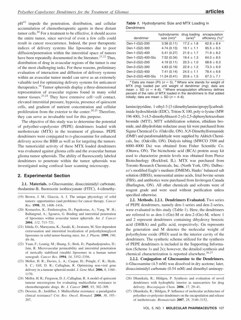
Table 1. Hydrodynamic Size and MTX Loading inDendrimers
dendrimerhydrodynamic
size (nm)adrug loading
(w/w)bencapsulationefficiency (%)c
Den-1-(G2)-200 3.58 (0.11) 17.2 ( 1.8 62.8 ( 8.4Den-1-(G2)-300 4.74 (0.13) 19.1 ( 1.1 65.5 ( 6.5Den-1-(G2)-400 5.41 (0.27) 21.9 ( 1.7 71.9 ( 8.2Den-1-(G2)-400-Glu 7.52 (0.34) 19.4 ( 1.3 64.0 ( 8.5Den-2-(G2)-200 4.19 (0.11) 20.3 ( 1.0 68.8 ( 6.3Den-2-(G2)-300 4.83 (0.19) 22.9 ( 1.2 73.3 ( 6.9Den-2-(G2)-400 7.41 (0.14) 24.5 ( 1.1 78.4 ( 6.6Den-2-(G2)-400-Glu 11.24 (0.41) 20.8 ( 1.5 67.3 ( 7.1
a Data are mean (PI) (n ) 5). b Where w/w stands for weight ofMTX (mg) loaded per unit weight of dendrimer (mg); data aremean ( SD (n ) 4–6). c Where encapsulation efficiency definespercent of the ratio of MTX loaded in the dendrimers to that addedinitially; data are mean ( SD (n ) 4–6).
Polyether-Copolyester Dendrimers for the Treatment of Gliomas articles
VOL. 5, NO. 1 MOLECULAR PHARMACEUTICS 107
ridine (2.3 mM) were added, and the reaction was allowedto continue for 48 h. Acetone was evaporated on a rotatoryevaporator, and the product was dissolved in methanol andpurified by extraction with ether (three times) and chloroform(three times). Subsequently, methanol was evaporated andproduct was dried under vacuum. The allyl pendant terminalgroups of dendrimers were oxidized to hydroxyl groups asreported previously.26 The succinimidyl carbonate derivativeof glucosamine (2.7 µM) was then conjugated to thesehydroxyl dendrimers (2.7 µM) by incubating them in dryacetone and DMF (10:1) at room temperature for 96 h.Product was purified by dialysis against water for 96 h usinga dialysis membrane of MWCO 6000–8000 Da. Conjugationof glucosamine to dendrimers was determined by 1HNMR.
2.2.3. Fluorescent Labeling of the Dendrimers. Theconjugation of rhodamine B to the dendrimers was carriedout as reported previously.28 In the case of den-1-(G2)-400,pendant allyl terminal functional groups were oxidized tohydroxyl groups26 to obtain the hydroxyl derivative of thedendrimers. Later, hydroxyl derivatives of den-1-(G2)-400or glucosamine conjugates of den-1-(G2)-400 (den-1-(G2)-400-Glu) were dissolved in DMF. EDC (0.1 mM), DMAP(0.08 mM), and rhodamine B (0.08 mM) were added to theflask and the reaction was allowed to occur for 48 h.Precipitate of EDC-urea was removed by filtration andproduct was purified by dialysis (MWCO 6000–8000 Da)against deionized water for 96 h. In Vitro stability ofrhodamine-dendrimer conjugates was assessed in PBS (pH7.4) at 37 °C. No dissociation of rhodamine from dendrimerswas observed for 3 days, indicating a stable linkage.
2.2.4. MTX Encapsulation. Dendrimers were dissolvedin DMF in screw-capped vials, MTX was added to thesevials and samples were stirred at room temperature for 48 h.Unencapsulated drug was removed by dialysis (MWCO 3500Da) for 4 h against 4 L of deionized water. The dialysatewas lyophilized to obtain freeze-dried dendrimer loaded withMTX. Drug loading was determined by adding freeze-drieddendrimers to DMF, sonicating them (550 Sonic dismem-brator, Fisher Scientific, Ottawa, ON) for 1 min, and lateragitating for 24 h at 200 rpm. MTX was analyzed using UVspectrophotometer U-2001 (Hitachi high technologies, Or-lando, FL) at 376 nm with appropriate blank corrections.
2.2.5. Internalization of Dendrimers by Glioma Cells. U87 MG and U 343 MGa cells (kind gift from J.-F. Bouchard,University of Montreal) ((American type Culture Collection(ATCC), Rockville, MD) (passage 15–20) were seeded at aconcentration of 1 × 10⁵ cells/mL (300 µL/well) into 24-well cell culture plates and were allowed to adhere to thewells for 24 h. Rhodamine-labeled dendrimers (100 µg/mL,in HBSS) were added to each well and incubated for 4 h at37 °C. Subsequently, cells were washed four times withHBSS to remove dendrimer adhering to the cellular surface.Cells were then lysed with 200 µL of 0.5% Triton-X
containing 0.1 N NaOH. Dendrimer concentrations in thecell lysate was determined by measuring rhodamine fluo-rescence (λex 550 nm and λem 625 nm) using Safire platereader (Tecan Austria GmbH, Salzburg, Austria). Standardcurve generated using lysed cells and rhodamine-labeleddendrimers was used for the quantification of fluorescence.Protein content of each well was estimated by using microBCA protein assay reagent.
2.2.6. Intracellular Localization of Dendrimers. U 343MGa cells (passage 15–20) were plated at the density of 0.5× 10⁶ cells/mL on glass coverslips treated with 0.05% w/vof poly-D-lysine solution. Cells were allowed to grow to 80%confluence. Later, rhodamine-labeled dendrimers (200 µg/mL) were added to each well and incubated for 4 h. Afterincubation, slides were washed four times with HBSS, fixedwith formaldehyde (1% w/v in PBS) for 30 min, and againwashed with HBSS two times. Coverslips were then mountedon slides using GelTol mounting medium (Thermo ElectronCorp., PA). Confocal laser scanning microscope images wereacquired at 100× using DMRXE microscope (Zeiss,Oberkochen, Germany) equipped with Leica TCS SP 2confocal system (Leica Microsystems, Heidelberg, Germany).
2.2.7. Development of MTX-Resistant U 87 MG Cells.U 87 MG cells were grown in DMEM supplemented with10% FBS, 1% nonessential amino acids, penicillin/strepto-mycin and 500 nM of MTX at 37 °C in a 5% CO2
atmosphere. The production of MTX resistance in cells wasfollowed by observing the size of the cells and nuclei, MTX-FITC accumulation in the cells, and dihydrofolate reductaseactivity.
(a) Enzyme Assay. Activity of dihydrofolate in the gliomacells was determined by preparing cell extracts as describedpreviously by Alt et al.29 Cells were harvested frommonolayer by trypsinization, washed three times with HBSS,and suspended in ice-cold 50 mM potassium phosphate (pH7.0). This suspension was disrupted with a probe sonicatorfor 5 min. Later, it was centrifuged at 100000g for 60 minat 4 °C to yield supernatant called cell extract. Folatereductase activity in the cell extract was determined usingdihydrofolate reductase assay kit. Protein content of the cellextract was determined by BCATM protein assay kit.
(b) MTX-FITC Accumulation. MTX was conjugatedwith FITC according to the procedure mentioned by Gapskiet al. 30 and Kaufman et al.31 U 87 MG cells (MTX resistantand sensitive) were plated into 24-well plates and wereallowed to adhere for 24 h. Later, they were incubated with
(28) Dhanikula, R.; Hildgen, P. In vitro evaluation of polyether-co-polyester dendrimers for delivery across the blood brain barrier.Biomacromolecules (Submitted).
(29) Alt, F. W.; Kellems, R. E.; Schimke, R. T. Synthesis anddegradation of folate reductase in sensitive and methotrexate-resistant lines of S-180 cells. J. Biol. Chem. 1976, 251, 3063–3074.
(30) Gapski, G. R.; Whiteley, J. M.; Rader, J. I.; Cramer, P. L.;Henderson, G. B.; Neef, V.; Huennekens, F. M. Synthesis of afluorescent derivative of amethopterin. J. Med. Chem. 1975, 18,526–528.
(31) Kaufman, R. J.; Bertino, J. R.; Schimke, R. T. Quantitation ofdihydrofolate reductase in individual parental and methotrexate-resistant murine cells. Use of a fluorescence activated cell sorter.J. Biol. Chem. 1978, 253, 5852–5860.
articles Dhanikula et al.
108 MOLECULAR PHARMACEUTICS VOL. 5, NO. 1
medium containing 30 µM MTX-FITC for 10 h at 37 °C.After incubation cells were washed four times with HBSSand lysed with 200 µL of 0.5% Triton-X containing 0.1 NNaOH. MTX-FITC concentrations in cell lysates weredetermined by measuring fluorescence (λex 504 nm and λem
538 nm) using a Safire plate reader (Tecan Austria GmbH,Salzburg, Austria). Standard curve generated using lysed cellsand MTX-FITC was used for the quantification of fluores-cence in the experiments. Protein content of each well wasestimated using micro BCA protein assay reagent.
2.2.8. Antiproliferative Activity of Dendrimers againstGlioma Cells. Human glioma cell lines, U 87 MG and U343 MGa, were cultured in DMEM supplemented with 10%FBS, 1% nonessential amino acids, and penicillin/strepto-mycin at 37 °C in a 5% CO2 atmosphere. MTX-resistant U87 MG cells were cultured as mentioned in the previoussection. Cells (passage 15–20) were seeded at the concentra-tion of 1 × 10⁴ cells/mL (100 µL/well) in 96-well cell cultureplates and were allowed to adhere to the wells for 24 h.Cellular growth inhibition was evaluated by MTT assay.Dendrimers loaded with MTX (2 to 1000 µM, in HBSS)were added to the cells and incubated for 72 h. Later, 10 µLof 5 mg/mL of MTT solution was added to each well,followed by incubation at 37 °C for 4 h. Formazan crystalsproduced by the cells were dissolved in 100 µL of MTTsolublization solution. Absorbance was measured at 570 nmusing Safire plate reader (Tecan Austria GmbH, Salzburg,Austria).
2.2.9. Transport of Dendrimers and MTX acrossBBB. Coculture of bEnd.3 (ATCC, Rockville, MD) andU373 MG cells was used as the model for BBB.28,32,33
bEnd.3 and U373 MG cells were seeded onto polycarbonateTranswell inserts at the density of 5 × 10⁵ cells/mL and 2× 10⁵ cells/mL, and confluent monolayers (9–10 days) wereused for the transport studies.28 Permeability was determinedin the apical to basolateral direction using HBSS as atransport medium. Rhodamine labeled dendrimers or MTXloaded dendrimers were placed in the donor compartmentand plates were incubated in a humidified, CO2 atmosphereat 37 °C. Samples were collected from the receiver compart-ment at predetermined time points. They were analyzed forrhodamine-labeled dendrimer using fluorescence plate reader(λex 550 nm and λem 625 nm). MTX was analyzed by HPLCusing C-18 column, mobile phase consisting of methanol,10 mM phosphoric acid, and 10 mM KH2PO4 (26:16:58 v/v,pH 4.5) at a flow rate of 0.9 mL/min and wavelength of 290nm using a Waters 717 system equipped with WatersTM486 tunable absorbance detector (Waters Corporation, Mil-
ford, MA). The effect of dendrimers on BBB integrity wasinvestigated by monitoring transendothelial electrical resis-tance (TEER) throughout the experiment.
2.2.10. Avascular Human Glioma Tumor Spheroids.Tumor spheroids of U 87 MG and U 343 MGa cells weregrown in Vitro using liquid overlay system.34,35 Agarosesolution was prepared in serum free DMEM (2% w/v) byheating it at 80 °C for 30 min. Each well of cell cultureplates was coated with a thin layer (0.3 mL) of this sterilizedsolution. Tumor cells at the density of 1 × 10⁵ cells/mL (incomplete medium) were seeded into each well. Subsequently,plates were gently agitated for 5 min on the first day ofseeding and tumor spheroids were allowed to grow for 7days at 37 °C in a humidified atmosphere. Medium of thewells was changed every 2–3 days.
(a) Growth Inhibition of the Tumor Spheroids. Forevaluating the inhibition of tumor growth, tumor spheroidswere incubated with PBS, MTX, or MTX-loaded dendrimers.Samples were prepared in PBS (pH 7.4) and 100 µL samplewas added per well to obtain a concentration of 200–400µM. Growth inhibition was monitored by measuring the sizeof tumor spheroids using an inverted phase microscope fittedwith an ocular micrometer. The major (dmax) and minor (dmin)diameters of each spheroid were determined and spheroidvolume was calculated as mentioned previously36 by usingthe following formula:
V)π × dmax × dmin
6(1)
(b) Determination of Cell Viability in the TumorSpheroids. Ethidium bromide was used as a fluorescentprobe for determining the dead cells in the tumor spheroidsafter treatment with MTX or MTX-loaded dendrimers. Forthis purpose, tumor spheroids were incubated with PBS,MTX, or MTX-loaded dendrimers for 7 days. Subsequently,they were incubated with ethidium bromide (50 µg/mL) for20 min at 4 °C. After incubation, they were washed fourtimes with PBS and disintegrated with 0.5% Triton-Xcontaining 0.1 N NaOH. Fluorescence in the cell lysate wasmeasured by using Safire plate reader (Tecan Austria GmbH,Salzburg, Austria). Protein content of tumor spheroids wasestimated using micro BCA protein assay reagent.
(c) Diffusion of Dendrimers into the TumorSpheroids. Tumor spheroids were incubated with rhodamine-labeled dendrimers (200 µg/mL) for 1, 2, 4, 6, 12, and 24 h.At specified time points, spheroids were washed four times
(32) Omidi, Y.; Campbell, L.; Barar, J.; Connell, D.; Akhtar, S.;Gumbleton, M. Evaluation of the immortalised mouse braincapillary endothelial cell line, b.End3, as an in vitro blood-brainbarrier model for drug uptake and transport studies. Brain Res.2003, 990, 95–112.
(33) Song, L.; Pachter, J. S. Culture of murine brain microvascularendothelial cells that maintain expression and cytoskeletal as-sociation of tight junction-associated proteins. In Vitro Cell DeV.Biol. Anim. 2003, 39, 313–320.
(34) Kobayashi, H.; Man, S.; Graham, C. H.; Kapitain, S. J.; Teicher,B. A.; Kerbel, R. S. Acquired multicellular-mediated resistanceto alkylating agents in cancer. Proc. Natl. Acad. Sci. U.S.A. 1993,90, 3294–3298.
(35) Rofstad, E. K.; Wahl, A.; Cde, L. D.; Brustad, T. Growthcharacteristics of human melanoma multicellular spheroids inliquid-overlay culture: comparisons with the parent tumourxenografts. Cell Tissue Kinet. 1986, 19, 205–216.
(36) Ballangrud, A.; Yang, W.-H.; Dnistrian, A.; Lampen, N.; Sgouros,G. Growth and characterization of LNCap prostate cancer cellspheroids. Clin. Cancer Res. 1999, 5, 3171s–3176s.
Polyether-Copolyester Dendrimers for the Treatment of Gliomas articles
VOL. 5, NO. 1 MOLECULAR PHARMACEUTICS 109
with HBSS, fixed with formaldehyde (10% w/v in PBS) for30 min, and placed in cavity microscope slides. Confocallaser scanning microscope images were acquired at 10 Xusing confocal microscope described above. Eight-micrometer-thick optical sections were acquired from the top toward thecenter of the spheroids approximately 136 µm deep into thespheroid.
3. Results
3.1. Conjugation of Glucosamine to the Dendrimers.Conjugation of glucosamine to the dendrimers was carriedout by using disuccinimidyl carbonate so that the aminogroup of the glucosamine was selectively activated to formsuccinimidyl derivative. This would in turn result in glu-cosamine conjugated to the dendrimers by a carbamatelinkage. Characterization of the products by FTIR and 1HNMR (Figure 1s, Supporting Information) proved the suc-cessful synthesis. The number of glucosamine moleculesattached to each dendrimer was also determined by 1H NMRspectroscopy. Three glucosamine molecules per dendrimerwere found to be conjugated in den-1-(G2)-400-Glu, whileden-2-(G2)-400-Glu had five glucosamine molecules con-jugated per dendrimer.
3.2. MTX Loading in the Dendrimers. MTX loading inthe dendrimers of den-1-series ranged between 17.2 and21.9% w/w, while in that of den-2-series ranged between20.3 and 24.5% w/w (Table 1). Dendrimers with PEO 400Da in the interior cavity encapsulated a higher amount ofMTX as compared to dendrimers with PEO 300 and 200Da in both den-1-series and den-2-series, with MTX loadingof 21.9% w/w in den-1-(G2)-400 and 24.5% w/w in den-2-(G2)-400. MTX loading in glucosylated dendrimers waslower than corresponding nonglucosylated dendrimers, withden-1-(G2)-400-Glu and den-2-(G2)-400-Glu encapsulating19.4% w/w and 20.8% w/w of MTX, respectively.
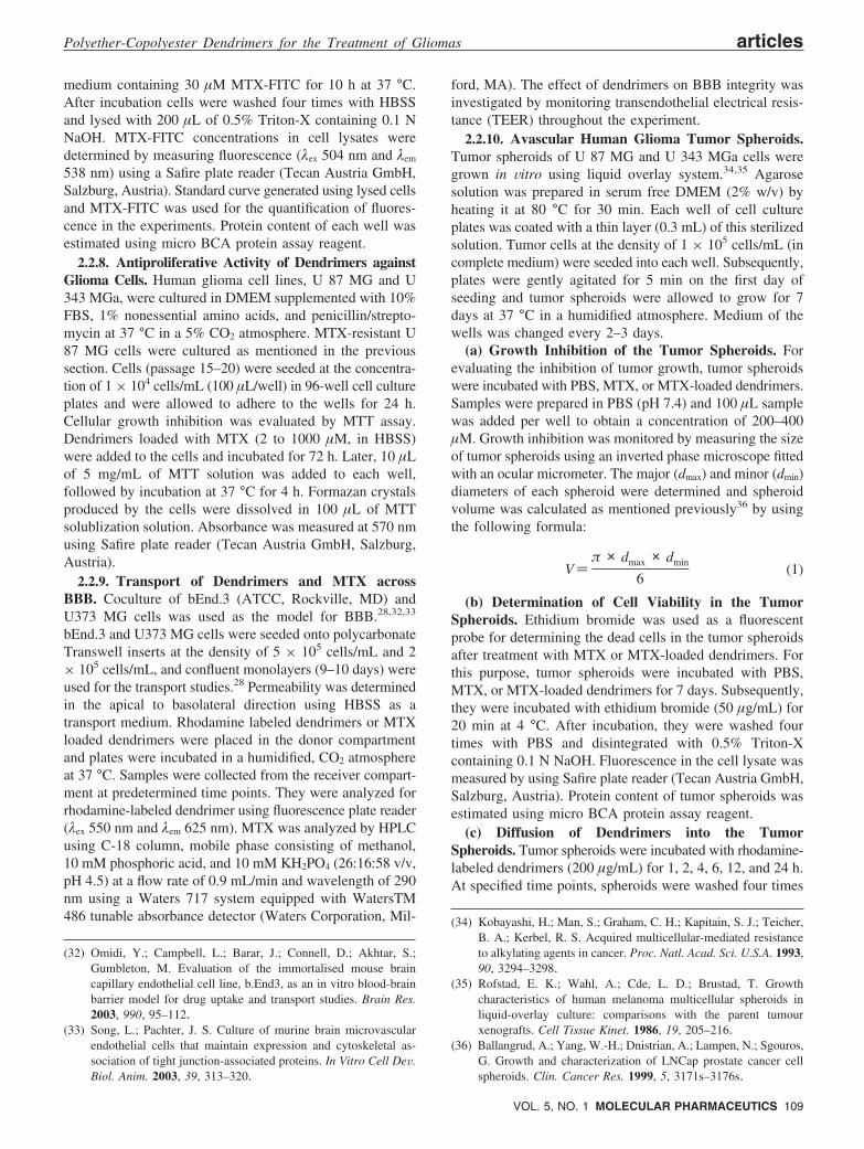
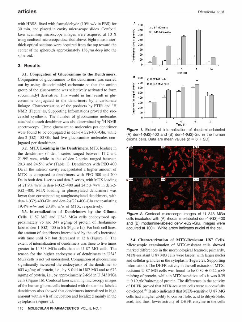
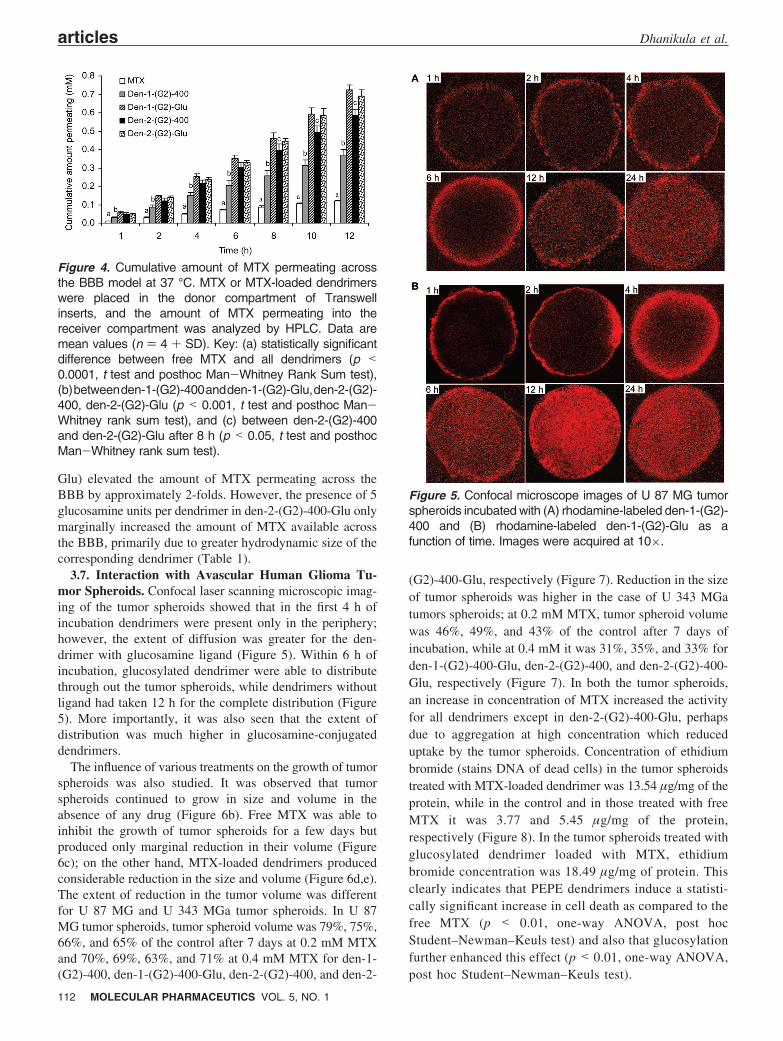
3.3. Internalization of Dendrimers by the GliomaCells. U 87 MG and U343 MGa cells endocytosed ap-proximately 76 and 347 µg/mg of protein of rhodamine-labeled den-1-(G2)-400 in 6 h (Figure 1a). For both cell lines,the amount of dendrimers internalized by the cells increasedwith time until 6 h but decreased at 12 h (Figure 1). Theextent of internalization of dendrimers was three to five timesgreater in U 343 MGa cells than in U 87 MG cells. Thereason for the higher endocytosis of dendrimers in U343MGa cells is not yet understood. Conjugation of glucosaminesignificantly increased the endocytosis of the dendrimer to603 µg/mg of protein, i.e., by 8-fold in U87 MG and to 672µg/mg of protein, i.e., by approximately 2-fold in U 343 MGacells (Figure 1b). Confocal laser scanning microscopy imagesof the human glioma cells incubated with rhodamine-labeleddendrimers also showed that dendrimers internalized in highamount within 4 h of incubation and localized mainly in thecytoplasm (Figure 2).
3.4. Characterization of MTX-Resistant U87 Cells.Microscopic examination of MTX-resistant cells showedmarked differences in the morphological features; primarily,MTX-resistant U 87 MG cells were larger, with larger nucleiand cellular granules in the cytoplasm (Figure 2s, SupportingInformation). The DHFR activity in the cell extracts of MTX-resistant U 87 MG cells was found to be 0.89 ( 0.22 µM/min/mg of protein, while in MTX-sensitive cells it was 0.39( 0.19 µM/min/mg of protein. The difference in the activityof DHFR proved that MTX-resistant cells were successfullydeveloped.29 It also indicated that MTX-sensitive U 87 MGcells had a higher ability to convert folic acid to dihydrofolicacid, and thus, lower activity of DHFR enzyme in the cells
Figure 1. Extent of internalization of rhodamine-labeled(A) den-1-(G2)-400 and (B) den-1-(G2)-Glu in the humanglioma cells. Data are mean values (n ) 6 + SD).
Figure 2. Confocal microscope images of U 343 MGacells incubated with (A) rhodamine-labeled den-1-(G2)-400and (B) rhodamine-labeled den-1-(G2)-Glu. Images wereacquired at 100×. White arrow indicates nuclei of the cell.
articles Dhanikula et al.
110 MOLECULAR PHARMACEUTICS VOL. 5, NO. 1
was one of the sources of MTX resistance. MTX-resistantcells accumulated MTX-FITC approximately two timeshigher than MTX-sensitive U 87 MG cells (Figure 3s,Supporting Information), suggesting the presence of higherlevels of DHFR in MTX-resistant cell lines.31
3.5. Antiproliferative Activity of Dendrimers againstGlioma Cells. The cytotoxicity of the dendrimers alone onthe brain endothelial cells28 and U87 MG cells wereevaluated to ascertain their safety. None of the dendrimersshowed significant cytotoxicity against bEnd.3 and U 87 MGcells at concentrations of 0.01-5 mg/mL (Figure 4s, Sup-porting Information). Inhibition in the growth of two gliomacell lines was tested in the present study to evaluate theirpotential in different types of gliomas. IC50 values of MTXtoward U 87 MG and U 343 MGa cells were 2.14 and 2.79µM, respectively (Table 2). In both the cell lines, MTXloaded in dendrimers had 1.5–5 times lower IC50 value thanthe free MTX. Against MTX resistant U 87 MG cells, MTXencapsulated in the dendrimers had IC50 values 9 to 15 timeslower (1.17 to 5.22 µM) than that of the free MTX (85.95µM). The conjugation of glucosamine to dendrimers furtherreduced the IC50 of MTX, but the extent of reduction wasdependent on the cell line. MTX loaded dendrimer-glu-cosamine conjugates were 3.5 to 4.5 times more potent thandendrimers alone toward U 87 MG cells with IC50 values of0.35 ( 0.08 and 0.43 ( 0.05 µM and 2 times more toxictoward U 343 MGa cells with IC50 values of 0.39 ( 0.13and 0.42 ( 0.15 µM (Table 2). This indicates that there isvariation in the extent of uptake of glucosylated dendrimersand, hence, expression of glucose transporters in these celllines. Even in the MTX-resistant cell lines, glucosylateddendrimers were two to five times more effective in inhibitingthe cell growth (Table 2). The influence of increasing thenumber of glucosamine units from 3 (den-1-(G2)-400-Glu)to 5 (den-2-(G2)-400-Glu) on potency was not apparent inthe IC50 values.
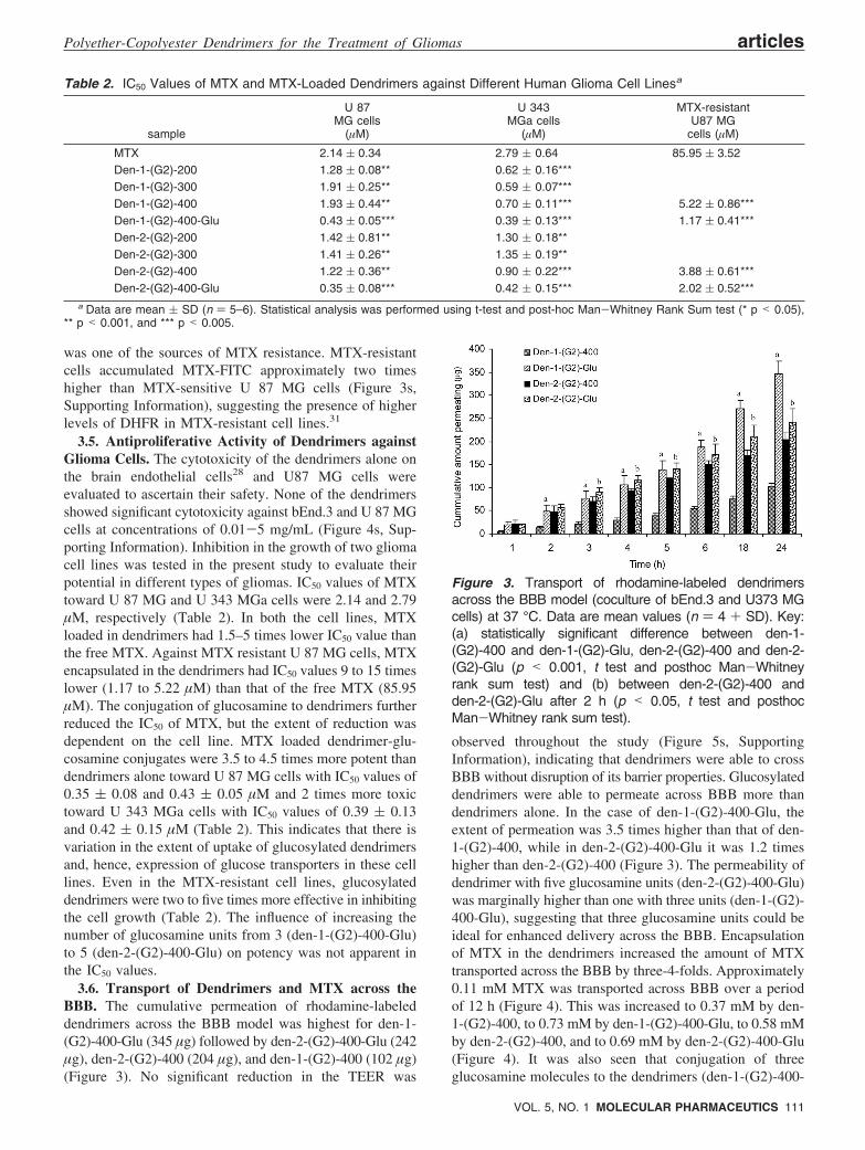
3.6. Transport of Dendrimers and MTX across theBBB. The cumulative permeation of rhodamine-labeleddendrimers across the BBB model was highest for den-1-(G2)-400-Glu (345 µg) followed by den-2-(G2)-400-Glu (242µg), den-2-(G2)-400 (204 µg), and den-1-(G2)-400 (102 µg)(Figure 3). No significant reduction in the TEER was
observed throughout the study (Figure 5s, SupportingInformation), indicating that dendrimers were able to crossBBB without disruption of its barrier properties. Glucosylateddendrimers were able to permeate across BBB more thandendrimers alone. In the case of den-1-(G2)-400-Glu, theextent of permeation was 3.5 times higher than that of den-1-(G2)-400, while in den-2-(G2)-400-Glu it was 1.2 timeshigher than den-2-(G2)-400 (Figure 3). The permeability ofdendrimer with five glucosamine units (den-2-(G2)-400-Glu)was marginally higher than one with three units (den-1-(G2)-400-Glu), suggesting that three glucosamine units could beideal for enhanced delivery across the BBB. Encapsulationof MTX in the dendrimers increased the amount of MTXtransported across the BBB by three-4-folds. Approximately0.11 mM MTX was transported across BBB over a periodof 12 h (Figure 4). This was increased to 0.37 mM by den-1-(G2)-400, to 0.73 mM by den-1-(G2)-400-Glu, to 0.58 mMby den-2-(G2)-400, and to 0.69 mM by den-2-(G2)-400-Glu(Figure 4). It was also seen that conjugation of threeglucosamine molecules to the dendrimers (den-1-(G2)-400-
Table 2. IC50 Values of MTX and MTX-Loaded Dendrimers against Different Human Glioma Cell Linesa
sample
U 87MG cells
(µM)
U 343MGa cells
(µM)
MTX-resistantU87 MG
cells (µM)
MTX 2.14 ( 0.34 2.79 ( 0.64 85.95 ( 3.52Den-1-(G2)-200 1.28 ( 0.08** 0.62 ( 0.16***Den-1-(G2)-300 1.91 ( 0.25** 0.59 ( 0.07***Den-1-(G2)-400 1.93 ( 0.44** 0.70 ( 0.11*** 5.22 ( 0.86***Den-1-(G2)-400-Glu 0.43 ( 0.05*** 0.39 ( 0.13*** 1.17 ( 0.41***Den-2-(G2)-200 1.42 ( 0.81** 1.30 ( 0.18**Den-2-(G2)-300 1.41 ( 0.26** 1.35 ( 0.19**Den-2-(G2)-400 1.22 ( 0.36** 0.90 ( 0.22*** 3.88 ( 0.61***Den-2-(G2)-400-Glu 0.35 ( 0.08*** 0.42 ( 0.15*** 2.02 ( 0.52***
a Data are mean ( SD (n ) 5–6). Statistical analysis was performed using t-test and post-hoc Man-Whitney Rank Sum test (* p < 0.05),** p < 0.001, and *** p < 0.005.
Figure 3. Transport of rhodamine-labeled dendrimersacross the BBB model (coculture of bEnd.3 and U373 MGcells) at 37 °C. Data are mean values (n ) 4 + SD). Key:(a) statistically significant difference between den-1-(G2)-400 and den-1-(G2)-Glu, den-2-(G2)-400 and den-2-(G2)-Glu (p < 0.001, t test and posthoc Man-Whitneyrank sum test) and (b) between den-2-(G2)-400 andden-2-(G2)-Glu after 2 h (p < 0.05, t test and posthocMan-Whitney rank sum test).
Polyether-Copolyester Dendrimers for the Treatment of Gliomas articles
VOL. 5, NO. 1 MOLECULAR PHARMACEUTICS 111
Glu) elevated the amount of MTX permeating across theBBB by approximately 2-folds. However, the presence of 5glucosamine units per dendrimer in den-2-(G2)-400-Glu onlymarginally increased the amount of MTX available acrossthe BBB, primarily due to greater hydrodynamic size of thecorresponding dendrimer (Table 1).
3.7. Interaction with Avascular Human Glioma Tu-mor Spheroids. Confocal laser scanning microscopic imag-ing of the tumor spheroids showed that in the first 4 h ofincubation dendrimers were present only in the periphery;however, the extent of diffusion was greater for the den-drimer with glucosamine ligand (Figure 5). Within 6 h ofincubation, glucosylated dendrimer were able to distributethrough out the tumor spheroids, while dendrimers withoutligand had taken 12 h for the complete distribution (Figure5). More importantly, it was also seen that the extent ofdistribution was much higher in glucosamine-conjugateddendrimers.
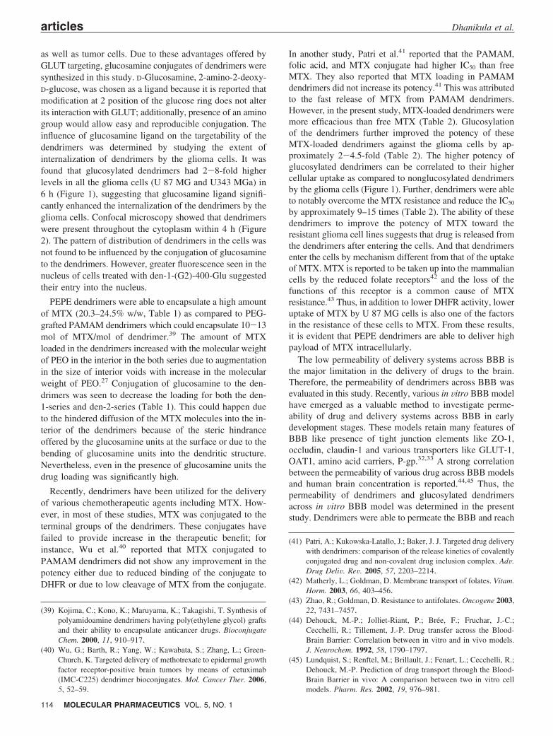
The influence of various treatments on the growth of tumorspheroids was also studied. It was observed that tumorspheroids continued to grow in size and volume in theabsence of any drug (Figure 6b). Free MTX was able toinhibit the growth of tumor spheroids for a few days butproduced only marginal reduction in their volume (Figure6c); on the other hand, MTX-loaded dendrimers producedconsiderable reduction in the size and volume (Figure 6d,e).The extent of reduction in the tumor volume was differentfor U 87 MG and U 343 MGa tumor spheroids. In U 87MG tumor spheroids, tumor spheroid volume was 79%, 75%,66%, and 65% of the control after 7 days at 0.2 mM MTXand 70%, 69%, 63%, and 71% at 0.4 mM MTX for den-1-(G2)-400, den-1-(G2)-400-Glu, den-2-(G2)-400, and den-2-
(G2)-400-Glu, respectively (Figure 7). Reduction in the sizeof tumor spheroids was higher in the case of U 343 MGatumors spheroids; at 0.2 mM MTX, tumor spheroid volumewas 46%, 49%, and 43% of the control after 7 days ofincubation, while at 0.4 mM it was 31%, 35%, and 33% forden-1-(G2)-400-Glu, den-2-(G2)-400, and den-2-(G2)-400-Glu, respectively (Figure 7). In both the tumor spheroids,an increase in concentration of MTX increased the activityfor all dendrimers except in den-2-(G2)-400-Glu, perhapsdue to aggregation at high concentration which reduceduptake by the tumor spheroids. Concentration of ethidiumbromide (stains DNA of dead cells) in the tumor spheroidstreated with MTX-loaded dendrimer was 13.54 µg/mg of theprotein, while in the control and in those treated with freeMTX it was 3.77 and 5.45 µg/mg of the protein,respectively (Figure 8). In the tumor spheroids treated withglucosylated dendrimer loaded with MTX, ethidiumbromide concentration was 18.49 µg/mg of protein. Thisclearly indicates that PEPE dendrimers induce a statisti-cally significant increase in cell death as compared to thefree MTX (p < 0.01, one-way ANOVA, post hocStudent–Newman–Keuls test) and also that glucosylationfurther enhanced this effect (p < 0.01, one-way ANOVA,post hoc Student–Newman–Keuls test).
Figure 4. Cumulative amount of MTX permeating acrossthe BBB model at 37 °C. MTX or MTX-loaded dendrimerswere placed in the donor compartment of Transwellinserts, and the amount of MTX permeating into thereceiver compartment was analyzed by HPLC. Data aremean values (n ) 4 + SD). Key: (a) statistically significantdifference between free MTX and all dendrimers (p <0.0001, t test and posthoc Man-Whitney Rank Sum test),(b)betweenden-1-(G2)-400andden-1-(G2)-Glu,den-2-(G2)-400, den-2-(G2)-Glu (p < 0.001, t test and posthoc Man-Whitney rank sum test), and (c) between den-2-(G2)-400and den-2-(G2)-Glu after 8 h (p < 0.05, t test and posthocMan-Whitney rank sum test).
Figure 5. Confocal microscope images of U 87 MG tumorspheroids incubated with (A) rhodamine-labeled den-1-(G2)-400 and (B) rhodamine-labeled den-1-(G2)-Glu as afunction of time. Images were acquired at 10×.
articles Dhanikula et al.
112 MOLECULAR PHARMACEUTICS VOL. 5, NO. 1
4. DiscussionThe basic GLUT is reported to play a major role in glucose
uptake by the tumor cells. Though glucose transporters areexpressed in all of the tissues, the major difference betweennormal and cancerous cells is the presence of facilitativeglucose transporter (GLUT) genes in the latter.16 Glucose-conjugated niosomes have been reported to have highertumor-to-muscle accumulation in the glioma xenografts;11
Kim et al.37 have also demonstrated that glucose-conjugatedPEI enhance gene delivery to the lung cancer. Additionally,targeting GLUT-1 can be potentially employed for enhanceddelivery of the carriers across BBB.9 Dendrimers conjugatedto glucose are small and mimic glucose; in fact, that theyhave been reported to compete with glucose in glucoseassay.38 Thus, they can be taken up by the GLUT on BBB
(37) Kim, H. W.; Park, I. K.; Cho, C. S.; Lee, K. H.; Beck, G. R. J.;Colburn, N. H.; Cho, M. H. Aerosol delivery of glucosylatedpolyethylenimine/phosphatase and tensin homologue deleted onchromosome 10 complex suppresses Akt downstream pathwaysin the lung of K-ras null mice. Cancer Res. 2004, 64, 7971–7976.
(38) Beier, H.; Ibey, B.; Pishko, M.; Cote, G. Use of glycosylateddendrimer macromolecules to fluorescently monitor glucoseconcentration. Proc. SPIE 2007, 6445, 644–504.
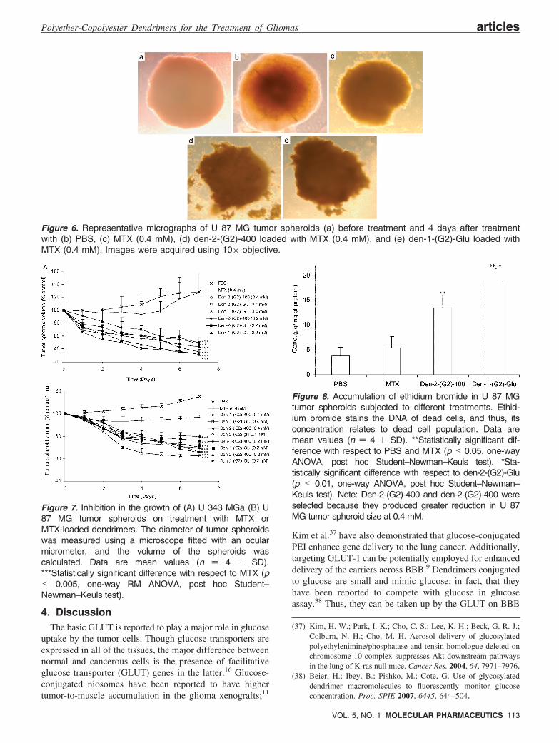
Figure 6. Representative micrographs of U 87 MG tumor spheroids (a) before treatment and 4 days after treatmentwith (b) PBS, (c) MTX (0.4 mM), (d) den-2-(G2)-400 loaded with MTX (0.4 mM), and (e) den-1-(G2)-Glu loaded withMTX (0.4 mM). Images were acquired using 10× objective.
Figure 7. Inhibition in the growth of (A) U 343 MGa (B) U87 MG tumor spheroids on treatment with MTX orMTX-loaded dendrimers. The diameter of tumor spheroidswas measured using a microscope fitted with an ocularmicrometer, and the volume of the spheroids wascalculated. Data are mean values (n ) 4 + SD).***Statistically significant difference with respect to MTX (p< 0.005, one-way RM ANOVA, post hoc Student–Newman–Keuls test).
Figure 8. Accumulation of ethidium bromide in U 87 MGtumor spheroids subjected to different treatments. Ethid-ium bromide stains the DNA of dead cells, and thus, itsconcentration relates to dead cell population. Data aremean values (n ) 4 + SD). **Statistically significant dif-ference with respect to PBS and MTX (p < 0.05, one-wayANOVA, post hoc Student–Newman–Keuls test). *Sta-tistically significant difference with respect to den-2-(G2)-Glu(p < 0.01, one-way ANOVA, post hoc Student–Newman–Keuls test). Note: Den-2-(G2)-400 and den-2-(G2)-400 wereselected because they produced greater reduction in U 87MG tumor spheroid size at 0.4 mM.
Polyether-Copolyester Dendrimers for the Treatment of Gliomas articles
VOL. 5, NO. 1 MOLECULAR PHARMACEUTICS 113
as well as tumor cells. Due to these advantages offered byGLUT targeting, glucosamine conjugates of dendrimers weresynthesized in this study. D-Glucosamine, 2-amino-2-deoxy-D-glucose, was chosen as a ligand because it is reported thatmodification at 2 position of the glucose ring does not alterits interaction with GLUT; additionally, presence of an aminogroup would allow easy and reproducible conjugation. Theinfluence of glucosamine ligand on the targetability of thedendrimers was determined by studying the extent ofinternalization of dendrimers by the glioma cells. It wasfound that glucosylated dendrimers had 2-8-fold higherlevels in all the glioma cells (U 87 MG and U343 MGa) in6 h (Figure 1), suggesting that glucosamine ligand signifi-cantly enhanced the internalization of the dendrimers by theglioma cells. Confocal microscopy showed that dendrimerswere present throughout the cytoplasm within 4 h (Figure2). The pattern of distribution of dendrimers in the cells wasnot found to be influenced by the conjugation of glucosamineto the dendrimers. However, greater fluorescence seen in thenucleus of cells treated with den-1-(G2)-400-Glu suggestedtheir entry into the nucleus.
PEPE dendrimers were able to encapsulate a high amountof MTX (20.3–24.5% w/w, Table 1) as compared to PEG-grafted PAMAM dendrimers which could encapsulate 10-13mol of MTX/mol of dendrimer.39 The amount of MTXloaded in the dendrimers increased with the molecular weightof PEO in the interior in the both series due to augmentationin the size of interior voids with increase in the molecularweight of PEO.27 Conjugation of glucosamine to the den-drimers was seen to decrease the loading for both the den-1-series and den-2-series (Table 1). This could happen dueto the hindered diffusion of the MTX molecules into the in-terior of the dendrimers because of the steric hindranceoffered by the glucosamine units at the surface or due to thebending of glucosamine units into the dendritic structure.Nevertheless, even in the presence of glucosamine units thedrug loading was significantly high.
Recently, dendrimers have been utilized for the deliveryof various chemotherapeutic agents including MTX. How-ever, in most of these studies, MTX was conjugated to theterminal groups of the dendrimers. These conjugates havefailed to provide increase in the therapeutic benefit; forinstance, Wu et al.40 reported that MTX conjugated toPAMAM dendrimers did not show any improvement in thepotency either due to reduced binding of the conjugate toDHFR or due to low cleavage of MTX from the conjugate.
In another study, Patri et al.41 reported that the PAMAM,folic acid, and MTX conjugate had higher IC50 than freeMTX. They also reported that MTX loading in PAMAMdendrimers did not increase its potency.41 This was attributedto the fast release of MTX from PAMAM dendrimers.However, in the present study, MTX-loaded dendrimers weremore efficacious than free MTX (Table 2). Glucosylationof the dendrimers further improved the potency of theseMTX-loaded dendrimers against the glioma cells by ap-proximately 2-4.5-fold (Table 2). The higher potency ofglucosylated dendrimers can be correlated to their highercellular uptake as compared to nonglucosylated dendrimersby the glioma cells (Figure 1). Further, dendrimers were ableto notably overcome the MTX resistance and reduce the IC50
by approximately 9–15 times (Table 2). The ability of thesedendrimers to improve the potency of MTX toward theresistant glioma cell lines suggests that drug is released fromthe dendrimers after entering the cells. And that dendrimersenter the cells by mechanism different from that of the uptakeof MTX. MTX is reported to be taken up into the mammaliancells by the reduced folate receptors42 and the loss of thefunctions of this receptor is a common cause of MTXresistance.43 Thus, in addition to lower DHFR activity, loweruptake of MTX by U 87 MG cells is also one of the factorsin the resistance of these cells to MTX. From these results,it is evident that PEPE dendrimers are able to deliver highpayload of MTX intracellularly.
The low permeability of delivery systems across BBB isthe major limitation in the delivery of drugs to the brain.Therefore, the permeability of dendrimers across BBB wasevaluated in this study. Recently, various in Vitro BBB modelhave emerged as a valuable method to investigate perme-ability of drug and delivery systems across BBB in earlydevelopment stages. These models retain many features ofBBB like presence of tight junction elements like ZO-1,occludin, claudin-1 and various transporters like GLUT-1,OAT1, amino acid carriers, P-gp.32,33 A strong correlationbetween the permeability of various drug across BBB modelsand human brain concentration is reported.44,45 Thus, thepermeability of dendrimers and glucosylated dendrimersacross in Vitro BBB model was determined in the presentstudy. Dendrimers were able to permeate the BBB and reach
(39) Kojima, C.; Kono, K.; Maruyama, K.; Takagishi, T. Synthesis ofpolyamidoamine dendrimers having poly(ethylene glycol) graftsand their ability to encapsulate anticancer drugs. BioconjugateChem. 2000, 11, 910–917.
(40) Wu, G.; Barth, R.; Yang, W.; Kawabata, S.; Zhang, L.; Green-Church, K. Targeted delivery of methotrexate to epidermal growthfactor receptor-positive brain tumors by means of cetuximab(IMC-C225) dendrimer bioconjugates. Mol. Cancer Ther. 2006,5, 52–59.
(41) Patri, A.; Kukowska-Latallo, J.; Baker, J. J. Targeted drug deliverywith dendrimers: comparison of the release kinetics of covalentlyconjugated drug and non-covalent drug inclusion complex. AdV.Drug DeliV. ReV. 2005, 57, 2203–2214.
(42) Matherly, L.; Goldman, D. Membrane transport of folates. Vitam.Horm. 2003, 66, 403–456.
(43) Zhao, R.; Goldman, D. Resistance to antifolates. Oncogene 2003,22, 7431–7457.
(44) Dehouck, M.-P.; Jolliet-Riant, P.; Brée, F.; Fruchar, J.-C.;Cecchelli, R.; Tillement, J.-P. Drug transfer across the Blood-Brain Barrier: Correlation between in vitro and in vivo models.J. Neurochem. 1992, 58, 1790–1797.
(45) Lundquist, S.; Renftel, M.; Brillault, J.; Fenart, L.; Cecchelli, R.;Dehouck, M.-P. Prediction of drug transport through the Blood-Brain Barrier in vivo: A comparison between two in vitro cellmodels. Pharm. Res. 2002, 19, 976–981.
articles Dhanikula et al.
114 MOLECULAR PHARMACEUTICS VOL. 5, NO. 1
the receiver compartment in high amounts (Figure 3). In theprevious study,28 we established that rhodamine-labeledPEPE dendrimers cross the BBB model and not merelyrhodamine crosses into the receiver compartment. Notably,the extent of permeation of den-2-(G2)-400 was 2 timeshigher than that of den-1-(G2)-400. It is speculated that thishigher permeation of the former across BBB was due to hi-gher number of PEG chains present on the surface.27 Den-1-(G2)-400-Glu had 3.5 times greater permeability in com-parison with den-1-(G2)-400, while Den-2-(G2)-400-Glu hadmoderately higher permeation (1.2 folds) than den-2-(G2)-400 (Figure 3). It is hypothesized that moderate augmentationin the permeability of den-2-(G2)-400-Glu as compared toden-2-(G2)-400 is because of the larger size of the gluco-sylated dendrimer due to the presence of 5 glucosaminemoiety which reduces its endocytosis (Table 1). Nonetheless,higher permeability of glucosylated dendrimers suggests thatglucosamine serves to increase the permeability of dendrim-ers across the BBB. The effect of higher permeability ofglucosylated dendrimers were reflected in the amount MTXdelivered by these dendrimers across BBB (Figure 4).Encapsulation of MTX in den-1-(G2)-400 and den-2-(G2)-400 increased the amount of MTX available across BBB 4and 6 times, respectively (Figure 4). Glucosylation of den-1-(G2)-400 and den-2-(G2)-400 increased the availability ofMTX across BBB by 2-folds in den-1-(G2)-400-Glu, but onlyby approximately 1-fold in den-2-(G2)-400-Glu (Figure 4).A lower increment in the MTX availability by the latter canbe explained by its lower permeability across the BBB(Figure 3). Interestingly, den-1-(G2)-400-Glu transported 7times higher amount of MTX as compared to the free MTX.Thus, conjugation of three glucosamine molecules perdendrimer seems to be ideal for enhancing the permeabilityof PEPE dendrimers across the BBB. Accordingly, thesePEPE dendrimers can be used for the delivery of a higheramount of MTX across BBB and the conjugation toglucosamine can further augment it.
Due to the poor permeation of delivery systems into thehypoxic and necrotic tumor regions distant from the vascularbed, the amount of drug accessing inside the solid tumors islow.20,22,46 As a consequence, the overall therapeutic effectof chemotherapeutic agents is restricted, leading to the relapseof cancer. These limitations of delivery systems are particu-larly dangerous in malignant gliomas which are one of themost refractory tumors.47,48 It is postulated that enhancementin the ability of the delivery system to penetrate deeper intothe tumor tissues can significantly reduce the tumor regrowthand augment the therapeutic benefit of the treatment. It hasbeen reported that tumor spheroids generated by liquidoverlay technique are not only aggregates of cells in closecontact but contain an organized extracellular matrix consist-ing of fibronectin, laminin, collagen, and GAG, suggestiveof the extracellular matrix of tumors in ViVo.49,50 Therefore,interstitial penetration and diffusion of dendrimers into
avascular regions of the solid tumors was evaluated usingtumor spheroids as a model. Rhodamine-labeled dendrimerswere able to reach the central necrotic region of the tumorspheroids within 12 h of incubation (Figure 5). Glucosylationwas found to enhance the rate and extent of diffusion of thedendrimers in the tumor spheroids (Figure 5). This is becausethe central regions of the solid tumors are hypoxic anddemonstrate a hypoxia related increase in the glucosetransport.10,51 Thus, greater demand of glucose in the hypoxicregions of the tumors drives the faster and enhanceddistribution of glucosylated dendrimers in the tumors sphe-roids. This finding suggests these PEPE dendrimers wouldprobably reach the avascular regions of the tumors within150–200 µm and thus would provide more effective controlof the tumor growth. Further, evaluation of the ability ofthese dendrimers to reach avascular regions of tumors wouldbe tested in in ViVo brain tumor models.
MTX-loaded dendrimers were able to considerably reducethe size of tumor spheroids during 7 days of the study(Figures 6 and 7). U 87 MG tumor spheroids volumes werereduced to 63–79% of the control, while U 343 MGa tumorspheroids volume was decreased to 31–49% of the control(Figure 7). The potency of the MTX loaded dendrimers wasalways higher than that of the free MTX (significantdifference, p < 0.005, one way RM ANOVA). However,the therapeutic gain obtained from glucosylated dendrimerson tumor spheroids was different depending on the type ofglioma cells. In both of the tumor spheroids, den-1-(G2)-400-Glu was more effective than den-1-(G2)-400, while den-2-(G2)-400-Glu was either more effective than or as effectiveas den-2-(G2)-400 at the tested concentrations (Figure 7).This could be due to the fact that den-2-(G2)-400-Glu hasgreater size than den-1-(G2)-400-Glu and hence lowerpenetration in the tumor spheroids (Table 1). The concentra-tion of ethidium bromide in the lysate of tumor spheroidstreated with MTX loaded nonglucosylated and glucosylateddendrimers dendrimer was significantly higher than in thosetreated with free MTX (p < 0.01, one-way ANOVA, posthoc Student–Newman–Keuls test) (Figure 8). Ethidiumbromide concentration is related to the population of dead
(46) Minchinton, A.; Tannock, I. Drug penetration in solid tumours.Nat. ReV. Cancer 2006, 6, 583–592.
(47) Desjardins, A.; Rich, J.; Quinn, J.; Vredenburgh, J.; Gururangan,S.; Sathornsumetee, S.; Reardon, D.; Friedman, A.; Bigner, D.;Friedman, H. Chemotherapy and novel therapeutic approaches inmalignant glioma. Front Biosci. 2005, 10, 2645–2668.
(48) Graham, C.; Cloughesy, T. Brain tumor treatment: chemotherapyand other new developments. Semin. Oncol. Nurs. 2004, 20, 260–272.
(49) Paulus, W.; Huettner, C.; Tonn, J. C. Collagens, integrins andthe mesenchymal drift in glioblastomas: a comparison of biopsyspecimens, spheroid and early monolayer cultures. Int. J. Cancer1994, 58, 841–846.
(50) De Lange Davies, C.; Müller, H.; Hagen, I.; Garseth, M.;Hjelstuen, M. H. Comparison of extracellular matrix in humanosteosarcomas and melanomas growing as xenografts, multicel-lular spheroids, and monolayer cultures. Anticancer Res. 1997,17, 4317–4326.
(51) Clavo, A. C.; Brown, R. S.; Wahl, R. L. Fluorodeoxyglucoseuptake in human cancer cell lines is increased by hypoxia. J. Nucl.Med. 1995, 36, 1625–1632.
Polyether-Copolyester Dendrimers for the Treatment of Gliomas articles
VOL. 5, NO. 1 MOLECULAR PHARMACEUTICS 115
cells in the sample, thereby suggesting that MTX loading inthe dendrimers significantly enhanced the percentage kill ofthe cells in the tumor spheroids. This is attributed to enhancedentry of dendrimers into tumor spheroids as well as slowrelease of MTX after loading in these dendrimers.27 Since,the tumor spheroids mimic the microenvironment of solidtumors, the higher efficacy of MTX loaded dendrimerssuggests that they would provide significantly higher thera-peutic benefit than MTX.
In a nutshell, this study shows that MTX-loaded PEPEdendrimers are more potent than free MTX as well aseffective against MTX-resistant glioma cells. They candeliver MTX across BBB in high amounts and can penetrateinto the central necrotic regions of avascular tumor spheroids.Additionally, glucosylation not only increases their potencybut also enhances their permeability across the BBB anddiffusion into the avsacular regions of the tumor tissue.Hence, glucosylated PEPE dendrimers can serve as effectivedelivery systems for the treatment of gliomas. Further in ViVostudies to test the proof-of-concept will be carried out in thefuture. It is speculated that these glucosylated dendrimers
with ability to counteract drug resistance and enhance drugdelivery to the central avascular regions of the tumor canserve as a step forward to improving the efficacy ofchemotherapy.
Acknowledgment. We thank Jean-Michel Rabanel fortechnical assistance with the confocal laser scanning mi-croscopy experiments. A graduate research scholarshipawarded to R.S.D. by Rx & D Health Research Foundationand Canadian Institutes of Health Research (CIHR) isgratefully acknowledged. P.H. thanks the Natural Sciencesand Engineering Research Council of Canada (NSERC) forthe research funding. This study was partially funded by aNSERC grant to J.-F.B. A.A. is supported by a CNIB-CIHRstudentship and J.-F.B. by a Rx & D Health ResearchFoundation and CIHR Scholar award.
Supporting Information Available: Schemes showing thesynthesis of the core and dendrimers; NMR spectra. This materialis available free of charge via the Internet at http://pubs.acs.org.
MP700086J
articles Dhanikula et al.
116 MOLECULAR PHARMACEUTICS VOL. 5, NO. 1