metastatic breast cancer - lieberman's...
TRANSCRIPT
Metastatic breast cancer The workup and radiologic characteristics of liver and skeletal metastases
Yolanda D. Tseng, MS 4Gillian Lieberman, MDJuly 21, 2008

Patient AK 53F with palpable mass in right breast: mammogram CC view
L CC R CC
PACS, BIDMC
Spiculated massBenign macrocalcification (oil cysts)
Biopsy clip from prior biopsySurface sticker to mark a mole
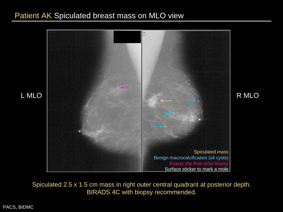
Patient AK Spiculated breast mass on MLO view
L MLO R MLO
PACS, BIDMC
Spiculated 2.5 x 1.5 cm mass in right outer central quadrant at posterior depth. BIRADS 4C with biopsy recommended.
Spiculated massBenign macrocalcification (oil cysts)
Biopsy clip from prior biopsySurface sticker to mark a mole
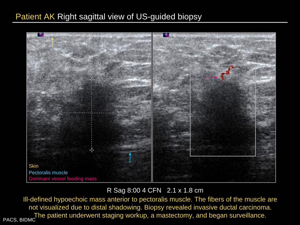
Patient AK Right sagittal view of US-guided biopsy
R Sag 8:00 4 CFN 2.1 x 1.8 cmIll-defined hypoechoic mass anterior to pectoralis muscle. The fibers of the muscle are
not visualized due to distal shadowing. Biopsy revealed invasive ductal carcinoma. The patient underwent staging workup, a mastectomy, and began surveillance.
PACS, BIDMC
SkinPectoralis muscleDominant vessel feeding mass
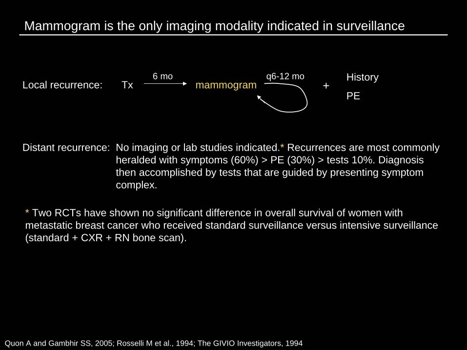
Mammogram is the only imaging modality indicated in surveillance
Quon A and Gambhir SS, 2005; Rosselli M et al., 1994; The GIVIO Investigators, 1994
Local recurrence: Tx6 mo
mammogramq6-12 mo History
PE+
Distant recurrence: No imaging or lab studies indicated.* Recurrences are most commonly heralded with symptoms (60%) > PE (30%) > tests 10%. Diagnosis then accomplished by tests that are guided by presenting symptom complex.
* Two RCTs have shown no significant difference in overall survival of women with metastatic breast cancer who received standard surveillance versus intensive surveillance (standard + CXR + RN bone scan).

Common sites of breast metastasis
Bone 38%
Lung/pleural 18%
Chest wall/skin 16%
Nodes 14%
Liver 6%
Breast 2%
CNS 1%
Other 4%
Pivot X et al., 2000
Our patient was one year s/p mastectomy when she presented with lower back pain. A MRI spine was obtained for evaluation.
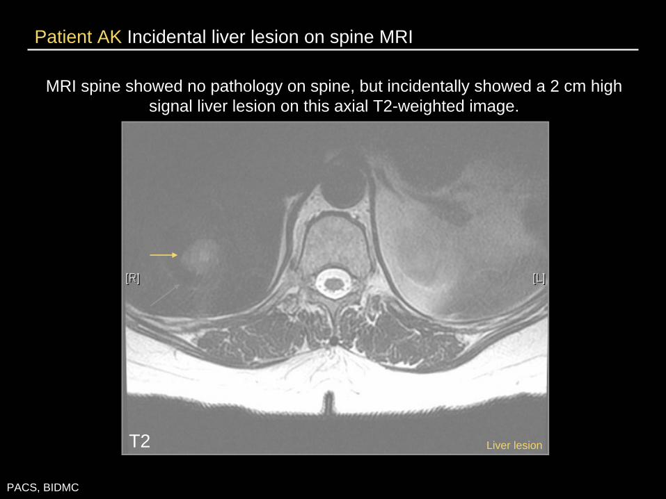
Patient AK Incidental liver lesion on spine MRI
PACS, BIDMC
MRI spine showed no pathology on spine, but incidentally showed a 2 cm high signal liver lesion on this axial T2-weighted image.
T2 Liver lesion
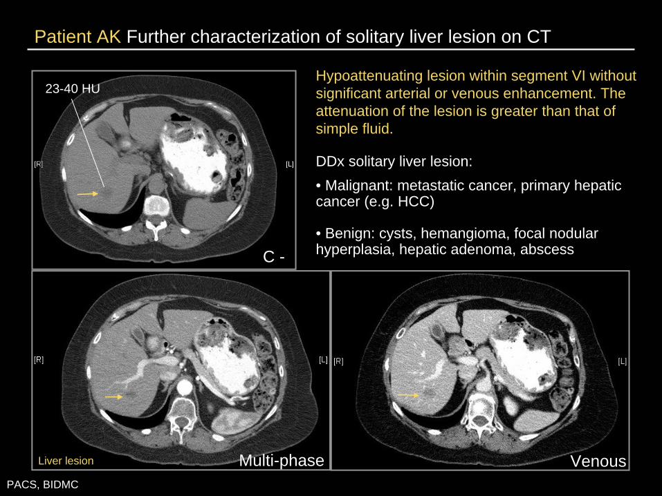
Patient AK Further characterization of solitary liver lesion on CT
Hypoattenuating lesion within segment VI without significant arterial or venous enhancement. The attenuation of the lesion is greater than that of simple fluid.
DDx solitary liver lesion: • Malignant: metastatic cancer, primary hepatic cancer (e.g. HCC)
• Benign: cysts, hemangioma, focal nodular hyperplasia, hepatic adenoma, abscess
PACS, BIDMC
Multi-phase Venous
C -
23-40 HU
Liver lesion
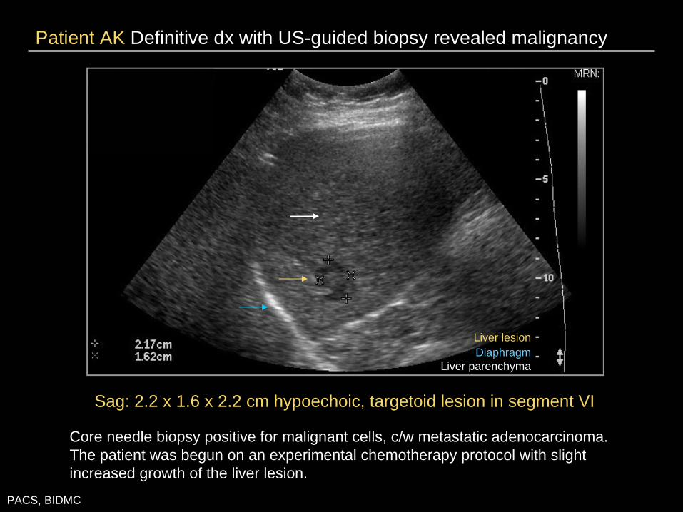
Patient AK Definitive dx with US-guided biopsy revealed malignancy
Sag: 2.2 x 1.6 x 2.2 cm hypoechoic, targetoid lesion in segment VI
Core needle biopsy positive for malignant cells, c/w metastatic adenocarcinoma. The patient was begun on an experimental chemotherapy protocol with slight increased growth of the liver lesion.
PACS, BIDMC
Liver lesionDiaphragm
Liver parenchyma
Companion patient Let’s look at another case with a different presentation of liver metastasis
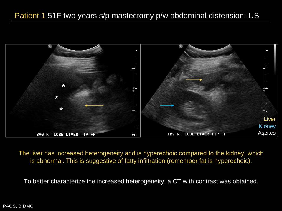
Patient 1 51F two years s/p mastectomy p/w abdominal distension: US
PACS, BIDMC
***
The liver has increased heterogeneity and is hyperechoic compared to the kidney, which is abnormal. This is suggestive of fatty infiltration (remember fat is hyperechoic).
LiverKidneyAscites
To better characterize the increased heterogeneity, a CT with contrast was obtained.
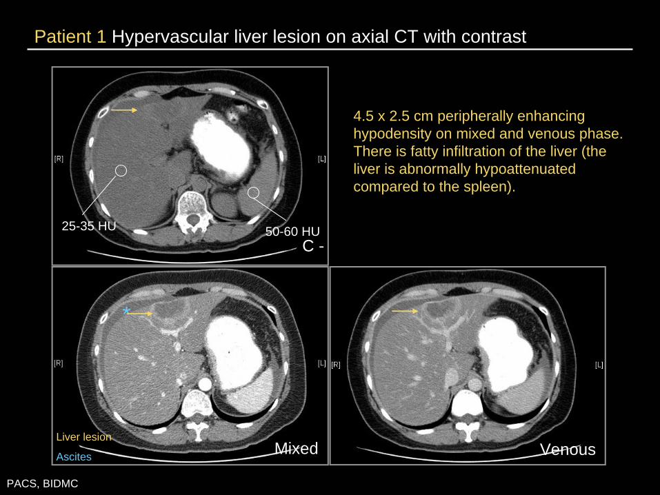
Patient 1 Hypervascular liver lesion on axial CT with contrast
PACS, BIDMC
4.5 x 2.5 cm peripherally enhancing hypodensity on mixed and venous phase. There is fatty infiltration of the liver (the liver is abnormally hypoattenuated compared to the spleen).
C -25-35 HU 50-60 HU
VenousMixed
*
Liver lesion
Ascites
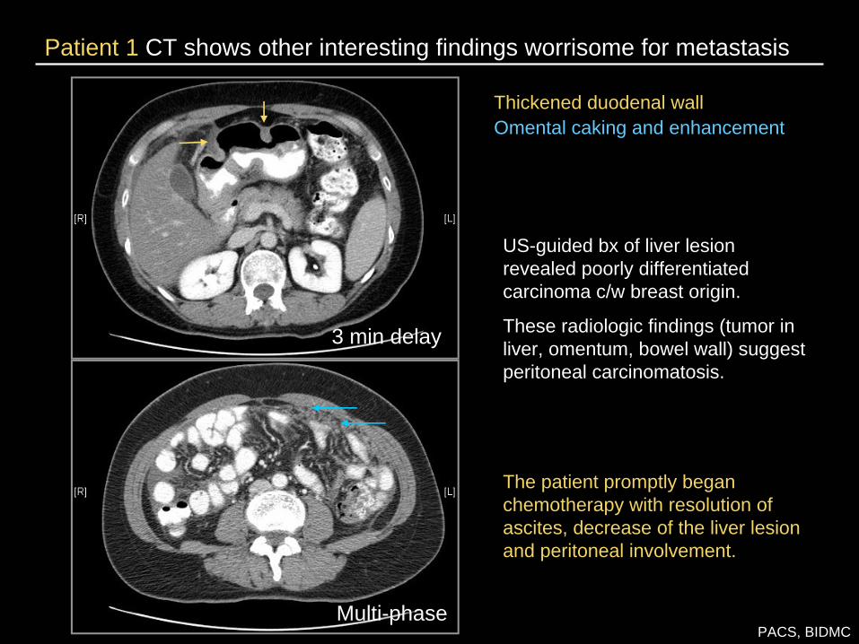
Patient 1 CT shows other interesting findings worrisome for metastasis
PACS, BIDMC
Thickened duodenal wallOmental caking and enhancement
US-guided bx of liver lesion revealed poorly differentiated carcinoma c/w breast origin.
These radiologic findings (tumor in liver, omentum, bowel wall) suggest peritoneal carcinomatosis.
The patient promptly began chemotherapy with resolution of ascites, decrease of the liver lesion and peritoneal involvement.
3 min delay
Multi-phase
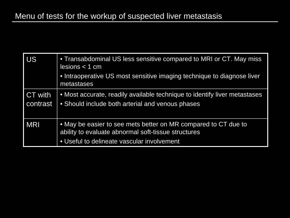
Menu of tests for the workup of suspected liver metastasis
US • Transabdominal US less sensitive compared to MRI or CT. May miss lesions < 1 cm• Intraoperative US most sensitive imaging technique to diagnose liver metastases
CT with contrast
• Most accurate, readily available technique to identify liver metastases• Should include both arterial and venous phases
MRI • May be easier to see mets better on MR compared to CT due to ability to evaluate abnormal soft-tissue structures• Useful to delineate vascular involvement
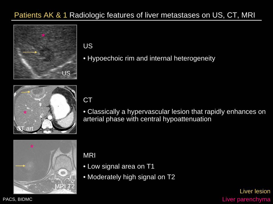
Patients AK & 1 Radiologic features of liver metastases on US, CT, MRI
US
• Hypoechoic rim and internal heterogeneity
CT
• Classically a hypervascular lesion that rapidly enhances on arterial phase with central hypoattenuation
MRI• Low signal area on T1• Moderately high signal on T2
Liver lesionLiver parenchyma
MRI T2
*
CT art
*
US
*
PACS, BIDMC

Metastatic breast cancer to the skeleton How breast cancer looks when it involves the bone, the most commonly affected organ
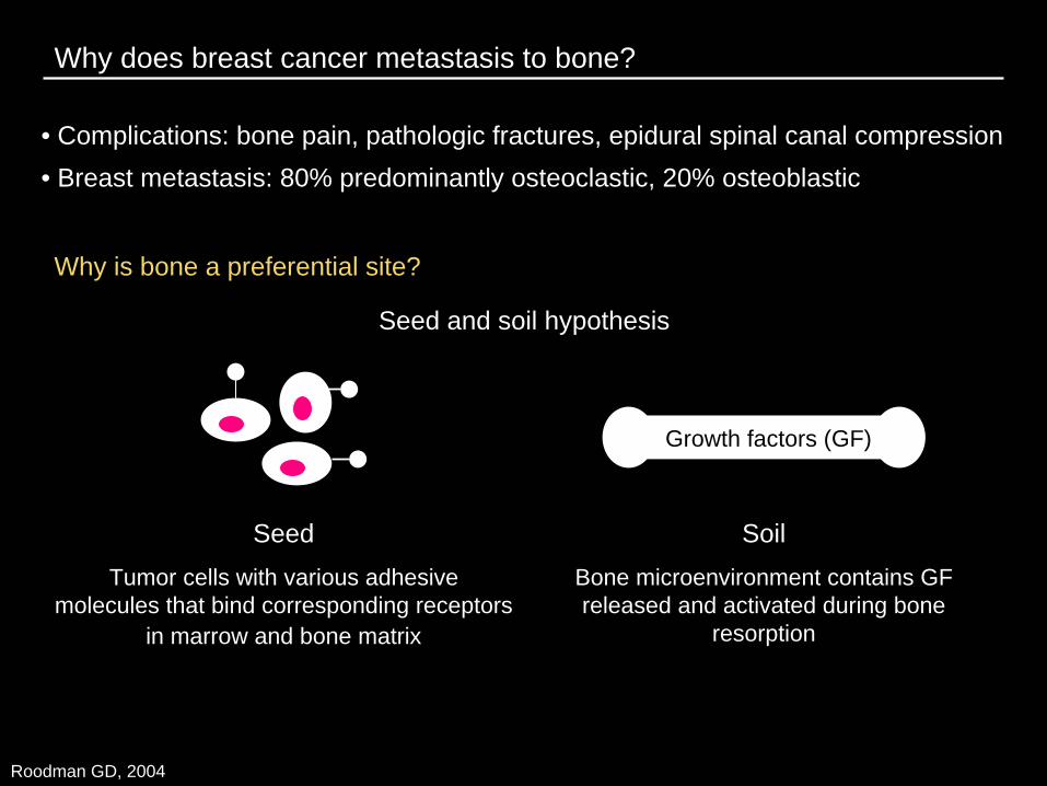
Why does breast cancer metastasis to bone?
• Complications: bone pain, pathologic fractures, epidural spinal canal compression• Breast metastasis: 80% predominantly osteoclastic, 20% osteoblastic
Why is bone a preferential site?
Seed Tumor cells with various adhesive
molecules that bind corresponding receptors in marrow and bone matrix
Soil Bone microenvironment contains GF released and activated during bone
resorption
Growth factors (GF)
Seed and soil hypothesis
Roodman GD, 2004
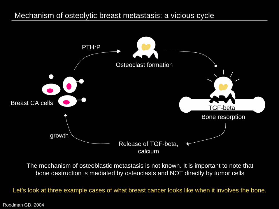
Mechanism of osteolytic breast metastasis: a vicious cycle
The mechanism of osteoblastic metastasis is not known. It is important to note that bone destruction is mediated by osteoclasts and NOT directly by tumor cells
Breast CA cellsTGF-beta
PTHrP
Osteoclast formation
Bone resorption
Release of TGF-beta, calcium
growth
Roodman GD, 2004
Let’s look at three example cases of what breast cancer looks like when it involves the bone.
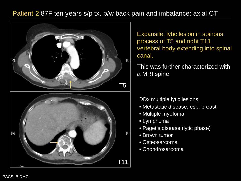
Patient 2 87F ten years s/p tx, p/w back pain and imbalance: axial CT
PACS, BIDMC
DDx multiple lytic lesions: • Metastatic disease, esp. breast• Multiple myeloma• Lymphoma• Paget’s disease (lytic phase)• Brown tumor• Osteosarcoma• Chondrosarcoma
T11
T5
Expansile, lytic lesion in spinous process of T5 and right T11 vertebral body extending into spinal canal.
This was further characterized with a MRI spine.

Patient 2 Epidural spinal cord compression on sagittal MRI spine
PACS, BIDMC
T2
T11
Spinal cord compression at T5Pathological fracture associated with T11
T1
T5
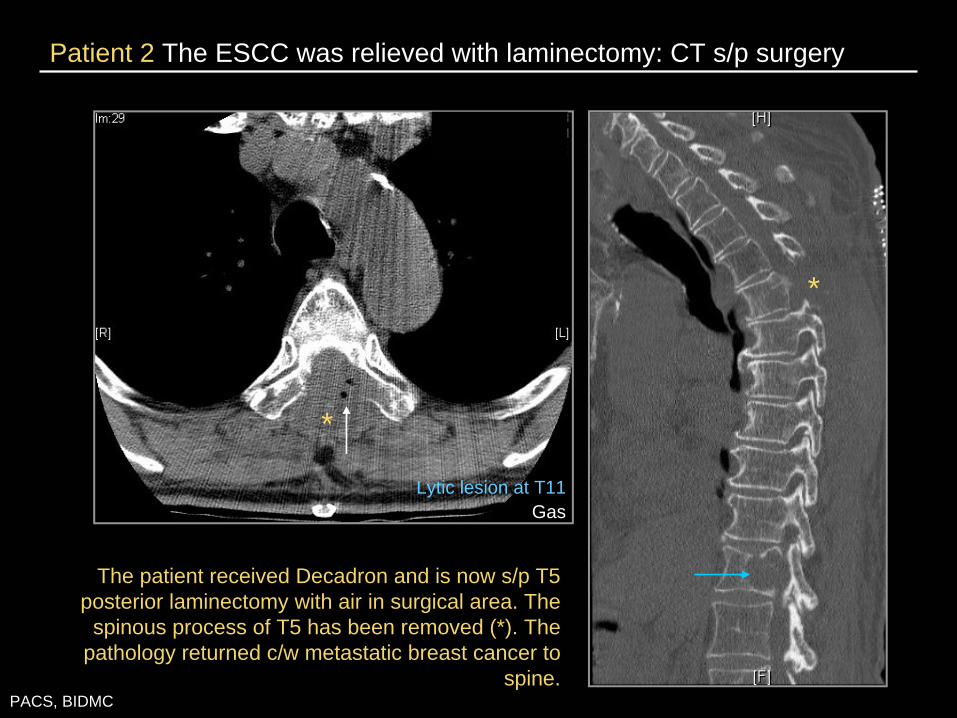
Patient 2 The ESCC was relieved with laminectomy: CT s/p surgery
PACS, BIDMC
The patient received Decadron and is now s/p T5 posterior laminectomy with air in surgical area. The
spinous process of T5 has been removed (*). The pathology returned c/w metastatic breast cancer to
spine.
*
*
Lytic lesion at T11Gas
Patients 3 & 4 Let’s look at other patients with different manifestations of bone metastasis
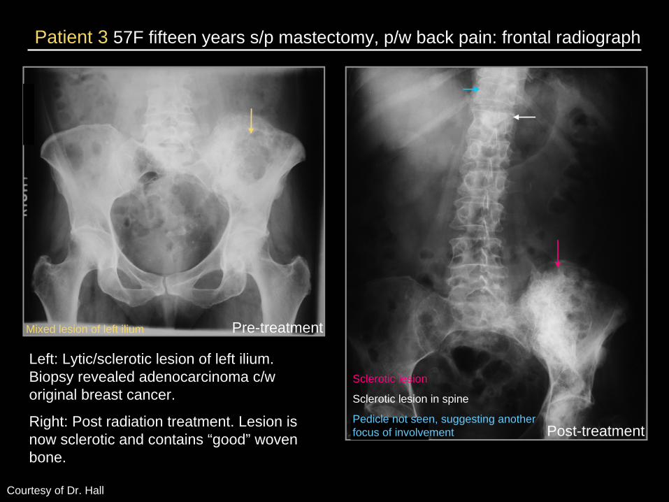
Patient 3 57F fifteen years s/p mastectomy, p/w back pain: frontal radiograph
Courtesy of Dr. Hall
Left: Lytic/sclerotic lesion of left ilium. Biopsy revealed adenocarcinoma c/w original breast cancer.
Right: Post radiation treatment. Lesion is now sclerotic and contains “good” woven bone.
Post-treatment
Sclerotic lesion
Sclerotic lesion in spine
Pedicle not seen, suggesting another focus of involvement
Mixed lesion of left ilium Pre-treatment
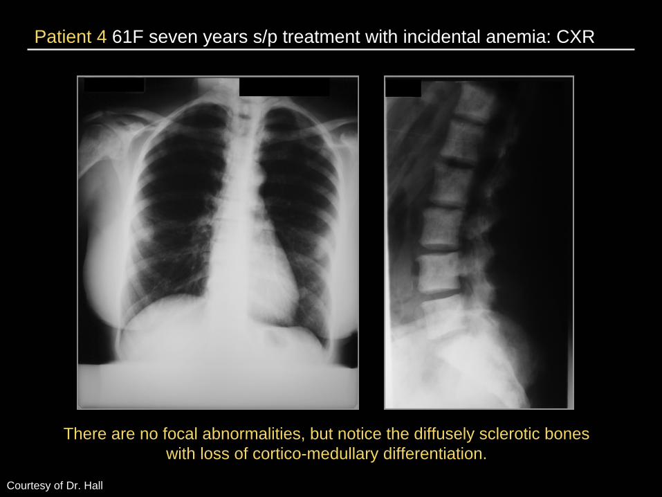
Patient 4 61F seven years s/p treatment with incidental anemia: CXR
Courtesy of Dr. Hall
There are no focal abnormalities, but notice the diffusely sclerotic bones with loss of cortico-medullary differentiation.
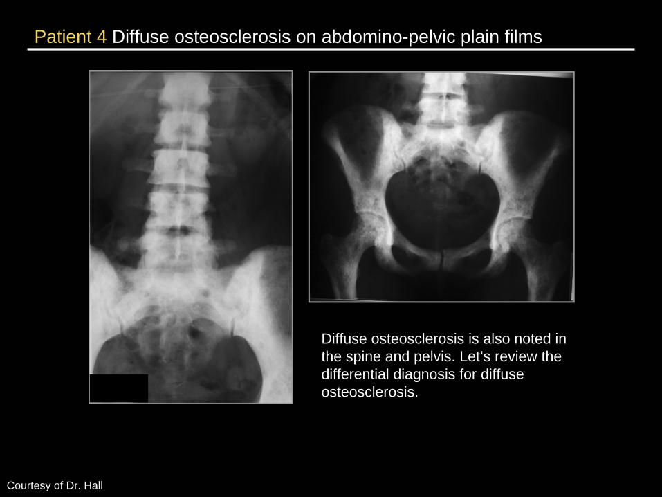
Patient 4 Diffuse osteosclerosis on abdomino-pelvic plain films
Courtesy of Dr. Hall
Diffuse osteosclerosis is also noted in the spine and pelvis. Let’s review the differential diagnosis for diffuse osteosclerosis.
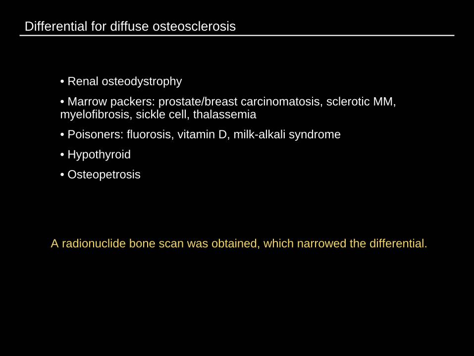
Differential for diffuse osteosclerosis
• Renal osteodystrophy
• Marrow packers: prostate/breast carcinomatosis, sclerotic MM, myelofibrosis, sickle cell, thalassemia
• Poisoners: fluorosis, vitamin D, milk-alkali syndrome
• Hypothyroid
• Osteopetrosis
A radionuclide bone scan was obtained, which narrowed the differential.
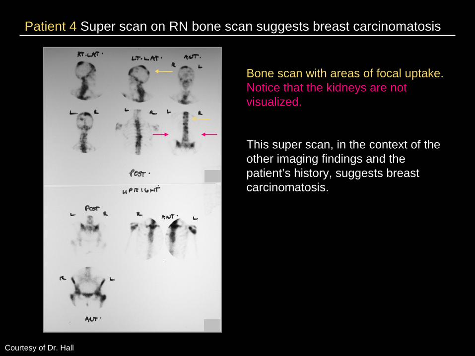
Patient 4 Super scan on RN bone scan suggests breast carcinomatosis
Courtesy of Dr. Hall
Bone scan with areas of focal uptake. Notice that the kidneys are not visualized.
This super scan, in the context of the other imaging findings and the patient’s history, suggests breast carcinomatosis.
Summary
• Mammography is the only imaging modality indicated in surveillance. There is no role for routine use of other studies to evaluate distant recurrence.
• US, CT with contrast, and MRI are often used to detect and evaluate a solitary liver lesion.
• Osteolytic breast metastasis, which predominates in 80% of cases with breast metastasis to the bone, is a vicious cycle of growth factor release and stimulation of tumor growth.
• Metastatic disease is most commonly heralded with symptoms. Workup is based on the constellation of presenting symptoms.
• We reviewed the various radiologic characteristics of breast metastasis to the liver and bone.
References
Esserman LJ and Joe BN. Diagnostic evaluation and initial staging work-up of women with suspected breast cancer. UpToDate; retrieved July 2008: http://utdol.com.
Novelline RA. Squire’s Fundamentals of Radiology. 6th ed. Harvard University Press, 2004.
Pivot X et al. A retrospective study of first indicators of breast cancer recurrence. Oncology 2000; 58:185-90.
Quon A and Gambhir SS. FDG-PET and beyond: molecular breast imaging. J Clin Oncol 2005; 23(8): 1664-73.
Roodman GD. Mechanisms of bone metastasis. NEJM 2004; 350(16):1655-64.
Rosselli M et al. Intensive diagnostic follow-up after treatment of primary breast cancer. A randomized trial. JAMA 1994; 271(20): 1593-7.
The GIVIO Investigators. Impact of follow-up testing on survival and health-related quality of life in breast cancer patients. A multicenter randomized controlled trial. JAMA 1994; 271(20): 1587-92.

Acknowledgements
Dr. Sham VenkataramanDr. Ferris HallDr. Kevin DonohoeDr. Fargol BooyaDr. Gerald KolodnyLarry BarbarasDr. Christina LeBedisDr. Rich Rana
Michael Larson
Maria LevantakisDr. Gillian Lieberman