metacaspase9 modulates autophagy to confine cell...
TRANSCRIPT
RESEARCH ARTICLE
METACASPASE9 modulates autophagy to confine cell death tothe target cells during Arabidopsis vascular xylem differentiationSacha Escamez, Domenique Andre*, Bo Zhang*, Benjamin Bollhoner, Edouard Pesquet§ andHannele Tuominen‡
ABSTRACTWe uncovered that the level of autophagy in plant cells undergoingprogrammed cell death determines the fate of the surrounding cells.Our approach consisted of using Arabidopsis thaliana cell culturescapable of differentiating into two different cell types: vasculartracheary elements (TEs) that undergo programmed cell death(PCD) and protoplast autolysis, and parenchymatic non-TEs thatremain alive. The TE cell type displayed higher levels of autophagywhen expression of the TE-specific METACASPASE9 (MC9) wasreduced using RNAi (MC9-RNAi). Misregulation of autophagy in theMC9-RNAi TEs coincided with ectopic death of the non-TEs, implyingthe existence of an autophagy-dependent intercellular signallingfrom within the TEs towards the non-TEs. Viability of the non-TEswas restored when AUTOPHAGY2 (ATG2) was downregulatedspecifically in MC9-RNAi TEs, demonstrating the importance ofautophagy in the spatial confinement of cell death. Our resultssuggest that other eukaryotic cells undergoing PCD might also needto tightly regulate their level of autophagy to avoid detrimentalconsequences for the surrounding cells.
KEY WORDS: Arabidopsis thaliana, Autophagy, Intercellularsignalling, Metacaspase, Programmed cell death, Tracheary element
INTRODUCTIONThe development of multicellular organisms involves programmedcell death (PCD). In land plants, PCD – followed by completecellular autolysis – likely represented the first evolutionary stepin the acquisition of specialized water-conducting structures thatwere necessary for survival in the dry atmosphere (Escamez andTuominen, 2014; Friedman and Cook, 2000). Nowadays higherplants possess an efficient vascular tissue called xylem (Brodribb,2009; Kenrick and Crane, 1997), in which the water-conductingtracheary element (TE) cells deposit cellulosic patterned secondarycell walls (SCW) before cell death (Oda and Fukuda, 2012; Odaet al., 2010; Pesquet et al., 2010; Torrey et al., 1971). TE cell death isbrought about by rupture of the central vacuole (Burgess andLinstead, 1984; Groover and Jones, 1999; Kuriyama, 1999), whichis believed to allow TE autolysis to occur by releasing certain
hydrolytic enzymes and by activating others when the acidiccontents of the vacuole leak into the cytoplasm (Bollhöner et al.,2013; Groover and Jones, 1999; Wodzicki and Brown, 1973). Nomolecular effectors of TE cell death have been identified, butTE post-mortem autolysis has been shown to involve specifichydrolases (Avci et al., 2008; Ito and Fukuda, 2002) includingArabidopsis thaliana METACASPASE9 (MC9) (Bollhöner et al.,2013).
Metacaspases (MCs) are cysteine proteases that are structurallyrelated to metazoan caspases (Uren et al., 2000). Plantmetacaspases are divided into two classes; type I, whichcontains enzymes with a prodomain consisting of both a proline-rich domain and a zinc finger, and type II, which contains MCfamily members without any prodomain. Besides the type IIArabidopsis MC9 which fosters TE post-mortem autolysis, severalmetacaspases have been shown to play a role in different plant celltypes undergoing PCD (Bozhkov et al., 2005; Coll et al., 2010; Heet al., 2008; Minina et al., 2013; Watanabe and Lam, 2011). Inparticular, the cells undergoing PCD in spruce somatic embryosexpress a type II MC which functions upstream of autophagy(Minina et al., 2013). Autophagy is a trafficking route commonlyused by cells for various purposes such as recycling of the cellularcontents during starvation (Mizushima et al., 2004; Mortimore andPoso, 1987; Thompson et al., 2005) and cellular differentiation(Alvarez et al., 2008; Kwon et al., 2010; Mizushima and Levine,2010). However its role in the regulation of cell death has beendebated (Lv et al., 2014). For example, the normal progression ofPCD in spruce embryos requires metacaspase controlledautophagy, although the cell death program itself is not executedby autophagy (Minina et al., 2013). Minina et al. (2013) alsoproposed that other plant cell types undergoing PCD could utilize asimilar process of metacaspase-regulated autophagy.
Autophagy has been claimed to play a crucial role in theprogression of TE PCD (Kwon et al., 2010). However, no publishedstudy has been able to determine whether TEs require autophagy toexecute PCD or whether autophagy is merely required to promoteTE differentiation. Furthermore, numerous studies on autophagyrely on mutants with increased or suppressed autophagy in all celltypes, which does not allow identification of specific regulators andfunctions of autophagy in a particular cell type. In the case of TEs,the function of autophagy remains poorly understood and apotential relation between autophagy and MCs has not beeninvestigated. We therefore hypothesized the existence of a linkbetween MC9 and autophagy during TE differentiation. To test thishypothesis, we utilized an Arabidopsis thaliana in vitro TE cellculture, which allows detailed and specific characterization of TEdifferentiation without interference from the other tissue types. Inthese cell cultures, hormonal stimulus is used to induce part of thecells to differentiate into TEs, while the other cells – hereafter callednon-TEs – stay alive (Pesquet et al., 2010). With the help of thisReceived 21 October 2015; Accepted 7 December 2015
Umeå Plant Science Centre, Department of Plant Physiology, Umeå University,Umeå 90187, Sweden.*Present address: Umeå Plant Science Centre, Department of Forest Genetics andPlant Physiology, Swedish University of Agricultural Sciences, Umeå S-901 83,Sweden. §Present address: Department of Ecology, Environment and PlantSciences, Stockholm University, SE-106 91 Stockholm, Sweden.
‡Author for correspondence ([email protected])
This is an Open Access article distributed under the terms of the Creative Commons AttributionLicense (http://creativecommons.org/licenses/by/3.0), which permits unrestricted use,distribution and reproduction in any medium provided that the original work is properly attributed.
122
© 2016. Published by The Company of Biologists Ltd | Biology Open (2016) 5, 122-129 doi:10.1242/bio.015529
BiologyOpen
by guest on July 16, 2018http://bio.biologists.org/Downloaded from
system we could observe that correct regulation of autophagy byMC9 in TEs is required for spatial confinement of cell death.
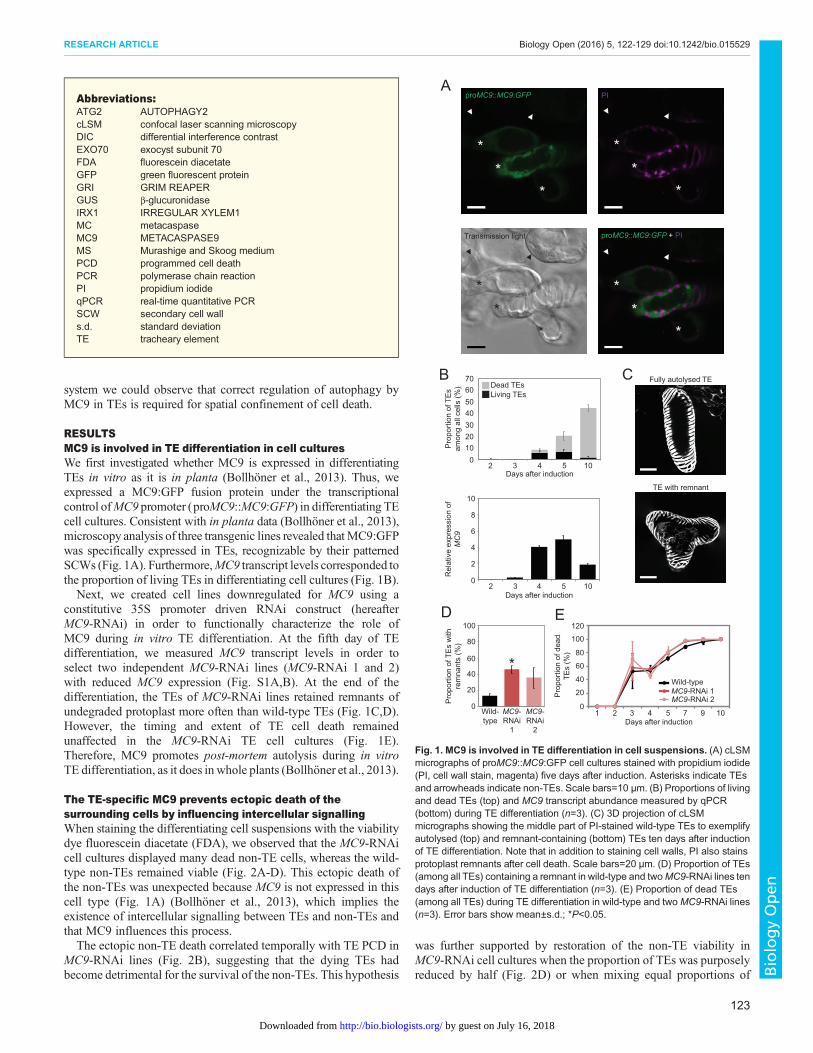
RESULTSMC9 is involved in TE differentiation in cell culturesWe first investigated whether MC9 is expressed in differentiatingTEs in vitro as it is in planta (Bollhöner et al., 2013). Thus, weexpressed a MC9:GFP fusion protein under the transcriptionalcontrol ofMC9 promoter (proMC9::MC9:GFP) in differentiating TEcell cultures. Consistent with in planta data (Bollhöner et al., 2013),microscopy analysis of three transgenic lines revealed thatMC9:GFPwas specifically expressed in TEs, recognizable by their patternedSCWs (Fig. 1A). Furthermore,MC9 transcript levels corresponded tothe proportion of living TEs in differentiating cell cultures (Fig. 1B).Next, we created cell lines downregulated for MC9 using a
constitutive 35S promoter driven RNAi construct (hereafterMC9-RNAi) in order to functionally characterize the role ofMC9 during in vitro TE differentiation. At the fifth day of TEdifferentiation, we measured MC9 transcript levels in order toselect two independent MC9-RNAi lines (MC9-RNAi 1 and 2)with reduced MC9 expression (Fig. S1A,B). At the end of thedifferentiation, the TEs of MC9-RNAi lines retained remnants ofundegraded protoplast more often than wild-type TEs (Fig. 1C,D).However, the timing and extent of TE cell death remainedunaffected in the MC9-RNAi TE cell cultures (Fig. 1E).Therefore, MC9 promotes post-mortem autolysis during in vitroTE differentiation, as it does in whole plants (Bollhöner et al., 2013).
The TE-specific MC9 prevents ectopic death of thesurrounding cells by influencing intercellular signallingWhen staining the differentiating cell suspensions with the viabilitydye fluorescein diacetate (FDA), we observed that the MC9-RNAicell cultures displayed many dead non-TE cells, whereas the wild-type non-TEs remained viable (Fig. 2A-D). This ectopic death ofthe non-TEs was unexpected because MC9 is not expressed in thiscell type (Fig. 1A) (Bollhöner et al., 2013), which implies theexistence of intercellular signalling between TEs and non-TEs andthat MC9 influences this process.The ectopic non-TE death correlated temporally with TE PCD in
MC9-RNAi lines (Fig. 2B), suggesting that the dying TEs hadbecome detrimental for the survival of the non-TEs. This hypothesis
was further supported by restoration of the non-TE viability inMC9-RNAi cell cultures when the proportion of TEs was purposelyreduced by half (Fig. 2D) or when mixing equal proportions of
**
*
**
*
**
*
**
*
A
B
2 5 10
2 5 10
Days after induction
Days after induction
010203040506070
0
2
4
6
8
10
Pro
porti
on o
f TE
sam
ong
all c
ells
(%)
Rel
ativ
e ex
pres
sion
of
MC
9
Dead TEsLiving TEs
proMC9::MC9:GFP
Transmission light
PI
proMC9::MC9:GFP + PI
3 4
3 4
Fully autolysed TE
TE with remnant
C
40
60
80
100
20
0Pro
porti
on o
f TE
s w
ith
rem
nant
s (%
)
Wild-type
MC9-RNAi
1
MC9-RNAi
2
40
60
80
120
20
0
Pro
porti
on o
f dea
dTE
s (%
)
100
1 2 3 4 5 7 9 10Days after induction
Wild-typeMC9-RNAi 1MC9-RNAi 2
*
D E
Fig. 1. MC9 is involved in TE differentiation in cell suspensions. (A) cLSMmicrographs of proMC9::MC9:GFP cell cultures stained with propidium iodide(PI, cell wall stain, magenta) five days after induction. Asterisks indicate TEsand arrowheads indicate non-TEs. Scale bars=10 µm. (B) Proportions of livingand dead TEs (top) and MC9 transcript abundance measured by qPCR(bottom) during TE differentiation (n=3). (C) 3D projection of cLSMmicrographs showing the middle part of PI-stained wild-type TEs to exemplifyautolysed (top) and remnant-containing (bottom) TEs ten days after inductionof TE differentiation. Note that in addition to staining cell walls, PI also stainsprotoplast remnants after cell death. Scale bars=20 µm. (D) Proportion of TEs(among all TEs) containing a remnant in wild-type and twoMC9-RNAi lines tendays after induction of TE differentiation (n=3). (E) Proportion of dead TEs(among all TEs) during TE differentiation in wild-type and twoMC9-RNAi lines(n=3). Error bars show mean±s.d.; *P<0.05.
Abbreviations:ATG2 AUTOPHAGY2cLSM confocal laser scanning microscopyDIC differential interference contrastEXO70 exocyst subunit 70FDA fluorescein diacetateGFP green fluorescent proteinGRI GRIM REAPERGUS β-glucuronidaseIRX1 IRREGULAR XYLEM1MC metacaspaseMC9 METACASPASE9MS Murashige and Skoog mediumPCD programmed cell deathPCR polymerase chain reactionPI propidium iodideqPCR real-time quantitative PCRSCW secondary cell walls.d. standard deviationTE tracheary element
123
RESEARCH ARTICLE Biology Open (2016) 5, 122-129 doi:10.1242/bio.015529
BiologyOpen
by guest on July 16, 2018http://bio.biologists.org/Downloaded from
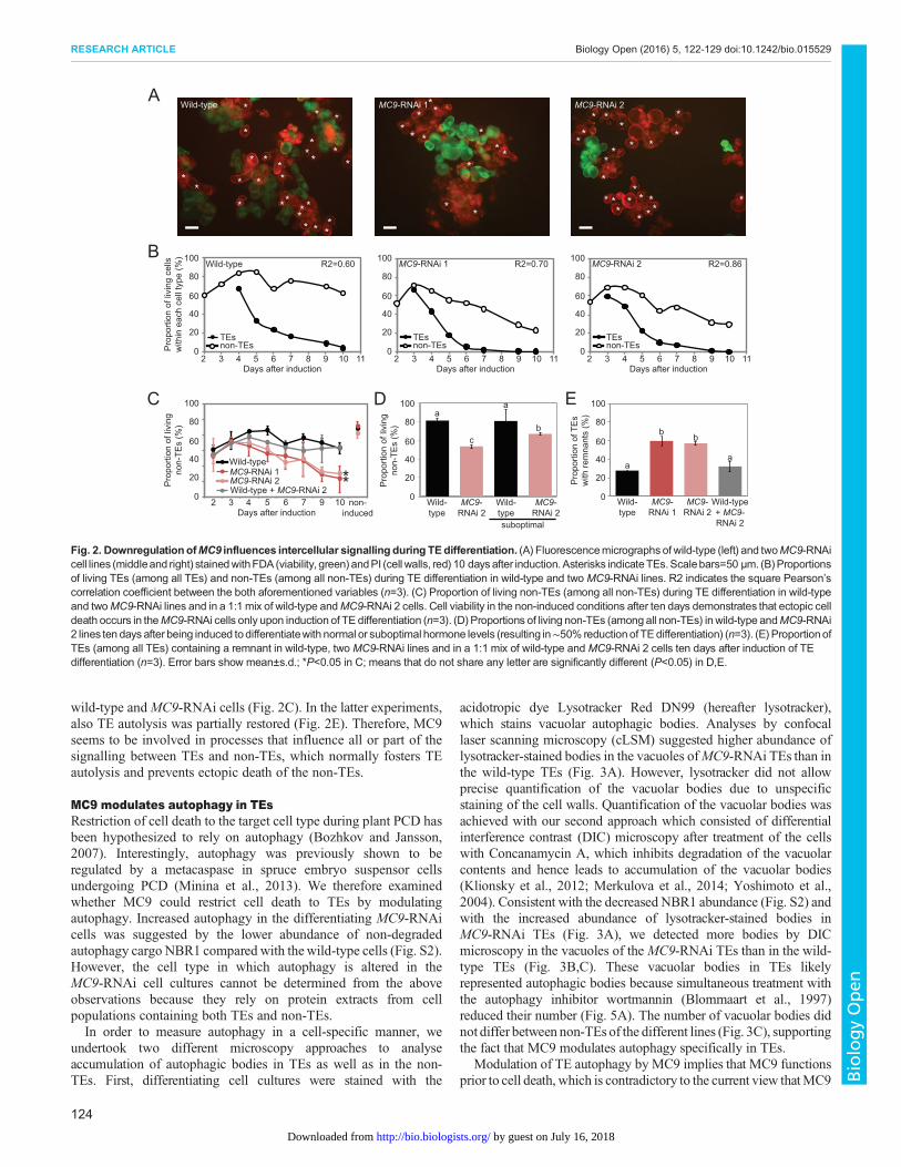
wild-type andMC9-RNAi cells (Fig. 2C). In the latter experiments,also TE autolysis was partially restored (Fig. 2E). Therefore, MC9seems to be involved in processes that influence all or part of thesignalling between TEs and non-TEs, which normally fosters TEautolysis and prevents ectopic death of the non-TEs.
MC9 modulates autophagy in TEsRestriction of cell death to the target cell type during plant PCD hasbeen hypothesized to rely on autophagy (Bozhkov and Jansson,2007). Interestingly, autophagy was previously shown to beregulated by a metacaspase in spruce embryo suspensor cellsundergoing PCD (Minina et al., 2013). We therefore examinedwhether MC9 could restrict cell death to TEs by modulatingautophagy. Increased autophagy in the differentiating MC9-RNAicells was suggested by the lower abundance of non-degradedautophagy cargo NBR1 compared with thewild-type cells (Fig. S2).However, the cell type in which autophagy is altered in theMC9-RNAi cell cultures cannot be determined from the aboveobservations because they rely on protein extracts from cellpopulations containing both TEs and non-TEs.In order to measure autophagy in a cell-specific manner, we
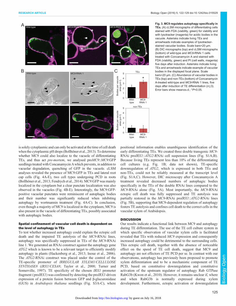
undertook two different microscopy approaches to analyseaccumulation of autophagic bodies in TEs as well as in the non-TEs. First, differentiating cell cultures were stained with the
acidotropic dye Lysotracker Red DN99 (hereafter lysotracker),which stains vacuolar autophagic bodies. Analyses by confocallaser scanning microscopy (cLSM) suggested higher abundance oflysotracker-stained bodies in the vacuoles ofMC9-RNAi TEs than inthe wild-type TEs (Fig. 3A). However, lysotracker did not allowprecise quantification of the vacuolar bodies due to unspecificstaining of the cell walls. Quantification of the vacuolar bodies wasachieved with our second approach which consisted of differentialinterference contrast (DIC) microscopy after treatment of the cellswith Concanamycin A, which inhibits degradation of the vacuolarcontents and hence leads to accumulation of the vacuolar bodies(Klionsky et al., 2012; Merkulova et al., 2014; Yoshimoto et al.,2004). Consistent with the decreased NBR1 abundance (Fig. S2) andwith the increased abundance of lysotracker-stained bodies inMC9-RNAi TEs (Fig. 3A), we detected more bodies by DICmicroscopy in the vacuoles of the MC9-RNAi TEs than in the wild-type TEs (Fig. 3B,C). These vacuolar bodies in TEs likelyrepresented autophagic bodies because simultaneous treatment withthe autophagy inhibitor wortmannin (Blommaart et al., 1997)reduced their number (Fig. 5A). The number of vacuolar bodies didnot differ between non-TEs of the different lines (Fig. 3C), supportingthe fact that MC9 modulates autophagy specifically in TEs.
Modulation of TE autophagy by MC9 implies that MC9 functionsprior to cell death, which is contradictory to the current view thatMC9
A
40
60
80
100
20
0Pro
porti
on o
f liv
ing
cells
w
ithin
eac
h ce
ll ty
pe (%
)
40
60
80
100
20
03 4 5 6 7 8 9 102
40
60
80
100
20
0
Days after induction
TEsnon-TEs
Wild-type MC9-RNAi 1 MC9-RNAi 2
TEsnon-TEs
TEsnon-TEs
113 4 5 6 7 8 9 102Days after induction
113 4 5 6 7 8 9 102Days after induction
11
B
Wild-typeMC9-RNAi 1MC9-RNAi 2Wild-type + MC9-RNAi 2
40
60
80
100
20
0
Pro
porti
on o
f liv
ing
non-
TEs
(%)
2 3 4 5 7 9 10Days after induction
6 Wild-type
MC9-RNAi 2
Wild-type
MC9-RNAi 2
suboptimal
40
60
80
100
20
0
Pro
porti
on o
f liv
ing
non-
TEs
(%)
40
60
80
100
20
0
Pro
porti
on o
f TE
sw
ith re
mna
nts
(%)
Wild-type
MC9-RNAi 1
MC9-RNAi 2
Wild-type + MC9-RNAi 2
**
C D Ea
a
cb b
b
aa
Wild-type MC9-RNAi 2
non-induced
R2=0.60 R2=0.70 R2=0.86
*
MC9-RNAi 1
* *
*
*
*
*
*
*
**
** **** * *
*
***
*
**
*
*
** *
**
*
*
*
*
*
*
*** *
*
**
*
*
***
* **
** **
*** *
Fig. 2. DownregulationofMC9 influences intercellular signalling during TEdifferentiation. (A) Fluorescencemicrographs of wild-type (left) and twoMC9-RNAicell lines (middleand right) stainedwithFDA (viability, green)andPI (cell walls, red) 10 daysafter induction.Asterisks indicateTEs.Scalebars=50 µm. (B)Proportionsof living TEs (among all TEs) and non-TEs (among all non-TEs) during TE differentiation in wild-type and two MC9-RNAi lines. R2 indicates the square Pearson’scorrelation coefficient between the both aforementioned variables (n=3). (C) Proportion of living non-TEs (among all non-TEs) during TE differentiation in wild-typeand twoMC9-RNAi lines and in a 1:1 mix of wild-type andMC9-RNAi 2 cells. Cell viability in the non-induced conditions after ten days demonstrates that ectopic celldeath occurs in theMC9-RNAi cells only upon induction of TE differentiation (n=3). (D) Proportions of living non-TEs (among all non-TEs) inwild-type andMC9-RNAi2 lines ten days after being induced todifferentiatewith normal or suboptimal hormone levels (resulting in∼50% reduction of TEdifferentiation) (n=3). (E) ProportionofTEs (among all TEs) containing a remnant in wild-type, two MC9-RNAi lines and in a 1:1 mix of wild-type and MC9-RNAi 2 cells ten days after induction of TEdifferentiation (n=3). Error bars show mean±s.d.; *P<0.05 in C; means that do not share any letter are significantly different (P<0.05) in D,E.
124
RESEARCH ARTICLE Biology Open (2016) 5, 122-129 doi:10.1242/bio.015529
BiologyOpen
by guest on July 16, 2018http://bio.biologists.org/Downloaded from
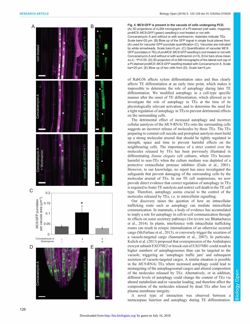
is solely cytoplasmic and can only be activated at the time of cell deathwhen the cytoplasmic pHdrops (Bollhöner et al., 2013). To determinewhether MC9 could also localize to the vacuole of differentiatingTEs, and thus act pre-mortem, we analysed proMC9::MC9:GFPseedlings treatedwith ConcanamycinAwhich prevents, in addition tovacuolar degradation, quenching of GFP in the vacuole. cLSManalyses revealed the presence of MC9:GFP in TEs and lateral rootcap cells (Fig. 4A-E), two cell types undergoing PCD in roots(Bollhöner et al., 2013; Fendrych et al., 2014).MC9:GFPwasmainlylocalized in the cytoplasm but a clear punctate localization was alsoobserved in the vacuoles (Fig. 4B-E). Interestingly, the MC9:GFP-positive vacuolar punctates were reminiscent of autophagic bodiesand their number was significantly reduced when inhibitingautophagy by wortmannin treatment (Fig. 4A-C). In conclusion,even though a majority of MC9 is localized in the cytoplasm,MC9 isalso present in the vacuole of differentiating TEs, possibly associatedwith autophagic bodies.
Spatial confinement of vascular cell death is dependent onthe level of autophagy in TEsTo test whether increased autophagy could explain the ectopic celldeath and the impaired TE autolysis of the MC9-RNAi lines,autophagy was specifically suppressed in TEs of the MC9-RNAiline 1. We generated an RNAi construct against the autophagy geneATG2 which is known to be a relevant target to efficiently modifyautophagy in plants (Hackenberg et al., 2013; Inoue et al., 2006).The ATG2-RNAi construct was placed under the control of theTE-specific promoter of IRREGULAR XYLEM1/CELLULOSESYNTHASE8 (IRX1/CESA8; Taylor et al., 2000; Turner andSomerville, 1997). TE specificity of the chosen IRX1 promoterfragment (proIRX1) was confirmed by detecting the proIRX1-drivenexpression of a protein fusion between GFP and β-glucuronidase(GUS) in Arabidopsis thaliana seedlings (Fig. S3A-C), where
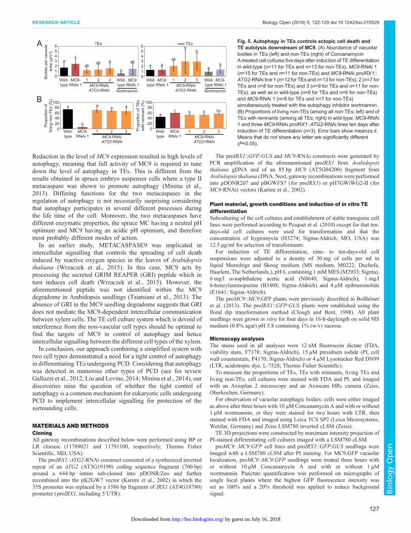
positional information enables unambiguous identification of theearly differentiating TEs. We created three double transgenicMC9-RNAi proIRX1::ATG2-RNAi cell suspension lines (Fig. S1A,B).Because living TEs represent less than 10% of the differentiatingcell cultures (e.g. Fig. 1B; data not shown), TE-specificdownregulation of ATG2, which is expressed in both TEs andnon-TEs, could not be reliably measured at the transcript level(Fig. S1A,C). However, DIC microscopy after Concanamycin Atreatment revealed decreased numbers of autophagic bodiesspecifically in the TEs of the double RNAi lines compared to theMC9-RNAi alone (Fig. 5A). Most importantly, the MC9-RNAiectopic cell death was fully suppressed and TE autolysis waspartially restored in the MC9-RNAi proIRX1::ATG2-RNAi lines(Fig. 5B), supporting that MC9-dependent regulation of autophagyfosters TE autolysis and confines cell death to the correct cells in thevascular xylem of Arabidopsis.
DISCUSSIONOur results indicate a functional link between MC9 and autophagyduring TE differentiation. The use of the TE cell culture system inwhich specific observation of vascular xylem cells is facilitatedrevealed that TEs with reduced MC9 expression and consequentlyincreased autophagy could be detrimental to the surrounding cells.This ectopic cell death, together with the absence of noticeableeffect on the speed of TE cell death, suggest that MC9 andautophagy are not effectors of TE PCD per se. In contrast with ourobservations, autophagy has previously been proposed to promotexylem differentiation and to be a mechanistic component of TEPCD, based on constitutive downregulation and constitutiveactivation of the upstream regulator of autophagy Rab GTPaseRabG3b (Kwon et al., 2010). However, it remains unclear if, whereand when RabG3b is normally expressed during xylemdevelopment. Furthermore, ectopic activation or downregulation
Wild-type MC9-RNAi 1 MC9-RNAi 2
0
1
2
3
4
5
0
1
2
3
4
5
Wild-type
MC9-RNAi 1
Wild-type
MC9-RNAi 1
TEs
non-TEs
Bod
ies
per v
acuo
le a
rea
(µm
)B
odie
s pe
r vac
uole
are
a (µ
m )
*
A
B CWild-type MC9-RNAi 1
*
*
*
*
* * *
*
FDA
+ ly
sotra
cker
FDA
+ P
I
-2-2
Fig. 3. MC9 regulates autophagy specifically inTEs. (A) cLSM micrographs of differentiating cellsstained with FDA (viability, green) for viability andwith lysotracker (magenta) for acidic bodies in thevacuole. Asterisks indicate living TEs andarrowheads indicate examples of lysotracker-stained vacuolar bodies. Scale bars=20 µm.(B) DIC micrographs (top) and cLSM micrographs(bottom) of wild-type and MC9-RNAi 1 cellstreated with Concanamycin A and stained withFDA (viability, green) and PI (cell walls, magenta)five days after induction. Asterisks indicate livingTEs and arrowheads indicate example of vacuolarbodies in the displayed focal plane. Scalebars=20 µm. (C) Abundance of vacuolar bodies inTEs (top) and non-TEs (bottom) of ConcanamycinA-treated wild-type and MC9-RNAi 1 lines, fivedays after induction of TE differentiation (n≥3).Error bars show mean±s.d.; *P<0.05.
125
RESEARCH ARTICLE Biology Open (2016) 5, 122-129 doi:10.1242/bio.015529
BiologyOpen
by guest on July 16, 2018http://bio.biologists.org/Downloaded from
of RabG3b affects xylem differentiation rates and thus clearlyaffects TE differentiation at an early time point, which makes itimpossible to determine the role of autophagy during later TEdifferentiation. We modified autophagy in a cell-type specificmanner after the onset of TE differentiation, which allowed us toinvestigate the role of autophagy in TEs at the time of itsphysiologically relevant activation, and to determine the need fora tight regulation of autophagy in TEs to prevent detrimental effectson the surrounding cells.
The detrimental effect of increased autophagy and incorrectcellular autolysis of the MC9-RNAi TEs onto the surrounding cellssuggests an incorrect release of molecules by these TEs. The TEspreparing to commit cell suicide and protoplast autolysis must buildup a strong molecular arsenal that should be tightly regulated instrength, space and time to prevent harmful effects on theneighbouring cells. The importance of a strict control over themolecules released by TEs has been previously illustrated indifferentiating Zinnia elegans cell cultures, where TEs becameharmful to non-TEs when the culture medium was depleted of aprotective extracellular protease inhibitor (Endo et al., 2001).However, to our knowledge, no report has since investigated thesafeguards that prevent damaging of the surrounding cells by themolecular arsenal of TEs. In our TE cell suspensions, we nowprovide direct evidence that correct regulation of autophagy in TEsis required to foster TE autolysis and restrict cell death to the TE celltype. Therefore, autophagy seems crucial to the control of themolecules released by TEs, i.e. to intercellular signalling.
Our discovery raises the question of how an intracellulartrafficking route such as autophagy can mediate intercellularcommunication. In mammals, a body of evidence has accumulatedto imply a role for autophagy in cell-to-cell communication throughits effects on some secretory pathways (for review see Bhattacharyaet al., 2014). In plants, interference with intracellular traffickingroutes can result in ectopic internalization of an otherwise secretedcargo (McFarlane et al., 2013), or conversely trigger the secretion ofa vacuole-targeted cargo (Sanmartín et al., 2007). In particular,Kulich et al. (2013) proposed that overexpression of the Arabidopsisexocyst subunit EXO70E2 or knock-out of EXO70B1 could result inhigher numbers of autophagosomes than can be targeted to thevacuole, triggering an ‘autophagic traffic jam’ and subsequentsecretion of vacuole-targeted cargos. A similar situation is possiblein the MC9-RNAi TEs where increased autophagy could lead tomistargeting of the autophagosomal cargos and altered compositionof the molecules released by TEs. Alternatively, or in addition,different levels of autophagy could change the content of TEs viaaltered metabolism and/or vacuolar loading, and therefore affect thecomposition of the molecules released by dead TEs after loss ofplasma membrane integrity.
A novel type of interaction was observed between ametacaspase function and autophagy during TE differentiation.
A
* *
*
*
+ +- +
D
B
E
Con
cana
myc
in A
Con
cana
myc
in A
Con
cana
myc
in A
+ W
ortm
anni
n
proM
C9:
:MC
9:G
FPpr
oMC
9::M
C9:
GFP
proM
C9:
:MC
9:G
FP +
PI
proM
C9:
:MC
9:G
FP +
PI
proM
C9:
:MC
9:G
FP +
PI
**
proM
C9:
:MC
9:G
FP +
PI
non-
treat
ed
proM
C9:
:MC
9:G
FP
0
0.05
0.1
0.15
0.2
MC
9:G
FP p
unct
ates
pe
r vac
uole
are
a (µ
m )
Concanamycin AWortmannin
+
--
C
*
-2
Fig. 4. MC9:GFP is present in the vacuole of cells undergoing PCD.(A) 3D projections of cLSM micrographs of a PI-stained (cell walls, magenta)proMC9::MC9:GFP (green) seedling’s root treated or not withConcanamycin A and without or with wortmannin. Asterisks indicate TEs.Scale bars=20 µm. (B) Blow up of the GFP signal in single focal planes from(A) used for vacuolar GFP-punctate quantification (C). Vacuoles are indicatedby white arrowheads. Scale bars=5 µm. (C) Quantification of vacuolar MC9:GFP punctates in TEs of proMC9::MC9:GFP seedling’s root treated or not withConcanamycin A and without or with wortmannin (n=3). Error bars showmean±s.d.; *P<0.05. (D) 3D projection of cLSMmicrographs of the lateral root cap ofa PI-stained proMC9::MC9:GFP seedling treated with Concanamycin A. Scalebar=20 µm. (E) Blow up of two cells from (D). Scale bar=5 µm.
126
RESEARCH ARTICLE Biology Open (2016) 5, 122-129 doi:10.1242/bio.015529
BiologyOpen
by guest on July 16, 2018http://bio.biologists.org/Downloaded from
Reduction in the level of MC9 expression resulted in high levels ofautophagy, meaning that full activity of MC9 is required to tunedown the level of autophagy in TEs. This is different from theresults obtained in spruce embryo suspensor cells where a type IImetacaspase was shown to promote autophagy (Minina et al.,2013). Differing functions for the two metacaspases in theregulation of autophagy is not necessarily surprising consideringthat autophagy participates in several different processes duringthe life time of the cell. Moreover, the two metacaspases havedifferent enzymatic properties, the spruce MC having a neutral pHoptimum and MC9 having an acidic pH optimum, and thereforemost probably different modes of action.In an earlier study, METACASPASE9 was implicated in
intercellular signalling that controls the spreading of cell deathinduced by reactive oxygen species in the leaves of Arabidopsisthaliana (Wrzaczek et al., 2015). In this case, MC9 acts byprocessing the secreted GRIM REAPER (GRI) peptide which inturn induces cell death (Wrzaczek et al., 2015). However, theaforementioned peptide was not identified within the MC9degradome in Arabidopsis seedlings (Tsiatsiani et al., 2013). Theabsence of GRI in the MC9 seedling degradome suggests that GRIdoes not mediate the MC9-dependent intercellular communicationbetween xylem cells. The TE cell culture system which is devoid ofinterference from the non-vascular cell types should be optimal tofind the targets of MC9 in control of autophagy and henceintercellular signalling between the different cell types of the xylem.In conclusion, our approach combining a simplified system with
two cell types demonstrated a need for a tight control of autophagyin differentiating TEs undergoing PCD. Considering that autophagywas detected in numerous other types of PCD (see for reviewGalluzzi et al., 2012; Liu and Levine, 2014;Minina et al., 2014), ourdiscoveries raise the question of whether the tight control ofautophagy is a common mechanism for eukaryotic cells undergoingPCD to implement intercellular signalling for protection of thesurrounding cells.
MATERIALS AND METHODSCloningAll gateway recombinations described below were performed using BP orLR clonase (11789021 and 11791100, respectively; Thermo FisherScientific, MD, USA).
The proIRX1::ATG2-RNAi construct consisted of a synthesized invertedrepeat of an ATG2 (AT3G19190) coding sequence fragment (700 bp)around a 644 bp intron sub-cloned into pDONR/Zeo and furtherrecombined into the pK2GW7 vector (Karimi et al., 2002) in which the35S promoter was replaced by a 1586 bp fragment of IRX1 (AT4G18780)promoter (proIRX1, including 5′UTR).
The proIRX1::GFP:GUS and MC9-RNAi constructs were generated byPCR amplification of the aforementioned proIRX1 from Arabidopsisthaliana gDNA and of an 85 bp MC9 (AT5G04200) fragment fromArabidopsis thaliana cDNA. Next, gateway recombinations were performedinto pDONR207 and pBGWFS7 (for proIRX1) or pH7GWiWG2-II (forMC9-RNAi) vectors (Karimi et al., 2002).
Plant material, growth conditions and induction of in vitro TEdifferentiationSubculturing of the cell cultures and establishment of stable transgenic celllines were performed according to Pesquet et al. (2010) except for that ten-days-old cell cultures were used for transformation and that theconcentration of hygromycin (H3274; Sigma-Aldrich, MO, USA) was12.5 µg/ml for selection of transformants.
For induction of TE differentiation, nine- to ten-days-old cellsuspensions were adjusted to a density of 30 mg of cells per ml inliquid Murashige and Skoog medium (MS medium, M0222; Duchefa,Haarlem, The Netherlands,), pH 6, containing 1 mMMES (M2933; Sigma),6 mg/l α-naphthalene acetic acid (N0640; Sigma-Aldrich), 1 mg/l6-benzylaminopurine (B3408; Sigma-Aldrich) and 4 μM epibrassinolide(E1641; Sigma-Aldrich).
The proMC9::MC9:GFP plants were previously described in Bollhöneret al. (2013). The proIRX1::GFP:GUS plants were established using thefloral dip transformation method (Clough and Bent, 1998). All plantseedlings were grown in vitro for four days in 16-h-daylength on solid MSmedium (0.8% agar) pH 5.8 containing 1% (w/v) sucrose.
Microscopy analysesThe stains used in all analyses were 12 nM fluorescein dictate (FDA,viability stain, F7378; Sigma-Aldrich), 15 µM presidium iodide (PI, cellwall counterstain, P4170; Sigma-Aldrich) or 4 µM Lysotracker Red DN99(LTR, acidotropic dye, L-7528; Thermo Fisher Scientific).
To measure the proportions of TEs, TEs with remnants, living TEs andliving non-TEs, cell cultures were stained with FDA and PI, and imagedwith an Axioplan 2 microscope and an Axiocam HRc camera (Zeiss,Oberkochen, Germany).
For observation of vacuolar autophagic bodies: cells were either imagedas above after three hours with 10 µM Concanamycin A and with or without1 µM wortmannin, or they were stained for two hours with LTR, thenstained with FDA and imaged using Leica TCS SP2 (Leica Microsystems,Wetzlar, Germany) and Zeiss LSM780 inverted cLSM (Zeiss).
TE 3D projections were constructed by maximum intensity projection ofPI-stained differentiating cell cultures imaged with a LSM780 cLSM.
proMC9::MC9:GFP cell lines and proIRX1::GFP:GUS seedlings wereimaged with a LSM780 cLSM after PI staining. For MC9:GFP vacuolarlocalization, proMC9::MC9:GFP seedlings were treated three hours withor without 10 µM Concanamycin A and with or without 1 µMwortmannin. Punctate quantification was performed on micrographs ofsingle focal planes where the highest GFP fluorescence intensity wasset as 100% and a 20% threshold was applied to reduce backgroundsignal.
0
2
4
6
1
Bod
ies
per v
acuo
le a
rea
(µm
)
Wild-type
MC9-RNAi 1
1 Wild-type
MC9-RNAi 1
ac
abab a
b
aba a
ab
bb
aa
TEs non-TEs
020406080
100
020406080
100
Pro
porti
on o
fliv
ing
non-
TEs
(%)
Pro
porti
on o
f TE
sw
ith re
mna
nt (%
)
a a a a
b
a b b bcc
A
BWortmannin
-2
MC9-RNAiATG2-RNAi
2 3 Wild-type
MC9-RNAi 1
1 Wild-type
MC9-RNAi 1
WortmanninMC9-RNAiATG2-RNAi
2 3
Wild-type
MC9-RNAi 1
1MC9-RNAiATG2-RNAi
2 3 Wild-type
MC9-RNAi 1
1MC9-RNAiATG2-RNAi
2 3
3
5
0
2
4
6
1
3
5
Fig. 5. Autophagy in TEs controls ectopic cell death andTE autolysis downstream of MC9. (A) Abundance of vacuolarbodies in TEs (left) and non-TEs (right) of ConcanamycinA-treated cell cultures five days after induction of TE differentiationin wild-type (n=11 for TEs and n=13 for non-TEs), MC9-RNAi 1(n=15 for TEs and n=11 for non-TEs) and MC9-RNAi proIRX1::ATG2-RNAi line 1 (n=12 for TEs and n=13 for non-TEs), 2 (n=7 forTEs and n=8 for non-TEs) and 3 (n=9 for TEs and n=11 for non-TEs), as well as in wild-type (n=6 for TEs and n=6 for non-TEs)and MC9-RNAi 1 (n=8 for TEs and n=7 for non-TEs)simultaneously treated with the autophagy inhibitor wortmannin.(B) Proportions of living non-TEs (among all non-TEs; left) and ofTEs with remnants (among all TEs; right) in wild-type, MC9-RNAi1 and three MC9-RNAi proIRX1::ATG2-RNAi lines ten days afterinduction of TE differentiation (n=3). Error bars show mean±s.d.Means that do not share any letter are significantly different(P<0.05).
127
RESEARCH ARTICLE Biology Open (2016) 5, 122-129 doi:10.1242/bio.015529
BiologyOpen
by guest on July 16, 2018http://bio.biologists.org/Downloaded from
Histochemical GUS staining of proIRX1::GFP:GUS seedlings wasperformed according to Bollhöner et al. (2013).
Semi-quantitative and quantitative PCR analysesTotal RNA was isolated from frozen cell homogenates using QiagenRNeasy Plant MiniKit (74904; Qiagen, Hilden, Germany). 0.5 µg totalRNA were used for cDNA synthesis using QuantiTect ReverseTranscription Kit (205313; Qiagen). qPCR reactions were run in a RocheLightCycler 480 (Roche, Basel, Switzerland) using 20 µl reactionscontaining 5 µl of 25 times diluted cDNA, 200 nM primers and iQSybrGreen Supermix (1708880; Bio-Rad, CA, USA). Forty cycles (95°C,10 s; 55°C, 20 s; 72°C, 30 s) were applied to all genes, and MC9 (forwardprimer: 5′-CGACATCGGCACACATCTAC-3′; reverse primer: 5′-TGGC-ACCTTCATTTTCGTTC-3′) expression was normalized to two referencegenes (combined by geometric mean): UBQ10 (AT4G05320; forwardprimer: 5′-GGCCTTGTATAATCCCTGAT-3′; reverse primer: 5′-AAAG-AGATAACAGGAACGGA-3′) and SAND (AT2G28390; forward primer:5′-AACTCTATGCAGCATTTGATCCACT-3′; reverse primer: 5′-TGAT-TGCATATCTTTATCGCCATC-3′).
Semi-quantitative PCR reactions were also run in 20 µl containing 5 µl of25 times diluted cDNA, 200 nM primers and Go Taq Green Master Mix(M7123; Promega Corporation, WI, USA). Using a T100 Thermal Cycler(Bio-Rad), 30 cycles (95°C, 15 s; 55°C, 15 s; 72°C, 30 s) were applied forUBQ10 reference gene (same primers as above) while 35 cycles wereapplied for MC9 (same primers as above) and for ATG2 (forward primer:5′-GTGCATGCTGCTGGAATCTA-3′; reverse primer: 5′-GTGTGCGG-ACTAATGCAGAA-3′). The corresponding products were loaded in 3%agarose gel containing 0.05% (v/v) Midori Green staining (MG-04; NipponGenetics Europe GmbH, Germany) for electrophoresis (30 min at 100 V).Gel imaging was performed using Techtum Gel Photo System (Techtum,Sweden) and Gel-Pro Analyzer (Media Cybernetics Inc, MD, USA).
Protein biochemistryAll analyses were performed from pools of three biological replicates.
Tomeasure abundance of NBR1 (Svenning et al., 2011), SDS-PAGEwasrun using 30 µg of total proteins. The proteins were stained in gel usingCoomassie Brilliant Blue R-250 (161-0435; Bio-Rad), or transferred onto anitrocellulose membrane (Bio-Rad) for immunoblotting. The membranewas probed with 1:1000 diluted anti-NBR1 antibodies, followed by probingwith 1:5000 diluted anti-rabbit horseradish peroxidase (HRP)-conjugatedantibodies (sc-2301; Santa Cruz Biotechnology, Inc., TX, USA). Theimmunoblot was revealed using SuperSignal West Pico ChemiluminescentSubstrate (Thermo Fisher Scientific) and imaged with a CCD camera(LAS-3000; FujiFilm, Japan).
Statistical analysesAll charts displaying error bars correspond to mean values and standarddeviations (s.d.) for at least three biological replicates. Data in Fig. 2D,E andFig. 5A,B were analysed by one-way ANOVA followed by Fisher’s test(two-tailed) using Minitab 17 (Cleverbridge AG, Germany). Means that donot share any letter are significantly different (P<0.05). For all other chartsdisplaying error bars and comparing wild-type (or control) andMC9-RNAilines (or treatments), Welch-corrected t-tests (two-tailed) were performedand an asterisk indicates significant difference compared to wild-type(P<0.05).
AcknowledgementsWe thank Dr Thomas Dobrenel for technical advice, and Dr Luiz Muniz, DelphineMenard and Hendrik Reuper for technical help. We thank Prof. Terje Johansen(University of Tromsø, Norway) for providing us with anti-NBR1 antibody.
Competing interestsThe authors declare no competing or financial interests.
Author contributionsB.B. created the proMC9::MC9-GFP construct and corresponding plants. B.Z.performed the protease activity assay. S.E and D.A. performed the experiments ofFigs 1 and 2. S.E. performed all other experiments. E.P., B.B. and B.Z. assisted S.E.
and H.T. with experimental design. E.P. and D.A. assisted S.E. and H.T. with writingthe manuscript.
FundingThis work was supported by Formas [grant number 232-2009-1698];Vetenskapsrådet [grant number 621-2013-4949]; and Swedish Energy Agency[grant number 2010-000431]. We also thank Umeå Plant Science Centre BerzeliiCentre in Forest Biotechnology, funded by the Swedish research councilVetenskapsrådet and the Swedish Governmental Agency for Innovation SystemsVinnova, as well as Bio4Energy – a strategic research environment appointed by theSwedish government – for financial support.
Supplementary informationSupplementary information available online athttp://bio.biologists.org/lookup/suppl/doi:10.1242/bio.015529/-/DC1
ReferencesAlvarez, V. E., Kosec, G., Sant’Anna, C., Turk, V., Cazzulo, J. J. and Turk, B.
(2008). Autophagy is involved in nutritional stress response and differentiation inTrypanosoma cruzi. J. Biol. Chem. 283, 3454-3464.
Avci, U., Petzold, H. E., Ismail, I. O., Beers, E. P. and Haigler, C. H. (2008).Cysteine proteases XCP1 and XCP2 aid micro-autolysis within the intact centralvacuole during xylogenesis in Arabidopsis roots. Plant J. 56, 303-315.
Bhattacharya, A., Prakash, Y. S. and Eissa, N. T. (2014). Secretory function ofautophagy in innate immune cells. Cell. Microbiol. 16, 1637-1645.
Blommaart, E. F. C., Krause, U., Schellens, J. P. M., Vreeling-Sindelarova, H.and Meijer, A. J. (1997). The phosphatidylinositol 3-kinase inhibitors wortmanninand LY294002 inhibit autophagy in isolated rat hepatocytes.Eur. J. Biochem. 243,240-246.
Bollhoner, B., Zhang, B., Stael, S., Denance, N., Overmyer, K., Goffner, D., VanBreusegem, F. and Tuominen, H. (2013). Post mortem function of AtMC9 inxylem vessel elements. New Phytol. 200, 498-510.
Bozhkov, P. and Jansson, C. (2007). Autophagy and cell-death proteases inplants: two wheels of a funeral cart. Autophagy 3, 136-138.
Bozhkov, P. V., Suarez, M. F., Filonova, L. H., Daniel, G., Zamyatnin, A. A.,Rodriguez-Nieto, S., Zhivotovsky, B. and Smertenko, A. (2005). Cysteineprotease mcII-Pa executes programmed cell death during plant embryogenesis.Proc. Natl. Acad. Sci. USA 102, 14463-14468.
Brodribb, T. J. (2009). Xylem hydraulic physiology: the functional backbone ofterrestrial plant productivity. Plant Sci. 177, 245-251.
Burgess, J. and Linstead, P. (1984). In-vitro tracheary element formation: structuralstudies and the effect of tri-iodobenzoic acid. Planta 160, 481-489.
Clough, S. J. and Bent, A. F. (1998). Floral dip: a simplified method forAgrobacterium-mediated transformation of Arabidopsis thaliana. Plant J. 16,735-743.
Coll, N. S., Vercammen, D., Smidler, A., Clover, C., Van Breusegem, F., Dangl,J. L. and Epple, P. (2010). Arabidopsis type I metacaspases control cell death.Science 330, 1393-1397.
Endo, S., Demura, T. and Fukuda, H. (2001). Inhibition of proteasome activity bythe TED4 protein in extracellular space: a novel mechanism for protection of livingcells from injury caused by dying cells. Plant Cell Physiol. 42, 9-19.
Escamez, S. and Tuominen, H. (2014). Programmes of cell death and autolysis intracheary elements: when a suicidal cell arranges its own corpse removal. J. Exp.Bot. 65, 1313-1321.
Fendrych,M., VanHautegem, T., VanDurme,M., Olvera-Carrillo, Y., Huysmans,M., Karimi, M., Lippens, S., Guerin, C. J., Krebs, M., Schumacher, K. et al.(2014). Programmed cell death controlled by ANAC033/SOMBRERO determinesroot cap organ size in Arabidopsis. Curr. Biol. 24, 931-940.
Friedman,W. E. and Cook, M. E. (2000). The origin and early evolution of tracheidsin vascular plants: integration of palaeobotanical and neobotanical data. Philos.Trans. R. Soc. Lond. B Biol. Sci. 355, 857-868.
Galluzzi, L., Vitale, I., Abrams, J. M., Alnemri, E. S., Baehrecke, E. H.,Blagosklonny, M. V., Dawson, T. M., Dawson, V. L., El-Deiry, W. S., Fulda,S. et al. (2012). Molecular definitions of cell death subroutines: recommendationsof the Nomenclature Committee on Cell Death 2012. Cell Death Differ. 19,107-120.
Groover, A. and Jones, A. M. (1999). Tracheary element differentiation uses anovel mechanism coordinating programmed cell death and secondary cell wallsynthesis. Plant Physiol. 119, 375-384.
Hackenberg, T., Juul, T., Auzina, A., Gwizdz, S., Małolepszy, A., Van Der Kelen,K., Dam, S., Bressendorff, S., Lorentzen, A., Roepstorff, P. et al. (2013).Catalase and NO CATALASE ACTIVITY1 promote autophagy-dependent celldeath in Arabidopsis. Plant Cell 25, 4616-4626.
He, R., Drury, G. E., Rotari, V. I., Gordon, A., Willer, M., Farzaneh, T., Woltering,E. J. and Gallois, P. (2008). Metacaspase-8 modulates programmed cell deathinduced by ultraviolet light and H2O2 in Arabidopsis. J. Biol. Chem. 283, 774-783.
Inoue, Y., Suzuki, T., Hattori, M., Yoshimoto, K., Ohsumi, Y. and Moriyasu, Y.(2006). AtATG genes, homologs of yeast autophagy genes, are involved in
128
RESEARCH ARTICLE Biology Open (2016) 5, 122-129 doi:10.1242/bio.015529
BiologyOpen
by guest on July 16, 2018http://bio.biologists.org/Downloaded from
constitutive autophagy in Arabidopsis root tip cells. Plant Cell Physiol. 47,1641-1652.
Ito, J. and Fukuda, H. (2002). ZEN1 is a key enzyme in the degradation of nuclearDNA during programmed cell death of tracheary elements. Plant Cell 14,3201-3211.
Karimi, M., Inze, D. and Depicker, A. (2002). GATEWAY™ vectors forAgrobacterium-mediated plant transformation. Trends Plant Sci. 7, 193-195.
Kenrick, P. andCrane, P. R. (1997). The origin and early evolution of plants on land.Nature 389, 33-39.
Klionsky, D. J., Abdalla, F. C., Abeliovich, H., Abraham, R. T., Acevedo-Arozena, A., Adeli, K., Agholme, L., Agnello, M., Agostinis, P., Aguirre-Ghiso,J. A. et al. (2012). Guidelines for the use and interpretation of assays formonitoring autophagy. Autophagy 8, 445-544.
Kulich, I., Pecenkova, T., Sekeres, J., Smetana, O., Fendrych, M., Foissner, I.,Hoftberger, M. and Žarský, V. (2013). Arabidopsis exocyst subcomplexcontaining subunit EXO70B1 is involved in autophagy-related transport to thevacuole. Traffic 14, 1155-1165.
Kuriyama, H. (1999). Loss of tonoplast integrity programmed in tracheary elementdifferentiation. Plant Physiol. 121, 763-774.
Kwon, S. I., Cho, H. J., Jung, J. H., Yoshimoto, K., Shirasu, K. and Park, O. K.(2010). The Rab GTPase RabG3b functions in autophagy and contributes totracheary element differentiation in Arabidopsis. Plant J. 64, 151-164.
Liu, Y. and Levine, B. (2014). Autosis and autophagic cell death: the dark side ofautophagy. Cell Death Differ. 157, 65-75.
Lv, X., Pu, X., Qin, G., Zhu, T. and Lin, H. (2014). The roles of autophagy indevelopment and stress responses in Arabidopsis thaliana. Apoptosis 19,905-921.
McFarlane, H. E., Watanabe, Y., Gendre, D., Carruthers, K., Levesque-Tremblay, G., Haughn, G. W., Bhalerao, R. P. and Samuels, L. (2013). Cellwall polysaccharides are mislocalized to the vacuole in echidna mutants. PlantCell Physiol. 54, 1867-1880.
Merkulova, E. A., Guiboileau, A., Naya, L., Masclaux-Daubresse, C. andYoshimoto, K. (2014). Assessment and optimization of autophagy monitoringmethods in Arabidopsis roots indicate direct fusion of autophagosomes withvacuoles. Plant Cell Physiol. 55, 715-726.
Minina, E. A., Filonova, L. H., Fukada, K., Savenkov, E. I., Gogvadze, V.,Clapham, D., Sanchez-Vera, V., Suarez, M. F., Zhivotovsky, B., Daniel, G.et al. (2013). Autophagy and metacaspase determine the mode of cell death inplants. J. Cell Biol. 203, 917-927.
Minina, E. A., Bozhkov, P. V. and Hofius, D. (2014). Autophagy as initiator orexecutioner of cell death. Trends Plant Sci. 19, 692-697.
Mizushima, N. and Levine, B. (2010). Autophagy in mammalian development anddifferentiation. Nat. Cell Biol. 12, 823-830.
Mizushima, N., Yamamoto, A., Matsui, M., Yoshimori, T. and Ohsumi, Y. (2004).In vivo analysis of autophagy in response to nutrient starvation using transgenicmice expressing a fluorescent autophagosome marker. Mol. Biol. Cell 15,1101-1111.
Mortimore, G. E. and Poso, A. R. (1987). Intracellular protein catabolism and itscontrol during nutrient deprivation and supply. Annu. Rev. Nutr. 7, 539-568.
Oda, Y. and Fukuda, H. (2012). Initiation of cell wall pattern by a Rho- andmicrotubule-driven symmetry breaking. Science 337, 1333-1336.
Oda, Y., Iida, Y., Kondo, Y. and Fukuda, H. (2010). Wood cell-wall structurerequires local 2D-microtubule disassembly by a novel plasma membrane-anchored protein. Curr. Biol. 20, 1197-1202.
Pesquet, E., Korolev, A. V., Calder, G. and Lloyd, C. W. (2010). The Microtubule-Associated Protein AtMAP70–5 regulates secondary wall patterning inArabidopsis wood cells. Curr. Biol. 20, 744-749.
Sanmartın, M., Ordon ez, A., Sohn, E. J., Robert, S., Sanchez-Serrano, J. J.,Surpin, M. A., Raikhel, N. V. and Rojo, E. (2007). Divergent functions of VTI12and VTI11 in trafficking to storage and lytic vacuoles in Arabidopsis. Proc. Natl.Acad. Sci. USA 104, 3645-3650.
Svenning, S., Lamark, T., Krause, K. and Johansen, T. (2011). Plant NBR1 is aselective autophagy substrate and a functional hybrid of the mammalianautophagic adapters NBR1 and p62/SQSTM1. Autophagy 7, 993-1010.
Taylor, N. G., Laurie, S. and Turner, S. R. (2000). Multiple cellulose synthasecatalytic subunits are required for cellulose synthesis in Arabidopsis.Plant Cell 12,2529-2539.
Thompson, A. R., Doelling, J. H., Suttangkakul, A. and Vierstra, R. D. (2005).Autophagic nutrient recycling in Arabidopsis directed by the ATG8 and ATG12conjugation pathways. Plant Physiol. 138, 2097-2110.
Torrey, J. G., Fosket, D. E. and Hepler, P. K. (1971). Xylem formation: a paradigmof cytodifferentiation in higher plants. Am. Sci. 59, 338-352.
Tsiatsiani, L., Timmerman, E., De Bock, P.-J., Vercammen, D., Stael, S., Van DeCotte, B., Staes, A., Goethals, M., Beunens, T., Van Damme, P. et al. (2013).The Arabidopsis metacaspase9 degradome. Plant Cell 25, 2831-2847.
Turner, S. R. and Somerville, C. R. (1997). Collapsed xylem phenotype ofArabidopsis identifies mutants deficient in cellulose deposition in the secondarycell wall. Plant Cell 9, 689-701.
Uren, A. G., O’Rourke, K., Aravind, L., Pisabarro, M. T., Seshagiri, S., Koonin,E. V. and Dixit, V. M. (2000). Identification of paracaspases and metacaspases:two ancient families of caspase-like proteins, one of which plays a key role inMALT lymphoma. Mol. Cell 6, 961-967.
Watanabe, N. and Lam, E. (2011). Calcium-dependent activation and autolysis ofArabidopsis metacaspase 2d. J. Biol. Chem. 286, 10027-10040.
Wodzicki, T. J. and Brown, C. L. (1973). Organization and breakdown of protoplastduring maturation of pine tracheids. Am. J. Bot. 60, 631-640.
Wrzaczek, M., Vainonen, J. P., Stael, S., Tsiatsiani, L., Help-Rinta-Rahko, H.,Gauthier, A., Kaufholdt, D., Bollhoner, B., Lamminmaki, A., Staes, A. et al.(2015). GRIM REAPER peptide binds to receptor kinase PRK5 to trigger celldeath in Arabidopsis. EMBO J. 34, 55-66.
Yoshimoto, K., Hanaoka, H., Sato, S., Kato, T., Tabata, S., Noda, T. and Ohsumi,Y. (2004). Processing of ATG8s, ubiquitin-like proteins, and their deconjugation byATG4s are essential for plant autophagy. Plant Cell 16, 2967-2983.
129
RESEARCH ARTICLE Biology Open (2016) 5, 122-129 doi:10.1242/bio.015529
BiologyOpen
by guest on July 16, 2018http://bio.biologists.org/Downloaded from