inhibition of soluble epoxide hydrolase modulates ... · inhibition of soluble epoxide hydrolase...
TRANSCRIPT
Inhibition of soluble epoxide hydrolase modulatesinflammation and autophagy in obese adipose tissueand liver: Role for omega-3 epoxidesCristina López-Vicarioa, José Alcaraz-Quilesa, Verónica García-Alonsoa, Bibiana Riusa, Sung H. Hwangb, Esther Titosa,c,Aritz Lopategia, Bruce D. Hammockb,1, Vicente Arroyoc,d, and Joan Clàriaa,c,e,1
aDepartment of Biochemistry and Molecular Genetics, dLiver Unit, Hospital Clínic, Institut d’Investigacions Biomèdiques August Pi i Sunyer (IDIBAPS), cCentrode Investigación Biomédica en Red Enfermedades Hepáticas y Digestivas, and eDepartment of Physiological Sciences I, University of Barcelona, Barcelona08036, Spain; and bDepartment of Entomology and Comprehensive Cancer Center, University of California, Davis, CA 95616
Contributed by Bruce D. Hammock, December 4, 2014 (sent for review July 25, 2014; reviewed by Karsten Gronert and Steve Watkins)
Soluble epoxide hydrolase (sEH) is an emerging therapeutic targetin a number of diseases that have inflammation as a commonunderlying cause. sEH limits tissue levels of cytochrome P450 (CYP)epoxides derived from omega-6 and omega-3 polyunsaturatedfatty acids (PUFA) by converting these antiinflammatory mediatorsinto their less active diols. Here, we explored the metabolic effectsof a sEH inhibitor (t-TUCB) in fat-1mice with transgenic expressionof an omega-3 desaturase capable of enriching tissues with endog-enous omega-3 PUFA. These mice exhibited increased CYP1A1,CYP2E1, and CYP2U1 expression and abundant levels of theomega-3–derived epoxides 17,18-epoxyeicosatetraenoic acid(17,18-EEQ) and 19,20-epoxydocosapentaenoic (19,20-EDP) in insulin-sensitive tissues, especially liver, as determined by LC-ESI-MS/MS.In obese fat-1mice, t-TUCB raised hepatic 17,18-EEQ and 19,20-EDPlevels and reinforced the omega-3–dependent reduction observedin tissue inflammation and lipid peroxidation. t-TUCB also pro-duced a more intense antisteatotic action in obese fat-1 mice, asrevealed by magnetic resonance spectroscopy. Notably, t-TUCBskewed macrophage polarization toward an antiinflammatoryM2 phenotype and expanded the interscapular brown adiposetissue volume. Moreover, t-TUCB restored hepatic levels of Atg12-Atg5 and LC3-II conjugates and reduced p62 expression, indicatingup-regulation of hepatic autophagy. t-TUCB consistently reducedendoplasmic reticulum stress demonstrated by the attenuation ofIRE-1α and eIF2α phosphorylation. These actions were recapitu-lated in vitro in palmitate-primed hepatocytes and adipocytes in-cubated with 19,20-EDP or 17,18-EEQ. Relatively similar but lesspronounced actions were observed with the omega-6 epoxide,14,15-EET, and nonoxidized DHA. Together, these findings identifyomega-3 epoxides as important regulators of inflammation andautophagy in insulin-sensitive tissues and postulate sEH as a drug-gable target in metabolic diseases.
obesity | inflammation | autophagy | omega-3–derived epoxides |soluble epoxide hydrolase
Cytochrome P450 (CYP) epoxygenases represent the thirdbranch of polyunsaturated fatty acid (PUFA) metabolism (1).
CYP epoxygenases add oxygen across one of the four double bondsof PUFA to generate three-membered ethers known as epoxides(1). In the case of arachidonic acid, CYP epoxygenases convert thisomega-6 PUFA into epoxyeicosatrienoic acids (EETs), which act asautocrine or paracrine factors in the regulation of vascular tone,inflammation, hyperalgesia, and organ and tissue regeneration(2, 3). In addition to omega-6s, CYP epoxygenases also convert theomega-3 PUFA eicosapentaenoic acid (EPA) and docosahexaenoicacid (DHA) into novel epoxyeicosatetraenoic (EEQs) and epoxy-docosapentaenoic (EDPs) acids, respectively (4, 5). These omega-3–derived epoxides also exert salutary actions and are even moreeffective and potent than omega-6–derived EETs (4–8).Because the predicted in vivo half-lives of fatty acid epoxides
(EpFA) are in the order of seconds (9), drugs that stabilize their
levels by targeting the enzyme soluble epoxide hydrolase (sEH)are currently under investigation. sEH is a cytosolic enzyme withepoxide hydrolase and lipid phosphatase activities that catalyzesthe rapid hydrolysis of EETs, EEQs and EDPs by adding waterto these EpFA and converting them into inactive or less active1,2-diols (10). Accordingly, inhibition of sEH exerts beneficialactions in controlling vascular tone, inflammation, and pain, andthis strategy has shown its therapeutic potential for long-termuse in hypertension, diabetes, renal disease, organ damage, andvascular remodeling (6, 9–12).The aim of the present study was to investigate the potential
metabolic benefits of sEH inhibition in obesity. Specifically, thisstudy addresses the question as to whether sEH inhibitionincreases the effectiveness of omega-3–derived epoxides in obeseadipose tissue and liver in the context of enriched omega-3 tissuecontent. fat-1 mice with transgenic expression of the Caeno-rhabditis elegans omega-3 fatty acid desaturase gene representa useful model to address this question because these mice haveabundant tissue omega-3 distribution from their embryonic stage
Significance
Our study demonstrates that stabilization of cytochrome P-450epoxides derived from omega-3 polyunsaturated fatty acidsthrough inhibition of the inactivating enzyme soluble epoxidehydrolase (sEH) exerts beneficial actions in counteractingmetabolic disorders associated with obesity. In addition, ourstudy sheds more light on the role of sEH in cellular homeo-stasis by providing evidence that omega-3 epoxides and sEHinhibition regulate autophagy and endoplasmic reticulumstress in insulin-sensitive tissues, especially the liver. Therefore,administration of a sEH inhibitor is a promising strategy toprevent obesity-related comorbidities.
Author contributions: C.L.-V., V.A., and J.C. designed research; C.L.-V., J.A.-Q., V.G.-A.,B.R., E.T., A.L., and J.C. performed research; C.L.-V., S.H.H., and B.D.H. contributed newreagents/analytic tools; C.L.-V. and J.C. analyzed data; and C.L.-V., B.D.H., and J.C. wrotethe paper.
Reviewers: K.G., University of California, Berkeley; and S.W., Lipomics Technologies Inc.
Conflict of interest statement: B.D.H. and S.H.H. are authors on a patent held by theUniversity of California on the synthesis of soluble epoxide hydrolase (sEH) inhibitors.B.D.H. founded a company, EicOsis, to move these inhibitors to the clinic to treat neuro-pathic and inflammatory pain. The published and freely available sEH inhibitor was pro-vided by University of California, Davis to the Claria group in Spain along with additionalreagents and data. The inhibitor was a key tool to test the hypothesis that the omega-6and 3 fatty acid epoxides were responsible for biological effects. It is conceivable that useof an sEH inhibitor could be of some benefit to EicOsis or GSK, both of which are workingto develop these materials clinically. However, numerous papers have been publishedalready implicating these inhibitors in diabetes treatment, and other inhibitors of similarstructure and potency are commercially available from Cayman Chemical and CalBio-chem. B.D.H. is an author on University of California patents in the area, has stock inEicOsis, which has licensed these patents, but has no salary from EicOsis.1To whom correspondence may be addressed. Email: [email protected] or [email protected].
This article contains supporting information online at www.pnas.org/lookup/suppl/doi:10.1073/pnas.1422590112/-/DCSupplemental.
536–541 | PNAS | January 13, 2015 | vol. 112 | no. 2 www.pnas.org/cgi/doi/10.1073/pnas.1422590112
Dow
nloa
ded
by g
uest
on
Janu
ary
13, 2
020
and throughout their lives (13, 14). This study builds on previouswork by our laboratory demonstrating that fat-1 mice replicatethe protection against insulin resistance and hepatic inflam-mation and steatosis observed in obese mice nutritionallyenriched with exogenous omega-3 PUFA (13, 15). The results ofthe present investigation indicate that inhibition of sEH whenthere is an increased content of omega-3 PUFA exerts a morefavorable role in counteracting the metabolic disorders associ-ated with obesity. In addition, our findings expand focus to in-clude EpFA to the protective actions described for those lipidmediators derived from omega-3s through lipoxygenase- andcycloxygenase-initiated pathways (i.e., resolvins, protectins, andmaresins) (16, 17).
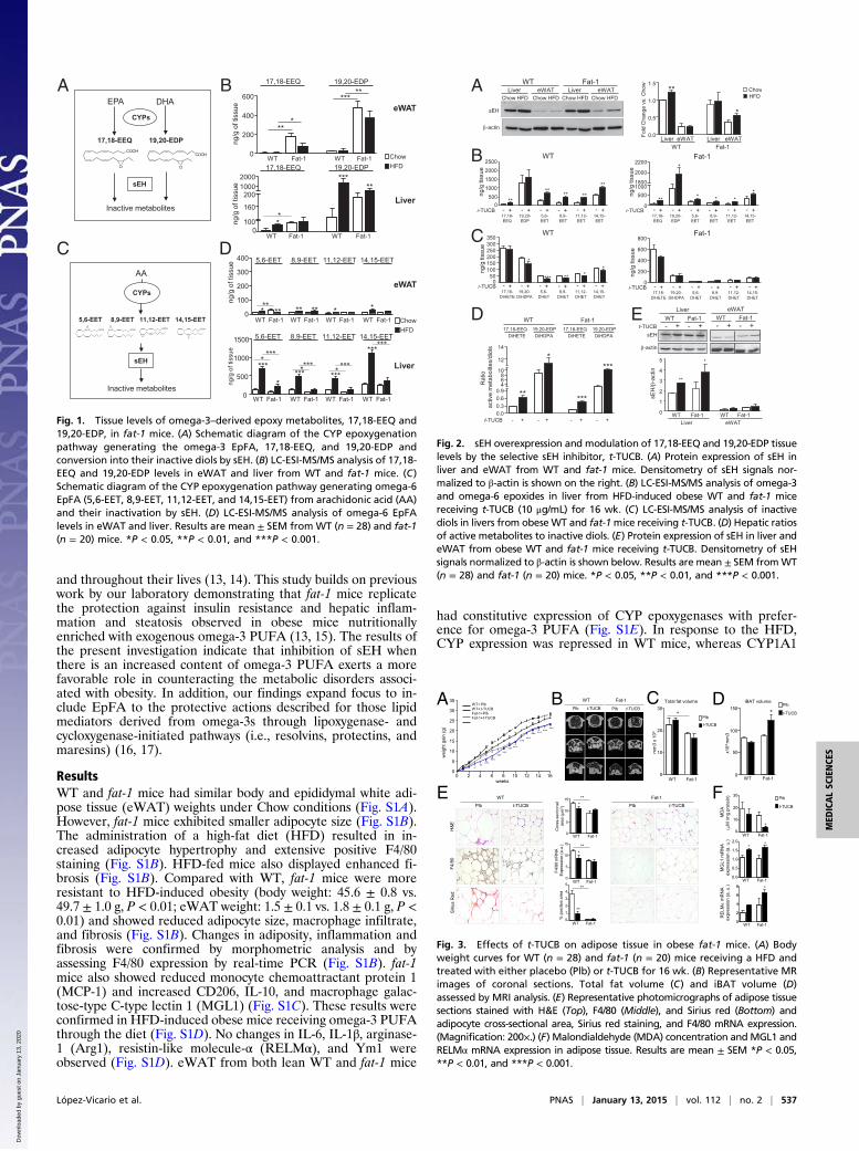
ResultsWT and fat-1 mice had similar body and epididymal white adi-pose tissue (eWAT) weights under Chow conditions (Fig. S1A).However, fat-1 mice exhibited smaller adipocyte size (Fig. S1B).The administration of a high-fat diet (HFD) resulted in in-creased adipocyte hypertrophy and extensive positive F4/80staining (Fig. S1B). HFD-fed mice also displayed enhanced fi-brosis (Fig. S1B). Compared with WT, fat-1 mice were moreresistant to HFD-induced obesity (body weight: 45.6 ± 0.8 vs.49.7 ± 1.0 g, P < 0.01; eWAT weight: 1.5 ± 0.1 vs. 1.8 ± 0.1 g, P <0.01) and showed reduced adipocyte size, macrophage infiltrate,and fibrosis (Fig. S1B). Changes in adiposity, inflammation andfibrosis were confirmed by morphometric analysis and byassessing F4/80 expression by real-time PCR (Fig. S1B). fat-1mice also showed reduced monocyte chemoattractant protein 1(MCP-1) and increased CD206, IL-10, and macrophage galac-tose-type C-type lectin 1 (MGL1) (Fig. S1C). These results wereconfirmed in HFD-induced obese mice receiving omega-3 PUFAthrough the diet (Fig. S1D). No changes in IL-6, IL-1β, arginase-1 (Arg1), resistin-like molecule-α (RELMα), and Ym1 wereobserved (Fig. S1D). eWAT from both lean WT and fat-1 mice
had constitutive expression of CYP epoxygenases with prefer-ence for omega-3 PUFA (Fig. S1E). In response to the HFD,CYP expression was repressed in WT mice, whereas CYP1A1
B
sEH
AA
5,6-EET 8,9-EET 11,12-EET 14,15-EET
Inactive metabolites
CYPs
COOH
O
COOH
O
COOHO
COOHO
D
ChowHFD
ng/g
of t
issu
e
WT Fat-1 WT Fat-1 WT Fat-1 WT Fat-10
500
1000
1500
***
**** ***
******
*********
** *
5,6-EET 8,9-EET 11,12-EET 14,15-EET
5,6-EET 8,9-EET 11,12-EET 14,15-EET
WT Fat-1 WT Fat-1 WT Fat-1 WT Fat-1
ng/g
of t
issu
e
0
100
200
300
400
**** ***** * *
eWAT
Liver
EPA DHA
17,18-EEQ
sEH
19,20-EDP
CYPs
COOH
O
COOH
O
Inactive metabolites
A
C
ChowHFD
eWAT
Liver
17,18-EEQ 19,20-EDP
ng/g
of t
issu
e
WT Fat-1 WT Fat-117,18-EEQ 19,20-EDP
0
200
400
600
***
*****
ng/g
of t
issu
eWT Fat-1 WT Fat-1
0100
160
2001000
*****
**
2000
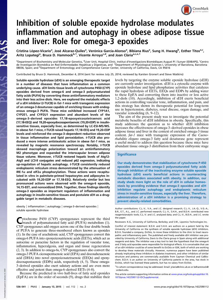
Fig. 1. Tissue levels of omega-3–derived epoxy metabolites, 17,18-EEQ and19,20-EDP, in fat-1 mice. (A) Schematic diagram of the CYP epoxygenationpathway generating the omega-3 EpFA, 17,18-EEQ, and 19,20-EDP andconversion into their inactive diols by sEH. (B) LC-ESI-MS/MS analysis of 17,18-EEQ and 19,20-EDP levels in eWAT and liver from WT and fat-1 mice. (C)Schematic diagram of the CYP epoxygenation pathway generating omega-6EpFA (5,6-EET, 8,9-EET, 11,12-EET, and 14,15-EET) from arachidonic acid (AA)and their inactivation by sEH. (D) LC-ESI-MS/MS analysis of omega-6 EpFAlevels in eWAT and liver. Results are mean ± SEM from WT (n = 28) and fat-1(n = 20) mice. *P < 0.05, **P < 0.01, and ***P < 0.001.
A WT Fat-1
sEH
β-actin
Liver eWATChow HFD Chow HFD
Liver eWATChow HFD Chow HFD
B0.0
0.5
1.0
1.5**
*
Fold
Cha
nge
vs. C
how
Liver eWAT Liver eWATWT Fat-1
ChowHFD
D
WT
0500
1000150020002500
****
******ng
/g ti
ssue
- + - +t-TUCB - + - + - + - +17,18-EEQ
19,20-EDP
5,6-EET
8,9-EET
11,12-EET
14,15-EET
Fat-1
0
50010001800
2000
2200
** ***
*
*
+ - +t-TUCB - + - + - + - +17,18-EEQ
19,20-EDP
5,6-EET
8,9-EET
11,12-EET
14,15-EET
-
ng/g
tiss
ue
050
100150200250300350
*** ** **
*
ng/g
tiss
ue
+ - +t-TUCB - + - + - + - +17,18-
DiHETE19,20-
DiHDPA5,6-
DHET8,9-
DHET11,12-DHET
14,15-DHET
- 0
200
400
600
800
+ - +t-TUCB - + - + - + - +17,18-
DiHETE19,20-
DiHDPA5,6-
DHET8,9-
DHET11,12-DHET
14,15-DHET
-
ng/g
tiss
ue
** ***
****
t-TUCB - + - + - + - +
WT Fat-1
Rat
io
activ
e m
etab
olite
s/di
ols
DiHETE DiHDPA17,18-EEQ 19,20-EDP
DiHETE DiHDPA17,18-EEQ 19,20-EDP
0.00.30.60.9
468
10
12
14
sEH
β-actin
WT Fat-1Liver
- + - + t-TUCBWT Fat-1
eWAT
- + - +
0
1
2
3
4
5
sEH
/β-a
ctin
WT Fat-1 WT Fat-1Liver eWAT
**
*
E
C WT Fat-1
Fig. 2. sEH overexpression and modulation of 17,18-EEQ and 19,20-EDP tissuelevels by the selective sEH inhibitor, t-TUCB. (A) Protein expression of sEH inliver and eWAT from WT and fat-1 mice. Densitometry of sEH signals nor-malized to β-actin is shown on the right. (B) LC-ESI-MS/MS analysis of omega-3and omega-6 epoxides in liver from HFD-induced obese WT and fat-1 micereceiving t-TUCB (10 μg/mL) for 16 wk. (C) LC-ESI-MS/MS analysis of inactivediols in livers from obeseWT and fat-1mice receiving t-TUCB. (D) Hepatic ratiosof active metabolites to inactive diols. (E) Protein expression of sEH in liver andeWAT from obese WT and fat-1 mice receiving t-TUCB. Densitometry of sEHsignals normalized to β-actin is shown below. Results are mean ± SEM fromWT(n = 28) and fat-1 (n = 20) mice. *P < 0.05, **P < 0.01, and ***P < 0.001.
A
0 2 4 6 8 10 12 14 160
5
10
15
20
25
30
35
weeks
wei
ght g
ain
(g)
WT+ PlbWT+ t-TUCBFat-1+PlbFat-1+t-TUCB
***************
******
***
******
******
***
BPlb
t-TUCB
Total fat volume
0
10
20
30
WT Fat-1
mm
3 x
103
*
E WT Fat-1
H&E
F4/8
0Si
rius
Red
0
1
2
3
4
5
**
**
% p
ositi
ve a
rea
WT Fat-1
0
5
10
15
*
F4/8
0 m
RN
A
Exp
ress
ion
(a.u
.)
WT Fat-1
**
0
5
10
15*
WT Fat-1
Cro
ss-s
ectio
nal
area
(μm
2 )
**
WT Fat-1
Plb t-TUCBPlb t-TUCB
Plb t-TUCB Plb t-TUCB
D
0
50
100
150*
WT Fat-1
x103
mm
3
iBAT volume
F Plb
t-TUCB
0
2
4
6
8 *
WT Fat-1
RE
LMα
mR
NA
expr
essi
on (a
. u.)
0.0
0.5
1.0
1.5
2.0* *
MG
L1 m
RN
A ex
pres
sion
(a. u
.)
WT Fat-1
0
10
20
30
MD
A(μ
M /m
g pr
otei
n)
*WT Fat-1
*
C Plb
t-TUCB
Fig. 3. Effects of t-TUCB on adipose tissue in obese fat-1 mice. (A) Bodyweight curves for WT (n = 28) and fat-1 (n = 20) mice receiving a HFD andtreated with either placebo (Plb) or t-TUCB for 16 wk. (B) Representative MRimages of coronal sections. Total fat volume (C) and iBAT volume (D)assessed by MRI analysis. (E) Representative photomicrographs of adipose tissuesections stained with H&E (Top), F4/80 (Middle), and Sirius red (Bottom) andadipocyte cross-sectional area, Sirius red staining, and F4/80 mRNA expression.(Magnification: 200×.) (F) Malondialdehyde (MDA) concentration andMGL1 andRELMα mRNA expression in adipose tissue. Results are mean ± SEM *P < 0.05,**P < 0.01, and ***P < 0.001.
López-Vicario et al. PNAS | January 13, 2015 | vol. 112 | no. 2 | 537
MED
ICALSC
IENCE
S
Dow
nloa
ded
by g
uest
on
Janu
ary
13, 2
020
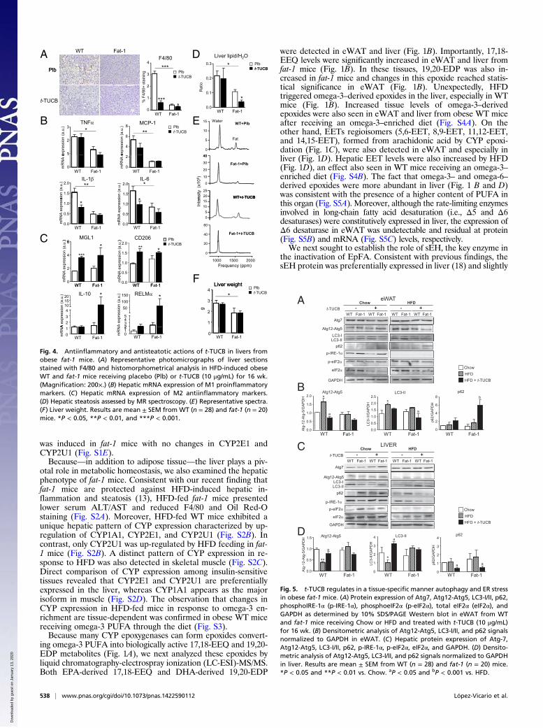
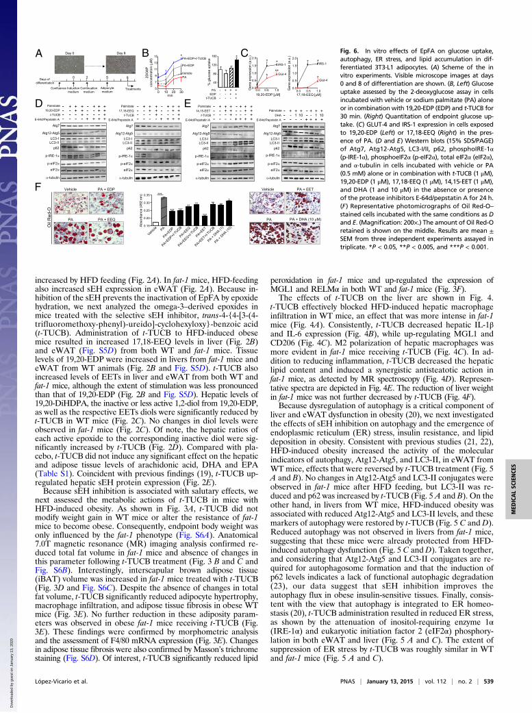
was induced in fat-1 mice with no changes in CYP2E1 andCYP2U1 (Fig. S1E).Because—in addition to adipose tissue—the liver plays a piv-
otal role in metabolic homeostasis, we also examined the hepaticphenotype of fat-1 mice. Consistent with our recent finding thatfat-1 mice are protected against HFD-induced hepatic in-flammation and steatosis (13), HFD-fed fat-1 mice presentedlower serum ALT/AST and reduced F4/80 and Oil Red-Ostaining (Fig. S2A). Moreover, HFD-fed WT mice exhibited aunique hepatic pattern of CYP expression characterized by up-regulation of CYP1A1, CYP2E1, and CYP2U1 (Fig. S2B). Incontrast, only CYP2U1 was up-regulated by HFD feeding in fat-1 mice (Fig. S2B). A distinct pattern of CYP expression in re-sponse to HFD was also detected in skeletal muscle (Fig. S2C).Direct comparison of CYP expression among insulin-sensitivetissues revealed that CYP2E1 and CYP2U1 are preferentiallyexpressed in the liver, whereas CYP1A1 appears as the majorisoform in muscle (Fig. S2D). The observation that changes inCYP expression in HFD-fed mice in response to omega-3 en-richment are tissue-dependent was confirmed in obese WT micereceiving omega-3 PUFA through the diet (Fig. S3).Because many CYP epoxygenases can form epoxides convert-
ing omega-3 PUFA into biologically active 17,18-EEQ and 19,20-EDP metabolites (Fig. 1A), we next analyzed these epoxides byliquid chromatography-electrospray ionization (LC-ESI)-MS/MS.Both EPA-derived 17,18-EEQ and DHA-derived 19,20-EDP
were detected in eWAT and liver (Fig. 1B). Importantly, 17,18-EEQ levels were significantly increased in eWAT and liver fromfat-1 mice (Fig. 1B). In these tissues, 19,20-EDP was also in-creased in fat-1 mice and changes in this epoxide reached statis-tical significance in eWAT (Fig. 1B). Unexpectedly, HFDtriggered omega-3–derived epoxides in the liver, especially in WTmice (Fig. 1B). Increased tissue levels of omega-3–derivedepoxides were also seen in eWAT and liver from obese WT miceafter receiving an omega-3–enriched diet (Fig. S4A). On theother hand, EETs regioisomers (5,6-EET, 8,9-EET, 11,12-EET,and 14,15-EET), formed from arachidonic acid by CYP epoxi-dation (Fig. 1C), were also detected in eWAT and especially inliver (Fig. 1D). Hepatic EET levels were also increased by HFD(Fig. 1D), an effect also seen in WT mice receiving an omega-3–enriched diet (Fig. S4B). The fact that omega-3– and omega-6–derived epoxides were more abundant in liver (Fig. 1 B and D)was consistent with the presence of a higher content of PUFA inthis organ (Fig. S5A). Moreover, although the rate-limiting enzymesinvolved in long-chain fatty acid desaturation (i.e., Δ5 and Δ6desaturases) were constitutively expressed in liver, the expression ofΔ6 desaturase in eWAT was undetectable and residual at protein(Fig. S5B) and mRNA (Fig. S5C) levels, respectively.We next sought to establish the role of sEH, the key enzyme in
the inactivation of EpFA. Consistent with previous findings, thesEH protein was preferentially expressed in liver (18) and slightly
APlb
WT Fat-1 D Liver lipid/H2O0.3 * Plb
t-TUCB4
***F4/80
Plb
t-TUCB
WT Fat-1
Rat
io
0.0
0.1
0.2
*
t-TUCBPlbt-TUCB
0
1
2
3
***% F
4/80
+ st
aini
ng
*
B
40
0
5
10
15 WT+PlbWater
Fat
ETNF
1
2
3*
expr
essi
on (a
.u.)
MCP-1
4
6
8ex
pres
sion
(a.u
.)
**Plbt-TUCB
WT Fat-1
nsity
(x10
6 )WT+t-TUCB
0
10
20
30
40
Fat-1+Plb
2025
0
1
mR
NA
e
WT Fat-1
IL-1
1.5
2.0
sion
(a.u
.) **IL-6
1.5
2.0
on (a
.u.) *
0
2
mR
NA
e
WT Fat-1
Inte
n WT+t TUCB
40
60
Fat-1+t-TUCB
05
1015
C MGL16)
0.0
0.5
1.0 *
mR
NA
expr
ess
WT Fat-10.0
0.5
1.0*
WT Fat-1
mR
NA
exp
ress
i
CD206) Plb
1000 1500 2000Frequency (ppm)
0
20
WT F t 10
2
4
6
***
*
mR
NA
expr
essi
on (a
.u.)
WT F t 10.0
0.5
1.0
1.5
2.0** *
mR
NA
expr
essi
on (a
.u.)
t-TUCB
FLiver weightWT Fat-1
IL-10
234
101520 *
A e
xpre
ssio
n (a
.u.) RELM
2345
50100150 *
A e
xpre
ssio
n (a
.u.)
WT Fat-1 Liver weight
1
2
3
4
g
*Plbt-TUCB
WT Fat-101
mR
NA
WT Fat-1012
mR
N
0WT Fat-1
Fig. 4. Antiinflammatory and antisteatotic actions of t-TUCB in livers fromobese fat-1 mice. (A) Representative photomicrographs of liver sectionsstained with F4/80 and histomorphometrical analysis in HFD-induced obeseWT and fat-1 mice receiving placebo (Plb) or t-TUCB (10 μg/mL) for 16 wk.(Magnification: 200×.) (B) Hepatic mRNA expression of M1 proinflammatorymarkers. (C) Hepatic mRNA expression of M2 antiinflammatory markers.(D) Hepatic steatosis assessed by MR spectroscopy. (E) Representative spectra.(F) Liver weight. Results are mean ± SEM from WT (n = 28) and fat-1 (n = 20)mice. *P < 0.05, **P < 0.01, and ***P < 0.001.
B
Chow HFD- + - +
WT Fat-1 WT Fat-1 WT Fat-1 WT Fat-1t-TUCB
LC3-ILC3-II
LIVER
p62
GAPDH
p-IRE-1α
p-eIF2α
Atg7
Atg12-Atg5
eIF2α
Chow HFD- + - +
WT Fat-1 WT Fat-1 WT Fat-1 WT Fat-1
eWAT
t-TUCB
LC3-ILC3-II
p62
GAPDH
p-IRE-1α
p-eIF2α
Atg7
Atg12-Atg5
eIF2α
A
C
D
Atg
-12-
Atg
-5/G
AP
DH
0.0
0.5
1.0
1.5
2.0 *
a
Atg12-Atg5
WT Fat-1
0.0
0.5
1.0
1.5
**
a
Atg12-Atg5
WT Fat-1
Atg
-12-
Atg
-5/G
AP
DH
LC3-II p62
0
1
2
3
4
a a
WT Fat-1
p62/
GA
PD
H
ChowHFDHFD + t-TUCB
0.0
0.5
1.0
1.5
2.0
2.5*
LC3-
II/G
AP
DH
LC3-II
WT Fat-1
a
0
2
4
6
8
p62/
GA
PD
H
p62
WT Fat-1
b
0
1
2
3
4
*
WT Fat-1
LC3-
II/G
AP
DH
a
ChowHFDHFD + t-TUCB
Fig. 5. t-TUCB regulates in a tissue-specific manner autophagy and ER stressin obese fat-1 mice. (A) Protein expression of Atg7, Atg12-Atg5, LC3-I/II, p62,phosphoIRE-1α (p-IRE-1α), phosphoeIF2α (p-eIF2α), total eIF2α (eIF2α), andGAPDH as determined by 10% SDS/PAGE Western blot in eWAT from WTand fat-1 mice receiving Chow or HFD and treated with t-TUCB (10 μg/mL)for 16 wk. (B) Densitometric analysis of Atg12-Atg5, LC3-I/II, and p62 signalsnormalized to GAPDH in eWAT. (C) Hepatic protein expression of Atg-7,Atg12-Atg5, LC3-I/II, p62, p-IRE-1α, p-eIF2α, eIF2α, and GAPDH. (D) Densito-metric analysis of Atg12-Atg5, LC3-I/II, and p62 signals normalized to GAPDHin liver. Results are mean ± SEM from WT (n = 28) and fat-1 (n = 20) mice.*P < 0.05 and **P < 0.01 vs. Chow. aP < 0.05 and bP < 0.001 vs. HFD.
538 | www.pnas.org/cgi/doi/10.1073/pnas.1422590112 López-Vicario et al.
Dow
nloa
ded
by g
uest
on
Janu
ary
13, 2
020
increased by HFD feeding (Fig. 2A). In fat-1 mice, HFD-feedingalso increased sEH expression in eWAT (Fig. 2A). Because in-hibition of the sEH prevents the inactivation of EpFA by epoxidehydration, we next analyzed the omega-3–derived epoxides inmice treated with the selective sEH inhibitor, trans-4-{4-[3-(4-trifluoromethoxy-phenyl)-ureido]-cyclohexyloxy}-benzoic acid(t-TUCB). Administration of t-TUCB to HFD-induced obesemice resulted in increased 17,18-EEQ levels in liver (Fig. 2B)and eWAT (Fig. S5D) from both WT and fat-1 mice. Tissuelevels of 19,20-EDP were increased in livers from fat-1 mice andeWAT from WT animals (Fig. 2B and Fig. S5D). t-TUCB alsoincreased levels of EETs in liver and eWAT from both WT andfat-1 mice, although the extent of stimulation was less pronouncedthan that of 19,20-EDP (Fig. 2B and Fig. S5D). Hepatic levels of19,20-DiHDPA, the inactive or less active 1,2-diol from 19,20-EDP,as well as the respective EETs diols were significantly reduced byt-TUCB in WT mice (Fig. 2C). No changes in diol levels wereobserved in fat-1 mice (Fig. 2C). Of note, the hepatic ratios ofeach active epoxide to the corresponding inactive diol were sig-nificantly increased by t-TUCB (Fig. 2D). Compared with pla-cebo, t-TUCB did not induce any significant effect on the hepaticand adipose tissue levels of arachidonic acid, DHA and EPA(Table S1). Coincident with previous findings (19), t-TUCB up-regulated hepatic sEH protein expression (Fig. 2E).Because sEH inhibition is associated with salutary effects, we
next assessed the metabolic actions of t-TUCB in mice withHFD-induced obesity. As shown in Fig. 3A, t-TUCB did notmodify weight gain in WT mice or alter the resistance of fat-1mice to become obese. Consequently, endpoint body weight wasonly influenced by the fat-1 phenotype (Fig. S6A). Anatomical7.0T magnetic resonance (MR) imaging analysis confirmed re-duced total fat volume in fat-1 mice and absence of changes inthis parameter following t-TUCB treatment (Fig. 3 B and C andFig. S6B). Interestingly, interscapular brown adipose tissue(iBAT) volume was increased in fat-1 mice treated with t-TUCB(Fig. 3D and Fig. S6C). Despite the absence of changes in totalfat volume, t-TUCB significantly reduced adipocyte hypertrophy,macrophage infiltration, and adipose tissue fibrosis in obese WTmice (Fig. 3E). No further reduction in these adiposity param-eters was observed in obese fat-1 mice receiving t-TUCB (Fig.3E). These findings were confirmed by morphometric analysisand the assessment of F4/80 mRNA expression (Fig. 3E). Changesin adipose tissue fibrosis were also confirmed by Masson’s trichromestaining (Fig. S6D). Of interest, t-TUCB significantly reduced lipid
peroxidation in fat-1 mice and up-regulated the expression ofMGL1 and RELMα in both WT and fat-1 mice (Fig. 3F).The effects of t-TUCB on the liver are shown in Fig. 4.
t-TUCB effectively blocked HFD-induced hepatic macrophageinfiltration in WT mice, an effect that was more intense in fat-1mice (Fig. 4A). Consistently, t-TUCB decreased hepatic IL-1βand IL-6 expression (Fig. 4B), while up-regulating MGL1 andCD206 (Fig. 4C). M2 polarization of hepatic macrophages wasmore evident in fat-1 mice receiving t-TUCB (Fig. 4C). In ad-dition to reducing inflammation, t-TUCB decreased the hepaticlipid content and induced a synergistic antisteatotic action infat-1 mice, as detected by MR spectroscopy (Fig. 4D). Represen-tative spectra are depicted in Fig. 4E. The reduction of liver weightin fat-1 mice was not further decreased by t-TUCB (Fig. 4F).Because dysregulation of autophagy is a critical component of
liver and eWAT dysfunction in obesity (20), we next investigatedthe effects of sEH inhibition on autophagy and the emergence ofendoplasmic reticulum (ER) stress, insulin resistance, and lipiddeposition in obesity. Consistent with previous studies (21, 22),HFD-induced obesity increased the activity of the molecularindicators of autophagy, Atg12-Atg5, and LC3-II, in eWAT fromWT mice, effects that were reversed by t-TUCB treatment (Fig. 5A and B). No changes in Atg12-Atg5 and LC3-II conjugates wereobserved in fat-1 mice after HFD feeding, but LC3-II was re-duced and p62 was increased by t-TUCB (Fig. 5 A and B). On theother hand, in livers from WT mice, HFD-induced obesity wasassociated with reduced Atg12-Atg5 and LC3-II levels, and thesemarkers of autophagy were restored by t-TUCB (Fig. 5 C and D).Reduced autophagy was not observed in livers from fat-1 mice,suggesting that these mice were already protected from HFD-induced autophagy dysfunction (Fig. 5 C and D). Taken together,and considering that Atg12-Atg5 and LC3-II conjugates are re-quired for autophagosome formation and that the induction ofp62 levels indicates a lack of functional autophagic degradation(23), our data suggest that sEH inhibition improves theautophagy flux in obese insulin-sensitive tissues. Finally, consis-tent with the view that autophagy is integrated to ER homeo-stasis (20), t-TUCB administration resulted in reduced ER stress,as shown by the attenuation of inositol-requiring enzyme 1α(IRE-1α) and eukaryotic initiation factor 2 (eIF2α) phosphory-lation in both eWAT and liver (Fig. 5 A and C). The extent ofsuppression of ER stress by t-TUCB was roughly similar in WTand fat-1 mice (Fig. 5 A and C).
D
Gen
e ex
pres
sion
(a.u
.)CA
Vehicle
PA+EDP
PA+EDP+t-TUCB
PA
min
2DG
6Pco
ncen
tratio
n ( μ
M)
0 10 20 305
7
9
11
13
15 *Day 0
Confluence
Days of differentiation
Inductionmedium
Continuationmedium
Adipocytemedium
Treatments
Day 8
0 2 5 840
80
120
160 *
% g
luco
se u
ptak
e
PA - + + +
t-TUCB - - - +EDP - - + +
B
Glut-4
IRS-1
0.0 0.5 1.00.0
0.5
1.0
1.5
2.0
19,20-EDP [μM]
* *
***
0.0 0.5 1.00.0
0.5
1.0
1.5
2.0
Gen
e ex
pres
sion
(a.u
.)
17,18-EEQ [μM]
Glut-4
IRS-1
*
* *
Atg7
LC3-ILC3-II
p-eIF2α
p62
α-tubulin
Atg12-Atg5
p-IRE-1α
eIF2α
E-64d/Pepstatin A
Palmitate19,20-EDP
t-TUCB
----
+---
+
--+
+
+-+
---
+--
+
-+
+
++
+ + + + E-64d/Pepstatin A
Palmitate17,18-EEQ
t-TUCB
Atg7
LC3-ILC3-II
p-eIF2α
p62
α-tubulin
Atg12-Atg5
p-IRE-1α
eIF2α
----
+---
+
--+
+
+-+
---
+--
+
-+
+
++
+ + + + E-64d/Pepstatin A
Palmitate14,15-EET
t-TUCB
Atg7
LC3-ILC3-II
p-eIF2α
p62
α-tubulin
Atg12-Atg5
p-IRE-1α
eIF2α
----
+---
+
--+
+
+-+
---
+--
+
-+
+
++
+ + + + E-64d/Pepstatin A
PalmitateDHA
Atg7
LC3-ILC3-II
p-eIF2α
p62
α-tubulin
Atg12-Atg5
p-IRE-1α
eIF2α
- + + + - + + +- - 1 10 - - 1 10- - - - + + + +
Vehicle
PA
PA + EDP
Oil
Red
-O
PA + EEQ
PA + EET
PA + DHA (10 μM)
Vehicle
PA
E
0.15
0.20
0.25
0.30
0.35 ****
** ********* ***
***
Abs
orba
nce
(492
nm
)
F
Fig. 6. In vitro effects of EpFA on glucose uptake,autophagy, ER stress, and lipid accumulation in dif-ferentiated 3T3-L1 adipocytes. (A) Scheme of the invitro experiments. Visible microscope images at days0 and 8 of differentiation are shown. (B, Left) Glucoseuptake assessed by the 2-deoxyglucose assay in cellsincubated with vehicle or sodium palmitate (PA) aloneor in combination with 19,20-EDP (EDP) and t-TUCB for30 min. (Right) Quantitation of endpoint glucose up-take. (C) GLUT-4 and IRS-1 expression in cells exposedto 19,20-EDP (Left) or 17,18-EEQ (Right) in the pres-ence of PA. (D and E) Western blots (15% SDS/PAGE)of Atg7, Atg12-Atg5, LC3-I/II, p62, phosphoIRE-1α(p-IRE-1α), phosphoeIF2α (p-eIF2α), total eIF2α (eIF2α),and α-tubulin in cells incubated with vehicle or PA(0.5 mM) alone or in combination with t-TUCB (1 μM),19,20-EDP (1 μM), 17,18-EEQ (1 μM), 14,15-EET (1 μM),and DHA (1 and 10 μM) in the absence or presenceof the protease inhibitors E-64d/pepstatin A for 24 h.(F) Representative photomicrographs of Oil Red-O–
stained cells incubated with the same conditions as Dand E. (Magnification: 200×.) The amount of Oil Red-Oretained is shown on the middle. Results are mean ±SEM from three independent experiments assayed intriplicate. *P < 0.05, **P < 0.005, and ***P < 0.001.
López-Vicario et al. PNAS | January 13, 2015 | vol. 112 | no. 2 | 539
MED
ICALSC
IENCE
S
Dow
nloa
ded
by g
uest
on
Janu
ary
13, 2
020
To provide direct evidence linking omega-3 EpFA to auto-phagy and ER stress, we next performed in vitro experiments inadipocytes incubated with the saturated fatty acid palmitate,a major contributor to lipotoxicity and insulin resistance (24). Aschematic diagram of the experimental in vitro procedure isshown in Fig. 6A. Incubation of adipocytes with 19,20-EDP, themost abundant omega-3 epoxide in eWAT, in the presence oft-TUCB, stimulated glucose uptake in adipocytes (Fig. 6B).Moreover, in these cells, 19,20-EDP and 17,18-EEQ induceda concentration-dependent up-regulation of insulin receptorsubstrate-1 (IRS-1) and glucose transporter-type 4 (GLUT-4)(Fig. 6C). Interestingly, in the presence of t-TUCB, both 19,20-EDP and 17,18-EEQ produced similar changes in autophagy tothose reported in vivo in eWAT from obese fat-1 mice receivingt-TUCB (Fig. 6D). With t-TUCB on board, reduction of IRE-1α and eIF2α phosphorylation in response to these omega-3EpFA also paralleled that seen in vivo (Fig. 6D). In these
experiments, the omega-6 EpFA, 14,15-EET, and the non-oxidized form of DHA were either ineffective or less activethan omega-3 EpFA in regulating authophagy and ER stress inadipocytes (Fig. 6E). All compounds tested significantly re-duced palmitate-induced accumulation of lipids in adipocytes,with the EpFA being more potent than the nonoxidized formof DHA (Fig. 6F). t-TUCB alone did not modify ER stress,autophagy, and intracellular lipid levels (Fig. S6E).Finally, we incubated hepatocytes with palmitate in the pres-
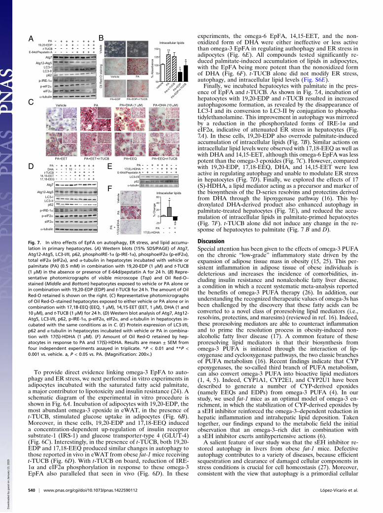
ence of EpFA and t-TUCB. As shown in Fig. 7A, incubation ofhepatocytes with 19,20-EDP and t-TUCB resulted in increasedautophagosome formation, as revealed by the disappearance ofLC3-I and its conversion to LC3-II by conjugation to phospha-tidylethanolamine. This improvement in autophagy was mirroredby a reduction in the phosphorylated forms of IRE-1α andeIF2α, indicative of attenuated ER stress in hepatocytes (Fig.7A). In these cells, 19,20-EDP also overrode palmitate-inducedaccumulation of intracellular lipids (Fig. 7B). Similar actions onintracellular lipid levels were observed with 17,18-EEQ as well aswith DHA and 14,15-EET, although this omega-6 EpFA was lesspotent than the omega-3 epoxides (Fig. 7C). However, comparedwith 19,20-EDP, 17,18-EEQ, DHA, and 14,15-EET were lessactive in regulating autophagy and unable to modulate ER stressin hepatocytes (Fig. 7D). Finally, we explored the effects of 17(S)-HDHA, a lipid mediator acting as a precursor and marker ofthe biosynthesis of the D-series resolvins and protectins derivedfrom DHA through the lipoxygenase pathway (16). This hy-droxylated DHA-derived product also enhanced autophagy inpalmitate-treated hepatocytes (Fig. 7E), and reduced the accu-mulation of intracellular lipids in palmitate-primed hepatocytes(Fig. 7F). t-TUCB alone did not induce any change in the re-sponse of hepatocytes to palmitate (Fig. 7 B and D).
DiscussionSpecial attention has been given to the effects of omega-3 PUFAon the chronic “low-grade” inflammatory state driven by theexpansion of adipose tissue mass in obesity (15, 25). This per-sistent inflammation in adipose tissue of obese individuals isdeleterious and increases the incidence of comorbidities, in-cluding insulin resistance and nonalcoholic fatty liver disease,a condition in which a recent systematic meta-analysis reportedthe benefits of omega-3 PUFA therapy (26). In addition, ourunderstanding the recognized therapeutic values of omega-3s hasbeen challenged by the discovery that these fatty acids can beconverted to a novel class of proresolving lipid mediators (i.e.,resolvins, protectins, and maresins) (reviewed in ref. 16). Indeed,these proresolving mediators are able to counteract inflammationand to prime the resolution process in obesity-induced non-alcoholic fatty liver disease (17). A common feature of theseproresolving lipid mediators is that their biosynthesis fromomega-3 PUFA is initiated through the interaction of lip-oxygenase and cyclooxygenase pathways, the two classic branchesof PUFA metabolism (16). Recent findings indicate that CYPepoxygenases, the so-called third branch of PUFA metabolism,can also convert omega-3 PUFA into bioactive lipid mediators(1, 4, 5). Indeed, CYP1A1, CYP2E1, and CYP2U1 have beendescribed to generate a number of CYP-derived epoxides(namely EEQs and EDPs) from omega-3 PUFA (4). In ourstudy, we used fat-1 mice as an optimal model of omega-3 en-richment, in which the stabilization of CYP-derived epoxides bya sEH inhibitor reinforced the omega-3–dependent reduction inhepatic inflammation and intrahepatic lipid deposition. Takentogether, our findings expand to the metabolic field the initialobservation that an omega-3–rich diet in combination witha sEH inhibitor exerts antihypertensive actions (6).A salient feature of our study was that the sEH inhibitor re-
stored autophagy in livers from obese fat-1 mice. Defectiveautophagy contributes to a variety of diseases, because efficientsequestration and clearance of damaged cellular components instress conditions is crucial for cell homeostasis (27). Moreover,consistent with the view that autophagy is a primordial cellular
Vehicle
B
E
A
Atg7
LC3-ILC3-II
p-eIF2α
p62
α-tubulin
Atg12-Atg5
p-IRE-1α
eIF2α
E-64d/Pepstatin A
PA19,20-EDP
t-TUCB
----
+---
+
--+
+
+-+
---
+--
+
-+
+
++
+ + + +
Vehicle
Vis
ible
Oil
Red
-O s
tain
ing
PA+EDP
PA
PA+EDP+t-TUCB
50
75
100
125
150 **
a
a
% A
bsor
banc
e
Intracellular lipids
DLC3-I/II
α-tubulinp62
E-64d/Pepstatin A
PA17(S)-HDHA
---
--
--
-+
+-+ -
--+
+
+
++ + + +
+
-20
0
20
40
60
% C
hang
e
PA - + +17(S)-HDHA - - +
a
*Intracelullar lipids
-
-
-
-
-
C PA+DHA (1 μM)PA
PA+EET+t-TUCBPA+EET PA+EEQ PA+EEQ+t-TUCB
200x
4x
Vehicle PA+DHA (10 μM)
F
Veh
icle
PA
PA +
17(S
)HD
HA
Atg7
LC3-ILC3-II
p-eIF2α
p62
α-tubulin
Atg12-Atg5
p-IRE-1α
eIF2α
PA - + + + + + + +DHA - - 1 10
t-TUCB
+- - - - -
- - - - + + +- -14,15-EET17,18-EEQ
- - - + + -- --- - - + +-- --
Fig. 7. In vitro effects of EpFA on autophagy, ER stress, and lipid accumu-lation in primary hepatocytes. (A) Western blots (15% SDS/PAGE) of Atg7,Atg12-Atg5, LC3-I/II, p62, phosphoIRE-1α (p-IRE-1α), phosphoeIF2α (p-eIF2α),total eIF2α (eIF2α), and α-tubulin in hepatocytes incubated with vehicle orpalmitate (PA) (0.5 mM) in combination with 19,20-EDP (1 μM) and t-TUCB(1 μM) in the absence or presence of E-64d/pepstatin A for 24 h. (B) Repre-sentative photomicrographs of visible microscope (Top) and Oil Red-O–
stained (Middle and Bottom) hepatocytes exposed to vehicle or PA alone orin combination with 19,20-EDP (EDP) and t-TUCB for 24 h. The amount of OilRed-O retained is shown on the right. (C) Representative photomicrographsof Oil Red-O–stained hepatocytes exposed to either vehicle or PA alone or incombination with 17,18-EEQ (EEQ, 1 μM), 14,15-EET (EET, 1 μM), DHA (1 and10 μM), and t-TUCB (1 μM) for 24 h. (D) Western blot analysis of Atg7, Atg12-Atg5, LC3-I/II, p62, p-IRE-1α, p-eIF2α, eIF2α, and α-tubulin in hepatocytes in-cubated with the same conditions as in C. (E) Protein expression of LC3-I/II,p62 and α-tubulin in hepatocytes incubated with vehicle or PA in combina-tion with 17(S)-HDHA (1 μM). (F) Amount of Oil Red-O retained by hep-atocytes in response to PA and 17(S)-HDHA. Results are mean ± SEM fromfour independent experiments assayed in triplicate. *P < 0.01 and **P <0.001 vs. vehicle. a, P < 0.05 vs. PA. (Magnification: 200×.)
540 | www.pnas.org/cgi/doi/10.1073/pnas.1422590112 López-Vicario et al.
Dow
nloa
ded
by g
uest
on
Janu
ary
13, 2
020
adaptive mechanism that mitigates ER-associated unfavorableconditions in insulin-sensitive tissues (20), sEH inhibition in fat-1mice was accompanied by an attenuated hepatic ER stress.Down-regulation of ER stress was also seen in conjunction withdecreased autophagy in adipose tissue from fat-1 mice receivingthe sEH inhibitor, suggesting dissociation between these twocellular processes in this tissue. Because inhibition of autophagicfunction in adipose tissue is related to reduced fat mass andimproved insulin sensitivity (28), our findings in adipose tissuecan be regarded as beneficial in terms of lipid homeostasis andmetabolic control. A strong asset of our study was that we wereable to recapitulate the effects on autophagy and ER stress seenin vivo following sEH inhibition, by exposing hepatocytes andadipocytes in vitro to the omega-3 epoxides 19,20-EDP and17,18-EEQ. Our findings are consistent with those reported byBettaieb et al., who showed attenuation of ER stress in adiposeand liver tissues in mice either receiving a sEH inhibitor or de-ficient for sEH (29). However, our data cannot exclude otherEpFA, such as the case of arachidonic acid-derived EETs aswell as the potential implication of other oxidized lipid medi-ators derived from omega-3 PUFA through the interaction oflipoxygenase and cyclooxygenase pathway. Additionally, our datacannot exclude the potential implication of nonoxidized DHA inthe observed favorable metabolic phenotype of fat-1 mice. In thisregard, although less potent than EpFA, DHA was active in ourcell bioassays. This finding is consistent with the reported bi-ological properties of DHA (30) and with findings reported byCaviglia et al. showing that DHA was able to rescue rat hepa-toma cells from palmitate-induced ER stress (31). However,these studies did not address whether the protective effects ofDHA were mediated by the parent nonoxidized molecule or byany other DHA oxidized metabolite (30, 31).
In summary, the results of the present study demonstrate thatstabilization of omega-3 epoxides through inhibition of sEHexerts beneficial actions in counteracting the metabolic disordersassociated with obesity. Of particular interest are the findingsdemonstrating the ability of sEH inhibition to restore autophagyin the liver with the consequent reduction of obesity-inducedliver ER stress. Our observations highlight the potentiality ofsmall bioactive lipid mediators to modulate autophagy, serving astemplates for the exploitation of this housekeeping cellularprocess for therapeutic interventions against obesity and obesity-related comorbidities, such as fatty liver disease.
Materials and MethodsStudies in fat-1 mice, hepatocytes and 3T3-L1 adipocytes, measurement of2-deoxyglucose uptake, mRNA and protein expression, and histology andimmunohistochemistry analysis, MR imaging and spectroscopy, and LC-ESI-MS/MS and gas chromatography analysis are described in detail in SI Materialsand Methods. The LC-MS/MS conditions used to profile the omega-3 andomega-6 epoxides are described in Table S2 and a schematic diagram of theexperimental design of the study can be found in Fig. S7.
ACKNOWLEDGMENTS. We thank Dr. J. X. Kang (Massachusetts GeneralHospital) for kindly providing fat-1 mice, Dr. M. Cofan for gas chromatog-raphy analysis, and Anabel Martínez-Puchol for assistance. This work wasconducted at the Centre Esther Koplowitz and was supported by SpanishMinisterio de Economía y Competitividad (MEC) (SAF12/32789 and PIE14/00045); the Research Investments in the Sciences and Engineering Programof the University of California, Davis; Grants R01 ES002710 and P42 ES004699from the National Institute of Environmental Health Sciences; fellowshipsfrom MEC (to V.G.-A. and B.R.); Marie Curie Action (A.L.); and an Emili Letangfellowship (to J.A.-Q.). Centro de Investigación Biomédica en Red EnfermedadesHepáticas y Digestivas is funded by the Instituto de Salud Carlos III. C.L.-V. wassupported by the Institut d’Investigacions Biomèdiques August Pi i Sunyer/Fundació Clínic.
1. Spector AA, Norris AW (2007) Action of epoxyeicosatrienoic acids on cellular function.Am J Physiol Cell Physiol 292(3):C996–C1012.
2. Zeldin DC (2001) Epoxygenase pathways of arachidonic acid metabolism. J Biol Chem276(39):36059–36062.
3. Panigrahy D, et al. (2013) Epoxyeicosanoids promote organ and tissue regeneration.Proc Natl Acad Sci USA 110(33):13528–13533.
4. Arnold C, et al. (2010) Arachidonic acid-metabolizing cytochrome P450 enzymes aretargets of ω-3 fatty acids. J Biol Chem 285(43):32720–32733.
5. Zhang G, et al. (2013) Epoxy metabolites of docosahexaenoic acid (DHA) inhibit an-giogenesis, tumor growth, and metastasis. Proc Natl Acad Sci USA 110(16):6530–6535.
6. Ulu A, et al. (2014) An omega-3 epoxide of docosahexaenoic acid lowers bloodpressure in angiotensin-II-dependent hypertension. J Cardiovasc Pharmacol 64(1):87–99.
7. Morisseau C, et al. (2010) Naturally occurring monoepoxides of eicosapentaenoic acidand docosahexaenoic acid are bioactive antihyperalgesic lipids. J Lipid Res 51(12):3481–3490.
8. Falck JR, et al. (2011) 17(R),18(S)-epoxyeicosatetraenoic acid, a potent eicosapentae-noic acid (EPA) derived regulator of cardiomyocyte contraction: structure-activityrelationships and stable analogues. J Med Chem 54(12):4109–4118.
9. Shen HC (2010) Soluble epoxide hydrolase inhibitors: A patent review. Expert OpinTher Pat 20(7):941–956.
10. Morisseau C, Hammock BD (2013) Impact of soluble epoxide hydrolase and epox-yeicosanoids on human health. Annu Rev Pharmacol Toxicol 53:37–58.
11. Schmelzer KR, et al. (2005) Soluble epoxide hydrolase is a therapeutic target for acuteinflammation. Proc Natl Acad Sci USA 102(28):9772–9777.
12. Shaik JS, et al. (2013) Soluble epoxide hydrolase inhibitor trans-4-[4-(3-adamantan-1-yl-ureido)-cyclohexyloxy]-benzoic acid is neuroprotective in rat model of ischemicstroke. Am J Physiol Heart Circ Physiol 305(11):H1605–H1613.
13. López-Vicario C, et al. (2014) Molecular interplay between Δ5/Δ6 desaturases andlong-chain fatty acids in the pathogenesis of non-alcoholic steatohepatitis. Gut 63(2):344–355.
14. Kang JX, Wang J, Wu L, Kang ZB (2004) Transgenic mice: fat-1 mice convert n-6 to n-3fatty acids. Nature 427(6974):504.
15. González-Périz A, et al. (2009) Obesity-induced insulin resistance and hepatic steatosisare alleviated by omega-3 fatty acids: A role for resolvins and protectins. FASEB J23(6):1946–1957.
16. Serhan CN (2014) Pro-resolving lipid mediators are leads for resolution physiology.Nature 510(7503):92–101.
17. Rius B, et al. (2014) Resolvin D1 primes the resolution process initiated by calorierestriction in obesity-induced steatohepatitis. FASEB J 28(2):836–848.
18. De Taeye BM, et al. (2010) Expression and regulation of soluble epoxide hydrolase inadipose tissue. Obesity (Silver Spring) 18(3):489–498.
19. Liu Y, et al. (2012) Inhibition of soluble epoxide hydrolase attenuates high-fat-diet–induced hepatic steatosis by reduced systemic inflammatory status in mice. PLoS ONE7(6):e39165.
20. Yang L, Li P, Fu S, Calay ES, Hotamisligil GS (2010) Defective hepatic autophagy inobesity promotes ER stress and causes insulin resistance. Cell Metab 11(6):467–478.
21. Nuñez CE, et al. (2013) Defective regulation of adipose tissue autophagy in obesity.Int J Obes (Lond) 37(11):1473–1480.
22. Jansen HJ, et al. (2012) Autophagy activity is up-regulated in adipose tissue of obeseindividuals and modulates proinflammatory cytokine expression. Endocrinology153(12):5866–5874.
23. Klionsky DJ, et al. (2012) Guidelines for the use and interpretation of assays formonitoring autophagy. Autophagy 8(4):445–544.
24. Van Epps-Fung M, Williford J, Wells A, Hardy RW (1997) Fatty acid-induced insulinresistance in adipocytes. Endocrinology 138(10):4338–4345.
25. Buckley JD, Howe PRC (2009) Anti-obesity effects of long-chain omega-3 poly-unsaturated fatty acids. Obes Rev 10(6):648–659.
26. Parker HM, et al. (2012) Omega-3 supplementation and non-alcoholic fatty liverdisease: A systematic review and meta-analysis. J Hepatol 56(4):944–951.
27. Schneider JL, Cuervo AM (2014) Liver autophagy: Much more than just taking out thetrash. Nat Rev Gastroenterol Hepatol 11(3):187–200.
28. Singh R, et al. (2009) Autophagy regulates adipose mass and differentiation in mice.J Clin Invest 119(11):3329–3339.
29. Bettaieb A, et al. (2013) Soluble epoxide hydrolase deficiency or inhibition attenuatesdiet-induced endoplasmic reticulum stress in liver and adipose tissue. J Biol Chem288(20):14189–14199.
30. Oh DY, et al. (2010) GPR120 is an omega-3 fatty acid receptor mediating potent anti-inflammatory and insulin-sensitizing effects. Cell 142(5):687–698.
31. Caviglia JM, et al. (2011) Different fatty acids inhibit apoB100 secretion by differentpathways: unique roles for ER stress, ceramide, and autophagy. J Lipid Res 52(9):1636–1651.
López-Vicario et al. PNAS | January 13, 2015 | vol. 112 | no. 2 | 541
MED
ICALSC
IENCE
S
Dow
nloa
ded
by g
uest
on
Janu
ary
13, 2
020