mengaku membenarkan tesis (sarjana muda/umpir.ump.edu.my/1116/1/suriani_abd_rahman.pdf ·...
TRANSCRIPT
UNIVERSITI MALAYSIA PAHANG
BORANG PENGESAHAN STATUS TESIS
JUDUL:
SESI PENGAJIAN:________________
Saya ________________________________________________________________
(HURUF BESAR)
mengaku membenarkan tesis (Sarjana Muda/Sarjana /Doktor Falsafah)* ini disimpan di
Perpustakaan dengan syarat-syarat kegunaan seperti berikut:
1. Tesis adalah hakmilik Universiti Malaysia Pahang (UMP).
2. Perpustakaan dibenarkan membuat salinan untuk tujuan pengajian sahaja. 3. Perpustakaan dibenarkan membuat salinan tesis ini sebagai bahan pertukaran antara institusi
pengajian tinggi.
4. **Sila tandakan ( )
(Mengandungi maklumat yang berdarjah keselamatan
SULIT atau kepentingan Malaysia seperti yang termaktub
di dalam AKTA RAHSIA RASMI 1972)
TERHAD (Mengandungi maklumat TERHAD yang telah ditentukan
oleh organisasi/badan di mana penyelidikan dijalankan)
TIDAK TERHAD
Disahkan oleh:
___________________________ ___________________________
(TANDATANGAN PENULIS) (TANDATANGAN PENYELIA)
Alamat Tetap:
1007 SIMPANG EMPAT MOHAMAD MAZWAN B. MAHAT CHENDERING 21080 ( Nama Penyelia )
KUALA TERENGGANU
Tarikh: 20 NOVEMBER 2009 Tarikh: : 20 NOVEMBER 2009
CATATAN: * Potong yang tidak berkenaan.
** Jika tesis ini SULIT atau TERHAD, sila lampirkan surat daripada pihak
berkuasa/organisasi berkenaan dengan menyatakan sekali tempoh tesis ini perlu
dikelaskan sebagai atau TERHAD.
Tesis dimaksudkan sebagai tesis bagi Ijazah doktor Falsafah dan Sarjana secara
Penyelidikan, atau disertasi bagi pengajian secara kerja kursus dan
penyelidikan, atau Laporan Projek Sarjana Muda (PSM).
2009/2010
SURIANI BINTI ABD RAHMAN (870730-46-5002)
THE EFFECT OF STENT VOID AREA ON
HEMODYNAMICS IN CEREBRAL ANEURYSM
THE EFFECT OF STENT VOID AREA ON HEMODYNAMICS IN CEREBRAL ANEURYSM
SURIANI BINTI ABD RAHMAN
Report submitted in partial fulfillment of the requirements
for the award of the degree of
Bachelor of Mechanical Engineering
Faculty of Mechanical Engineering
UNIVERSITI MALAYSIA PAHANG
NOVEMBER 2009

UNIVERSITI MALAYSIA PAHANG
FACULTY OF MECHANICAL ENGINEERING
We certify that the project entitled “The Effect of Stent Void Area on Hemodynamics in
Cerebral Aneurysm” is written by Suriani binti Abd Rahman. We have examined the
final copy of this project and in our opinion; it is fully adequate in terms of scope and
quality for the award of the degree of Bachelor of Mechanical Engineering. We
herewith recommend that it is accepted in partial fulfillment of the requirements for the
degree of Bachelor of Mechanical Engineering.
Mr Muhamad Zuhairi Sulaiman
Examiner Signature
SUPERVISOR’S DECLARATION
We hereby declare that we have checked this project report and in our opinion
this project is satisfactory in terms of scopes and quality for the award of the
degree of Bachelor of Mechanical Engineering.
Signature :
Name of Supervisor : Mohamad Mazwan Bin Mahat
Position : Lecturer
Date : 24 November 2009

STUDENT’S DECLARATION
I hereby declare that the work in this report is my own except for quotations and
summaries which have been duly acknowledge. The report has not been
accepted for any degree and is not concurently submitted for award of other
degree.
Signature :
Name : Suriani binti Abd Rahman
ID Number : MA06060
Date : 24 November 2009

ACKNOWLEDGEMENTS
Alhamdulillah, thank you to Allah for His greatness and graciousness, allowing
me to complete this thesis.
First of all, I would like to express my most sincere gratitude and appreciation to
my Final Year Project supervisor, Mr. Mohamad Mazwan Bin Mahat, for generously
spending his precious time and offering his available guidance and encouragement
during the preparation of this thesis.
Sincere appreciation to my beloved family, my father and my late mother, my
sisters and brothers for unfaltering support and encouragement throughout this
challenging study. Also to my precious friends who help and support me all the time.
I‟ll keep all the memories as time goes on.
Finally, I would like to thank for those who involved directly or indirectly in
order to complete this thesis. May Allah bless you all.

ABSTRACT
The purpose of this study is to develop modeling methodologies to
understand how different stent void area alter the aneurysmal flow. Four
difference stents design with difference void area were created with suitable
geometry for Computational Fluid Dynamics (CFD) analysis. The velocity
profile and pressure distribution after installing the device had been identified
from the selected stent. These four types of design will be referred as stent type
I, II, III, and IV and these stent was tested in an aneurysm model. The simulation
of the model was studied under incompressible, Newtonian, viscous, non
pulsatile condition. In this study, we need to determine the correlation between
stent porous area and blood flow and to determine the flow behavior in stented
aneurysm. We found the stent design affected a cerebral aneurysm
hemodynamic. The different of stent structural pattern produces the different
results of flow field around the stented aneurysm. The minimum velocity had
improved after stents insertion and the type IV with less void area results most
optimized. Other than that, the peak pressure will also decrease after the stent
implantation. As expected, the lowest peak pressure results from stent type IV.
The result shows that the stent with the lowest void area gives the best
performance.
ABSTRAK
Tujuan kajian ini adalah untuk meningkatkan kaedah permodelan untuk
memahami bagaimana keluasan stent yang berbeza mengubah pengaliran darah
dalam aneurism . Empat rekabentuk stent dengan keluasan yang berbeza telah
dihasilkan dengan geometri yang sesuai dianalisis menggunakan program
dinamik bendalir tiga dimensi. Profil halaju dan taburan tekanan diperolehi hasil
dari implant stent yang terpilih. Empat jenis rekabentuk stent yang dikaji ini
dirujuk sebagai jenis I, jenis II,jenis III, dan jenis IV dan stent-stent ini telah
dimasukkan ke dalam aneurism dan dianalisis. Simulasi model dikaji dengan
parameter aliran mampat, Newtonian, bendalir likat dan keadaan tiada denyut
menggunakan program dinamik bendalir tiga dimensi. Kami mendapati
rekabentuk stent memberi kesan terhadap hemodinamik darah. Perbezaan
struktur telah menghasilkan bentuk aliran yang berbeza disekitar aneurism.
Halaju minimum telah di pertingkatkan selepas implant stent dibuat dan stent
jenis IV yang mempunyai keluasan yang paling rendah mengahsilkan keputusan
yang paling optimum. Selain itu, tekanan meksimum juga akan menurun selepas
pemasangan stent. Seperti yang dijangka, tekanan maksimum yang paling
rendah dihasilkan oleh stent jenis IV. Keputusan daripada kajian ini
menunjukkan stent yang mempunyai keluasan terendah memberikan
keberkesanan yang terbaik.

TABLE OF CONTENTS
Page
SUPERVISOR’S DECLARATION ii
STUDENT DECLARATION iii
ACKNOWLEDGEMENTS iv
ABSTRACT v
ABSTRAK vi
TABLE OF CONTENTS vii
LIST OF TABLES x
LIST OF FIGURES xi
LIST OF SYMBOLS xiii
LIST OF ABBREVIATIONS xiv
CHAPTER 1 INTRODUCTION
1.1 Aneurysm 1
1.2 Stent Technology 8
1.3 Effect of stent design 12
1.4 Objectives 14
1.5 Scopes 14
CHAPTER 2 LITERATURE REVIEW
2.1 Flow behavior in aneurysm 15
2.1.1 Flow pattern in Abdominal Aortic Aneurysm 16
2.2.2 Flow pattern in Cerebral Aneurysm 18
2.2.3 Flow pattern in Thoracic Aortic Aneursym 22
2.2 Flow in stented aneurysm 27

2.3 Summary 29
CHAPTER 3 METHODOLOGY
3.1 Geometry of model 30
3.2 Governing equation of blood flow 33
3.3 Assumption and parameter 34
3.4 Boundary condition 35
3.4.1 Peak pressure systole and diastole 35
3.5 Computational Fluid Dynamics 37
3.5.1 Method in CFD 38
3.5.2 Discretization methods 39
CHAPTER 4 RESULTS AND DISCUSSION
4.1 Result 40
4.2 Velocity Profile 41
4.3 Pressure 48
CHAPTER 5 CONCLUSION AND RECOMMENDATIONS
5.1 Conclusion 54
5.2 Recommendations 55
REFERENCES 56
APPENDICES 58

LIST OF TABLE
Table No. Title Page
1.1 General shape of aneurysm 1
1.2 Types of aneurysms 2
1.3 Current stent design 9
3.1 Parameters used in the simulation 35
4.1 Minimum velocity for all stent types 42
4.2 Percentage of Minimum Velocity for all stent types 43
4.3 Peak Pressure for all stent types 49

LIST OF FIGURES
Figure No Title Page
1.1 CT scan image of cerebral aneurysm 3
1.2 Brain Aneurysm detected by X ray 4
1.3 Variety of aneurysm clips 5
1.4 A titanium clip is placed across the neck of an aneurysm. 6
1.5 Process of Endovascular Embolization 7
1.6 Stented Troracic 7
1.7 Stented AAA 7
1.8 Process for endovascular repair 8
1.9 Examples of type of stent link 10
1.10 The balloon expansions phase for Palmaz stent 11
1.11 7-mm long stent graft 13
2.1 Overview of the aneurysm hemodynamics 16
2.2 Blood flow pattern in AAA 17
2.3 Artery wall pressure for non-stented and stented AAA 18
2.4 Vortex and complex flow pattern in cerebral aneurysm 19
2.5 Velocity reduction in aneurysm area 20
2.6 Velocity distributions in cerebral aneurysm. 21
2.7 The patient-specific thoracic aortic aneurysm computational model. 23
2.8 Velocity streamlines in TAA 24
2.9 Velocity vectors in the TAA 25
2.10 Time-averaged wall shear stress (TAWSS) contours in the TAA 26
2.11 Oscillatory shear index (OSI) contours in the TAA 26
2.12 Turbulence intensity iso-surfaces in the TAA 27
2.13 Flow pattern in a stented aneurysm with a fast hemodynamic 28
2.14 Flow pattern in a stented aneurysm with a slow hemodynamic 29
2.15 Void surface area in stent 29
3.1 The geometry model of aneurysm 30
3.2 The geometry model of stent type 1 31
3.3 The geometry model of aneurysm with stent 31
3.4 K.M. Khanafer et al model of aneurysm 31
3.5 Model of stent type 2 32
3.6 Model of stent type 3 32
3.7 Model of stent type 4 32
3.8 Pressure distribution during peak systole 36
3.9 Pressure distribution during peak diastole 36
3.10 Pressure boundary condition 37
3.11 Method in CFD 38
4.1 Velocity profile in aneurysm region 41
4.2 Percentage of velocity for 4 types of stents 44
4.3 Velocity Band Width for 4 Types of Stents 44
4.4 Aneurysm with type 1 stent 45
4.5 Velocity streamlines for type 1 stent 45
4.6 Aneurysm with stent type 2 46
4.7 Velocity Streamlines for stent type 2 46
4.8 Aneurysm with stent Type 3 46
4.9 Velocity Streamlines for stent type 3 47
4.10 Aneurysm with stent type 4 47
4.11 Velocity Streamlines for stent type 4 47
4.12 Pressure distribution for all stented aneurysms 49
4.13 Correlation of peak pressure 53
4.14 Pressure Contour for type 1 stent 54
4.15 Pressure contour for type 2 stent 54
4.16 Pressure contour for type 3 stent 54
4.17 Pressure contour for type 4 stent 55

LIST OF SYMBOLS
ui : velocity in the i-th direction
P : pressure
fi : body force
ρ : density
μi : viscosity
δij : Kronocker delta

LIST OF ABBREVIATIONS
3DRA : Three-dimensional rotational angiography
AAA : Abdominal Aortic Aneurysm
CFD : Computational Fluid Dynamics
CT : Computer-assisted tomographic
DSA : Medical substraction angiography
FEFLO : Incompressible flow solver
GTA : Computer tomographic angiography
ICA : Intracranial aneurysm
LBM : Lattice-Boltzmann Method
MRA : Magnetic resonance angiography
MRI : Magnetic resonance Imaging
OSI : Oscillatory shear index
SPH : Smooth-particle hydrodynamics
TAA : Thoracic Aortic Aneurysms
TAWSS : Time-averaged wall shear stress
WSSG : Wall Shear Stress Gradient

CHAPTER 1
INTRODUCTION
1.1 Aneurysm
An aneurysm can be defined as a weak area in the wall of a blood vessel that
causes the arteries to swelling or balloon out. Arteries are blood vessels that carry
oxygen-rich blood from the heart to other parts of the body. An aneurysm that grows
and becomes large enough can burst, causing dangerous, often fatal, and bleeding inside
the body. Generally, aneurysms can be found either in saccular or fusiform shape as
shown in Table 1.1.
Table 1.1: General shape of aneurysm
No Shape of Aneurysm Figure
1
Saccular
2
Fusiform
There are several main types of aneurysm which are cerebral aneurysm, thoracic
aortic aneurysm, abdominal aortic aneurysm (AAAs), and dissecting aortic aneurysm as
summarized in table 1.2
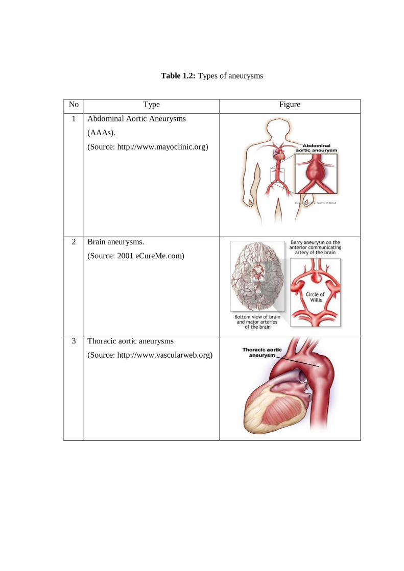
Table 1.2: Types of aneurysms
No Type Figure
1 Abdominal Aortic Aneurysms
(AAAs).
(Source: http://www.mayoclinic.org)
2 Brain aneurysms.
(Source: 2001 eCureMe.com)
3 Thoracic aortic aneurysms
(Source: http://www.vascularweb.org)
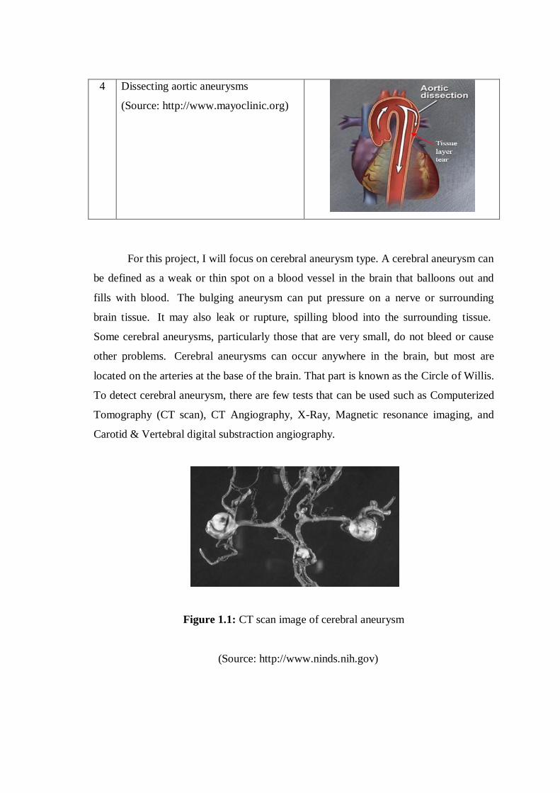
4 Dissecting aortic aneurysms
(Source: http://www.mayoclinic.org)
For this project, I will focus on cerebral aneurysm type. A cerebral aneurysm can
be defined as a weak or thin spot on a blood vessel in the brain that balloons out and
fills with blood. The bulging aneurysm can put pressure on a nerve or surrounding
brain tissue. It may also leak or rupture, spilling blood into the surrounding tissue.
Some cerebral aneurysms, particularly those that are very small, do not bleed or cause
other problems. Cerebral aneurysms can occur anywhere in the brain, but most are
located on the arteries at the base of the brain. That part is known as the Circle of Willis.
To detect cerebral aneurysm, there are few tests that can be used such as Computerized
Tomography (CT scan), CT Angiography, X-Ray, Magnetic resonance imaging, and
Carotid & Vertebral digital substraction angiography.
Figure 1.1: CT scan image of cerebral aneurysm
(Source: http://www.ninds.nih.gov)
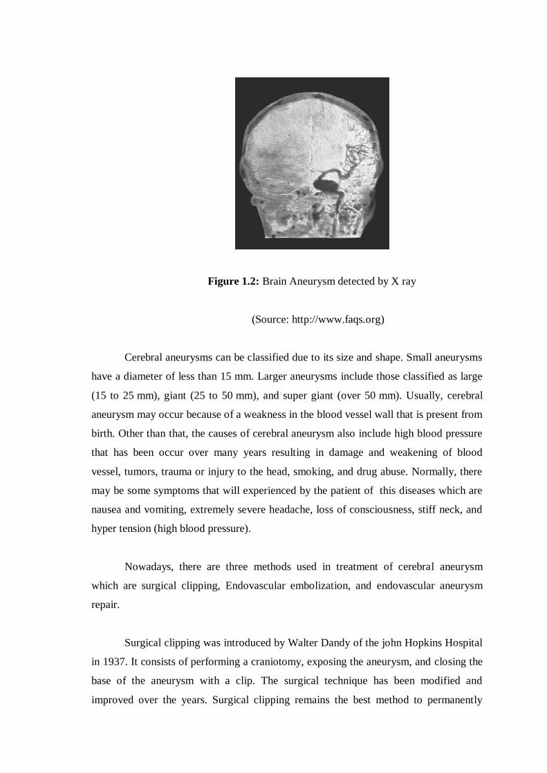
Figure 1.2: Brain Aneurysm detected by X ray
(Source: http://www.faqs.org)
Cerebral aneurysms can be classified due to its size and shape. Small aneurysms
have a diameter of less than 15 mm. Larger aneurysms include those classified as large
(15 to 25 mm), giant (25 to 50 mm), and super giant (over 50 mm). Usually, cerebral
aneurysm may occur because of a weakness in the blood vessel wall that is present from
birth. Other than that, the causes of cerebral aneurysm also include high blood pressure
that has been occur over many years resulting in damage and weakening of blood
vessel, tumors, trauma or injury to the head, smoking, and drug abuse. Normally, there
may be some symptoms that will experienced by the patient of this diseases which are
nausea and vomiting, extremely severe headache, loss of consciousness, stiff neck, and
hyper tension (high blood pressure).
Nowadays, there are three methods used in treatment of cerebral aneurysm
which are surgical clipping, Endovascular embolization, and endovascular aneurysm
repair.
Surgical clipping was introduced by Walter Dandy of the john Hopkins Hospital
in 1937. It consists of performing a craniotomy, exposing the aneurysm, and closing the
base of the aneurysm with a clip. The surgical technique has been modified and
improved over the years. Surgical clipping remains the best method to permanently

eliminate aneurysms. Depending on the shape and location of the aneurysm, surgical
clipping has been a very effective treatment, and typically, aneurysms that have been
completely clipped do not recur. Surgical clipping minimizes the necessity for periodic
follow-up angiographic studies. Furthermore, surgical clipping provides controlled
access to difficult anatomy and allows for arterial reconstruction when aneurysms have
complicated shapes and wide necks. Typically, aneurysm clips are made from titanium.
Titanium is selected because of their characteristics which are strong, has a low density,
and has a highly corrosion-resistant metal alloy. The clip has a spring mechanism that
allows the jaws of the clip to be placed on the aneurysm neck, thus occluding the
aneurysm from the feeding blood vessel. Figure 1.3 shown the variety of aneurysm clips
and Figure 1.4 shown a titanium clip is placed across the neck of an aneurysm.
Figure 1.3: Variety of aneurysm clips
(Source: http://www.mayfieldclinic.com)

Figure 1.4: A titanium clip is placed across the neck of an aneurysm
(Source: http://www.mayfieldclinic.com)
Endovascular embolization is an alternative to surgery. This treatment has been
offered at Toronto Western Hospital since 1992. This is done in the neuroangiography
suite under fluoroscopy. The Neurointerventional radiologist will make a small incision
in the groin through which a tiny catheter is guided through the femoral artery into the
brain vessels (1st stage). The catheter is carefully guided into a site of aneurysm (2
nd
stage). Soft platinum coils are deposited through the microcatheter into the aneurysm
(3rd
stage). When in this position, the coil is released by an application of a very low
voltage current causing the coil to detach from the pusher wire. The softness of the
platinum allows the coil to conform to the often irregular shape of the aneurysm. An
average of 5-6 coils are required to completely pack an aneurysm (4th
stage) The goal of
this treatment is to prevent the blood flow into the aneurysm sac by filling the aneurysm
with coils and thrombus (5th
stage). This treatment would prevent the aneurysm
bleeding or re-bleeding.
(1st stage) (2
nd stage) (3
rd stage) (4
th stage) (5
th stage)
Figure 1.5: Process of Endovascular Embolization
(Source: http://brainavm.oci.utoronto.ca)
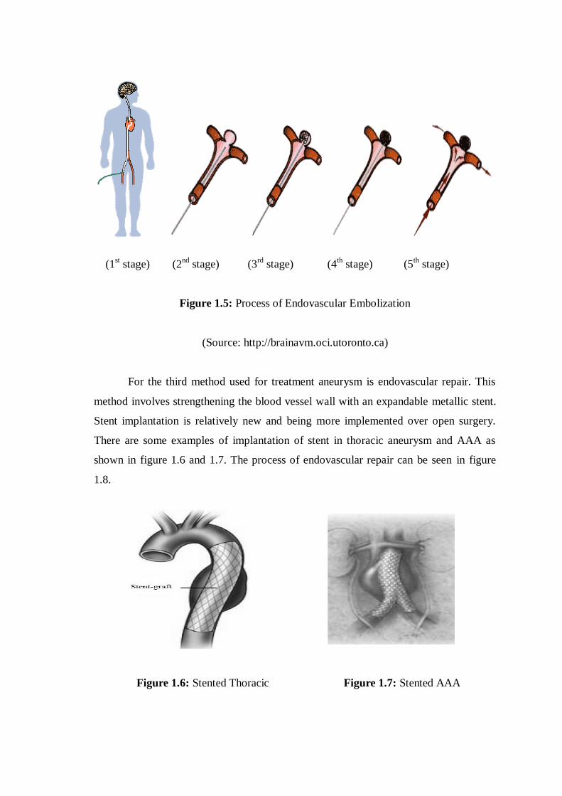
For the third method used for treatment aneurysm is endovascular repair. This
method involves strengthening the blood vessel wall with an expandable metallic stent.
Stent implantation is relatively new and being more implemented over open surgery.
There are some examples of implantation of stent in thoracic aneurysm and AAA as
shown in figure 1.6 and 1.7. The process of endovascular repair can be seen in figure
1.8.
Figure 1.6: Stented Thoracic Figure 1.7: Stented AAA

Figure 1.8: Process for endovascular repair
(Source: http://www.hpcbd.com)
1.2 Stent technology
Stent is a wire metal mesh tube inserted into a weakened area in a blood vessel
to help the blood flow smoothly and avoid aneurysm from burst. There are two types of
stents: bare stents (wire mesh) and covered stents (also commonly called stent grafts).
As many as 30 different stent designs are in use in the world. Stents may be classified
based on their predeployed repeating „cell‟ pattern of metal construction (slotted tube,
coil, or mesh) and nature of the stent delivery systems (self-expandable or balloon-
expandable). With the abundance of new stents, there is an urgent need for a how to
manual to help interventional cardiologist through the plethora of stent brands, types,
structures, patterns, strengths, diameters and lengths.
Currently, a stent classification design was proposed by Jost which was based on
structural characteristics of the stents. This includes original slotted tube stents (eg.
Palmaz-Schatz) as shown in figure 1.9, second generation tubular stents such as Crown,
MultiLink, and NIR, self-expanding stents such as Wallstent and Tristar stents, coil
stents such as Crossflex and Gianturco-Roubin, and Modular Ziagzag stents (eg. AVE
GFX).

In general, slotted-tube systems, characterized by the PT stent, are characterized
by Palmaz-Schatz stent, characterized by high vessel surface area coverage, high radial
strength, and consistent circumferential deployment pattern. Coil stent provide for
greater flexibility, conformability to the target vessel tortuosity, and access to side-
branches but have significant intrinsic recoil. Mesh design stent, found in many of the
second generation tubular stents, are hybrid of slotted tube and coil features. They
possess the sizing strategies and deployment mechanics of slotted tube stents; and
flexibility, conformability and side-branch access of the coil stents. A summary of
current stent designs is shown in Table 1.3. (H C Tan, Y T Lim, 1999). The figure of
linkage example shows in Figure 1.9.
Table 1.3: Current stent design
Type Product Name Material
Self expanding stents
Wallstent
Radius
Cobalt alloy
Nitinol
Balloon expandable
Gianturco-Roubin
Stainless steel
Coil stents
Crossflex
Wiktor
Stainless steel
Tantalum
Balloon expandable
Palmaz-schatz
Stainless steel
Tubular stents
Crown
NIR
MultiLink
Bestent
Stainless steel
Stainless steel
Stainless steel
Stainless steel
Balloon expandable
Crossflex LC
Stainless steel