memory b cells in the lung participate in protective ... · memory b cells in the lung participate...
TRANSCRIPT

Memory B cells in the lung participate in protectivehumoral immune responses to pulmonary influenzavirus reinfectionTaishi Onoderaa, Yoshimasa Takahashia,1, Yusuke Yokoia,b, Manabu Atoa, Yuichi Kodamaa, Satoshi Hachimurab,Tomohiro Kurosakic,d, and Kazuo Kobayashia
aDepartment of Immunology, National Institute of Infectious Diseases, Shinjuku-ku, Tokyo 162-8640, Japan; bResearch Center for Food Safety, GraduateSchool of Agricultural and Life Sciences, University of Tokyo, Bunkyo-ku, Tokyo 113-8657, Japan; cLaboratory of Lymphocyte Differentiation, World PremierInternational Immunology Frontier Research Center, and Graduate School of Frontier Biosciences, Osaka University, Suita, Osaka 565-0871, Japan; anddLaboratory for Lymphocyte Differentiation, RIKEN Research Center for Allergy and Immunology, Tsurumi-ku, Yokohama, Kanagawa 230-0045, Japan
Edited* by Michel C. Nussenzweig, The Rockefeller University, New York, NY, and approved December 29, 2011 (received for review September 18, 2011)
After pulmonary virus infection, virus-binding B cells ectopicallyaccumulate in the lung. However, their contribution to protectiveimmunity against reinfecting viruses remains unknown. Here, weshow the phenotypes and protective functions of virus-bindingmemory B cells that persist in the lung following pulmonaryinfection with influenza virus. A fraction of virus-binding B-cellpopulation in the lung expressed surface markers for splenicmature memory B cells (CD73, CD80, and CD273) along with CD69and CXCR3 that are up-regulated on lung effector/memory T cells.The lung B-cell population with memory phenotype persisted formore than 5 mo after infection, and on reinfection promptlydifferentiated into plasma cells that produced virus-neutralizingantibodies locally. This production of local IgG and IgA neutral-izing antibody was correlated with reduced virus spread inadapted hosts. Our data demonstrates that infected lungs harbora memory B-cell subset with distinctive phenotype and ability toprovide protection against pulmonary virus reinfection.
lung memory B cells | viral immunity
B-cell memory is bipartite, consisting of both long-lived plasmacells and memory B cells. Immediate protection against re-
infection is mediated by long-lived plasma cells that are presentin the bone marrow and secrete antibodies in an antigen-in-dependent fashion. Recall responses are mediated by memoryB cells that rapidly proliferate and differentiate in response toantigenic stimulation (1, 2). The accessibility of memory B cellsto reinfecting pathogens is, therefore, likely a significant factorin determining the effectiveness of humoral protection againstreinfection. Thus, it is fundamentally important to determine theprotective functions of pathogen-specific memory B cells thatreside at the sites of infection after the resolution of a pri-mary infection.In the case of influenza virus, the initial infection and replica-
tion occur in the respiratory tract. These elicit immune responsesin associated secondary lymphoid organs, e.g., mediastinal lymphnodes (MLNs), which support the initial rounds of B-cell primingthat follow a pulmonary infection (3). In addition, nonlymphoidorgans, including the lungs, participate in these primary respon-ses. Indeed, after primary infection with influenza virus, infectedlungs often support the development of ectopic tertiary lymphoidstructures known as induced bronchus-associated lymphoid tissue(iBALT) that contain germinal centers (GCs) and plasma cells(4). Moreover, infected lungs harbor the precursors of virus-binding plasma cells as revealed after in vitro stimulation of lungcells (5, 6), suggesting the existence of virus-binding memory Bcells in the lungs. However, virus-binding memory B cells in thelungs have not been indentified at cellular level, thereby theirphenotypic and functional characterization is still lacking.In this study, we characterized the phenotypes and functions of
class-switched, influenza-specific B cells in the lungs. We show that
a fraction of class-switched, influenza-specific B cells in the lungspossess amemory phenotype, persist for a long period, and respondto virus reinfection by promoting rapid viral clearance. Our datademonstrate that local tissues are important sites for the mainte-nance and reactivation of protective humoral memory responses.
ResultsInfection with Influenza Virus Induces Antigen-Specific Memory-LikeB-Cell Population in Lung. Pulmonary infection with influenza vi-ruses induces precursors of antigen-specific, class-switched plasmacells in the lungs (5, 6); however, the phenotypes of these cellshave not been determined. To characterize the phenotype andpersistence of influenza-specific B cells in lungs, we labeled Bcells recovered from lung tissue with recombinant hemagglutinin(rHA) conjugated to PE. Non-B cells, transitional B cells, B1cells, and plasma cells were excluded from our analyses bycolabeling with 12 mAbs specific for their surface markers (SIMaterials and Methods and Fig. S1). IgM/D+ cells were also ex-cluded from the present analysis to reduce the risk of includingnaïve HA-binding B cells present in the preimmune repertoire(7). This staining procedure resulted in the clear visualization ofHA-binding, class-switched B cells in mice infected with the X31influenza virus but not with other influenza virus subtypes (Fig.S1), confirming our methods’ specificity and sensitivity. Amongthe HA-binding IgM/D− lung B cells was a CD38+ subset thatcould represent a memory B-cell population (8, 9). We firsttraced the numbers of both CD38+ and CD38− B cells in lung,MLN, and spleen for 160 d after a primary infection (Fig. 1 Aand B). The numbers of HA-binding IgM/D−CD38− B cellsrapidly but transiently increased in lung, MLN, and spleen.However, CD38− B cells in MLNs persisted for a longer periodthan those in lungs and spleens, similar to splenic or MLN GCsfollowing vesicular stomatitis or influenza virus infection, re-spectively (10, 11).HA-binding IgM/D−CD38+ B cells were found in the lung,
MLN, and spleen, but lung CD38+ B cells required more time toreach equilibrium than that required for CD38+ B cells in otherorgans (Fig. 1B). This process resembles the slower accumula-tion of plasma cells in the lungs after influenza virus infection(11) and may reflect a requirement for structural alteration and/or niche formation in the infected lungs for B-cell localization.
Author contributions: Y.T., M.A., S.H., T.K., and K.K. designed research; T.O., Y.T., Y.Y.,and Y.K. performed research; T.O. and Y.T. analyzed data; and Y.T., T.K., and K.K. wrotethe paper.
The authors declare no conflict of interest.
*This Direct Submission article had a prearranged editor.1To whom correspondence should be addressed. E-mail: [email protected].
This article contains supporting information online at www.pnas.org/lookup/suppl/doi:10.1073/pnas.1115369109/-/DCSupplemental.
www.pnas.org/cgi/doi/10.1073/pnas.1115369109 PNAS | February 14, 2012 | vol. 109 | no. 7 | 2485–2490
IMMUNOLO
GY

Once generated, HA-binding IgM/D−CD38+ B cells stably per-sisted for 160 d after infection in all organs.Although definitive markers of murine memory B cells remain
to be identified (12, 13), CD73, CD80, and CD273 (PD-L2) areexpressed at higher levels on splenic memory than on naïve Bcells (14, 15). It is also postulated that the proportions of CD80+
and/or CD273+ cells reflect the maturation of memory B cells,because CD80+ and/or CD273+ cells express isotype-switched,somatically mutated B cell receptors more frequently thanCD80−CD273− cells (15). HA-binding IgM/D−CD38+ B cells inthe lung, MLN, and spleen expressed increased levels of CD73,CD80, and CD273 than naïve B cells (IgD+CD38+B220+) fromthe same tissue (Fig. S2). The phenotypic similarity of these HA-binding B cells to hapten-binding memory B cells (14, 15)
supports our hypothesis that HA-binding IgM/D−CD38+ B cellsrepresent a memory population. Significantly, HA-binding IgM/D−CD38+ lung B cells expressed CD73, CD80, and CD273 athigher frequencies than comparable populations in the MLN andspleen (Fig. S2); however, CD80 expression in the lung and MLNdid not differ significantly. Considering the slow and steady ac-cumulation of HA-binding IgM/D−CD38+ lung B cells, they maysuggest that these lung B cells develop after progressive acqui-sition of mature memory phenotypes.Further characterization revealed distinctive features of HA-
binding IgM/D−CD38+ lung B cells. These lung B cells showedelevated expression of CD69 and CXCR3, a chemokine receptorgoverning effector T-cell migration to inflamed lung tissue (16),compared with the comparable populations in MLN and spleen(Fig. 1C and Fig. S3). Notably, Lee et al. (17) recently suggestedthat CD69 regulates lung localization of CD8+ T cells followinginfluenza virus infection. Thus, HA-binding IgM/D−CD38+ lungB cells expressed elevated levels of localization factors that directthe infiltration and residence of T cells in response to lung in-flammation; however, the contribution of these to lung B-celllocalization is not yet known. Together, phenotypic character-ization of HA-binding IgM/D−CD38+ lung B cells revealed theirunique phenotypes sharing surface markers for murine memoryB cells with lung localization factors. Hereafter, we putativelydefine HA-binding IgM/D−CD38+ B-cell population as memory-like B-cell population.After pulmonary influenza virus infection, IgA-secreting plasma
cells develop in the lung concomitantly with the presence of IgAAb in bronchoalveolar lavage fluids (BALFs) (6, 18). To know therelative distribution of virus-specific IgA+ B cells in lung andother organs, we compared the frequencies of IgA+ cells amongHA-binding IgM/D−CD38+ B cells in lung, MLN, and spleen. Asexpected, the memory-like B-cell population in lung expressedIgA isotype more frequently than the comparable populations inMLN and spleen; however, the average frequency of IgA+ cellsrepresented only 7% of the lung B-cell population (Fig. 1C andFig. S3). The minor composition of IgA+ cells among IgM/D−
memory-like B cells in lungs is also supported by the previousestimation of IgA:IgG ratio (∼1:10) in the precursors of plasmacells in lungs (6). This result suggests that IgA switching is en-hanced but is not a major event during the development of thelung memory-like B-cell population following primary infection.
Memory-Like B-Cell Population in Lung Rapidly Differentiates intoIgG- or IgA-Secreting Plasma Cells on Pulmonary Challenge. Accel-erated responses to antigen challenge are a defining feature ofmemory B cells. To examine whether the memory-like B-cellpopulation in lung are indeed responding to secondary infection,we detected lung B cells proliferating shortly after virus chal-lenge by BrdU-incorporation assay. The memory-like B-cellpopulation in the lungs did not incorporate detectable levels ofBrdU at day 80 after primary infection (labeling period: 2 d)(Fig. 2A), consistent with the maintenance as a stable B-cellpopulation in the absence of frequently dividing precursors. Incontrast, by 2–3 d after secondary infection, BrdU+ memory-likeB-cell populations appeared in the lungs and MLNs, but not inthe spleens (Fig. 2A). By day 4 postinfection, HA-binding IgM/D−CD138+ cells expressing plasmablasts/plasma cell markersaccumulated in the lungs, and about half of these expressed IgA(Fig. 2B). ELISPOT analysis confirmed the prompt appearanceof plasma cells in the lungs consisting of comparable frequenciesof IgG- or IgA-secreting plasma cells (Fig. S4). These resultssuggest that, in response to virus reinfection, the memory-like B-cell population in lung divided and developed into plasma cells.In addition, regardless of infrequent expression of surface IgAon the memory-like B-cell populations, HA-binding class-switched plasma cells expressed IgG or IgA isotypes at similarfrequencies following secondary challenge.
A
Spl
Lung
Naive D20 D160
MLN
0
0
0.2<0.1
0
0
0.24.9
0
0
0.63.7
0
0
0.30.3
0
0
<0.1<0.1
0
0
0.16.1
0
0
6.14.5
0
0
0.9<0.1
0
0
11.5<0.1
B C
Num
bers
of H
A-b
indi
ng B
cel
ls (p
er 1
06 cel
ls)
103
102
10
104
1
105
0 100 15050 200
103
102
10
104
1
105
0 100 15050 200
103
102
10
104
1
105
0 100 15050 200
CD38-CD38+
Lung MLN Spl
100
80
60
40
20
0Freq
uenc
y of
CD
69+ (
%)
Lung MLN Spl
100
80
60
40
20
0
Freq
uenc
y of
CX
CR
3+ (%
)
Lung
10
8
6
4
2
0Lung MLN Spl
Freq
uenc
y of
IgA
+ (%
)
MLN
Spl
HA
bind
ing
CD38
Fig. 1. Kinetics and surface phenotypes of HA-binding IgM/D− B-cell pop-ulations. (A) Cells recovered at the indicated time points after infection weresubjected to flow-cytometric analysis (n = 4–5). Representative flow data forHA-binding/CD38 expression by IgM/D−dump− B cells (Fig. S1) are shown. (B)Absolute cell number of CD38+ and CD38− cells within the lymphocyte gatewas plotted. (C) The frequencies of cells expressing CD69, CXCR3, or IgA areplotted. In B and C, each circle represents the result for an individual mouse(lung and spleen) and two to four pooled mice (MLN). **P < 0.01.
2486 | www.pnas.org/cgi/doi/10.1073/pnas.1115369109 Onodera et al.

Protective Function of the Memory-Like B-Cell Population in Lung. Toexamine the protective capacity of the memory-like B-cell pop-ulation against reinfection, HA-binding IgM/D−CD38+ B cellswere highly purified from the lungs and spleens of the mice >2mo after primary infection, and then transferred into scid micetogether with CD4+ T cells isolated from the same donors.MLNs provided too few cells for adoptive transfer experimentsand were not used. Accumulating evidence indicates that iBALTserves not only a site for initiating respiratory immune responsesbut also as a homing site for plasma cells (18, 19). Therefore, weconsidered that preforming iBALT structure might be requiredfor reconstitution of local, secondary Ab responses to virus in-fection in adoptive hosts. To generate iBALTs in recipient micebefore memory B-cell transfer, recipient scid mice were sub-jected to intranasal CpG treatment and i.v. transfer of spleencells. As expected, this treatment generated peribronchial B-cellclusters 10 d later (Fig. 3 A and B). The treated mice were in-oculated with challenging viruses 1 d after transfer of purifiedHA-binding IgM/D−CD38+ B cells (Fig. 3C), and then we de-termined virus titers in BALFs 6 d after infection. Remarkably,the mice reconstituted with the memory-like B-cell population inlung significantly reduced virus titers in BALFs, whereas thosegiven splenic counterparts were similar to controls (Fig. 3D).Moreover, the protective ability of lung memory-like B-cellpopulation was not observed in recipient mice without pre-CpGtreatment (Fig. 3E).To address whether adoptive transfer of the memory-like B-
cell population in lung accelerated the Ab production in therespiratory tract, the numbers of HA-binding lung plasma cellsand the levels of anti-HA Abs in BALFs were evaluated at thesame time point. Adoptive transfer of lung memory-like B-cellpopulation generated sixfold more IgG- and IgA-secretingplasma cells in the lungs compared with splenic counterparts ina manner depending on pre-CpG treatment of recipient mice(Fig. 3 F and G). Consistent with the results in secondary chal-lenged BALB/c mice, the frequencies of IgA-secreting plasmacells were comparable to those of IgG-secreting plasma cells.These results support the contention that the enhanced IgA re-sponse following secondary infection reflects the increased sup-ply of IgA-secreting plasma cells from memory and not naïve B
cells. In accordance with ELISPOT data, anti-HA IgG and IgAAb titers in BALFs were elevated in the mice reconstituted withthe lung B-cell population after CpG treatment but not in thosegiven the splenic B-cell population (Fig. 3H). Together, thesedata indicate that HA-binding IgM/D−CD38+ lung B cells areable to generate large amounts of Abs at the site of virus repli-cation to confer protection more effectively than splenic coun-terparts. Moreover, given the successful reconstitution of local,secondary Ab responses in adoptive hosts, we conclude that HA-binding IgM/D−CD38+ B cells represent memory B cells in bothphenotypes and functions.
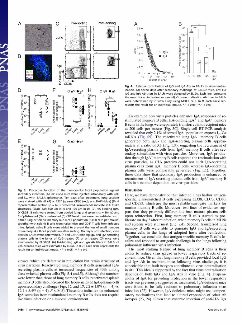
Protective Functions of IgG Versus IgA Abs in Respiratory Tracts.Although it is established that secretory IgA can provide pro-tection more effectively than IgG in upper respiratory tracts (20,21), it is not known whether IgA in lower respiratory tractscontributes to protection in the presence of IgG. To estimate thecontribution of IgG and IgA Abs in BALFs to virus neutraliza-tion, we first determined the titers of HA-binding IgG and IgAAbs. Consistent with comparable accumulation of IgG- and IgA-secreting plasma cells in the lungs (Figs. 2 and 3), BALFs insecondary challenged mice contained both HA-binding IgG andIgA Abs (Fig. 4A). The relative contributions of IgG and IgA tovirus neutralization were estimated using BALFs depleted ofeither IgG or IgA Abs by affinity chromatography. We observedthat removal of either IgG or IgA reduced virus-neutralizingactivity of BALFs by two- to threefold (Fig. 4B), indicatingpartial, but not complete, reduction in activity. Moreover, re-moval of both IgG and IgA reduced virus-neutralizing activity tolevels close to the detection limit. These data suggest that con-comitant production of IgG and IgA Abs is required to achievemaximum neutralization activity.
Virus Particles Enhance Secondary IgA Response by Recruiting IgA-Secreting Plasma Cells from IgA− Memory B Cells. To explore themechanisms for enhanced IgA secretion following secondary in-fection, we first examined whether the enhanced IgA responsedepends on intranasal infection by live viruses. Similar to Fig. 3,scidmice were reconstituted with either lung or splenic memory Bcells and then subjected to i.p. boosting with formalin-inactivated
A
BrdU
Lung MLN Spl
D3
D2
% of Max
0
0
14.4 ± 5.6
0
0
9.1 ± 1.7
Pre
0
0
0
0
0
2.3 ± 0.5
0
0
0
0
0
0
0
0
0
0
0
0
0
0
0.2 ± 0.3
B
0
0
0
0
0
0
0
0
0
0
0
0
<0.01
0.03 ±0.01
<0.010.29 ± 0.17
0.88 ±0.22
IgA
CD138
HA
bind
ing
Lung MLN Spl
1.11 ±0.21
Pre
D4
0
0
52.3 ± 1.2
0
0
57.5 ± 8.6
Fig. 2. Reactivation and terminal differentiation of the memory-like B-cell population in lung following secondary infection. (A) BALB/c mice were infectedtwice, with an 80-d interval. Mice were treated intraperitoneally with 1 mg BrdU for 48 h before analysis, and cells from the indicated organs were subjectedto intracellular analysis for BrdU. Representative flow data for BrdU staining of HA-binding IgM/D−CD38+ B cells are presented (n = 3). (B) HA-binding/CD138expression among B220dulldump− cells and IgA expression among HA-binding CD138+ cells are presented (n = 3–5).
Onodera et al. PNAS | February 14, 2012 | vol. 109 | no. 7 | 2487
IMMUNOLO
GY
viruses, which are defective in replication but retain structure ofvirus particles. Reactivated lung memory B cells generated IgA-secreting plasma cells at increased frequencies of 40% amongclass-switched plasma cells (Fig. 5A andB). Although the numberswere lower than those of lung memory B cells, reactivated splenicmemory B cells also increased the frequencies of IgA-plasma cellsupon secondary challenge (Figs. 1C and 5B; 2.2 ± 1.0% (n= 4) vs.12.1 ± 5.4% (n= 4), P < 0.05). These data indicate that enhancedIgA secretion from restimulated memory B cells does not requirelive virus infection or a mucosal environment.
To examine how virus particles enhance IgA responses of re-stimulated memory B cells, HA-binding IgA+ and IgA− memoryB cells in the lungs were separately transferred into recipient miceat 200 cells per mouse (Fig. 5C). Single-cell RT-PCR analysisrevealed that only 2.1% of sorted IgA− population express JH-CαmRNA (Fig. S5). The reactivated lung IgA− memory B cellsgenerated both IgG- and IgA-secreting plasma cells approxi-mately at a ratio of 3:1 (Fig. 5D), suggesting the recruitment ofIgA-secreting plasma cells from IgA− memory B cells after sec-ondary stimulation with virus particles. Moreover, IgA produc-tion through IgA−memory B cells required the restimulation withvirus particles, as rHA proteins could not elicit IgA-secretingplasma cells from IgA− memory B cells, whereas IgG-secretingplasma cells were comparably generated (Fig. 5E). Together,these data show that secondary IgA production is enhanced byrecruitment of IgA-secreting plasma cells from IgA− memory Bcells in a manner dependent on virus particles.
DiscussionHere, we have demonstrated that infected lungs harbor antigen-specific, class-switched B cells expressing CD38, CD73, CD80,and CD273, which are the most reliable surrogate markers formurine memory B cells. Moreover, two lines of evidences sup-port that they promptly differentiate into mature plasma cellsupon reinfection. First, lung memory B cells started to pro-liferate on day 2 after reinfection, when memory B cells in MLNsand spleens were still inert (Fig. 2A). Second, transferred lungmemory B cells were able to generate IgG and IgA-secretingplasma cells in the lungs of adopted hosts after reinfection.Together, we conclude that antigen-specific memory B cells lo-calize and respond to antigenic challenge in the lungs followingpulmonary influenza virus infection.The most striking feature of lung memory B cells is their
ability to reduce virus spread in lower respiratory tracts of re-cipient mice. Given that lung memory B cells provided local IgGand IgA Ab in recipient mice following virus challenge, it isconceivable that both isotypes contribute to virus neutralizationin situ. This idea is supported by the fact that virus neutralizationdepends on both IgG and IgA Abs in vitro (Fig. 4). Dispens-ability of IgA for providing protection in the lower respiratorytracts was previously suggested as vaccinated, IgA-deficient micewere found to be fully resistant to pulmonary influenza virusinfection (22). However, IgA-deficient mice might use compen-satory mechanisms that lead to altered expression of other Abisotypes (23, 24). Given that systemic injection of anti-HA IgA
Fig. 3. Protective function of the memory-like B-cell population againstsecondary infection. (A) CB17-scid mice were injected intranasally with CpGand i.v. with BALB/c splenocytes. Ten days after treatment, lung sectionswere stained with HE (A) or B220 (green), CD90 (red), and DAPI (blue) (B). Arepresentative section (n = 6) is presented. Arrowheads indicate iBALT-likestructures. (Scale bar: 500 μm in A and 100 μm in B). (C) HA-binding IgM/D−CD38+ B cells were sorted from pooled lungs and spleens (n = 10). (D andE) CpG-treated (D) or untreated (E) CB17-scid mice were reconstituted witheither lung or splenic memory-like B-cell population (3,000 cells per mouse)together with splenic B cells from naïve mice and CD4+ T cells from infectedmice. Splenic naïve B cells were added to prevent the loss of small numbersof memory-like B-cell population after sorting. On day 6 postinfection, virustiters in BALFs were determined. (F and G) HA-binding IgG and IgA-secretingplasma cells in the lungs of CpG-treated (F) or untreated (G) mice wereenumerated by ELISPOT. (H) HA-binding IgG and IgA Ab titers in BALFs ofCpG-treated mice were estimated by ELISA. In D–H, each circle represents theresult for an individual mouse. *P < 0.05; **P < 0.01.
A
10410310210 105
A49
0
0
1.0
2.0
3.0
3.5
2.5
1.5
0.5
Dilution of BALFCTRL -IgG -IgA -IgG
-IgA
Viru
s-ne
utra
lizat
ion
Ab
titer
0
20
100
120
80
60
40
140
∗∗∗
∗∗
B
IgAIgG
Fig. 4. Relative contribution of IgG and IgA Abs in BALFs to virus-neutral-ization. (A) Seven days after secondary challenge of BALB/c mice, anti-HAIgG and IgA Ab titers in BALFs were detected by ELISA. Each line representsthe result for an individual mouse. (B) Virus-neutralization Ab titers in BALFswere determined by in vitro assay using MDCK cells. In B, each circle rep-resents the result for an individual mouse. *P < 0.05; **P < 0.01.
2488 | www.pnas.org/cgi/doi/10.1073/pnas.1115369109 Onodera et al.

mAbs was effective to prevent the initial infection in lung airways(25), we prefer the idea that both IgG and IgA Abs in lungairways contribute to virus neutralization.Themechanism underlying the ability of lungmemory B cells to
supply local IgG and IgA Abs remains an important question tobe addressed. Lung memory B cells express CD69 and CXCR3,possible mediators of lung localization. Thus, one possibility isthat transferred lung memory B cells home back to the lung,wherein they could generate IgG- and IgA-secreting plasma cellsat virus replication sites. This possibility is supported by re-quirement for intranasal CpG treatment before memory B-cell
transfer. CpG-induced inflammation would trigger the local ex-pression of several ligands (e.g., CXCL9 and CXCL10) for che-mokine and/or homing receptors expressed on lung memoryB cells. However, without pre-CpG treatment, the reduction ofvirus spread was not observed in the mice reconstituted with lungmemory B cells (Fig. 3 D–G). The alternative possibility is thattransferred lung memory B cells home to the MLNs, where theysupply plasma cells in the lungs upon reinfection. Although wemade tremendous efforts for in vivo tracing of transferredmemory B cells, our attempts were unsuccessful due to a paucityof memory B cells.B-cell intrinsic recognition of intact viruses is often hampered
by the tissue tropisms of virus replication in nonlymphoid organs.Following pulmonary influenza virus infection, lung localizationof memory B cells is one way to facilitate B cell intrinsic recog-nition for shaping the magnitude and quality of protective effectorfunctions. Better understanding of the generation, maintenance,and reactivation of memory B cells in lung provides importantinsights for the development of vaccines for protection againstinfluenza virus and other respiratory pathogens.
Materials and MethodsMice and Viruses. Mice and viruses used in this study are described in SIMaterials and Methods.
Cell Preparation and Flow Cytometry. Lung cells were isolated by Percollgradient centrifugation after digestion with collagenase D and DNase I.Single-cell suspensions from lungs, MLNs, and spleens were stained withmixtures of biotinylated mAbs, followed by fluorescence-conjugated mAbs.BrdU-labeled cells were detected by using BrdU Flow kit (BD Biosciences).Stained cells were analyzed or purified using FACS Canto II or FACS Aria (BDBioscience). Detailed methods are included in SI Materials and Methods.
Quantification of Anti-HA Abs and Plasma Cells. HA-binding Abs and plasmacells were quantified by ELISA and ELISPOT using rHA as coating antigensand anti-mouse IgG- or IgA Abs as secondary Abs. Virus-neutralization Abtiters were quantified by microneutralization assay using MDCK cells andX31 virus (100 TCID50) (26). Detailed methods are included in SI Materialsand Methods.
Statistical Analyses of Data. Statistical significance was determined by anunpaired two-tailed Student t test. P < 0.05 were considered significant.
ACKNOWLEDGMENTS. We thank Dr. G. Kelsoe (Duke University) for criticalreading of the manuscript; Dr. T. Tsubata (Tokyo Medical and DentalUniversity) for providing the X31 virus; and Ms. E. Watanabe, E. Izumiyama,and K. Fukuhara for technical assistance. This study was supported by grantsfrom the Ministry of Education, Culture, Sports, Science, and Technology inJapan (to Y.T. and T.K.), the Japan Science and Technology Agency, CoreResearch for Evolutional Science and Technology (to Y.T. and T.K.); and theEmerging and Re-Emerging Infectious Diseases and Regulatory Science ofPharmaceuticals and Medical Devices of the Ministry of Health, Labour andWelfare in Japan (to Y.T. and K.K.).
1. Dörner T, Radbruch A (2007) Antibodies and B cell memory in viral immunity. Im-munity 27:384–392.
2. Sallusto F, Lanzavecchia A, Araki K, Ahmed R (2010) From vaccines to memory andback. Immunity 33:451–463.
3. Coro ES, Chang WL, Baumgarth N (2006) Type I IFN receptor signals directly stimulatelocal B cells early following influenza virus infection. J Immunol 176:4343–4351.
4. Moyron-Quiroz JE, et al. (2004) Role of inducible bronchus associated lymphoid tissue(iBALT) in respiratory immunity. Nat Med 10:927–934.
5. Jones PD, Ada GL (1987) Persistence of influenza virus-specific antibody-secreting cellsand B cell memory after primary murine influenza virus infection. Cell Immunol 109:53–64.
6. Joo HM, He Y, Sangster MY (2008) Broad dispersion and lung localization of virus-specific memory B cells induced by influenza pneumonia. Proc Natl Acad Sci USA 105:3485–3490.
7. Baumgarth N, et al. (1999) Innate and acquired humoral immunities to influenza virusare mediated by distinct arms of the immune system. Proc Natl Acad Sci USA 96:2250–2255.
8. Ridderstad A, Tarlinton DM (1998) Kinetics of establishing the memory B cell pop-ulation as revealed by CD38 expression. J Immunol 160:4688–4695.
9. TakahashiY,OhtaH,TakemoriT (2001)Fas is requiredfor clonal selection ingerminal centersand the subsequent establishment of the memory B cell repertoire. Immunity 14:181–192.
10. Bachmann MF, Odermatt B, Hengartner H, Zinkernagel RM (1996) Induction of long-lived germinal centers associated with persisting antigen after viral infection. J ExpMed 183:2259–2269.
11. Rothaeusler K, Baumgarth N (2010) B cell fate decisions following influenza virusinfection. Eur J Immunol 40:366–377.
12. Bhattacharya D, et al. (2007) Transcriptional profiling of antigen-dependent murine Bcell differentiation and memory formation. J Immunol 179:6808–6819.
13. Tomayko MM, et al. (2008) Systematic comparison of gene expression between mu-rine memory and naive B cells demonstrates that memory B cells have unique sig-naling capabilities. J Immunol 181:27–38.
14. Anderson SM, Tomayko MM, Ahuja A, Haberman AM, Shlomchik MJ (2007) Newmarkers for murine memory B cells that define mutated and unmutated subsets. J ExpMed 204:2103–2114.
15. TomaykoMM, Steinel NC, Anderson SM, Shlomchik MJ (2010) Cutting edge: Hierarchyof maturity of murine memory B cell subsets. J Immunol 185:7146–7150.
16. Kohlmeier JE, et al. (2009) CXCR3 directs antigen-specific effector CD4+ T cell mi-gration to the lung during parainfluenza virus infection. J Immunol 183:4378–4384.
BA
Freq
uenc
y of
IgA
+ pla
sma
cells
amon
g Ig
G+ a
nd Ig
A+ p
lasm
a ce
lls (%
) D
ECPre-sorting
IgA+
IgA-IgA
CD38
∗∗
∗IgAIgG
memoryNo
Lung
Spleen
Anti-
HA
plas
ma
cells
(per
106 s
plen
ocyt
es)
103
10
102
104
10
0
20
30
40
50
Lung
Spleen
memoryNo
IgA+
IgA-
Anti-
HA
plas
ma
cells
(per
106 s
plen
ocyt
es)
103
10
102
104 IgAIgG
IgAIgGAn
ti-H
A pl
asm
a ce
lls (p
er 1
06 spl
enoc
ytes
)103
10
102
104
∗
Virion rHA
∗∗∗∗
0
0
5.8
76.4
0
0
96.4
3.6
0
0
<1.4
98.6
Fig. 5. Virus-dependent recruitment of IgA-secreting plasma cells from IgA−
memory B cells. (A) CB17-scid mice were reconstituted similarly to Fig. 3Dand boosted with inactivated viruses. At day 7 after boosting, the numbersof HA-binding IgG+ and IgA+ plasma cells were enumerated and plotted. (B)The frequencies of IgA+ plasma cells among IgG+/IgA+ plasma cells areshown. (C) IgA+ and IgA− memory B cells were sorted from pooled lung cellsand splenocytes (n = 10). (D) The recipient mice were reconstituted with 200IgA+ or IgA− memory B cells in the lungs together with B cells and CD4+ Tcells, and the numbers of IgG+ and IgA+ plasma cells were determined afterboosting. (E) Mice were reconstituted with IgA− memory B cells in the lungs,and the numbers of IgG+ and IgA+ plasma cells were determined afterboosting with inactivated viruses or rHA. In A, B, D, and E, each circle rep-resents the result for an individual recipient mouse. *P < 0.05; **P < 0.01.
Onodera et al. PNAS | February 14, 2012 | vol. 109 | no. 7 | 2489
IMMUNOLO
GY

17. Lee YT, et al. (2011) Environmental and antigen receptor-derived signals supportsustained surveillance of the lungs by pathogen-specific cytotoxic T lymphocytes.J Virol 85:4085–4094.
18. GeurtsvanKessel CH, et al. (2009) Dendritic cells are crucial for maintenance of tertiarylymphoid structures in the lung of influenza virus-infected mice. J Exp Med 206:2339–2349.
19. Halle S, et al. (2009) Induced bronchus-associated lymphoid tissue serves as a gen-eral priming site for T cells and is maintained by dendritic cells. J Exp Med 206:2593–2601.
20. Renegar KB, Small PA, Jr. (1991) Passive transfer of local immunity to influenza virusinfection by IgA antibody. J Immunol 146:1972–1978.
21. Renegar KB, Small PA, Jr., Boykins LG, Wright PF (2004) Role of IgA versus IgG in thecontrol of influenza viral infection in the murine respiratory tract. J Immunol 173:1978–1986.
22. Mbawuike IN, et al. (1999) Mucosal immunity to influenza without IgA: an IgAknockout mouse model. J Immunol 162:2530–2537.
23. Harriman GR, et al. (1999) Targeted deletion of the IgA constant region in mice leadsto IgA deficiency with alterations in expression of other Ig isotypes. J Immunol 162:2521–2529.
24. Arulanandam BP, et al. (2001) IgA immunodeficiency leads to inadequate Th cellpriming and increased susceptibility to influenza virus infection. J Immunol 166:226–231.
25. Palladino G, Mozdzanowska K, Washko G, Gerhard W (1995) Virus-neutralizing an-tibodies of immunoglobulin G (IgG) but not of IgM or IgA isotypes can cure influenzavirus pneumonia in SCID mice. J Virol 69:2075–2081.
26. Takahashi Y, et al. (2009) Protective immunity afforded by inactivated H5N1 (NIBRG-14) vaccine requires antibodies against both hemagglutinin and neuraminidase inmice. J Infect Dis 199:1629–1637.
2490 | www.pnas.org/cgi/doi/10.1073/pnas.1115369109 Onodera et al.