melatonin: fundamental non-ionizing … fundamental non-ionizing electromagnetic settings for...
TRANSCRIPT
Melatonin:
Fundamental Non-Ionizing Electromagnetic Settings for Optimal Human Performance
by Katharina Gustavs
Environmental and Occupational Health Study Program
University of Victoria August 2001

Melatonin: Fundamental Non-Ionizing EM Settings for Optimal Human Performance 2
© 2001 Katharina Gustavs www.buildingbiology.ca
Table of Contents
TABLE OF CONTENTS .............................................................................................. 2
TABLES ................................................................................................................... 3
INTRODUCTION ...................................................................................................... 4
BREAST CANCER INCIDENCE AND ETIOLOGY.......................................................... 6
MELATONIN ............................................................................................................ 8
MELATONIN - ITS ANTITUMOR PROPERTIES......................................................................... 9 PRIMARY FACTOR: LIGHT - VISIBLE NON-IONIZING EMR ..................................................... 11 SECONDARY FACTOR: INVISIBLE NON-IONIZING EMR ......................................................... 12 EXPOSURE CHARACTERISTICS OF NON-IONIZING ELECTROMAGNETIC RADIATION AND OCCURRENCE OF
MELATONIN REDUCTION EFFECT.................................................................................... 13
MELATONIN-EMR INFORMATION NETWORK ......................................................... 23
SITING AND MEASURING CAPACITY OF DATA COLLECTION SITES ............................................. 24 REAL WORLD MEASUREMENTS IN RESIDENTIAL SETTINGS AT NIGHT ......................................... 25 MULTIVARIATE ANALYSIS ............................................................................................ 27
CONCLUSIONS ...................................................................................................... 28
REFERENCES ......................................................................................................... 31
REFERENCES FOR TABLES 4 THROUGH 12 ........................................................................ 33 MELATONIN RESEARCH UPDATE WITH REGARDS TO ELECTROMAGNETIC FIELD EXPOSURE (2005) ....... 38
Melatonin: Fundamental Non-Ionizing EM Settings for Optimal Human Performance 3
© 2001 Katharina Gustavs www.buildingbiology.ca
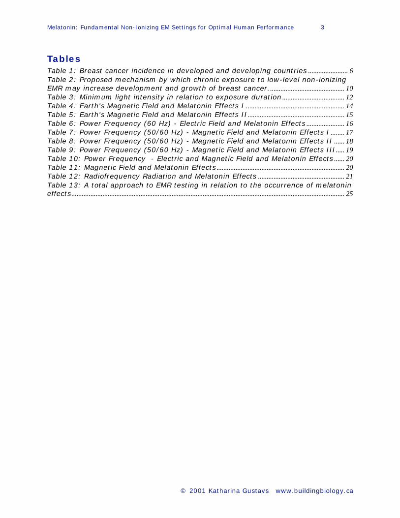
Tables
Table 1: Breast cancer incidence in developed and developing countries ....................... 6
Table 2: Proposed mechanism by which chronic exposure to low-level non-ionizing
EMR may increase development and growth of breast cancer............................................ 10
Table 3: Minimum light intensity in relation to exposure duration .................................... 12
Table 4: Earth's Magnetic Field and Melatonin Effects I ......................................................... 14
Table 5: Earth's Magnetic Field and Melatonin Effects II........................................................ 15
Table 6: Power Frequency (60 Hz) - Electric Field and Melatonin Effects ...................... 16
Table 7: Power Frequency (50/60 Hz) - Magnetic Field and Melatonin Effects I ........ 17
Table 8: Power Frequency (50/60 Hz) - Magnetic Field and Melatonin Effects II ...... 18
Table 9: Power Frequency (50/60 Hz) - Magnetic Field and Melatonin Effects III..... 19
Table 10: Power Frequency - Electric and Magnetic Field and Melatonin Effects ...... 20
Table 11: Magnetic Field and Melatonin Effects .......................................................................... 20
Table 12: Radiofrequency Radiation and Melatonin Effects .................................................. 21
Table 13: A total approach to EMR testing in relation to the occurrence of melatonin effects.............................................................................................................................................................. 25
Melatonin: Fundamental Non-Ionizing EM Settings for Optimal Human Performance 4
© 2001 Katharina Gustavs www.buildingbiology.ca
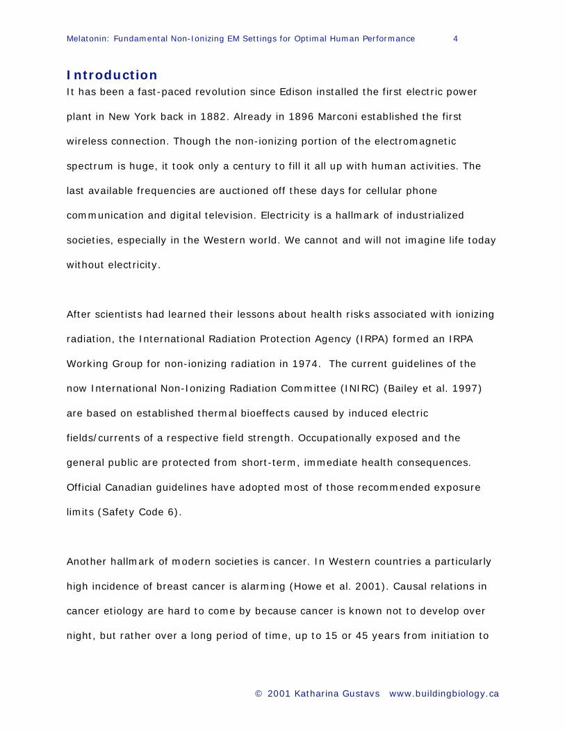
Introduction
It has been a fast-paced revolution since Edison installed the first electric power
plant in New York back in 1882. Already in 1896 Marconi established the first
wireless connection. Though the non-ionizing portion of the electromagnetic
spectrum is huge, it took only a century to fill it all up with human activities. The
last available frequencies are auctioned off these days for cellular phone
communication and digital television. Electricity is a hallmark of industrialized
societies, especially in the Western world. We cannot and will not imagine life today
without electricity.
After scientists had learned their lessons about health risks associated with ionizing
radiation, the International Radiation Protection Agency (IRPA) formed an IRPA
Working Group for non-ionizing radiation in 1974. The current guidelines of the
now International Non-Ionizing Radiation Committee (INIRC) (Bailey et al. 1997)
are based on established thermal bioeffects caused by induced electric
fields/currents of a respective field strength. Occupationally exposed and the
general public are protected from short-term, immediate health consequences.
Official Canadian guidelines have adopted most of those recommended exposure
limits (Safety Code 6).
Another hallmark of modern societies is cancer. In Western countries a particularly
high incidence of breast cancer is alarming (Howe et al. 2001). Causal relations in
cancer etiology are hard to come by because cancer is known not to develop over
night, but rather over a long period of time, up to 15 or 45 years from initiation to
Melatonin: Fundamental Non-Ionizing EM Settings for Optimal Human Performance 5
© 2001 Katharina Gustavs www.buildingbiology.ca
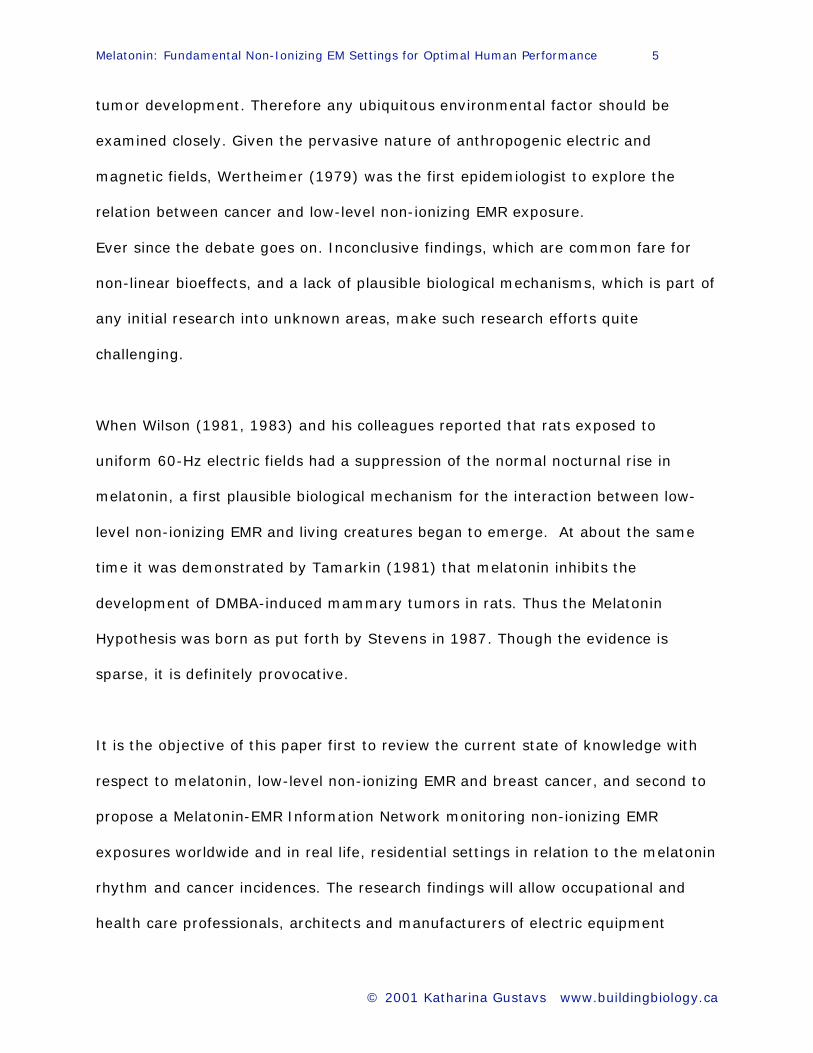
tumor development. Therefore any ubiquitous environmental factor should be
examined closely. Given the pervasive nature of anthropogenic electric and
magnetic fields, Wertheimer (1979) was the first epidemiologist to explore the
relation between cancer and low-level non-ionizing EMR exposure.
Ever since the debate goes on. Inconclusive findings, which are common fare for
non-linear bioeffects, and a lack of plausible biological mechanisms, which is part of
any initial research into unknown areas, make such research efforts quite
challenging.
When Wilson (1981, 1983) and his colleagues reported that rats exposed to
uniform 60-Hz electric fields had a suppression of the normal nocturnal rise in
melatonin, a first plausible biological mechanism for the interaction between low-
level non-ionizing EMR and living creatures began to emerge. At about the same
time it was demonstrated by Tamarkin (1981) that melatonin inhibits the
development of DMBA-induced mammary tumors in rats. Thus the Melatonin
Hypothesis was born as put forth by Stevens in 1987. Though the evidence is
sparse, it is definitely provocative.
It is the objective of this paper first to review the current state of knowledge with
respect to melatonin, low-level non-ionizing EMR and breast cancer, and second to
propose a Melatonin-EMR Information Network monitoring non-ionizing EMR
exposures worldwide and in real life, residential settings in relation to the melatonin
rhythm and cancer incidences. The research findings will allow occupational and
health care professionals, architects and manufacturers of electric equipment
Melatonin: Fundamental Non-Ionizing EM Settings for Optimal Human Performance 6
© 2001 Katharina Gustavs www.buildingbiology.ca
including the general public to create a built environment at work and at home,
which is designed for long-term health.
Breast Cancer Incidence and Etiology
According to the Annual Report to the Nation on the Status of Cancer (Howe 2001),
total death rates declined in males and females during 1992 through 1998.
Incidence rates in females, however, increased slightly, "largely because of breast
cancer increases". The Atlas of Cancer Mortality in the United States shows an
interesting geographic pattern for breast cancer. Breast cancer clusters have
persisted across the northeast of the US, especially in urban centers, for over four
decades (Kulldorff et al. 1997). In the sunny south we find a much lower incidence.
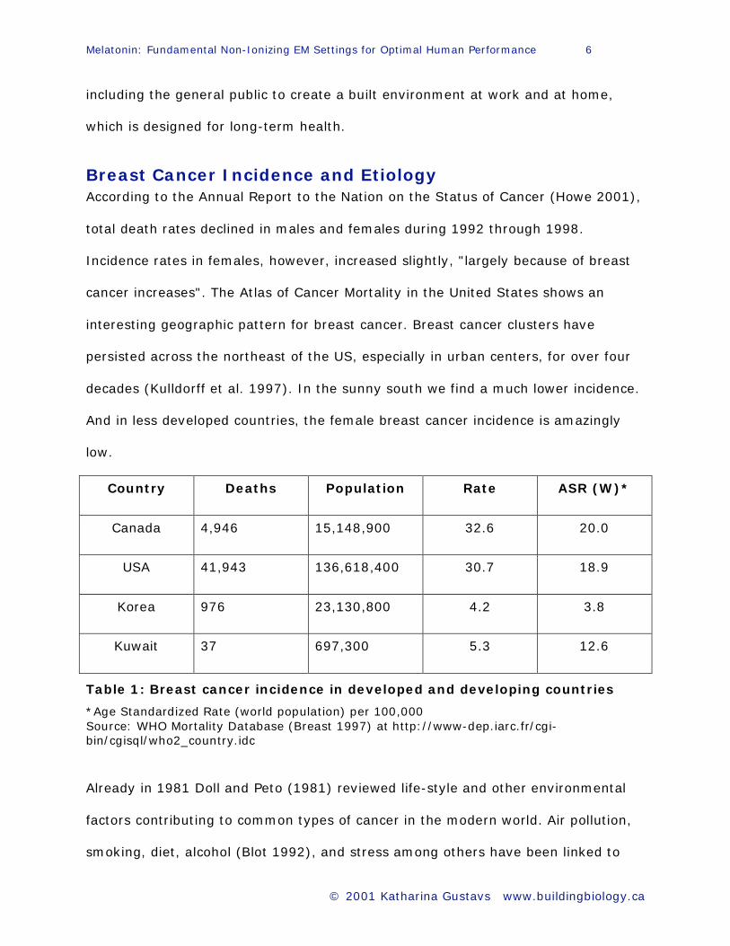
And in less developed countries, the female breast cancer incidence is amazingly
low.
Country Deaths Population Rate ASR (W)*
Canada 4,946 15,148,900 32.6 20.0
USA 41,943 136,618,400 30.7 18.9
Korea 976 23,130,800 4.2 3.8
Kuwait 37 697,300 5.3 12.6
Table 1: Breast cancer incidence in developed and developing countries
*Age Standardized Rate (world population) per 100,000
Source: WHO Mortality Database (Breast 1997) at http://www-dep.iarc.fr/cgi-
bin/cgisql/who2_country.idc
Already in 1981 Doll and Peto (1981) reviewed life-style and other environmental
factors contributing to common types of cancer in the modern world. Air pollution,
smoking, diet, alcohol (Blot 1992), and stress among others have been linked to
Melatonin: Fundamental Non-Ionizing EM Settings for Optimal Human Performance 7
© 2001 Katharina Gustavs www.buildingbiology.ca
cancer. In the case of breast cancer, experts agree that early age at onset of
menarche, late age at onset of menopause, first full-term pregnancy after age 30, a
history of pre-menopausal breast cancer for mother and a sister, and obesity are
associated with an increased risk of breast cancer (CancerNet 2001). In addition,
urban residence is again quoted as a risk factor.
In contrast to Doll and Peto, Schmahl (1989) from the Cancer Research Center in
Germany emphasizes that only one-third of the cancer deaths registered in
Germany can be assigned to known exogenous carcinogenic agents or lifestyle.
What is it then beside unhealthy lifestyles and polluted air that causes so much
trouble in urban areas? According to the National Cancer Institute (NCI 2001), "a
female born today has a 1 in 8 chance of developing breast cancer sometime during
her life." This is unacceptable. Modern life ought not to take such a high toll.
Kerenyi (1990) from the University of Toronto points his finger at the change of
light exposure, which occurred during the last 100 years in modern society to
account for the rapid growth rate of cancer. His postulated "light pollution" is very
much in line with the observations of the many researchers dedicated to
substantiate the Melatonin Hypothesis (1997), which extends the exogenous agent
of visible electromagnetic energy across the whole non-ionizing portion of the
spectrum including e.g. power frequencies.
Melatonin: Fundamental Non-Ionizing EM Settings for Optimal Human Performance 8
© 2001 Katharina Gustavs www.buildingbiology.ca
Melatonin
Melatonin is a universal substance, having been found in every animal and plant
studied to date. The human pineal gland, a pea-size organ at the exact center of
the skull, was thought to be superfluous for a long time. The persistent research
effort of Lerner (1958) and his associates let them discover a new hormone in
1958, which finally made this inconspicuous organ to become known as a master
gland.
The release of melatonin follows a strong diurnal pattern, with high melatonin levels
at night (ca. 30 - 120 pg/ml) and low melatonin levels during the day (ca. 10
pg/ml). There is also a distinct pattern throughout a human life cycle. It begins with
a minimal melatonin production in newborns, goes into a huge peak in early
childhood, and starts to decline with puberty, which continues through middle age
down to negligible amounts in older people above 60. (Reiter et al. 1995)
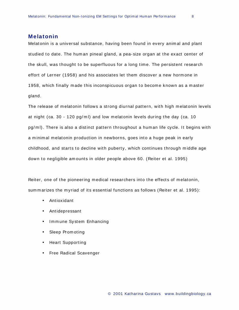
Reiter, one of the pioneering medical researchers into the effects of melatonin,
summarizes the myriad of its essential functions as follows (Reiter et al. 1995):
• Antioxidant
• Antidepressant
• Immune System Enhancing
• Sleep Promoting
• Heart Supporting
• Free Radical Scavenger

Melatonin: Fundamental Non-Ionizing EM Settings for Optimal Human Performance 9
© 2001 Katharina Gustavs www.buildingbiology.ca
Melatonin - Its Antitumor Properties
There are many biological pathways by which melatonin is able to protect against
cancer.
One is its ability to neutralize free radicals involved in many cancers, protecting
nuclear DNA from oxidative damage because it is able to enter the cell nucleus. This
was, for example, demonstrated when Reiter and associates injected rats with
safrole, a known carcinogen, and half of them also with melatonin. A robust effect
could be observed because the melatonin-treated rats had sustained only 1% as
much damage as the controls, whose DNA had been severely damaged (Tan et al.
1993). Melatonin is 5 times more effective than glutathione and 15 times more
effective than mannitol (Reiter 1994).
Interestingly, Lai (1997) and coworkers observed that radiofrequency-radiation-
induced increases in single and double strand DNA breaks in rat brain cells could be
blocked by treating the rats with melatonin. This finding, too, suggests in their
opinion that RF exposure causes an increase in free radicals, which can then be
neutralized by melatonin.
Another pathway is its inhibitory effect on estrogen. Or put another way, when
melatonin levels drop, reproductively active hormones such as estrogen and
prolactin rise as a consequence. The growth of breast cancer, for example, is
stimulated in the presence of excessive levels of prolactin and estrogen. The
Melatonin Hypothesis (1997) by Stevens is based on this pathway.

Melatonin: Fundamental Non-Ionizing EM Settings for Optimal Human Performance 10
© 2001 Katharina Gustavs www.buildingbiology.ca
Low-Level EMR Exposure and Breast Cancer
Low-level non-ionizing EMR exposure
(Single magnetic or electric field component or combination)
Pineal Gland: Reduced nocturnal melatonin production
Ovary: Increased estrogen production
Pituitary: Increased prolactin production
DNA: Increased oxidative damage
DNA: Increased risk of being damaged by carcinogenic agents
Mammary Gland:
Increased proliferation of breast epithelial stem cells to carcinogens
such as DMBA
Immune System: Suppressed immune response to tumor formation
Consequence: Increased risk of breast cancer formation
Table 2: Proposed mechanism by which chronic exposure to low-level non-
ionizing EMR may increase development and growth of breast cancer.
Modified from the following sources: Stevens et al. 1987, Reiter 1994, Löscher et al. 1997.
Melatonin: Fundamental Non-Ionizing EM Settings for Optimal Human Performance 11
© 2001 Katharina Gustavs www.buildingbiology.ca
Many in vivo and in vitro studies demonstrate significant antiproliferative effects of
melatonin: Hill et al. 1992, Cos et al. 1991, Cos et al. 1994, Blask et al. 1991. The
cell proliferation in human breast cancer cells (MCF-7) in culture can be inhibited as
much as 60 - 78% by the addition of physiological concentrations of melatonin as
found during the evening hours (Hill et al.1988).
However, it could also be demonstrated that its oncostatic effect on breast cancer
cell proliferation is blocked by 60-Hz magnetic fields (Liburdy et al.1993). Those
results could by replicated recently by Blackman (2001).
Primary Factor: Light - Visible Non-Ionizing EMR
Beyond any doubt, visible light between 700 nm and 400 nm or rather its absence
notifies the pineal gland mainly via ocular pathways to know when melatonin is to
be released. Ocular light is the major determinant of the circadian rhythm (Skene
et al. 1999). The time and amount of melatonin to be released at night depends on
many factors (e.g. alcohol and drug consumption, exercise, food intake) but the
light intensity, its spectral distribution, and duration of light exposure seem to be
the primary factors. First, it was thought that only bright light (2500 lux) at night
would have an adverse effect on the pineal gland. Subsequent research, however,
revealed that dim light as low as 250 lux can already reduce melatonin to below
detectable levels (Trinder et al. 1996). Even single pulses of bright light can shift
the timing of the melatonin rhythm (Shanahan et al. 1997).
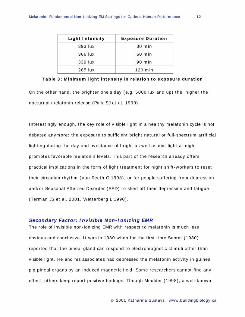
Minimum intensities of light at night (LAN) found to suppress nocturnal melatonin
levels could be related to the duration of light exposure (Aoki 1998).
Melatonin: Fundamental Non-Ionizing EM Settings for Optimal Human Performance 12
© 2001 Katharina Gustavs www.buildingbiology.ca
Light Intensity Exposure Duration
393 lux 30 min
366 lux 60 min
339 lux 90 min
285 lux 120 min
Table 3: Minimum light intensity in relation to exposure duration
On the other hand, the brighter one's day (e.g. 5000 lux and up) the higher the
nocturnal melatonin release (Park SJ et al. 1999).
Interestingly enough, the key role of visible light in a healthy melatonin cycle is not
debated anymore: the exposure to sufficient bright natural or full-spectrum artificial
lighting during the day and avoidance of bright as well as dim light at night
promotes favorable melatonin levels. This part of the research already offers
practical implications in the form of light treatment for night shift-workers to reset
their circadian rhythm (Van Reeth O 1998), or for people suffering from depression
and/or Seasonal Affected Disorder (SAD) to shed off their depression and fatigue
(Terman JS et al. 2001, Wetterberg L 1990).
Secondary Factor: Invisible Non-Ionizing EMR
The role of invisible non-ionizing EMR with respect to melatonin is much less
obvious and conclusive. It was in 1980 when for the first time Semm (1980)
reported that the pineal gland can respond to electromagnetic stimuli other than
visible light. He and his associates had depressed the melatonin activity in guinea
pig pineal organs by an induced magnetic field. Some researchers cannot find any
effect, others keep report positive findings. Though Moulder (1998), a well-known
Melatonin: Fundamental Non-Ionizing EM Settings for Optimal Human Performance 13
© 2001 Katharina Gustavs www.buildingbiology.ca
radiation oncologist, maintains that the "weak to non-existent" link between ELF
EMR and cancer and the "biochemical plausibility" of its effect "is not only
unproven, but rather unlikely," there is more and more data accumulating, which
should not be ignored.
The whole situation is quite complicated because small differences in experiment
protocols can have a huge effect on the reported observations: temperature of
tissue during exposure (Blackman 1991), geomagnetic densities at a given
laboratory (Blackman 1985b), lighting conditions (Aoki et al. 1998). The following
tables list studies with positive findings to date. They are classified according to the
applied electric, magnetic or electromagnetic field.
Exposure Characteristics of Non-Ionizing Electromagnetic Radiation and Occurrence of Melatonin Reduction Effect
Earth's Magnetic Field and Melatonin Effects I - II
Power Frequency (60 Hz) Electric Field and Melatonin Effects I - III
Power Frequency (50/60 Hz) Magnetic Field and Melatonin Effects
Power Frequency - Electric and Magnetic Field and Melatonin Effects
Magnetic Field and Melatonin Effects
Radiofrequency Radiation and Melatonin Effects
Abbreviations in the Tables
6-OHMS - 6-hydroxymelatonin sulfate MEL - Melatonin
CPW - Continuous Polymer Wire MF - Magnetic Field
DMBA - Dimethylbenz(a)anthracene NAT - N-acetyl-5-methoxytryptamine
EF - Electric Field SD - Sprague-Dawley
EMF - Electromagnetic Field WK - Wistar King
EMR - Electromagnetic Radiation LE - Long Evans
Melatonin: Fundamental Non-Ionizing EM Settings for Optimal Human Performance 14
© 2001 Katharina Gustavs www.buildingbiology.ca
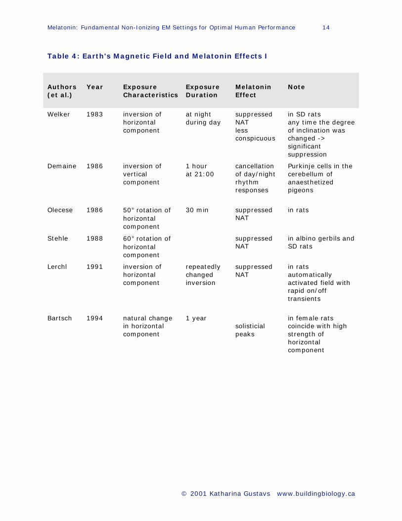
Table 4: Earth's Magnetic Field and Melatonin Effects I
Authors
(et al.)
Year
Exposure
Characteristics
Exposure
Duration
Melatonin
Effect
Note
Welker 1983 inversion of
horizontal
component
at night
during day
suppressed
NAT
less
conspicuous
in SD rats
any time the degree
of inclination was
changed ->
significant
suppression
Demaine 1986 inversion of
vertical
component
1 hour
at 21:00
cancellation
of day/night
rhythm
responses
Purkinje cells in the
cerebellum of
anaesthetized
pigeons
Olecese
1986
50° rotation of
horizontal
component
30 min
suppressed
NAT
in rats
Stehle 1988 60° rotation of
horizontal
component
suppressed
NAT
in albino gerbils and
SD rats
Lerchl 1991 inversion of
horizontal
component
repeatedly
changed
inversion
suppressed
NAT
in rats
automatically
activated field with
rapid on/off
transients
Bartsch 1994 natural change
in horizontal
component
1 year
solisticial
peaks
in female rats
coincide with high
strength of
horizontal
component
Melatonin: Fundamental Non-Ionizing EM Settings for Optimal Human Performance 15
© 2001 Katharina Gustavs www.buildingbiology.ca
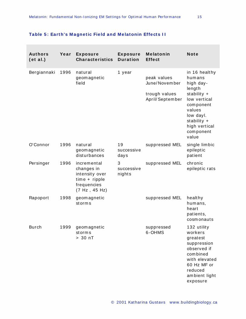
Table 5: Earth's Magnetic Field and Melatonin Effects II
Authors
(et al.)
Year
Exposure
Characteristics
Exposure
Duration
Melatonin
Effect
Note
Bergiannaki 1996 natural
geomagnetic
field
1 year
peak values
June/November
trough values
April/September
in 16 healthy
humans
high day-
length
stability +
low vertical
component
values
low dayl.
stability +
high vertical
component
value
O'Connor 1996 natural
geomagnetic
disturbances
19
successive
days
suppressed MEL single limbic
epileptic
patient
Persinger 1996 incremental
changes in
intensity over
time + ripple
frequencies
(7 Hz , 45 Hz)
3
successive
nights
suppressed MEL chronic
epileptic rats
Rapoport 1998 geomagnetic
storms
suppressed MEL healthy
humans,
heart
patients,
cosmonauts
Burch 1999 geomagnetic
storms
> 30 nT
suppressed
6-OHMS
132 utility
workers
greatest
suppression
observed if
combined
with elevated
60 Hz MF or
reduced
ambient light
exposure
Melatonin: Fundamental Non-Ionizing EM Settings for Optimal Human Performance 16
© 2001 Katharina Gustavs www.buildingbiology.ca
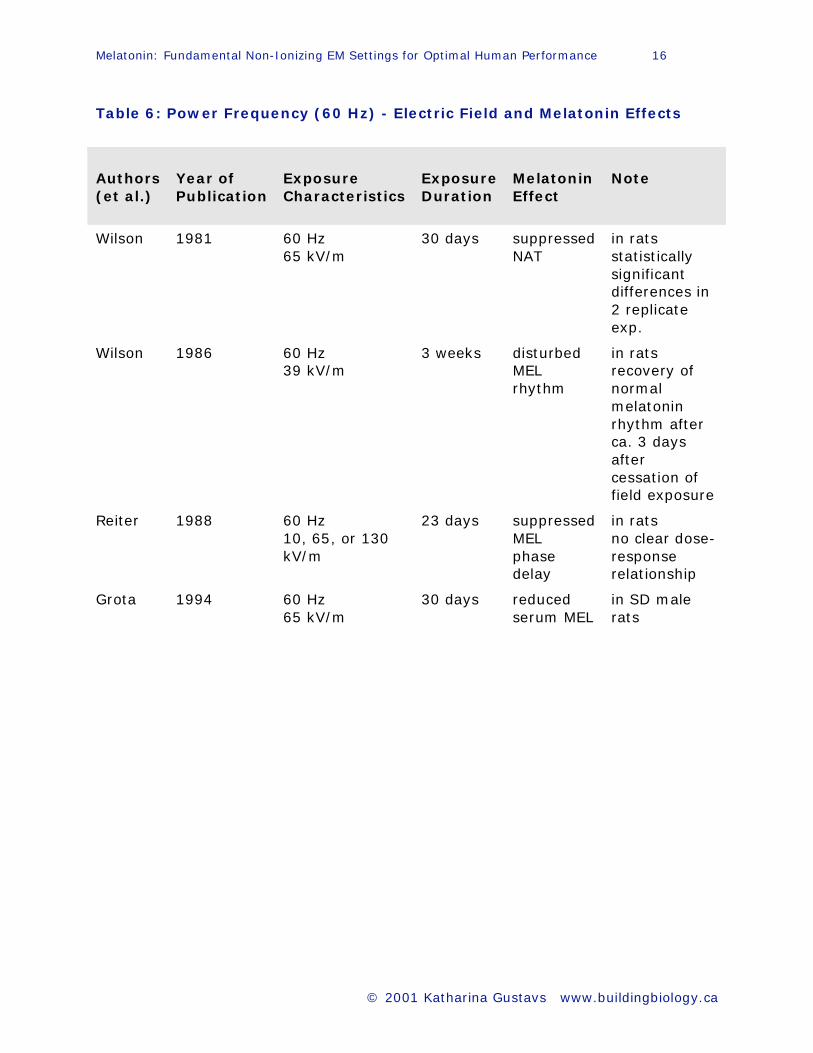
Table 6: Power Frequency (60 Hz) - Electric Field and Melatonin Effects
Authors
(et al.)
Year of
Publication
Exposure
Characteristics
Exposure
Duration
Melatonin
Effect
Note
Wilson 1981 60 Hz
65 kV/m
30 days suppressed
NAT
in rats
statistically
significant
differences in
2 replicate
exp.
Wilson 1986 60 Hz
39 kV/m
3 weeks disturbed
MEL
rhythm
in rats
recovery of
normal
melatonin
rhythm after
ca. 3 days
after
cessation of
field exposure
Reiter 1988 60 Hz
10, 65, or 130
kV/m
23 days suppressed
MEL
phase
delay
in rats
no clear dose-
response
relationship
Grota 1994 60 Hz
65 kV/m
30 days reduced
serum MEL
in SD male
rats

Melatonin: Fundamental Non-Ionizing EM Settings for Optimal Human Performance 17
© 2001 Katharina Gustavs www.buildingbiology.ca
Table 7: Power Frequency (50/60 Hz) - Magnetic Field and Melatonin
Effects I
Authors
(et al.)
Year of
Publication
Exposure
Characteristics
Exposure
Duration
Melatonin
Effect
Note
Kato 1993 50 Hz
circularly
polarized MF
1, 5, 50, 250 μT
6 weeks
(2 h
break per
day)
suppressed
MEL
WK albino
rats
Kato 1994 50 Hz
circularly
polarized MF
1 μT
6 weeks suppressed
MEL
rats
normal
melatonin
levels 1 week
after
cessation of
field exposure
Kato 1994 50 Hz
1 μT
0.02 μT
(controls)
6 weeks suppressed
MEL
LE rats (albino
+ pigmented)
slight MEL
suppression
also occurred
in controls
Yellon 1994 60 Hz
1 mG
15 min
(2 hours
before
lights off)
suppressed
MEL
hamsters
experiment
was repeated
6 months
later
Baum 1995 50 Hz
100 μT
24 h/day
for
91 days
increased
mammary
tumor
growth
216 DMBA
rats
Löscher 1995 50 Hz
0.3 - 1 μT
10 μT
50 μT + 100 μT
24 h/day
for
13 weeks
no
increase
non-
significant
significant
increase
in
mammary
tumor
ca. 200 DMBA
rats
significant
linear relation
between flux
density and
increase in
tumor
incidence
Melatonin: Fundamental Non-Ionizing EM Settings for Optimal Human Performance 18
© 2001 Katharina Gustavs www.buildingbiology.ca
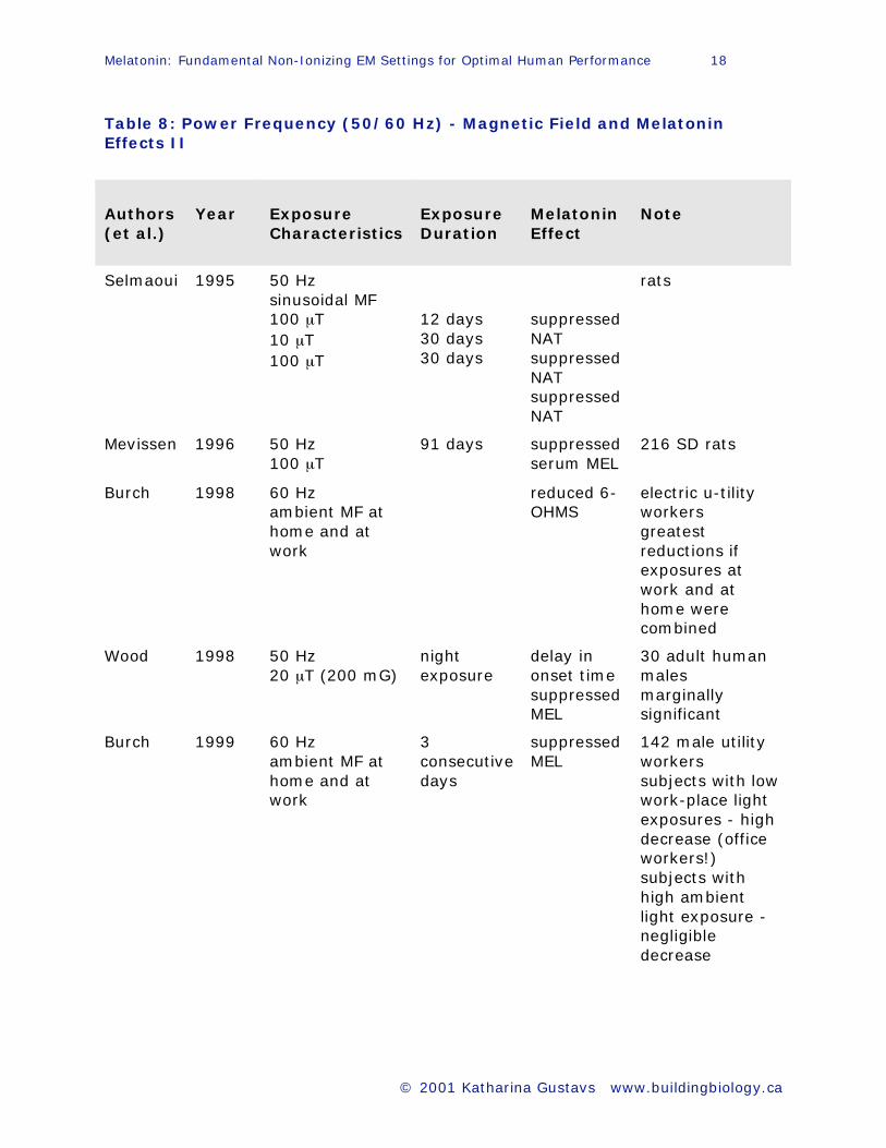
Table 8: Power Frequency (50/60 Hz) - Magnetic Field and Melatonin
Effects II
Authors
(et al.)
Year
Exposure
Characteristics
Exposure
Duration
Melatonin
Effect
Note
Selmaoui 1995 50 Hz
sinusoidal MF
100 μT
10 μT
100 μT
12 days
30 days
30 days
suppressed
NAT
suppressed
NAT
suppressed
NAT
rats
Mevissen 1996 50 Hz
100 μT
91 days suppressed
serum MEL
216 SD rats
Burch 1998 60 Hz
ambient MF at
home and at
work
reduced 6-
OHMS
electric u-tility
workers
greatest
reductions if
exposures at
work and at
home were
combined
Wood 1998 50 Hz
20 μT (200 mG)
night
exposure
delay in
onset time
suppressed
MEL
30 adult human
males
marginally
significant
Burch 1999 60 Hz
ambient MF at
home and at
work
3
consecutive
days
suppressed
MEL
142 male utility
workers
subjects with low
work-place light
exposures - high
decrease (office
workers!)
subjects with
high ambient
light exposure -
negligible
decrease
Melatonin: Fundamental Non-Ionizing EM Settings for Optimal Human Performance 19
© 2001 Katharina Gustavs www.buildingbiology.ca
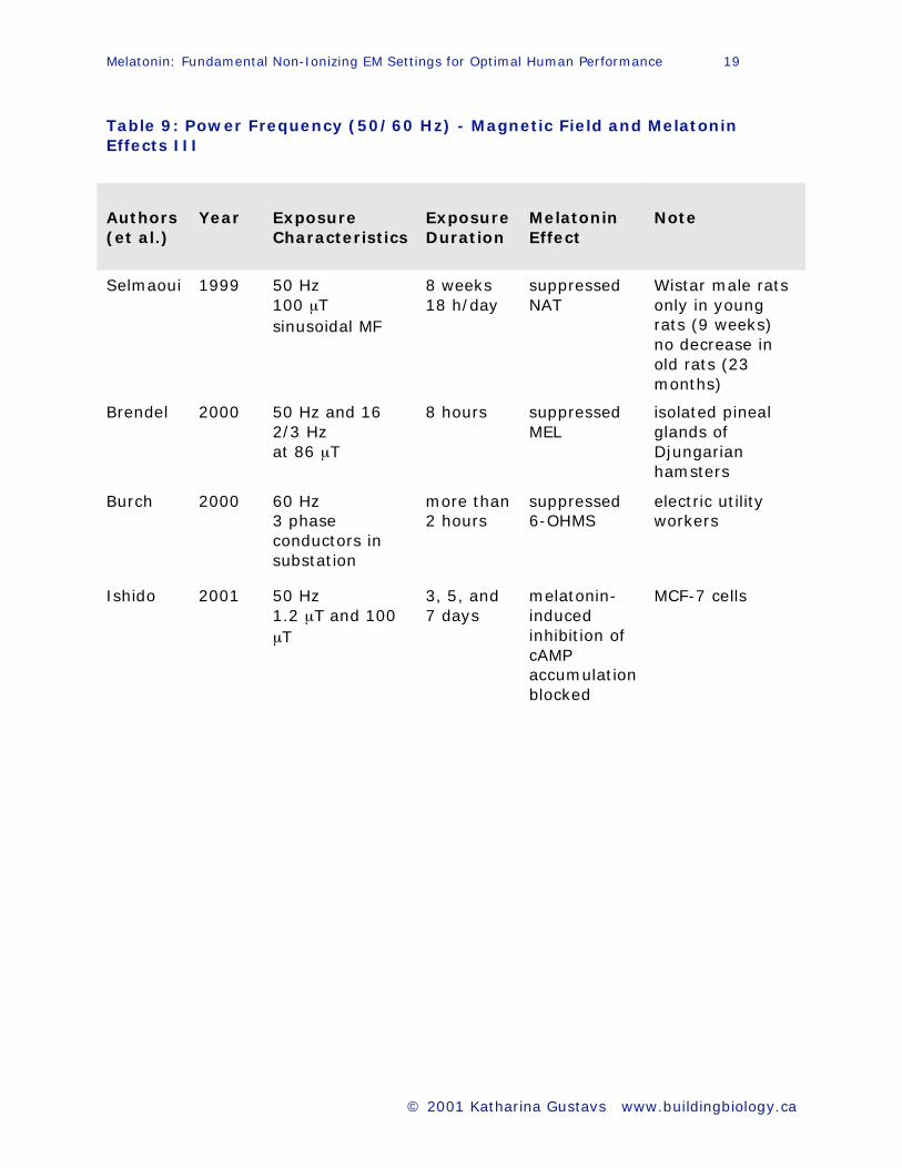
Table 9: Power Frequency (50/60 Hz) - Magnetic Field and Melatonin
Effects III
Authors
(et al.)
Year
Exposure
Characteristics
Exposure
Duration
Melatonin
Effect
Note
Selmaoui 1999 50 Hz
100 μT
sinusoidal MF
8 weeks
18 h/day
suppressed
NAT
Wistar male rats
only in young
rats (9 weeks)
no decrease in
old rats (23
months)
Brendel 2000 50 Hz and 16
2/3 Hz
at 86 μT
8 hours suppressed
MEL
isolated pineal
glands of
Djungarian
hamsters
Burch 2000 60 Hz
3 phase
conductors in
substation
more than
2 hours
suppressed
6-OHMS
electric utility
workers
Ishido 2001 50 Hz
1.2 μT and 100
μT
3, 5, and
7 days
melatonin-
induced
inhibition of
cAMP
accumulation
blocked
MCF-7 cells

Melatonin: Fundamental Non-Ionizing EM Settings for Optimal Human Performance 20
© 2001 Katharina Gustavs www.buildingbiology.ca
Table 10: Power Frequency - Electric and Magnetic Field and Melatonin
Effects
Authors
(et al.)
Year of
Publication
Exposure
Characteristics
Exposure
Duration
Melatonin
Effect
Note
Wilson 1990 60 Hz EMF
ca. 2 - 7 mG
ca. 8
weeks
suppressed
6-OHMS
excretion
32 women/10
men
CPW electric
blanket users
Rogers 1995 60 Hz
rapid on/off
changes
electric field
transients
suppressed
nocturnal
serum MEL
two baboons
Table 11: Magnetic Field and Melatonin Effects
Authors
(et al.)
Year of
Publication
Exposure
Characteristics
Exposure
Duration
Melatonin
Effect
Note
Richardson 1992 pulsed
0.4 G
static magnetic
field
1 hour suppressed
NAT
in vitro rat
pineal
glands
Lerchl 1998 1 Hz
40 μT
200 ms on
800 ms off
increased
nocturnal
serum MEL
brook trout
Melatonin: Fundamental Non-Ionizing EM Settings for Optimal Human Performance 21
© 2001 Katharina Gustavs www.buildingbiology.ca
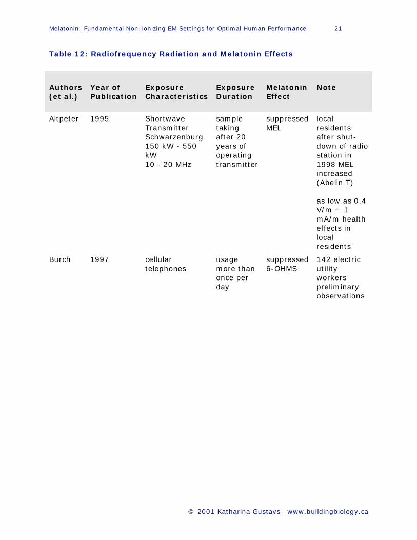
Table 12: Radiofrequency Radiation and Melatonin Effects
Authors
(et al.)
Year of
Publication
Exposure
Characteristics
Exposure
Duration
Melatonin
Effect
Note
Altpeter 1995 Shortwave
Transmitter
Schwarzenburg
150 kW - 550
kW
10 - 20 MHz
sample
taking
after 20
years of
operating
transmitter
suppressed
MEL
local
residents
after shut-
down of radio
station in
1998 MEL
increased
(Abelin T)
as low as 0.4
V/m + 1
mA/m health
effects in
local
residents
Burch 1997 cellular
telephones
usage
more than
once per
day
suppressed
6-OHMS
142 electric
utility
workers
preliminary
observations
Just because plausible biological mechanisms are difficult to elucidate, the
observations of actual bioeffects do not go away. Harvey and French (1999)
were puzzled to see that the lower the microwave power levels tended to be,
with which they exposed a human mast cell line (HMC-1), the larger the
induced stress response genes were upregulated. Back in the 80s it had
already been observed that bioeffects caused by ELF electromagnetic
radiation in the non-thermal range follow a "window" pattern (Adey 1980).
This mystery remains until today: at one specific frequency a large response
can be seen and only a little further up or down hardly any (Berg 1999).
Since the calculations by King and Wu (1998), we now know that spherical
cells are effectively shielded by their membrane against incoming exogenous
fields. However, in cylindrical cells, which are long compared to their radius
such as muscle or nerve cells, this shielding effect does not seem to take
place. Therefore non-thermal effects of induced electrical fields appear to be
possible in those cells at much lower levels.
Brainard (1999) from the Department of Neurology in Philadelphia argues
that "given the ubiquitous nature of EMR and artificial light exposure along
with the high incidence of breast cancer, even a small risk would have a
substantial public health impact."
Melatonin: Fundamental Non-Ionizing EM Settings for Optimal Human Performance 23
© 2001 Katharina Gustavs www.buildingbiology.ca
Melatonin-EMR Information Network
As can be seen by looking through table 4 to 12, there are various non-
ionizing frequency bands in which nocturnal melatonin suppression occurs in
addition to too much visible light at night (LAN) and too little light during the
day. Though the evidence is just beginning to emerge, one thing becomes
clear: melatonin activity in humans, rodents and other mammals can be
influenced by various measures of non-ionizing electromagnetic energy,
regardless of being visible or not.
The significance of field strengths and modulations and combinations thereof
are much less clear. Because a myriad of possibilities for the promotion of an
adverse melatonin effect exist, it is of utmost importance to take precise
measurements in the real world. "Everything" should be captured according
to Hansen (1997), including high-band-width, high-dynamic range, and long-
duration data logging. At first glance, this suggestion looks cost prohibitive.
However, that is the only way to find out.
So far mostly occupational and residential studies have observed significant
changes in melatonin activity (Graham et al 2000). This does not take
wonder because in the real world there is no control over relative orientation
of the fields, the waveforms are not sinusoidal, sources turn off and on at
random, transients are everywhere, artificial lighting conditions prevail. I,
hereby, propose a network of data collection sites around the globe to
monitor the change throughout the entire non-ionizing portion of the non-
Melatonin: Fundamental Non-Ionizing EM Settings for Optimal Human Performance 24
© 2001 Katharina Gustavs www.buildingbiology.ca
ionizing spectrum. Just like scientific institutions worldwide take the "pulse"
of the earth by monitoring the geomagnetic activity (Geological Survey of
Canada).
Siting and Measuring Capacity of Data Collection Sites
Since the geographic distribution of breast cancer incidence follows a specific
pattern as shown above, the stationary data collection sites should be placed
accordingly: one in an urban center of Northeastern US with the highest
incidence, one in a rural area in the same state with the lowest incidence;
one in a southern US state with the lowest incidence, one in a southern US
state with an increasing trend; one in an urban area of Korea with the
highest incidence, one in a rural area of Korea with the lowest incidence. The
data collection site should be stationed where people live, one also close to a
power line and/or cell phone tower. Testing probes should be attached at
ground level and at the highest floor level inhabited in a particular city.
The following frequency bands should be monitored 24 hours a day, most of
them have been shown to influence melatonin rhythm (see tables 4 - 12).

Melatonin: Fundamental Non-Ionizing EM Settings for Optimal Human Performance 25
© 2001 Katharina Gustavs www.buildingbiology.ca
EMR Frequency Field Characteristics
Static Magnetic
Field
0 • all geomagnetic disturbances > 30
nT from the nearest geological
survey site
• locating magnetostatic disturbances
in bed (e.g. metal springs, steel
trusses)
Electrostatic Field 0 • atmospheric electricity from the
nearest meteorological survey site
• atmospherics activity, if possible
AC Magnetic Field
ELF
VLF
20 Hz - 2 kHz
2 - 400 kHz
• intensity, temporal stability, energy
spikes
AC Electric Field
ELF
VLF
20 Hz - 2 kHz
2 - 300 kHz
• intensity, temporal stability,
transients
RF Radiation 300 kHz -
300 GHz
• intensity, modulation characteristic
(amplitude, phase, frequency)
Table 13: A total approach to EMR testing in relation to the
occurrence of melatonin effects
Each frequency band would have to be covered with two sets of equipment:
one being able to detect the original natural background radiation, if still
possible, and one being able to register human-made background radiation.
The testing equipment should also be able to keep track of maximum energy
spikes, transients and modulation characteristics and store them as such.
Mean values are of little value with respect to bioeffects. The superposition of
a static field to an alternating one can cause dramatic changes in the
response of intracellular signal transduction pathways (Kaiser 1992).
Real World Measurements in Residential Settings at Night
The natural background radiation occurs in the nano and pico range whereas
exposure from human-made sources in the residential setting can often
Melatonin: Fundamental Non-Ionizing EM Settings for Optimal Human Performance 26
© 2001 Katharina Gustavs www.buildingbiology.ca
range somewhere from micro to milli or higher. In order to determine
whether it is advisable to stick closer to the natural range for long term well-
being or if it is possible to change those settings without any co-promoting
cancer effect in the future, it is suggested here to start monitoring exposures
in residential bedrooms.
Since the field characteristics of each frequency band are different, there are
two sets of measurements necessary. First, women suffering from breast
cancer and healthy matched controls in the same high-risk age group 35 -45
are equipped with portable meters. Those portable meters should be able to
monitor the ELF, VLF and RF range plus a light sensor because visible light
exposure during the day or at night has a significant effect on the nocturnal
melatonin serum level (Burch 1999). In addition, actual field measurements
(Hansen 1997) covering all the frequency bands listed in table 13 should also
be taken in the bedroom (8 hour data logging to detect energy spikes + spot
measurements to capture transients), at best once a month or at least once
each season. Due to electric heating, for example, the exposure in the ELF
magnetic field range is usually higher during winter compared to summer.
Void morning urine samples can be used to perform a radioimmunassay for
6-OHMS, a melatonin metabolite. Creatine-adjusted concentrations give
reliable data to determine nocturnal melatonin serum level. To determine
phase delays, samples would have to be taken throughout the night, which is
reserved for laboratory studies.
Melatonin: Fundamental Non-Ionizing EM Settings for Optimal Human Performance 27
© 2001 Katharina Gustavs www.buildingbiology.ca
Self-administered questionnaire surveys should ask for known risk factors
and melatonin-relevant parameters: alcohol, stimulant & drug consumption;
time, type and duration of exercise; time of last meal every day; essential
fatty acid consumption; type of bed and mattress (with or without metal).
The stationary data collection sites should continue to monitor the
background radiation as an ongoing business. So that this type of data will
always be available. The measurements in the bedroom would ideally be
taken every day. To cut costs but being able to cover a whole year,
measurements could be taken once a week. It should be alternated between
weekdays and weekend.
Multivariate Analysis
Each field should be analyzed for the intensity (geometric time weighted
average), temporal stability (standardized rate of change metric), and any
unusual field characteristic such as transients in relation to 6-OHMS
concentrations. The paramaters of each field should also be analyzed for the
relation between external, ambient measurements from the stationary sites
and the mostly indoor measurements from the portable readings.
First, each single field analysis in combination with the melatonin metabolite
levels should be related to light intensity during the day and light at night
(LAN). After that each of the fields can be paired and correlated to see which
field parameters seem to produce the highest effect when combined.
Melatonin: Fundamental Non-Ionizing EM Settings for Optimal Human Performance 28
© 2001 Katharina Gustavs www.buildingbiology.ca
Finally, all the related data will have to be adjusted for age, known risk
factors, and seasonal fluctuations in the melatonin level.
Conclusions
Given the drastic change in the energy level of the non-ionizing portion of the
electromagnetic spectrum in industrialized societies over the past 100 years,
we as users of this natural resource share the responsibility to find out what
consequences our applications have in the long term. In addition to
laboratory work, the practical approach to monitor the "unpleasantries"
(Hansen 1997) of electromagnetic exposure in everyday life with focus on the
vulnerable sleep phase will reveal much needed data. Key is the exact field
measurement across as many frequency bands as possible. Even if the
numbers do not strike us particularly significant at the moment, they are
indispensable to settle the EMR issue some day. Fundamental science is
always about keeping good records.
If it turns out that breast cancer sufferers compared to healthy females in
the same age group are exposed significantly more frequently to a higher
field strength or combination of fields and modulations occurring in their
bedroom, we would have a starting point to redesign our built
electromagnetic environment based on health. However, the task is daunting
because the overall exposure in an urban area is similar to all inhabitants.
There are no real controls! That notwithstanding, the actual exposure can
Melatonin: Fundamental Non-Ionizing EM Settings for Optimal Human Performance 29
© 2001 Katharina Gustavs www.buildingbiology.ca
vary greatly in an individual case depending on their lifestyle, computer use,
cell phone use, etc.
It became obvious that exogenous non-ionizing electromagnetic stimuli exert
an influence on human melatonin cycle, be it subtle or great. Given the
importance of melatonin as an oncostatic agent in the human body,
anything contributing to an optimization of its rhythm and amount should be
given appropriate attention.
The proposed Melatonin-EMR Information Network including its data
collection sites, analytical tools and focus on real life situations, will serve
this purpose.
Occupational health officers will have new data to support occupations
susceptible to disrupted melatonin rhythms including shift workers, flight
attendants and office workers by optimizing work schedules and adjusting
the electromagnetic work environment accordingly. Professional health care
providers will be able to practice preventive medicine with regard to optimal
visible and non-visible "light" settings. Architects, builders and electrical
contractors can begin to create spaces that are most beneficial to their
inhabitants. Manufacturers of electric and electronic equipment already follow
guidelines for electromagnetic compatibility with regards to electronic
equipment and appliances, finally this will also include human parameters of
tolerance. Subgroups with low melatonin levels such as is common in
Melatonin: Fundamental Non-Ionizing EM Settings for Optimal Human Performance 30
© 2001 Katharina Gustavs www.buildingbiology.ca
depression, SAD, sleep disorders, cancer, diabetes, arthritis will also benefit
tremendously from preventive optimal settings of EMR.
Melatonin: Fundamental Non-Ionizing EM Settings for Optimal Human Performance 31
© 2001 Katharina Gustavs www.buildingbiology.ca
References
Adey WR 1980: Frequency and power windowing in tissue interactions with
weak electromagnetic fields. Proc IEEE 1980; 68: 119-125.
Aoki H, Yamada N, Ozeki Y, Yamane H, Kato N: Minimum light intensity
required to suppress nocturnal melatonin concentration in human
saliva. Neurosci Lett 1998; 252 (2): 91-94.
Atlas of Cancer Mortality in the United States: 1950-94: Breast. http://www.
dceg.ims.nci.nih.gov/cgi-bin/atlas/site-discuss?site=bre
Baily WH, Steave HS, Dan Bracken T, Kavet R: Summary and evaluation of
guidelines for occupational exposure to power frequency electric and
magnetic fields. Health Physics Society 1997; 73 (3): 433-453.
Berg H: Problems of weak electromagnetic field effect in cell biology.
Bioelectrochem Bioenerg 1999; 48 (2): 355-360.
Blackman CF, Benane SG, House DE: The influence of 1.2 microT, 60 Hz
magnetic fields on melatnin- and tamoxifen-induced inhibition of MCF-
7 cell growth. Biolectromagnetics 2001; 22 (2): 122-128.
Blackman CF, Benane SG, House DE: The influence of temperature during
electric- and magnetic-field-induced alteration of calcium-ion release
from in vitro brain tissue. Bioelectromagnetics 1991; 12: 173-182.
Blackman CF, Benane SG, Rabinowitz JR, House DE, Joines WT: A role for the
magnetic field in the radiation-induced efflux of calcium ions from
brain tissue, in vitro. Bioelectromagnetics 1985b; 6: 327-337.
Blask DE, Pelletier DB, Hill SM, Lemus Wilson A, Grosso DS, Wilson ST, Wise
ME: Pineal melatonin inhibition of tumor promotion in the N-nitroso-N-
methylurea model of mammary carcinogenesis: potential involvement
of antiestrogenic mechanisms in vivo. J Cancer Res Clin Oncol 1991;
117 (6): 526-532.
Blot WJ: Alcohol and cancer. Cancer Res 1992; 52: 2119s-2123s.
Brainard GC, Kavet R, Kheifets LI: The relationship between electromagnetic
field and light exposures to melatonin and breast cancer risk: a review
of the relevant literature. J Pineal Res 1999; 26 (2): 65-100.
CancerNet at http://cancernet.nci.nih.gov/seer/Breast-cancer.html.
Cos S, Blask DE, Lemus-Wilson A, Hill AB: Effects of mealtonin on the cell
cycle kinetics and "estrogen-rescue" of MCF-7 human breast cancer
cells in culture. J Pineal Res 1991; 10 (1): 36-42.
Cos S, Blask DE: Melatonin modulates growth factor activity in MCF-7 human
breast cancer cells. J Pineal Res 1994; 17 (1): 25-32.
Geological Survey of Canada: National Geomagnetism Program:
http://geolab.nrcan.gc.ca/geomag.
Graham C, Cook MR, Saste A, Riffle DW, Gerkovich MM: Multi-night exposure
to 60 Hz magnetic fields: effects on melatonin and its enzymatic
metabolite. J Pineal Res 200; 28: 1-8.
Hansen NH: Understanding dose: implications for bioelectromagnetics
research. In: The Melatonin Hypothesis: breast cancer and use of
electric power/ edited by RG Stevens, BW Wilson, LE Anderson.
Battelle Press: Columbus, Richland 1997, 81-108.

Melatonin: Fundamental Non-Ionizing EM Settings for Optimal Human Performance 32
© 2001 Katharina Gustavs www.buildingbiology.ca
Harvey C, French PW: Effects of protein kinase C and gene expression in a
human mast
cell line, HMC-1, following microwave exposure. Cell Biol Int 1999; 23
(11): 739-748.
Hill SM, Blask DE: Effects of the pineal hormone melatonin on the
proliferation and morphological characteristics of human breast cancer
cells (MCF-7) in culture. Cancer Res 1988; 48 (21): 6121-6126.
Hill SM, Spriggs LL, Simon MA, Muraoka H, Blask DE: The growth inhibitory
action of melatonin on human breast cancer cells is linked to the
estrogen response system. Cancer Lett 1992; 64 (3): 249-256.
Howe HL, Wingo PA, Thun MJ, Ries LAG, Rosenberg HM, Feigal EG, Edwards
BK: The annual report to the nation on the status of cancer (1973
through 1998): featuring cancers with recent increasing trends.
Journal of the National Cancer Institute 2001; 93 (11):824-842.
Kaiser F, Eichwald C: Biologische Systeme und nichtlineare Dynamik:
periodische Prozesse unter dem Einfluss schwacher externer Felder
[Biological systems and non-linear dynamics: periodic processes under
the influence of weak external fields]. Kleinheubacher Berichte 1992;
35: 301-308.
Kerenyi NA, Pandula E, Feuer G: Why the incidence of cancer is increasing:
the role of "light pollution". Med Hypotheses 1990; 33 (2): 75-78.
King RWP, Wu TT: Electric field induced in cells in the human body when this
is exposed to low-frequency electric fields. Physical Review E 1998; 58
(2): 2363-2369.
Kulldorff M, Feuer EJ, Miller BA, Freedman LS: Breast cancer clusters in the
northeast United States: a geographic analysis. Am J Epidemiol 1997;
146 (2): 161-170.
Lai H, Carino MA, Singh NP: Naltrexone blocked RFR-induced DNA double
strand
breaks in rat brain cells. Wireless Networks Journal 1997; 3:471-476;
1997.
Lai H, Singh NP: Melatonin and N-tert-butyl-alpha-phenylnitrone block 60 Hz
magnetic field-induced DNY single and double strand breaks in rat
brain cells. J Pineal Res 1997; 22 (3): 152-162.
Lerner A: J Am Chem Soc 1958.
Liburdy RP, Sloma TR, Sokolic R, Yaswen P: ELF magnetic fields, breast
cancer, and melatonin: 60 Hz fields block melatonin's oncostatic action
on ER+ breast cancer cell proliferation. J Pineal Res 1993; 14 (2): 89-
97.
Löscher W, Mevissen M: Magnetic fields and breast cancer. In: The Melatonin
Hypothesis: breast cancer and use of electric power/ edited by RG
Stevens, BW Wilson, LE Anderson. Battelle Press: Columbus, Richland
1997, 557.
Moulder JE: Power-frequency fields and cancer. Crit Rev Biomed Eng 1998;
26 (1-2): 1-116.
Park SJ, Tokura H: Bright light exposure during the daytime affects circadian
thythms of urinary melatonin and salivary immunoglobulin A.
Chronobiol Int 1999; 16 (3): 359-371.
Melatonin: Fundamental Non-Ionizing EM Settings for Optimal Human Performance 33
© 2001 Katharina Gustavs www.buildingbiology.ca
Reiter RJ, Robinson J: Melatonin. Bantam Books: New York, 1995.
Reiter RJ: Melatonin suppression by static and extremely low frequency
electromagnetic fields: relationship to the reported increased incidence
of cancer. Reviews on Environmental Health 1994; 10 (3-4): 171-186.
Safety Code 6 - Limits of human exposure to radiofrequency electromagnetic
fields in the frequency range from 3 kHz to 300 GHz/ prepared by the
Radiation Protection Bureau of Health Canada. 1999, 75 p.
Semm P, Schneider T Vollrath L: Effects of an earth-strength magnetic field
on electrical activity of pineal cells. Nature 1980; 288 (5791): 607-
608.
Schmahl D, Preussmann R, Berger MR: Causes of cancer - an alternative
view to Doll and Peto (1981). Klin Wochenschr 1989; 67 (23): 1169-
1173.
Skene DJ, Lockley SW, Thapan K, Arendt J: Effects of light on human
circadian rhythms. Reprod Nutr Dev 1999; 39 (3): 295-304.
Stevens RG: Electric power use and breast cancer: a hypothesis. Am J
Epidemiol 1987 125 (4): 556-561.
Tamarkin L, Cohen M, Roselle D, Reicher C, Lippman M, Chabner B:
Melatonin inhibition and pinealectomy enhancement of 7,12-
dimethylbenz(a)anthracene-induced mammary tumors in the rat.
Cancer Res 1981; 41 (11 Pt 1): 4432-4436.
Tan DX, Poeggeler B, Reiter RJ: the pinial hormone melatonin inhibits DNA-
adduct formation induced by the chemical carcinogen safrole in vivo.
Cancer Letters 1993; 70: 65-71.
The Melatonin Hypothesis: breast cancer and use of electric power/ edited by
RG Stevens, BW Wilson, LE Anderson. Battelle Press: Columbus,
Richland 1997, 760 p.
Trinder J, Armstrong SM, O'Brien C, Luke D, Martin MJ: Inhibition of
melatonin secretion onset by low levels of illumination. J Sleep Res
1996 5 (2): 77-82.
Van Reeth O: Sleep and circadian disturbances in shift work: strategies for
their management. Horm Res 1998; 49 (3-4): 158-162.
Wertheimer N, Leeper E: Electric wiring configurations and childhood cancer.
Int J Epidemiol 1979; 109: 273-284.
Wetterberg L: Lighting: nonvisual effects. Scand J Work Environ Health
1990; 16 Suppl 1: 26-28.
Wilson BW, Anderson LE, Hilton DI, Phillips RD: Chronic exposure to 60 Hz
electric field: effects on pineal function in the rat. Bioelectromagnetics
1981; 2: 371-380.
Wilson BW, Anderson LE, Hilton DI, Phillips RD: Chronic exposure to 60 Hz
electric field: effects on pineal function in the rat. Bioelectromagnetics
1983; 4: 293.
References for Tables 4 through 12
(The following studies are sorted by year of publication in accordance with
the tables.)
Melatonin: Fundamental Non-Ionizing EM Settings for Optimal Human Performance 34
© 2001 Katharina Gustavs www.buildingbiology.ca
Table 4
Welker HA et al.: Effects of an artificial magnetic field on serotonin N-
acetyltransferase activity and melatonin content of the rat pineal
galnd. Exp Brain Res 1983; 50 (2-3): 426-432.
Deamine C et al.: Magnetic fields abolish nychthemeral rhythmicity of
responses of Purkinje cells to the pineal hormone melatonin in the
pigeon's cerebellum. Neurosci Lett 1986; 72 (2): 158-162.
Olcese J et al.: Magnetic field effects on pineal gland melatonin synthesis:
comparitive studies on albino and pigmented rodents. Brain Res 1986;
396(1-2): 365-368.
Stehle J et al.: Magnetic field effects on pineal N-acetyltransferase activity
and melatonin content in the gerbil--role of pigmentation and sex.
Physiol Behav 1988; 44 (1): 91-94.
Lerchl A et al.: Pineal gland "magnetosensitivity" to static magnetic fields is a
consequence of induced electric currents (eddy currents). J Pineal Res
1991; 10 (3): 109-116.
Bartsch H et al.: Seasonality of pineal melatonin production in the rat:
possible synchronization by the geomagnetic field. Chronobiol In 1994;
11 (1): 21-26.
Table 5
Bergiannaki J et al.: Seasonal pattern of melatonin excretion in humans:
relationship to daylength variation rate and geomagnetic field
fluctuations. Experientia 1996; 52 (3): 253-258.
O'Connor RP et al.: Increases in geomagnetic activity are associated with
increases in thyroxine levels in a single patient: implications for
melatonin levels.
Int J Neurosci 1996; 88 (3-4): 243-247.
Persinger MA: Enhancement of limbic seizures by nocturnal application of
experimental magnetic fields that simulate the magnitude and
morphology of increases in geomagnetic activity. Int J Neurosci 1996;
86 (3-4): 271-280.
Rapaport SI et al.: Magnetic storms as a stress factor. Biofizika 1998; 43 (4):
632-639.
Burch JB et al.: Geomagnetic disturbances are associated with reduced
nocturnal excretion of a melatonin metabolite in humans. Neurosc Lett
1999; 266: 209-212.
Table 6
Wilson BW et al.: Chronic exposure to 60-Hz electric fields: effects on pineal
function in the rat. Bioelectromagnetics 1981; 2 (4):371-380.
Wilson BW et al.: 60-Hz electric-field effects on pineal melatonin rhythms:
time course for onset and recovery. Bioelectromagnetics 1986; 7 (2):
239-242.
Reiter RJ et al.: Reduction of the nocturnal rise in pineal melatonin levels in
rats exposed to 60-Hz electric fields in utero and for 23 days after
birth. Life Sci 1988; 42 (22):2203-2206.
Melatonin: Fundamental Non-Ionizing EM Settings for Optimal Human Performance 35
© 2001 Katharina Gustavs www.buildingbiology.ca
Grota LJ et al.: Electric field exposure alters serum melatonin but not pineal
melatonin synthesis in male rats. Bioelectromagnetics 1994; 15 (5):
427-437.
Table 7
Kato M et al.: Effects of exposure to a circularly polarized 50 Hz magnetic
field on plasma and pineal melatonin levels in rats.
Bioelectromagnetics 1993; 14 (2): 97-106.
Kato M et al.: Recovery of nocturnal melatonin concentration takes place
within one week following cessation of 50 Hz circularly polarized
magnetic field exposure for six weeks. Bioelectromagnetics 1994; 15
(5): 489-492.
Kato M et al.: Circularly polarized 50-Hz magnetic field exposure reduces
pineal gland and blood melatonin concentrations of Long-Evans rats.
Neurosci Lett 1994; 166 (1): 59-62.
Yellon SM: Acute 60 Hz magnetic field exposure effects on the melatonin
rhythm in the pineal gland and circulation of the adult Djungarian
hamster. J Pineal Res 1994; 16 (3): 136-144.
Baum A et al.: A histopathological study on alterations in DMBA-induced
mammary carcinogenesis in rats with 50 Hz, 100 μT magnetic field
exposure. Carcinogensisis 1995; 16 (1): 119-125.
Löscher W et al.: Linear relationship between flux density and tumor co-
promoting effect of prolonged magnetic field exposure in a breast
cancer model. Cancer Lett 1995; 96 (2): 175-180.
Table 8
Selmaoui B et al.: Sinusoidal 50-Hz magnetic fields depress rat pineal NAT
activity and serum melatonin. Role of duration and intensity of
exposue. Life Sci 1995; 57 (14): 1351-1358.
Mevissen M et al.: Study on pineal function and DMBA-induced breast cancer
formation in rats during exposure to a 100 mG, 50 Hz magnetic field. J
Toxicol Environm Health 1996; 48 (2): 169-185.
Burch JB et al.: Nocturnal excretion of a urinary melatonin metabolite among
electric utility workers. Scand J Work Environ Health 1998; 24 (3):
183-189.
Wood AW et al.: Changes in human plasma melatonin profiles in response to
50 Hz magnetic field exposure. J Pineal Research 1998; 25 (2): 116-
127.
Burch JB: Reduced excretion of a melatonin metabolite in workers exposed to
60 Hz magnetic fields. Am J Epidemiol 1999; 150 (1): 27-36.
Table 9
Selmaoui B et al.: Age-related differences in serum melatonin and pineal NAT
activity and in the response of rat pineal to a 50 Hz magnetic field. Life
Sci 1999; 64 (24): 2291-2297.
Brendel H et al.: Direct suppressive effects of weak magnetic fields (50 Hz
and 16 2/3 Hz) on melatonin synthesis in the pineal gland of
Melatonin: Fundamental Non-Ionizing EM Settings for Optimal Human Performance 36
© 2001 Katharina Gustavs www.buildingbiology.ca
Djungarian hamsters (Phodopus sungorus). J pineal Res 2000; 29 (4):
228-233.
Burch JB et al.: Melatonin metabolite levels in workers exposed to 60 Hz
magnetic fields: work in substations and with 3-phase conductors. J
Occup Environ Med 2000; 42 (2): 136-142.
Ishido M et a.: Magnetic fields (MF) of 50 Hz at 1.2 uT as well as 100 uT
cause uncoupling of inhibitory pathways of adenylyl caclase mediated
by melatonin 1a receptor in MF-sensitive MCF-7 cells. Carcinogenesis
2001; 22 (7): 1043-1048.
Table 10
Wilson BW et al.: Evidence for an effect of ELF electromagnetic fields on
human pineal gland function. J Pineal Res 1990; 9: 259-269.
Rogers WR et al.: Rapid-onset/offset, variably scheduled 60 Hz electric and
magnetic field exposure reduces nocturnal serum melatonin
concentration in nonhuman primates. Bioelectromagnetics 1995; Suppl
3: 119-122.
Melatonin: Fundamental Non-Ionizing EM Settings for Optimal Human Performance 37
© 2001 Katharina Gustavs www.buildingbiology.ca
Table 11
Richardson BA et al.: Pulsed magnetic field effects on in-vitro pineal
indoleamine metabolism. Biochem Biophys Acta 1992; 1137 (1): 59-
64.
Lerchl A et al.: The effects of pulsing magnetic fields on pineal melatonin
synthesis in a teleost fish (brook trout, Salvelinus fontinalis). Neurosci
Lett 1998; 256 (3): 171-173.
Table 12
Altpeter E et al.: Study on health effects of the shortwave transmiter station
of Schwarzenburg. Department of Social and Preventive Medicine:
University of Bern, 1995.
Burch JB et al.: Cellular telephone use and excretion of a urinary melatonin
metabolite. In: Annual review of research in biological effects of
electric and magnetic fields from the generation, delivery and use of
electricity. San Diego, CA, Nov 9-13 1997, P-52.

Melatonin: Fundamental Non-Ionizing EM Settings for Optimal Human Performance 38
© 2001 Katharina Gustavs www.buildingbiology.ca
Melatonin Research Update with regards to Electromagnetic
Field Exposure (2005)
Over the past 4 years melatonin research has continued. The picture is still
the same. As always there are studies that do not support the melatonin
hypothesis (e.g. Travis 2004) , other observations, however, confirm that
electromagnetic fields and waves have the potential of suppressing nocturnal
melatonin levels. Please find a selection of recent studies with positive
findings listed below:
Burch JB, Reif JS, Noonan CW, Ichinose T, Bachand AM, Koleber TL, Yost MG.
Melatonin metabolite excretion among cellular telephone users. Int J
Radiat Biol. 2002 Nov; 78(11): 1029-36.
Cocco P, Cocco ME, Paghi L, Avataneo G, Salis A, Meloni M, Atzeri S, Broccia
G, Ennas MG, Erren TC, Reiter RJ: Urinary 6-sulfatoxymelatonin
excretion in humans during domestic exposure to 50 hertz
electromagnetic fields. Neuro Endocrinol Lett. 2005 Apr; 26(2): 136-42.
Davis S, Kaune WT, Mirick DK, Chen C, Stevens RG: Residential magnetic
fields, light-at-night, and nocturnal urinary 6-sulfatoxymelatonin
concentration in women. Am J Epidemiol. 2001 Oct 1; 154(7): 591-
600.
Ichinose TY, Burch JB, Noonan CW, Yost MG, Keefe TJ, Bachand A,
Mandeville R, Reif JS: Immune markers and ornithine decarboxylase
activity among electric utility workers. Occup Environ Med. 2004 Feb;
46(2): 104-12.
Ishido M, Nitta H, Kabuto M: Magnetic fields (MF) of 50 Hz at 1.2 microT as
well as 100 microT cause uncoupling of inhibitory pathways of adenylyl
cyclase mediated by melatonin 1a receptor in MF-sensitive MCF-7 cells.
Carcinogenesis. 2001 Jul; 22(7): 1043-8.
Levallois P, Dumont M, Touitou Y, Gingras S, Masse B, Gauvin D, Kroger E,
Bourdages M, Douville P: Effects of electric and magnetic fields from
high-power lines on female urinary excretion of 6-sulfatoxymelatonin.
Am J Epidemiol. 2001 Oct 1; 154(7): 601-9.
Noonan CW, Reif JS, Burch JB, Ichinose TY, Yost MG, Magnusson K:
Relationship between amyloid beta protein and melatonin metabolite in
a study of electric utility workers. J Occup Environ Med. 2002 Aug;
44(8): 769-75.
Rodriguez M, Petitclerc D, Burchard JF, Nguyen DH, Block E: Blood melatonin
and prolactin concentrations in dairy cows exposed to 60 Hz electric
and magnetic fields during 8 h photoperiods. Bioelectromagnetics. 2004
Oct; 25(7): 508-15.
Melatonin: Fundamental Non-Ionizing EM Settings for Optimal Human Performance 39
© 2001 Katharina Gustavs www.buildingbiology.ca
Simko M, Mattsson MO: Extremely low frequency electromagnetic fields as
effectors of cellular responses in vitro: possible immune cell activation.
J Cell Biochem. 2004 Sep 1; 93(1): 83-92.
Suchinda Jarupat, Atsuko Kawabata, Hiromi Tokura and Alicja Borkiewicz:
Effects of the 1900 MHz Electromagnetic Field Emitted from Cellular
Phone on Nocturnal Melatonin Secretion. J of Physiological Antropology
and Applied Human Science Vol. 22; 61-63 (2003).
Zwirska-Korczala K, Adamczyk-Sowa M, Polaniak R, Sowa P, Birkner E,
Drzazga Z, Brzozowski T, Konturek SJ: Influence of extremely-low-
frequency magnetic field on antioxidative melatonin properties in AT478
murine squamous cell carcinoma culture. Biol Trace Elem Res. 2004
Winter; 102(1-3): 227-43.