meeting abstracts 23rd european workshop for …
TRANSCRIPT
S1
Available online http://arthritis-research.com/supplements/5/S1
Autoantibodies and antigens
1
Rheumatoid arthritis — class prediction byautoreactivity profilesR Bergholz1, F Schumann1, S Behrens1, U Ungethüm1, G Valet2,WA Schmidt3, GR Burmester1, JM Engel4, WJ van Venrooij5,G Steiner6, S Bläß1
1Department of Rheumatology & Clinical Immunology, CharitéUniversity Clinic, Berlin, Germany2MPI Biochemistry, Munich, Germany3Clinic for Rheumatology Berlin Buch, Berlin, Germany4Rheumaklinik, Bad Liebenwerda, Germany5Department of Biochemistry, University of Nijmegen, The Netherlands6Divison of Rhematology, Department Internal Medicine III, ViennaGeneral Hospital, AustriaArthritis Res Ther 2003, 5 (suppl 1):1
Heterogeneity and multifactoriality complicate diagnostics and ourunderstanding of pathogenesis of rheumatoid arthritis (RA). The onlyaccepted serologic parameter (rheumatoid factor [RF]) is not diseasespecific, nor are any of several novel RA autoantibodies. We aimed atidentifying profiles instead of individual autoreactivities allowing forunambiguous prediction of RA.Selected RA autoantigens were tested by ELISA (RF and anti-cycliccitrullinated peptide [anti-CCP]) or Western blot (heavy-chain-bindingprotein [BiP], heterogeneous ribonucleoprotein particle A2 [RA33/hnRNP A2], calpastatin and calreticulin). Antibody reactivities wereassayed from serum samples of 149 RA patients and 132 patientswith other rheumatic diseases and from synovial fluids (SF) (58 RA,65 non-RA).No single autoreactivity was sufficient for unambiguous prediction ofRA. Frequencies of multiparameter profiles consisting of 3, 4, 5 and 6autoreactivites were determined. Fifteen six-parameter serum profileswere exclusively expressed in RA patients, representing a cumulativesensitivity of 59%. Twelve SF profiles were exclusively expressed in64% of RA patients. The self-learning classification algorithmCLASSIF1 was capable of accurately predicting RA when these profileswere present. Data profile analysis of RF/CCP/BiP/calpastatin/calretic-ulin/RA33 provided specific discrimination of 64% of RA. Most impor-tantly, RA specific profiles were observed in 64% of patients with earlydisease (<12 months).For the first time, the accurate prediction of the class RA has beenachieved by the use of multiparametric autoreactivity profiles. Becauseof early expression in disease, these profiles make it possible to start adisease-modifying therapy long before irreversible bone and jointdestruction may develop. Additional RA-specific profiles are required tocover the entire group of RA patients.
2Investigation of the reactivity patterns ofantifilaggrin antibodies in sera and synovial fluidsfrom patients with rheumatoid arthritis usingcitrullinated synthetic peptidesM Brózik1, J Szakonyi2, A Magyar3, B Rojkovich1, R Tobi3,F Hudecz3, P Gergely2, K Merétey1
1National Institute of Rheumatology, Frankel Leo u 25, Budapest,Hungary2Central Laboratory of Immunology, Faculty of Medicine, SemmelweissMedical University, Budapest, Hungary3Peptide Chemistry Research Group, Eötvös Lóránd University,Budapest, HungaryArthritis Res Ther 2003, 5 (suppl 1):2
Antifilaggrin antibodies comprise a heterogeneous population of anti-bodies directed to citrullinated proteins.Recent studies have shown that their production is highly specific forrheumatoid arthritis (RA) and the initial antigenic trigger for theseautoantibodies can be localised to the inflammed synovial tissue.The aim of our study was to compare the reactivity and specificity of anti-bodies in sera and synovial fluids towards citrullinated epitopes. Peptidesequence corresponding to human profilaggrin (amino acid residues306–324) and sequences with citrulline substitution at different posi-tions were synthetised by mutipin peptide synthesis on solid supports.Shortened versions of the peptide were also produced by removal ofamino acid residues from its N and C terminals. Completely citrullinatedvariant of the 14-mer peptide was also prepared. Peptide with no cit-rulline replacement was used as a control antigen. We found significantdifferences in the sensitivity for RA of 19 individual peptides tested (from5% to 68%), reflecting previous results that the surrounding amino acidsplay an important role in creation of an autoantigenic epitope. Further, wetested the reactivities of paired serum and synovial fluids and found verysimilar peptide recognition patterns in serum and synovial IgG from thesame individuals. Studies on larger number of samples are in progress toevaluate the results statistically that may support further evidence of thesynovial origin of antifilaggrin autoantibodies. Acknowledgement: This work was supported by the Hungarian grantOTKA T037876.
3Analysis of the peptidylarginine deiminase V gene inrheumatoid arthritisL Caponi1, E Petit-Teixeira3, M Sebbag2, F Bongiorni1, S Moscato1,F Pratesi1, J Osorio1, M Guerrin-Weber3, F Cornelis3, G Serre2,P Migliorini for European Consortium for Rheumatoid ArthritisFamilies (ECRAF)1Clinical Immunology Unit, University of Pisa, Pisa, Italy2INSERM U563, Toulouse, France3ECRAF and Genople EVRY, FranceArthritis Res Ther 2003, 5 (suppl 1):3
A number of rheumatoid arthritis (RA) sera contain antibodies specificfor peptides in which arginine is substituted by the deiminated form cit-
Meeting abstracts23rd European Workshop for Rheumatology ResearchMarseille, France27 February – 2 March 2003
Received: 14 January 2003 Published: 24 February 2003
© 2003 BioMed Central Ltd (Print ISSN 1478-6354; Online ISSN 1478-6362)

S2
rulline (AKA). These antibodies are a marker of RA, as they are absentin other disorders. The enzyme responsible for the generation of cit-rulline residues, peptidylarginine deiminase (PAD), has different iso-forms, with a specific tissue distribution. PAD V, expressed inmonocytes, might be responsible for the deimination of arginineresidues of synovial proteins and thus be involved in the generation ofepitopes for RA-specific antibodies. The ECRAF genome scan showedsuggestive linkage evidence at PAD V locus on chromosome 1(P < 0.005). We decided to analyze PAD V as a candidate gene for RA,studying a cohort of 100 RA patients (tested for AKA) and their unaf-fected parents. Investigation (by single-strand conformation polymor-phism [SSCP] analysis and sequencing) of the 16 exons, 5′ and 3′regions of the PAD V gene provided polymorphisms in the 5′, exons3, 4, and 7 and 3′ regions. Analysis used the transmission disequilib-rium test and the haplotype relative risk for alleles and haplotypes withAnalyze and Genhunter2 programs.We found an association between RA and one PAD V haplotype (38%in RA versus 17% in controls) (P < 0.007). The association was alsoobserved in the AKA+ RA subgroup (41%) (P < 0.03).In conclusion, the PAD V gene may be considered one of the geneticfactors that confer susceptibility to RA. Studies are in progress toclarify the relationship between the PAD V haplotypes, the enzymeactivity and the production of anticitrulline antibodies.
4Subclass distribution of IgG autoantibodies todeiminated fibrinogen in rheumatoid arthritisS Chapuy-Regaud, L Nogueira, C Clavel, M Sebbag, C Vincent,G Serre
Department of Epidermis Differentiation and RheumatoidAutoimmunity, INSERM U563, Toulouse, FranceArthritis Res Ther 2003, 5 (suppl 1):4
Background: Antifilaggrin autoantibodies, previously known as ‘antik-eratin’ antibodies or antiperinuclear factor, are serum IgG that consti-tute the most specific diagnostic markers of rheumatoid arthritis (RA).We showed that they specifically recognise deiminated forms of theα and β chains of fibrin in the rheumatoid synovium. Subsequently, wedeveloped a new ELISA for these autoantibodies, using in vitro deimi-nated human fibrinogen as immunosorbent (AhFibA-ELISA). We evalu-ated its diagnostic performance in a cohort of 617 patients withwell-characterised rheumatic diseases, including 181 patients withestablished RA: at a diagnostic specificity of 98.5%, the ELISA pre-sents a diagnostic sensitivity of 76%. It is to date the most efficient testfor the diagnosis of RA.Objective: On the basis of this test, we undertook to determine thesubclass distribution of AhFibA.Methods: From the AhFibA-ELISA, four ELISAs using monoclonal anti-bodies to each IgG subclass (IgG1, IgG2, IgG3 and IgG4) were devel-oped. The ELISAs were adjusted to allow the respective proportions ofeach AhFibA subclass to be determined in each serum sample tested.141 RA patients positive for AhFibA were analysed.Results: For each IgG subclass, the titres (optical density [OD] values)in the whole population of patients were found to be significantly corre-lated with those obtained with the AhFibA-ELISA. IgG1 AhFibAreached the highest OD values (range 0.137–3.028, median: 1.125),followed by IgG4 AhFibA (range 0–2.846, median 0.043), IgG3AhFibA (range 0–1.448, median 0.034) and lastly IgG2 AhFibA (range0–0.695, median 0.040). The predominance of IgG1 AhFibA was alsoobserved at the individual level since, among the 141 AhFibA-positivesera, all but one contained at least 40% of IgG1 AhFibA. In 39.7% ofthe sera, one or several other subclasses accounted for more than10% of total AhFibA. IgG4 AhFibA was the most frequently associatedsubclass, 25% of the sera containing more than 10% of these antibod-ies. Only 10.7 and 7.9% of the sera contained more than 10% of IgG2and IgG3 AhFibA, respectively.Conclusion: These results confirm and extend those previouslyobtained by indirect immunofluorescence for ‘antikeratin’ antibodies.
The predominance of IgG1 AhFibA, and their frequent association withIgG4 AhFibA, raises the question of the Th1/Th2 balance in RA. More-over, the predominance of IgG1 AhFibA is compatible with effectormechanisms involving complement activation and/or the engagementof Fc gamma receptors.
5Peptidylarginine deiminase isoforms expressed inthe synovial membrane of rheumatoid arthritispatientsS Chapuy-Regaud1, M Sebbag1, R Nachat1, D Baeten2, V Foulquier1, M Simon1, T Senshu3, M Yamada3, H Takahara4,F De Keyser2, G Serre1
1Department of Epidermis Differentiation and RheumatoidAutoimmunity, INSERM U563, Toulouse, France2Department of Rheumatology, Ghent University Hospital, Ghent, Belgium3Yokohama City University, Yokohama, Japan4Ibaraki University, Ibaraki, JapanArthritis Res Ther 2003, 5 (suppl 1):5
Background: Antifilaggrin autoantibodies (AFAs) are highly specific forrheumatoid arthritis and are probably involved in its pathophysiology.We showed that they are synthesised in the rheumatoid synovial mem-brane and that their target antigens in the tissue correspond to variantsof the α and β chains of fibrin. The variants are generated by deimina-tion, i.e. transformation of their arginine into citrulline residues. Deimina-tion, mediated by a peptidylarginine deiminase (PAD) activity,generates the epitopes recognised by AFA/antifibrin autoantibodies.Four PAD isoforms (or types), have been identified and cloned inhumans and rodents (mouse and rat). Expression of one or several ofthese isoforms has been reported in numerous tissues, but their targetsare still poorly known.Objective: Since fibrin deimination occurs in the rheumatoid synovialtissue, we undertook to identify which PAD types are expressed in thetissue.Methods: By immunising rabbits with peptides situated in the most vari-able regions of the otherwise highly conserved PAD type sequences(three synthetic peptides per PAD), we produced antisera specific foreach of the four PAD isoforms. The antisera were affinity-purified on thecorresponding peptides. Each set of anti-peptide antibodies was con-firmed to be specific for one isoform by immunoblotting on recombinantor purified PADs. Additional antisera or purified antibodies to wholehuman PADs II, III and V were used to confirm the results obtained withthe anti-peptide antibodies.The synovial tissue from seven patients with rheumatoid arthritis wasanalysed. In all the tissues, the presence of deiminated proteins andparticularly of deiminated fibrin was demonstrated by immunoblottingand/or immunohistology. Then low-salt extracts of the tissues wereanalysed by immunoblotting with all the immunological tools to PADs.Results: Expression of PAD type V was clearly detected in all sevenpatients. Expression of PAD type II was observed in six patients. No reac-tivities were observed with antibodies specific for PAD types I and III.Conclusion: Of the four PAD isoforms, only the types II and V are sig-nificantly expressed in the synovial tissue of patients with rheumatoidarthritis. Their respective roles in deimination of fibrin in the tissueremain to be determined.
6Paratope diversity of anti-prothrombin antibodiesS Cucnik, T Kveder, B Bozic
Department of Rheumatology, University Medical Centre, Ljubljana,SloveniaArthritis Res Ther 2003, 5 (suppl 1):6
To ascertain the heterogeneity of anti-prothrombin antibodies (aPT), wecompared three in-house aPT ELISAs: A) medium binding plates, phos-phatidylserine, prothrombin, Tris-buffered saline, calcium; B) highbinding plates, prothrombin, Tris-buffered saline; and C) high binding
Arthritis Research & Therapy Vol 5 Suppl 1 Abstracts of the 23rd European Workshop for Rheumatology Research

S3
plates, prothrombin, PBS. One serum, exhibiting high positive aPT in allthree ELISAs, was selected as the calibrator. Sera from 47 patients(41 with SLE, 4 with pAPS, 2 with arterial thromboses) were tested forIgG and IgM aPT.The results showed six different patterns: 1) similar results wereobtained with A, B and C (similar analytical sensitivity); 2) similar resultswere obtained with A and B while C showed lower analitycal sensitivity;3) B and C seemed analytically less sensitive than A; 4) A and B wereanalytically less sensitive than C; 5) A showed very low analytical sensi-tivity; 6) A and C showed lower analytical sensitivity than B.The analysis of all the presented patterns showed noncomparableresults of the three ELISAs. In our experiments, prothrombin was rec-ognized by relevant antibodies when the protein was bound to phos-phatidylserine-coated microtiter plates using calcium ions or when itwas bound to high binding plates with or without calcium ions. Never-theless, detected aPT values were not of the same fine specificity. Dif-ferent binding conditions for prothrombin exposed different epitopes,resulting in the detection of various subgroups of aPT.
7
High-affinity antibodies against ββ2-glycoprotein IS Cucnik, T Kveder, A Ambrozic, B Borut
Department of Rheumatology, University Medical Centre, Ljubljana,SloveniaArthritis Res Ther 2003, 5 (suppl 1):7
Background: Antibodies against β2-glycoprotein I (anti-β2GPI) arebelieved to be of low affinity and thus unable to bind to the free antigenin a solution.Objective: The aim of our study was to determine the affinity of IgGanti-β2GPI, isolated by affinity chromatography.Methods: A β2GPI affinity column was prepared by CNBr-activatedagarose without spacer arms and human purified unnicked β2GPI. TheIgG fraction from the protein G column was applied to the column andbound antibodies were eluted with various solutions: A) 0.1 M glycine /0.5 M NaCl / 0.1% Tween 20 pH 2.5; B) 0.1 M glycine / 4 M NaCl /0.1% Tween 20 pH 2.5; C) 0.1 M sodium borate pH 10 and D) 25%ethylene glycol. Eluted fractions containing anti-β2GPI antibodies wereneutralised and analysed by ELISA using various binding buffers. Thelevel of anti-β2GPI antibodies in each sample was derived from thestandard curve according to the defined dilutions of monoclonal anti-bodies (AUG are arbitrary units of IgG monoclonals).Results: Increased concentrations of sodium ions in the binding solu-tion from 0.15, 0.25, 0.50, 1.11, 2.07 and 4.0 M NaCl did not com-pletely prevent the binding between isolated antibodies and β2-GPI(79.8, 65.3, 36.1, 19.9, 12.0 and 8.1 AUG, respectively).Conclusion: In contrast to the common opinion that all anti-β2GPIautoantibodies are of low affinity, we clearly showed that at least onesubset among them was of high affinity.
8
Anti-Ro/SSA antibodies in rheumatoid arthritis (RA)F Franceschini, I Cavazzana, F Malacarne, P Airò, R Cattaneo,N Del Papa, A Radice, RA Sinico
Piazzale Spedali Civili, Brescia, ItalyArthritis Res Ther 2003, 5 (suppl 1):8
Background: AntiRo are found in 5–15% of RA. Significant associa-tions were reported with sicca, vasculitis, hypergammaglobulins, ANA,high-titer RF, toxicity to D-penicillamine and gold salts treatment. Aim ofthe study: to evaluate clinical features, radiologic progression andresponse to disease-modifying antirheumatic drugs (DMARDs) inantiRo-RA patients.Patients and methods: We studied 210 patients with RA: antiRowere determined by CIE, with human spleen extract, and by ELISA withrecombinant Ro proteins (Pharmacia). Cutoff values for ELISA weredetermined testing 177 sera from routine.
Results: AntiRo were detected in 27 patients (F:M 12.5:1). Two groups(antiRo+ and antiRo–) did not show any difference with regard to diseaseduration, arthritis onset and articular erosions. AntiRo were associatedwith xerophthalmia (P<0.0000001), xerostomia (P=0.0012), oral ulcers(P=0.0067), scleritis (P=0.0067) and amyloidosis (P=0.042).Rheumatoid factor, antiperinuclear factor and anticitrulline were recordedin 70% in both groups; hypergammaglobulinemia, ANA, anti-dsDNA andAMA were frequently detected in antiRo+ patients. Patients were given amean of 3.93 DMARDs, with no statistical difference between antiRo+
and antiRo–: hydroxychloroquine, methotrexate and gold salts are themost frequently used. Patients who were antiRo– were more frequentlytreated with hydroxychloroquine and infliximab, while D-penicillamine wasused more frequently in those who were antiRo+. DMARD toxicity wasdetected in 9.3% of antiRo+, with no statistically significant differencebetween the two groups.Conclusion: AntiRo, found in 12.8% of patients with RA, is associatedwith extra-articular features and with an autoantibody profile unusual forRA. No difference with respect to DMARD toxicity was found in anti-Ro+ patients.
9Presence of anti-RNP-A and anti-RNP-C antibodies isinversely associated with renal symptoms of systemiclupus erythematosusI Hoffman1, I Peene1, L Meheus2, K De Bosschere2, F Hulstaert2,TWJ Huizinga3, L Cebecauer4, D Isenberg5, EM Veys1, F De Keyser1
1UZG, Ghent, Belgium2Innogenetics, Ghent, Belgium3Leiden University Medical Center, Leiden, The Netherlands4Research Institute for Rheumatic Diseases, Piestany, Slovakia5University College London, London, UKArthritis Res Ther 2003, 5 (suppl 1):9
Background: Systemic lupus erythematosus (SLE) is an autoimmunerheumatic disease characterised by the production of autoantibodies.The most common serious feature of SLE is renal involvement. Anassociation with anti-RNP antibodies remains controversial.Aim: To identify associations of autoantibodies and renal symptoms ina consecutive cohort of SLE patients.Methods: Sera and clinical data from 235 consecutive SLE patients,fulfilling the ACR criteria for SLE, were collected in four centres. Thepresence of renal disease was defined as the presence of cellularcasts in the urine, proteinuria (>0.5 g/day), or glomerulonephritis duringthe course of the disease. Autoantibody profiles were determined bythe INNO-LIATM ANA Update (a line immunoassay with recombinantand/or native antigens, including SmB, SmD, RNP-70, RNP-A, RNP-C,Ro52, Ro60, SSB, and ribosomal P) and anti-dsDNA antibodies byindirect immunofluorescence on Crithidia luciliae. Odds ratios (ORs)and their 95% confidence intervals (CI) were computed to determinethe associations between antibodies and renal symptoms. No correc-tion was made for multiple testing.Results: The presence of anti-RNP-A and anti-RNP-C appeared to beprotective against renal involvement (OR = 0.445, CI = 0.210–0.942and OR = 0.484, CI = 0.243–0.964, respectively). Concerning the indi-vidual symptoms, anti-RNP-C was associated with a lower occurrenceof proteinuria (OR = 0.470, CI = 0.236–0.938), cellular casts(OR = 0.324, CI = 0.150–0.696) and glomerulonephritis (OR = 0.460,CI = 0.226–0.934), whereas anti-RNP-A was only significantly associ-ated with a lower occurrence of cellular casts (OR = 0.303,CI = 0.129–0.716). In contrast, antibodies to dsDNA were associatedwith a higher risk for cellular casts (OR = 2.014, CI = 1.057–3.839).We found no associations between renal symptoms and other specificantinuclear reactivities. More specifically, for anti-RNP-70 a trend wasonly detected for the association with the presence of cellular casts(OR = 0.411, CI = 0.153–1.106).Conclusion: Anti-RNP-A and anti-RNP-C antibodies appear to beassociated with a lower risk for renal disease.
Available online http://arthritis-research.com/supplements/5/S1

S4
10
Cultured salivary gland epithelial cells releaseexosomes that contain the Sjögren’s-syndrome-associated autoantigenic ribonucleoproteins Ro/SSAand La/SSBEK Kapsogeorgou, ID Dimitriou, RF Abu-Helu,HM Moutsopoulos, MN Manoussakis
Department of Pathophysiology, National University of Athens,75 Mikras Asias Street, Athens, GreeceArthritis Res Ther 2003, 5 (suppl 1):10
Sjögren’s syndrome (SS) is characterized by exocrine gland destruc-tion associated with lymphocytic infiltrations and chronic autoimmuneantigen-driven responses against the intracellular Ro/SSA and La/SSBribonucleoproteins. Epithelial cells, which are the main targets ofautoimmune responses, appear to have a central role in the pathogene-sis of SS. In recent years we have presented evidence indicating thatsalivary gland epithelial cells (SGECs) of SS patients are inherentlycapable of functioning as antigen-presenting cells. Recently, a novel,cell-free mechanism of antigen presentation has been identified. Thismechanism involves exosomes, which are small (30–200 nm) mem-brane vesicles of endosomal origin secreted by a variety of cell types,such as reticulocytes, B lymphocytes and dendritic cells. In the presentstudy we investigated the capacity of cultured SGEC lines establishedfrom SS patients and disease controls to release exosomal vesiclesthat contain intracellular ribonucleoproteins. Membrane vesicles wereisolated by differential centrifugation from SGEC culture supernatantsand their nature was confirmed by electron microscopy. CulturedSGECs from patients and controls secreted significant amounts of exo-somal vesicles, in a manner largely indistinguishable from other exosome-secreting cells. Exosome release was not associated with apoptosis orother cellular destruction processes. SGEC-derived exosomes werefound by Western blot analysis to contain Ro/SSA, La/SSB, and Smribonucleoproteins. Our results indicate that SGECs are capable ofsecreting exosomes. This mechanism may represent a pathway throughwhich intracellular epithelial antigens are exported and subsequently pre-sented to the immune system. In this context, exosomes produced byepithelial cells may have a role in the pathogenesis of SS.
11
Low frequency of phosphatidylserine/prothrombincomplex antibodies in a cohort of patients withanticardiolipin antibodies and recent thrombosisH Locht
Department of Autoimmunology, Statens Serum Institut, Copenhagen,DenmarkArthritis Res Ther 2003, 5 (suppl 1):11
Objective: Clinical data from patients who were positive for anticardi-olipin antibodies (ACAs), tested on a routine basis in an autoimmunelaboratory, were obtained by questionnaires from the referring physi-cians. One hundred and sixty-two individuals had experienced a recent(within <6 months) thromboembolic event. All sera were tested for IgGand IgM antibodies against cardiolipin, β2-glycoprotein 1 (β2-glp 1),and the complex of phosphatidylserine/prothrombin (PPC) by in-houseELISA methods.Results: Among the 162 ACA-positive patients, 31 (19%) were alsopositive for antibodies against PPC. In the group with ACA only, 73%of the patients had no pre-existing rheumatic condition, compared with32% in the PPC group (P = 0.00002). Thirty-two percent in the PPCgroup had SLE, vs 12% in the ACA group (P = 0.016). The fractions ofpatients with deep venous thrombosis (DVT), pulmonary embolism(PE), or myocardial infarction (MI) were equal, whereas cerebrovascularincidents (CVI) were more frequent among ACA patients; 51% vs 26%(P = 0.02). Antibodies against β2-glp 1 were also more frequent in thePPC group 61% vs 30% (P = 0.002).
Conclusion: Antibodies against both ACA and PPC seem to define asubset of patients with autoimmune thrombophilia. More patients withPPC antibodies had SLE and also tested positive for antibodiesagainst β2-glp 1. The distribution of thrombotic manifestations differedbetween the two populations in that CVI was more frequent in theACA-only group, whereas the fractions with DVT, PE, and MI wereequal.
12Anticardiolipin antibodies of IgG and IgM isotypesreflect different forms of recent thromboemboliceventsH Locht, A Wiik
Department of Autoimmunology, Statens Serum Institut, Copenhagen,DenmarkArthritis Res Ther 2003, 5 (suppl 1):12
Objective: To correlate the distribution and levels of anticardiolipin(ACA) and anti-β2-glycoprotein 1 (a-β2-glp 1) antibodies of IgG andIgM isotypes to the clinical spectrum of recent (within <6 months)thromboembolic events.Method: During one year, all sera positive for IgG or IgM ACA submit-ted on a routine basis from hospitals or primary-care physicians from allparts of Denmark were recorded. Information about thromboembolicevents and any underlying rheumatic disease was obtained by ques-tionnaires from the referring physicians. Sera were analysed forIgG/IgM ACA and a-β2-glp 1 antibodies by in-house ELISA assays, andthe results were expressed in arbitrary units.Results: One hundred and sixty-two patients fulfilled criteria for recentthromboembolic disease. Cerebrovascular infarction (CVI) was presentin 82 patients, deep venous thrombosis (DVT) in 34, pulmonaryembolism (PE) in 14, myocardial infarction (MI) in 4, and other throm-boses in 28 patients. Isolated IgG ACA was found in 31 of 48 patientswith DVT+PE (65%), but in only 21 of 82 patients with CVI (26%)(P = 0.00002). In contrast, isolated IgM ACA was found in 9 (19%) ofpatients with DVT+PE, but in 46 (56%) CVI patients (P = 0.00007).IgG a-β2-glp 1 antibodies were found in 13 (16%) CVI patients and23 (48%) DVT+PE patients (P = 0.0002).Conclusion: IgG and IgM ACA isotypes seem to define different clini-cal subsets of patients with recent thromboembolic events, with IgGACA being most prevalent in the group having DVT+PE whereas IgMACA is found primarily among CVI patients. There was a linear correla-tion between levels of IgG a-β2-glp 1 antibodies and IgG ACA.
13Autoantibodies in osteoarthritisJ Menard1, J Neidel2, C Perka2, M Sparmann3, B Mueller1
1Deutsches RheumaForschungs-Zentrum, Berlin, Germany2Charité, Humboldt-University, Berlin, Germany3Immanuelkrankenhaus, Berlin, GermanyArthritis Res Ther 2003, 5 (suppl 1):13
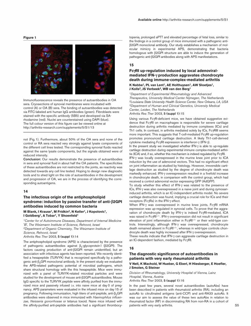
Objective: We have previously shown that inflammatory cytokines areup-regulated in chondrocytes of osteoarthritis (OA) patients. However,the inflammatory responses associated with OA are still ill defined. Toinvestigate in more detail the involvement of the immune system in thepathogenesis of OA, we here analyzed patients for the presence ofautoantibodies.Methods: Both sera and synovial fluids obtained from 85 OA patientsat the time of joint replacement were tested for autoreactivity. Autoanti-bodies reacting against synovial membranes were detected performingimmunofluorescence on cryosections. Autoantibodies recognizinglysate components of various cell lines (B-cell, T-cell, monocyte, fibrob-last and chondrosarcoma) were shown by the use of Western blotanalyses. In preparation for the identification of the respective autoanti-gens, we started to immunoprecipitate the proteins of interest.Results: Sera from the OA patients reacted strongly with cells in thesynovial membranes whereas sera obtained from healthy donors did
Arthritis Research & Therapy Vol 5 Suppl 1 Abstracts of the 23rd European Workshop for Rheumatology Research
S5
not (Fig. 1). Furthermore, about 50% of the OA sera and none of thecontrol or RA sera reacted very strongly against lysate components ofthe different cell lines tested. The corresponding synovial fluids reactedagainst the same lysate components, but the signals obtained were ofreduced intensity.Conclusion: Our results demonstrate the presence of autoantibodiesin sera and synovial fluid in about half the OA patients. The specificitiesof these autoantibodies are not restricted to the joints, as reactivity wasdetected towards any cell line tested. Hoping to design new diagnostictools and to shed light on the role of autoantibodies in the developmentand progression of OA, we are in the process of identifying the corre-sponding autoantigens.
14
The infectious origin of the antiphospholipidsyndrome: induction by passive transfer of anti-ββ2GPIantibodies induced by common bacteriaM Blank, I Krause1, M Fridkin2, N Keller1, J Kopolovic1, I Goldberg1, A Tobar1, Y Shoenfeld1
1Center for of Autoimmune Diseases, Department of Internal Medicine'B', The Weizmann Institute of Science, Rehovot, Israel2Department of Organic Chemistry, The Weizmann Institute ofScience, Rehovot, IsraelArthritis Res Ther 2003, 5 (suppl 1):14
The antiphospholipid syndrome (APS) is characterized by the presenceof pathogenic autoantibodies against β2-glycoprotein I (β2GPI). Thefactors causing production of anti-β2GPI remain unidentified, but anassociation with infectious agents has been reported. We recently identi-fied a hexapeptide (TLRVYK) that is recognized specifically by a patho-genic anti-β2GPI monoclonal antibody. In the present study we evaluatedthe APS-related pathogenic potential of microbial pathogens, whichshare structural homology with the this hexapeptide. Mice were immu-nized with a panel of TLRVYK-related microbial particles and werestudied for the development of mouse anti-β2GPI autoantibodies. MouseIgG specific to the TLRVYK peptide were affinity purified from the immu-nized mice and passively infused i.v. into naive mice at day 0 of preg-nancy. APS parameters were evaluated in the infused mice on day 15 ofpregnancy. Following immunization, high titers of anti-peptide, anti-β2GPIantibodies were observed in mice immunized with Haemophilus influen-zae, Neisseria gonorrhoeae or tetanus toxoid. Naive mice infused withthe affinity-purified anti-peptide antibodies had a significant thrombocy-
topenia, prolonged aPTT and elevated percentage of fetal loss, similar tothe findings in a control group of mice immunized with a pathogenic anti-β2GPI monoclonal antibody. Our study establishes a mechanism of mol-ecular mimicry in experimental APS, demonstrating that bacteriahomologous with β2GPI structure are able to induce the generation ofpathogenic anti-β2GPI antibodies along with APS manifestations.
15FcγγRI up-regulation induced by local adenoviral-mediated IFN-γγ production aggravates chondrocytedeath during immune-complex-mediated arthritisK Nabbe1, PL van Lent1, AE Holthuysen1, AW Sloetjes1,J Kolls2, JS Verbeek3, WB van den Berg1
1Department of Experimental Rheumatology and AdvancedTherapeutics, University Medical Center Nijmegen, The Netherlands2Louisiana State University Health Science Center, New Orleans, LA, USA3Department of Human and Clinical Genetics, University MedicalCenter, Leiden, The NetherlandsArthritis Res Ther 2003, 5 (suppl 1):15
Using various FcγR-deficient mice, we have obtained suggestive evi-dence that FcγRI on macrophages is responsible for severe cartilagedestruction during arthritis mediated by immune complexes (ICs) andTh1 cells. In contrast, in arthritis mediated solely by ICs, FcγRIII seemsmore important. This suggests that T-cell-mediated FcγRI up-regulationpromotes pronounced cartilage destruction. A likely Th1-cell-derivedcytokine mediating FcγRI expression is interferon-γ (IFN-γ).In the present study we investigated whether IFN-γ is able to up-regulatecartilage destruction during experimental immune complex-mediated arthri-tis (ICA) and, if so, whether this mechanism is indeed regulated by FcγRI.IFN-γ was locally overexpressed in the murine knee joint prior to ICAinduction by the use of adenoviral vectors. This had no significant effecton joint inflammation as studied by histology. However, irreversible carti-lage destruction as studied by the degree of chondrocyte death wasmarkedly enhanced. IFN-γ overexpression resulted in a fivefold increasein chondrocyte death, in comparison with the control group, which hadreceived a control adenoviral vector expressing GFP (AdGFP).To study whether this effect of IFN-γ was related to the presence ofICs, IFN-γ was also overexpressed in a naive joint and during zymosan-induced arthritis, which is an IC-independent arthritis model. No severecartilage destruction was found, implying a crucial role for ICs and theirreceptors (FcγRs) in the IFN-γ effect.When IFN-γ was overexpressed in murine knee joints, FcγRI mRNAexpression was up-regulated in synovial cells. To prove that the aggra-vation of chondrocyte death by IFN-γ is indeed FcγRI-mediated, ICAwas raised in FcγRI–/–. IFN-γ overexpression did not result in significantelevation of joint inflammation either in FcγRI–/– or their wild-type con-trols. Interestingly, although IFN-γ was overexpressed, chondrocytedeath remained absent in FcγRI–/–, whereas in wild-type controls chon-drocyte death was highly increased after IFN-γ overexpression.These results indicate that IFN-γ can aggravate cartilage destruction inan IC-dependent fashion, mediated by FcγRI.
16The diagnostic significance of autoantibodies inpatients with very early rheumatoid arthritisV Nell, K Machold, W Hueber, G Eberl, H Hiesberger, E Hoefler,J Smolen, G Steiner
Division of Rheumatology, University Hospital of Vienna, LainzHospital, Vienna, AustriaArthritis Res Ther 2003, 5 (suppl 1):16
In the past few years, several novel autoantibodies (autoAbs) havebeen described in patients with rheumatoid arthritis (RA), including anautoAb to citrullinated antigens (anti-CCP) and anti-RA33 autoAb. Itwas our aim to assess the value of these two autoAbs in relation torheumatoid factor (RF) in discriminating RA from non-RA in a cohort ofpatients with very early arthritis.
Available online http://arthritis-research.com/supplements/5/S1
Figure 1
Immunofluorescence reveals the presence of autoantibodies in OAsera. Cryosections of synovial membranes were incubated withcontrol (A) or OA (B) sera. The binding of autoantibodies was detectedvia FITC-labeled anti human IgG antibodies (green). Fibroblasts werestained with the specific antibody (5B5) and developed via SA-rhodamine (red). Nuclei are counterstained using DAPI (blue). The full colour version of this figure can be viewed online athttp://arthritis-research.com/supplements/5/S1/13
S6
Ninety-four patients with arthritis of less than 3 months’ duration wereincluded in this prospective study. Follow-up was for at least 1 year.Sixty-one patients had a final diagnosis of RA and 33 had other arthri-tides. Among the RA patients, RF was present in 34 (56%), anti-CCPin 18 (30%), and anti-RA33 in 15 (25%) at their first visit. Amongthe 33 non-RA patients, 6 were RF-positive, 3 had anti-RA33, and1 had anti-CCP. Thus, anti-CCP was very specific for RA with a posi-tive predictive value (PPV) of 95%, while RF and anti-RA33 weresomewhat less specific, with PPVs of 85% and 83%, respectively.However, the co-occurrence of anti-RA33 and RF was observed exclu-sively in RA patients and thus had a PPV of 100% in this relativelysmall cohort of patients. In conclusion, these data suggest that thedetermination of autoantibodies such as anti-CCP and anti-A2/RA33 inaddition to RF may be quite helpful in the early diagnosis of RA.
17A rapid ELISA based method to determine peptidyl-arginine deiminase activity in biological samplesS Nijenhuis, AJW Zendman, JMH Raats, GJM Pruijn, WJ van Venrooij
Department of Biochemistry, University of Nijmegen, Nijmegen, The NetherlandsArthritis Res Ther 2003, 5 (suppl 1):17
Peptidylarginine deiminases (PADs; EC 3.5.3.15) are a family ofcalcium-dependent enzymes that convert peptidylarginine into peptidyl-citrulline. The recent finding that patients with rheumatoid arthritis (RA)produce autoantibodies against citrulline-containing epitopes greatlyincreased the interest in the PAD enzymes and their activities. It is notyet known whether there is a causative relationship between the gener-ation of antibodies targeting citrullinated epitopes and the developmentof the disease. Characterisation of the structure and function of PADsmay help to understand the production process of citrullinated antigensand possibly also the aetiology of RA.Several assays are known to monitor PAD activity in biologicalsamples. However, these assays either have a low sensitivity or arelaborious. Here, we describe the development of a simple, rapidmethod for the simultaneous analysis of many PAD-containing samples.This new method is based on the binding of an antibody specificallyrecognising a citrulline-containing epitope in a defined peptide. Weshow that this method is very sensitive and can be applied to monitorPAD activity in many types of biological samples, such as bacteriallysates, mammalian cell extracts and tissue extracts.
18Autoantibodies to deiminated fibrinogen are themost efficient serological criterion for earlyrheumatoid arthritis diagnosisL Nogueira1, S Chapuy-Regaud1, A Constantin1, C Clavel1,M Sebbag1, A Cantagrel2, C Vincent1, G Serre1
1Department of Epidermis Differentiation and RheumatoidAutoimmunity, INSERM U 563, Toulouse, France2Department of Rheumatology, Rangueil Hospital, Toulouse, FranceArthritis Res Ther 2003, 5 (suppl 1):18
Background and objectives: Autoantibodies to deiminated proteinsare known to be the most specific serological marker for the diagnosisof rheumatoid arthritis (RA). We recently showed that deiminated fibrinis the major target of this family of autoantibodies in rheumatoid syn-ovial tissue. We subsequently developed, and validated on a largeseries of patients with established rheumatic diseases, an ELISA forthe detection of circulating autoantibodies to deiminated human fibrino-gen (AhFibA). The test was shown to be the most efficient (specificand sensitive) serological criterion for the diagnosis of RA.Methods: We collected 352 sera from patients with arthritides ofrecent onset (disease duration <1 year). The diagnosis was estab-lished after at least 2 years’ follow-up. The patients were then classifiedinto two groups, 175 with RA and 177 with non-RA inflammatory
rheumatic diseases. The previously developed ELISA, using in vitrodeiminated human fibrinogen as immunosorbent, was used for detec-tion and titration of AhFibA. Antibodies to cyclic citrullinated peptide(CCP) were sought in accordance with the manufacturer’s procedure(Immunoscan RA, Euro-diagnostica). Rheumatoid factor (RF) wastitrated by nephelometry (RF-reagent for Image, Beckman Coulter).Results: The diagnostic sensitivity of AhFibA was found to be signifi-cantly higher than those of CCP (P < 0.05) and RF (P < 0.001)(Table 1). The positive predictive values (PPV) of the three tests wereall found to be very high and were not significantly different. The nega-tive predictive values (NPV) were too low to be diagnostically useful.Among the AhFibA-positive RA sera 83% were RF-positive, whileamong the AhFibA-negative, only 13% were RF-positive.
Table 1
Diagnostic indices computed at thresholds allowing 0.985 specificity tobe reached
Antibodies to Se PPV
AhFibA 0.646 0.974
CCP 0.543 0.969
RF 0.274 0.941
PPV, positive predictive value.
Conclusion: Unlike the case with RF, autoantibodies to deiminatedproteins are confirmed to be of diagnostic value in early arthritides. Thedetection of these autoantibodies by ELISA using their synovial target(deiminated fibrin) appears the most efficient method for the diagnosisof early RA.
19Anti-BIP antibodies in rheumatoid arthritisGS Panayi, M Bodman-Smith, V Corrigall
GKT School of Medicine, Guy’s Hospital, London, UKArthritis Res Ther 2003, 5 (suppl 1):19
Background: We have implicated the human chaperone protein BiP inthe pathogenesis of rheumatoid arthritis (RA). Increased immunoglobu-lin binding of RA sera to BiP is seen on Western blot analysis.Methods: We now describe an ELISA developed to enable rapidscreening of sera for antibody reactivity to BiP.Results: Specificity of the assay has been shown by free ligand com-petition and extensive correlations with other immunological parame-ters. We show no correlation between anti-BiP and rheumatoid factoror with cyclic citrullinated peptide. We confirm the increased binding ofimmunoglobulin to BiP in the sera of a group of patients with RA(n = 96) in comparison with controls (n = 96). Our data show a speci-ficity of 71% and a sensitivity of 73% for RA. Furthermore, these datashow that antibody will bind to a nonglycosylated form of BiP, since theprotein is produced in an Escherichia coli expression system.Conclusion: We have developed a robust protocol for the detection ofantibodies to the human chaperone molecule BiP. Our data show anelevated antibody response to BiP in RA patients and hence support arole for this molecule in the disease.
20Antineutrophil cytoplasmic antibodies in synovialfluid from patients with early rheumatoid arthritisM Puszczewicz, I Zimmermann-Górska, G Bialkowska-Puszczewicz, E Tatarkiewicz
University of Medical Sciences, Poznañ, PolandArthritis Res Ther 2003, 5 (suppl 1):20
Background: Antineutrophil cytoplasmic antibodies (ANCAs) occur invasculitides, including Wegener’s granulomatosis and Churg-Strauss
Arthritis Research & Therapy Vol 5 Suppl 1 Abstracts of the 23rd European Workshop for Rheumatology Research
S7
syndrome. Their presence in sera of patients with some otherrheumatic diseases has been well documented. ANCAs have alsobeen detected in synovial fluid (SF) of patients with RA; however, theirpresence and potential role in early stages of the disease is not known.Objective: The aim of the study was to evaluate the prevalence andspecificity of ANCAs in SF in early RA.Methods: SF samples were obtained from 114 patients with early RA.Control SF were taken from 76 patients with knee osteoarthritis (OA).ANCAs were detected by indirect immunofluorescence and by ELISA.Proteinase 3, myeloperoxidase, elastase, lactoferrin and lysozyme, aswell as cathepsin G, were used as antigens in ELISA method. At thesame time antinuclear antibodies (ANAs) and rheumatoid factor (RA)were detected in SF samples under study.Results: ANCAs were found by indirect immunofluorescence in SF of26/114 (22.8%) patients with early RA and 2/76 (2.6%) patients withOA. A perinuclear pattern (p-ANCA) was detected in 22/26 cases(84.6%), and atypical pattern (a-ANCA) in 4/26 (15.3%). In patientswith OA, p-ANCAs only were observed. We did not observe a cyto-plasmic pattern (c-ANCA) in indirect immunofluorescence or in thereactivity against proteinase 3 in ELISA. p-ANCAs yielded reactivityagainst lactoferrin in SF from 15/26 (57%) and against myeloperoxi-dase in 5/26 (19.2%) SF samples from RA patients. a-ANCAs indi-cated reactivity against cathepsin G in 3/26 cases (11.5%) andagainst lysozyme in 1/26 (3.8%). RF was present in 18/26 (69.2%)and ANA in 14/114 (12.2%) SF samples from patients with RA.Conclusion: ANCAs are present in SF of over 20% of patients withearly RA We think that these antibodies in SF could be one of the earlyRA markers. Their role in this stage of synovitis should be clarified.
21
Biphasic decline/increase for anticitrulline andmonophasic decline in anti-type II collagen antibodylevels in recently diagnosed RA patientsJ Rönnelid1,2, S Rogberg1, B Nordmark1, J Lampa1, I Dahlbom3,T Hansson1,3, L Klareskog1
1Unit of Rheumatology, Karolinska Hospital, Stockholm2Unit of Clinical Immunology, Uppsala University, Uppsala, Sweden3Pharmacia Diagnostics, UppsalaArthritis Res Ther 2003, 5 (suppl 1):21
Objective: Antibodies against both type II collagen and citrullineresidue containing peptides can be found in rheumatoid arthritis (RA)patients. We have noted that serum levels of both anticollagen andanticitrulline antibodies show significant decline after inclusion in anearly arthritis clinic (EAC) cohort, and wanted to compare their kineticpatterns of disappearance.Methods: Two hundred and fifty-five EAC patients and 80 RA patientswere followed for 1 and 5 years, respectively, from the time of initialreferral. Anticollagen and anticitrulline antibodies were determined withELISA at inclusion and after 3 months and 1, 2, 3 and 5 years.Results: At the patients’ inclusion, anticollagen and anticitrulline anti-bodies were found in 6.3% and 48.2%, respectively, of EAC patientsand in 11.5% and 61.0% of the patients with definite RA. Serum levelsof antibodies against type II collagen declined continously for the entirestudy period in both groups, being significant already at 3 months afterinclusion. Serum levels of anticitrulline antibodies, on the other hand,showed a biphasic pattern. A significant decline from inclusion (for EACpatients at 1 year, and for RA patients at 3 months and at 1 and 2 years)was followed by increased serum levels (significant for RA patientsbetween 2 and 5 years). No correlation was found between changes inserum levels of anticollagen and anticitrulline antibodies at any time.Conclusion: These findings suggest that different immunologicalevents predispose to immunity against collagen type II and against cit-rulline-containing peptides in RA.
22Zinc finger domain of Ro60kD autoantigen is essentialfor binding of Ro52kD and autoantibodiesJG Routsias, A Makri, C Sakarellos, M Sakarellos-Daitsiotis,A Kosmopoulou, HM Moutsopoulos, AG Tzioufas
Department of Pathophysiology, School of Medicine, University ofAthens, Athens, GreeceArthritis Res Ther 2003, 5 (suppl 1):22
The Ro60kD polypeptide is associated with both RNA and the Ro52kDprotein. The specific protein–RNA and protein–protein interactions arethought to occur through the RNP and zinc-finger secondary structureelements located on the 92–161 and 301–327 regions of Ro60kDprotein, respectively. The zinc finger domain of Ro60kD is a good can-didate to hold a conformational epitope, because both the binding ofzinc and the redox conditions can induce specific conformationalchanges. In this study, we investigated the presence of antibodiesagainst synthetic peptides corresponding to the zinc finger domain ofRo60kD protein (Zif-1b), to a truncated form possessing zinc-bindingregions but lacking the intermediate loop (Zif-2b) and to the intermedi-ate loop (310–319) of the zinc finger domain (Zif-3b). We found thatthe peptide Zif-1b, corresponding to the native sequence (301–327aa) of Ro60kD, is recognized by antibodies from the majority (83%) ofanti-Ro/La-positive patients with primary Sjögren’s syndrome (pSS), inthe absence of zinc ions. The same sera failed to react with Zif-1b inthe presence of Zn2+ (2.5%). Its truncated form (Zif-2b) did not reactagainst the same sera, while the peptide corresponding to loop310–319 (Zif-3b) exhibited high reactivity (85%). The presence of zincions was necessary for binding of Zif-1b to recombinant Ro52kD, indi-cating that discrete conformational states of the Ro60kD zinc fingerdomain are employed in interaction with Ro52kD protein and autoanti-bodies. The two different states of the zinc finger domain of Ro60kDmay reflect the existence of the Ro60kD autoantigen in different redoxenvironments (e.g. in the interior of the cell and cell membrane), wherethe Cys residues have different capacities to coordinate zinc ions.
23Differential expression of IgVH mRNAs in human RAsynovium detected by single-cell RT-PCRS Ruzickova1,3, Z Cimburek1, J Niederlova1, O Horvath2,O Krystufkova1, T Dörner2, J Vencovsky1
1Institute of Rheumatology and Laboratory of Gene Expression, CzechAcademy of Sciences, Prague, Czech Republic2Institute of Microbiology, Czech Academy of Sciences, Prague,Czech Republic3Department of Rheumatology/Immunology, Medical Faculty, Charité,Humboldt University, Berlin, GermanyArthritis Res Ther 2003, 5 (suppl 1):23
Introduction: In rheumatoid arthritis (RA), the synovial membranescontain lymphocytic infiltrates sometimes resembling germinal centres.The production of clonally related immunoglobulin (Ig) transcripts, thepresence of plasma cells within RA synovial tissue, somatic mutationsand isotype switching in RF-specific synovial B lymphocytes have beenobserved. In addition, the expression of recombination-activatinggenes 1 and 2 (Rag1 and 2) has been detected in RA synovial B cells.Objective: Analysis of the presence of plasma cells, mutational fre-quencies of their Ig heavy-chain transcripts, the signs of isotype switch-ing and expression of Rag1 and 2 genes in inflamed RA synovial tissue.Methods: Synovial tissue sections were labeled with anti-CD19, anti-CD138 and anti-CD38 antibodies and visualized using the alkalinephosphatase technique. Individual CD19+ CD38+ plasma cells wereisolated from digested synovium of two caucasian RA patients usingsingle-cell deposition. The cDNA from each single B cell was gener-ated, and nested polymerase chain reaction specific for VH genes andRag1 and Rag2 genes was performed. After sequencing, the VBASEdatabase was used to assign VH, DH and JH gene segments andsomatic mutations.
Available online http://arthritis-research.com/supplements/5/S1
S8
Results: Three different subsets of CD19+ CD38+ plasma cells weredetected. The first subset represents cells expressing only IgM tran-scripts (IgM+, 13.5%), the second expressed only IgG transcripts(IgG+, 48.7%) and the third produced both IgM and IgG mRNAs (IgM+
IgG+, 37.8%). All of these detected IgVH mRNAs contained mutatedsequences, indicating their memory cell origin. However, the differ-ences of mutational frequencies between these subsets were statisti-cally significant (IgM+ plasma cells 3.8%, IgG+ plasma cells 11.2% andIgM+ IgG+ cells 6.3%). Interestingly, either Rag1 or Rag2 mRNA wasobserved in 83.3% of all analyzed CD19+CD38+ plasma cells, withthe highest frequency in the IgM+ IgG+ subset of plasma cells (71.4%).Conclusion: The population of CD19+ CD38+ plasma cells differentiallyexpressing mutated IgVH mRNAs and reinducing Rag 1 and 2 geneswas observed in RA synovium. The IgM+ IgG+ cells might represent cellsswitching from IgM to IgG isotype and IgG+ plasma cells might corre-spond to post-switched cells producing high-affinity (auto)antibodies.
24Ability of second-generation anti-cyclic citrullinatedpeptide (CCP) to predict rheumatoid arthritis inpatients with early arthritisA Saraux, JM Berthelot, P Le Goff, P Youinou
Department of Rheumatology, Centre Hospital Universitaire, Brest, FranceArthritis Res Ther 2003, 5 (suppl 1):24
Objective: We previously studied the diagnostic value of biological tests inRA and found that IgG-AKA, first generation of anti-CCP, IgM-RF byELISA, and the latex test is the best combination in diagnosing RA. Thegoal of the present study was 1) to study the diagnostic value of thesecond generation of anti-CCP and 2) to determine the diagnostic value ofsecond-generation anti-CCP used in combination in the same population.Methods: A cohort of 270 patients with early arthritis underwent a stan-dardized examination, laboratory tests and radiographs in 1995–1997.The final diagnosis was evaluated by a panel of five rheumatologistsbetween June and December, 1999. The respective diagnostic valuesof antiperinuclear factor, antikeratin antibody, and anti-CCP (commercialkits: Eurodiagnostica first generation [EFG], Eurodiagnostica secondgeneration [ESG], and Axis-Shield [AS]) carried out on sera taken at thepatients’ first visit in discriminating between patients who did (98/270;36%) and did not have RA at the last visit was evaluated using receiv-ing-operator characteristic curves. To evaluate the combination of anti-CCP with other laboratory tests in discriminating between patients withand without RA, a multiple logistic regression with backward selectionusing the likelihood ratio test was applied.Results: 1) Anti-CCP, APF and IgG AKA were not perfectly correlatedwith each other. For anti-CCP EFG (cutoff 53 UI), ESG (cutoff0.120 OD), and AS (cutoff 0.320 OD), sensitivity and specificity were47%–93%, 57%–94% and 58%–94%, respectively. 2) By the use ofa multiple logistic regression, second generation anti-CCP, IgG-AKA,the latex test and IgM-RF ELISA were selected.Conclusion: ESG and AS are the best tests for predicting RA. Com-bining one of these tests with IgG-AKA, IgM-RF, and the latex testslightly increases the diagnostic value.
25Autoantibodies to hnRNP-A2 in SLE: identification ofdisease-specific linear epitopes and correlation withdisease activity and clinical featuresG Schett1, F Monneaux2, E Hoefler3, R Fritsch1, M Tohidast-Akrad3,J Smolen1,3, S Muller2, G Steiner1,3
1Division of Rheumatology, University of Vienna, Austria2Institut de Biologie Moléculaire et Cellulaire, CNRS, Strasbourg, France3Ludwig-Boltzmann-Institute for Rheumatology, Vienna, AustriaArthritis Res Ther 2003, 5 (suppl 1):25
The autoantigen hnRNP-A2 (RA33) is targeted by autoantibodies(autoAbs) of patients with rheumatoid arthritis (RA), systemic lupus
erythematosus (SLE) and mixed connective tissue disease (MCTD).To define the humoral autoimmune response in more detail, a series ofoverlapping peptides covering the N-terminal part hnRNP-A2 knownto harbour the major epitopes was studied by ELISA in sera frompatients with SLE (n = 40), RA (n = 50), MCTD (n = 11) and otherrheumatic diseases (n = 86) and from healthy subjects (n = 29). Anti-peptide reactivities were detected in 25% of SLE sera but only rarelyin patients with RA or MCTD. Since most of the SLE patients investi-gated had inactive disease, we next studied sequential sera of 15 indi-vidual patients. Of those, only three patients were completely negativefor hnRNP-A2 antibodies, while all the other patients showed at leastone positive reaction during the observation period. These weredirected to the full-length protein and/or to 4 of the 13 peptides used:p35–55, p50–70, p90–116 and p155–175. Interestingly, autoreactiv-ities to the first three peptides were significantly associated with eachother but not with reactivity to p155–175 or to the complete protein.This cluster of reactivity was also not linked to any clinical marker ofdisease. In contrast, p155–175 was strongly correlated with reactivityto the complete protein (P < 0.001) and both reactivities were associ-ated with autoAb to dsDNA and correlated significantly with clinicaldisease activity, skin involvement and proteinuria (P < 0.01). Remark-ably, immunohistochemical analysis revealed overexpression ofhnRNP-A2 in affected skin of SLE patients. These data, together withpreviously published findings in murine SLE models, are suggestive ofan involvement of hnRNP-A2 autoimmunity in the pathogenesis ofSLE.
26Fcγγ receptors complement interaction: new aspects inpathogenesis and treatment of vasculititisRE Schmidt
Clinical Immunology, Hannover Medical School, Hannover, GermanyArthritis Res Ther 2003, 5 (suppl 1):26
For a long time, detailed mechanisms of immune-complex-mediatedvasculitis have been unknown. The advent of gene knockout technol-ogy and the characterization of the various Fc and complement recep-tors as well as cytokines now allows the various pathogenetic elementsin vasculitic inflammation to be distinguished.Using various murine knockout models, in particular Fcγ-receptor-defi-cient animals, mast-cell-defective animals, and complement-deficientanimals in recent years, we have shown that FcγRIII and C5a receptorare critical for induction of immune-complex-mediated vasculitis. Inseveral studies, it became clear that mast cells have a critical role in theinitiation of the inflammatory process. On these mast cells, again theFcγRIII is a critical activating receptor used by immune complexes.When examining the effects of immune complexes in glomerularmesangial cells and GBM nephritis, we showed that IgG immune com-plexes had opposing regulatory effects on FcγRII and FcγRIII receptorsin glomerular mesangial cells. Whereas activation by TNF-α/IL-1βinduces substantial FcγRII expression, IFN-γ showed a complete down-regulation of FcγRII. At the same time, IFN-γ induced the Fc receptorγ-chain as well as the low-affinity IgG receptor FcγRIII. Triggering ofFcγRIII again induced chemoattractant protein 1, MCP-1, MCP-5 andRANTES.Examining the regulatory role in the cooperation of Fcγ receptors andcomplement, we showed that C5a is critical in amplifying the inflam-matory response to IgG. C5a is important on the one hand in down-regulating FcγRII, and on the other hand in inducing the activatingFcγRIII.Altogether, distinguishing the various components of vasculitis patho-genesis allows for new strategies to intervene in this inflammatoryprocess.
Arthritis Research & Therapy Vol 5 Suppl 1 Abstracts of the 23rd European Workshop for Rheumatology Research
S9
27
Linear epitopes of two different autoantigens (La/SSBand myelin basic protein) with a high degree ofmolecular similarity cause different humoral responsesA Terzoglou, JG Routsias, C Sakarellos, M Sakarellos-Daitsiotis, HM Moutsopoulos, AG Tzioufas
Department of Pathophysiology, School of Medicine, University ofAthens, Athens, GreeceArthritis Res Ther 2003, 5 (suppl 1):27
Backgkround: Sequences 147–154aa of La/SSB and 139–146aa ofhuman myelin basic protein (MBP) present 83% sequence similarity.Objective: We investigated the immune response of both epitopes inrabbits and in sera from patients with autoimmune diseases.Methods: Peptides 147–154aa of La/SSB and 139–146aa of MBPwere used for immunizations of New Zealand White rabbits. Spreadingto the other epitopes of La/SSB (289–308aa, 349–364aa) as well asthe recombinant human MBP (hMBP) and La/SSB (recLa) was identi-fied using ELISA assays. Sera from 49 patients with systemic lupuserythematosus (SLE), 44 patients with Sjögren’s syndrome (pSS),and 18 with rheumatoid arthritis (RA) with anti-Ro\La reactivity weretested against the two peptides and the hMBP.Results: Rabbits immunized with the La epitope developed early anti-bodies against all three La/SSB peptides, hMBP, and recLa. In con-trast, rabbits immunized with the MBP peptide developed a lateimmune response to other La epitopes, hMBP, and recLa. Inhibitionexperiments using the MBP peptide as inhibitor against the hMBPshowed that the 79% of reactivity was abolished, indicating that thispeptide is the major antibody target in MBP. Twenty percent of pSS,27% of SLE, and none from RA patients reacted with the 147–154aaLa epitope; 28% of pSS, 22% of SLE, and 17% of RA sera reactedwith the MBP peptide. Finally, 17% of pSS, 37% of SLE, and 30% ofRA sera reacted with the hMBP.Conclusion: La 147–154aa peptide when used for animal immuniza-tions can induce immediate epitope spreading while the mimickingepitope MBP 139–146aa induces a delayed response against the otherLa epitopes. A significant proportion of human sera reacted with bothpeptides and hMBP. Thus, despite the fact that these two peptidespresent molecular similarity, they induce different immune responses.
28
Autoantibodies predict progression to rheumatoidarthritis in undifferentiated arthritis: a prospectivecohort studyFA van Gaalen, SP Linn-Rasker, WJ van Venrooij, BA de Jong,FC Breedveld, CL Verweij, REM Toes, TWJ Huizinga
Department of Rheumatology, Leiden University Medical Center,Leiden, The NetherlandsArthritis Res Ther 2003, 5 (suppl 1):28
Background: Early intervention in rheumatoid arthritis (RA) reduceslong-term disability. However, early diagnosis of RA can be difficult, asthe disease may initially be indistinguishable from other forms of arthri-tis. Recent studies indicate that autoantibodies can be detected yearsbefore clinical symptoms develop. In a cohort with recent-onset arthri-tis, we aimed to assess the value of autoantibodies in predicting thedevelopment of RA in patients with undifferentiated arthritis (UA).Methods: IgM rheumatoid factors (IgM-RF) and anti-cyclic citrullinatedpeptide (CCP) antibody tests were performed at baseline in 936 con-secutive, newly referred patients with recent-onset arthritis. Two weeksafter inclusion arthritis, patients who could not be properly classifiedwere categorized as UA. Patients with UA were followed for 3 yearsand evaluated for progression to RA.Results: At 2 weeks, 346 of 936 patients (37%) with recent-onsetarthritis were classified as having UA. After 3 years of follow-up, 127UA patients (40%) had progressed to RA. However, RA had devel-oped in 64 of 69 UA patients with a positive anti-CCP test, giving a
positive predictive value (PPV) of 93% and a negative predictive value(NPV) of 74%. Progression to RA was observed in 51 of 68 UApatients with IgM-RF antibodies at baseline (PPV 75%, NPV 70%).Conclusion: Up to 3 years before a diagnosis of RA was made,autoantibodies were detected in patients with UA. Detection of anti-CCP antibodies had a high predictive value for the progression to RA.Thus, screening for anti-CCP antibodies in UA allows physicians topredict progression to RA.
29Autoantibodies to GPI are predominantly present inextra-articular complications of human rheumatoidarthritisFA van Gaalen, REM Toes, HJ Ditzel, M Schaller, CL Verweij,TWJ Huizinga
Department of Rheumatology, Leiden University Medical Center,Leiden, The NetherlandsArthritis Res Ther 2003, 5 (suppl 1):29
Several years ago, Benoist et al. described in Cell a T-cell transgenicmouse that spontaneously developed severe arthritis (KRN model).Subsequent reports demonstrated that in this model, disease is causedby antibodies against glucose-6-phosphate isomerase (GPI).A link between this mouse model and human disease was made whenSchaller et al. reported that antibodies against GPI are present in 64%of human rheumatoid arthritis (RA) (Nat Immunol 2001). However, thisfinding remains controversial, since several other groups could notreproduce these results. Given these apparently conflicting findings,we hypothesized that GPI antibodies are present in a specific subset ofRA patients and set out to determine at what point autoantibodies toGPI occur in RA. GPI antibodies were detected in only few sera of ofuncomplicated RA (2%)and healthy controls (3%). However, in RApatients with disease manifestation outside their joints, GPI antibodieswere more common. 18% of RA patients with skin inflammation(rheumatoid nodules) and 45% of RA patients with vascular inflamma-tion had anti-GPI antibodies. Yet, in RA patients with arthritis compli-cated by decreased numbers of circulating granulocytes (Felty’ssyndrome) 92% had GPI antibodies. But, since the GPI antigen wasnot detected on RA granulocytes, we propose that GPI antibodies arenot likely to be directly involved in granulocyte destruction.In summary, we conclude that anti-GPI antibodies that cause diseasein the KRN mouse model are common in human RA complicated byinflammation outside the joint, demonstrating the relevance of themouse model to human disease.
30Citrullination of synovial proteins in murine modelsof rheumatoid arthritisE Vossenaar, S Nijenhuis, MM van Helsen, A van der Heijden,WB van den Berg, WJ van Venrooij, L Joosten
Department of Biochemistry, University of Nijmegen, Nijmegen, TheNetherlandsArthritis Res Ther 2003, 5 (suppl 1):30
Antibodies directed to citrulline-containing proteins are highly specificfor rheumatoid arthritis (RA) and can be detected in up to 80% of RApatients. Citrulline is an unnatural amino acid that can be incorporatedinto proteins only by post-translational modification of arginine by pep-tidylarginine deiminase (PAD) enzymes. We investigated the presenceof anticitrulline antibodies, PAD enzymes and citrullinated antigens inan acute and a chronic destructive mouse model for arthritis: strepto-coccal-cell-wall arthritis and collagen-induced arthritis. In both mousemodels, PAD2 mRNA is present in the synovium but not translated intoPAD2 protein. In contrast, PAD4 mRNA, although absent from healthysynovia, is readily transcribed and translated by polymorphonuclearneutrophils infiltrating the synovial tissue during inflammation. As a con-sequence, several synovial proteins are subjected to citrullination. Oneof these proteins was identified as fibrin, which has been reported to
Available online http://arthritis-research.com/supplements/5/S1

S10
be citrullinated also in the synovia of RA patients. Although the genera-tion of citrullinated antigens during synovial inflammation in the micewas eminent, no anticitrullinated protein antibodies could be detected.In conclusion, the citrullination of synovial antigens is an active processduring joint inflammation both in mouse and man, but the induction ofautoantibodies directed against these proteins is a more specific phe-nomenon detectable only in RA patients.
31Fibrinogen-specific T cells in rheumatoid arthritisE Vossenaar1, R Bergholz2, F Schumann2, GR Burmester2,JM Engel3, WJ van Venrooij1, S Bläß2
1Department of Biochemistry, University of Nijmegen, The Netherlands2Department of Rheumatology & Clinical Immunology, CharitéUniversity Clinic, Berlin, Germany3Rheumaklinik, Bad Liebenwerda, GermanyArthritis Res Ther 2003, 5 (suppl 1):31
Rheumatoid arthritis (RA) is characterized by the occurence of autoreactiveantibodies and T cells. The family of antibodies directed to citrulline-contain-ing antigens (anti-filaggrin, anti-CCP and anti-Sa) have the highest speci-ficity (>98%) for RA. To investigate the presence of citrulline-specificT cells in RA patients, we analyzed T-cell reactivity to unmodified and citrul-linated filaggrin and fibrinogen in modified T-cell proliferation assays.No T-cell responses were observed when either unmodified or citrulli-nated filaggrin was used as the stimulating antigen in serum-free T-cellproliferation assays. With unmodified fibrinogen, however, T-cell prolifer-ation was observed in 8/15 (53%) of RA patients, while only 1/14control patients (SLE, SSc, PsoA, Sjö) displayed clearly positive T-cellproliferation. The difference was highly significant (P<0.0001). Thesefindings were even more pronounced when using citrullinated fibrinogenas stimulating antigen: 10/15 (67%) RA patients were positive for T-cellproliferation and again only 1/14 control patients was clearly positive(P<0.0001). T-cell reactivity in RA patients was significantly (P<0.05)higher against citrullinated as compared with unmodified fibrinogen.We conclude that fibrinogen (citrullinated or not) can induce proliferationof RA T cells, thereby further substantiating its pathogenic relevance.
32Elevated anti-serum amyloid P component (SAP)antibodies in SLE patientsG Zandman-Goddard, M Blank, P Langevitz, M Pras, Y Levy,T Witte, A Doria, J Rovensky, Y Shoenfeld
Center for Autoimmune Diseases, Sheba Medical Center, Tel Hashomer, IsraelArthritis Res Ther 2003, 5 (suppl 1):32
Background: Serum amyloid P component (SAP) binds to DNA andchromatin and plays a role in the clearance of apoptotic debris. Onepostulated mechanism in the dysregulation of the immune system insystemic lupus erythematosus (SLE) is the aberrant clearance of apop-totic cells. We hypothesize that binding of SAP to anti-SAP antibodymay alter SAP function.Objective: The aim of the study was to determine the presence of anti-SAP antibodies in SLE patients and investigate the correlation withclinical disease.Methods: Samples from 481 subjects (357 SLE patients, 124 normalcontrols) were screened for the presence of elevated anti-SAP anti-body titers by the ELISA method (optical density 405 nm above 3 SDwere considered elevated). Clinical parameters and SLEDAI scoreswere assessed from the review of files.Results: Elevated anti-SAP antibody titers were detected in 47% ofSLE samples, versus 2% of the control group. In a representativegroup (n = 112), 62% of patients had elevated anti-SAP antibodies andanti-dsDNA antibody titers. SLEDAI scores were assessed in 83patients, of whom 54% had elevated anti-SAP antibody titers: 59%had a SLEDAI score of at least 8, an indication of severe disease. Thedistribution of 35 clinical manifestations in 34/135 patients with ele-
vated anti-SAP antibody titers did not reveal a specific pattern. Serialsampling of two representative SLE patients revealed a decrease inanti-SAP antibody titers after treatment with IVIg that correlated with adecrease in anti-dsDNA antibody titers and with clinical improvement.Conclusion: Elevated anti-SAP antibody titers were detected in SLEpatients and correlated with disease activity. In SLE patients, elevatedanti-SAP antibody titers may serve as an additional diagnostic andprognostic marker.
Cytokines
33Clinical and immunological effects of anti-TNFtherapy in systemic lupus erythematosus (SLE)M Aringer, G Steiner, WB Graninger, E Höfler, H Hiesberger,C-W Steiner, J Smolen
Rheumatology, Internal Medicine III, University of Vienna, AustriaArthritis Res Ther 2003, 5 (suppl 1):33
Background: Tumor necrosis factor (TNF) is increased in the sera ofpatients with SLE and in lupus glomerulonephritis and is associatedwith disease activity. We investigated clinical and immunological out-comes in a pilot trial of TNF blockade in SLE.Methods: Within an open safety study, SLE patients with nephritis orarthritis receive the humanized chimeric anti-TNF antibody infliximab plusazathioprine or methotrexate. Serum TNF (sTNF)was measured byELISA, the percentages of TNF-positive lymphocytes by fluorocytometry.Results: In the first two lupus nephritis patients treated with infliximab,proteinuria fell from 1.2 to 0.3 g/24 h and from 5.7 to 1.1 g/24 h,respectively, within 3 months after the start of therapy; the (normal) cre-atinine serum levels remained stable. Arthritis in a third SLE patientremitted under therapy but relapsed 8 weeks after the last infusion.Anti-dsDNA IgG antibodies increased transiently in all three patients ataround week 10 of therapy. An increase in anti-histone antibodies (aswell as anti-chromatin antibodies in two of the three patients) predatedthe increase in anti-dsDNA, which was not associated with increaseddisease activity. Interestingly, even the increase in anti-histone antibod-ies was predated by an increase in sTNF (mean ± SD of peak value168 ± 114 pg/ml), while the percentage of lymphocytes carrying TNFdecreased at the same time.Conclusion: Anti-TNF therapy improves SLE glomerulonephritis andarthritis but leads to a transient increase in autoantibodies, which wasnot associated with flares in our patients. The observation that anti-dsDNA antibodies are predated by anti-histone antibodies and that thisincrease follows the changes in TNF suggests that TNF-blockingtherapy is directly associated with an increase in anti-histone and asubsequent transient increase in anti-dsDNA antibodies.
34Therapy with soluble TNF receptor (etanercept) inducesapoptosis in rheumatoid arthritis (RA) synoviumAI Catrina, C Trollmo, J Lampa, E af Klint, Y Hermansson,L Klareskog, A-K Ulfgren
Rheumatology Unit, Department of Medicine, KarolinskaHospital/Institutet, Stockholm, SwedenArthritis Res Ther 2003, 5 (suppl 1):34
Objectives: This study evaluates modulation of rheumatoid arthritis(RA) synovial apoptosis by therapy with the soluble tumor necrosisfactor (TNF) alpha receptor (etanercept).Methods: Apoptosis (TUNEL [transferase-mediated UTP end labeling]combined with morphology), cell-surface markers (CD3, CD68,CD163, Fas), FLIP and granzyme B were evaluated by immunohisto-chemistry in synovial biopsies from 12 RA patients before and after8 weeks of treatment with etanercept. Moreover, the in vitro effect ofetanercept on FLIP expression and on the death of mononuclear cells(MNCs) derived from synovial fluid (SF) was determined in five RApatients by flow cytometry.
Arthritis Research & Therapy Vol 5 Suppl 1 Abstracts of the 23rd European Workshop for Rheumatology Research

S11
Results: Etanercept treatment increased synovial apoptosis anddecreased the number of CD68-positive and CD163-positive mono-cyte/macrophages and FLIP expression (P < 0.05). No significantchanges were observed for the expression of CD3, Fas andgranzyme B. In vitro, low concentrations of etanercept (1 and 10 µg/ml)increased cell death (P < 0.05) in the SF CD14-positive monocyte/macrophage population after 24 hours’ incubation, while higher con-centrations (100 µg/ml) had no effect. FLIP expression in this popula-tion did not change after in vitro culture with either low or high doses.The in vitro culture with etanercept did not induce any changes in FLIPexpression or apoptosis level in SF CD3-positive cells.Conclusion: Therapy with etanercept at clinically relevant concentra-tions increased RA synovial monocyte/macrophage apoptosis, sug-gesting an alternate pathway to explain the decrease in synovialcellularity observed after anti-TNF therapy.
35The effect of methotrexate and mycophenolic acid onmonokine production in vitroS de Lathouder1, AH Gerards2, ER de Groot1, LA Aarden1
1Immunopathology, Sanquin Research at CLB, Amsterdam, The Netherlands2Rheumatology, VU medical center, Amsterdam, The NetherlandsArthritis Res Ther 2003, 5 (suppl 1):35
Methotrexate (MTX) and mycophenolic acid (MPA) are used clinicallyfor their immunosuppressive properties. MTX is widely used for thetreatment of RA. MPA is used to prevent graft rejection and is nowexperimentally used in SLE and RA. The precise mechanism of actionis still debated. Both drugs, though in different ways, inhibit thede novo synthesis of DNA and RNA. We have analysed cytokine pro-duction in short cell cultures in whole blood and isolated cells byELISA. We have shown before that both drugs inhibit the production ofseveral cytokines after T-cell stimulation, and we concluded that MTXleads to irreversible elimination of activated T cells by apoptosis,whereas MPA reversibly prevents activation of resting T cells.We now show that when monocytes are stimulated by SAC orlipopolysaccharide, both MTX and MPA decrease TNF-α, IL-6 and IL-8production. However the inhibition is not as profound as after T-cellstimulation. An exception is the effect on the production of the proin-flammatory cytokine IL-1β. The production of IL-1β is not influenced byMTX after SAC or LPS stimulation, whereas the production is increasedby MPA when cells are stimulated with LPS, but not with SAC. We haveinvestigated the cause for this increase. The expression of IL-1β mRNAis not influenced by MPA. Immunoprecipitation and ELISA show thatMPA leads to a decrease in the intracellular pro- form of IL-1β. We con-clude that MPA leads to enhanced cleavage of pro-IL-1β.
36Changes in rheumatoid factor reflect the inflammatoryresponse (CRP and ESR) to infliximab treatmentL De Rycke, E Kruithof, N Van Damme, IEA Hoffman, F Van denBosch, EM Veys, F De Keyser
Department of Rheumatology, Ghent University Hospital, Ghent, BelgiumArthritis Res Ther 2003, 5 (suppl 1):36
Background: We showed previously that there is a reduction in bothrheumatoid factor (RF) Waaler Rose (WR) and RF latex fixation (LF)titres after infliximab treatment, but that no differences are seen for anti-cyclic citrullinated peptide antibodies.Objectives: To analyze changes in C-reactive protein (CRP) and ery-throcyte sedimentation rate (ESR) in relation to changes in RF.Patients and methods: Sixty-two patients with refractory rheumatoidartritis (RA) were treated with infliximab in an early-access program.They received 3 mg/kg infliximab IV at weeks 0, 2 and 6 and every8 weeks thereafter in combination with methotrexate. Serum sampleswere obtained at baseline and at week 30 and tested for RF WR, RFLF, CRP and ESR. For statistical analysis, Mann–Whitney tests wereused. P-values ≤ 0.05 were considered significant.
Results: Patients with an increase in RF WR titre (n= 9) were likely tohave an increase in CRP (median CRP increase of 0.59 mg/dl) andESR (median ESR increase of 9 mm/h), whereas patients with noincrease in RF WR titre (n= 53) were more likely to have a decrease inCRP (median CRP decrease of 0.89 mg/dl) and ESR (median ESRdecrease of 5.5 mm/h) during infliximab treatment. The changes in CRPand ESR were significantly different when comparing patients with anincrease in RF WR titre and patients with no increase in RF WR titre(P = 0.046 for CRP and P = 0.032 for ESR). When additionally also theRF LF titre increased in the group of patients with an increase in RFWR titre, all patients (n= 3) had an increase in CRP (median CRPincrease of 2.3 mg/dl) and ESR (median ESR increase of 16 mm/h).Conclusion: After infliximab treatment, RA patients with an increase inRF WR titre are more likely to have increased inflammatory parameters,in comparison with patients who had no increase in RF WR. Especiallywhen both RF titres increased, an increase of CRP and ESR waspresent in all patients.
37Promoter polymorphisms in the IL-18 gene areassociated with rheumatoid arthritis in twoindependent clinical cohortsJA Gracie1, N Koyama2, M Field1, F McGarry1, A Schobel2,IB McInnes1, B Moller2
1Centre for Rheumatic Diseases, University of Glasgow, Glasgow, UK2Rheumatology, University of Frankfurt, Frankfurt, GermanyArthritis Res Ther 2003, 5 (suppl 1):37
Introduction: IL-18 in synovial tissues of patients with rheumatoidarthritis (RA) promotes local inflammation via effects on innate andadaptive immune responses. Promoter polymorphisms may modulateIL-18 expression. Using two independent clinical cohorts, we deter-mined the frequency of single nucleotide polymorphisms (SNPs) in theIL-18 promoter for RA patients.Methods: DNA was extracted from peripheral blood mononuclear cellsof 102 RA patients and 114 healthy donors in Frankfurt and from 153RA patients and 187 healthy donors in Glasgow. In Frankfurt, DNA wasamplified by polymerase chain reaction (PCR) and the presence ofIL-18 SNP at positions -607 and -137 was determined by the restric-tion fragment length technique. Independently, in Glasgow, allele-spe-cific PCR was performed for the same SNP sites.Results: Frankfurt cohort: The -607 C and -137 G alleles were signifi-cantly more frequent in the RA population. Highly significant associa-tions (P < 0.001) for RA were found for the homozygous C genotype inpositions -607 and -137. Glasgow cohort: The -137 C allele was morefrequent in RA patients (P < 0.01), but no effect was seen at position -607. Moreover, the -137CC genotype was more frequent in RA(P < 0.01). However in contrast to the Frankfurt dataset, no effect wasseen at the -607 site.Conclusion: SNPs at the -137 position in the IL-18 promoter appear tocontribute to the genetic background in RA pathogenesis. Importantly,this has been independently identified in two clinical cohorts, by theuse of distinct methodologies, in Germany and the UK. IL-18 is apromising therapeutic target and, as such, defining factors that modu-late its regulation in rheumatoid tissues is important.
38Combination of the proinflammatory cytokines IL-1,TNF-αα and IL-17 leads to enhanced expression andadditional recruitment of AP-1 family members, Egr-1and NF-κκB in osteoblast-like cellsC Granet, P Miossec
INSERM U403, Department of Immunology and Rheumatology,Hôpital E Hérriot, Lyon, FranceArthritis Res Ther 2003, 5 (suppl 1):38
Objectives: To determine the contribution of IL-1, TNF-α and IL-17 onAP-1, NF-κB and Egr-1 activation in cytokine-induced bone destruction
Available online http://arthritis-research.com/supplements/5/S1

S12
as in rheumatoid arthritis, we investigated the effect of these proinflam-matory cytokines on transcription factor activation in osteoblasts.Methods: Osteoblast-like ROS 17/2.8 cells were cultured with IL-1,TNF-α and IL-17 alone and in combination. Effects of each cytokinewere explored by RT-PCR and immunocytochemistry.Results: IL-1 and TNF-α induced most of these transcription factorswhile IL-17 had a weak effect. IL-1 induced egr-1 and all AP-1 memberexpression, except fosB and junD. TNF-α also induced AP-1 memberexpression, with a longer expression for fra-1 and fra-2. These twocytokines individually induced nuclear translocation at 30 min, exceptfor JunB. IL-17 enhanced fra-2 and egr-1 mRNA between T30-T120with a peak at T90, while nuclear localisation was noticed at T30 forFra-1, JunD and NF-κB. More importantly, when these cytokines wereused at low concentrations with no effect when used alone, their com-binations showed a synergistic effect on transcription and nucleartranslocation of AP-1 members, Egr-1 and NF-κB. Moreover, cytokinecombinations allowed an enhanced recruitment of factors not expressby cytokines used alone.Conclusion: AP-1, Egr-1 and NF-κB pathways in osteoblast cells arevery sensitive to the combined effect of proinflammatory cytokinesthrough additive or synergistic mechanisms.
39Expression of chemokines and their receptors insynovial tissue of patients with rheumatoid arthritis,osteoarthritis and reactive arthritisJJ Haringman, TJM Smeets, MC Kraan, PP Tak
Clinical Immunology and Rheumatology, Academic MedicalCenter/University of Amsterdam, Amsterdam, The NetherlandsArthritis Res Ther 2003, 5 (suppl 1):39
Background: Chemokine receptors and their ligands play a crucial rolein the recruitment of leukocyte subsets into inflamed tissue. Their exactexpression in synovial tissue (ST) of patients with various forms ofarthritis has yet to be determined.Objective: The objective of this study was to determine the expressionof an extensive number of chemokines and their receptors in patientswith rheumatoid arthritis (RA) and other forms of arthritis.Methods: Synovial biopsies were obtained with a Parker–Pearsonneedle from patients with RA (n= 23), osteoarthritis (OA) (n= 16), andreactive arthritis (ReA) (n= 8). ST was studied in patients with bothearly and late stages of disease. Sections were analyzed by immuno-histochemistry using monoclonal antibodies against CD3 (T-cells),CD68 (macrophages), CD13 (aminopeptidase N), CCR1, CCR2b,CCR5, CXCR4, CCL2 (MCP-1), CCL8 (MCP-2), CCL7 (MCP-3),CCL14 (hCC-1), CCL15 (hCC-2), CCL16 (hCC-4), and CCL5(RANTES). Digital image analysis was used to quantify the staining andfor statistical analysis the Kruskal–Wallis H test and the Mann–WhitneyU test were used.Results: All chemokines and chemokine receptors were detected ininflamed synovium. There was abundant expression of especiallyCD13, CCR1, CXCR4, CCR5, CCL7, and CCL8 in all forms of arthri-tis. The ligands CCL7, CCL8, CCL14, CCL15, and CCL16, whichhave not previously been described in ST, were all expressed ininflamed ST. The expression of CCL5 (RA, 11182 ± 2239 (mean inte-grated optical density ± SEM); OA, 8759 ± 2930; ReA, 2081 ± 1181;P = 0.010) and CCL15 (RA, 4993 ± 1601 (mean integrated opticaldensity ± SEM); OA, 1738 ± 573; ReA, 1252 ± 1919; P = 0.043) wassignificantly higher in rheumatoid ST compared with disease controls.Conclusion: These results suggest a potentially important role for avariety of chemokines and their receptors in the migration of inflamma-tory cells towards the synovial compartment. Disruption of thechemokine network might represent a novel therapeutic approach inRA, but also in other arthritides.
40Osteoprotegerin protects from generalized bone lossin TNF-transgenic miceS Hayer1, K Redlich1, J Zwerina1, B Bolon2, C Dunstan2, B Görtz1,H Bergmeister3, G Kollias4, G Steiner1, J Smolen1, G Schett1
1Division of Rheumatology, Department of Internal Medicine III,University of Vienna, Vienna, Austria2Department of Pathology, Amgen, Inc., Thousand Oaks, CA, USA3Center for Biomedical Research, University of Vienna, Vienna, Austria4Institute of Immunology, Alexander Fleming Biomedical SciencesResearch Center, Vari, GreeceArthritis Res Ther 2003, 5 (suppl 1):40
Background: Chronic inflammatory conditions, such as rheumatoidarthritis, are characterized by generalized loss of bone mass. Proinflam-matory cytokines, such as TNF, are believed to play a central role in thisprocess by increasing bone resorption.Objective: We have investigated systemic bone changes in humanTNF-transgenic (hTNFtg) mice, which spontaneously develop severeinflammatory arthritis.Results: Osteodensitometry revealed a significant decrease of trabec-ular bone mineral density (BMD) (–37%) in hTNFtg mice, and histomor-phometry revealed a dramatic loss of bone volume (–85%) incomparison with wild-type controls. Osteoclast-covered bone surfaceand serum levels of deoxypyridinolin crosslinks were significantly ele-vated, suggesting increased osteoclast-mediated bone resorption inhTNFtg mice. Osteoprotegerin (OPG) completely blocked TNF-medi-ated bone loss by increasing BMD (+89%) and bone volume (+647%).Most strikingly, formation of primary spongiosa was dramaticallyincreased (+563%) in hTNFtg mice after OPG treatment. Osteoclast-covered bone surface and serum levels of deoxypyridinolin crosslinkswere significantly decreased by OPG, suggesting effective blockade ofosteoclast-mediated bone resorption. OPG did not influence levels ofhTNF, TNF-receptor-1, IL-1β and IL-6. However, OPG decreased boneformation parameters, which were elevated in hTNFtg mice. In contrastto OPG, bisphosphonates and anti-TNF treatment did not affect gener-alized bone loss in hTNFtg mice.Conclusion: These data indicate that TNF-mediated generalized boneloss is primarily dependent on RANKL/RANK signaling and can beblocked by OPG. Thus, OPG may represent a potent tool to preventgeneralized loss of bone mass in chronic inflammatory disorders, espe-cially rheumatoid arthritis.
41Influence of anti-TNF therapy on monocyte geneexpression in rheumatoid arthritisM Hernandez, L Martinez, O Kiesslich, N Tandon, M Janitz,H Lehrach, F Wagner, T Häupl, GR Burmester, B Stuhlmüller
Department of Rheumatology, Charité, Berlin, GermanyArthritis Res Ther 2003, 5 (suppl 1):41
Background and objective: The joint in rheumatoid arthritis (RA) ischaracterized by pannus formation and cartilage/bone destruction. InRA, not only tissue macrophages but also blood monocytes (MO) areknown to be activated and spontaneously release inflammatory media-tors. However, genes and functional pathways involved in RA mono-cyte activation are recognized and characterized only in part.Of all techniques for differential mRNA expression analysis, DNA arraysraise most expectations and are applied for drug discovery and compoundtesting in preclinical or clinical studies. This technique may also enable theidentification of genes and expression patterns associated with the type ofdisease, stage of activation, disease progression and molecular therapeu-tic mechanisms. For functional interpretation, the experimental concept isimportant. Considering drawbacks and benefits, analysis of defined cellpopulations is advantageous for functional interpretation.Methods: Therefore, we analyzed the expression profile of untouchednegatively separated MO using the Affymetrix U133A/B array system.MO were obtained from normal donors, patients with active RA before
Arthritis Research & Therapy Vol 5 Suppl 1 Abstracts of the 23rd European Workshop for Rheumatology Research

S13
and after several months of anti-TNF antibody therapy. Bioinformaticanalysis of gene expression included background correction, normaliza-tion, comparative statistics and cluster analysis.Results: So far, six characteristic clusters have been obtained, includ-ing genes relevant to RA and induced or decreased upon therapy. Fur-thermore, RA regulated genes were identified, which returned to‘normal’ upon anti-TNF treatment, indicating selective molecular effectsof the drug.Conclusion: These promising results could open new avenues for theidentification of novel genes for a general definition of MO/macrophageactivation patterns and for pathways activated or blocked during differ-ent stages of the disease or therapy-induced remission in RA.
42Endogenous IL-12p40 is crucial for antigen-inducedarthritis and is partly involved in chronic relapsingstreptococcal-cell-wall arthritisM Jacobs, L Joosten, MMA Helsen, E Lubberts, WB van denBerg
Rheumatology Research Laboratory, University Medical CenterNijmegen, Nijmegen, The NetherlandsArthritis Res Ther 2003, 5 (suppl 1):42
Background: Interleukin 12 (IL-12) is a proinflammatory cytokine withimportant immunoregulatory activities and is critical in determining thedifferentiation and generation of Th1 cells.Objectives: For the present study, we investigated the role of endoge-nous IL-12p40, component of both IL-12 and IL-23, in the pathogenesisof antigen- and chronic relapsing streptococcal-cell-wall-(SCW-)induced arthritis. To this end we used C57Bl6 (wild-type; WT) andIL-12p40-deficient (IL-12 ko) mice. Chronic SCW was induced by intra-articular (i.a.) injection of 25 µg SCW fragments at days 0, 7, 14 and 21.Joint swelling and histology were examined at day 28 after the first injec-tion. Antigen-induced arthritis (AIA) was initiated by i.a. injection of60 µg mBSA in preimmunized mice. Joint swelling was measured atdays 3, 7 and 14, whereas histology was examined at days 7 and 14.Results: At day 28, joint swelling of the mainly macrophage-drivenchronic SCW arthritis was significantly reduced in IL-12p40 ko mice incomparison with WT mice (P = 0.0008). Although cell influx wasdecreased (P = 0.0194), no reduction in cartilage proteoglycan deple-tion was found. In a predominantly T-cell-mediated process such asAIA, joint swelling was significantly suppressed at days 3 and 7 inIL-12p40 ko mice in comparison with WT (P = 0.0021 and P = 0.0260respectively). Moreover, histopathology was drastically reduced inIL-12p40 ko mice; the number of inflammatory cells was strongly sup-pressed in IL-12p40 ko mice in comparison with WT animals (1.6 ± 0.4vs 0.2 ± 0.2 and P < 0.0001). In line with these findings matrix proteo-glycan depletion was completely absent in IL-12p40 ko mice(1.9 ± 0.35 vs 0.05 ± 0.11 and P < 0.0001).Conclusion: These results indicate that IL-12p40 plays a pivotal role inantigen-induced arthritis and a minor role in chronic relapsing strepto-coccal-cell-wall-induced arthritis.
43IL-18 directly promotes joint inflammation andinduces cartilage destruction through IL-1L Joosten, E Lubberts, MJM Wagenmans, MMA Helsen,FAJ van de Loo, WB van den Berg
Rheumatology Research Laboratory, University Medical CenterNijmegen, Nijmegen, NetherlandsArthritis Res Ther 2003, 5 (suppl 1):43
Background: Interleukin-18 is a member of the IL-1 family of proteinsthat exerts proinflammatory effects and induces cartilage destructionin vitro.Objective: The goal of the present study was to investigate whetherIL-18 mediates joint destruction in vivo, directly or via induction of othercytokines.
Results: To this end, we performed both in vitro and in vivo kineticstudies. 35S-labeled explants of C57Bl/6 mice were cultured for 24 to72 hours in either IGF-1, IGF-1/IL-18 or IGF-1/IL-1β in combinationwith or without IL-1 receptor antagonist (IL-1Ra) or an ICE inhibitor.Wild-type, TNFα-deficient and IL-1α,β-deficient mice were used forin vivo IL-18 exposure studies. Mice were injected intra-articularly with107 pfu mIL-18 adenovirus at day 0, and histopathology was examinedat days 4, 7 and 14. In vitro IL-18 exposure for 24 or 48 hours did notinduce cartilage degradation, determined as release of prelabeled pro-teoglycans. Cartilage degradation by IL-18 was only found after a72-hour culture period. Blocking of IL-1 with IL-1Ra or ICE-inhibitorresulted in almost complete protection against IL-18 mediated cartilagedegradation in vitro. Application of 107 pfu AdmIL-18 resulted in pro-longed elevated levels of IL-18, for up to 14 days. Histology atdays 4, 7, and 14 revealed that local overexpression of IL-18 resulted inincreasing joint inflammation and cartilage destruction in the wild-typemice. Of high interest, IL-18 gene transfer in IL-1α,β–/– mice did notshow cartilage damage at the various time points, although joint inflam-mation was similar to that in the wild-type animals. Overexpression ofIL-18 in TNFα-deficient mice showed that TNFα was partly involved inIL-18-induced joint inflammation.Conclusion: Here we showed that IL-18 induces joint inflammationindependently of IL-1. In addition, we showed that IL-1 generation, dueto IL-18 exposure, was essential for marked cartilage degradation bothin vitro and in vivo. These findings imply that IL-18 contributes, throughseparate pathways, to joint inflammation and cartilage destruction.
44
TNF polymorphisms are associated with thedevelopment of joint erosions in psoriatic arthritisD Kane1, J Balding2, W Livingstone2, L Mynett-Johnson2,B Bresnihan1, O Smith2, O Fitzgerald1
1Department of Rheumatology, St Vincent’s University Hospital, Dublin,Irish Republic2Department of Genetics, Trinity College, Dublin, Irish RepublicArthritis Res Ther 2003, 5 (suppl 1):44
Objective: To determine whether functional cytokine gene polymor-phisms influence disease susceptibility and phenotype in patients withpsoriatic arthritis (PsA).Methods: DNA was obtained from 147 PsA patients (mean age ofarthritis onset = 35.2 ± 13.4 years; oligoarticular = 43 [29%], polyarticu-lar = 104 [71%]) and 389 healthy Irish blood donors. Seven functionalproinflammatory (IL-1β +3953, IL-6 -174, TNF-α -308, TNF-β +252)and anti-inflammatory (IL-10 -1082, IL-10 -592, IL-1Ra [intron 2, 86bpVNTR]) gene polymorphisms were detected by PCR and RFLP assays.Results: No significant difference in genotype frequencies wasobserved between the control population and the PsA population, andno association with ACR functional class, disease classification (pol-yarticular or oligoarticular), the presence of spinal involvement, or ageof PsA onset was observed. Age of onset of psoriasis was significantlyassociated with the TNF-β (P = 0.0011) and TNF-α (P = 0.01) polymor-phisms. The TNF-β B2/B2 and TNF-α -308 AA genotypes were associ-ated with the earliest mean age of psoriasis onset. Plain radiographs ofthe hands and feet were obtained in 114 patients (64 with joint ero-sions, 48 with periostitis). The presence of joint erosions was signifi-cantly associated with the TNF-α -308 A (P < 0.0001) and TNFB1(P = 0.0009) alleles. Sequential radiographs were obtained in 52/61patients who presented with early PsA (<2 years’ duration) with 19/52(37%) increasing the number of joint erosions (progressors) over amedian interval of 24 months. The TNF-α -308 A allele and theTNFB B1 allele were significantly increased in the progressor group(P = 0.014 and P = 0.048 respectively).Conclusion: The TNF-α -308 and TNFB polymorphisms are signifi-cantly associated with the presence of joint erosions in PsA and withthe progression of joint erosions in early PsA.
Available online http://arthritis-research.com/supplements/5/S1

S14
45Defect of Th1 immune response of whole blood cellsfrom active patients with rheumatoid arthritis (RA)M Kawashima, P Miossec
Department of Immunology and Rheumatology and INSERM U-403,Hôpital Edouard Hérriot, Lyon, FranceArthritis Res Ther 2003, 5 (suppl 1):45
Background: Cases of severe tuberculosis have been reported inpatients with RA during anti-TNF-alpha (TNF-α) treatment. In additionto TNF-α, IFN gamma (IFN-γ) and the cellular immunity are known to beimportant to prevent the development of tuberculosis infection.Objective and methods: We examined mRNA expression of IFN-γ,IL-4, T-bet, GATA-3, and TNF-α by peripheral whole blood from RApatients (n= 28) and healthy controls (n= 16), using real-time RT-PCR.Results: IFN-γ, IL-4, and TNF-α expression was significantly higher inRA than in healthy blood (P = 0.0023, 0.003, and 0.0004, respec-tively). In RA samples, T-bet expression correlated with that of IFN-γ(r = 0.738, P < 0.0001) and negatively correlated with serum CRPlevels (r = –0.516, P = 0.004). No correlation between T-bet and IFN-γexpression was observed in healthy blood. Next, we separated RApatients into two groups according to their serum CRP levels. In com-parison with healthy or mild RA (CRP <40 mg/l, n= 19), active RAblood (CRP >40 mg/l, n= 9) showed significantly lower ratio ofT-bet/β-actin (P = 0.014, P = 0.0009) or T-bet/GATA-3 (P = 0.0035,P = 0.0085, respectively). Active RA blood expressed significantlylower IFN-γ mRNA in comparison with that of mild RA (P = 0.014).There was no difference of IFN-γ expression between active RA andhealthy blood, but active RA showed higher expression of IL-4 than thatof healthy samples (P = 0.025). TNF-α expression by active RA bloodwas lower than that of mild RA (P = 0.013) and healthy blood, thoughnot significantly so.Conclusion: These findings demonstrate that IFN-γ, T-bet, or TNF-αexpression by whole blood cells may be influenced by disease activityof RA, and the combination of the defect of TNF-α and Th1 immuneresponse indicated by low IFN-γ and T-bet with high IL-4 expression inthe active RA blood, with the additional effect of blockade of TNF acti-vation on T cells, may represent critical conditions for the developmentof tuberculosis.
46TNF-αα dependency of IL-17-induced joint pathologydiffers under naive and arthritis conditions in vivoM Koenders, E Lubberts, L Joosten, B Oppers, L van denBersselaar, J Kolls, WB van den Berg
University Medical Center Nijmegen, Nijmegen, The NetherlandsArthritis Res Ther 2003, 5 (suppl 1):46
Background: T-cell IL-17 is a proinflammatory cytokine present in thesynovium of RA patients. IL-17 is an inducer of other cytokines, such asIL-1 and TNF. It can have both additive and synergistic effects oncytokine induction and tissue destruction with these cytokines, but mayhave direct pathological effects as well.Objective and methods: In the present study, we examined thedependency of TNF-α in the IL-17-induced joint inflammation and carti-lage damage under naive and arthritis conditions using an adenoviralvector expressing mIL-17 (AdIL-17).Results: IL-17 overexpression in the knee joint of naive mice resulted injoint inflammation and cartilage proteoglycan depletion, which graduallyincreased with time. No effects were noted with the same dose of thecontrol vector. IL-17 induced elevated expression of IL-1 mRNA levelsin the synovium in comparison with the control group. However, no dif-ference in IL-17-induced joint pathology was noted in IL-1α/β-deficientmice. Of high interest, using TNF-α deficient mice, the IL-17-inducedjoint inflammation and cartilage damage were almost completelyabsent. This strongly indicates that under naive conditions in vivo theIL-17-induced joint inflammation and cartilage destruction are mediatedby TNF and not necessarily by IL-1.
Since it is known that in streptococcal-cell-wall (SCW) arthritis, TNFand IL-1 induction can be uncoupled, we did similar experiments in thisacute SCW arthritis model. Overexpression of T-cell IL-17 in thismacrophage-mediated model results in an elevation of joint inflammationand cartilage proteoglycan depletion in comparison with the controlvector group. Furthermore, this T-cell cytokine turns this acute modelinto a more chronic one. Although knocking out TNF-α did have someeffect on inflammation and cartilage damage, the dependency of TNF inIL-17 induced pathology was not as strong as under naive conditions.Conclusion: These data show a direct relation of IL-17 and TNF-αinduction under naive conditions in vivo. However, the presence of IL-1during arthritis conditions and the collaboration of IL-17 with IL-1 mod-ulates the TNF-α dependency during arthritis.
47Differential effects of leflunomide and methotrexateon cytokine production in RAMC Kraan1, TJM Smeets1, MJ van Loon1, FC Breedveld2,BAC Dijkmans3, PP Tak1
1Division of Clinical Immunology and Rheumatology, AcademicMedical Center/University of Amsterdam, Amsterdam, The Netherlands2Department of Rheumatology, Leiden University Medical Center, Leiden,The Netherlands3Department of Rheumatology, Free University, Amsterdam, TheNetherlandsArthritis Res Ther 2003, 5 (suppl 1):47
Background: T cells are considered to be pivotal cells in the pathogen-esis of rheumatoid arthritis (RA) and therefore represent a potentialtarget for treatment. The novel disease-modifying antirheumatic drug(DMARD) leflunomide inhibits pyrimidine biosynthesis. T cells are espe-cially susceptible to inhibition of this enzyme due to increased demandfor pyrimidines after activation, together with the absence of a salvagepathway.Objective: We investigated the effects of leflunomide on cytokine pro-files in vivo and in vitro to provide more insight into the mechanism ofaction of leflunomide in RA.Methods: Serum samples from 100 RA patients, treated with eitherleflunomide (n= 50) or methotrexate (n= 50), were collected at base-line, after 12 weeks and after 1 year of treatment. In these samples,serum levels of interleukin-6 (IL-6) and interferon gamma (IFN-γ) weredetermined by ELISA. The effects of the active metabolite of lefluno-mide (A77-1726; 0–200 µM) on IL-6 and IFN-γ production by periph-eral blood mononuclear cells (PBMCs) from healthy volunteers (n= 6)and RA patients (n= 3) were studied by ELISA after activation (withphytohemagglutinin, lipopolysaccharide, and αCD3/αCD28) by ELISA.In addition, monocytes and lymphocytes were isolated from two healthyvolunteers by density-gradient centrifugation methods, and effects ofA77-1726 on IL-6 production after activation (with phytohemagglutininor lipopolysaccharide) were measured by ELISA and PCR. Effects oncell proliferation (3H-thymidine incorporation), were measured as well.Results: Serum levels of IFN-γ were significantly reduced after lefluno-mide treatment (baseline 43pg/ml±10 [mean±SEM]; 1 year 29±7[P=0.015]), whereas we did not observe a change in IL-6 concentra-tions in serum (baseline 158±41, 4 months 151±48). In contrast, bothIFN-γ and IL-6 serum levels were significantly reduced after methotrex-ate treatment. Consistent with these data, in vitro experiments revealeda dose-dependent inhibition of IFN-γ production by activated PBMCs inboth healthy volunteers and RA patients in the presence of A77-1726,without a clear-cut effect on IL-6 production. IL-6 production by mono-cytes was not inhibited (measured by ELISA and PCR). Production ofIFN-γ by lymphocytes was inhibited by A77-1726.Conclusion: The results presented here show inhibition of IFN-γ pro-duction by leflunomide without a clear-cut effect on IL-6 production.This differential effect supports the hypothesis that leflunomide prefer-entially affects activated T cells. The effects on T cells could beexplained by both DHODH inhibition and effects on signal transductionpathways.
Arthritis Research & Therapy Vol 5 Suppl 1 Abstracts of the 23rd European Workshop for Rheumatology Research

S15
48Alveolar macrophages in scleroderma lung diseaseare activated by TGF-ββ and IL-4 but not TNF-ααI Luzina, S Atamas, B White
University of Maryland, Baltimore, MD, USAArthritis Res Ther 2003, 5 (suppl 1):48
Background: We reported previously that alveolar macrophages fromscleroderma patients with lung inflammation are characterized by anactivated gene expression profile.Objective: The purpose of this study was to identify the molecularmediators that have activated alveolar macrophages in sclerodermalung disease, through an analysis of transcription factor activation.Methods: Freshly isolated alveolar macrophages were negativelyselected by depleting CD3+ cells from BAL cells of six patients withlung inflammation. Nuclear translocation of 55 transcription factors wasevaluated using a protein/DNA array system (Panomics). Data wereanalyzed using hierarchical clustering.Results and conclusion: Three distinct clusters of translocated tran-scription factors were notable. One cluster included Smad3/4 as wellas other factors involved in or activated by TGF-β signaling, includingCREB/p300, Fast-1, the glucocorticoid receptor element, andGAS/ISRE. A second cluster included Stat5/6 and other factors acti-vated by IL-4 signaling, including GATA, Sp1, and early-responseelement. The third cluster included factors that were translocated tothe nucleus in low levels in only a few samples. This cluster includedNF-κB, which is critical in signaling by TNF-α and IL-1, and severalother transcription factors activated by TNF-α signaling. In animalmodels, macrophages that are activated by TGF-β or IL-4 have beenassociated with the subsequent development of fibrosis. Thus, wedirectly demonstrate cellular activation by the profibrotic factors TGF-βand IL-4 in involved tissues from scleroderma patients.
49Circulating bioactive TNF in rheumatoid arthritispatients treated with infliximab: link to clinical responseH Marotte, P Miossec
Clinical Immunology Unit, Departments of Immunology andRheumatology, Hôpital Edouard Hérriot, Lyon, FranceArthritis Res Ther 2003, 5 (suppl 1):49
Background: Infliximab is an anti-tumor-necrosis-factor (anti-TNF) anti-body used for the treatment of rheumatoid arthritis.Objective: To clarify heterogeneity in patient response, a bioassay wasestablished to measure functional circulating tumor necrosis factor (TNF).Results: Culture of TNF activated synoviocytes with plasma samplesbefore and after infliximab treatment showed an increased productionof IL-6 that was strongly reduced by treatment. This pattern was asso-ciated with a good clinical response, and bioactive TNF levels detectedwith this bioassay were correlated with TNF levels measured by ELISA.This was not observed in patients with a poor response. The functionalcirculating bioactive TNF was significantly (P < 0.001) more elevated insubset with a good biological response than bad responders. No cor-relation was observed with levels of soluble receptors.Conclusion: This result suggests the importance of systemic bioactiveTNF in such response.
50Shared epitope and rheumatoid arthritis severity:association with infliximab treatment in apostmarketing studyH Marotte, P Gaudin, C Alexandre, P Miossec
Clinical Immunology Unit, Departments of Immunology andRheumatology, Hôpital Edouard Hérriot, Lyon, FranceArthritis Res Ther 2003, 5 (suppl 1):50
Background: Modalities of infliximab treatment for active rheumatoidarthritis (RA) have been established following randomised clinical trials.
Objective: In a postmarketing study, we compared the clinical presen-tation of RA patients from the same geographic area treated by inflix-imab or not. We checked for markers linked with selection for infliximabtreatment focusing on the shared epitope (SE).Methods: RA patients (897 ) from the Rhone-Alpes area, France, wereenrolled. Clinical indices of disease activity and joint destruction,including age, sex, disease duration, Ritchie articular index, and rightLarsen wrist index, were collected. Biological data included erythrocytesedimentation rate, rheumatoid factor (RF), and SE status. SE determi-nation was performed by enzyme-linked oligosorbent assay.Results and conclusion: All patients had the typical clinical and biologi-cal features of RA. The patients were predominantly white women and64.6% were RF-positive. The distribution of the SE was 44.74% for 0copy, 43.33% for 1 copy, and 11.93% for 2 copies. The risk of develop-ing joint destruction was associated with the presence of the SE, with adose effect. SE heterozygote patients were almost twice as likely todevelop erosions as SE-negative patients (OR=2.18; 95% CI1.48–3.22, P<0.001). Patients with two copies of the SE were morelikely to develop erosions (OR=4.73, CI 2.67–8.36, P<0.001). Whenpatients treated with infliximab were isolated, RA severity parameters,such as the Ritchie and Larsen indices, RF, and the presence of the SE,were associated with infliximab treatment. The frequency of the SE in RApatients treated with infliximab was approximately twice as high for SEheterozygote carriers (OR=2.18; 95% CI 1.43–3.31, P<0.001), and 4times as high for SE homozygotes (OR=3.88; 95% CI 2.25–6.68,P<0.001). Thus RA patients with the SE were more often selected forinfliximab treatment extending the link between SE and severity.
51
High-molecular-weight PEG precipitates fromsynovial fluid induce more TNF-αα than those fromserum of RA patients, which is in contrast to patientswith other inflammatory arthritidesL Mathsson1, N Mohammad Noor1, G ElGhazali1, O Sjöberg1,L Bengtsson1, K Nilsson Ekdahl1, B Nilsson1, J Rönnelid1,2
1Unit of Clinical Immunology, Uppsala University, Stockholm, Sweden2Unit of Rheumatology, Karolinska Institute, Stockholm, SwedenArthritis Res Ther 2003, 5 (suppl 1):51
Background: We have earlier shown that polyethylene glycol (PEG)precipitates from sera of active SLE patients can induce monokine pro-duction, possibly through immune complexes.Objective: We have now investigated monokine production induced byPEG precipitates from arthritis patients.Methods: Paired synovial fluid (SF) and serum samples from 26 RApatients and 20 patients with other inflammatory arthritides were sub-jected to combined PEG precipitation and purification. The IgGcontent was determined by ELISA. Precipitates were added to periph-eral blood mononuclear cell (PBMC) cultures, and supernatant levels ofTNF-α and IL-10 determined by ELISA after 20 hours.Results: In both the investigated PBMC donors, the highest medianvalues of TNF-α were found in SF-PEG cultures. In one of the PBMCdonors, this difference was significant in paired analysis for all patientsand for the RA group only, but not for non-RA patients only. The corre-lation between the TNF-α and IL-10 production was higher for RA thanfor non-RA patients, and higher for SF than for serum precipitates. Nocorrelation was noted between the IgG content of the precipitates andcytokine levels.Conclusion: In this preliminary study, PEG precipitates from RA SFinduced higher production of TNF-α than did corresponding serum pre-cipitates, possibly through stimulation by immune complexes in SF. Sinceno correlation was observed between the IgG content and cytokineinduction, it is likely that immune-complex composition rather than theIgG content of the precipitates determines the cytokine response.
Available online http://arthritis-research.com/supplements/5/S1

S16
52
A novel therapeutic approach to cytokine modulationin articular inflammation using filarial nematodederived ES-62IB McInnes1, JA Gracie1, BP Leung1, M Harnett1, FY Liew1,W Harnett2
1Centre for Rheumatic Diseases and Department of Immunology,University of Glasgow, Glasgow, Scotland, UK2Department of Immunology, University of Strathclyde, Glasgow,Scotland, UKArthritis Res Ther 2003, 5 (suppl 1):52
Background: Discovering safe, novel immunomodulators that areeffective in RA is currently a major therapeutic objective. Long-termimmune system deviation is most striking in the host–parasite relation-ship, in which microbes may coexist with a human host.Objective: We have investigated our previously discovered filarialderived ES-62 for therapeutic potential in RA.Methods: ES-62 was administered during collagen-induced arthritis(CIA) either at immunization (prophylactic) or 24 hours after the onset ofclinically evident disease (therapeutic). Cytokine and antibody responseswere measured in ex vivo cultures. RA synovial cultures were also made.Results: ES-62 exhibited powerful immunomodulation of CIA, preventinginitiation of inflammatory arthritis. Crucially, ES-62 suppressed even estab-lished disease. These effects were due to inhibition of cytokine release,specifically TNF-α, and reversal of collagen specific Th1 responses associ-ated with reduced expression of IFN-γ. The physiologic relevance of theseobservations was confirmed, as ES-62 down-regulated the release ofproinflammatory cytokines (TNF-α and IL-6) from patient-derived samples.Conclusion: The host–parasite relationship has long promised thera-peutic potential. Our data clearly show that ES-62, a filarial-derivedmoiety, can profoundly alter cytokine expression in vitro and in vivo ininflammatory arthritis. This in turn suggests an intriguing role for ES-62in treating chronic inflammatory diseases. Finally, our data have implica-tions for the mechanisms that might underpin previously reported epi-demiologic interactions between chronic infection and autoimmunity.
53
Crosstalk between synovial fibroblasts andT lymphocytes in rheumatoid arthritis: effect ofmethotrexateE Miranda, A Balsa, M Benito, C de Cózar, D Pascual-Salcedo,E Martín-Mola
Hospital La Paz, Madrid, SpainArthritis Res Ther 2003, 5 (suppl 1):53
Objective: Our objective was to characterize the molecules responsi-ble for crosstalk between synovial fibroblasts (SFibs) and T lympho-cytes (TLs) in RA and test the effect of methotrexate (MTX).Methods: SFibs were obtained from RA patients. TLs were isolatedfrom peripheral blood of healthy controls and RA patients, and from RAsynovial fluid by Ficoll–Hypaque gradient and magnetic depletion ofnon-T cells; purity was 99%. Cocultures were established in six-wellplates; adhesion molecules and surface and intracellular cytokineswere determined by flow cytometry.Results: Synovial fluid TLs (SFTLs) from RA patients (n=10) and periph-eral blood TLs (PBTLs) from untreated recent-onset RA (n=10) inducedan up-regulation of intercellular adhesion molecule (ICAM)-1, intracellularIL-8, IL-15 and surface IL-15 in SFibs. SFTLs and PBTLs showed an up-regulation of IFN-γ, IL-17, CD25 and CD69 when cocultured with SFibs.Freshly isolated PBTLs from healthy controls (n=10) did not induce SFibcytokine production. Pretreatment of SFs with MTX (0.1–1µM) pre-vented the TL-stimulated increase of IL-8 and IL-15. Pretreatment of TLswith MTX (0.1–1µM) prevented the SFib-induced up-regulation of IFN-γ,IL-17, CD25 and CD69. Blocking MoAbs to IL-15, but not an isotype-control antibody, prevented the increased TL secretion of IFN-γ and IL-17induced by SFibs. Blocking MoAbs to IL-17 significantly decreased the
TL-induced up-regulation of IL-8 and IL-15 in SFibs. Cocultures of TLsand SFibs using 0.4µM plate inserts did not induce a significant up-regu-lation of adhesion molecules or cytokines. Paraformaldehide-fixed TLs didup-regulate IL-8 and IL-15 in SFibs. This suggests that direct cell contactis an important contributor to the observed effects.Conclusion: SFib production of IL-8 and IL-15 may contribute to theinflux and proliferation of TL in the RA joint. In turn, TL cytokines IFN-γand IL-17 stimulate the production of IL-8 and IL-15 by Sfibs, therebycreating a feedback loop that leads to persistent synovial inflammation.MTX seems to disrupt this loop, acting at both the SFib and TL level.
54Plasma-cell like morphotype with loss of CD3expression by Th1 cytokine producing cellsG Page1, A Sattler2, A Thiel2, A Radbruch2, P Miossec1
1INSERM U403 and Departments of Immunology and Rheumatology,Hôpital Edouard Hérriot, Lyon, France2Department for Humoral Immunology, German Arthritis ResearchCentre, Berlin, GermanyArthritis Res Ther 2003, 5 (suppl 1):54
Background and objective: Activated T helper CD4+ T cells, by theirproduction and secretion of cytokines, seem to have functional similari-ties with plasmocytes secreting immunoglobulins. The morphotype andphenotype of IL-17- and IFN-γ-producing cells in both in vitro andin vivo situations remain to be clarified. Oligoclonal activation of normalperipheral blood mononuclear cells with the superantigen SEB andpolyclonal activation with PMA/PHA for 4, 24, and 48 hours were usedas in vitro models. This study was extended to various in vivo situationssuch as RA, dermatomyositis (DM), and normal activated lymph nodes.Methods: We looked at the phenotype of IL-17 and IFN-γ-producing cellsby immunohistochemistry, using the CD3 and CD4 T-cell markers, theCD20 B-cell marker, the CD38, and the kappa and lambda light-chainimmunoglobulins plasmocytes markers. We also focused on the expressionof two chemokine receptors, CCR6 and CCR7, involved with their associ-ated ligands CCL20 and CC19/CCL21 in the migration of T lymphocytes.Results and conclusions: IL-17 and IFN-γ-producing cells acquire aplasma-cell-like morphotype associated with the reduced expression ofCD3 and the persistence of CD4 after both polyclonal and oligoclonalactivation. In RA, DM, and activated lymph nodes, IL-17- and IFN-γ-pro-ducing cells presented the same morphotype. In RA synovium, theseplasmacytoid-like Th1-producing cells still express the CD4 marker butnot the CD3 and are CD20–, CD38–, kappa– and lambda–. In bothin vitro and in vivo conditions, some of the Th1-producing cells canproduce the CCR6 and CCR7 chemokine receptors, a finding whichsupports the argument that CCL20/CCR6 and CCL19,21/CCR7 playa role in the homing of T cells.
55Effects of leflunomide on interleukin-1 receptorantagonist (IL-1Ra) synthesis in chondrocytes andsynovial fibroblastsG Palmer, D Burger, F Mezin, C Gabay, J-M Dayer, P-A Guerne
Divisions of Rheumatology and Immunology, University Hospital,Geneva, SwitzerlandArthritis Res Ther 2003, 5 (suppl 1):55
Objectives: We analyzed the effects of the active metabolite of lefluno-mide, A77-1726, on IL-1Ra production by chondrocytes and synovialfibroblasts, with regards to the relevance of its use in RA and other car-tilage damaging diseases.Methods and results: Cartilage was obtained from joints replaced forosteoarthritis or broken femoral neck. Synovial membranes wereobtained from joints replaced for osteoarthritis. Cells were isolated bycollagenase digestion; chondrocytes were used either directly (primarycells) or between passages 1 and 5 (dedifferentiated cells). IL-1Ra con-centrations in cell conditioned media (CM) or cell lysates were assessedusing a sandwich ELISA. In chondrocytes and synoviocytes, A77-1726
Arthritis Research & Therapy Vol 5 Suppl 1 Abstracts of the 23rd European Workshop for Rheumatology Research
S17
dose-dependently enhanced the stimulatory effects of IL-1β or TNF on IL-1Ra production (average 2 fold increase in primary chondrocytes, 10 foldincrease in synoviocytes). A maximal response was observed at 100 µM.In synoviocytes, A77-1726 increased the production of soluble (s; asmeasured in CM) and intracellular (ic; as measured in cell lysates) IL-1Ra; in contrast, no significant amounts of icIL-1Ra were recovered fromprimary chondrocytes. We next investigated putative pathways involved,first focusing on known effects of A77-1726 on uridine andprostaglandin E2 (PGE2) synthesis. The addition of exogenous uridine didnot modify the effects of A77-1726. The addition of PGE2 partiallyreversed the effects of A77-1726 in chondrocytes and synoviocytes.Indomethacin also increased IL-1Ra production, although less potentlythan A77-1726, and its effects were completely annulled by low doses ofPGE2. A77-1726 at 100 µM inhibited by 95-98% the production ofPGE2 induced by IL-1β and TNF-α; this was not significantly differentfrom the inhibition achieved by optimal doses of indomethacin.Conclusion: A77-1726 may thus possess chondroprotective effectsby increasing IL-1Ra release by chondrocytes and synoviocytes. Theeffects of A77-1726 seem to arise partially through inhibition of PGE2synthesis, but other mechanisms likely concur to its stimulatory effecton IL-1Ra production.
56The Th2 cytokine IL-10 inhibits hyaluronan mRNAaccumulation in fibroblast-like synoviocytesC Pollaschek, KM Stuhlmeier
Ludwig Boltzmann Institute for Rheumatology, Vienna, AustriaArthritis Res Ther 2003, 5 (suppl 1):56
Background: IL-10 has been shown to be beneficial in a series of ail-ments, inflammatory arthritis among them.Objective: In an attempt to study possible mechanisms we investi-gated the effect of IL-10 on mRNA accumulation of genes encodinghyaluronan. Hyaluronan and/or its degradation products have beenimplicated in the many detrimental effects associated with disease pro-gression. Here we report that IL-10 suppresses noninduced as well asinduced mRNA accumulation of certain genes encoding hyaluronan.Methods: Human fibroblast-like synovial cells (FS) were studied fortheir potential to synthesize hyaluronan synthase (HAS) mRNA. Expres-sion levels of mRNA for HAS1, HAS2 and HAS3 were monitored byRT-PCR. IL-10 was added to FS cultures for 6 hours.HAS3 is constitutively expressed in FS. We found that treating FS withIL-10 inhibited HAS3 mRNA accumulation in a dose-dependent manner.Similar to HAS3, HAS2 mRNA was readily detectable in unstimulated FS.Our experiments show that in contrast to HAS3, IL-10 has no significanteffect on HAS2 mRNA. Furthermore, in contrast to HAS3 and HAS2mRNA levels in untreated FS, HAS1 mRNA is below or close to the detec-tion limit in unstimulated FS but can be readily induced by stimulating thesecells with TGF-β (1ng/ml for 6 hours). We tested the potential of IL-10 toinhibit TGF-β-induced HAS1 mRNA synthase and found that IL-10 alsoinhibited TGF-β induced HAS1 mRNA, albeit to a lesser degree.Conclusion: IL-10 can be a potent inhibitor of induced as well as non-induced HAS activation. While 10 ng/ml of IL-10 can reduce HAS3levels in FS by more than 90%, the effect of IL-10 on TGF-β inducedHAS1 levels is less pronounced. Taken together, it is tempting to spec-ulate that the observed beneficial effects of IL-10 in rheumatoid arthritisare at least in part due to its inhibitory effect on HAS mRNA activation.
57Adjustment in anti-TNF therapy after initial response:long-term follow-up in patients with rheumatoid arthritisP Sidiropoulos1, G Kakavouli1, DT Boumpas2, K Voudouris1
1First General Hospital of Thessaloniki, Thessaloniki, Greece2University of Crete, Heraklion, GreeceArthritis Res Ther 2003, 5 (suppl 1):57
Background and objectives: Although treatment with anti-TNFtherapy is effective in controlling disease activity in patients with refrac-
tory rheumatoid arthritis (RA), approximately 30% of patients do notrespond. Moreover, in certain patients, adjustments in therapy areneeded after an initial response. We sought to better delineate this in acohort of RA patients receiving anti-TNF therapy.Patients and methods: Sixty-four patients (50 females and 14 males,mean age 52.6 years and mean disease duration 10.3 years), withactive rheumatoid arthritis despite treatment with disease-modifyingantirheumatic drugs (DMARDs) received infliximab. In these patients,treatment with a mean of 2.9 DMARDs had previously failed. Treatmentwith methotrexate was continued in 48 patients (mean dose10.1 mg/wk) and with various DMARDs in 6. Concomitant pred-nisolone (mean dose 8.5 mg/d) was used in 43 (67.2%). The DASindex (44 joints) was applied to assess clinical efficacy. The dose ofinfliximab was 3 mg/kg per infusion administered every 8 weeks afterthe third dose on week 6.Results: In this cohort, 19 (29.7%) patients discontinued therapy(42% for lack of insurance coverage, 26% because of inefficacy, 21%because of infections and 11% because of allergic reactions). Nopatient discontinued therapy for any reason after the 30th week. Allclinical parameters were significantly improved (P < 0.01) after thefourth infusion. Forty-five patients completed 6 infliximab infusions(mean 30 weeks) and 29 of them 12 infusions (mean 75 weeks oftreatment). In the last group, clinical response was sustained through-out the 75 weeks of treatment and the dose of prednisolone was signif-icantly decreased (P < 0.0001) while 24% (6 of 25) of patientsdiscontinued steroids. To achieve clinical response, the frequency ofinfliximab infusions was increased to every 6 weeks in 31% of patients,while methotrexate was significantly increased in 38% of patients(21 patients, mean dose from 10.1 to 12.3 mg/wk, P < 0.04).Conclusion: The clinical response of RA patients treated with inflix-imab is sustained during 75 weeks of treatment. However, in approxi-mately one-third of patients, the frequency of infliximab infusions has toincrease after initial response in order to sustain it.
58Infliximab treatment reduces synovial cellularity asearly as 48 hours after initiation of treatment, but notby induction of apoptosisTJM Smeets, MC Kraan, PP Tak
Division of Clinical Immunology and Rheumatology, Academic MedicalCenter/University of Amsterdam, Amsterdam, The NetherlandsArthritis Res Ther 2003, 5 (suppl 1):58
Background: The mechanism of action of TNF-α-targeted therapies,which may have a beneficial effect soon after initiation of treatment inrheumatoid arthritis (RA) patients, is as yet not completely understood.Previous work has shown that TNF-α blockade results in decreasedcellularity in the synovium, which can be explained in part by reducedcell migration. In the present study we investigated whether treatmentwith the chimeric anti-TNF-α antibody infliximab could also reduce cel-lularity by induction of apoptosis in the RA synovium.Methods: Twenty-four RA patients with active disease (DAS 4.8 orhigher), who had failed at least 2 DMARDs, were randomized toreceive either infliximab (3 mg/kg) (n= 12) or placebo (n= 12) intra-venously. All 24 patients were subjected to an arthroscopic synovialbiopsy directly before initiation of treatment. In all 24 patients, a secondarthroscopic synovial biopsy of the same index joint was performed48 hours after the first procedure. After the second arthroscopy, thepatients who had initially received placebo were also treated with inflix-imab (3 mg/kg) in an extension study. A third arthroscopy was per-formed in all 24 patients on day 28. Immunohistologic analysis wasperformed using monoclonal antibodies (mAbs) against CD3 (T cells),CD22 (B cells), CD38 (plasma cells), CD68 (macrophages), CD55(fibroblast-like synoviocytes [FLS]), and granzyme B. In situ detectionof apoptosis was performed by TUNEL (transferase-mediated UTP endlabeling) assay. Sections were analysed by computer-assisted imageanalysis. For statistical analysis, the Wilcoxon signed-rank test and theMann–Whitney U test were used.
Available online http://arthritis-research.com/supplements/5/S1

S18
Results: As early as 48 hours after initiation of infliximab treatment,there was a significant reduction in the number of intimal macrophages(–49 cells/mm2 ± 67 [median ± SEM]; P = 0.009), which was notobserved in the placebo group (+10 ± 54). The number of T cellstended to be decreased in patients treated with infliximab (–52 ± 32)but not in the placebo group (+10 ± 25); this difference did not reachstatistical significance. The numbers of B cells, plasma cells, andgranzyme B+ cells were unchanged 48 hours after treatment with eitherinfliximab or placebo.Of interest, the TUNEL assay did not reveal any increase in the numberof apoptotic cells after infliximab treatment (–0.7 ± 13.6 in the infliximabgroup; +6.9 ± 10.8 in the placebo group; both not significant). Simi-larly, the extension study in 24 patients revealed that there was noincrease in apoptosis in the synovium on day 28.Conclusion: Infliximab therapy may reduce the number of inflammatorycells in rheumatoid synovial tissue as early as 48 hours after initiation oftreatment, but apparently not by induction of apoptosis. Presumably,decreased cell infiltration primarily results from early inhibition of cellmigration.
59Secreted IL-1Ra exerts a protective effect onatherosclerotic lesion development inapolipoprotein-E-deficient miceF Soussi1,2, BR Kwak2, G Pelli2, F Mach2, C Gabay1,2
1Division of Rheumatology, University Hospital and Department ofPathology, School of Medicine, University of Geneva, Geneva,Switzerland2Division of Cardiology, University Hospital, Geneva, SwitzerlandArthritis Res Ther 2003, 5 (suppl 1):59
Background: A large body of evidence implicates interleukin (IL)-1, aproinflammatory cytokine, in the pathogenesis of a variety of humaninflammatory disorders such as rheumatoid arthritis (RA). RA patientsexperience a markedly increased frequency of cardiovascular disease.IL-1 is expressed within the endothelium of atherosclerotic plaques andmay participate in inflammatory mechanisms of atherogenesis. IL-1receptor antagonist (IL-1Ra) is a natural IL-1 inhibitor. The term ‘IL-1Ra’refers to four different isoforms, one of which is secreted (sIL-1Ra) andthe other three of which are intracellular (icIL-1Ra1, 2, 3). sIL-1Ra com-petitively inhibits receptor binding of IL-1. In patients with RA, theadministration of recombinant human sIL-1Ra ameliorates the clinicalparameters of disease activity and decreases the progression of jointdamage.Objective: The purpose of this study was to determine whethersIL-1Ra influences the formation of atherosclerotic plaques in vivo.Methods: Transgenic mice expressing high amounts of human sIL-1Rawere crossed with apolipoprotein-E-deficient mice (ApoE–/–), whichdevelop atherosclerotic lesions when fed a high-cholesterol diet, inorder to obtain ApoE–/– IL-1Ra-wild-type and ApoE–/– IL-1Ra-trans-genic mice of identical genetic background. Twelve-week-old males ofboth groups (n= 6 per group) were fed a cholesterol-rich diet (1.25%)for 10 weeks. Increase in serum lipid profiles (total cholesterol, triglyc-erides) did not differ between both groups. Development of atheroscle-rotic lesions was measured as the extent of sudanophilic lesions, bycomputer image analysis.Results: The average lesion area within aortic roots was significantlydecreased (47%, P < 0.001) in transgenic mice (0.49 ± 0.06 mm2) ascompared with controls (0.93 ± 0.05 mm2). The extent of sudanophiliclesions was also reduced within the thoracoabdominal aorta in trans-genic mice (–36%). Preliminary analysis of the cellular composition ofatherosclerotic plaques showed a decrease in the percentage ofmacrophage-positive areas (–49%) in IL-1Ra transgenic mice as com-pared with control mice.Conclusion: Our results show that sIL-1Ra exerts a protective effecton the progression of atherosclerosis in mice. These findings suggestthat the administration of IL-1Ra may also exert beneficial effects oncardiovascular complications in patients with RA.
60Anti CD4 antibodies directly suppress TNF-ααproduction in human Th1 T-cell clones but not IL-4production in Th2 clonesS Tschiedel, Y Schwitalle, F Emmrich
Institute for Clinical Immunology and Transfusion Medicine, Universityof Leipzig, GermanyArthritis Res Ther 2003, 5 (suppl 1):60
Background: Anti-CD4 antibodies have been described for treatmentof certain autoimmune diseases. In vitro experiments have shown thatanti-CD4 antibodies are able to inhibit immune responses mediated byT helper cells.Objective: However, it became unclear whether or not these experi-ments performed in bulk cultures represent merely regulatory effects orcell biological differences between functionally different populations ofT helper cells.Methods: Therefore, we established human Th1 T-cell and Th2 T-cellclones, which were treated with anti-human CD4 antibodies. The T-cellclones were stimulated via the T-cell receptor and the CD28 molecule,and cytokine production was determined for TNF-α, IFN-γ and IL-4.Results: TNF-α and Interferon-γ are produced exclusively by Th1-T cellsand inhibition of cytokine production by anti-CD4 was nearly total forTNF-α production but much less pronounced for IFN-γ. No effect of anti-CD4 on IL-4 production in human Th2 T-cell clones was observed.Conclusion: Our data may help us to understand the mechanisms ofanti-CD4-mediated modulation of immune responses.
61A GATA-3 binding site in the first intron of theinterleukin-4 gene defines a critical element for thememory expression of interleukin-4 in Th lymphocytesL Tykocinski, P Hajkova, T Stamm, O Sözeri, M Löhning,B Friedrich, J Hu-Li, C Pannetier, WE Paul, G Grütz, J Walter,A Radbruch
Deutsches Rheumaforschungszentrum, Berlin, GermanyArthritis Res Ther 2003, 5 (suppl 1):61
T lymphocytes can become imprinted for the expression of particularcytokines. This functional memory may be an essential and basic com-ponent in the control of rheumatic inflammation. We have shown beforethat epigenetic modification of cytokine genes is involved in the estab-lishment of a cytokine memory for IL-4.Here we report identification of a region of the first intron of the inter-leukin-4 (IL-4) gene that is methylated in naive Th cells and in Th1 cells,but demethylated in IL-4 expressing Th2 cells. This intron sequence(CIS) is conserved in the IL-4 genes of all species analysed so far. TheCIS contains two GATA-binding sites. Both binding sites, their orienta-tion and distance are required for specific binding of GATA-3 from Th2cells. Th cells in which part of the IL-4 gene, including the CIS, isreplaced by a gfp-gene, are impaired in their memory for IL-4.The CIS thus provides a GATA-3 specific nucleus of epigenetic modifi-cation of the IL-4 gene, imprinting it for memory expression upon res-timulation of the T cell by antigen alone.
62A polymorphism in the IL-10 promoter affectsautoantibody production and subsequent jointdestruction in rheumatoid arthritisFA van Gaalen, L Lard, J Schonkeren, E Pieterman, G Stoeken,K Vos, R Nelissen, R Westendorp, R Hoeben, FC Breedveld,R Toes, TWJ Huizinga
Department of Rheumatology, Leiden University Medical Center,Leiden, The NetherlandsArthritis Res Ther 2003, 5 (suppl 1):62
Background: Rheumatoid arthritis (RA) is a multifactorial, polygenicdisease, in which the relevant pathways involved in its various stages
Arthritis Research & Therapy Vol 5 Suppl 1 Abstracts of the 23rd European Workshop for Rheumatology Research

S19
are poorly understood. Interleukin-10 (IL-10) is known for its widerange of both anti-inflammatory and inflammatory effects on various celltypes involved in RA.Objective: To gain a better understanding of the role of IL-10 in RA, weanalyzed in a prospective cohort of RA patients (n=283) the –2849A/G IL-10 promoter polymorphism, as this polymorphism is highly asso-ciated with high (AG/GG variants) or low (AA variant) IL-10 production.Results: In our cohort, the IL-10 genotype was, in comparison withcontrols, not a risk factor for RA. But,RA patients with the –2849 geno-types associated with high IL-10 production, had, at baseline, signifi-cantly higher titers of IgG autoantibody (anti-CCP and IgG rheumatoidfactor), but not of IgM autoantibody (IgM rheumatoid factor). Moreover,a higher rate of joint destruction was observed in RA with genotypesassociated with high IL-10 production. However, invasiveness offibroblast-like synoviocytes, which are thought to be important effectorsof joint destruction, was not correlated with IL-10 genotypes.Conclusion: The –2849 IL-10 promoter polymorphims associated withhigh IL-10 production were found in RA patients with high IgG autoanti-body titers and an increased rate of joint destruction. As IL-10 is cruciallyinvolved in class-switching and function of B-cells, we propose that genet-ically determined differences in IL-10 production mediate their effect onjoint destruction via increased autoantibody production by B cells.
63Association of single nucleotide polymorphismswithin cytokine genes to juvenile idiopathic arthritisin Czech childrenJ Vavrinec1, O Cinek1, P Vavrincova1, T Slavcev2, H Malcova1,S Radova1
12nd Department of Pediatrics, Prague, Czech Republic2Institute of Clinical and Experimental Medicine, Prague, Czech RepublicArthritis Res Ther 2003, 5 (suppl 1):63
Objective: We tested the association of certain cytokine gene poly-morphisms with juvenile idiopathic arthritis (JIA) in Czech Caucasians.Methods: In a case–control study, genotypes of 130 patients with JIA(63 males, 67 females; age at onset 7.6 ± 4.4 years; 43 oligoarticular,72 polyarticular, 15 systemic form) were compared with those of 102healthy unrelated blood donors. Using polymerase chain reaction withsequence-specific primers designed by J Mytilineos, 24 single-nucleotide polymorphisms were tested within 13 different genes forcytokines (IL-1α, IL-1β, IL-2, IL-4, IL-6, IL-10, IL-12, TNF-α, TGF-1β,INF-γ) and related molecules (IL-1R, IL-1RA, IL-4Rα). Risk wasexpressed using OR calculated from allelic, genotypic, and phenotypicfrequencies, and the P values were corrected for 13 tested molecules.Results: Two polymorphisms were significantly associated with JIA.The IL4 -1098 G phenotype was present in 10% of cases and 25% ofcontrols (OR = 0.32, CI 95% 0.16–0.67, P corrected = 0.039). TheIL-1β +3962 C phenotype was present in 96% of cases and 84% ofcontrols (OR = 4.65, CI 95% 1.64–13.2, P corrected = 0.05). Therewas also a tendency towards a lower prevalence of the TNF-α -238A phenotype in cases (5%) than in controls (16%), but it becameinsignificant after correction for the number of tested molecules.Conclusions: Our associations with IL-1β or with IL-4 have not beenyet reported from other populations.Acknowledgement: Supported by grant MSMT CR 111 300 003 andMZ CR 0000 6042.
64Molecular cross-talk between the TRAIL and TGF-ββpathways in human lung fibroblastsVV Yurovsky
University of Maryland School of Medicine, Baltimore, MD, USAArthritis Res Ther 2003, 5 (suppl 1):64
Tumor-necrosis-factor-related apoptosis-inducing ligand (TRAIL) hasbeen closely associated with regulation of the immune response andpathogenesis of autoimmune diseases. The expression of TRAIL was
found in CD8 T cells that had undergone oligoclonal expansion in thelungs of patients with systemic sclerosis and were able to stimulate col-lagen production in lung fibroblasts. Soluble, multimerized TRAIL andnormal human lung fibroblasts were used as a model of selective TRAILinfluence on fibroblasts. TRAIL was found to increase both α2(I) colla-gen mRNA expression and total soluble collagen secretion, with peakresponse at 1ng/ml TRAIL. DNA microarray analysis suggested thatfibroblast response to TRAIL might be mediated by c-Jun N-terminalkinase (JNK). The increase in JNK1 expression and phosphorylation aswell as phosphorylation of its specific substrate were confirmed byWestern blotting. The treatment of fibroblasts with TRAIL also enhancedthe expression of a number of genes involved in tissue remodeling,including those related to the transforming growth factor (TGF) betapathway. The increase in TGF-β1 mRNA level was confirmed by North-ern blotting and by measurements of active TGF-β1 in culture super-natants. In addition, antibody-mediated blocking of TGF-β was shown toabrogate TRAIL-induced collagen synthesis. These data suggest thatTRAIL can enhance extracellular matrix production in fibroblasts by trig-gering expression of TGF-β, which acts in an autocrine manner. If thisprocess continues uncontrolled, it may contribute to the development offibrosis, particularly in the lungs of patients with systemic sclerosis.
65
Isolated and combined inhibition of TNF-, IL-1 andRANKL-pathways in TNF-induced arthritis: effects onsynovial inflammation, bone erosion and cartilagedestructionJ Zwerina1, S Hayer1, K Redlich1, U Feige2, C Dunstan2,H Bergmeister3, G Kollias4, G Steiner1, J Smolen1, G Schett1
1Division of Rheumatology, Department of Internal Medicine III,University of Vienna, Vienna, Austria2Department of Pathology, Amgen, Inc., Thousand Oaks, CA, USA3Center for Biomedical Research, University of Vienna, Vienna, Austria4Institute of Immunology, Alexander Fleming Biomedical SciencesResearch Center, Vari, GreeceArthritis Res Ther 2003, 5 (suppl 1):65
To investigate the efficacy of isolated and combined blockade of TNF-,IL-1- and RANKL-pathways on synovial inflammation, bone erosion andcartilage destruction in a TNF-driven arthritis model, human TNF trans-genic (hTNFtg) mice were treated with anti-TNF (infliximab), IL-1 recep-tor antagonist (IL-1Ra) or osteoprotegerin (OPG). In addition, each ofthree possible double combination therapies and a triple combinationtherapy was applied. Therapy was followed by histological assessmentof synovial inflammation, bone erosion and cartilage damage.Synovial inflammation was inhibited by anti-TNF (–51%), but not IL-1Raor OPG monotherapy. Combinations of anti-TNF with IL-1Ra (–91%)or OPG (–81%) were additive and almost completely blocked inflam-mation. Bone erosion was effectively blocked by anti-TNF (–79%) andalso by OPG (–60%), but not by IL-1Ra monotherapy. Combination ofanti-TNF and IL-1Ra, however, completely blocked bone erosion(–98%). The effects on inhibition of bone erosion were accompaniedby reduction of osteoclast numbers within synovial tissue. Cartilagedestruction was inhibited by anti-TNF (–43%), weakly, but not signifi-cantly by IL-1Ra, and not at all by OPG monotherapy. Combination ofanti-TNF with IL-1Ra was the most effective double combinationtherapy in preventing cartilage destruction (–80%). In all analyses, atriple combination of anti-TNF, IL-1Ra and OPG was not superior todouble combination therapy with anti-TNF and IL-1Ra. Articularchanges caused by chronic TNF overexpression are not completelyblockable by monotherapies, which target TNF, IL-1 or RANKL.However, combined approaches that reduce TNF load and block IL-1or RANKL as downstream mediators of TNF can lead to a completeremission. Differences in their efficacy to block synovial inflammation,bone erosion and cartilage destruction further strengthen the rationalefor combined blockade of more than one proinflammatory pathway.
Available online http://arthritis-research.com/supplements/5/S1

S20
MHC and autoimmune disease
66Interaction between hsp73 and rheumatoid-arthritis-associated HLA-DR allelesI Auger, J Roudier
INSERM E9940-Faculté de Médecine, Marseille, FranceArthritis Res Ther 2003, 5 (suppl 1):66
Background: The QKRAA amino acid motif of HLA-DRB1*0401 helpsthe development of rheumatoid arthritis by an unknown mechanism. Inhuman B cells, we have observed that the QKRAA motif carries originalproperties. The constitutive 70-kDa heat-shock protein hsp73 bindsHLA-DRB1*0401 and targets it directly from endoplasmic reticulum tolysosomes, bypassing the normal route through golgi and endosomes.Objective: To evaluate how well different HLA-DRB1 alleles boundhsp73, we developed quantitative assays.Results: By precipitation assay using total and lysosomal proteinextracts, we found that alleles associated with rheumatoid arthritisbound hsp73 better than alleles not associated with rheumatoid arthri-tis. HLA-DRB1*0401 is the best hsp73 binder. By a direct bindingassay with purified protein, we confirmed that HLA-DRB1*0401 thatcarries the QKRAA motif is the best hsp73 binder.Conclusion: These properties may help particular antigen processing andpresentation by HLA-DRB1*0401, potentially leading to autoimmunity.
67The presentation of the immunodominant epitope ofHC gp-39 in the context of the RA-associated HLAclass II molecules is specific for RA synoviumD Baeten1, P Steenbakkers2, A Boots2, EM Veys1, F De Keyser1
1Rheumatology, Ghent University Hospital, Ghent, Belgium2Pharmacology, NV Organon, Oss, The NetherlandsArthritis Res Ther 2003, 5 (suppl 1):67
Objective: Peptide 263–275 is the immunodominant epitope ofHC gp-39, a candidate autoantigen in rheumatoid arthritis (RA). Thepresent aim is to analyse HLA-DR SE/HC gp-39263–275 complexes insynovium of RA patients versus inflammatory controls.Methods: Synovial biopies were obtained from swollen knee joints in65 RA patients and 67 controls (30 with spondylarthropathy, 16 withpsoriatic arthritis, 10 with crystal-induced arthritis, 7 with osteoarthritis,and 4 with PMR), and from knee joints without synovitis in 9 RApatients. Synovial sections were stained with mAb 12A, which recog-nizes specifically HLA-DR SE/HC gp-39263–275 complexes.Results: mAb 12A revealed mononuclear cells located near lympoidaggregates and positive for CD1a, a marker of dendritic cells. Positive cellswere found in 40 of 65 (61.5%) inflamed RA synovia but only in 2 of 30with spondylarthropathy and 0 of 37 other controls (3.0%) (P<0.001). InRA, there were no differences between the 12A+ and 12A– patients fordisease duration, rheumatoid factor, SE, C-reactive protein, erythrocytesedimentation rate, nonsteroidal anti-inflammatory drugs, disease-modifyingantirheumatic drugs, and corticosteroid. Histological analysis of 12A+
versus 12A– RA synovia indicated a higher score for global inflammation(P=0.025), CD3 (P=0.013), CD1a (P=0.032) and a trend for CD20(P=0.066). Accordingly, no 12A+ cells were detected in the nine RA kneejoints without effusion (6 were SE+). HC gp-39 expression was higher in12A+ versus 12A– samples (P=0.001). Finally, intracellular citrullinatedpeptides, which are another specific histological marker for RA synovium,were found in only half of the 12A+ samples but also in some 12A–
samples, indicating that the two markers are independent.Conclusion: HLA-DR SE/HC gp-39263–275 complexes are frequentlydetected in inflamed RA synovia but not in inflamed control samplesand in noninflamed RA samples. This suggests that dendritic cellsactively present the immunodominant epitope of HC gp-39 at theprimary site of inflammation in RA. Moreover, the specificity of the stain-ing offers a new and independent tool for the histopathological diagno-sis of rheumatoid arthritis.
68
Localisation of MHC Class II/HC gp-39 complexes insynovia of rheumatoid arthritis patients usingcomplex-specific monoclonal antibodiesD Baeten1, PGA Steenbakkers2, E Rovers2, EM Veys1,AMW Rijnders2, J Meijerink2, F De Keyser1, A Boots2
1Rheumatology, Ghent University Hospital, Ghent, Belgium2Pharmacology, NV Organon, Oss, The NetherlandsArthritis Res Ther 2003, 5 (suppl 1):68
Objective: Recently HC gp-39 was identified as a candidate autoanti-gen in RA. The aim of this study is to generate and validate monoclonalantibodies (mAbs) to HLA DRαβ1*0401/HC gp-39263–275 complexesto further investigate the relevance of this antigen in RA.Methods: Spleen cells from mice immunised with HLADRαβ1*0401/HC gp-39263–275 complexes were selected and submit-ted to a B-cell culture and minielectrofusion procedures. The resultingset of mAbs were characterised for their specificity by ELISA andFACS. Functionally, they were tested for inhibition of HC gp-39263–275-specific DRαβ1*0401-restricted T-cell hybridomas. Finally, one of themAbs was used for staining of specific complexes in RA synovium.Results: We have generated a set of five mAbs to a combination epitopeof the specific MHC/peptide complexes. FACS studies revealed thatthese mAbs recognize specific complexes on homozygous DRαβ1*0401-positive BLCL pulsed with HC gp-39263–275. The best mAb, 12A, wasfurther characterized using a set of irrelevant DRαβ1*0401-binding pep-tides, and truncated/elongated versions of HC gp-39263–275 itself. Theminimal epitope recognized in combination with DRαβ1*0401 wasHC gp-39263–273. Peptides not encompassing HC gp-39263–273 were notrecognized. Three out of five mAbs were able to inhibit (up to 90%) theresponse of HC gp-39263–275-specific DRαβ1*0401-restricted T-cellhybridomas to peptide-pulsed antigen-presenting cells or purified com-plexes. Moreover, mAb 12A localised and identified dendritic cells thatpresent DRαβ1*0401/HC gp-39263–275 complexes in synovial tissue ofDRαβ1*0401-positive RA patients.Conclusions: These data indicate local presentation of theHC gp-39263–275 epitope in the inflamed target tissue by professionalantigen-presenting cells and support a role of HC gp-39 in the localautoimmune response that leads to chronic inflammation and jointdestruction.
69
Impaired T-cell stimulation by antigen-presentingcells results from defective accessory cell function inHLA-B27 transgenic ratG Falgarone, C Hacquard-Bouder, A Bosquet, D Monnet, M Breban
Rhumatologie, Hôpital Avicenne, Bobigny, FranceArthritis Res Ther 2003, 5 (suppl 1):69
HLA-B27/hb2-microglobulin transgenic rats spontaneously develop amultisystem inflammatory disease resembling the human spondy-loarthropathies. This disease in rats is dependent on the presence of abacterial flora and mediated by T cells, but antigen-presenting cells(APCs) may also play a critical role. Splenic dendritic cells (DCs) havebeen reported to be defective in allogeneic mlR in this model.Here, we investigated the functional defect of APCs. We observed adefective stimulation of allogeneic and syngeneic CD4+, and CD8+ LNT lymphocytes, by DC and B cells, from disease-prone 33-3 HLA-B27transgenic, but not from healthy homozygous 120-4 HLA-B7 trans-genic rats, even if stimulation was driven in the presence of Con A oranti-TCR mAbs. Pretreatment of HLA-B27 DCs with lipopolysaccha-ride or IFN-γ did not enhance their stimulatory capacity. In competitiveculture, and transwell assay, we found no evidence for an inhibitoryfactor or a lack of diffusible factor produced by HLA-B27 DCs. Whencomparing class II MHC, CD2, B7, ICAM1, and LFA-1 molecules at thesurface of DC from 33-3, 120-4, and nontransgenic rats, we found nodifference. However, the proportion of conjugates formed between
Arthritis Research & Therapy Vol 5 Suppl 1 Abstracts of the 23rd European Workshop for Rheumatology Research

S21
33-3 DC and allogeneic or syngeneic T cells was decreased, whichcould account for a defective stimulation of those T cells. We haveconfirmed a defective stimulation of T cells by APCs in disease-prone33-3 rats, the mechanism of which appears to implicate APC/T-cellcontact, independently of TCR engagement. It must involve accessorycell function, and could explain a loss of tolerance towards microbialflora in this model of spondyloarthropathy.
70HLA DRB1*01 and macrophagic myofasciitisS Guis1, JF Pellissier2, F Nicoli3, D Reviron4, JP Mattei1,RK Gherardi5, J Pelletier2, G Kaplanski1, D Figarella-Branger2,J Roudier1
1Service de Rhumatologie, Hôpital de La Conception, Marseille,France2Service d’Anatomie-Pathologie et de Neuropathologie, Hôpital de laTimone, Marseille, France3Service de Neurologie, Hôpital Sainte Marguerite, Marseille, France4Laboratoire d’Immunogénétique, Etablissement Français du Sang‘Alpes-Méditerranée’, Marseille, France5Service de Médecine Interne, Hôpital Henri Mondor, Créteil, FranceArthritis Res Ther 2003, 5 (suppl 1):70
Background: Macrophagic myofasciitis (MMF) is characterised byspecific myopathological alterations with inflammatory infiltrates of themonocyte/macrophage lineage containing aluminic inclusions.Objective: The aim of this study was to analyse the HLA-DRB1 poly-morphism in determining genetic susceptibility to MMF.Methods: HLA-DRB1 alleles were defined by polymerase chain reac-tion SSOP/SSP genotyping in 10 European Mediterranean MMFpatients and 230 unrelated healthy controls.Results: An increased frequency of HLA-DRB1*01 was observed inMMF patients (66%) in comparison with controls (17%) (OR 9.8, CI2.0–62.2) (P < 0.002, Pc < 0.03). There was no significant difference inthe distribution of the remaining HLA-DRB1 alleles frequenciesbetween MMF and controls.Conclusion: In Southern France, HLA-DRB1*01 is significantly associ-ated with MMF.
71Influence of HLA-DRB1 P4 binding pocket electriccharge on rheumatoid arthritis (RA) susceptibilityL Michou1,2, S Tezenas du Montcel3, E Petit-Texeira2, C Pierlot2,I Lemaire4, J Osorio2, W Frigui2, S Lasbleiz1, P Quillet4,T Bardin1, F Clerget3, B Prum5, F Cornélis1,2 for ECRAF1Unité de Génétique Clinique, Hôpital Lariboisière, Paris, France2GenHotel/Laboratoire de Recherche Européen pour la PolyarthriteRhumatoïde, ECRAF-Universités d’Evry et de Paris VII, Evry-Genopole,France3INSERM U535, Le Kremlin Bicêtre, France4 Laboratoire de Biologie, Centre Hospitalier Sud Francilien, CorbeilEssonne, France5Laboratoire Statistique et Génome, Genopole, Evry, FranceArthritis Res Ther 2003, 5 (suppl 1):71
Background: HLA-DRB1 alleles coding for the shared epitope (SE),an amino acid motif in the third hypervariable region of the beta 1 chainof HLA-DR, are associated with RA. The underlying biological mecha-nism is unknown. A model was recently proposed (Reviron et al. Arthri-tis Rheum 2001; 44:535–540), based on the electric charge (EC) ofthe fourth antigenic peptide-binding pocket (P4) of the HLA-DR mole-cule. SE alleles carrying a strong positive EC at P4, the model postu-lates that the negative EC of the P4 has a protective effect for RA thatmodulates the effect of the SE.Objective: To test that model, taking advantage of familial-based asso-ciation analysis.Methods: The DNA of 100 Caucasian French families with one RApatient and both parents were genotyped for HLA-DRB1. Genotypes
were stratified in susceptible (S), neutral (N) and protective (P) groups:S for the genotype SE/SE and the genotype SE/EC+ (non SE allelewith a positive EC); N for the genotype SE/EC– (non SE allele with anegative EC) and the genotype EC+/EC+; P for the genotype EC–/ EC–
and the genotype EC–/EC+.The analysis was performed with a genotype relative-risk method com-paring the RA patient genotypes with the virtual control genotypesreconstructed from parental untransmitted alleles. The expectationsfrom the model were: S genotype more frequent in RA, N genotypewith similar frequency in RA and controls and P genotype more fre-quent in controls.Results: The S genotype was more frequent in RA (55% versus 29%,P = 1.9×10⁻⁴), the N genotype had similar frequencies (33% in RAversus 29% in controls, not significant) and the P genotype was morefrequent in controls (42% versus 12%, P = 1.8×10⁻⁶).Conclusion: Our results are in keeping with a protective effect of thenegative EC of the P4 HLA-DR pocket modulating the SE effect on RAsusceptibility.
72CD25+ regulatory cells from HLA-DQ8 transgenicmice are capable of modulating collagen-inducedarthritisME Morgan, R Sutmuller, HJ Witteveen, RRP de Vries,REM Toes
Leiden University Medical Center, Leiden, The NetherlandsArthritis Res Ther 2003, 5 (suppl 1):72
Background: In mice, CD25+ CD4+ T regulatory cells have been impli-cated in the control of immune responses and may have the potential tobe used in therapies for autoimmune diseases, such as rheumatoidarthritis.Objective: In order to evaluate their involvement in chronic arthritis, wedepleted CD25+ cells in DBA/1 mice, which are susceptible to colla-gen-induced arthritis (CIA).Results: Significant increases in severity and collagen-specific anti-bodies were seen in mice that were depleted before immunization withcollagen. In vitro tests with splenocytes from depleted mice alsoshowed increased proliferation of collagen-specific T cells, indicatingthat CD25+ regulatory cells are involved in CIA. In humans, theHLA-DQ8 molecule is strongly associated with rheumatoid arthritis.Since CD25+ CD4+ T regulatory cells are probably selected in thethymus, specific MHC class II molecules could hinder their develop-ment, which could explain the association between HLA and diseasesusceptibility. In order to explore this possibility, we performed similarexperiments in HLA-DQ8 transgenic mice. We found that depletion ofCD25+ cells had the same effect on CIA as seen in the DBA/1 mice.Furthermore, DQ8-restricted CD25+ CD4+ T cells were found to be ascapable of suppressing the proliferation of PHA stimulated splenocytesas CD25+ CD4+ T regulatory cells isolated from DBA/1 mice.Conclusion: These findings indicate that CD25+ regulatory cells canmodulate arthritis and that HLA-DQ8 does not interfere in the genera-tion of functional CD25+ CD4+ T regulatory cells.
Gene and cell therapy
73Effect of synovial fluid on adeno-associated virusmediated gene transfer to chondrocytesV Cottard1, G Falgarone1,2, D Lutomski1, MC Boissier1,2, N Bessis1
1UPRES EA -3408 and Rheumatology Department, Université ParisXIII, Paris, France2Avicenne Hospital, Bobigny, FranceArthritis Res Ther 2003, 5 (suppl 1):73
Background: Direct intra-articular gene tranfer with adeno-associatedvirus (AAV) vectors is a promising strategy to allow efficient therapeutictransgene expression within the joint. However, over 90% of humans
Available online http://arthritis-research.com/supplements/5/S1
S22
demonstrate antibodies against AAV which could alter AAV transduc-tion efficiency in vivo.Objective: Our objective was to determine the influence of synovialfluid (SF) and serum from arthritic patients on AAV-mediated genetransfer to chondrocytes in vitro.Methods: SF and sera derived from 21 patients suffering from various(inflammatory or mechanical) origins were collected. Neutralizing activ-ity against AAV/IL-4 was determined by assessing the ability of SF orserum to inhibit transduction of AAV/IL-4 into the C20A4 human chon-drocyte cell line. Total IgGs were purified from SF by salt-dependentchromatography.Results: In SF and sera, inhibition of AAV-mediated gene transfer tochondrocytes was observed in all arthritic patients. Purified IgG fromSF exhibited inhibition patterns similar to those seen with unfraction-ated SF. A correlation was observed between levels of inhibition by theSF and the serum (P < 0.0001, r = 0.813). Lastly, we have shown thatthe inhibition of AAV/IL-4 infection on C20A4 cells by SF and seracould be reversed by increasing the number of AAV/IL-4 particles, witha dose-dependent effect.Conclusion: SF and sera from all patients show a neutralizing activityagainst AAV infection on chondrocytes. Neutralizing factors were IgGantibodies present in the SF. This inhibition can be reversed by increas-ing AAV doses. In the future, these data might be useful to adapt intra-articular AAV gene therapy to each individual patient.
74
Tetracycline transcriptional silencer (tTS) tightlycontrols transgene expression in the skeletal muscle:in vivo intramuscular IL-10 DNA electrotransferapplication to arthritisN Perez1, P Plence2, V Millet2, C Minot1, D Noël2, O Danos1,C Jorgensen2,3, F Apparailly2
1Genethon III CNRS URA 1923, Evry, France2INSERM U475, Immunopathologie des maladies tumorales et auto-immunes, Montpellier, France3Service d’Immuno-rhumatologie, Centre Hospitalier UniversitaireLapeyronie, Montpellier, FranceArthritis Res Ther 2003, 5 (suppl 1):74
Background: The Dox-inducible reverse tetracycline transactivator(rtTA) is often used to control gene activity in cells and transgenicanimals. However, the Tet-on system is limited by the high levels oftransgene expression in the absence of doxycycline administration.Objective and methods: To overcome this unregulated expression, weused the tetracycline-dependent transcriptional silencer (tTS) thatbinds the tetO inducible promoter in the absence of Dox. Controlledgene expression was analyzed in vivo by delivering combinations of theDox-regulated luciferase reporter construct, the rtTA and the tTSexpression plasmids into the tibialis anterior muscles of adult mice,using electrotransfer.Results: Elevated luciferase expression levels were observed in theabsence of doxycycline, and a 10-fold induction was obtained afterdrug administration. In contrast, when tTS was added, backgroundexpression was dramatically lowered, by three to four orders of magni-tude, and induction was maintained. The tTS system was then used tocontrol expression of a therapeutic gene in experimental arthritis. Thetibialis anterior muscle of DBA1 mice was coinjected with plasmidsencoding the anti-inflammatory cytokine viral interleukin-10 (vIL-10)under the control of the tetO promoter, the rtTA and tTS. Electropora-tion of injected muscles resulted in a dose-dependent increase invIL-10 expression, maintained over a 3-month period. The Dox-regu-lated vIL-10 secretion showed significant inhibitory effects on collagen-induced arthritis, as paw swelling was reduced and the onset ofdisease was delayed in the Dox-treated group in comparison with thecontrol group without Dox.Conclusion: The use of tTS significantly improves the utility of the rtTAsystem for somatic gene transfer by reducing background activity.
75Immunotherapy through injection of immaturedendritic cells prevents collagen-induced arthritisP Plence, R Pfannes, D Noël, F Apparailly, J Sany, C Jorgensen
Immuno Rheumatology, University Hospital Montpellier, FranceArthritis Res Ther 2003, 5 (suppl 1):75
Background: Dendritic cells (DCa) play an important role for initiationand regulation of immune response in arthritis. Immature DCs with alow expression of MHC class II and of coactivation molecules CD86have a tolerigenic potential in vivo. Previous studies have shown thatgenetically modified DCs and TNF-α-matured DCs may preventautoimmune diseases or allograft rejection.Objective: Therefore, we explored the immunological effects of differ-entially matured DCs on the development of collagen-induced arthritisin mice.Methods: Murine bone-marrow-derived DCs where cultured in thepresence of granulocyte/macrophage-colony-stimulating factor andIL-4 and incubated with collagen type II (CII). Maturation of the DCswas blocked with IL-10, or induced with TNF-α or lipopolysaccharide(LPS). The DC differentiation and maturation steps were controlled byCD11c and CD86 expression, which was assessed by flow cytometry.Seven days before immunisation with bovine CII, mice were injectedintraperitoneally with 500,000 DCs obtained at different stages of mat-uration. Mice were boosted on day 21 after arthritis induction and thedisease course was monitored until day 43. DC function was assessedthrough mlR assays.Results: While injection with IL-10-blocked or LPS-matured DCs hadno significant influence on the onset and development of arthritis,administration of TNF-matured DCs resulted in a reduction of arthritisseverity in comparison with the control group. In contrast, mice treatedwith immature DCs showed a delayed arthritis onset and a decrease inthe arthritis clinical score.Conclusions: Collagen-induced arthritis may be prevented by pretreat-ment with antigen-specific immature DCs or TNF-matured DCs. Fur-thermore, the principle of DC-mediated tolerance may have promisingimmunotherapeutic potential in arthritis.
76Gene therapy of destructive arthritis in SCID miceU Sack1, A Hirth1, A Moessner1, M Grapenthin1, F Kahlenberg1,J Lehmann2, U Anderegg3, K Ueberla4, F Emmrich1
1Institute of Clinical Immunology and Transfusion Medicine, Universityof Leipzig, Leipzig, Germany2Institute of Veterinary Immunology, University of Leipzig, Leipzig,Germany3Clinic of Dermatology, University of Leipzig, Leipzig, Germany4Department of Molecular and Medical Virology, University of Bochum,Bochum, GermanyArthritis Res Ther 2003, 5 (suppl 1):76
Background and objective: Synovial membrane fibroblasts in rheuma-toid arthritis induce the destruction of cartilage and bone. We have iso-lated a fibroblast cell line which induces a rapid destruction of articularcartilage following intraarticular instillation in SCID mice and in vitro.Both approaches can be modulated by gene transfer.Methods: As a cytokine with functions directed to inflammatory andimmune cells as well as fibroblasts, interleukin-11 was selected tomodulate invasive behaviour of fibroblasts. For transduction, a retroviralvector system, particle-mediated transfer (gene gun), and a lentiviralvector based on simian immunodeficiency virus were investigated.Cells (500,000) were injected directly into the knee joinst of mice withsevere combined immunodeficiency (SCID). Mice were monitored forjoint swelling, serological parameters and by radiological methods.In vitro, cells were coincubated with cartilage for 1 week. Finally, thehistology of cartilage destruction and signs of apoptosis were studied.Results: The efficacy of gene transfer was <10% for retroviral transferor gene gun but >85% for lentiviral transfer. Therefore, animal testing
Arthritis Research & Therapy Vol 5 Suppl 1 Abstracts of the 23rd European Workshop for Rheumatology Research

S23
and in vitro assays were performed with lentiviral-transfected cells incomparison with nontransfected invasive fibroblasts and control cells.Cartilage destruction within 10 days was induced by instillation of non-transfected cells. Morphology revealed invasion of fibroblast-like cellsinto the articular cartilage. Transfection with interleukin-11 did not acton swelling but slightly reduced cartilage invasion. In vitro, this effectwas caused by reduction of apoptosis in chondrocytes.Conclusion: Induction of cartilage destruction by intraarticular applica-tion of fibroblast like cells into SCID mice can be modulated by genetransfer. Lentiviral vectors systems offer a novel approach for highly effi-cient transduction of these cells. This model provides the opportunityto check novel therapeutic strategies for reduction of cartilage erosion.
77Treatment with OPG and cellular gene therapy withIL-4 decrease bone resorption-associatedinflammation in collagen-induced arthritisN Saidenberg-Kermanac’h1, N Bessis1, M-C de Vernejoul2,MC Boissier1, M Cohen-Solal2
1Department of Rheumatology, Avicenne Hospital and University ofParis 13, Bobigny, France2INSERM U348, Lariboisier Hospital, Paris, FranceArthritis Res Ther 2003, 5 (suppl 1):77
Background: Interleukin-4 (IL-4) has anti-inflammatory effects in colla-gen-induced arthritis (CIA). Osteoprotegerin (OPG) inhibits boneresorption by binding the receptor activator of nuclear factor κB ligand(RANKL) and thereby inhibits binding of RANKL to RANK (receptoractivator of nuclear factor κB) receptors of osteoclast precursors.Objectives: To evaluate the action of OPG and IL-4 on the inflamma-tory process and the bone resorption associated with the inflammation.Methods: CIA was induced in DBA/1 mice by immunization withbovine type II collagen (CII). Mice were treated by subcutaneous injec-tions of OPG-Fc or with IL-4 DBA/1-transfected fibroblasts or bothOPG-Fc+IL-4. Control groups received DBA/Tst or I1gG or saline. Inone group CIA was not induced. Urinary deoxypyridinolin levels andtotal-body bone mineral density (BMD, Piximus Lunar) were measuredat baseline and at sacrifice, allowing the measure of bone gain (BMD).Spleen cells were cultured after stimulation with CII, and cytokineswere measured in the supernatants using ELISA techniques.Results: CIA significantly improved in the IL-4 groups. OPG had noeffect on arthritis clinical scores but histologic scores were reduced inOPG, IL-4 and OPG+IL-4 groups versus nontreated CIA mice. Treat-ment with OPG significantly increased BMD in the total body. More-over, the combination of IL-4 and OPG exerted an additive effect ofBMD. D-pyridinolin levels decreased by 45% in OPG-treated mice andby 68% when mice were treated with both OPG and IL-4. INF-γ pro-duction was inhibited in IL-4 treated mice in comparison with controls.Interestingly, OPG also had a moderate effect on IFN-γ but potentiatedthe inhibitory effect observed in IL-4-treated mice.Conclusion: OPG and IL-4 prevent bone loss in CIA in the mousemodel and could have additive effects.
78Gene transfer using artificial chromosomescontaining a marker gene in primary cells and ratadjuvant arthritisMJBM Vervoordeldonk1, J Adriaansen1, S Vanderbyl2, PP Tak1
1Division of Clinical Immunology and Rheumatology, AcademicMedical Center, Amsterdam, The Netherlands2Chromos Molecular Systems Inc., Burnaby, BC, CanadaArthritis Res Ther 2003, 5 (suppl 1):78
Background and objective: Most antirheumatic therapies are givensystemically, which may result in complications because of the high
dosage needed to achieve therapeutic levels in the joints. Gene therapymight provide a more efficient system to deliver therapeutic compoundsat the site of inflammation. The artificial chromosome expression system(ACes) is a unique, nonintegrating, nonviral gene-expression system,which functions like a natural chromosome. This technology offersadvantages over current expression systems because it allows stableexpression of genes producing single or multiple proteins over longperiods. We are developing ex vivo gene therapy using artificial chromo-somes containing reporter genes (LacZ or RFP) for local delivery ofgenes in rats with adjuvant arthritis. The aim of this study was to evalu-ate the transfection efficiency in cell lines or primary cells, including ratskin fibroblasts (RSFs) and fibroblast-like synoviocytes (FLSs). Further-more, we investigated the feasibility of local delivery of a marker gene tothe joints of rats with adjuvant arthritis (AA) by ex vivo gene therapy.Methods: Transfer efficiency and optimal dose of transfection agentwere determined using iododeoxyuridine (IdUrd)-incorporated ACescomplexed to Superfect (Qiagen) (Cytometry Vol 44:100-105, 2001).Following transfection, ACes were antibody-labeled and cells were ana-lyzed by flow cytometry and fluorescent photomicroscopy. Using opti-mized transfection conditions and after growing the cells underselection, we expanded the colonies and determined reporter geneexpression. Cell lines or primary cells transfected with Aces containingthe LacZ gene or control cells were injected into the right ankle joints ofrats with adjuvant arthritis on day 12 after adjuvant immunization. Jointswere harvested 2 and 7 days later and assayed for beta-gal activity.Results: The delivery of intact artificial chromosomes was detectedwithin 24 h post transfection. Maximum delivery rates of 27% wereobserved. Flow cytometry data correlated well with microscopic analy-sis. After growing cell lines and primary cells under selection, clonesexpressing LacZ and RFP were identified. Ex vivo transfer of the LacZgene to the ankle joints of rats with AA resulted in expression of themarker gene in the synovium. In the other organs, LacZ expression wasnot detected two days after injection of the cells.Conclusion: The results demonstrate that IdUrd-labelled ACes can bedetected 24 h after transfection. Both cell lines and primary cells canbe efficiently transfected with ACes and express the transgene. Inaddition, we show successful local ex vivo gene transfer in a rat AAmodel using ACes containing a marker gene. This work demonstratesthe potential feasibility of treating arthritis and other connective tissuediseases utilizing ACes as nonviral vectors for gene therapy.
Genetics
79The mutation P392L of the sequestosome 1 gene inPaget’s disease of bone is frequent in the FrenchpopulationL Michou1, JL Laplanche2, P Orcel3, P Hilliquin4, N Philip6,J Roudier7, P Quillet5, T Bardin1,3, JM Launay2, F Cornélis1
1Unité de Génétique Clinique, Hôpital Lariboisière, Paris, France2Service de Biochimie, Hôpital Lariboisière, Paris, France3Fédération de Rhumatologie, Hôpital Lariboisière, Paris, France4Service de Rhumatologie, Centre Hospitalier Sud Francilien, CorbeilEssonne, France5Laboratoire de Biologie, Centre Hospitalier Sud Francilien, CorbeilEssonne, France6Service de Génétique, Hôpital de la Timone, Marseille, France7INSERM E 9940, Faculté de médecine, Marseille, FranceArthritis Res Ther 2003, 5 (suppl 1):79
Background: Paget’s disease of bone (PDB) is a chronic disease of theskeleton that affects 3% of the Caucasian population aged over40 years. PDB often segregates as an autosomal dominant trait.Genetic heterogeneity has been demonstrated, with at least 2 PDB loci,on chromosomes 18 (18q23) and 5 (5q35-qter). At the chromosome 5locus, (Laurin et al. Am J Hum Genet 2002; 70:1582–1588) identifiedin the sequestosome 1 (SQSTM1) gene, involved in the RANK (recep-
Available online http://arthritis-research.com/supplements/5/S1

S24
tor activator of NF-κB) pathway, a recurrent mutation (P392L) present in16% of the sporadic PDB patients in the French Canadian population.Genetic testing is clinically relevant, as it could lead, in families carryingthe mutation, to early diagnosis at the clinically asymptomatic stage,when early treatment could be attempted to prevent complications.However, it is not known whether this mutation is specific to the Cana-dian population or is frequent in other Caucasian populations.Objective: To evaluate the frequency of the SQSTM1 P392L mutationin the French Caucasian population.Methods: Nineteen French Caucasian patients with sporadic PDBunderwent genetic testing. The search for the mutation relied on aPCR-RFLP assay.Results: The P392L mutation of SQSTM1 was detected in 2 patientsout of 19 (11%).Conclusion: The P392L mutation of the SQSTM1 gene in PDB is fre-quent in the French Caucasian population. Investigation of other Euro-pean populations would be of interest. Genetic testing of PDB patientsin France is indicated, aiming at early diagnosis in relatives of patientscarrying the mutation.
80A novel juvenile idiopathic arthritis (JIA) susceptibilitygene in HLA class I region marked by microsatelliteD6S265A Smerdel, BA Lie, C Finholt, R Ploski, Ø Førre, DE Undlien,E Thorsby
Institute of Immunology, Department of Rheumatology, RikshospitaletUniversity Hospital, Oslo, NorwayArthritis Res Ther 2003, 5 (suppl 1):80
Objective: Juvenile idiopathic arthritis (JIA), a chronic inflammatory jointdisease, is associated with particular alleles at three different HLA loci:HLA-A, DR-DQ, and DP. We have recently found that a gene in the vicin-ity of microsatellite (MS) marker D6S265 in the HLA class I region maycontribute to a predisposition to JIA. To further evaluate the implicatedregion containing marker D6S265, we analyzed a denser set of polymor-phic markers covering ~1.8Mb of the HLA region around D6S265.Methods: We investigated 102 patients with JIA classified accordingto the ACR criteria. As controls we selected 207 healthy, unrelatedindividuals. Only patients and controls carrying the DR8-DQ4 haplo-type were included in this study. We used 13 MS markers covering aregion ~1 Mb and ~0.8 Mb centromeric and telomeric, respectively, toD6S265. An EM- algorithm was used to estimate haplotypes, and thedistribution of MS alleles on the DR8-DQ4 haplotype was comparedbetween patients and controls, in order to exclude associations sec-ondary to linkage disequilibrium (LD) with this high-risk haplotype.Results: The strongest association with JIA was observed for allele 5at D6S265 (OR = 3.1; Pc < 10–4) (~100 kb centromeric to HLA-A). LDand haplotype analyses showed that the D6S265*5 association wasnot secondary to an association to HLA-A*0201, previously reported tobe associated with JIA. Positive associations with JIA showed also:allele 7 at D6S2704 (Pc < 0.05) (~160 kb centromeric to D6S265) andallele 11 at D6S2707 (Pc < 0.005) (~300 kb telomeric to D6S265).This strongly suggests that our initially observed association toD6S265*5 is not a false positive result, but reflects the presence of atrue disease-susceptibility gene in this region. Furthermore, the analysisof three-marker rolling haplotypes demonstrated a peak of associationclose to D6S265.Conclusion: This hitherto unidentified gene in the HLA class I region,marked by D6S265*5 and distinct from HLA-A2, influences the risk forJIA conferred by the DR8-DQ4 haplotype. The gene in questionremains to be identified.
81RA-specific expression profiles and new candidategenesU Ungethüm1,4, T Häupl2, D Koczan3, H Huber2, T v Helversen2,P Ruiz4, H Witt4, M Drungowski4, HJ Zacher5, C Seyfert6,J Neidel7, V Krenn8, GR Burmester2, HJ Thiesen3, H Lehrach4,S Bläß2
1Core Facility GeneExpression, Charité University Clinic, Berlin, Germany2Rheumatology and Clinical Immunology, Charité University Clinic, Berlin,Germany3Proteome Center, Rostock, Germany4Max-Planck Institute for Molecular Genetics, Berlin, Germany5Helios Kliniken, Berlin, Germany6Department of Orthopedics, KMG Kliniken, Kyritz, Germany7Department of Orthopedics, Charité University Clinic, Berlin, Germany8Institute of Pathology, Charité University Clinic, Berlin, GermanyArthritis Res Ther 2003, 5 (suppl 1):81
Objective: To identify rheumatoid arthritis- (RA)-specific profiles of dif-ferentially expressed genes.Methods: Synovial tissues from RA and osteoarthritis (OA) patients andfrom normal joints were selected according to their disease-characteris-tic histology. Gene expression was analyzed using DNA microarrays(GeneChip; Unigene-array) and representational difference analysis(RDA). Data were validated on larger cohorts of patients by RT-PCR.Results: Nine hundred and eighty genes were significantly regulated inRA synovial tissue as compared with non-RA. Specialized cluster analy-sis identified a set of 312 genes as sufficient of unequivocally discrimi-nating RA from non-RA patterns (class discovery). Genes of highestregulation were associated with leukocyte activation (chemokines,chemokine receptors, B- and T-cell genes), endothelial and angiogenicactivation, tissue destruction and remodelling [MMP-3, BMP-4, TIMPs].Interestingly, a large set of genes was down-regulated in RA (TGF-βsuperfamily, apoptosis-related genes, transcription factors). Osteopon-tin-like genes (n = 46) — up-regulated in RA — and glutathione peroxi-dase-3-like genes (n = 85) — down-regulated in RA — yielded thehighest correlation coefficients (>0.94). Megakaryocyte stimulatingfactor (MSF), down-regulated in a subset of RA, may hold the key tosubclassification: a loss-of-function mutation in the MSF-encodinggene leads to synovial hyperplasia in camptodactyly–arthropathy–coxavara–pericarditis syndrome, and, as in RA, also to pericardial involve-ment. A further candidate, vitamin-D3-up-regulated protein-1 (VDUP-1),is regulated like MSF and predisposes to premature coronary arterydisease when mutated, again a feature of a subset of RA.Conclusion: RA specific gene profiles were identified and are useful toimprove diagnostics of the disease. Novel gene candidates not yet inthe focus of RA pathogenesis have been identified that are likely tofurther the understanding of RA.
82Association between ASCA-positivity and carrier of aCARD-15 mutation in patients with Crohn’s diseaseB van der Cruyssen1, F De Keyser1, H Peeters1, M van den Berghe1, D Marichal1, I Hoffman1, T Van Oostveldt1,D Laukens2, E Remaut2, L Steidler2, D Elewaut1, C Cuvelier1, M de Vos1, H Mielants1, E Veys1
1Ghent University Hospital Belgium2Department of Molecular Biomedical Research, Ghent, BelgiumArthritis Res Ther 2003, 5 (suppl 1):82
Objective: We investigated whether there is a link between twomarkers for Crohn’s disease (CD): the CD susceptibility mutations inthe CARD15 gene and the serologic anti-Saccharomyces cerevisiae(ASCA) antibodies. Both markers may, in some way, also be associ-ated with spondyloarthropathy (SpA), which is known to be related toinflammatory bowel disease.Methods: Serum samples of 109 CD patients were analysed for IgAand IgG ASCA with a commercial ELISA kit. A RFLP-PCR technique
Arthritis Research & Therapy Vol 5 Suppl 1 Abstracts of the 23rd European Workshop for Rheumatology Research

S25
was used to genotype all patients for three single-nucleotide polymor-phisms (SNPs) in the CARD15 gene: R702W, G908R and 1007fs.Every SNP was verified by direct sequencing.Results: The overall carrier frequency of a CARD15 mutation in CDwas 50.5%. ASCA-positivity for IgA, IgG and IgG or IgA was found inrespectively 31.2%, 31.2% and 43.1% .Chi-square testing showed a positive association between CARD15mutation and ASCA IgA (P = 0.001, OR = 4.167, CI 1.709–10.157),IgG (P = 0.005, OR = 3.406, CI 1.428–8.124) and IgA or IgG(P = 0.005, OR = 3.068, CI 1.391–6.764).Conclusion: CARD15 mutations and ASCA are not independent riskfactors for CD. Instead, they are significantly associated with eachother in a population of CD patients (both for ASCA IgA and IgGisotype). This could be interpreted in a double way. CARD15 mutationsand ASCA may be both associated with a particular, as-yet-undefined,phenotypic subset of CD. Or CARD15, as an immune-response gene,may in some way modulate humoral immunity, predisposing to the gen-erating of ASCA.
Infection
83
Epstein–Barr virus load is higher in rheumatoidarthritis patients than in normal controlsN Pieri-Balandraud1, J Baptiste Meynard1, I Auger1, H Sovran1,B Mugnier1, D Reviron2, J Roudier1, C Roudier1
1INSERM EMI 9940, Hôpital de la Conception2Etablissement Français du Sang, Marseille, FranceArthritis Res Ther 2003, 5 (suppl 1):83
Objective: For 20 years the Epstein–Barr virus (EBV) has been sus-pected to contribute to the pathogenesis of rheumatoid arthritis (RA).RA is strongly associated with shared epitope positive HLA-DR alleles.EBV load has been extensively studied in RA patients, using semiquan-titative PCR. Inconsistent results reflect the lack of sensitivity and accu-racy of this technique. We quantified EBV in peripheral bloodmononuclear cells by real time PCR, to determine whether EBV load ishigher in RA patients than in controls and to test whether HLA-DRalleles or treatment influences EBV load.Methods: Eighty-four patients fulfilling the 1987 ACR criteria for RAand 22 patients with rheumatic conditions other than RA were studied.Sixty-nine healthy controls were chosen from bone marrow donors atthe Marseille blood transfusion center. HLA-DR genotyping of patientsand controls was performed by PCR-SSP. Real-time PCR was per-formed using a Roche LightCycler. A 214-bp fragment from the highlyconserved long internal repeat IR1 was amplified. Two specifichybridization probes were used to recognize adjacent internalsequences within the target. EBV-positive Burkitt’s lymphoma cell linewas used as an external standard.Results and conclusion: EBV load is expressed in EBV genome copynumber per 500 ng of human genomic DNA (1.5 × 10⁵ cells). Wefound that patients with RA have a higher EBV load (median 8.84) thannon-RA patients (median 1.56) and healthy controls (median 0.6). EBVload is not obviously influenced by disease-modifying antirheumaticdrugs nor by HLA-DR and is stable over time.
84
Fast and sensitive method for the detection ofbacteria in synovial fluid by multiplex PCRR Stuhlmeier, H Broell, KM Stuhlmeier
Ludwig Boltzmann Institute for Rheumatology, Vienna, AustriaArthritis Res Ther 2003, 5 (suppl 1):84
Background: Bacterial infections are common and are known to beinvolved in many diseases including arthritis. While some methods fordetecting bacteria are time-consuming, others are faster but lack speci-ficity and/or sensitivity.
Methods and results: We employed a new method for DNA isolationand compared the results with those of an established method. Weshow that an equivalent of fewer than 10 bacteria of Staphylococcusaureus (SA), methicillin-resistant Staphylococcus aureus, Staphylococ-cus epidermitis (SE) or bacteria in general can be detected in less than2 hours.We found that lyses in a SDS/β-mercaptoethanol buffer and exposureto microwaves is as well suited for detecting fewer than 10 microor-ganisms per sample as a longer, more elaborate and more expensivemethod using specific enzymes. We further confirmed the sensitivity ofour multiplex PCR method by recovery experiments using knownamounts of bacteria. Such experiments showed that bacteria present insingle-digit quantities can be detected even in viscous solutions suchas synovial fluid. We further showed that the chosen primer pairs aresuited for multiplex PCR, the simultaneous testing of samples for thepresence of SA, SE, and/or bacteria in general. We also showed thatthe assay conditions are suitable for detecting the presence of methi-cillin-resistant forms of SA. The specificity of the PCR was confirmedby comparison of the actual size with the calculated length of the frag-ment, and by sequencing of the PCR product.Conclusion: Taken together, our method of rapid DNA isolation andthe optimized PCR program make it possible to confirm the presenceor absence of minute amounts of bacteria. Employing real-time PCRwould shorten this procedure even further. This method might thereforecontribute to more timely and specific interventions.
85Increased frequency of T cells specific for EBV gp110in the synovial fluid of patients with rheumatoidarthritisE Toussirot1,3, D Wendling1, J Luka2, P Tiberghien3, J Roudier4
1Rheumatology, Hôpital Minjoz, Besançon, France2Eastern Virginia Medical School, Norfolk, Virginia, USA3Immunogenetics, INSERM E0119 UPRES EA 2284, BesançonFrance4Immuno-rheumatology, INSERM EMI 9440, Marseille, FranceArthritis Res Ther 2003, 5 (suppl 1):85
Background: The shared epitope (SE) helps the development ofrheumatoid arthritis (RA) by an unknown mechanism. The Epstein–Barrvirus (EBV) glycoprotein gp110, a major target in the immune control ofEBV replication, contains a copy of the SE. We previously showed thatfrequencies of T-cell precursors specific to EBV gp110 are influencedby HLA-DR polymorphism and that RA patients have low frequenciesof T cells specific for EBV gp110.Objective: To determine the frequency of T cells (limiting dilution analysis)specific for EBV gp110 and a control antigen (Escherichia coli proteinextract) in the synovial fluid of patients with RA or various forms of arthritis.Methods: Nine RA patients (ACR 1987 criteria; mean age61.1 ± 7.3 years) and 10 controls with inflammatory rheumatic diseases(mean age 42.5 ± 17.4 years) were studied. For RA patients, the fre-quency of both peripheral blood and synovial T cells specific for EBVgp110 was evaluated.Results: In the synovial fluid, the frequency of T cells specific for EBVgp110 was significantly higher in RA than in non-RA patients(mean ± SD 2.5 ± 1.6 × 10⁻⁶ vs 0.7 ± 0.9 × 10⁻⁶; P = 0.02) (no differ-ence for the control antigen: 1.9 ± 3.3 × 10⁻⁶ vs 2.1 ± 2.0 × 10⁻⁶;P = 0.3). In RA patients, the frequency of T cells proliferating to gp110was higher in the synovial fluid than in peripheral blood, but withoutachieving statistical significance (2.5 ± 1.6 × 10⁻⁶ vs 2.0 ± 1.8 × 10⁻⁶;P = 0.7) (no difference for the control antigen).Conclusion: A high frequency of T cells specific for EBV gp110 canbe detected in the synovial fluid of RA patients, providing furthersupport for the argument that EBV is involved in RA pathogenesis.
Available online http://arthritis-research.com/supplements/5/S1

S26
Muscle disease
86
Covariation of MHC class I and CD163 positivemacrophages in skeletal muscle tissue ofpolymyositis and dermatomyositis patients aftertreatment with high-dose intravenousimmunoglobulin (IVIg)S Barbasso, M Dastmalchi, H Alexandersson, IE Lundberg
Rheumatology Unit, Department of Medicine, Karolinska Institutet,Stockholm, SwedenArthritis Res Ther 2003, 5 (suppl 1):86
Background: Polymyositis (PM) and dermatomyositis (DM) are charac-terised by symmetrical muscle weakness, inflammatory infiltrates inskeletal muscle tissue and skin rash (DM).Objective: Our aim was to investigate the effects of high-dose intra-venous immunoglobulin (IVIg) treatment of PM/DM patients on expres-sion of various immunological markers in skeletal muscle tissue, e.g.major histocompatibility complex (MHC) class I and macrophages; onclinical signs; and on muscle status.Methods: Six PM and four DM patients were treated intravenously withhigh-dose IVIg. Skeletal muscle biopsies were collected from thesepatients on three occasions: before treatment, and 24–48 hours and3 months after treatment. Consecutive sections immunohistochemicallystained for HLA-ABC and CD163 were analysed both manually by con-ventional microscope and by computerized image analysis. The per-centage of positively stained areas and the number of cells per mm2
were assessed.Results: The skeletal muscle tissue of IVIg-treated PM/DM patientsrevealed a heterogeneous change and covariation in expression ofMHC class I and macrophages. Correlations with clinical parametersand muscle function will be determined.Conclusions: IVIg treatment affects the expression of MHC class I andCD163 positive macrophages in skeletal muscle tissue of PM/DMpatients in a heterogenous and covarying fashion.
87
HMGB1, a new proinflammatory molecule of interestin polymyositis and dermatomyositis patientsC Grundtman, A-K Ulfgren, K Borg, U Andersson, H Erlandsson-Harris, IE Lundberg
Rheumatology Unit, Department of Medicine, Karolinska Institutet,Stockholm, SwedenArthritis Res Ther 2003, 5 (suppl 1):87
Background: Polymyositis (PM) and dermatomyositis (DM) are chronicinflammatory muscle disorders of unknown origin. The ubiquitouslyexpressed nuclear protein high mobility group box chromosomalprotein 1 (H MGB1) will, when released extracellularly, act as a potentproinflammatory molecule.Objective: To investigate H MGB1 expression in muscle tissue fromPM and DM patients with and without inflammatory infiltrates present intheir muscle biopsy.Methods: Muscle biopsy specimens from 10 patients (6 PM, 4 DM)with and 14 patients (8 PM, 6 DM) without inflammatory cell infiltratesand 8 healthy controls were investigated. H MGB1 expression wasstudied by immunohistochemical staining.Results: Patients with inflammatory cell infiltrates show H MGB1 stain-ing mainly with a cytoplasmic localization in vascular endothelial cells,infiltrating rounded mononuclear cells of macrophage-like morphologyfor all patients and in muscle fibres in 6 of 10 patients. In patientswithout inflammatory infiltrates, H MGB1 was detected in 3 of 14patients, all with DM, in endothelial cells with a cytoplasmic manner,there was no H MGB1 expression in the muscle fibres. No cytoplasmicor nuclear H MGB1 expression was found in the muscle tissue speci-mens from healthy controls.
Conclusion: H MGB1 was detected in muscle biopsies from PM andDM patients with inflammatory cell infiltrates. H MGB1 could play a rolein maintaining the chronic inflammation. The major cell source ofH MGB1 production in myositis seems to be the mononuclear inflam-matory cells. H MGB1 could be a molecule of potential interest for newtargeted therapy in myositis.
88
Rhabdomyolysis and myalgia associated withanticholesterolemic treatment as potential signs ofsusceptibility to malignant hyperthermiaS Guis1, D Bendahan1, JP Mattei1, G Kozak-Ribbens1, D Figarella-Branger2, S Trefouret3, V Bernard4, A Lando4, J Cozzone1
1Centre de Résonance Magnétique Biologique et Médicale, UMRCNRS 6612, Faculté de Médecine, Marseille, France2Service d’Anatomie-Pathologie et de Neuropatholgie, Hôpital de laTimone, Marseille, France3Service de Neurologie, Hôpital de la Timone, Marseille, France4Département d’Anesthésie-Réanimation, Hôpital Nord, Marseille,FranceArthritis Res Ther 2003, 5 (suppl 1):88
Background: Antilipemic agents such as statins are extensively usedfor the acute and long-term treatment of cardiovasular disease.Adverse muscle effects commonly include myalgias and elevatedserum creatine kinase whereas a few fatal cases associated with myo-globinuria and renal failure have been described recently.Objective: The purpose of the present study was to investigate musclecontraction using in vitro contracture tests (IVCT) in patients withadverse effects from anticholesterolemic treatment.Methods: Two patients with statin-therapy-induced mylagias and rhab-domyolysis were included in the study. They were investigated6 months after discontinuation of statin treatment, when signs of rhab-domyolysis had disappeared. In vitro contracture tests in the presenceof halothane or caffeine were performed in accordance with the guide-lines to the European Malignant Hyperpyrexia Group.Results: Histological and immunohistochemical analyses showedsigns of rhabdomyolysis. Abnormal muscle contracture was measuredwith both halothane and caffeine in the first patient. For the secondpatient, muscle samples developed abnormal contracture withhalothane only. Both patients were diagnosed as having malignanthyperthermia syndrome.Conclusion: Our results further confirm the myotoxic effects of treat-ment with statins. We report for the first time these effects in patientswith malignant hyperthermia syndrome for whom calcium homeostasisis impaired in skeletal muscle. This impairment is commonly attributedto a genetic defect affecting the ryanodine receptor, one of the calciumchannels in the sarcoplasmic reticulum membrane. Our result suggestthat toxic rhabdomyolysis could be a sign of susceptibility to malignanthyperthermia and that muscle contraction should be investigated, usingIVCT, in subjects who develop myalgia with rhabdomyolysis duringtreatment with statins.
Miscellaneous
89
The diagnostic value of synovial histopathology inundifferentiated inflammatory arthritis: a prospectivestudy in 154 consecutive patientsD Baeten, E Kruithof, IEA Hoffman, L De Rycke, B Vandooren,EM Veys, F De Keyser
Department of Rheumatology, Ghent University Hospital, Ghent, BelgiumArthritis Res Ther 2003, 5 (suppl 1):89
Objective: To explore prospectively the diagnostic value of synovialhistopathology in undifferentiated inflammatory arthritis by analysing the
Arthritis Research & Therapy Vol 5 Suppl 1 Abstracts of the 23rd European Workshop for Rheumatology Research

S27
positive predictive value (PPV) of single histological markers and theadditive value of multiparameter models.Methods: Synovial biopsies were obtained in 154 consecutive patientspresenting for diagnostic work-out: 67 were clinically classified asrheumatoid arthritis (RA), spondyloarthropathy (SpA), or other diseaseat time of arthroscopy (cohort 1). Patients with undifferentiated arthritis(n = 87) were re-evaluated after 6 months, yielding a diagnosis in53 (cohort 2). Macroscopic, histological, and immunohistochemicalsynovial parameters with diagnostic value were identified in cohort 1.Subsequently, the PPV of single parameters and multiparametermodels was tested in cohort 2.Results: Four single parameters yielded PPVs of more than 95% incohort 1: anticitrulline staining, mAb 12A staining (recognising a spe-cific MHC/peptide complex), and lining layer hyperplasia of more thanfive cell layers for RA and crystal depositions for other diseases. Inundifferentiated arthritis, only anticitrulline staining, mAb 12A staining,and crystal depositions had a PPV of more than 90%. Using theseparameters, 39.6% of the undifferentiated patients were classified, witha PPV of 90.5%.Using cohort 1 as a learning file, two decision trees were developedwhich made a prediction in 74.6% and 67.2% of the patients, withPPVs of 88.0% and 86.7%, respectively. When this was applied tocohort 2, a diagnostic prediction was made in 79.2% and 71.7% of thepatients, with a PPV of 81.0% and 86.8%, respectively.Conclusion: This study supports the idea that synovial histopathologycan be used for the early diagnosis of undifferentiated arthritis and thatmultiparameter models have an additive value compared with singleparameters.Acknowledgments: The authors thank Annemieke Boots and PeterSteenbakkers, Organon NV, for providing mAb 12A.
90Local production of soluble CD163 in thespondyloarthropathic jointD Baeten1, HJ Møller2, J Delanghe3, EM Veys1, SK Moestrup4, F De Keyser1
1Rheumatology, Ghent University Hospital, Ghent, Belgium2Clinical Biochemistry, Aarhus University Hospital, Aarhus, Denmark3Clinical Biochemistry, Ghent University Hospital, Ghent, Belgium3Medical Biochemistry, University of Aarhus, Aarhus, DenmarkArthritis Res Ther 2003, 5 (suppl 1):90
Objective: We recently described an increase of macrophagesexpressing the scavenger receptor CD163 in synovium and gut of SpApatients. CD163 is a receptor for haemoglobin–haptoglobin (Hp) com-plexes with highest affinity for the multimeric 2-2 Hp phenotype.CD163 can be shed from the macrophage membrane as a solubleprotein (sCD163), which has been shown to inhibit lymphocyte prolif-eration in vitro. The present aim is to find out if the increased presenceof CD163+ macrophages in SpA is associated with Hp polymorphismand/or local production of sCD163 in the joint.Methods: CD163+ macrophages in synovium and sCD163 in synovialfluid (SF) (normalized for albumin levels) were assessed by immunohis-tochemistry and ELISA, respectively, in 26 SpA and 23 RA patients.Serum sCD163 was analysed in 100 SpA patients, 23 RA patients,and 20 healthy controls (HCs). Serum sCD163 was reassessed after12 weeks of treatment with infliximab in 20 SpA patients. Finally, Hppolymorphisms were determined in 130 SpA and 23 RA patients.Results: The number of CD163+ macrophages was increased in bothsynovial lining (P < 0.001) and sublining (P < 0.001) in SpA patientsversus RA patients. Within the SpA group, HLA-B27+ patients hadhigher scores than HLA-B27– (P = 0.022 and P = 0.016 for lining andsublining, respectively). SF median sCD163 levels were high in bothSpA (8.04 mg/l) and RA (8.79 mg/l), and approximately three timeshigher than the corresponding serum levels. Interestingly, in SpA,sCD163 in SF correlated with that in the synovial lining (r = 0.43,P = 0.034) and in serum (r = 0.33, P = 0.002). Serum sCD163 wasincreased in SpA (2.2 mg/l) (P = 0.008) and RA (3.0 mg/l) (P < 0.001)
versus HCs. In SpA, serum sCD163 correlated weakly with CRP andESR, but was unaffected by infliximab treatment. Finally, Hp polymor-phisms in SpA were characterised by a slight underrepresentation ofthe 1-1 and overrepresentation of the 2-1 phenotype. Hp polymor-phisms were not associated with serum or SF sCD163 levels.Conclusion: These data confirm an increased presence of CD163+
macrophages in the SpA joint and indicate local production ofsCD163. The increased levels of sCD163 may contribute to localimpairment of T-cell proliferation and activation in SpA.
91The use of neural network algorithms for theclassification of undifferentiated arthritis by synovialhistopathologyD Baeten1, B Wyns2, I Hoffman1, S Sette2, L Boullart2, F De Keyser1
1Department of Rheumatology, Ghent University Hospital, Ghent,Belgium2Department of Electrical Energy, Systems and Automation, GhentUniversity, Ghent, BelgiumArthritis Res Ther 2003, 5 (suppl 1):91
Objective: We indicated recently that synovial histopathology can beused for early diagnosis of undifferentiated arthritis (UA), with multipara-meter models such as decision trees being superior to single parame-ters. The present aim is to build a prediction method based on a neuralnetwork approach and to compare this to the decision tree algorithm.Methods: Synovial histopathology (including14 parameters) was per-formed on 120 synovia of patients with knee synovitis presenting fordiagnostic workout. In 67 patients, a definite diagnosis was made atthe time of arthroscopy (32 RA, 22 SpA, 13 other diseases); thiscohort was used as learning file for the algorithms. The remaining53 patients had real undifferentiated arthritis (UA) at presentation and adefinite diagnosis was obtained by clinical follow-up (19 RA, 21 SpA,13 other diseases) 6 months later; this cohort was used as test file tovalidate the models. A self-organizing neural network was developedusing modified Kohonen mapping in combination with a case-basedlearning evaluation criterion.Results: Classifying all 53 UA, the global positive predictive value(PPV) of the neural network was 64.2%, including a PPV of 78.9% forRA, 57.9% for SpA, and 63.2% for other diseases. Using the decisiontree model on the same cohort, the global PPV was 69.8%, with a PPVof 66.7% for RA, 70.0 % for SpA, and 77.8% for other diseases. Sinceit is clinically more important to classify a subset of UA with a high PPV(>80%) than to classify all patients (resulting in a lower overall PPV), adistance criterion was placed on top of the case-based learning evalua-tion algorithm, resulting in the classification of 50.9% of the sampleswith a PPV of 81.5%. Using the same PPV requirement for the decisiontree, 79.2% of the samples were classified with a PPV of 81.0%.Conclusion: Although the global performance of the neural networkwas slightly inferior to the decision tree model, the neural network wassuperior for the classification of RA. These results warrant furtherresearch in the neural network approach, especially for classificationsystems using a high number of parameters.
92CXCL13 and CCL21 play a key role in ectopiclymphoneogenesis in Sjögren’s syndromeF Barone, M Blades, A Manzo, P Morgan, M Montecucco,G Valesini, C Pitzalis
GKT School of Medicine, London, UKArthritis Res Ther 2003, 5 (suppl 1):92
Objective: The aim of this study was to characterise the organisation ofneolymphoid structures in glands of patients with Sjögren’s syndromeand to correlate their maturation with CXCL13 and CCL21 expression.Methods: Salivary-gland biopsies from 28 patients with SS and 12 withsialoadenitis were examined using immunohistochemistry (IHC). Infil-
Available online http://arthritis-research.com/supplements/5/S1

S28
trates were characterised according to the score (grade 1, <50 periduc-tal lymphocytes; grade 2, >50 lymphocytes; grade 3= grade 1 or 2 plusGC). Follicular maturation was assessed with respect to T/B-cell segre-gation, CD45RO and CD45RA, CD21+ FDCs, GC and MECA79+
HEVs. CXCL13 and CCL21 expression was also assessed.Results: Grade 1 showed predominance of CD3+/CD45RO+ infiltrat-ing lymphocytes. Grade 2 showed increased number of CD20+/CD45RA+ and a variable degree of organization (54.55% not segre-gated, 21.21% atypically segregated, 24.24% segregated). Grade 3showed CD20+/CD45RA+ predominance with typical segregation.Within grades 2 and 3, we detected CD21+ FDC clusters. MECA79+
vessels were detected on the edge of the aggregates. CXCL13expression was detected in 4.07% of grade 1, 46.16% of grade 2 and100% of grade 3 aggregate and in some infiltrated ducts. CCL21expression was detected in 2.5% of grade 1, 17.85% of grade 2 and47.62% of grade 3 aggregates.Conclusions: The presence of fully formed lymphoid follicles charac-terized by GC, B/T segregation, HEVs and FDCs within the targetorgans for SS, and the association of these structures with chemokinesacting as regulator of lymphoneogenesis, suggest a key role of thesemolecules in the pathogenesis and evolution of the disease process.
93
Delayed resolution of acute inflammation duringzymosan-induced arthritis in leptin-deficient miceE Bernotiene1,2, G Palmer1,2, D Talabot-Ayer1,2, I Szalay-Quinodoz2, C Gabay1,2
1Division of Rheumatology, University Hospital, and Department ofPathology, University of Geneva School of Medicine, Geneva,Switzerland2Division of Clinical Pathology, University Hospital, Geneva,SwitzerlandArthritis Res Ther 2003, 5 (suppl 1):93
Background: Proinflammatory or anti-inflammatory roles have beenascribed to leptin, depending on the experimental model investigated.We recently observed decreased severity of antigen-induced arthritis(AIA) in leptin-deficient ob/ob mice. However, joint inflammation in AIAdepends on the immune response to an exogenous antigen, which isimpaired in ob/ob mice.Objective: The aim of the present study was to investigate potentialeffects of leptin in zymosan-induced arthritis (ZIA), a model of acuteinflammatory arthritis, which is not dependent on the adaptive immuneresponse.Methods and results: Arthritis was induced in ob/ob and control +/?C57BL/6 mice by injection of zymosan A (Zy) into the knee joint cavity.Increased blood flow, reflecting the severity of joint inflammation, wasquantified by uptake of technetium 99 m. It was similar 6 and 24 hoursafter Zy injection in ob/ob and control mice. However, it persisted onday 3 in ob/ob animals, while subsiding in controls. Histology repeat-edly showed similar severity of arthritis in ob/ob mice and controls onday 3, but arthritis always tended to be more severe in ob/ob than in+/? mice at later time points (days 14 and 21). Similar observationswere made using leptin-receptor-deficient db/db mice, suggesting thatthe effects of leptin are mediated via the Ob-Rb receptor. The acute-phase response was examined in ob/ob and control mice after injectionof Zy by measuring circulating levels of interleukin (IL)-6 and serumamyloid A (SAA). Serum IL-6 increased in all animals during the first24 hours after Zy injection, but IL-6 levels were significantly higher inob/ob mice than in controls. SAA increased in all animals during thefirst 3 days after Zy injection, but SAA levels were significantly higheron days 1 and 3 in ob/ob animals than in controls. The acute-phaseresponse thus appeared to be more pronounced in ob/ob mice,although corticosterone levels were significantly elevated in theseanimals at baseline and throughout the experimentConclusion: These data suggest that the resolution of acute inflamma-tion during ZIA is delayed in leptin-deficient animals. In this model,
leptin thus appears to display anti-inflammatory properties, limiting theacute phase response and the chronicity of arthritis.
94Chondrocyte number and proteoglycan synthesis inthe ageing and osteoarthritic human articular cartilageK Bobacz, A Soleiman, L Erlacher, J Smolen, WB Graninger
Department of Rheumatology, Internal Medicine III, University ofVienna, Vienna, AustriaArthritis Res Ther 2003, 5 (suppl 1):94
Objective: To correlate the number of chondrocytes in healthy andosteoarthritic human articular cartilage with age and to evaluate theinfluence of senescence on total proteoglycan synthesis.Methods: Chondrocytes were isolated from human articular cartilagederived from hip joints with and without osteoarthritic lesions. Cellnumber was assessed by light microscopy and normalized to cartilagesample wet weight. Chondrocytes were grown as monolayer culturesfor 7 days in a chemically defined serum-free basal medium. Total pro-teoglycan synthesis was measured by [35S]sulfate incorporation intonewly synthesized macromolecules.Results: Chondrocyte numbers in healthy cartilage decreased signifi-cantly with advancing age (r = –0.69, P < 0.0001). In contrast tohealthy specimens, chondrocyte numbers were decreased in OA carti-lage irrespective of and unrelated to age and differed markedly, by anaverage of 38%, from the cell numbers found in healthy cartilaginoustissue (P < 0.0001). Regarding proteoglycan synthesis, no depen-dence on patient’s age, either in healthy or in OA specimens, wereobserved in our culture settings.Conclusions: Chondrocytes from healthy and OA joints synthesized com-parable amounts of PG, independent of age or underlying OA disease. Asthe cell count in our monolayer cultures was constant, we conclude thatthe decreased chondrocyte number found in the ageing and OA jointscould be a crucial factor in limiting tissue replenishment in vivo.
95Expression and regulation of discoidin domain receptor1 (DDR1) — a novel receptor for collagen — in normalhuman renal cells and patients with lupus nephritisDT Boumpas1, T Kuroiwa2
1University of Crete, Medical School, Heraklion, Greece2Gunma University, Medical School, Kuroiwa, Japan Arthritis Res Ther 2003, 5 (suppl 1):95
Background and objective: Pathogenic accumulation of extracellularmatrix (ECM) is a common feature of progressive renal diseases.Cytokines such as TGF-β as well as aberrant communication of renalcells with ECM via specific cell-surface receptors are thought to play apivotal role in this process. Recently, a novel receptor tyrosine kinasesubfamily has been identified and designated as discoidin domainreceptor tyrosine kinase type 1 (DDR1) and type 2 (DDR2). These arefunctional collagen receptors, whose tyrosine kinase is activated uponbinding to collagen. We sought to explore the expression and regula-tion of DDR1 in the human kidney in both normal and diseased states.Methods: We used cultured human mesangial and tubular cells andrenal biopsies from patients with lupus nephritis or normal kidneysobtained from nephrectomies. DDR1 expression was studied byimmunohistochemistry and Northern and Western blot analyses. Tyro-sine phosphorylation was detected by immunoblotting using antiphos-photyrosine blots.Results: High levels of DDR1 expression were detected by immunohis-tochemistry in glomerular epithelial and mesangial cells (MCs) ofnormal human kidney specimens. DDR1 was also expressed in renaltubular epithelial cells, albeit at lower levels. As compared with normalcontrols, DDR1 expression was significantly decreased in mesangialareas obtained from patients with proliferative lupus nephritis (n = 7),while it was up-regulated in tubular epithelial cells surrounded by signif-icant interstitial fibrosis. Cultured human MCs expressed DDR1 in vitro,
Arthritis Research & Therapy Vol 5 Suppl 1 Abstracts of the 23rd European Workshop for Rheumatology Research

S29
a finding verified by Western and Northern blot analysis. Stimulation ofMCs with collagen induced tyrosine phosphorylation of DDR1, indicat-ing that mesangial DDR1 is a functional collagen receptor. Interest-ingly, platelet-derived growth factor and basic fibroblast growth factor,both of which are potent mitogens for MCs, significantly decreased theexpression of DDR1 both in mRNA and protein levels. TGF-β did notexert such an effect.Conclusion: DDR1 participates in the interaction of MCs with mesan-gial ECM. Decreased expression of DDR1 in MCs may represent animportant process for mesangial proliferation.
96Human platelets stimulate mesangial cells toproduce monocyte chemoattractant protein-1 via theCD40/CD40-ligand pathway and may amplifyglomerular injuryDT Boumpas1, T Kuroiwa2
1University of Crete, Medical School, Heraklion, Greece2Gunma University, Medical School, Kuroiwa, JapanArthritis Res Ther 2003, 5 (suppl 1):96
Background and objective: Depletion of platelets reduces influx ofhemopoietic cells into the glomeruli, decreases resident cell prolifera-tion and improves renal function in animal models of glomerulonephri-tis. However, the precise mechanisms by which platelets promoteglomerular injury are poorly understood. We explored whether plateletsinduce production of monocyte chemoattractant protein-1 (MCP-1) bycultured human mesangial cells (MCs) and the mechanisms involved.Methods: Platelets were isolated from normal human donors andcocultured with MCs at various ratios. The contribution of cell-to-cellcontact was examined by a double-chamber culture system. MCP-1synthesis in cocultures was determined by semiquantitative reversetranscription/real-time PCR and ELISA. Signal transduction wasstudied by mobility gel-shift assays and phosphotyrosine immunoblots.Results: Platelets, at 1:100 ratio (MC : platelets), induced a more than10-fold increase in mesangial MCP-1 production, both in mRNA andprotein levels. Using a double-chamber culture system, we demon-strated that the direct cell-to-cell contact between platelets and MC isindispensable for MCP-1 production. Importantly, blockade of theCD40–CD40 ligand (CD40L) pathway with neutralizing antibodiesdecreased MCP-1 production by approximately 60%. Immunostainingconfirmed the expression of CD40 by cultured MCs. When added intoMC cultures, recombinant CD40L in combination with recombinantIFN-γ, enhanced MCP-1 production twofold (relative to the basal levelsin response to recombinant IFN-γ alone), confirming that CD40 wasfunctionally expressed on MCs. Using gel-shift assay and a specificinhibitors, we showed that the contact of MCs with platelets inducedNF-κB activation, which was essential for MCP-1 synthesis. In addi-tional experiments, we found that activation of p38 mitogen-activatedprotein kinase (MAPK) and protein tyrosine kinases (PTKs) are involvedin platelet-mediated MCP-1 induction by MCs.Conclusion: Platelet/MC contact stimulates the production of MCP-1and may contribute to glomerular inflammatory responses by recruitingleukocytes from the peripheral blood.
97Activation of respiratory burst in polymorphonuclearleukocytes upon contact with stimulated T cells andinhibition by high-density lipoproteins (HDLs)P Cettour-Rose, J-M Dayer, D Burger, P Roux-Lombard
Immunology and Allergy Division, University Hospital, Geneva,SwitzerlandArthritis Res Ther 2003, 5 (suppl 1):97
Background: Polymorphonuclear neutrophils (PMNs) are attractedearly at sites of inflammation, where they are in close proximity withother types of immune cells. Cellular contact with stimulated T cells are
known to prime PMNs for respiratory burst, and direct cell–cell contactbetween T cells and monocytes is a major mechanism that triggers theproduction of inflammatory cytokines.Methods: We analysed the direct induction of the respiratory burst inhuman PMNs upon contact with stimulated T cells. The production ofreactive oxygen species (ROS) by PMNs was monitored in vitro by theluminol-enhanced chemiluminescence assay.Results: Membrane from PHA/PMA-stimulated human T lymphocytesand from T-cell line HUT-78 induced a dose-dependent ROS productionby PMNs, demonstrating that stimulated T-cells are able to stimulatePMN respiratory burst by direct cell–cell contact. Neither TNF solublereceptors nor IL-1 receptor antagonist interfered with ROS productioninduced by T-cell membranes. Monoclonal antibodies directed towardCD-18 inhibited 60% of PMN respiratory burst induced by stimulatedT-cell membrane, while antibodies to CD11a,b,c and CD69 had noeffect. These results suggest a partial role for β-2-integrin CD18 in PMNactivation. Furthermore, HDL inhibited stimulated T-cell induced PMNrespiratory burst in a dose-dependent manner, allowing a complete inhi-bition for 200µg/ml. Similar inhibitory effects of HDL have previouslybeen described in activation of monocytes by direct cell–contact.Conclusion: These results emphasize the importance of direct cell–cellcontact in inflammatory processes and strongly suggest that at thesurface of T cells, similar molecules, although affected by differentcoactivators, are involved in the activation of both monocytes–macrophages and PMNs.
98TNF suppresses CD4+ T-CELL responses byattenuating calcium/NFAT-dependent but not ERK-dependent signal transduction pathwaysJM Clark, K Aleksiyadis, N Panesar, AP Cope
The Kennedy Institute of Rheumatology Division, Faculty of Medicine,Imperial College, London, UKArthritis Res Ther 2003, 5 (suppl 1):98
Background: Chronic exposure of CD4+ T cells to picomolar concen-trations of tumour necrosis factor alpha (TNF) induces a hyporespon-sive phenotype, as measured by T-cell-receptor-(TCR)-stimulatedproliferation and cytokine production, which is similar to that of T cellsrecovered from inflamed rheumatoid synovial joints. We have reportedpreviously that chronic exposure to TNF impairs assembly of theTCR/CD3 complex at the cell surface by down-regulating expression ofthe signal amplification module TCR zeta (TCR-ζ). TCR-induced tyro-sine phosphorylation of ZAP-70, LAT and PLC-γ1 are attenuated inTNF-treated cells as a consequence.Objective and results: Here we report studies of the effects of TNF ondownstream signalling pathways using a murine T-cell hybridoma. Weshow that signalling from the TCR via the extracellular signal-regulatedkinase (ERK) pathway is similar in control and TNF-treated cells, asdetermined by induction of GTP-bound Ras, by phosphorylation onERK1/2 and the ERK substrate Elk-1, and by ERK-dependent expres-sion of c-fos and CD69. By contrast, TCR-induced calcium flux isgreatly attenuated in TNF-treated cells. Despite preservation of ERKsignalling, induction of cytokine mRNA transcripts, whose expressiondepends upon calcium signals and the generation of NFAT (functionalnuclear factor of activated T cells) or NFAT/AP-1 complexes, ismarkedly suppressed in TNF-treated T cells. Further experiments revealthat TNF alters signalling through the calcium/NFAT pathway indepen-dently of its effects on receptor-proximal events. For example, we findthat whereas calcium responses induced by ionomycin or thapsigarginare similar in control and TNF-treated cells, IL-2 production is greatlyreduced in TNF-treated cells as compared with controls after stimula-tion with phorbol ester and ionomycin. As is the case for stimulation viaTCR, activation of the Ras/ERK pathway by phorbol ester is preservedin TNF-treated T cells.Conclusion: These data are consistent with a model in which TNFalters NFAT function through effects on nuclear translocation, DNAbinding or transcriptional activation. The model also predicts that TNF
Available online http://arthritis-research.com/supplements/5/S1

S30
may promote T-cell effector responses indirectly by suppressing NFAT-dependent genes whose expression is required for immunoregulationand peripheral tolerance.
99
Myeloid-related protein (MRP) 8 and MRP 14 inrheumatoid arthritis and spondyloarthropathyL De Rycke1, D Baeten1, D Foell2, EM Veys1, F De Keyser1,J Roth2
1Department of Rheumatology, Ghent University Hospital, Ghent,Belgium2Department of Pediatrics, University of Münster, Münster, GermanyArthritis Res Ther 2003, 5 (suppl 1):99
Background: Myeloid-related protein (MRP) 8 (S100A8) and MRP 14(S100A9) are calcium-binding proteins belonging to the S100 family.Their expression in leukocytes is restricted to neutrophils and early dif-ferentiation stages of monocytes. In several inflammatory diseases,serum concentrations and expression of MRP 8 and MRP 14 by infil-trating myelomonocytic cells correlate with disease activity, indicatingthat both proteins are released during inflammatory processes.Objective: To analyze MRP 8/14 concentrations in serum and synovialfluid (SF), and expression of MRP 8 and MRP 14 in synovial tissue inRA and SpA.Methods: Paired serum, SF and synovial tissue samples of 20 RA and20 SpA patients and sera of 20 healthy controls (HCs) were analyzed.Concentrations of MRP 8/14 in serum and SF were determined byELISA. The expression of MRP 8 and MRP 14 in synovium was investi-gated using immunohistochemistry on frozen sections. Sections werescored on a semiquantitative 4-point scale. For statistical analysis, theMann–Whitney test and Spearman’s Rho correlations were performed.P values ≤0.05 were considered significant. Bonferroni correctionswere applied when necessary.Results: Serum concentrations of MRP 8/14 were significantly higherin RA (medians 1285 ng/ml [210–11390]) and SpA (medians570 ng/ml [230–2730]) than in HCs (medians 280 ng/ml [130–680])(RA versus HCs: P < 0.001; SpA versus HCs: P = 0.002). Moreover,MRP 8/14 serum concentrations in RA were increased, in comparisonwith SpA (P = 0.036). Also for SF concentrations, MRP 8/14 levelswere significantly higher in RA than in SpA (medians 11,720 ng/ml[0–95,000] versus 2445 ng/ml [0–33,250], P = 0.049). MRP 8 stain-ing in the lining layer was more prominent in RA than in SpA(medians 2 [0–3] versus 1 [0–3], P = 0.019), and MRP 14 staining inthe sublining layer was more abundant in RA than in SpA (medians 2[0–3] versus 1 [0–2], P = 0.026). In RA, MRP 8/14 concentrations inserum correlated well with SF concentrations (r = 0.650, P = 0.002).Conclusion: We found significantly higher MRP 8 and 14 serum con-centrations, MRP 8 and 14 SF concentrations, and MRP 8 and 14expression in synovial tissue in RA than in SpA. In both diseases,MRP 8 and 14 serum levels were higher than in HCs.
100
Human chondrocytes, reactive oxygen speciesproduction and NoxL Grange, MJ Stasia, S Vergnaud, JS Lu, C Trocme, P Gaudin,F Morel
GREPI EA 2938 UJF, Enzymology Laboratory and RheumatologyDepartment, Centre Hospitalier Universitaire A Michallon, Grenoble,FranceArthritis Res Ther 2003, 5 (suppl 1):100
Joint destruction is a key point in many inflammatory and degenerativediseases. This is mainly mediated by inflammation cells and residentcells such as chondrocytes. Matrix metalloproteinases and reactiveoxygen species are widely involved.
This work shows that three different human immortalized chondrocyte(HIC) cell lines (a gift from M Goldring) and native chondrocytes cangenerate superoxide anions (O2–) constitutively and after activationwith PMA or ionomycin. The O2– produced by chondrocyte NADPHoxidase is about 0.1% of the respiratory burst measured in phagocyticcells such as neutrophils. All the components of NADPH oxidase weredetected by Western blot analysis: the cytosolic factors p47phox,p67phox and p40phox and the two subunits of the cytochrome b558,gp91phox (Nox2) and p22phox. The presence of cytochrome b558 wasalso demonstrated by reduced minus oxidized differential spectra in1% Triton X-100-soluble extract from the three HIC cell lines. Thecytochrome b558 content in chondrocytes was about 10 pmol/mg ofproteins: this amount is lower than in neutrophils (200 pmol/mg of pro-teins). The presence of Nox4 and Nox2 mRNA in the three HIC celllines was demonstrated by RT-PCR and Northern blot analysis.The three HIC cell lines and native chondrocytes contain phagocyteNADPH oxidase components, and Nox2 (gp91phox) analogs as Nox4and Nox5. The low level of superoxide anions O2– produced by chon-drocytes in comparison with neutrophils may be involved in signaltransduction and may be implicated in the structural and functionalalterations of cartilage matrix observed in arthritis and osteoarthritis.
101Predictive markers of disease outcome in synovialtissue: T cells, synovial fibroblasts, and granzyme B+
cytotoxic cells predict unfavorable outcome inpatients with recent-onset rheumatoid arthritisMC Kraan¹, JJ Haringman¹, EC Barg1,2, MD Smith³, MJ Ahern³,TJM Smeets 1, FC Breedveld², PP Tak¹
¹Division of Clinical Immunology and Rheumatology, Department ofInternal Medicine, Academic Medical Center/University of Amsterdam,Amsterdam, The Netherlands²Department of Rheumatology, Leiden University Medical Center,Leiden, The Netherlands³Department of Rheumatology, Repatriation General Hospital,Adelaide, South AustraliaArthritis Res Ther 2003, 5 (suppl 1):101
Objective: To determine immunohistological markers in synovial tissueof early rheumatoid arthritis (RA) patients, which could be used as pre-dictors for disease outcome.Methods: Synovial tissue was obtained from 36 RA patients within1 year after the initial symptoms and before the initiation of treatmentwith any disease-modifying antirheumatic drug. Clinical, laboratory, andradiological assessment (Larsen score) was performed at the time ofthe biopsy and at the end of follow-up (mean 58 months, range38–72 months). Immunohistological analysis was performed to detectT cells, B cells, plasma cells, fibroblast-like synoviocytes (FLS),macrophages, and granzyme B+ cytotoxic cells. The sections wereevaluated by digital image analysis.Results: Patients were divided into two groups based upon the radio-logical progression per year of follow-up: group I with moderate pro-gression (n = 20) (by <2 points/year, Larsen score) and group II withpronounced progression (n = 16) (by ≥2 points/year, Larsen score).Regression analysis with a univariate model revealed that femalegender (RR = 10.7, P = 0.015) and numbers of granzyme B+ cytotoxiccells (RR = 12, P = 0.003), T cells (RR = 10.1, P = 0.013), and FLS(RR = 10, P = 0.020) discriminated between groups I and II. A multivari-ate model demonstrated that female gender (RR = 78.42, P = 0.017)and numbers of T cells (RR = 1.2, P = 0.015) and FLS (RR = 1.4,P = 0.013) were independent discriminators between groups I and II.Conclusion: The numbers of granzyme B+ cytotoxic cells, T cells, andFLS in synovial tissue of RA patients are related to the severity of jointdamage and therefore may be useful prognostic markers of diseaseoutcome in patients with recent-onset RA. In addition, the observationssuggest a pathogenic role of these cells in the process of joint damage.
Arthritis Research & Therapy Vol 5 Suppl 1 Abstracts of the 23rd European Workshop for Rheumatology Research

S31
102OPG and RANKL in serum and synovial fluids ofpatients with rheumatoid arthritis, osteoarthritis andspondylarthropathyO Krystufkova1, J Niederlova1, V Senolt1, M Hladikova2,S Ruzickova1, J Vencovsky1
1Institute of Rheumatology, Prague, Czech Republic2Institute of Medical Informatics, 2nd Medical Faculty, CharlesUniversity, Prague, Czech RepublicArthritis Res Ther 2003, 5 (suppl 1):102
Background: Osteoprotegerin (OPG) and receptor activator ofnuclear factor κB ligand (RANKL) are the key regulators of osteoclasto-genesis in inflammatory diseases, such as RA. OPG binds to RANKL,prevents its ligation to receptor activator of nuclear factor κB (RANK)and inhibits differentiation and activation of osteoclasts. The imbalanceof this system results in the predominance of osteoresorption and boneerosions.Objective: The aim of study was to compare the levels of OPG andsoluble RANKL (sRANKL) in serum (S) and synovial fluid (SF) inrheumatoid arthritis (RA), osteoarthritis (OA) and spondylartropathies(SpA) and to correlate these levels with inflammatory parameters. Wehypothesized that there is a higher local synthesis of both factors andpositive correlation of sRANKL in synovial fluid with the disease activityin RA.Methods: The paired S and knee SF samples were collected from 105patients with RA (n = 45), OA (n = 46) and SpA (n = 14). The OPG andsRANKL levels were measured by sandwich ELISA (Biomedica).Results: Concentrations of OPG and sRANKL were significantlyhigher in SF than in S in all groups (Table 1). RA and SpA groups hadS-OPG and SF-OPG lower than OA. RA and OA groups differed sig-nificantly. S-sRANKL was negative in most cases in all groups. The sig-nificant negative correlation with serum CRP and SF leukocyte countwas found in S-OPG, SF-OPG and SF-sRANKL.
Table 1
The mean OPG and sRANKL levels in patient cohorts
RA OA SpA
S-OPG (pmol/l) 1.9 (1.6; 2.3) 2.4 (2.2; 2.7) 1.8 (0.9; 2.7)
SF-OPG (pmol/l) 3.8 (3.1; 4.4) 8.4 (7.5; 9.3) 3.5 (2.1; 4.8)
SF-sRANKL (pmol/l) 3.36 (2.5; 4.2) 3.41 (2.9; 3.8) 1.9 (1.5; 2.5)
Values are means (95% confidence interval).
Conclusion: The increased local production of OPG and sRANKL inactive joints reflects the production by inflammatory cells. Lower localand systemic concentrations of OPG in RA and SpA, but not in OA,are in agreement with the theory that OPG is insufficiently protective ininflammatory arthritis. The finding that there is no difference in SFsRANKL in RA and OA suggests that the local osteoresorption resultsfrom cell-bound RANKL rather than from the activity of the soluble form.Acknowledgement: Supported by grant NK/7293-3 MH CR.
103Identification of homing peptides specific for humansynovium by in vivo phage display selectionL Lee, C Buckley, M Blades, G Panayi, AJ George, C Pitzalis
Department of Rheumatology, GKT School of Medicine, London, UKArthritis Res Ther 2003, 5 (suppl 1):103
Background: The microvascular endothelium (MVE) plays a major rolein the pathogenesis of rheumatoid arthritis (RA). There is evidence tosuggest that tissue-specific MVE determinants are involved in selectiverecruitment of pathogenic immune cells to sites of inflammation.
Objective: The aim of this work was to target the synovial MVE intissue grafts transplanted into mice with SCID (severe combinedimmunodeficiency), using phage display.Methods: Human synovium and skin were transplanted into SCIDmice. A disulfide-constrained seven-amino-acid peptide phage librarywas injected intravenously into the animals and synovial homing phagewas recovered from grafts. DNA sequencing of homing phage clonesallowed the identification of specific peptides.Results: Synovial homing phages that distinctively bind to synovial butnot skin or mouse MVE were isolated. They retained their tissuehoming specificity in vivo independently from the phage component,the original pathology of the transplanted tissue and the degree ofhuman/murine graft vascularisation. One such peptide (CKSTHDRLC)maintained synovial homing specificity both when presented by thephage or as a free synthetic peptide. The synthetic peptide also com-peted and inhibited in vivo the binding of the parent phage to thecognate synovial MVE ligand.Conclusions: We report the isolation of peptides with homing proper-ties specific for human synovial MVE. The identification of such pep-tides opens the possibility of using these sequences to constructjoint-specific drug delivery systems that may have a considerableimpact in the treatment of arthritic conditions.
104Increased susceptibility of osteoarthritis tenocytes toFasL-induced apoptosis is associated with elevatedexpression of Fas receptor but no alterations in theexpression of Sentrin-1/SUMO-1A Machner1, A Baier1,2, A Drynda2, S Drynda2, G Pap1, S Gay3,W Neumann1, T Pap2,3
1Department of Orthopedic Surgery, Otto-von-Guericke-University,Magdeburg, Germany2Division of Experimental Rheumatology, Otto-von-Guericke-University,Magdeburg, Germany3Center of Experimental Rheumatology, University Hospital Zürich,Zürich, SwitzerlandArthritis Res Ther 2003, 5 (suppl 1):104
Background and objective: Alterations in periarticular tendons havebeen described in osteoarthritis (OA), but the specific properties of OAtenocytes and particularly the regulation of apoptosis in these cellsremain poorly understood. Therefore, we analyzed the expression ofFas and sentrin-1/SUMO-1, a small ubiquitin-like modulator of Fas sig-naling, in tenocytes of patients with knee OA and studied the suscepti-bility of these cells to FasL-induced apoptosis.Methods: Tenocyte cultures were established from tendon specimensof the quadriceps femoris muscle of OA patients and characterized byFACS analysis with antibodies against fibroblast markers (AS02, 5B5)and monocyte markers (CD45, CD68). Expression levels of Fas andsentrin-1/SUMO-1 were determined by quantitative real-time PCR(TaqMan, Applied Biosystems). Levels of soluble Fas (sFas), andsoluble TNF receptor I (sTNFRI) were measured by ELISA. To induceapoptosis, tenocytes were stimulated with 100 ng/ml rhFasL for16 hours. Apoptosis was determined through the detection of cytoplas-matic mononucleosomes and oligonucleosomes (Cell Death ELISA,Roche). The effects of TNF-α were studied by stimulation with 1, 10 or100 ng/ml TNF-α alone or 24 hours prior to the induction of apoptosis.Tenocytes from non-OA patients were used as controls.Results: FACS analysis confirmed the fibroblast-like nature of teno-cytes and excluded the presence of monocyte-derived cells in the cul-tures. Quantitative PCR showed significantly higher levels of FasmRNA in OA tenocytes in comparison with healthy controls. As demon-strated by ELISA, tenocytes from OA patients produced lower levels ofapoptosis-inhibiting sFas. No differences were seen in the expressionof sentrin-1/SUMO-1 and sTNFRI. After stimulation with rhFasL, teno-cytes from OA patients exhibited higher rates of apoptosis than controlcells. TNF-α prevented FasL-induced apoptosis in OA tenocytes buthad no effects in normal tenocytes.
Available online http://arthritis-research.com/supplements/5/S1

S32
Conclusion: The data suggest an association between knee OA andhigher susceptibility of periarticular tenocytes to FasL-induced apoptosis.Increased expression of Fas receptor and lower levels of sFas mayprovide the basis for these alterations that in turn may promote the ruptureof tendons. The antiapoptotic effects of TNF-α in OA tenocytes but not innormal cells most likely reflect regenerative attempts. They should be con-sidered when anti-TNF-α strategies are discussed for the treatment of OA.
105BCA-1 (CXCL13) and SLC (CCL21) production andlymphoid neogenesis in rheumatoid synovitisA Manzo, M Carulli, S Paoletti, M Blades, F Barone, G Yanni,R Caporali, C Montecucco, M Uguccioni, C Pitzalis
GKT School of Medicine, London, UKArthritis Res Ther 2003, 5 (suppl 1):105
Objective: To relate the production of BCA-1 and SLC to lymphoidneogenesis in the rheumatoid arthritis (RA) synovium, in order toanalyse their expression in relationship to the development of lymphoidfeatures (T/B cell segregation, follicular dendritic cells [FDC] andexpression of peripheral node addressin [PNAd]).Methods: Fifty synovial-membrane samples from 20 RA patients werestudied using immunohystochemistry and in situ hybridization. Sampleswere stained for T, B and FDC, PNAd, BCA-1 and SLC. In situ hybridi-sation was performed for BCA-1 and SLC. Grading analysis wasassessed according to the radial cell count from the central bloodvessel to the point of widest infiltration: grade 1 (2–5 cells), grade 2(6–10) and grade 3 (>10). The percentage of aggregates positive foreach factor within each grading group was calculated.Results: Samples from 17 patients showed the presence of grade 2and/or 3 aggregates; in three cases only perivascular cuffing wasdemonstrated. Fully formed follicular-like structures, with centrallylocated CD21+ FDC and T/B segregation, were seen in 7 of the20 patients (35%). BCA-1 and SLC were expressed within lympho-cytic clusters in 18 of 20 and 15 of 20 patients, respectively. BCA-1and SLC expression was associated with mature follicular organisa-tions. However they were detected also in the absence of fully formedlymphoid-like structures. Production of BCA-1 and SLC was estab-lished by in situ hybridisation to localize within lymphocytic clusters buteven in the absence of mature follicles.Conclusions: This study confirms that the expression of BCA-1 andSLC is associated with the ectopic lymphoid organisation seen in RA.Furthermore, it demonstrates by in situ hybridisation the precise site ofproduction of these chemokines. Finally, grading analysis indicates arelationship between their expression and the progressive enlargementand organisation of infiltrating cell.
106Modulation of VCAM-1 expression with antisenseoligonucleotides targeting signalling moleculesJ Morel, CC Park, B Combe, K Zhu, AE Koch
Centre Hospitalier Universitaire Montpellier, Montpellier, FranceArthritis Res Ther 2003, 5 (suppl 1):106
In a recent report, we implicated a PI3-kinase and nuclear-factor-κB-(NF-κB)-dependent pathway in IL-18-induced expression of vascular celladhesion molecule-1 (VCAM-1) expression in RA synovial fibroblasts. Weshowed that IL-18 activates PI3 kinase/Akt, NF-κB, c-Src and Erk1/2.Here, we tested various oligonucleotides (ODNs) targeting signallingmolecules induced by IL-18 signalling pathways. We also tested theseantisense ODNs on IL-18-induced VCAM-1 expression. We evaluatedIL-18-activated signalling molecules by Western blot and VCAM-1expression by flow cytometry. Treatment with antisense c-Src ODNreduced IL-18-induced Erk1/2 expression by 32% in comparison withcontrol treatment, suggesting the upstream role of src in Erk1/2 activa-tion. In addition, antisense c-Src ODN treatment also inhibited Aktexpression by 74% in comparison with that in controls. Blocking PI3-kinase through treatment with antisense PI3-kinase ODN almost com-pletely inhibited its known effector Akt expression. In an alternative
pathway, inhibition of IRAK with antisense ODN to IRAK reduced IL-18-induced expression of the transcription factor NF-κB, which was notaffected by inhibition of c-Src by antisense ODN. Finally, the IL-18-induced expression of VCAM-1 was inhibited by treatment with anti-sense ODN to c-Src, NF-κB, PI3-kinase and Erk1/2 to 43%, 57%, 59%and 68% versus control treatment with the respective sense ODNs.Conclusion: These data support the role of IL-18 in the activation oftwo novel pathways during RA synovial fibroblast stimulation, in addi-tion to signalling through NF-κB. They suggest that these two novelpathways are controlled by Src kinase. The potential benefit of anti-sense ODN as selective inhibitors in down-regulating signalling eventsis also supported by the real decrease in IL-18-induced VCAM-1expression seen after treatment of the RA synovial fibroblasts with anti-sense ODNs. Antisense ODNs constitute an interesting approach toblock intracellular signalling pathways.
107Inhibition of MMP1, MMP2 and MMP8 by mepacrineC Pollaschek, KM Stuhlmeier
Ludwig Boltzmann Institute for Rheumatology, Vienna, AustriaArthritis Res Ther 2003, 5 (suppl 1):107
Background: Matrix metalloproteinases (MMPs) play a pivotal role inmany diseases. Inhibition of these enzymes is therefore also an impor-tant therapeutic target in rheumatoid arthritis and osteoarthritis, inwhich elevated levels of MMPs contribute to joint destruction. Wereported earlier that mepacrine may effect MMPs by interfering with theactivation of the transcription factor AP-1. Here we demonstrate thatmepacrine may exert certain beneficial effects by suppressing MMPup-regulation in mononuclear cells.Objective and methods: THP-1 and blood cells freshly isolated bydensity-gradient centrifugation were used. This cell population was pre-treated with mepacrine and was subsequently stimulated with PMA for4 hours to induce MMPs. RT-PCR was used to monitor the effect ofmepacrine on cytokine- and PMA-induced MMP1, MMP2, MMP3 andMMP8 up-regulation.Mepacrine has been used successfully to tether certain forms ofrheumatic diseases. In order to account for these findings, we investi-gated possible effects of mepacrine on TNF as well as PMA-inducedMMP up-regulation.Results: Mepacrine dose-dependently inhibited the PMA-induced up-regulation of MMP1, MMP2 and MMP8. The observed effects are spe-cific, since mepacrine did not interfere with MMP3 activation. Westernblot and RT-PCR experiments showed that treatment of mononuclearcells (MNCs) with mepacrine blocked the MMP up-regulation at thetranscriptional level. Furthermore, electrophoretic mobility shift assays,using the AP-1 consensus element and MNC nuclear extract, demon-strated that, similar to human synovial fibroblast-like cells, mepacrineblocked AP-1-DNA interactions.Conclusion: These effects of mepacrine on MMP up-regulation inMNCs may contribute to the beneficial effects of this drug seen in thepast. To what degree events upstream of AP-1 are affected bymepacrine is currently under investigation.
108The new B-cell-specific exon 1B of the CD5 mRNAfavors autoimmunity by preventing the membraneexpression of CD5Y Renaudineau, N Haget, P Le Goff, P Youinou
Brest University Medical School, Laboratory of Immunology andDepartment of Rheumatology, Brest, FranceArthritis Res Ther 2003, 5 (suppl 1):108
Background and objective: The B-cell population comprises B1 andB2; the B1 population is subdivided into B1a, which express CD5, andB1b, which do not. B1 cells contribute to autoimmune disease, and anegative regulation has been ascribed to CD5. We recently describedan alternative exon1 (referred as exon1B) and pursued the issue of theinterplay of two exons 1 in the pathogenesis of lupus.
Arthritis Research & Therapy Vol 5 Suppl 1 Abstracts of the 23rd European Workshop for Rheumatology Research

S33
Methods: B cells, including the B1a, B1b and B2 subpopulations, andT cells, were sorted from periperal blood (PB), tonsils, cord blood (CB)and CLL samples. Jurkat and Daudi cells were used as positive andnegative controls, respectively. Conventional, nested and real-timePCR, in situ hybridization and EBV transformation were used, and CD5molecules were counted.Results: Exon 1B was shown to encode an additional mRNA in B cellsand to be functional. It was never found in T cells, including those acti-vated by anti-CD3 plus IL-2 or PMA. There was a gradient expression ofexon 1A: PB T cells >B CLL>tonsil B (B1a>B1b>B2), >PB B>CBB cells. This correlated with the CD5 molecule at the membrane. In situhybridization of cytospan cells and germinal center sections confirmedthis statement. Exon 1A substituted to exon 1B in activated (anti-µ plusIL-2), but not in further advanced (anti-µ plus IL-4) B cells. The leaderpeptide of exon-1B-induced protein was hydrophilic (not expressed)while that of exon-1A-induced protein was hydrophobic (expressed).Conclusion: An aberrant exon 1B usage prevents transportation ofCD5 to the membrane. Given the negative control exerted by CD5 inthe vicinity of the BCR, B-cell-mediated autoimmune disease mightthus occur as a failure to express CD5.
109Pro-MMP levels and severity of RAI Tchetverikov, L Lard, R Hanemaaijer, TWJ Huizinga
Leiden University Medical Center, TNO, Leiden, The NetherlandsArthritis Res Ther 2003, 5 (suppl 1):109
Objective: The aim of the present study was to analyze the relationshipbetween systemic levels of proMMP3, MMP8, and MMP9 and MMPactivity in α2M/MMP complexes and the progression of joint destruc-tion in patients with early rheumatoid arthritis (RA).Methods: One hundred and nine patients diagnosed with probable ordefinite RA of recent onset were entered in this study. The patientswere followed up for 2 years; clinical data, blood samples and radi-ographs were obtained at baseline, at 1 year and at 2 years. MMPlevels were quantified using ELISA and MMP activity assays.Results: During the 2-year study, joint damage progressed from0 to 10 (median Sharp score, P < 0.001) and the Disease ActivityScore (DAS) decreased from 3.4 to 2.5 (P < 0.001). Stable levels ofproMMP3 and α2M/MMP and a significant decrease in proMMP8 andproMMP9 levels was observed throughout the 2 years. Regressionanalysis showed that serum proMMP3 levels at the disease onset wereindependently associated with the progression of joint damage(B: 0.37, 95% CI 0.13–0.61, P= 0.003).On the basis of the rate of joint destruction, the patients were dividedinto two subgroups: according to whether the progression of jointdamage was mild or severe. The proMMP3 levels were significantlyhigher in the severe vs mild group at all time points. Mild progressorsshowed a decrease in the levels of proMMP8, proMMP9 andα2M/MMP complexes (P = 0.01 and P < 0.001, P = 0.009, respec-tively), whereas no changes in proMMP8, proMMP9 and α2M/MMPlevels were found in the severe group.Conclusion: ProMMP3 levels at the onset of disease are predictive ofjoint damage progression.
110Fibroblast-like synoviocytes obtained from differentjoints at different times from one patient showsimilar invasiveness in vitroTCA Tolboom1, R Nelissen2, REM Toes1, TWJ Huizinga1
1Department of Rheumatology, Leiden University Medical Center,Leiden, The Netherlands2Department of Orthopedic Surgery, Leiden University Medical Center,Leiden, The NetherlandsArthritis Res Ther 2003, 5 (suppl 1):110
Background: In rheumatoid arthritis (RA), fibroblast-like synoviocytes(FLSs) have an aberrant behaviour. We have shown that FLSs from
patients with RA display a different expression profile of matrix metallo-proteinases, a different rate of proliferation and a different behaviour inan in vitro invasion model as compared with FLS from patients withosteoarthritis (OA) or fractures. Others have shown that FLSs inpatients with RA show characteristics of transformation, such as anincreased rate of proliferation, anchorage-independent growth andinvasiveness in a SCID mouse invasion model. Interestingly, microdis-section studies recently showed that RA synovial tissue sections fromdifferent regions of the synovium were characterised by different p53transition mutations, suggesting that oxidative stress leads to differentmutations and can lead to different behaviour of FLSs. This observationsuggests that even within a joint, the inflammatory stress can lead todifferences in FLS behaviour.Objective: We therefore wished to evaluate the differences betweenFLS populations obtained from different joints of the same patient, togain a better understanding of the potential differences in invasivebehaviour of FLSs within one patient. To do this, we analysed the inva-siveness of FLSs obtained from different joints from one patient in anin vitro model.Methods: Synovium was obtained from 11 patients (8 with RA and3 with OA) at joint replacement surgery and FLSs were isolated fromthe tissue using enzymatic digestion. When the cells had grown to con-fluence, invasion was tested in transwells coated with Matrigel.Results: The results show that the variation within one patient is smallerthan the variation between patients (Fig.1). These data suggest that theinflammatory process that drives the transition of synoviocytes to inva-sive FLSs is similar in different joints taken from the same patient.
111Serum cartilage oligomer proteins in psoriatic arthritisI Ujfalussy1, M Brózik2, J Kelemen1, Z Tarján1, É Koó1
1Polyclinic of the Hospitaler Brothers of St John of God, Budapest,Hungary2National Institue of Rheumatology and Physiotherapy, Budapest,HungaryArthritis Res Ther 2003, 5 (suppl 1):111
Background: In the cartilage of the joint, the matrix proteins are verypronounced in relation to the scattered chondrocytes in the tissue. Inrecent years, many different small molecules have been described withclose relation to the collagen fibers and with activities that could beimportant in protecting the collagen fiber structure. One of these pro-teins is cartilage oligometric protein (COMP).
Available online http://arthritis-research.com/supplements/5/S1
Figure 1
Invasiveness of FLSs (in Matrigel) of two different joints from onepatient.
OA 1OA 2
OA 3RA 1
RA 2RA 3
RA 4RA 5
RA 6RA 7
RA 8
Inva
sive
ness
2500
5000
7500
10000
+
+
+
+ +
+
+
+
+
+
+
+
+
+
+
+
+
+
++
+
+

S34
Objective: The aim of our study was to investigate the serum level ofCOMP in psoriatic arthritis, to determine whether cartilage destructionis taking place.Methods: Thirty-six psoriatic arthritis patients (22 women, 14 men), withan avarage disease duration of 7 years, were examined. For every patient,the disease activity on a visual analogue scale, the number of painful andswollen joints and the Ritchie index were recorded, as well as the sedi-menation rate and CRP. The level of COMP was measured. Correlationsbetween the activity parameters and the level of COMP were evaluated.Results: A good correlation was found between the Ritchie index andthe serum level of COMP (r = 0.4) . Low correlation was seen betweenthe level of COMP and the number of painful joints (r = 0.1). No correla-tion was found between COMP and any of CRP, sedimentation rate ornumber of swollen joints.Conclusion: The serum level of COMP can be used as one of the activityindicators of arthritis activity, though the correlation with other activityparameters in PsA is not so strong as in rheumatoid arthritis. The reasonsfor this difference may lie in the additional activity of the skin disease.
112The inhibitory receptor FcγγRII reduces jointinflammation in immune complex arthritis not only byinhibition of activatory FcγγR but also by efficientclearance of immune complexesPL van Lent1, K Nabbe1, AB Blom1, AE Holthuysen1,S Verbeek2, WB van den Berg1
1Department of Experimental Rheumatology and AdvancedTherapeutics, University Medical Center, Nijmegen, The Netherlands2Department of Human Genetics, University Medical Center,Nijmegen, The NetherlandsArthritis Res Ther 2003, 5 (suppl 1):112
Background: In earlier studies, we found that using FcγRII–/– mice, theinhibiting FcγRII is an important regulator of joint inflammation and carti-lage destruction during immune-complex arthritis (ICA) and that thisreceptor probably acts by down-regulating activatory FcγR functions.Objective: In the present study, we investigated the in vivo role ofFcγRII during ICA in the absence of activatory FcγR, using FcγRI/III–/–
and FcγRI/II/III–/– mice.Results: At day 3 after ICA induction, a strongly elevated inflammation(exudate and infiltrate respectively 2200% and 270%) was found inknee joints of FcγRI/II/III–/– as determined by histology whereas inflam-mation was almost absent in knee joints of FcγRI/III–/–. Eight hours afterinjection of heat-aggregated IgG, significantly more IgG was detectedin knee joints of FcγRI/II/III–/– in comparison with wild-type controlswhich was mainly associated with the lining layer. Although inflamma-tion was much higher in knee joints of FcγRI/II/III–/– and the inflamma-tory cells still expressed an inflammatory phenotype, severe cartilagedestruction (matrix-metalloproteinase-mediated neoepitopes in thematrix and chondrocyte death) was completely prevented, in contrastto the marked destruction observed in the wild type.Conclusion: Our study indicates that FcγRII reduces joint inflammation inthe absence of activating FcγR by promoting clearance of immune com-plexes from the joint. Infiltrating cells that fail to express FcγR, although theystill become activated, are no longer able to induce cartilage destruction.
113The molecular chaperone BiP (GRP 78) inhibits thedifferentiation of normal human monocytes intoimmature dendritic cellsO Vittecoq, V Corrigall, M Bodman-Smith, G Panayi
Department of Rheumatology, King’s College London, Guy’s Hospital,London, UKArthritis Res Ther 2003, 5 (suppl 1):113
Objective: To evaluate the effects of BiP, which has recently beenshown to prevent collagen-induced arthritis, on the differentiation ofmonocytes into immature dendritic cells (IDCs).
Methods: Monocytes from healthy blood donors were cultured for3 days with GM-CSF and IL-4 to generate IDCs, either alone or in thepresence of BiP (20 µg/ml) or β-galactosidase (20 µg/ml) as a control.TNF-α and IL-10 levels were measured by ELISA from the culturesupernatants collected at 24 and 72 hours. Neutralisation experimentswith IL-10 blockers were performed. Immunoflurescent staining andFACS analysis were carried out on cells harvested at day 3. Mixedleukocyte reactions were performed by coculturing the different mono-cyte populations with purified allogenic T cells (ratio 1:10) for 5 days.Results: Addition of BiP to monocytes cultured with GM-CSF and IL-4i) significantly inhibited down-regulation of CD14 and up-regulation ofHLA-DR, CD80, CD86 and CD1a, the pattern of surface markersusually consistent with IDC differentition, ii) induced a significantlyhigher production of IL-10 within the supernatants and iii) stronglyinhibited the proliferation of allogeneic T cells. Many of the effectsmediated by BiP can be reversed by the addition of anti-IL10 antibody.Conclusion: These findings support the concept that BiP may haveimmunomodulatory properties that could have therapeutic benefit inrheumatoid arthritis.
114A subset of memory effector T cells in peripheral blooddefined by differential expression of the TCR-ζζ chainZ Zhang, N Panesar, AP Cope
Kennedy Institute of Rheumatology Division, Faculty of Medicine,Imperial College, London, UKArthritis Res Ther 2003, 5 (suppl 1):114
Background: The T-cell receptor zeta (TCR-ζ) chain plays an impor-tant role not only in the assembly of the TCR/CD3 complex but also incoupling cell surface receptors to downstream intracellular signallingpathways. We have recently identified a subset of T cells in the periph-eral blood that expresses low levels of TCR zeta (TCR-ζ-dim). Sinceseveral groups have documented reduced expression of TCR-ζ chainin synovial-joint T cells from patients with rheumatoid arthritis, we setout to test the hypothesis that peripheral blood TCR-ζ-dim T cells rep-resent a subset of circulating memory effector T cells.Objective: To characterise the cell surface markers and cytokine-pro-ducing capacity of the TCR-ζ-dim T cells.Methods: Three-colour staining and flow cytometry (FACS) were per-formed on fixed and permeabilised cells to study cell-surface markersand the expression of intracellular TCR-ζ. Intracellular TNF-α and IFN-γexpression were also detected by FACS after stimulation with phorbolester and ionomycin in the presence of monensin, and the expressionin TCR-ζ-bright and TCR-ζ-dim populations was compared.Results: Engagement of TCR on peripheral blood T cells in vitro led todown-regulation of TCR-ζ. Furthermore, levels of TCR-ζ from inflamedjoints were also dramatically reduced in comparison with that from theperipheral blood. As expected, synovial-fluid and membrane T cellsexpressed more CD45RO, CD45RB-dull and CD62L-neg than periph-eral blood T cells. In comparison with the CD3+ TCR-ζ-bright popula-tion, peripheral blood TCR-ζ-dim T cells were also enriched forCD45RO expression, and expressed the CD45RB-dull and CD62L-neg markers of memory T cells. A significant proportion of TCR-ζ-dimT cells were found to be CD28-negative. After stimulation in vitro,TCR-ζ-dim cells were found to be enriched for TNF-α and IFN-γ pro-ducers in comparison with the TCR-ζ-bright T-cell subset from thesample individual. Preliminary data suggest that TCR-ζ-bright andTCR-ζ-dim T-cell subsets can be distinguished on the basis of their rel-ative expression of CD52.Conclusion: CD3+ TCR-ζ-dim cells are characteristic of T cells acti-vated through the TCR and are enriched for cells capable of express-ing TNF-α and IFN-γ. We postulate that this T-cell subset mayrepresent circulating effector cells that may play a role in promoting thechronic inflammatory process.
Arthritis Research & Therapy Vol 5 Suppl 1 Abstracts of the 23rd European Workshop for Rheumatology Research
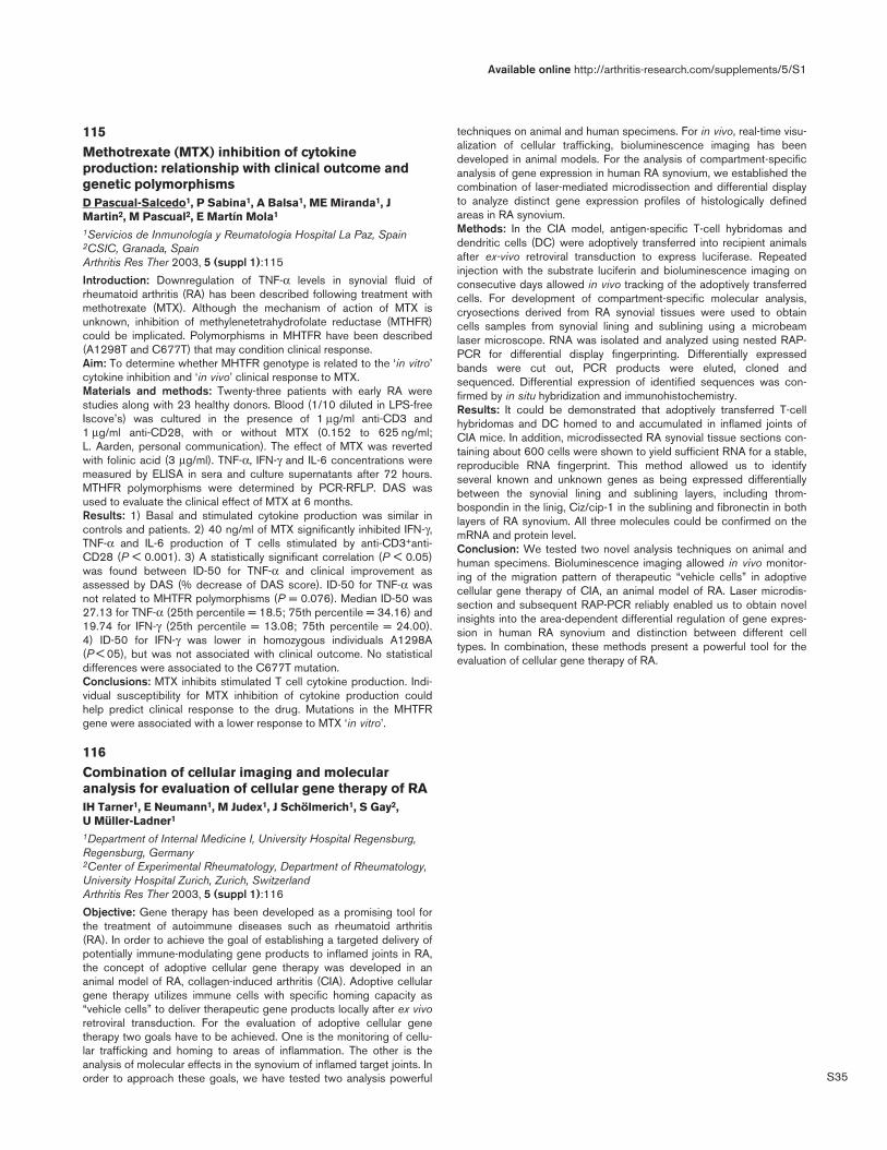
S35
115Methotrexate (MTX) inhibition of cytokineproduction: relationship with clinical outcome andgenetic polymorphismsD Pascual-Salcedo1, P Sabina1, A Balsa1, ME Miranda1, JMartin2, M Pascual2, E Martín Mola1
1Servicios de Inmunología y Reumatologia Hospital La Paz, Spain2CSIC, Granada, SpainArthritis Res Ther 2003, 5 (suppl 1):115
Introduction: Downregulation of TNF-α levels in synovial fluid ofrheumatoid arthritis (RA) has been described following treatment withmethotrexate (MTX). Although the mechanism of action of MTX isunknown, inhibition of methylenetetrahydrofolate reductase (MTHFR)could be implicated. Polymorphisms in MHTFR have been described(A1298T and C677T) that may condition clinical response.Aim: To determine whether MHTFR genotype is related to the ‘in vitro’cytokine inhibition and ‘in vivo’ clinical response to MTX.Materials and methods: Twenty-three patients with early RA werestudies along with 23 healthy donors. Blood (1/10 diluted in LPS-freeIscove’s) was cultured in the presence of 1 µg/ml anti-CD3 and1 µg/ml anti-CD28, with or without MTX (0.152 to 625 ng/ml; L. Aarden, personal communication). The effect of MTX was revertedwith folinic acid (3 µg/ml). TNF-α, IFN-γ and IL-6 concentrations weremeasured by ELISA in sera and culture supernatants after 72 hours.MTHFR polymorphisms were determined by PCR-RFLP. DAS wasused to evaluate the clinical effect of MTX at 6 months.Results: 1) Basal and stimulated cytokine production was similar incontrols and patients. 2) 40 ng/ml of MTX significantly inhibited IFN-γ,TNF-α and IL-6 production of T cells stimulated by anti-CD3+anti-CD28 (P < 0.001). 3) A statistically significant correlation (P < 0.05)was found between ID-50 for TNF-α and clinical improvement asassessed by DAS (% decrease of DAS score). ID-50 for TNF-α wasnot related to MHTFR polymorphisms (P = 0.076). Median ID-50 was27.13 for TNF-α (25th percentile = 18.5; 75th percentile = 34.16) and19.74 for IFN-γ (25th percentile = 13.08; 75th percentile = 24.00). 4) ID-50 for IFN-γ was lower in homozygous individuals A1298A(P < 05), but was not associated with clinical outcome. No statisticaldifferences were associated to the C677T mutation.Conclusions: MTX inhibits stimulated T cell cytokine production. Indi-vidual susceptibility for MTX inhibition of cytokine production couldhelp predict clinical response to the drug. Mutations in the MHTFRgene were associated with a lower response to MTX ‘in vitro’.
116Combination of cellular imaging and molecularanalysis for evaluation of cellular gene therapy of RAIH Tarner1, E Neumann1, M Judex1, J Schölmerich1, S Gay2, U Müller-Ladner1
1Department of Internal Medicine I, University Hospital Regensburg,Regensburg, Germany2Center of Experimental Rheumatology, Department of Rheumatology,University Hospital Zurich, Zurich, SwitzerlandArthritis Res Ther 2003, 5 (suppl 1):116
Objective: Gene therapy has been developed as a promising tool forthe treatment of autoimmune diseases such as rheumatoid arthritis(RA). In order to achieve the goal of establishing a targeted delivery ofpotentially immune-modulating gene products to inflamed joints in RA,the concept of adoptive cellular gene therapy was developed in ananimal model of RA, collagen-induced arthritis (CIA). Adoptive cellulargene therapy utilizes immune cells with specific homing capacity as“vehicle cells” to deliver therapeutic gene products locally after ex vivoretroviral transduction. For the evaluation of adoptive cellular genetherapy two goals have to be achieved. One is the monitoring of cellu-lar trafficking and homing to areas of inflammation. The other is theanalysis of molecular effects in the synovium of inflamed target joints. Inorder to approach these goals, we have tested two analysis powerful
techniques on animal and human specimens. For in vivo, real-time visu-alization of cellular trafficking, bioluminescence imaging has beendeveloped in animal models. For the analysis of compartment-specificanalysis of gene expression in human RA synovium, we established thecombination of laser-mediated microdissection and differential displayto analyze distinct gene expression profiles of histologically definedareas in RA synovium.Methods: In the CIA model, antigen-specific T-cell hybridomas anddendritic cells (DC) were adoptively transferred into recipient animalsafter ex-vivo retroviral transduction to express luciferase. Repeatedinjection with the substrate luciferin and bioluminescence imaging onconsecutive days allowed in vivo tracking of the adoptively transferredcells. For development of compartment-specific molecular analysis,cryosections derived from RA synovial tissues were used to obtaincells samples from synovial lining and sublining using a microbeamlaser microscope. RNA was isolated and analyzed using nested RAP-PCR for differential display fingerprinting. Differentially expressedbands were cut out, PCR products were eluted, cloned andsequenced. Differential expression of identified sequences was con-firmed by in situ hybridization and immunohistochemistry.Results: It could be demonstrated that adoptively transferred T-cellhybridomas and DC homed to and accumulated in inflamed joints ofCIA mice. In addition, microdissected RA synovial tissue sections con-taining about 600 cells were shown to yield sufficient RNA for a stable,reproducible RNA fingerprint. This method allowed us to identifyseveral known and unknown genes as being expressed differentiallybetween the synovial lining and sublining layers, including throm-bospondin in the linig, Ciz/cip-1 in the sublining and fibronectin in bothlayers of RA synovium. All three molecules could be confirmed on themRNA and protein level.Conclusion: We tested two novel analysis techniques on animal andhuman specimens. Bioluminescence imaging allowed in vivo monitor-ing of the migration pattern of therapeutic “vehicle cells” in adoptivecellular gene therapy of CIA, an animal model of RA. Laser microdis-section and subsequent RAP-PCR reliably enabled us to obtain novelinsights into the area-dependent differential regulation of gene expres-sion in human RA synovium and distinction between different celltypes. In combination, these methods present a powerful tool for theevaluation of cellular gene therapy of RA.
Available online http://arthritis-research.com/supplements/5/S1