mediated (nonactive) transport of glucose in mammalian cells and
TRANSCRIPT

Mediated (Nonactive) Transport of Glucose in
Mammalian Cells and its Regulation
C. R. PARK, O. B. CROFFORD, and T. KONO
From the Department of Physiology, Vanderbilt University School of Medicine, Nashville,Tennessee 37203
ABSTRACT Mediated (nonactive) transport of glucose in mammalian cells ischaracterized by saturation kinetics, stereospecificity, sensitivity to inhibitionby phlorizin and certain sulfhydryl-blocking agents, a temperature coefficientof about 2, an inability to utilize metabolic energy, and countertransport. Coun-tertransport can be explained by the development of carrier gradients in thecell membrane and provides the best evidence for carrier mobility. Efforts toidentify and isolate chemical components of the transport system have not beensuccessful. Transport among different types of mammalian cells shows a widerange of activities (V.,,, values differ by three or more orders of magnitude)and different sensitivities to hormones. Glucose enters the liver cell by mediatedtransport, as shown by a difference in the penetration rates of D- and L-glucoseand sensitivity to phlorizin. The activity of the system is one of the highestknown. Transport in muscle is the most important rate-controlling step for glu-cose utilization and is strongly accelerated by hypoxia, work, and insulin. Theeffect of work or insulin is strongly inhibited by metabolism of fatty acids.Insulin also stimulates glucose transport in adipose tissue. Using isolated fatcells, it could be shown that insulin is rapidly bound to sites on the cell surface.The effect is lost within a few minutes after the exogenous hormone is removed.The bound insulin is not released as such, but is metabolized to unknown prod-ucts. Binding is prevented by preexposure of cells to maleimide, which presum-ably blocks certain sulfhydryl groups at or near the insulin-binding site. Pre-treatment with insulin protects against maleimide. Digestion of the cell withtrypsin eliminates the acceleration of glucose transport and the inhibition oflipolysis by insulin. The glucose transport and adenyl cyclase systems are notgrossly affected by trypsin, indicating that the insulin effector system is a sepa-rate entity.
In mammalian tissues, glucose passes across the cell membranes by a processwhich has been called mediated transport, facilitated diffusion, or nonactivetransport. There are objections to all of these terms, but they serve to dis-tinguish the process from active transport. The most important difference
296 s
The Journal of General Physiology

PARK, CROFFORD, AND KONO Glucose Transport
between mediated and active transport is that the nonactive process cannotuse metabolic energy to move glucose against a concentration gradient.Mediated transport occurs in most, if not all, mammalian cells, whereasactive transport is seen prominently only in the kidney and gut.
This paper will review the basic concepts of mediated transport in mam-malian cells and will mention some recent efforts to determine the componentsof the system. It will discuss some aspects of the regulation of the process, andwill describe some recent observations on the mode and site of insulin action.
The basic concepts of nonactive transport were developed in the late 1940sby LeFevre in this country, Widdas in England, and Wilbrandt and Rosen-berg in Switzerland (see references 1 and 2 for review). Their studies, carriedout with the human erythrocyte, assume great significance today when it isrealized that this was one of the first clear expositions of membrane transportas we now understand it. Fig. 1 shows the simplest representation of thecarrier system which is in accord with the experimental findings. Glucose onthe outside, G, complexes with some membrane component, X, to form acomplex, GX, which moves across the membrane to discharge the glucose,on the inside, G . The system is freely reversible at all points. The six mostimportant observations which establish this concept of transport are as follows.
Saturation Kinetics It has been observed that the initial rate of entry orexit of glucose approaches a maximum above a certain concentration of sugar.This phenomenon, termed saturation, argues strongly that glucose mustcomplex with some membrane component, analogous to X in Fig. 1, duringthe permeation process. The combining site will be continuously occupiedfrom a statistical view when bombarded by a sufficiently high concentrationof glucose molecules. Permeation at this point becomes maximal and islimited by the number of carriers and their motility.
Stereospecificity The selectivity of the transport process for certain sugarsgreatly strengthens the idea of a combining site. It suggests further that thesite is in a protein, since only proteins have the necessary complexity ofstructure to discriminate among closely related small molecules such as thesugars. The stereospecificity of the system has been studied particularly byLeFevre and Marshall (3), who concluded that the three-dimensional con-formation of the sugar was the most important factor determining affinity.D-Glucose, the pyranose sugar which exists to the greatest extent in a chairconformation of the so-called C-1 type, has the highest affinity. Other hexosesand pentoses can be arranged in a descending order of affinity which corre-lates with the decline in the stability of the C-1 conformation in water. L-
Glucose, which is extremely close to D-glucose in most of its chemical andphysical properties, does not exist at all in the C-1 conformation, but is
297 s

TRANSPORT ACROSS CELL MEMBRANES
largely in the mirror image conformation, 1-C. L-Glucose has a very pooraffinity indeed for the transport site and hardly penetrates most cells.
An important consequence of stereospecificity, which is very useful in dis-tinguishing transport from subsequent metabolic steps, is the characteristicpattern of competition for transport among the common pentoses and hexoses.
Inhibition by Certain Agents Inhibition by certain compounds is a thirdcharacteristic of the system. Phloretin, phlorizin, and related compoundshave long been known as moderately specific inhibitors (1, 2). Transport inmany cells is also very sensitive to low levels of sulfhydryl-blocking agents (4),
OUTSIDE CELL MEMBRANE INSIDE
FREE GLUCOSE COMBINEDGLUCOSE FREE GLUCOSE1 ONLY
o+ + G,
GX. - GX 1 FiouRE 1. Schematic repre-sentation of the nonactive trans-port system for glucose. See thetext for description.
G = GLUCOSE o=ON OUTSIDE SURFACEX=UNOCCUPIED "CARRIER" I=ON INSIDE SURFACEGX=OCCUPIED "CARRIER"
'= REACTION CLOSE TO EQUILIBRIUM
-= RATE-LIMITING PROCESS
a point we will mention again later, and to dinitrofluorobenzene (5). Inhi-
bition by these substances also suggests participation of protein components.
Temperature Coefficient The coefficient is about 2 for the temperature
range of 27-37°C. This value is well above that for simple aqueous diffusion
and suggests, but by no means proves, that transport involves the formationand breaking of chemical bonds.
Energy Requirements Nonactive transport cannot utilize metabolic
energy. This means, of course, that no transport against a concentrationgradient is possible, although a special case, which looks superficially like
active transport, will be discussed in the next paragraph. Mediated transportis freely reversible and tends simply to equilibrate sugar concentrations acrossthe cell membranes; phosphorylation is not involved, and free sugar is theproduct of transport in either direction.
298 s

PARK, CROFFORD, AND KONO Glucose Transport
Countertransport This phenomenon provides the best evidence that thesugar complex has mobility across the membrane, an essential feature in theconcept of a carrier. Countertransport was predicted on kinetic grounds byWiddas (6); it was first demonstrated in our laboratory (7) and has subse-quently become a common test for a mobile carrier.
C,
w
,,
Z_t
OU
0
O
O
tn
!0
-J
OI.-W
0
ro
C-
4nt,
Uo0z
4X
-I
TIME, min
FIGURE 2. Countertransport of 3-O-methyl D-glucose and L-arabinose in the isolated,perfused rat heart on addition of nD-glucose to the perfusate. Hearts were perfused for thetimes indicated with oxygenated buffer containing 3-0-methyl D-glucose- 4C (0.75 m)or L-arabinose (13 m) (). In two groups of hearts perfused with 3-0-methyl glucose,perfusion was switched after 10 min to buffer containing 21 mM D-glucose in addition,and perfusion was continued for either 20 or 35 min (). Additional groups were per-fused with 0.75 mM 3-0-methyl glucose and 21 mrM glucose from zero time (). Onegroup of hearts, perfused with L-arabinose for 10 min, was perfused for an additional 20min with buffer containing L-arabinose (13 mM) and n-glucose (27 mM) (). At leastsix hearts were perfused for each point. The vertical line through each point indicatestwo standard errors of the mean. Figure reprinted by permission from the Journal of BiologicalChemistry, 1964, 239: 369.
Fig. 2 demonstrates countertransport in studies using the isolated rat heartpreparation (8, 9). In these experiments, the tissue was perfused with thenonmetabolized sugar 3-0-methyl glucose (left panel) until a substantial risein the concentration of intracellular sugar had occurred, as reflected by theincrease in sugar space above the extracellular space (about 325 jdl/g oftissue). At this point, a high concentration of glucose was added to the me-dium, causing a rapid drop in the intracellular 3-0-methyl glucose concen-tration. Since the latter sugar could not be metabolized, it must have beentransported out of the cell, although the extracellular concentration was higherthan the intracellular concentration. Similar results were obtained using thenonmetabolized pentose, L-arabinose, as shown in the panel on the right.
299 s

TRANSPORT ACROSS CELL MEMBRANES
The explanation for this phenomenon can be understood by reference toFig. 3. (The following discussion applies equally to the case of 3-0-methylglucose.)
The external medium, containing glucose and L-arabinose molecules, isshown on the left. In the center, the cell membrane is represented withmobile carriers, most of which have sugar molecules attached. On the rightis the intracellular water, which contains L-arabinose almost exclusively,since glucose, on entering the cell, has been rapidly transformed to glucose-6-Pand other metabolic products. As shown, glucose competes favorably for thecarrier at the external surface of the cell and cuts off arabinose entrance,whereas arabinose meets no competition at the inside surface and continues
x x
* 0x x
x x·
Oxo
x
x
xx x
x
FIGURE 3. Schematic repre-X X sentation of sugar-carrier com-
0 X plexes in a cell membrane dur-X ing counterflow of L-arabinose
x (X) induced by D-glucose (e).X The figure is explained in the
text.
MEMBRANEWITH
CARRIERS
to be transported out. Thus, the net flow of arabinose is outward. Under theseconditions, a gradient for glucose-loaded carrier is established within themembrane from outside to inside. As glucose is moved into the cell, emptycarriers become available at the inner side of the membrane for arabinose andcreate a gradient of arabinose-loaded carriers from inside to outside. Thelatter is the immediate source of energy to move arabinose out of the cell intoa higher external concentration. The ultimate source of energy, however, isin the movement of glucose down its concentration gradient with the carriergradients mediating the energy transfer. The conclusion that the carriersmust be mobile derives from this concept of changing concentration gradientsof the carrier in the membrane. Movement is also implicit in the conclusionthat the carrier can be accessible to sugars only at one side of the membrane
300 s

PARK, CROFFORD, AND KONO Glucose Transport
at a given time. If the carrier were bombarded simultaneously from bothsides, glucose on the outside would compete with arabinose on the inside forthe carrier. As a consequence, glucose would inhibit rather than enhance theflow of arabinose outward.
As regards the composition of the transport system, no components havebeen isolated and identified to date. The reason for this failure lies in ourinability to assay for transport activity after a cell is broken. Nevertheless,Bobinski and Stein (10, 11) and Langdon and Sloan (12) have recentlysearched for transport components by looking for membrane proteins whichwould combine with monosaccharides in a manner corresponding to thecharacteristic stereospecificity and kinetics of the transport system. To datethese efforts have not yielded convincing results (13), presumably becauseglucose can react reversibly with many proteins through their free aminogroups. This huge background of nonspecific reactivity makes it very difficultto isolate a specific reaction, particularly when the component involved maybe present in an extremely small amount. Another effort has gone into investi-gating the long-held proposition that glucose might cross the membrane as acomplex with some lipoid substance, possibly even a phospholipid. Severalyears ago we noted that erythrocyte membrane phospholipids would carryglucose from an aqueous into a lipid phase, and that phlorizin inhibited thistransport (14). This phenomenon has been studied in more detail byLeFevre et al. (15). Recent experiments by Wood (16), however, with bilayermembrane of the Rudin-Mueller type (see reference 16 a) made with thephospholipid from ghosts of human erythrocytes, suggest that phospholipidsin this geometrical arrangement cannot transport glucose. The possibilityremains, nevertheless, that lipid components in conjunction with specificprotein(s) may be part of the transport system.
Mediated transport of glucose has been seen in all mammalian cells thathave been studied, and it is probably an essential feature of the cell. Theprocess shows a number of variations among different cells, some of whichare noted below.
There is a remarkable range of transport activity among cell types, amongspecies, and at different ages within a given species. Transport is so fast in thehuman red blood cell that it is best measured by optical systems which canfollow the very rapid osmotic shrinkage or swelling of a cell as sugar enters orleaves. By contrast, nonprimate erythrocytes, such as those of the rabbit,were for many years considered to be impermeable to glucose because verylittle or no sugar could be found inside the cell no matter how high the externalconcentration might be. These erythrocytes are not impermeable, in fact, buttransport is relatively very slow, as shown in Table I. The Vma, for the rabbitcell is only about 0.4% of that for the human cell. As a consequence, transportis so slow that the sugar is phosphorylated as quickly as it enters and intra-
30 s

TRANSPORT ACROSS CELL MEMBRANES
cellular free glucose remains nearly undetectable. The K values of thehuman cells and rabbit systems are about the same, but human cells arevery sensitive to certain sulfhydryl-blocking agents whereas rabbit cells arenot. The stereospecificities appear to be similar, except that fructose ap-parently does not employ the glucose system in the rabbit cell, as judged fromthe absence of competition with glucose. The system in the rabbit cell showscountertransport between sugars, as in the human cell. It would thus appearthat the rabbit cell has the basic elements of the carrier system but is deficientin some element(s) which confers a high rate, a degree of specificity, andsensitivity to sulfhydryl-blocking agents. A related observation is that ofWiddas (17), who found some years ago that fetal erythrocytes of nonprimateshave a very fast rate of glucose transport, like that in human cells. About thetime of birth, synthesis of the component that confers this high rate apparentlyceases. Thus nature has provided in these nonprimate fetal and adult erythro-
TABLE I
SOME PROPERTIES OF GLUCOSE TRANSPORTIN HUMAN AND RABBIT ERYTHROCYTES
Property Human Rabbit
Vmax 600* 0.15*Km 8 m 6 mMInhibition by SH blockers Yes NoCompetition: glucose vs. fructose Yes No
* Millimoles per liter of cell water per minute.
cytes, cells which are analogous to transport mutants in bacteria and whichmight be profitably studied to dissect out components of the transport system.
The liver has long been known to be very permeable to glucose, since theconcentration of the sugar has been found to be almost the same in the tissuewater and blood under various conditions. Cahill and associates (18), whostudied hepatic permeability to monosaccharides in some detail a number ofyears ago, concluded that glucose entered by free diffusion. However, recentwork of Williams et al. (19) indicates that this is not the case and that glucosepenetrates by an extremely fast transport system. The following two experi-ments support this conclusion. In the first, an isolated liver preparation wasperfused with medium without glucose for about 15 min to establish steadystate conditions (Fig. 4). At this point, the perfusion was switched to anotherreservoir of medium of the same composition, except that it contained D-
glucose and L-glucose. At intervals, samples of the liver were taken to deter-mine its content of these two sugars. As can be seen, the concentration ofD-glucose in the liver was approaching that in the perfusate at 2 min, whereasthe concentration of L-glucose was much less than in the medium. Since
302 s

PARK, CROFFORD, AND KONO Glucose Transport
D-glucose and L-glucose are almost identical in size, solubility, and otherphysical properties, the difference in permeability indicates very stronglythat a process other than simple aqueous diffusion is involved. The secondexperiment to suggest transport was the finding that phlorizin inhibitspermeation (Fig. 5). Phlorizin is a well-known inhibitor of the monosac-charide transport system in other cells. As can be seen, it inhibited D-glucoseand, more strongly, the entry of L-glucose. While the effect of phlorizin on theD-isomer was small, kinetic analysis from other, more extensive experiments
3uI tn rinrI 14'i L -I iuCOSf
1.00
0.80
LIVERPERFUSATE
0.60
cpm/ml H20
0.40
0.20
A'
--
8
T
4
4
2 10 2 10 40
TIME (MIN)
FIGURE 4. Penetration of D-glucose and L-glucose into the cells of the perfused rat liverThe experiment is described in the text, and details will be published (19). D-Glucoselabeled with tritium and L-glucose labeled with 4C were used to facilitate analysis in thetissue and the perfusion medium. Both sugars were added to the medium at zero timein tracer concentration. Livers were fixed by freeze-clamping, followed by cold per-chlorate extraction. Distribution of the sugars in extracellular space only would give aratio of about 0.25.
made it apparent that transport is so fast that the major limiting step forD-glucose permeation in liver tissue is actually extracellular transfer, aprocess that is not affected by phlorizin. Since transport of L-glucose is muchslower, it is more limiting for permeation and the phlorizin effect is there-fore larger. In contrast to muscle and fat cells, to be discussed below, trans-port in liver is not sensitive to insulin, although the hormone affects otherparameters of liver function.
In muscle cells, a striking feature of mediated transport is its regulation byfactors such as hypoxia, muscular work, fatty acids, and certain hormones,notably insulin. These controls have been elucidated in a number of lab-oratories, including those of Randle, Kipnis, Cori, and Narahara as well as
303
.. . _ _______

TRANSPORT ACROSS CELL MEMBRANES
our own. The data chosen in this paper to illustrate some of these controlshave been collected by Morgan and associates in our laboratory using theisolated, perfused rat heart as the test object. An experiment demonstratingthe effects of insulin and muscle work on transport is shown in Table II.Transport was evaluated by determining the accumulation of L-arabinosewithin the cell when the heart was perfused for 10 min with medium con-taining the pentose. The sugar enters cardiac cells by the glucose transportsystem, as shown by competition studies, but is not metabolized (22). Atthe end of the perfusion, the tissue content of arabinose was determined, and,after correcting for sugar in the extracellular space, the intracellular con-
== CONTROL
LIVERPERFUSAT
cpm/ml H
3H (D-GLUCOSE) 14C(L-GLUCOSE)
FIGURE 5. Effect of phlorizin on the distribution of labeled D- and L-glucose in theperfused rat liver. Livers were exposed to phlorizin (10 m) by perfusion for 15 minbefore the sugars were added in tracer concentrations. The tissue was taken for analysis10 min later. Details will be published (19).
centration could be calculated (9). The term "Equilibration (%)" indicatesthe extent to which the intracellular concentration reached the extracellularor perfusate concentration. In the aerobic heart, the transport of L-arabinosewas very slow in the absence of insulin and was stimulated strongly by in-sulin addition. When the tissue was made severely hypoxic, transport wasgreatly accelerated, even in the absence of insulin, and could be increasedfurther to a modest extent on addition of the hormone. Similarly, transportwas strongly stimulated by muscular work with or without insulin. The workeffect did not appear to be due to hypoxia, since measurements of oxygentension and lactate production in other experiments indicated that oxygen-ation was adequate (23). It was also clear that work (or hypoxia) increasedthe sensitivity of transport to suboptimal concentrations of insulin. The
3o4 s

PARK, GROFFORD, AND KONO Glucose Transport
combination of transport acceleration and increased insulin sensitivity mayexplain the improved glucose tolerance of diabetic individuals with exercise.
As would be expected from the above, muscular work can cause a sub-stantial increase in glucose uptake, as shown in Table III. In these experi-ments, work was increased by raising the perfusion pressure in the aorta of a
TABLE II
EFFECT OF INSULIN, HYPOXIA, AND WORKON THE TRANSPORT OF L-ARABINOSE
IN THE PERFUSED RAT HEART
After a preliminary 10 min perfusion with arabinose-free buffer, perfusionwas switched to buffer containing pentose (13 m) and sorbitol-3 H. Thisbuffer was recirculated for an additional 10 min. Insulin was addedonly to the recirculating buffer. The aerobic or anaerobic nonworking heartwas a Langendorff preparation perfused retrogradely via the aorta. It wasnot a completely "nonworking" preparation, since contractions occurredagainst fluid contained in the left ventricle at the aortic pressure (about 60mm Hg). The working heart was perfused by introducing the perfusion me-dium into the left auricle at the left auricular pressure indicated. The leftventricle pumped out the fluid (about 130 ml g- min-1), developing a peakpressure of about 95 mm Hg. The oxygen consumption was approximatelydoubled by the work load (20). The data shown are from Morgan et al. (21).
Perfusion conditions Insulin added Equilibrium
(units/ml) X 106 %
Aerobic 0 44-4*26 04:4
100 44-3300 204-5900 29i4
Anaerobic 0 284-326 414-3
100 414-5300 4044900 464-3
Aerobic-working, 10 mm Hg atrial pressure 0 234:3100 484-5300 624-5900 614-7
Langendorff isolated heart preparation, as a consequence of which the pres-sure against which the left ventricle contracted was proportionately increased(see legend to Table II). On going from 60 to 100 mg Hg perfusion pressure,no free intracellular glucose could be detected at any time. The absence offree glucose indicates, as discussed in detail elsewhere (7, 9), that phosphoryl-ation kept pace with sugar entry and that transport was the rate-limitingstep for the uptake process. Table III also shows an important effect of fatty
3o5 s

TRANSPORT ACROSS CELL MEMBRANES
acid on transport. At a low level of cardiac work (60 mm Hg perfusion pres-
sure) addition of palmitate to the medium caused a modest reduction in
glucose uptake, but at the higher work load the fatty acid suppressed the
transport acceleration completely. Fatty acid had a similar inhibitory effect
on the stimulation of glucose transport by insulin, as shown by Randle et al.
(25, 26) and recently confirmed by Neely, Bowman, and Morgan (24). Evi-
dence to date suggests that the inhibition is not a direct effect of the fatty
acid on the transport system, but occurs indirectly as a consequence of fatty
acid oxidation (24, 26).There are a number of other physiological agents concerned with trans-
port control in addition to those mentioned above. In most instances, how-
TABLE III
EFFECTS OF WORK AND FATTY ACID ON GLUCOSEUPTAKE AND INTRACELLULAR FREE
GLUCOSE IN THE PERFUSED RAT HEART
The hearts of 18-hr fasted rats were perfused with Krebs bicarbonate buffer
containing 3% bovine albumin with or without bound palmitate for 1 hr at
37C. The work load was varied by changing the perfusion pressure (see
legend to Table II) in a Langendorff perfusion apparatus. The additional
work load increased oxygen consumption by 50%. Oxygen consumption was
not affected by the addition of the fatty acid. The data shown are from Neely
et al. (24).
Palmitate. Free intracellularPerfusion pressure Glucose, 15 mm 1.6 mm Glucose uptake glucose detected
mm Hg pmoles g-l hr-l mM
60 + 0 874-14 0+ + 58+:10 0
100 + 0 2324-8 0+ + 35-7 0
ever, the mechanisms involved are probably the same. Epinephrine, forexample, stimulates glucose uptake (27) and, by inference, transport in theheart, but this may be largely secondary to its inotropic and chronotropiceffects. In fat tissue, where mechanical activity is not involved, the stimu-lation of sugar transport by epinephrine is very small. The sensitivity of
transport to insulin in muscle is reduced by growth hormone and/or theglucocorticoids, and in certain forms of diabetes. This reduction may besecondary, however, to the increased availability and metabolism of fat,as suggested originally by Randle et al. (25) as a part of the "glucose-fattyacid cycle." In all cases of transport control, with the possible exception ofthat by insulin, acceleration or inhibition is coordinated with regulation ofglycolytic enzyme activities, particularly phosphorylase, hexokinase, and
306 s

PARK, CROPFFORD, AND KONO Glucose Transport
phosphofructokinase. The most critical control, however, remains that ex-erted on transport, since this is the predominately rate-limited step forglucose utilization by muscle under virtually all circumstances.
In the fat cell, transport of glucose is also strongly stimulated by insulin(28). An opportunity to examine some of the initial steps in this action of thehormone has been provided by Rodbell's (29) development of an isolatedcell preparation of adipose tissue. In this connection, Crofford (30), usingthe Rodbell cell preparation, has reexamined the question of how insulinbinds to the cell. He could thus avoid a major difficulty in earlier studiesemploying intact tissues, in which it was not possible to distinguish satis-factorily between binding to the cell and trapping in the interstitial spaces.Crofford also employed native rather than labeled insulin, since the bio-logical activity and binding qualities of labeled insulin are probably altered.In Fig. 6 is shown the time course of binding by the fat cells. Two separateexperiments are shown in each panel. The cells were placed in medium con-taining a physiological level of insulin, and the fall in concentration wasfollowed. There was an immediate uptake of insulin, presumably reflectingbinding to the cell surface, followed by a relatively very slow, progressiveuptake, reflecting utilization of the hormone (Fig. 6 A). This interpretationwas supported by showing that the initial uptake was not reduced when theincubation temperature was dropped from 37°C to 17°C (Fig. 6 B), but thatthe utilization was greatly slowed. As shown by the experiment of Fig. 6 C,the utilization of insulin required the presence of the cells. Fig. 6 D showsthat the initial binding could be prevented by a short prior exposure of thecells to maleimide, although this agent did not suppress the subsequent rateof insulin destruction. The effects of maleimide in the fat cell' are similarto those observed earlier in heart muscle (31, 32) and may be summarizedas follows. A brief exposure to maleimide (or N-ethylmaleimide), which is asulfhydryl-blocking agent with rather high specificity under the conditionsemployed, can suppress the stimulatory effect of insulin on glucose trans-port without destroying the activity of the transport system itself, and with-out poisoning intracellular glycolytic enzymes, such as 3-phosphoglyceralde-hyde dehydrogenase (21). The effect of the blocking agent is prevented,however, if the cells are exposed first to insulin. The simplest interpretationof these observations is that the binding and/or action of insulin requires theintegrity of a sulfhydryl group which is at or near the cell surface and isclosely associated with the hormone-binding site.
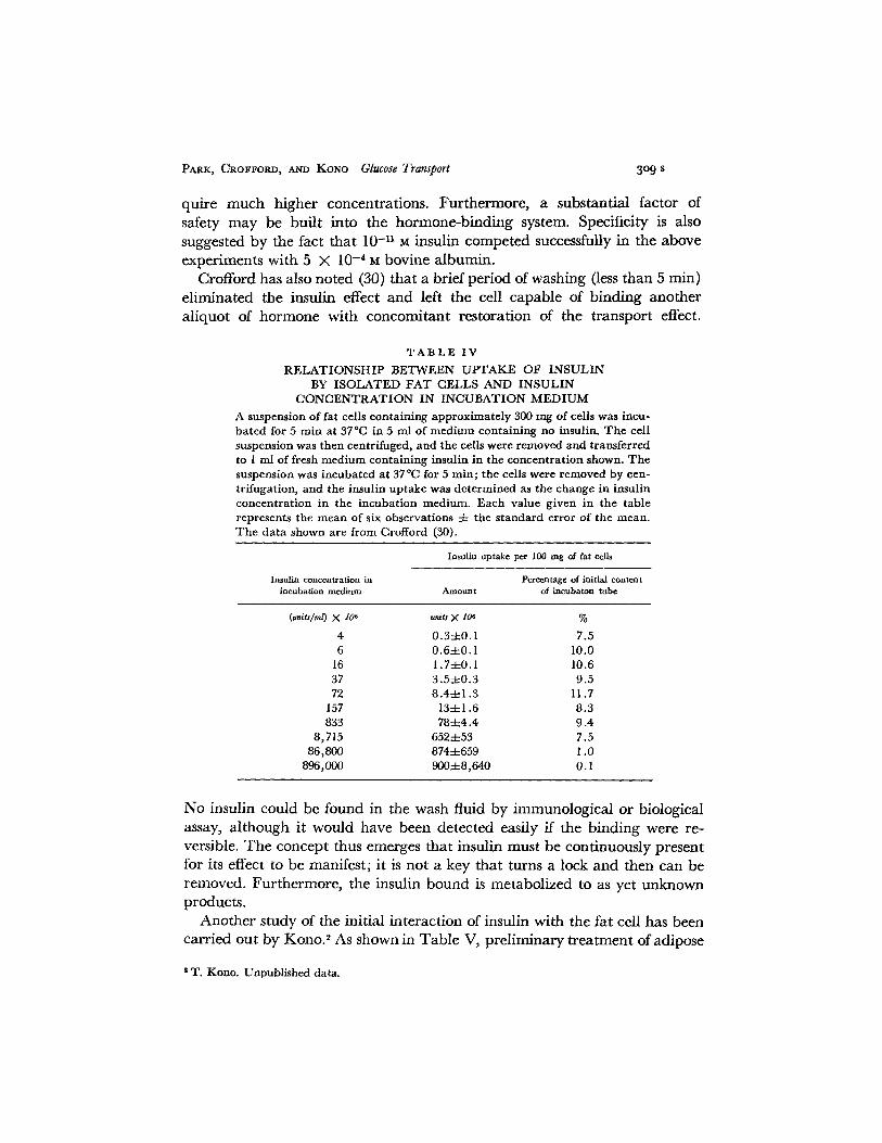
Crofford (30) has studied the quantitative relationship between the initialuptake and the insulin concentration of the medium (Table IV). The "bind-ing" remained linearly related to concentration up to about 10 milliunits/
1 0. B. Crofford. Unpublished observations.
307

TRANSPORT ACROSS CELL MEMBRANES
ml, a concentration considerably in excess of the concentration giving amaximal stimulation of transport, at which point binding appeared to beapproaching a maximum. The fact that the transport effect reached a
40
30
20
10
0 10 20 30 40 50 60 0 10 20 30 40 50 60
MINUTES
FIGURE 6. Time course of disappearance of insulin from the incubation medium. In
each of the experiments, approximately 1200 mg of isolated fat cells were incubated in4 ml of medium containing between 30 and 45 microunits (MU) of insulin per ml and1 mm glucose. 30 sec before each sampling time, the incubation mixture was centrifugedso that approximately 0.3 ml of cell-free incubation medium could be aspirated fromthe tube and used for the assay of immunoreactive insulin. In each panel, the data fromtwo experiments are shown; one by the solid curve and a duplicate by the dashed curve.A. Results of experiments performed at 37°C. B. at 17.50C. C. Cells were incubated at
37C for 60 min, removed from the incubation medium, and discarded. At zero time,insulin was added to the medium in which the fat cells had been incubated, the incuba-
tion was continued for 60 min, and the cell-free medium was sampled at the timesindicated. D. The fat cells were given a 30 sec exposure to 10- 3 M maleimide (C4H3NO2)immediately after collagenase treatment. The maleimide was then removed by washingthe cell suspension four times in fresh buffer, and the cells were used for experimentsidentical with those of A. Figure reprinted by permission from The Journal of Biological Chem-
istry, 1968, 243: 362.
plateau before the binding reached saturation does not mean necessarilythat the binding was nonspecific. Transport acceleration is not the primaryor only response to insulin (see below), and other actions of insulin may re-
_0____- _ t ---
I _ , .. C
zo
zwozo0
-
C,z
B
I .'0..~~~~~~~~~~~~~~~~~
_·
308
PARK, CROFFORD, AND KONO Glucose Transport
quire much higher concentrations. Furthermore, a substantial factor ofsafety may be built into the hormone-binding system. Specificity is alsosuggested by the fact that 10-11 M insulin competed successfully in the aboveexperiments with 5 X 10- 4 M bovine albumin.
Crofford has also noted (30) that a brief period of washing (less than 5 min)eliminated the insulin effect and left the cell capable of binding anotheraliquot of hormone with concomitant restoration of the transport effect.
TABLE IV
RELATIONSHIP BETWEEN UPTAKE OF INSULINBY ISOLATED FAT CELLS AND INSULIN
CONCENTRATION IN INCUBATION MEDIUM
A suspension of fat cells containing approximately 300 mg of cells was incu-bated for 5 min at 37°C in 5 ml of medium containing no insulin. The cellsuspension was then centrifuged, and the cells were removed and transferredto 1 ml of fresh medium containing insulin in the concentration shown. Thesuspension was incubated at 37C for 5 min; the cells were removed by cen-trifugation, and the insulin uptake was determined as the change in insulinconcentration in the incubation medium. Each value given in the tablerepresents the mean of six observations 4- the standard error of the mean.The data shown are from Crofford (30).
Insulin uptake per 100 mg of fat cells
Insulin concentration in Percentage of initial contentincubation medium Amount of incubaton tube
(units/ml) X 106 units X 106 %
4 0.34-0.1 7.56 0.6-0.1 10.0
16 1.7--0.1 10.637 3.540.3 9.572 8.441.3 11.7
157 131.6 8.3833 7844.4 9.4
8,715 652453 7.586,800 874-659 1.0
896,000 90048,640 0.1
No insulin could be found in the wash fluid by immunological or biologicalassay, although it would have been detected easily if the binding were re-versible. The concept thus emerges that insulin must be continuously presentfor its effect to be manifest; it is not a key that turns a lock and then can beremoved. Furthermore, the insulin bound is metabolized to as yet unknownproducts.
Another study of the initial interaction of insulin with the fat cell has beencarried out by Kono.2 As shown in Table V, preliminary treatment of adipose
2 T. Kono. Unpublished data.
309 s

TRANSPORT ACROSS CELL MEMBRANES
tissue with trypsin under appropriate conditions virtually eliminated theacceleratory effect of insulin on glucose utilization. Appropriate controlsshowed that this was not due simply to destruction of insulin by the enzyme,but appeared rather to involve a modification of the cell surface. The ability
TABLE V
LOSS OF INSULIN EFFECT ON GLUCOSEUTILIZATION BY ADIPOSE TISSUE AFTER
TREATMENT WITH TRYPSIN
Rat epididymal fat pads were first incubated in Krebs bicarbonate buffer at37°C with 1 mg/ml crystalline trypsin without albumin. The fat pads werethen washed extensively with two changes of buffer containing albumin(2%) and trypsin inhibitor (0.1 mg/ml). A third washing was carried outwithout the inhibitor. The second incubation was carried out for 30 min at37 C in Krebs bicarbonate buffer with 2% albumin. The medium containeduniformly labeled glucose-14 C (0.3 mM) and insulin (0.1 unit/ml) and phlore-tin (1 mM) where indicated. The data shown are from unpublished experi-ments of T. Kono.2
Glucose: CO, ratio
Treatment Glucose in incubation Control Insulin
mM mLmotes g- hr-
None 0.3 46 644Trypsin 0.3 38 56None 3 540 2680Trypsin 3 360 468Trypsin 3 plus phloretin 54
<: o
w .'W
LuE
6001
400-
200-
n_
X (LW (..
O Z
NO TREAT-MENT
TRYPSIN
FIGURE 7. Persistence of response to epinephrine and ACTH in adipose tissue aftertreatment with trypsin. Rat epididymal fat pads were treated with trypsin and washedas described in Table V. They were then incubated in Krebs bicarbonate buffer with2% albumin for 30 min at 37C. Epinephrine (EPI) was added in a concentration of1 /Ag/ml with I mm caffeine, and ACTH was added in a concentration of 1 Azg/ml with1 mM caffeine. From unpublished results of T. Kono. 2
3o s
v

PARK, CROFFORD, AND KONO Glucose Transport
of the cell to transport and metabolize glucose was not destroyed by trypsintreatment, however, since these functions could be strongly stimulated simplyby raising the concentration of glucose in the medium. Glucose utilization,furthermore, could be markedly inhibited by phloretin, indicating thatthe cell membrane was not made "leaky" by the enzyme treatment.
As shown in Fig. 7, the same trypsin treatment did not destroy the lipolyticresponse of the cell to epinephrine or to ACTH. It did, however, eliminatethe antilipolytic response of the cell to insulin, as shown in Fig. 8. This anti-lipolytic action has been recently explained by the observation (33) thatinsulin lowers the tissue level of 3' ,5'-adenosine monophosphate (cyclicadenylate), the compound which mediates the action of ACTH and epineph-rine on lipolysis (34). The effect of insulin on transport, however, does not
o 600-U)
uw 0 400-
_J 0M
W E 200-
U5 -
z_+
-J + -
W J
gW 8~~~~~~ ._1.o .o :~H o.
NO TREAT- TRYPSINMENT
FIGURE 8. Loss of insulin effect on lipolysis in adipose tissue after treatment with tryp-sin. Rat epididymal fat pads were treated with trypsin and washed as described in TableV. They were then incubated in Krebs bicarbonate buffer containing 2% albumin for1 hr at 37C with the additions shown, but no glucose. The concentrations employedwere as follows: epinephrine (EPI), 0.1 g/ml; insulin (INS), I milliunit/ml. Fromunpublished results of T. Kono. 2
appear to be altered by a change in cyclic adenylate levels (35, 36). It seemslikely, from Kono's results,2 that a part of the insulin effector system is atthe cell surface and contains peptide elements; it is apparently distinct fromthe glucose transport and the adenyl cyclase systems. How interaction ofinsulin with this system accelerates transport and reduces levels of cyclicadenylate in the cell remains unknown.
REFERENCES
I. LEFEvRE, P. G. 1961. Sugar transport in the red blood cell; structure-activity relationshipsin substrates and antagonists. Pharmacol. Rev. 13:39.
2. WILBRANDT, W., and T. ROSENBERG. 1961. The concept of carrier transport and its corol-laries in pharmacology. Pharmacol. Rev. 13:109.
3. LEFEvRE, P. G., and J. K. MARSHALL. 1958. Conformational specificity in a biologicalsugar transport system. Am. J. Physiol. 194:333.
31I s

TRANSPORT ACROSS CELL MEMBRANES
4. LEFEvRE, P. G. 1947. Evidence of active transfer of certain nonelectrolytes across thehuman red cell membrane. J. Gen. Physiol. 31:505.
5. BOWYER, F., and W. F. WIDDAS. 1958. The action of inhibitors on the facilitated hexosetransfer system in erythrocytes. J. Physiol., (London). 141:219.
6. WIDDAS, W. F. 1952. Inability of diffusion to account for placental glucose transfer in thesheep and consideration of the kinetics of a possible carrier transfer. J. Physiol., (London).118:23.
7. PARK, C. R., R. L. POST, C. F. KALMAN, J. H. WRIGHT, JR., L. H. JOHNSON, and H. E.
MORGAN. 1956. The transport of glucose and other sugars across cell membranes andthe effects of insulin. Ciba Found. Colloq. Endocrinol. 9:240.
8. MORGAN, H. E., D. M. REGEN, and C. R. PARK. 1964. Identification of a mobile carrier-mediated sugar transport system in muscle. J. Biol. Chem. 239:369.
9. MORGAN, H. E., M. J. HENDERSON, D. M. REGEN, and C. R. PARK. 1961. Regulation ofglucose uptake in muscle. I. The effects of insulin and anoxia on glucose transport andphosphorylation in the isolated perfused heart of normal rats. J. Biol. Chem. 236:253.
10. BOBINSKI, H., and W. D. STEIN. 1966. Isolation of a glucose binding component fromhuman erythrocyte membranes. Nature. 211:1366.
11. STEIN, W. D. 1967. Some properties of carrier substances isolated from bacterial anderythrocyte membranes. Biochem. J. 105:3P.
12. LANGDON, R. G., and H. R. SLOAN. 1967. Formation of imine bonds between transportsugars and lysyl residues of specific membrane proteins of erythrocytes and fat cells.Proc. Natl. Acad. Sci. U.S. 57:401.
13. LEFEvRE, P. G. 1967. Imine-bonding in membrane transport of monosaccharides: in-validity of kinetic evidence. Science. 158:274.
14. PARK, C. R. 1961. Discussion. In Membrane Transport and Metabolism. A. Kleinzeller andA. Kotyk, editors. Academic Press, New York. 453.
15. LEFEvRE, P. G., K. I. HABICH, H. S. HEss, and M. R. HUDSON. 1964. Phospholipid sugarcomplexes in relation to cell membrane monosaccharide transport. Science. 143:955.
16. WOOD, R. E. 1968. Model systems for the study of membrane permeability. DoctorateThesis. Vanderbilt University, Nashville, Tennessee.
16 a. TIEN, H. T. 1968. Black lipid membranes at bifaces. Formation characteristics, opticaland some thermodynamic properties. J. Gen. Physiol. 52(1, Pt. 2):125 s.
17. WIDDAS, W. F. 1955. Hexose permeability of foetal erythrocytes. J. Physiol., (London).127:318.
18. CAHILL, G. F., JR., J. ASMORE, A. S. EARLE, and S. ZOTTU. 1958. Glucose penetrationinto liver. Am. J. Physiol. 192:491.
19. WILLIAMS, T. F., J. H. EXTON, C. R. PARK, and D. M. REGEN. 1968. Stereospecific trans-port of glucose in the perfused rat liver. Am. J. Physiol. In press.
20. NEELY, J. R., H. LIEBERMEISTER, E. J. BATTERSBY, and H. E. MORGAN. 1967. Effect ofpressure development on oxygen consumption by isolated heart. Am. J. Physiol. 212:804.
21. MORGAN, H. E., J. R. NEELY, R. W. WOOD, C. LIEBECQ, H. LIEBERMEISTER, and C. R.PARK. 1965. Factors affecting glucose transport in heart muscle and erythrocytes. Fed-
eration Proc. 24:1040.22. PARK, C. R., D. REINWEIN, J. J. HENDERSON, E. CADENAS, and H. E. MORGAN. 1959. The
action of insulin on the transport of glucose through the cell membrane. Am. J. Med.26:674.
23. NEELY, J. R., H. LIEBERMEISTER, and H. E. MORGAN. 1967. Effect of pressure develop-ment on membrane transport of glucose in isolated rat heart. Am. J. Physiol. 212:815.
24. NEELY, J. R., R. H. BOWMAN, and H. E. MORGAN. 1968. Regulation of glycogenolysis inthe perfused rat heart developing intraventricular pressure. In Control of GlycogenMetabolism. W. J. Whelan, editor. University Press, Universitetsforlaget, Oslo.
25. RANDLE, P. J., P. B. GARLAND, C. N. HALES, and E. A. NEWSHOLME. 1963. The glucosefatty acid cycle. Its role in insulin sensitivity and the metabolic disturbances of diabetesmellitus. Lancet. 1(13 April):785.
312

PARK, CROFFORD, AND KONO Glucose Transport
26. RANDLE, P. J., E. A. NEWSHOLME, and P. B. GARLAND. 1964. Regulation of glucose uptakeby muscle. 8. Effects of fatty acids, ketone bodies and pyruvate and of alloxan diabetesand starvation on the uptake and metabolic fate of glucose in rat heart and diaphragmmuscle. Biochem. J. 93:652.
27. WILLIAMSON, J. R. 1964. Metabolic effects of epinephrine in the isolated, perfused ratheart. J. Biol. Chem. 239:2721.
28. CRoFFORD, O. B., and A. E. RENOLD. 1965. Glucose uptake by incubated rat epididymaladipose tissue. Rate-limiting steps and site of insulin action. J. Biol. Chem. 240:14.
29. RODBELL, J. 1964. Metabolism of isolated fat cells. I. Effects of hormones on glucosemetabolism and lipolysis. J. Biol. Chem. 239:375.
30. CROFFORD, O. B. 1968. The uptake and inactivation of native insulin by isolated fat cells.J. Biol. Chem. 243:362.
31. CADENAS, E., H. KAJI, C. R. PARK, and H. RASMUSSEN. 1961. Inhibition of the insulineffect on sugar transport by N-ethylmaleimide. J. Biol. Chem. 236:PC63.
32. PARK, C. R., H. E. MORGAN, H. KAJI, and M. SMITH. 1964. Effects of insulin on transportin heart muscle. In The Biochemical Aspects of Hormone Action. A. B. Eisenstein,editor. Little, Brown and Company, Boston. 18.
33. BUTCHER, R. W., J. G. T. SNEYD, C. R. PARK, and E. W. SUTHERLAND. 1966. Effect ofinsulin on adenosine 3',5'-monophosphate in the rat epididymal fat pad. J. Biol. Chem.241:1651.
34. BUTCHER, R. W., R. J. Ho, H. C. MENG, and E. W. SUTHERLAND. 1965. Adenosine 3',5'-monophosphate in biological materials. II. The measurement of adenosine 3',5'-mono-phosphate in tissues and the role of the cyclic nucleotide in the lipolytic response of fat toepinephrine. J. Biol. Chem. 240:4515.
35. RODBELL, M. 1967. Metabolism of isolated fat cells. VI. The effects of insulin, lipolytichormones, and theophylline on glucose transport and metabolism in "ghosts." J. Biol.Chem. 242:5751.
36. SNEYD, J. G. T., J. CORBIN, and C. R. PARK. 1968. Glucose transport and adenosine3' ,5-phosphate (cyclic adenylate) in adipose tissue. To be published.
Discussion
Dr. Kennedy: I should like to call upon Dr. Crane to begin a discussion of Dr. Park'spresentation.
Dr. Robert K. Crane: I would like to show you three by now rather ancient slides toillustrate the concept of gradient coupling in membrane transport, which I think hassome relevance to what Dr. Park has been talking about. Fig. 1, Discussion, simplyshows the mobile carrier as we draw it. It does not differ at all from Dr. Park's con-cept. It has all the features that he listed as characteristic of mobile carrier transportssystems. A carrier such as shown in Fig. 2, Discussion is a useful description of thekinetic properties of the transport system in the intestine for sugars, amino acids, bilesalts, and several other compounds, and, in most animal cells at least, for amino acids.Again, this is a mobile carrier with properties just like those that Dr. Park has de-scribed, but with the one additional property that the carrier appears to associate notonly with substrate but also with sodium ion, and transports both across the membraneat the same time.
Now, I don't have time to give you any evidence for it and I don't want to get intothat area; the consequences of the presence of such a carrier are what I think we areinterested in. These are illustrated in Fig. 3, Discussion.
313 s

314 S TRANSPORT ACROSS CELL MEMBRANES
On the one hand, we may consider a red blood cell with its sodium ion pump in the
membrane which operates to maintain a low intracellular sodium ion concentration,
and a mobile carrier system with a single substrate-specific site. The net result for
substrate is what we would call passive transport, where at equilibrium the substrate
0
Clpll membrane
MC
FIGURE 1
CELL MEMBRANE
0>CELL FLUID
4A1
0MEDIUM
25
%oFIGURE 2
concentration inside equals the substrate concentration outside. In the gut epithelial
cell, on the contrary, the carrier also associates with sodium ion. Because of the low
concentration of sodium ion maintained within the cell, the substrate concentration
inside the cell at equilibrium is necessarily going to be higher than the substrate
concentration outside the cell. This is what we would nominally call active transport.
I remind you that there is no essential difference in the carriers, except that one has
-a (
I

PARK, CROFFORD, AND KONO Glucose Transport
an association with two specific substances, one of which has an asymmetry across themembrane imposed upon it by the action of some other process at a different locationwithin the cell.
The particular relevance of this comparison for the latter part of Dr. Park's talk isthat I had the opportunity last May in Bruges, at the 15th Colloquium on the Protidesof the Biological Fluids, to hear a paper presented by Dr. Letarte, who had beenworking with Albert Renold in Geneva. The substance of that paper was that glucoseentry into the isolated fat cell in the absence of insulin was associated with ioniceffects which were not easily rationalized. In the presence of insulin, however, glucoseentry into the isolated fat cell could be analyzed in terms of a sodium-dependent,gradient-coupled system. They saw a specific sodium ion dependence for the entryof the glucose in the presence of insulin.
RED BLOOD CELL GUT EPITHELIAL CELL
MEDIUM MEDIUM
NA+ NA+ So
PUMP LEAK CARRIER
NA+ NA+ Si
kJ-
NA NA'So
PUMP CARRIER 2
NA+ NASi
KJCELL CONTENTS CELL CONTENTS
PASSIVE TRANSPORT ACTIVE TRANSPORT
Si = So Si > SoFIGURE 3
Question from the Floor: The insulin effect very clearly activates the glucose systemin such a way that it will move glucose out of the cell just to facilitate outward move-ment as well as inward, and this also, of course, is an immediate effect of insulin. Iwonder if this could conceivably fit in with a sodium gradient effect?
Dr. Park: Sodium-dependent glucose transport systems are usually systems ofactive transport. The transport process in the fat cell is probably simply a "nonactive,"mediated transport system. If this is true, and if it is like transport in muscle, insulinwould accelerate transport in both directions. This would then seem to exclude theparticipation of a sodium gradient across the membrane in the insulin effect. I wouldlike to emphasize, however, that the above comments are speculative and experimentsshould be done along the lines suggested by the question.
Dr. Ivan Bihler: I would like to add to the question of ion effects on insulin-stimu-lated transport in muscle. We have recently studied the transport of 3-methyl glucose,a nonmetabolized glucose analogue, in the "intact" rat hemidiaphragm and have
315

TRANSPORT ACROSS CELL MEMBRANES
found that whenever the sodium pump is inhibited by cardiac glycosides, by low levelsof K+ in the incubation medium, or by other means, sugar transport is increased.This effect is additive to that of a submaximal dose of insulin. In contrast, a high -K +
medium is known to activate the sodium pump and to reduce its sensitivity to inhibi-tion by ouabain; in such a medium sugar transport is decreased and is less stimulatedby ouabain. Furthermore, diphenylhydantoin, a drug supposed to activate the sodiumpump, was also able to reduce sugar transport, provided that the sodium pump wasnot inhibited by other means. It seems as though, in addition to insulin, muscularwork, and anoxia, sugar transport in muscle is also regulated by the activity of thesodium pump and/or the related intracellular ionic levels. These results suggest to usthat perhaps the effects of anoxia and muscular work are also mediated by alterationsin the levels of cations in the cell or in a particular cellular compartment.
Dr. Park: There is likely to be some common explanation for the effect on transportof muscle work, hypoxia, and other acceleratory agents such as the uncouplers de-scribed by Randle and Smith,' but I think it is not at all clear what this commonmechanism is. It is conceivable that it could be an ionic influence.
Dr. Zierler: One of the things I hoped very much would come out of this symposiumwas that the physiologists and biophysicists, who largely spoke yesterday morning;the physical chemists, who spoke yesterday afternoon and this morning; and thebiochemists, who spoke this afternoon, would find a common understanding andwould appreciate one another's data and understand one another's problems, andthat somehow some of us would be smart enough to bring them all together. At themoment I feel more confused than able to synthesize what I have learned, and perhapsDr. Kennedy will bring this all together.
I would like to discuss some of the things that I think may be a little more simple.I don't disagree that one can construct all kinds of plausible models of movement ofsubstances across these interfaces, and as you said, Dr. Kennedy, the question is whichof them is really going on. Some of the things that emerge from the study of thinfilms I think have to be considered in all models, because some of this material ispresent, and whenever a simple physical chemical phenomenon can be shown we haveto assume either that it proceeds in living tissue or that something in nature preventsit. One of the things that impressed me about Dr. Blank's data was the fact that in apurely simple system in which there were no carriers he could demonstrate somethingwhich kinetically would have fit the first five constraints that Dr. Park listed as evi-dence for facilitated diffusion. Everything except countertransport could be demon-strated in Dr. Blank's model. I proposed a few years ago that one could obtain theseresults in any poorly permeable membrane. If one has a large number of potentiallydiffusing molecules competing with one another for the opportunity to get through abarrier, where the probability, per molecule, of getting through the barrier is verysmall, one can mimic such kinetics. One can account for everything except counter-transport in this model.
I think, therefore, that the only unique argument that one can make for a carrierfrom Dr. Park's data lies in countertransport. With so much reservation about the ex-
1RANDLE, P. J., and G. H. SMITH. 1958. Biochem. J. 70:490, 501.
3i6 s

PARK, CROFFORD, AND KONO Glucose Transport
planation of competition and saturation, I find myself willing to keep an open mindabout the possibility that some day we may be clever enough to find an alternativeexplanation for countertransport. With respect to Dr. Crane's comments about thelink between the sodium and glucose uptake, I doubt that it is a universally operativemechanism. There are many experimental situations in which one can clearly disso-ciate the action of insulin on glucose uptake from its action on potassium uptake, andon the outward movement of sodium from cells. These seem to me to be two com-pletely independent phenomena, and in living tissues of whole animals, sensitivity toinsulin with respect to potassium uptake is an order of magnitude greater than toglucose uptake, which seems to me to argue for their dissociation.
Also, I have a question for Dr. Park which arises from our very crude calculationsof the number of molecules of insulin required to exert an effect per cell in intact man.We concluded that as few as 30 molecules/cell were required. In your studies, ofcourse, using Rodbell's preparation, you had an opportunity to make that calculation,and it would be interesting to hear what sort of number you got.
Dr. Park: Dr. O. B. Crofford (personal communication) has calculated that aminimal detectable insulin effect in a fat cell would involve binding of about 100 mole-cules. This would mean roughly 1 molecule/30 42 of cell surface. Thus the insulinmolecules would be very far apart.
Dr. Zierler: The further point being that I doubt it is itself incorporated into thecarrier system. An alternative possibility is that it modulates the average configurationof some other membrane components.
Dr. David Satchell: Is it possible that the effects of increased work or anoxia on theuptake of glucose by isolated, perfused hearts might be complicated by the release ofendogenous catecholamines? Catecholamines cause marked increases in glucose uptakein this preparation.
Dr. Park: I do not know of any studies of the effects of work or hypoxia on trans-port that have been carried out with reserpine-treated tissues. In the heart, epineph-rine stimulates transport strongly, but this may be secondary to its inotropic andchronotropic effects. In fat tissue, epinephrine has little effect on transport. Further-more, in heart, neither work nor hypoxia promotes any substantial conversion ofphosphorylase b to a whereas epinephrine does so very strongly. These considerationssuggest, but certainly do not prove, that the work and hypoxia effects on transport donot depend on catecholamine release.
Mr. Erold R. Diller: You made your experiment with trypsin, and I think you con-cluded that this removed some protein which is a receptor for insulin. On the otherhand, couldn't the trypsin attack the insulin? Have you studied the effect of trypsininhibitors?
Dr. Park: Yes, the experiments are carried out with trypsin inhibitor. (Note: Theuse of inhibitor is now described in the written text although it was not described inthe oral presentation.)
Dr. Thomas K. Hathhorn: Perhaps some of the confusion, as previously noted, isassociated with the physical evaluation of the membrane being performed in differentmedia. Dr. Ponder demonstrated 20 years ago that there were antisphering agents in
317 s

TRANSPORT ACROSS CELL MEMBRANES
plasma, which included albumin. Recently, in Toronto, cholesterol proved to beanother one.
Dr. Rand's evaluation of the red cells' viscoelastic properties was done in a choles-terol-albumin-free medium (saline). This is certainly not an antisphering condition,and thus perhaps might not represent the true physical characteristics of the mem-brane. I suspect that similar discrepancies are present that distort analysis in otherstudies of the interface.
Dr. Kennedy: If I may be permitted a few sentences of summary, I should like tosuggest that we may be erecting a false dichotomy between alternative models of themembrane. On the one hand, the work of Maddy, Huang, and Thompson2 and ofMueller, Rudin, Tien, and Wescott3 shows that artificial lipid bilayer membranesmay mimic some of the most striking characteristics of the membranes of living cells.On the other hand, it is a great principle emerging from contemporary biochemistrythat highly specific enzymatic and transport functions require highly specific proteins,and here the model suggested by Dr. Korn is much more helpful to the biochemistthan the previous model of Davson and Danielli. I know of no evidence, however,that actual membranes in living cells have an entirely uniform, monotonous aspect.It may well be that large areas of the membrane are organized essentially as suggestedby the Davson-Danielli model, while other areas contain functional globular proteinsintercalated directly into the membrane.
2 MADDY, A. H., C. HuANO, and T. E. THOMPSON. 1966. Studies on lipid bilayer membranes: amodel for the plasma membrane. Federation Proc. 25:933.
3a MUELLER, P., D. O. RUDIN, G. T. TIEN, and W. C. WESCOTT. 1962. Reconstitution of cell mem-
brane structure in vitro and its transformation into an excitable system. Nature. 194:979.
318