mechanisms of dorsal-ventral axis determination in - development
TRANSCRIPT

INTRODUCTION
The generation of the dorsal-ventral polarity in Drosophilaembryos requires twelve maternal components encoded bythe eleven dorsal group genes and cactus (for review, seeGovind and Steward, 1991). These components constitutea signal transduction pathway. The receptor protein of thepathway is encoded by the Toll (Tl) gene (Anderson et al.,1985a,b; Hashimoto et al., 1988). Toll is a transmembraneprotein with homology to the interleukin-1 receptor in itscytoplasmic domain (Schneider et al., 1991). Toll proteinis evenly distributed in the cell membrane of syncytial blas-toderm embryos (Hashimoto et al., 1991). The genes snake,easter and spätzle encode proteins that are secreted into theperivitelline cleft, a fluid-filled space surrounding theembryo. snake and easter code for serine proteases and maybe involved in processing the putative Toll ligand (DeLottoand Spierer, 1986; Chasan and Anderson, 1989; Stein andNüsslein-Volhard, 1992). Three of the dorsal group genes,pipe, nudel and windbeutel, are required in the somatic tis-sues of the ovary that produce the egg coverings (Stein etal., 1991, Schüpbach et al., 1991). Therefore, they may pro-
vide spatial cues in the vitelline membrane that influencethe production of Toll ligand (Chasan et al., 1992).
Toll receptor activation leads to the spatially regulatednuclear transport of dorsal protein, a NF-κB/rel-like tran-scription factor (Steward et al., 1988). This results in theformation of a nuclear concentration gradient of dorsal pro-tein along the dorsal-ventral axis (Roth et al., 1989;Rushlow et al., 1989; Steward, 1989). dorsal protein func-tions as a concentration-dependent transcriptional activatoror repressor of the zygotic genes that specify the dorsal-ventral anlagen (Ip et al., 1991; Jiang et al., 1991; Pan etal., 1991; Thisse et al., 1991).
The embryonic dorsal-ventral axis has an invariable ori-entation with respect to the egg shell. Since the dorsal-ven-tral polarity is not derived from a cytoplasmic determinant(Anderson et al., 1985b), a transfer of spatial informationfrom the vitelline membrane to the embryo has to occur(Schüpbach, 1987; Schüpbach et al., 1991). It is, however,not clear how much spatial information relevant for the gen-eration of the nuclear dorsal protein gradient is already pre-sent in the extraembryonic compartment. If the extraem-bryonic compartment contained an elaborated prepattern,
1385Development 117, 1385-1396 (1993)Printed in Great Britain © The Company of Biologists Limited 1993
The establishment of the dorsal-ventral pattern inDrosophila embryos depends on a signal transductionprocess: a putative extracellular ligand released into theperivitelline space surrounding the embryo binds to theToll receptor. Toll activation triggers the formation ofthe nuclear gradient of dorsal protein, the morphogenof the dorsal-ventral axis. Here, I analyse the dorsal pro-tein distribution and the expression of zygotic dorsal-ventral genes in Toll embryos that have been injectedwith wild-type cytoplasm under a variety of differentinjection conditions. Injections into two positions withina single embryo lead to the formation of two dorsal-ven-tral patterns in one embryo, allowing the analysis ofinteractions between pattern-forming processes. Theresults of single and double injections suggest that thespatial information for the embryonic dorsal-ventralaxis is largely derived from spatial cues present in theextraembryonic compartment, which restrict the releaseof the putative Toll ligand. They argue against a Toll-
dependent pattern-formation process employing localself-enhancement and lateral inhibition to enhance aweak initial asymmetry. The putative Toll ligandappears to originate from a ventrally restricted zonewhich extends along the entire anterior-posterior axis.Ligand diffusion or its graded release are required todetermine the slope of the nuclear dorsal protein gradi-ent. Both the Toll receptor and the putative ligand ofToll are in excess in wild-type embryos. Since spatialinformation for the embryonic dorsal-ventral axis isalready present in the vitelline membrane or the periv-itelline space, it is most likely generated during oogen-esis. Oogenic pattern formation is also responsible forthe perpendicular orientation the dorsal-ventral axismaintains with respect to the anterior-posterior axis.
Key words: pattern formation, lateral inhibition, origin of spatialinformation, pattern regulation, Toll
SUMMARY
Mechanisms of dorsal-ventral axis determination in Drosophila embryos
revealed by cytoplasmic transplantations
Siegfried Roth
Department of Molecular Biology, Princeton University, Princeton, NJ 08544, USA

1386
the putative Toll ligand might be initially produced and dis-tributed in a way that would determine the shape of thenuclear dorsal protein gradient. This process would notrequire additional steps of pattern formation. If, on the otherhand, only a weak dorsal-ventral asymmetry were presentin the vitelline membrane or perivitelline space, a systemwith an autonomous pattern-forming capacity would benecessary to generate the spatial information of the nucleardorsal protein gradient. The extraembryonic environmentwould only determine the orientation of the gradient.
Pattern-formation mechanisms that are able to enhanceweak spatial asymmetries have two features in common:local activation and lateral inhibition (Gierer and Mein-hardt, 1972). The process of local activation is a self-enhancement or positive feedback process. Lateral inhibi-tion refers to the suppression of new centers of activationin the vicinity of existing activation centers. I will use theterm ‘autonomous pattern formation’ to refer to local acti-vation/lateral inhibition processes with the capacity to gen-erate spatial information.
Previously, two types of tranplantation experiments hadbeen performed to elucidate properties of the dorsal-ventralpattern-formation process: cytoplasmic and perivitellinefluid injections. If wild-type cytoplasm is injected into thedorsal side of wild-type or Toll embryos, a new dorsal-ventral pattern can be induced only in Toll embryos whilein wild-type embryos there is no effect. The new patterninduced in Toll embryos has its ventralmost region at thesite of injection (Anderson et al., 1985b). The induction ofventral structures is probably due to Toll mRNA in thetransplanted material (Anderson and Nüsslein-Volhard,1984a; Hashimoto et al., 1988), which leads to the inser-tion of Toll receptor molecules in a small region of theplasma membrane surrounding the injection site. Theseexperiments suggest that Toll receptor molecules present atthe dorsal side can only be activated in Toll embryos, butnot in wild-type embryos. If the ligand is produced only atthe ventral side, a simple explanation for this inhibition phe-nomenon is that in wild-type embryos the evenly distrib-uted Toll receptor binds all the available ligand ventrally,preventing it from reaching and activating Toll receptormolecules on the dorsal side (Stein at al., 1991). However,it is also possible that the ventral activation of Toll initi-ates a process of lateral inhibition which is at least partiallyresponsible for the inactivation of Toll-dependent processesin more lateral and dorsal positions.
Transplantations of perivitelline fluid from embryos lack-ing the Toll receptor showed that the perivitelline fluid con-tains an activity that restores the dorsal-ventral pattern ofembryos mutant for the somatic dorsal group genes (Steinet al., 1991). Importantly, the positioning of the injectedperivitelline fluid determines the polarity of the new pat-tern. The polarizing activity probably represents the ligandof the Toll receptor because it can only be recovered fromToll embryos. Presumably, this ligand is only released intothe ventral region of the perivitelline space. In wild-typeembryos, the ligand is immediately bound and leads to ven-tral activation of the Toll receptor, but in Toll embryos itdiffuses freely. Although these observations demonstratethat the orientation of the dorsal-ventral axis depends on anextracellular signal, they cannot assess the degree of spa-
tial information originally present in the extraembryoniccompartment. Thus, we cannot rule out the possibility ofan active pattern-formation process required to enhance aweak spatial asymmetry present in vitelline membrane orperivitelline space.
To test whether such an autonomous pattern-formationprocess exists and whether it involves the Toll product, Iperformed cytoplasmic transplantations into Toll embryosunder a variety of different injection conditions. In partic-ular, I demonstrate that two dorsal-ventral patterns can begenerated within a single embryo by the transplantation ofwild-type cytoplasm to two different positions. The two pat-terns induced within one embryo were analysed to detectpossible lateral interactions.
MATERIALS AND METHODS
Fly strainsThe wild-type stock was Oregon R. All injections were performedinto embryos derived from Tl5BRE/Df(3R)roXB3 females (Andersonet al., 1985a).
Cytoplasmic transplantationsCytoplasmic transplantations were performed essentially accord-ing to Santamaria and Nüsslein-Volhard (1983).
For the dilution experiments, the cytoplasmic content of onewild-type embryo was distributed to 5, 10, 15 or 20 Toll embryos.The entire cytoplasm of five Toll embryos, which contained either1/5, 1/10, 1/15, or 1/20 of the whole cytoplasmic content of awild-type embryo, was taken up into the injection needle. Themixing was performed by reinjecting the cytoplasm into the emptyegg cases and taking it up into the needle again three times. Sub-sequently, the mixed cytoplasmic content of one egg case wastransplanted to 30 recipient Toll embryos.
For double injections, needles were prepared with flame-pol-ished tips using a microforge.
For non-simultaneous double injections, the first injection wasperformed before pole cell formation (preblastoderm stage) andthe second injection at various time intervals following pole cellformation. The embryos were oriented randomly so that the firsttransplantation occurred either at the dorsal or at the ventral side.Induction of twist expression in response to a second transplanta-tion is possible until the beginning of cycle 14. This is the lateststage of responsiveness to a single injection.
For the formation of stripe-like depositions of cytoplasm, theinjection needle was introduced into the embryo from the anteriorand moved through the entire embryo to the posterior tip. Duringretraction of the needle, the cytoplasm was released continuously(Fig. 8A). While the formation of dorsal stripes is relatively easy,the deposition of ventral stripes is hindered by the ventral curva-ture of the egg. Therefore, in many ventrally injected embryos,the twist stripe does not extend along the entire anterior-posterioraxis.
AntibodiesThe production of antibodies against twist and dorsal protein isdescribed in Roth et al., 1989. Antibodies against zen protein wereobtained from C. Rushlow (Rushlow et al., 1987), and anti-even-skipped antibodies were supplied by M. Frasch (Frasch et al.,1987). Immunological staining of whole-mount embryos withbiotinylated HRP-avidin complexes bound to biotinylated sec-ondary antibody (Vector Laboratories, Avidin/Biotin ABC
S. Roth

1387Dorsal-ventral pattern formation
system) was carried out as described by MacDonald and Struhl(1986), with the modification that during the washes I added 100mM NaCl to the solutions. For sectioning, stained embryos weredehydrated (10 minutes 70% ethanol, 2× 10 minutes 100%ethanol, 2× 100% acetone) and mounted in Durcupan-ACM(Fluka). A complete series of transverse sections (10 µm) was pre-pared to study changes of the staining pattern along the anterior-posterior body axis.
Whole-mount in situsWhole-mount in situs using a digoxigenin-labeled sim probe weredone essentially as described by Tautz and Pfeifle, 1989, withminor modifications. Stained embryos were dehydrated (10 min-utes 70% ethanol, 2× 10 minutes 100% ethanol, 2× 100% ace-tone) and mounted in Durcupan-ACM (Fluka).
Cuticle preparations of embryosFor the observation of cuticular structures, differentiated embryoswith vitelline membrane or dissected out of the vitelline mem-brane were mounted in a mixture of Hoyer’s medium (Van derMeer, 1977) and lactic acid (1:1).
RESULTS
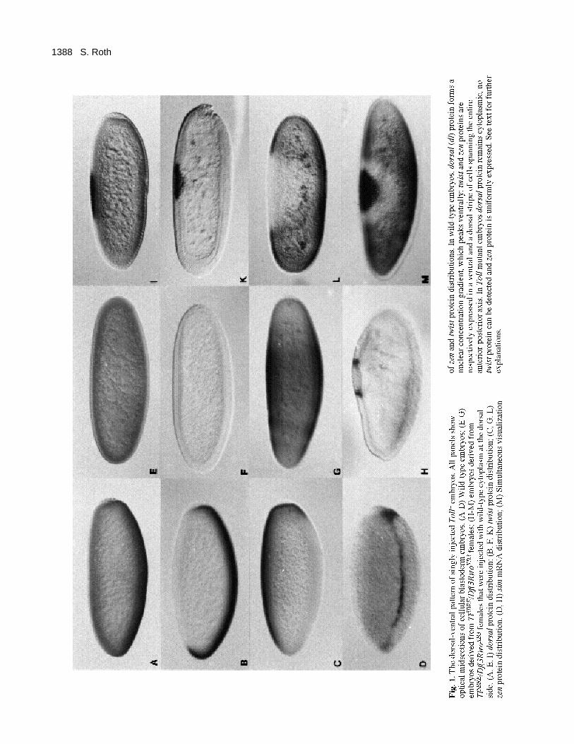
The dorsal-ventral pattern of Toll embryos withone local transplantation of wild-type cytoplasmThe dorsal-ventral pattern of injected embryos was analysedby studying the dorsal (Roth et al., 1989; Rushlow et al.,1989; Steward, 1989), zerknüllt (zen, Rushlow et al., 1987)and twist (twi) protein (Thisse et al., 1988) distributions andthe distribution of single minded (sim) transcripts (Thomaset al., 1988). (See Fig. 1 for expression patterns of thesemarkers in wild-type and Toll embryos.) The transplanta-tion of wild-type cytoplasm to a dorsal site in a Tollembryo leads to a local nuclear accumulation of dl proteinat the site of injection (Fig. 1I). In this region, zen proteinis absent (Fig. 1L) and twist shows a narrow domain ofexpression surrounding the site of injection (Fig. 1K). Iftwist and zen expression are detected simultaneously ininjected embryos (Fig. 1M), the three main subdivisions ofthe induced dorsal-ventral pattern are visible: the regionexpressing twist protein which will give rise to mesoderm,the surrounding region expressing neither zen nor twistwhich will contribute to the neuroectoderm, and finally thearea of the zen-expressing cells which will differentiate asdorsal epidermis and amnioserosa.
To test at the molecular level whether finer subdivisionsof the dorsal-ventral pattern are established in injectedT o l l embryos, the single minded (sim) RNA distributionwas examined. Correct s i m expression is only indirectlydependent on the maternal gradient; it is directly regulatedby the zygotic genes t w i s t and s n a i l (Leptin, 1991; Raoet al., 1991; Kosman et al., 1991). In wild-type embryos,s i m expression is confined to a single line of ventrolateralcells which will form the mesectoderm (Fig. 1D). InjectedT o l l embryos show a single line of sim expression sur-rounding the site of injection (Fig. 1H). This demonstratesthat the pattern induced by cytoplasmic transplantationundergoes processes of refinement similar to the wild-typepattern.
The dorsal protein gradient of injected embryos issteeper than that of wild-type embryosThe size of the twist-expressing region that forms in a Tollembryo after injection depends on the relative amount oftransplanted wild-type cytoplasm. Thus, twist-expressingpatches with diameters between 10 and 50 cells could beproduced. Despite their variability in size, the twist-express-ing patches are always surrounded by a domain withouttwist and zen expression which comprises about 7 cells(Table 1). This domain is considerably smaller than that ofwild-type embryos where it comprises approximately 14cells (Fig. 2B,D). The observed expression patterns werenot significantly influenced by the age of the recipientembryos as long as the injections were performed beforethe beginning of cellularization (early cycle 14, data notshown).
The narrower zen repression domain of injected embryosprobably results directly from a change in the slope of thenuclear dorsal protein gradient. In wild-type embryos, thedorsal protein has highest nuclear concentrations in a ven-tral region which is 12-14 cells wide. Lateral to this regionthe nuclear concentrations decrease rapidly (Rushlow et al.,1989). Within an approximately 16-cell span, they go fromhighest to undetectable levels (Roth et al., 1989). In injectedembryos, this region of decreasing nuclear concentrationscomprises only approximately 8 cells (Fig. 2A,C).
These results demonstrate that domains of twist expres-sion and zen repression whose sizes differ considerablyfrom those found in wild-type embryos, can still be stable.Furthermore, the size of the domain without zen and twistexpression is independent from the size of the twist expres-sion domain. Thus, there is apparently no size regulation atthe time of early zygotic dorsal-ventral pattern formation.Rather, the observed patterns directly reflect the shape ofthe nuclear dorsal protein gradient.
Toll product is present in excess in wild-typeembryosThe injection of a small amount of wild-type cytoplasm(approximately 1% of total egg contents) is sufficient toform a complete set of dorsal-ventral pattern elements in aregion spanning about 50% of the anterior-posterior axis(Anderson et al., 1985b). Therefore, either the activated Tollproduct induces a process of self-enhancement or it is pre-sent in vast excess in wild-type embryos. To address thisquestion, injection experiments were performed withdiluted wild-type cytoplasm. (see Materials and Methods).The cuticle phenotype of the injected embryos was scoredfor the presence of ventral denticles and filzkörper whichserve as markers for ventrolateral and dorsolateral fates,respectively (Fig. 3). The formation of ventral denticles inmore than 50% of the injected embryos can be achievedusing cytoplasm diluted up to15-fold. With 20-fold dilutedcytoplasm no ventral denticles were observed, but dorso-lateral stuctures (filzkörper) still formed. In the case of 10-fold diluted cytoplasm, injected embryos were also stainedfor twist expression. 26 out of 42 injected embryos showeda patch of twist-expressing cells, demonstrating that 10-folddiluted cytoplasm still leads to the formation of the ven-tralmost structures in a majority of injected embryos.
1388 S. Roth

1389Dorsal-ventral pattern formation
The varying degrees of rescue observed after injectionof different amounts of T o l l product show that a gradedrelation exists between T o l l concentration and the numberof dorsal-ventral pattern elements formed in injectedembryos. This result argues against a strong self-enhance-ment process initiated by the T o l l product since such aprocess should cause an all-or-none response rather thana graded response upon injection. It is therefore morelikely that the rescue response reflects a vast excess of
Toll product present in wild-type embryos. Assuming thatToll RNA in the injected cytoplasm is as efficiently trans-lated as in wild type, the local concentration of Toll p r o-tein in wild-type embryos is in approximately 10-foldexcess.
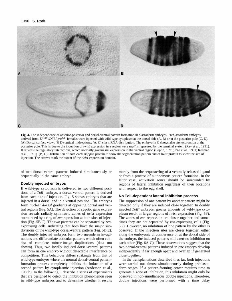
No interaction between dorsal-ventral andanterior-posterior pattern-forming systemsThe shapes of the patterns induced by injection often showradial symmetry (Figs 1M, 5B,C). In Fig. 4A, this propertyis demonstrated using sim RNA as a marker. The radialsymmetry implies that these embryos possess dorsal-ven-tral gradients not only perpendicular, but at all angles tothe anterior-posterior axis. The blastoderm-stage Tollembryo seems to have no axial bias with respect to the ori-entation and spreading of the induced dorsal-ventral pat-tern. This can be demonstrated more dramatically by inject-ing wild-type cytoplasm into the anterior or posterior tip ofToll embryos. Embryos injected posteriorly form a poste-rior cap of twist expression (Fig. 4D) and a sim stripe encir-cling the embryo (Fig. 4C). Therefore, in these embryos thedorsal-ventral axis is parallel to the anterior-posterior axis.Despite this fact, the early anterior-posterior pattern for-mation of dorsally or posteriorly injected embryos is notsignificantly disturbed, since they form seven even-skippedexpression stripes (Frasch et al., 1987) with normal orien-tation (Fig. 4B,D).
In summary, the spatial and temporal properties of thesingle injection experiments demonstrate that, in Tollembryos, all cells of preblastoderm and syncytial blasto-derm stages respond to small amounts of transplanted wild-type cytoplasm in a similar way. The developing patternsshow no preference in orientation with respect to the ante-rior-posterior or dorsal-ventral egg axes. These character-istics, together with the ability to manipulate the size of theinduced patterns, make it feasible to study the interaction
Fig. 2. dorsal protein gradient and zen repression domain ofinjected embryos. (A, B) Transverse sections of wild-typeembryos. (C, D) Transverse sections of embryos derived fromTl5BRE/Df(3R)roXB3 females and injected with wild-typecytoplasm at the dorsal side. For comparison with the wild-typepattern, the injected embryos are oriented with the ventralmostregion of the induced pattern downward. (A, C) Embryos stainedusing dorsal antibodies. Only the lower left (A) or the lower rightsector (C) of the transverse sections is shown to facilitate thecomparison of the relevant parts of the dorsal protein gradients.The arrows indicate the extension of the dorsal protein gradients.(B, D) Embryos stained simultaneously with zen and twistantibodies. (C, D) Embryos injected at preblastoderm stage. Thearrows indicate the extension of the zen repression domain.
Fig. 3. Injections with diluted wild-type cytoplasm. Preblastodermembryos derived from Tl5BRE/Df(3R)roXB3 females were injectedwith nondiluted or diluted wild-type cytoplasm (see Materials andMethods). The different dilutions are plotted on the horizontalaxis using a logarithmic scale. The numbers of injected embryosscored for the differentiation of ventral epidermis (VE) andfilzkörper (Fk) are indicated.
Table 1. The size of the domain without zen and twistexpression is independent of the number of twist-
expressing cellsNumber of cells
Number of twi- Number showing neither zenexpressing cells of embryos nor twi expression
10-20 7 6-8 (average 7.0)20-30 12 5-9 (average 7.0)30-40 7 6-9 (average 7.6)
Preblastoderm embryos derived from Tl5BRE/Df(3R)roXB3 females wereinjected with varying amounts of wild-type cytoplasm, simultaneouslystained with zen and twist antibodies and sectioned. The number of twist-expressing cells and the number of cells expressing neither twist nor zen(zen repression domain) was counted along the embryonic circumference.
1390
of two dorsal-ventral patterns induced simultaneously orsequentially in the same embryo.
Doubly injected embryosIf wild-type cytoplasm is delivered to two different posi-tions of a Toll embryo, a dorsal-ventral pattern is derivedfrom each site of injection. Fig. 5 shows embryos that areinjected in a dorsal and in a ventral position. The embryosform nuclear dorsal gradients at opposing dorsal and ven-tral regions (Fig. 5A). The detection of zygotic gene expres-sion reveals radially symmetric zones of twist expressionsurrounded by a ring of zen repression at both sites of injec-tion (Fig. 5B,C). The two patterns can be separated by zen-expressing cells, indicating that both have the major sub-divisions of the wild-type dorsal-ventral pattern (Fig. 5D,E).The doubly injected embryos form two mesoderm invagi-nations and differentiate cuticular patterns which often con-sist of complete mirror-image duplications (data notshown). Thus, two locally induced dorsal-ventral patternscan form in one embryo without detectable interference orcompetition. This behaviour differs strikingly from that ofwild-type embryos where the normal dorsal-ventral pattern-formation process completely inhibits the induction of asecond pattern by cytoplasmic injection (Anderson et al.,1985b). In the following, I describe a series of experimentsthat are designed to detect the inhibition phenomenon seenin wild-type embryos and to determine whether it results
merely from the sequestering of a ventrally released ligandor from a process of autonomous pattern formation. In thelatter case, activation zones should be surrounded byregions of lateral inhibition regardless of their locationswith respect to the egg shell.
No Toll-dependent lateral inhibition processThe suppression of one pattern by another pattern might bedetected only if they are induced close together. In doublyinjected Toll embryos, greater amounts of wild-type cyto-plasm result in larger regions of twist expression (Fig. 5F).The zones of zen repression are closer together and some-times they are not separated by zen-expressing cells (Fig.5G). However, no inhibition of one pattern by the other isobserved. If the injection sites are closer together, eitheralong the embryonic circumference or at the dorsal side ofthe embryo, the induced patterns still exert no inhibition oneach other (Fig. 6A-C). These observations suggest that thetwo dorsal-ventral patterns induced in one embryo developindependently if far enough apart and overlap if generatedclose together.
In the transplantations described thus far, both injectionswere carried out almost simultaneously during preblasto-derm stages. If a pattern-forming center requires time togenerate a zone of inhibition, this inhibition might only beobserved in non-simultaneous double injections. Therefore,double injections were performed with a time delay
S. Roth
Fig. 4. The independence of anterior-posterior and dorsal-ventral pattern formation in blastoderm embryos. Preblastoderm embryosderived from Tl5BRE/Df(3R)roXB3 females were injected with wild-type cytoplasm at the dorsal side (A, B) or at the posterior pole (C, D).(A) Dorsal surface view; (B-D) optical midsections. (A, C) sim mRNA distribution. The embryo in C shows also sim expression at theposterior pole. This is due to the induction of twist expression in a region were snail is repressed by the terminal system (Ray et al., 1991).It reflects the regulatory interactions, which normally govern sim expression in the ventral region (Leptin, 1991; Rao et al., 1991; Kosmanet al., 1991). (B, D) Distribution of both even-skipped protein to show the segmentation pattern and of twist protein to show the site ofinjection. The arrows mark the extent of the twist-expression domain.

1391Dorsal-ventral pattern formation
betweentransplantations (see Materials and Methods). If thesecond injection was performed before cycle 14, theinduced pattern was not consistently smaller than thatderived from the first injection, regardless of the time delaybetween both injections (Fig. 7). Furthermore, the resultswere not influenced by the site (ventral or dorsal) of thefirst injection. Thus, non-simultaneous double injectionsalso reveal no sign of lateral inhibition.
If the strength of inhibition depends on the size of theinduced pattern, a larger pattern might suppress the devel-opment of a smaller pattern. Furthermore, since inhibitionwas observed only in wild-type embryos, patterns approach-ing the size of the wild-type pattern might be required. InFig. 8, an injection technique is described that allows theformation of stripe-like regions of twist expression in
injected Tl embryos (see also Materials and Methods). Ifstripes are produced that extend along the entire anterior-posterior axis, wild-type-like embryos can be generated. Inthe case of the delivery of cytoplasm in a stripe along thedorsal midline, the embryos develop in the egg case withreversed orientation. 50% of the ‘upside down’ embryosshow a complete or almost complete restoration of thedorsal-ventral cuticle pattern (Fig. 8D).
If embryos with a dorsal stripe of injected cytoplasm alsoreceive a ventral spot-like injection, both the dorsal stripeand the ventral spot of cytoplasm give rise to twist-express-ing regions (Fig. 8E,F; Table 2). The larger dorsal patternwas never observed to repress the smaller ventral pattern,indicating that there is no detectable repressive activitydependent on the size of the pattern. This result holds also
Fig. 5. Embryos with two dorsal-ventral patterns. Preblastoderm embryos derived from Tl5BRE/Df(3R)roXB3 females were injectedsimultaneously at the dorsal and ventral side with wild-type cytoplasm. (A) dorsal protein distribution; (B-G) zen and twist proteindistribution. (A) Optical midsection; (B) dorsal surface view; (C) ventral surface view of the embryo shown in B; (D, F) opticalmidsections; (E, G) lateral surface views of the respective embryos shown in D and F.

1392
when the ventral injection was performed substantially laterthan dorsal injection (data not shown). Thus, although theseembryos can form a wild-type-like pattern, they do notbehave like wild-type embryos with respect to a secondcytoplasmic injection.
An inhibition dependent on the orientation withrespect to the egg shellThe embryos injected with a dorsal stripe of cytoplasmdiffer in one major respect from wild-type embryos: theirdorsal-ventral pattern forms inside the egg shell with oppo-site orientation as compared to wild type. To reconstructthe wild-type situation fully, I also produced Toll embryoswith a ventral stripe of wild-type cytoplasm (Fig. 8G).Despite some experimental constraints (see Materials andMethods), double injections with a ventral stripe and dorsalspot of cytoplasm lead to results significantly different fromthe experiments with the reverse orientation. While a dorsalstripe never inhibited twist induction on the opposite side,in 38% of the embryos with a ventral stripe, a second dorsalinjection failed to induce a zone of twist expression (Table2). Interestingly, a correlation could be seen between theanterior-posterior extension of the ventral twist stripe andthe ability to induce a dorsal pattern. The inhibition wasusually only observed if the twist stripe extended along the
entire anterior-posterior axis (compare Fig. 8G and H).Therefore, the inhibition of a second pattern seems todepend on both the size and orientation of the other patternwith respect to the egg shell. In summary, the observedinhibition phenomenon is not an intrinsic property of thepattern-formation process inside the embryo, but ratherreflects a strong spatial asymmetry present in the vitellinemembrane or the perivitelline space.
Not only the Toll protein, but also the Toll ligandis in excessInterestingly, the doubly injected embryos often have moretwist-expressing cells than wild-type embryos. 15 embryosinjected with a dorsal stripe and a ventral spot of cytoplasmwere subsequently sectioned to estimate the size of theinduced mesodermal regions. In 13 of the sectionedembryos, the dorsal twist stripe was 25 to 30 cells wide and
S. Roth
Fig. 6. Variation of the distance between sites of injection. Tollembryos were injected at two different sites with wild-typecytoplasm and later stained with twist antibodies. (A) Embryowith a dorsal and a lateral injection. (B) Embryo with two dorsalinjections placed closely together. (C) The injections wereperformed as in B, but larger amounts of cytoplasm weretransplanted. The twist expression zones derived from the twosites of injections have fused.
100
80
60
40
20
0
n=23 n=33 n=30 n=75n=30
n=18
n=25
0
10 11 12 13 14
20 40 60 80 100
Time of injection in min after pole cell formation at 18°C
Beginning of cycle
Beginning ofcellularization
Fig. 7. Non-simultaneous double injections. Toll embryos wereinjected with wild-type cytoplasm at a dorsal and a ventralposition with a delay between injections. The first injection wasperformed before pole cell formation, the second at different timeintervals after pole cell formation. The doubly injected embryoswere stained using twist and zen antibodies. For each time intervalthe number of embryos scored for a second patch of twist-expressing cells is indicated. Below the graph: Lateral surfaceview of a doubly injected embryo with two dorsal-ventral patternsof similar size. The ventral injection was performed 60 minutesafter the dorsal injection.

1393Dorsal-ventral pattern formation
thus broader than the 20- to 22-cell-wide twist expressionzone of wild-type embryos. Fig. 8F shows an example inwhich the dorsal twist-expressing zone constitutes as muchas 60% of the embryonic circumference (55 cells). If the
zone of twist expression formed by the ventral spot-likecytoplasmic injections is also included, the total area oftwist expression is often considerably larger than in wild-type embryos. The ability to induce more ventral structures
Fig. 8. Double injections with stripe-like deposition of cytoplasm. (A, B) The injection technique. (A) The injection of a dorsal stripe ofcytoplasm is followed by a ventral spot-like injection. (B) The injection of a ventral stripe of cytoplasm is followed by a dorsal spot-likeinjection. (C, E, F, G, H) Injected Toll embryos stained using twist antibodies. (C, D) Toll embryo injected with a dorsal stripe of wild-type cytoplasm. (C) The mesoderm formation occurs dorsally and the germband extends towards the ventral side. (D) Dark-fieldphotograph of the cuticle produced by an injected embryo. The cuticle pattern resembles that of a wild-type larva. The larva is orientedwith the ventral side down, although it developed inside the egg shell in the opposite direction. (E, F) Toll embryos injected with a dorsalstripe of wild-type cytoplasm followed by a ventral spot-like injection as depicted in (A). (E) Optical midsection. (F) Transverse section.Arrows indicate the extension of the twist-expressing regions. In this specific case the dorsal twist-expressing region comprises about 60%of the embryonic circumference. (G, H) Toll embryos injected with a ventral stripe of cytoplasm followed by a dorsal spot-like injectionas depicted in B. (G) In this embryo, both injections cause regions of twist expression. (H) In this embryo, the dorsal injection does notlead to a region of twist expression.
A

1394
than present in a wild-type embryo suggests that, like theToll receptor, the ligand of Toll is present in excess (forfurther explanation see discussion).
DISCUSSION
The pattern-forming capacity of the dorsal-ventralsystem in Drosophila embryosThe existence of an autonomous pattern-formation processthat controls the dorsal-ventral axis formation in lowerinsects has been suggested on the basis of classical embry-ological studies (Sander, 1976). For example, in experi-ments with the cicada Euscelis, longitudinal ligations sep-arating left and right or dorsal and ventral egg halves causedpattern duplications producing two complete embryos fromthe two separated fragments (Sander, 1971). These self-reg-ulatory properties not only rule out the existence of prelo-calized cytoplasmic determinants, but also are incompati-ble with a strict dependence of the embryonic dorsal-ventralaxis on a prepattern present in the extraembryonic com-partment. However, they can be explained by a mechanismof lateral activation of mutually exclusive states (Meinhardtand Gierer, 1980) or by a local activation/lateral inhibitionmechanism (Meinhardt, 1989).
As shown by perivitelline fluid injections, the extracel-lular signal that is required to establish the dorsal-ventralaxis of Drosophila embryos has a polarizing influence andthus confers some spatial information (Stein et al., 1991).From these experiments, however, it is not clear whetherthe extracellular spatial cues fully contain the spatial infor-mation present in the final pattern. The situation could besimilar to the cell fate determination of the vulval precur-sor cells in C. elegans, which requires both a graded induc-tive signal from the anchor cell and a lateral inhibitionbetween the cells receiving the signal (for review seeHorvitz and Sternberg, 1991). Therefore, a Toll-dependentlocal activation/lateral inhibition process might exist. Thespatial cues present in the vitelline membrane or periv-itelline space may trigger a process, which then would leadautonomously to the elaboration of the final pattern.
However, the described experiments presented here failto demonstrate either a Toll-dependent local self-enhance-ment process or a Toll-dependent lateral inhibition mecha-
nism. Only Toll embryos that had received a ventral stripe-like injection of wild-type cytoplasm show an inhibitionphenomenon. If the process under investigation has anautonomous pattern-forming capacity, its behaviour shouldnot depend strongly on orientation with respect to the eggshell. My results argue against an autonomous pattern-for-mation mechanism occurring at the level of Toll activationor downstream of Toll. Therefore, it is unlikely thatDrosophila embryos possess the regulatory capacity foundfor the dorsal-ventral pattern-formation process in moreprimitive insects (Sander, 1976).
The transfer of spatial information from theextraembryonic compartment to the embryoThe failure to detect autonomous pattern formation,strongly supports the previously proposed model (Stein etal., 1991; Schüpbach et al., 1991) that the Toll ligand isreleased from a restricted ventral region of the perivitellinespace and that its amount is limited with respect to thenumber of Toll-binding sites. Under these conditions, bind-ing and sequestering of the ligand prevents its furtherspreading to more lateral regions thereby limiting the sizeof the activation zone. This mechanism attributes two func-tions to the Toll receptor: Toll is not only required to trans-mit the extracellular signal to the cytoplasm, but Toll, uni-formly distributed in the plasma membrane, is alsonecessary to localize the extracellular signal to the ventralside.
A similar model has been proposed for the terminalsystem of the anterior-posterior axis. Here, the torso pro-tein, a receptor tyrosine kinase, is uniformly distributed inthe plasma membrane (Sprenger et al., 1989; Casanova andStuhl, 1989). The torso receptor binds a putative extracel-lular ligand, which is released from the terminal region ofthe perivitelline space or the vitelline membrane (Stevenset al., 1990; Stevens and Nüsslein-Volhard, 1991). Theinjection of torso RNA into central regions of torso−
embryos leads to the formation of terminal structures at thesite of injection (Sprenger and Nüsslein-Volhard, 1992).Similar results have been obtained by a non-uniformexpression of torso protein in torso− embryos (Casanovaand Struhl, 1993). Thus, as in the dorsal-ventral system, theligand for the terminal system can diffuse and its localiza-tion to the terminal regions depends on a uniform distrib-ution of the receptor protein.
If this model applies, receptor density and amount ofligand are important parameters. The Toll gene shows nodetectable dosage sensitivity (Anderson et al., 1985a)implying that Toll product is present at least in two-foldexcess. The dilution experiments with wild-type cytoplasmindicate that Toll is in approximately 10-fold excess. Supris-ingly, it seems that not only the Toll receptor, but also theligand of Toll is produced in excess. By injection of dorsalstripes of cytoplasm, embryos can be produced that havesignificantly more twist-expressing cells than wild-typeembryos (Fig. 8F). Since twist expression requires theligand-dependent activation of Toll, the increased amountsof mesodermal cells indicate that more ligand is presentthan is necessary to form a mesoderm of normal size. Underinjection conditions, the density of Toll protein in theplasma membrane may be lower than in wild type, so that
S. Roth
Table 2. Double injections with stripe-like depositions ofcytoplasm
Number of embryos with a spot-like secondinjection resulting in
Spot-like No Notwist twist assignable
expression expression pattern
Dorsal stripe 40 (72%) 0 (0%) 16 (28%)ventral spot
Ventral stripe 46 (47%) 37 (38%) 14 (14%)dorsal spot
For explanation of injection technique see Fig. 8. ‘no assignable pattern’includes embryos with an irregular twist-expression domain that mighthave formed by a fusion of the dorsally and ventrally induced patterns.

1395Dorsal-ventral pattern formation
the same amount of ligand may activate a larger area ofreceptor molecules. This implies that at the ventral side ofa wild-type embryo more ligand is bound than is necessaryto achieve the highest nuclear concentrations of dorsal pro-tein. Thus, it is not the absolute amount of the ligand, butthe relative concentrations of ligand and receptor moleculesthat determine the size of the activation zone.
The final shape of the nuclear dorsal protein gradient,and especially its slope, might depend on events occurringat several different levels. Gradient formation by diffusioncould occur downstream of Toll in the cytoplasm orupstream of Toll in the perivitelline space. It is even pos-sible that the slope of dorsal gradient is already determinedby spatial cues in the vitelline membrane. If gradient for-mation occurred entirely downstream of Toll in the cyto-plasm, the nuclear gradient of injected embryos should haveeither the same slope as in wild-type embryos or a reducedslope due to the lateral diffusion of Toll RNA in the cyto-plasm or Toll receptor molecules in the plasma membrane.The fact that the dorsal gradient of injected embryos issteeper than in wild type strongly suggests that extracellu-lar events contribute to the gradient formation in wild-typeembryos.
The role of oogenesis and the perpendicularorientation of the body axesSince no active Toll-dependent mechanisms of pattern-sharpening seem to govern the formation of the nucleardorsal protein gradient, the question arises how the spatialinformation contained in the extracellular signal for Toll isgenerated. Given the apparent precision of this extracellu-lar prepattern, it is likely that processes upstream of Tollexist that have properties of autonomous pattern-formationsystems. These processes may involve activities of dorsalgroup genes, which are necessary to produce the ligand.They may occur in the perivitelline space after egg depo-sition or in the follicular epithelium during oogenesis. Inthe latter case, they would be responsible for defining theventral region of the vitelline membrane where the pro-duction of the ligand can be initiated. In fact, pattern dupli-cations in embryos that are derived from ventralized tor -pedo, gurken or cornichon eggs suggest the existence of anautonomous pattern-formation system operating upstreamof Toll (Roth et al., unpublished data).
A feature of the dorsal-ventral patterns induced in Tollembryos is the complete lack of a bias that restricts theirorientation with respect to the anterior-posterior axis. Itseems that the blastoderm embryo is isotropic with respectto the orientation of dorsal-ventral gradients. Thus, no gen-eral mechanism operates in blastoderm embryos thataccounts for or stabilizes the perpendicular orientation ofthe body axes and the interpretation of dorsal-ventral andanterior-posterior positional information occurs largelywithout interference. This idea has been proposed previ-ously based on the observation that the majority of mater-nal-effect and zygotic mutations disrupt either the anterior-posterior or the dorsal-ventral pattern (Anderson andNüsslein-Volhard, 1984b; Nüsslein-Volhard et al., 1987).
An interesting exception is the interaction between ter-minal and dorsal-ventral system, which influences theexpression pattern of dorsal-ventral zygotic genes in termi-
nal regions of the embryo (see for example the terminal simexpression in Fig. 4C; Ray et al., 1991; Casanova, 1991).However, this exception does not affect the general con-clusion: the Cartesian coordinate system reflected in theexpression pattern of early zygotic genes depends strongly,if not entirely, on the correct localization within the egg ofthe RNAs that determine the anterior and posterior pattern(for review see St Johnston and Nüsslein-Volhard, 1992)and on the correct positioning in the egg shell of the extra-cellular signals that determine the terminal and dorsal-ven-tral pattern. The process of axis orientation relies, therefore,on a not yet understood pattern-formation system, whichoperates during oogenesis and determines with high preci-sion the 90° angle between the dorsal-ventral and anterior-posterior axes by defining the relative positions of cyto-plasmic determinants and extracellular signals in thegrowing egg.
This work was started in the lab of C. Nüsslein-Volhard at theMax-Planck-Institut für Entwicklungsbiologie in Tübingen andfinished in the lab of T. Schüpbach in Princeton. I wish to thankC. Nüsslein-Volhard and T. Schüpbach for support and stimulat-ing discussions. I am grateful to H. Meinhardt and D. Stein fornumerous discussions. I wish to thank the following for their con-tributions to this work. C. Rushlow, for providing the zen anti-body. M. Frasch, for providing the even-skipped antibody. S. T.Crews, for the gift of sim DNA. A. Bejsovec, M. Costa, M. Kon-solaki, M. Postner, C. A. Rushlow, E. Schejter, and T. Schüpbachfor discussions and critical reading of the manuscript. I was sup-ported by a fellowship from the Human Frontiers Science Pro-gram.
REFERENCES
Anderson, K. V., Jürgens, G. and Nüsslein-Volhard, C. (1985a).Establishment of dorsal-ventral polarity in the Drosophila embryo:genetic studies on the role of the Toll gene product. Cell 42, 779-789.
Anderson, K.V., Bokla, L. and Nüsslein-Volhard, C. (1985b).Establishment of dorsal-ventral polarity in the Drosophila embryo: theinduction of polarity by the Toll gene product. Cell 42, 791-798.
Anderson, K. V. and Nüsslein-Volhard, C. (1984a). Information for thedorsal-ventral pattern of the Drosophila embryo is stored as maternalmRNA. Nature 311, 223-227.
Anderson, K. V. and Nüsslein-Volhard, C. (1984b). Genetic analysis ofdorsal-ventral embryonic pattern in Drosophila. In Pattern Formation: APrimer in Developmental Biology (ed. G. M. Malacinski and S. V.Bryant), pp. 269-289. New York: Macmillan.
Casanova, J. (1991) Interaction between torso and dorsal, two elements ofdifferent transduction pathways in the Drosophila embryo. Mechan. Dev36, 41-45.
Casanova, J. and Struhl, G. (1989). Localized surface activity of torso, areceptor tyrosine kinase, specifies terminal body patterns in Drosophila.Genes Dev 3, 2025-2038.
Casanova, J. and Struhl, G. (1993). The torso rreceptor localizes as well astransduces the spatial signal specifying terminal body pattern inDrosophila Nature (In press).
Chasan, R. and Anderson, K. V. (1989). The role of easter, an apparentserine protease, in organizing the dorsal-ventral pattern of the Drosophilaembryo. Cell 56, 391-400.
Chasan, R., Jin, Y. and Anderson, K. V. (1992). Activation of the easterzymogen is regulated by five other genes to define dorsal-ventral polarityin the Drosophila embryo. Development 115, 607-616.
DeLotto, R. and Spierer, P. (1986). A gene required for the specification ofdorsal-ventral pattern in Drosophila appears to encode a serine protease.Nature 323, 688-692.
Frasch, M., Hoey, T., Rushlow, C., Doyle, H. and Levine, M. (1987).

1396
Characterization and localization of the even-skipped protein inDrosophila embryogenesis. EMBO J. 6, 749-759.
Gierer, A. and Meinhardt, H. (1972). A theory of biological patternformation. Kybernetik 12, 30-39.
Govind, S. and Steward, R. (1991). Dorsoventral pattern formation inDrosophila: signal transduction and nuclear targeting. Trends in Genet.7,119-125.
Hashimoto, C., Gertulla, S. and Anderson, K. V. (1991) Plasmamembrane localization of the Toll protein in the syncytial Drosophilaembryo: importance of transmembrane signaling for dorsal-ventralpattern formation. Development 111, 1021-1028.
Hashimoto, C., Hudson, K. L. and Anderson, K. V. (1988). The Toll geneof Drosophila, required for dorsal-ventral embryonic polarity, appears toencode a transmembrane protein. Cell 52, 269-279.
Horvitz, R. H. and Sternberg, P. W. (1991). Multiple intercellularsignalling systems control the development of the Caenorhabditiselegans vulva. Nature 351, 535-541.
Ip, Y. T., Kraut, R., Levine, M. and Rushlow, C. A. (1991). The dorsalmorphogen is a sequence-specific DNA-binding protein that interactswith a long-range repression element in Drosophila. Cell 64, 439-446.
Jiang, J., Kosman, D., Ip, Y.T. and Levine, M. (1991). The dorsalmorphogen gradient regulates the mesoderm determinant twist in earlyDrosophila embryos. Genes Dev. 5, 1881-1891.
Kosman, D., Ip, Y. T., Levine, M. and Arora, K. (1991). Establishment ofmesoderm-neuroectoderm boundary in the Drosophila embryo. Science254, 118-122.
Leptin, M. (1991). twist and snail as positive and negative regulators duringDrosophila mesoderm development. Genes Dev. 5, 1568-1576.
MacDonald, P. M. and Struhl, G. (1986). A molecular gradient in earlyDrosophila embryos and its role in specifying the body pattern. Nature324, 537-545.
Meinhardt, H. (1989). Models for positional signaling with application tothe dorsoventral patterning of insects and segregation into different celltypes. Development 107 Supplement, 169-180.
Meinhardt, H. and Gierer, A. (1980). Generation and regeneration ofsequences of structures during morphogenesis. J. Theor. Biol. 85, 429-450.
Nüsslein-Volhard, C., Frohnhofer, H. G. and Lehmann, R. (1987).Determination of anteroposterior polarity in Drosophila. Science 238,1675-1681.
Pan, D., Huang, J.-D. and Courey, A. J. (1991). Functional analysis of theDrosophila twist promotor reveals a dorsal-binding ventral activatorregion. Genes Dev. 5, 1892-1901.
Rao, Y., Vaessin, H., Jan, L. Y. and Jan, Y.-N. (1991). Neuroectoderm inDrosophila embryos is dependent on the mesoderm for positioning butnot for formation. Genes Dev. 5, 1577-1588.
Ray, R. P., Arora, K., Nüsslein-Volhard, C. and Gelbart, W. (1991) Thecontrol of cell fate along the dorsal-ventral axis of the Drosophila embryo.Development 113, 35-54.
Roth, S., Stein, D. and Nüsslein-Volhard, C. (1989). A gradient of nuclearlocalization of the dorsal protein determines dorsoventral pattern in theDrosophila embryo. Cell 59, 1189-1202.
Rushlow, C. A., Frasch, M., Doyle, H. and Levine, M. (1987). Maternalregulation of zerknüllt: a homeobox gene controlling differentiation ofdorsal tissues in Drosophila. Nature 330, 583-586.
Rushlow, C. A., Han, K., Manley, J. L. and Levine, M. (1989). Thegraded distribution of the dorsal morphogen is initiated by selectivenuclear transport in Drosophila. Cell 59, 1165-1177.
Sander, K. (1971). Pattern formation in longitudinal halves of leaf hoppereggs (Homoptera) and some remarks on the definition of ‘embryonicregulation’. Wilhelm Roux´s Arch. EntwMech. Org. 167, 336-352.
Sander, K. (1976). Specification of the basic body pattern in insectembryogenesis. Adv. Ins. Physiol.12, 125-238.
Santamaria, P. and Nüsslein-Volhard, C. (1983). Partial rescue of dorsal,a maternal effect mutation affecting the dorso-ventral pattern of theDrosophila embryo, by the injection of wild-type cytoplasm. EMBO J. 2,1695-1699.
Schneider, D. S., Hudson, K. L., Lin, T.-Y. and Anderson, K. V. (1991).Dominant and recessive mutations define functional domains of Toll, atransmembrane protein required for dorsal-ventral polarity in theDrosophila embryo. Genes Dev. 5, 797-807.
Schüpbach, T. (1987). Germ line and soma cooperate during oogenesis toestablish the dorsoventral pattern of egg shell and embryo in Drosophilamelanogaster. Cell 49, 699-707.
Schüpbach, G., Clifford, R. J., Manseau, L. J. and Price, J. V. (1991).Dorsoventral signal processes in Drosophila oogenesis. In Cell-CellInteractions in Early Development (ed. J. Gerhart), pp 163-174. NewYork: Wiley-Liss Inc.
Sprenger, F. and Nüusslein-Volhard, C. (1992). Torso receptor activity isregulated by a diffusible ligand produced at the extracellular terminalregions of the Drosophila egg. Cell 71, 987-1001.
Sprenger, F., Stevens, M.L. and Nüsslein-Volhard, C. (1989). TheDrosophila gene torso encodes a putative receptor tyrosine kinase.Nature 338, 478-483.
Stein, D. and Nüsslein-Volhard, C. (1992). Multiple extracellularactivities in Drosophila egg perivitelline fluid are required forestablishment of embryonic dorsal-ventral polarity. Cell 68, 429-440.
Stein, D., Roth, S., Vogelsang, E. and Nüsslein-Volhard, C. (1991). Thepolarity of the dorsoventral axis in the Drosophila embryo is defined byan extracellular signal. Cell 65, 725-735.
Stevens, L. and Nüsslein-Volhard, C. (1991). Development of the terminalanlagen of the Drosophila embryo depends upon interactions between thegermline and the somatic follicle cells. In Cell-Cell Interactions in EarlyDevelopment (ed. J. Gerhart), pp. 145-162. New York: Wiley-Liss. Inc.
Stevens, L., Frohnhöfer, H., Klingler, M. and Nüsslein-Volhard, C.(1990). Localized requirement for torsolike expression in follicle cells forthe development of terminal anlagen of the Drosophila embryo. Nature346, 660-663.
Steward, R.(1989). Relocalization of the dorsal protein from the cytoplasmto the nucleus correlates with its function. Cell 59, 1179-1188.
Steward, R., Zusman, S. B., Huang, L. H. and Schedl, P. (1988). Thedorsal protein is distributed in a gradient in early Drosophila embryos.Cell 55, 487-495.
StJohnston, R. D. and Nüsslein-Volhard, C. (1992).The origin of patternand polarity in the Drosophila embryo. Cell 68, 1-20.
Tautz, D. and Pfeifle, D. (1989). A nonradioactive in situ hybridizationmethod for the localization of specific RNAs in Drosophila embryosreveals a translational control of the segmentation gene hunchback.Chromosoma 98, 81-85.
Thisse, C., Perrin-Schmitt, F., Stoetzel, C. and Thisse, B. (1991).Sequence-specific transactivation of the Drosophila twist gene by thedorsal gene product. Cell 65, 1-20.
Thisse, B., Stoetzel, C., Gorostiza-Thisse, C. and Perrin-Schmitt, F.(1988). Sequence of the twist gene and nuclear localization of its proteinin endomesodermal cells of early Drosophila embryos. EMBO J. 7, 2175-2183.
Thomas, J. B., Crews, S. T. and Goodman, C. S. (1988). Moleculargenetics of the single-minded locus: A gene involved in the developmentof the Drosophila nervous system. Cell 52, 133-141.
Van der Meer, J. M. (1977). Optical clean and permanent whole mountpreparations for phase-contrast microscopy of cuticular structures ofinsect larvae. Dros. Info. Serv. 52, 160.
(Accepted 8 January 1993)
S. Roth