mechanism of amphotericin b resistance in clinical...
TRANSCRIPT
Mechanism of Amphotericin B Resistance in Clinical Isolatesof Leishmania donovani
Bidyut Purkait,a Ashish Kumar,a Nilay Nandi,c Abul Hasan Sardar,a Sushmita Das,a Sudeep Kumar,d Krishna Pandey,b
Vidyananda Ravidas,b Manish Kumar,a Tripti De,e Dharmendra Singh,a and Pradeep Dasa
Department of Molecular Biologya and Department of Clinical Medicine,b Rajendra Memorial Research Institute of Medical Sciences, Agamkuan, Patna, Bihar, India; UTSouthwestern Medical Center, Dallas, Texas, USAc; Institute of Global Health, Internal Medicine, University of New Mexico, Albuquerque, New Mexico, USAd; and IndianInstitute of Chemical Biology, Kolkata, Indiae
The clinical value of amphotericin B, the mainstay therapy for visceral leishmaniasis in sodium antimony gluconate-nonresponsive zones of Bihar, India, is now threatened by the emergence of acquired drug resistance, and a comprehensive un-derstanding of the underlying mechanisms is the need of the hour. We have selected an amphotericin B-resistant clinical isolatewhich demonstrated 8-fold-higher 50% lethal doses (LD50) than an amphotericin B-sensitive strain to explore the mechanism ofamphotericin B resistance. Fluorimetric analysis demonstrated lower anisotropy in the motion of the diphenylhexatriene fluo-rescent probe in the resistant strain, which indicated a higher fluidity of the membrane for the resistant strain than for the sensi-tive strain. The expression patterns of the two transcripts of S-adenosyl-L-methionine:C-24-�-sterol methyltransferase and theabsence of ergosterol, replaced by cholesta-5,7,24-trien-3�-ol in the membrane of the resistant parasite, indicate a decreasedamphotericin B affinity, which is evidenced by decreased amphotericin B uptake. The expression level of MDR1 is found to behigher in the resistant strain, suggesting a higher rate of efflux of amphotericin B. The resistant parasite also possesses an up-regulated tryparedoxin cascade and a more-reduced intracellular thiol level, which helps in better scavenging of reactive oxygenspecies produced by amphotericin B. The resistance to amphotericin B was partially reverted by the thiol metabolic pathway andABC transporter inhibitors. Thus, it can be concluded that altered membrane composition, ATP-binding cassette transporters,and an upregulated thiol metabolic pathway have a role in conferring amphotericin B resistance in clinical isolates of Leishma-nia donovani.
Leishmaniasis, or kala azar, is a group of diseases caused by aprotozoan parasite of the genus Leishmania. Kala azar is a
symptomatic infection of liver, spleen, and bone marrow. Theglobal estimates for the incidence and prevalence of kala azar casesper year are 0.5 and 2.5 million, respectively (57). In India, visceralleishmaniasis (VL) has been reported in parts of West Bengal,Uttar Pradesh, and Bihar. It poses a major health problem in thestate of Bihar, which accounts for nearly 90% of the total cases inIndia (51). Chemotherapy has proven to be the only effective wayof controlling infections and is highly dependent upon antimony-containing drugs, such as sodium stibogluconate (Pentostam).Amphotericin B (AmB) is used as the drug of choice when ac-quired drug resistance emerges for traditional antimony therapy,and nearly 64% of cases in regions of Bihar where VL is hyperen-demic are now resistant to antimonials (28). Hexadecylphospho-choline (miltefosine) is the first orally administered drug for VLand the latest to enter the market. The dosing scheme of hexade-cylphosphocholine is 100 mg/kg of body weight/day for 28 days inadults weighing �50 kg, 50 mg/kg/day in adults weighing �50 kg,and 2.5 mg/kg/day in children (maximum dose, 100 mg/day).Major concerns about the wide use of hexadecylphosphocholineinclude its teratogenic potential and its long half-life (approxi-mately 150 h), which may facilitate the emergence of resistance.This agent is associated with high efficacy rates, even in cases un-responsive to antimonials (14, 49). Paromomycin (aminosidine)is an aminoglycoside with antileishmanial activity. In a phase IIIstudy of VL in India, this drug was associated with 94.6% curerates, similar to that of amphotericin B (54). Adverse effects weremore frequent in the paromomycin-treated group than in theamphotericin B-treated group (6% versus 2%, respectively);
paromomycin-related adverse effects included elevated hepatictransaminases, ototoxicity, and pain at the injection site (54).Combinations of hexadecylphosphocholine with AmB, paromo-mycin, or pentavalent antimonials have been evaluated in an invivo model, which revealed that the combinations of hexadecyl-phosphocholine with AmB or paromomycin were efficacious(50).
AmB is a polyene antifungal drug often used intravenously forsystemic fungal infections. It was originally extracted from Strep-tomyces nodosus, a filamentous bacterium (33, 15). Currently, thedrug is available as plain AmB, as a cholesteryl sulfate complex, asa lipid complex, and as a liposomal formulation (33). The latterformulations have been developed to improve tolerability for thepatient but may show pharmacokinetic characteristics consider-ably different from those of plain AmB (33, 46).
The mechanism(s) of AmB resistance among fungi varies. Theprimary target of AmB is ergosterol in the cell membrane of fungi.The binding of AmB to sterols (ergosterol-containing membranesare more sensitive than cholesterol-containing membranes [9])
Received 10 January 2011 Returned for modification 23 March 2011Accepted 30 October 2011
Published ahead of print 28 November 2011
Address correspondence to Pradeep Das, [email protected].
B. Purkait and A. Kumar contributed equally to this article.
Supplemental material for this article may be found at http://aac.asm.org/.
Copyright © 2012, American Society for Microbiology. All Rights Reserved.
doi:10.1128/AAC.00030-11
0066-4804/12/$12.00 Antimicrobial Agents and Chemotherapy p. 1031–1041 aac.asm.org 1031
on June 11, 2018 by guesthttp://aac.asm
.org/D
ownloaded from

incorporated in artificial or cellular membranes results in disor-ganization of the membrane (5), possibly by formation of specificpores composed of small aggregates of AmB and sterol (56). Thesedefects cause depolarization of the membrane and an increase inmembrane permeability for protons and monovalent cations (8,32). Recently, however, researchers found evidence that pore for-mation is not necessarily linked to cell death (2). The actual mech-anism of action of AmB may be more complex and multifaceted.Another mechanism by which AmB could affect the cells is itsauto-oxidation and subsequent formation of free radicals (32).AmB-induced cell injury might be associated with both ion move-ment and oxidative effects (8). If AmB-induced cell damage islinked to the generation of reactive forms of oxygen, the ability todecompose them should affect cell resistance to damage (7, 4).Kinetoplastids, comprising parasites of the genera Crithidia,Trypanosoma, and Leishmania, are devoid of catalase and gluta-thione peroxidase (6, 17, 19), and the removal of hydroperoxidesin these parasites is presumed to rely mainly on the tryparedoxinpathway to regulate oxidative stress (39, 44). This pathway con-sists of a cascade of low-molecular-weight, thiol-specific oxi-doreductases acting in an NADPH-dependent manner to detoxifyperoxides in the following order: trypanothione reductase (TR) ¡trypanothione (TSH) ¡ tryparedoxin ¡ tryparedoxin peroxi-dase (11, 55). In antimonial resistance, it was shown that overex-pression of tryparedoxin peroxidase is linked with resistanceagainst oxidative stress (36). In trypanosomatids, a reduced formof trypanothione is involved in the maintenance of the intracellu-lar reducing environment, substituting for the glutathione (GSH)found in most other organisms (18). The enzyme trypanothionereductase, absent from mammalian cells, is mainly responsible forkeeping trypanothione reduced (17, 31). The rate-limiting en-zymes for trypanothione biosynthesis are �-glutamylcysteine syn-thetase (�-GCS; encoded by the gsh1 gene) and ornithine decar-boxylase (ODC) (47). In Leishmania tarentolae isolates resistant toarsenite, buthionine sulfoximine (BSO), an inhibitor of �-GCS,can partially revert the resistance phenotype (24, 47). Also, treat-ment of a glucantime-resistant Leishmania tropica line with BSOproduced a thiol depletion that was accompanied by a substantialincrease in the chemosensitivity to glucantime (1).
The ATP-binding cassette (ABC) transporters represent thebiggest known superfamily of proteins, being present in all studiedorganisms, from archaebacteria to higher eukaryotes (26). In ad-dition to their physiological function, translocating a high varietyof substrates across the cellular membrane, ABC proteins haveenormous medical relevance. Some of them are responsible forthe multidrug resistance (MDR) phenotype during the treatmentof cancer and infectious diseases, and others are involved in im-portant genetic diseases. In Leishmania spp., three different classesof ABC transporters are known. It has been reported that twotypes of ABC transporters are involved in drug resistance mecha-nisms in Leishmania spp. (47): P-glycoprotein A (PgPA), which ishomologous with the mammalian MDR-associated protein(MRP) cluster (involved in drug sequestration) (45), and MDR1,which is homologous with the mammalian PgP cluster (involvedin drug efflux) (25). It has also been demonstrated that cotrans-fection of gsh1 and PgPA in the revertant resulted in resistancelevels that were higher than expected from the individual contri-bution of either gene (24).
Although AmB chemotherapy has been proven to be very suc-cessful in treatment of VL in India, due to the very high frequency
of its use, emergence of drug-resistant cases is expected (53). Wehave encountered some AmB-unresponsive cases at the RajendraMemorial Research Institute of Medical Sciences (RMRIMS), Bi-har, India. Microbiological evolution of one such clinical isolateshowed resistance in in vitro as well as ex vivo studies. Until now,no study of any AmB-resistant clinical isolate to understand themechanism of resistance has been done. Therefore, the major ob-jective of the present investigation is to understand the molecularmechanism of AmB resistance of the clinical isolate by investigat-ing the involvement of membrane composition, drug efflux ma-chinery, and the peroxide elimination cascade using clinical iso-lates of Leishmania donovani.
MATERIALS AND METHODSClinical isolates. Clinical isolates of VL were obtained from the splenicaspirates of AmB-responsive and -nonresponsive patients in the indoorward facility of Rajendra Memorial Research Institute of Medical Sci-ences, Patna, Bihar, India. Briefly, the collected splenic aspirates wereincubated in RPMI-1640 medium (Gibco) (pH 7.4) supplemented with10% fetal bovine serum (FBS) (Gibco) and 1% of penicillin (50 U/ml)-streptomycin (50 mg/ml) solution (Sigma) at 25°C. The amastigotes fromsplenic aspirates were transformed into promastigotes, and they weremaintained further in RPMI-1640 medium supplemented with FBS.
Cell line. The THP1 cell line was maintained in a 25-cm2 flask (Corn-ing) in Dulbecco’s modified Eagle medium (DMEM) (Sigma) supple-mented with 10% FBS and antibiotics (streptomycin and penicillin) in ahumidified 5% CO2/air atmosphere at 37°C.
Characterization and clonal selection of clinical isolates. Clinicalisolates were confirmed as Leishmania spp. by amplification of kinetoplastDNA (kDNA) using a kDNA gene-specific primer (F, 5=-TCTGTGGCCCATTTGTTGTA-3=, and R, 5=-CATTTTTGGGTTTTCGGAGA-3=). Theisolates were then clonally selected by growing them on NNN agar slantmedium. The single colonies formed on the agar slant were further grownseparately in RPMI-1640 medium.
In vitro drug sensitivity assay. In vitro drug sensitivity was deter-mined by incubating 2 � 106 parasites in RPMI-1640 medium (supple-mented with 10% FBS) with different concentrations of AmB and at 1-dayintervals for 6 consecutive days. Parasites were not treated with AmB inthe control experimental set. Viable cells were counted in a hemocytom-eter (Rohem) by the trypan blue (Sigma) (0.5 mg ml�1) exclusionmethod, and the 50% lethal doses (LD50) were determined for both theAmB-resistant and AmB-sensitive strains. There were three replicates ineach test, and the data are the means and the standard deviations (SDs) ofthree experiments.
MTT assay. The 3-(4,5-dimethyl-2-thiazolyl)-2,5-diphenyl-2H-tetra-zolium bromide (MTT) assay is a quantitative colorimetric assay for mea-surement of metabolically active cells. This assay is based on cleavage ofthe yellow tetrazolium salt, MTT (Sigma), which forms water-insoluble,dark blue formazan crystals, and this cleavage happens in living cells onlybecause of the mitochondrial enzyme succinate dehydrogenase. To deter-mine the LD50 of AmB using an in vitro drug sensitivity assay, 10 �l ofMTT solution (5 mg/ml) was added for each 100 �l of untreated or drug-treated parasite culture. After addition of MTT, the cultures were incu-bated at 22.4°C for 3 h and subsequently incubated with 200 �l of MTTsolubilization buffer. Absorbance was recorded at 570 nm using a UV-visible spectrophotometer (Hitachi, Japan). The MTT assay was also per-formed to quantify the proportion of metabolically active cells after theaddition of inhibitors (both the thiol metabolic pathway and ABC trans-porter inhibitors) and a reactive oxygen species (ROS) scavenger,6-hydroxy-2,5,7,8-tetramethylchroman-2-carboxylic acid (Trolox) (20mM) (Sigma), for untreated and AmB-treated parasites. There were threereplicates in each test, and the data are the means and SDs of three exper-iments.
Purkait et al.
1032 aac.asm.org Antimicrobial Agents and Chemotherapy
on June 11, 2018 by guesthttp://aac.asm
.org/D
ownloaded from

Cell cytotoxicity assay. THP1 cells were counted in an improved Neu-bauer chamber using the vital stain trypan blue, and 104 cells/well wereplaced in a 96-well plate with different concentrations of AmB. After 48 hof incubation, the medium was removed, 200 �l of fresh supplementedmedium and 20 �l of alamarBlue (Sigma) were added, and the absorbancewas measured at 550 nm. There were three replicates in each test, and thedata are the means and SDs of three experiments.
Ex vivo assay. An ex vivo assay was performed using a modification ofthe method described by Mendez et al. (40). Briefly, 104 THP1 cells/wellwere cultured in 8-well Lab-Tek chambers (Nunc, Roskilde, Denmark).Once macrophages were adhered, 105 stationary-phase Leishmania pro-mastigotes were added to each well and maintained at 33°C in 5% CO2
overnight. Noninternalized promastigotes were eliminated, and differentconcentrations of AmB were added to the wells and incubated for 48 h.Slides were then fixed and stained (Giemsa), and the number of amasti-gotes/100 cells was determined. LD50 values for both resistant and sensi-tive parasites were calculated. There were three replicates in each test, andthe data are the means and SDs of three experiments.
K�i level. The intracellular K� (K�i) levels of AmB-treated (0.125�g/ml) resistant and sensitive parasites were determined using 5 mMpotassium-binding benzofuran isophthalate acetoxymethyl (PBFI-AM;Sigma) as a cell-permeant probe and an LS55 spectrofluorimeter (PerkinElmer). Fluorescence in the parasites was analyzed by excitation at 370nm, and emission was registered at 540 nm.
Membrane fluidity. Diphenylhexatriene (DPH) has been used as afluorescence probe to measure the fluidity of membranes (34, 35). Inbrief, a stock solution of the probes (2 mM DPH in tetrahydrofuran) wasstored at 4°C in the dark. For labeling, the 2 mM final concentration ofDPH was added to both AmB-resistant and AmB-sensitive promastigotes(2 � 106 cells/ml) and incubated for 45 min at 37°C. The suspension wascentrifuged, washed, resuspended in phosphate-buffered saline (PBS; pH7.2), and fixed with 4% paraformaldehyde. The fluorescence anisotropywas immediately measured in an LS55 spectrofluorimeter (Perkin Elmer).The excitation and emission wavelengths were 360 nm and 430 nm, re-spectively. Fluorescence anisotropy was calculated by the following equa-tion: Ivv � (GF � Ivh)/Ivh � (2 � GF � Ivh). Here, Ivv and Ivh are theintensities with polarizers that are vertical and vertical (excitation andemission, respectively) and vertical and horizontal, respectively. The grat-ing factor (GF) is specific for the instrument and is defined by the ratio ofIhv to Ihh, where Ihv and Ihh are the intensities with polarizers that arehorizontal and vertical (excitation and emission, respectively) and hori-zontal and horizontal, respectively. The polarization value of DPH fluo-rescence was used as a measure of membrane fluidity, since decreasedanisotropy indicates an increase in membrane fluidity (29).
Extraction and analysis of free sterols. Promastigotes were grown asdescribed above in three Erlenmeyer flasks in a total volume of 1 liter ofculture medium. For cell treatment, a volume of the drug correspondingto an appropriate final concentration was added to 10% dimethyl sulfox-ide in water, and cultures were incubated at 27°C for various times at 150rpm in an orbital incubator (Gallenkamp). Cells were harvested, washed,and pooled, and the pellet was resuspended in 20 ml of dichloromethane-methanol (2:1, vol/vol) for about 24 h at 4°C. After centrifugation(11,000 � g, 1 h, 4°C), the extract was evaporated under vacuum. Theresidue and the pellet were saponified with 30% KOH in methanol at 80°Cfor 2 h. Sterols were extracted with n-hexane, which was thereafterevaporated, and the residue was dissolved in dichloromethane. Analiquot of clear yellow sterol solution was added to 2 volumes of N,O-bis(trimethylsilyl)trifluoroacetamide (BSTFA), and the sealed tubes wereheated at 80°C for 1 h. The trimethylsilyl (TMS) ethers of the sterols weresubjected to gas chromatography/mass spectrometry (GC/MS) analysisby following previously described methods with some modifications (21,30). GC was performed with a Varian model 3400 chromatographequipped with DB5 columns (methyl-phenylsiloxan ratio, 95/5; dimen-sions, 30 m by 0.25 mm). The gas carrier was He (1 ml/min). Analysisconditions were as follows: the column was kept at 270°C, the injector was
kept at 300°C (splitless), and the detector was kept at 300°C (isothermicconditions for sterols). The linear gradient for methyl esters was from 150to 180°C at 10°C/min. The MS conditions were 280°C, 70 eV, and 2.2 kV.
Determination of intracellular AmB content in treated promasti-gotes. AmB-resistant and -sensitive promastigotes were cultured in 25-cm2 flasks at an initial concentration of 1 � 106 cells/ml in RPMI-1640medium supplemented with 10% FBS. Flasks were kept at 22.5°C with andwithout AmB (0.125 �g/ml) for the desired time periods. Promastigoteswere harvested by centrifugation and washed twice with large volumes ofcold PBS (pH 7.2). Pellets were resuspended in an aqueous solution of 20mM cholic acid for 24 h at 4°C and then centrifuged (11,000 � g, 1 h, 4°C).An equal volume of cold ethanol was added to the supernatant, and themixture was kept in an ice bath for 1 h to precipitate the proteins. Thesamples were then centrifuged (11,000 � g, 1 h, 4°C), and the clear super-natants were analyzed by high-performance liquid chromatography(HPLC) (Hitachi) for AmB determination.
HPLC analysis. Reverse-phase chromatography analysis on a C18
reversed-phase column (4.6 by 250 mm) (Zorbax) was performed with anHPLC (Hitachi). The detection was at 408 nm, corresponding to the max-imum absorbance for AmB. An isocratic mobile phase of acetonitrile-water-acetic acid (45:51:5) was delivered at a flow rate of 1 ml/min. Stan-dard concentrations of 0.0125, 0.125, 0.25, 0.5, 1, and 5 �g/�l were madeby dissolving AmB in the mobile phase or in methanol.
Drug efflux measurement. Leishmania promastigotes treated with thedrug were harvested as described above and transferred to fresh, drug-freemedium after different time points and incubated for 2 h prior to extrac-tion. The AmB efflux was measured using HPLC (Hitachi) in a mannersimilar to that described above.
Semiquantitative RT-PCR and real-time PCR. Reverse transcriptionwas performed using 0.2 mg total RNA from untreated L. donovani cellsand 0.125 �g/ml AmB-treated L. donovani cells incubated for 8 h using ananchored oligo(dT) (H-dT11M, where M represents A, C, or G; Gen-Hunter). The synthesized cDNAs were amplified by PCR for specific genes,viz., MDR1, PgPA, ODC, spermidine synthase (SPS), �-GCS, trypanothionesynthetase (TryS), TR, cytoplasmic tryparedoxin (cTXN), cytoplasmictryparedoxin peroxidase (CTP), mitochondrial tryparedoxin (mTXN),mitochondrial tryparedoxin peroxidase (MTP), and S-adenosyl-L-methionine:C-24-�-sterolmethyltransferase (SCMT) A and B. The for-ward (F) and reverse (R) primers of SCMT A and SCMT B were designedon the basis of the 3= untranslated region (UTR) (48). In all PCRs afterinitial denaturation at 94°C for 5 min, targeted genes were amplified by 25amplification cycles (94°C for 30 s, 56°C or 58°C for 30 s, and 72°C for 1min), followed by a final extension at 72°C for 5 min. All PCRs wereperformed for 25 cycles, which was within the linear range of amplifica-tion of the corresponding mRNA species. The products were run on 1.5%agarose gel, stained with ethidium bromide, and finally documented andquantified using the Bio-Rad gel documentation system and associatedQuantity One software. All the reverse transcription PCR (RT-PCR)products were normalized with respect to the alpha-tubulin (a-tub) RT-PCR product. These semiquantitative data were validated by quantitativereal-time PCR, which was performed in the LightCycler 480 (Roche) us-ing SYBR green (Roche) chemistry. The cycling conditions were as fol-lows: 1 cycle at 95°C for 3 min and 40 cycles of 95°C for 15 s (denatur-ation), 58°C for 30 s (annealing), and 72°C for 30 s (extension). Thefluorescence signal was captured at the end of each cycle using the SYBRchannel (490-nm wavelength for excitation and 525-nm wavelength foremission). Results are the target/reference ratios of each sample, normal-ized by the target/reference ratio of the calibrator. Here, the target/refer-ence value of sensitive parasites was used as the calibrator and the alpha-tubulin was used as the reference.
The primers used for both semiquantitative RT-PCR and quantitativereal-time PCR were as follows: for MDR1, 5=-AATGCTCTTGAGCCTCA-3= (F) and 5=-CTTCCAGTTGACTACATTCC-3= (R); for PgPA, 5=-TCAATCAGTCAAATGTCTTGC-3= (F) and 5=-GAATGTCAACTTTCCTACTCC-3= (R); for ODC, 5=-GCACGCGCTTCTCATGAACGTATT-3= (F)
Amphotericin B Resistance in Leishmania donovani
February 2012 Volume 56 Number 2 aac.asm.org 1033
on June 11, 2018 by guesthttp://aac.asm
.org/D
ownloaded from

and 5=-CGAAGAGGATGCAGTTGAAGCTGT-3= (R); for SPS, 5=-ACTTCTACACGAATGTGCTCCGCA-3= (F) and 5=-CATTGCGTACTTGACCGTGGCAAA-3= (R); for �-GCS, 5=-AGCGATAAACCGCTCGTACTGTGA-3= (F) and 5=-ATGTTGTCAAAGTGCTCCGTGTGC-3= (R); forTryS, 5=-TGTCATGAGCGAATGACCAACCGAT-3= (F) and 5=-GCTTGCCATTCAACAAACGTCAGGT-3= (R); for TR, 5=-AATGAGGACGGCTCGAATCACGTT-3= (F) and 5=-ATGGCGTAGATGTTGTCCACCGAT-3= (R); for cTXN, 5=-AAGCTAAACACGCAGGTTGTTGCG-3= (F)and 5=-ATACCGGATTCCTCGATCAGCACA-3= (R); for CTP, 5=-CCAACGGCAGCTTCAAGAAGATCA-3= (F) and 5=-TGAAGTCGAGCGGGTAGAAGAAGA-3= (R); for mTXN, 5=-GCTTCACTCCGAAGCTCGTTGAAT-3= (F) and 5=-AGTACTCCATGAAAGCGTCGGCCT-3= (R); forMTP, 5=-AAGCTAAACACGCAGGTTGTTGCG-3= (F) and 5=-TCGATCAGCACACCATAGTCACGA-3= (R); for SCMT A, 5=-CATCTTCCCTCCCTTTCCTC-3= (F) and 5=-CCGCATGAACAACAGAGAGA-3= (R);and for SCMT B, 5=-TGTCCAGTAGTCCCGGAAAC-3= (F) and 5=-CGGCGATGATAGTGATGTTG-3= (R).
Determination of total intracellular reduced thiol content. The levelof total intracellular thiol was measured in deproteinized cell extracts forboth resistant and sensitive strains. The late-log-phase cells were har-vested, washed with a buffer (0.14 M Na3PO4, 0.14 M K3PO4, 0.14 MNaCl, and 3 mM KCl) (pH 7.4), and suspended in 0.6 ml of 25% trichlo-roacetic acid. After 10 min on ice, the denatured protein and cell debriswere removed by centrifugation in a microcentrifuge for 10 min at 4°C.The thiol content of the supernatant solution was determined with 0.6mM 5,5=-dithio-bis(2-nitrobenzoic acid) (DTNB, Ellman’s reagent) (16)in 0.2 M Na3PO4 buffer (pH 8.0). The concentration of DTNB derivativesof thiols was estimated spectrophotometrically at 412 nm. There werethree replicates in each test, and the data are the means and SDs of threeobservations.
Detection of accumulation of reactive oxygen species. Intracellularoxidant levels were determined by the use of 2=,7=-dichlorodihydro-fluorescein diacetate (H2DCFDA) (Sigma), which is oxidized inside thecell to the fluorescent dichlorofluorescein (DCF). AmB-treated (0.125�g/ml) sensitive and resistant parasites (2 � 106 of each set) were incu-bated after the desired time intervals with 0.4 mM (final concentration)H2DCFDA for 15 min in the dark. The cells were washed once in PBS (pH7.2) and were harvested and lysed in lysis buffer (1% SDS and 1%Triton X-100 in 10 mM Tris), and the fluorescence intensity was mea-sured in the supernatant using an LS55 spectrofluorimeter (PerkinElmer), with excitation measured at 504 nm and emission at 529 nm.The measured fluorescence intensity was directly proportional to theaccumulation of ROS. The reagent blank was prepared with 0.4 mM(final concentration) H2DCFDA in lysis buffer. The accumulation ofROS has also been measured in the presence of the ROS scavenger6-hydroxy-2,5,7,8-tetramethylchroman-2-carboxylic acid (Sigma).
There were three replicates in each test, and the data are the means andSDs of three observations.
Inhibitor assay. Verapamil, an ABC transporter inhibitor, BSO, aninhibitor of �-GCS, and difluoromethylornithine (DFMO), an inhibitorof ODC, were added at concentrations of 5 �M each to study the effect ofABC transporters and thiol metabolic pathway inhibitors in strain rever-sal. They were added to the resistant parasites and incubated for 2 h at23°C in a biological oxygen demand (BOD) incubator prior to AmB treat-ment. The parasites were subsequently washed with PBS (pH 7.2) andtreated with AmB. Three experimental sets were prepared for the inhibitorassay: parasites preincubated with only verapamil, parasites pretreatedwith BSO and DFMO, and parasites pretreated with verapamil, BSO, andDFMO simultaneously. The LD50 of AmB, intracellular thiol content,intracellular AmB content, and efflux of AmB in a drug-free medium wererecorded. There were three replicates in each test, and the data are themeans and SDs of these three observations.
RESULTSCharacterization of clinical isolates. The parasites isolated fromthe splenic aspirates of VL patients were confirmed as Leishmaniaspecies by kDNA amplification (Fig. 1), and they were also dem-onstrated to be axenic, as they are clonally selected in NNN bloodagar medium.
Confirmation of AmB-resistant and -sensitive nature of clin-ical isolates. The LD50 value for the resistant strain was found tobe 8-fold higher than that of the sensitive strain, as demonstratedby both in vitro and ex vivo drug sensitivity assays (Table 1). Wehave also studied the cross-resistance property of AmB-resistantclinical isolates to structurally and functionally unrelated drugsand showed no significant cross-resistance toward other drugs,such as hexadecylphosphocholine, sodium antimony gluconate(SAG), paromomycin, and sodium stibogluconate (data notshown).
Increased membrane fluidity for the resistant strain. Thechange in plasma membrane sterol content and composition mustalter the physical state of the plasma membrane. DPH, a hydro-phobic probe, shows a double hard-cone wobbling movement toincorporate a hydrocarbon inside the membrane bilayer, and ori-entation of the probe in the membrane is related to the physicalstate of the lipid bilayer (10). The emission anisotropy value ofDPH fluorescence was used as a measure of membrane fluidity.Figure 2A demonstrated that the emission anisotropy value ofAmB-resistant Leishmania promastigotes was �2.5 times lowerthan that of the sensitive promastigotes, indicating differences inthe trimethylammonium (TMA)-DPH environment within theplasma membranes of these cells. The membrane of AmB-resistant cells is therefore more fluid than that of AmB-sensitivecells. This change in membrane fluidity is probably a consequenceof the resistance-induced modification of membranous lipid me-tabolism.
Decreased K�i level for the sensitive strain. The AmB-treatedsensitive promastigotes demonstrated an �2-fold decrease in the
FIG 1 kDNA amplification of both resistant and sensitive parasites. Lane M, S,and R represent a 100-bp marker, amplified kDNA from a sensitive parasite,and amplified kDNA from a resistant parasite, respectively.
TABLE 1 Comparison of LD50 of AmB-resistant and -sensitive clinicalisolates in the presence of AmB
Drug sensitivityassay
LD50a (�g/ml) of indicated parasites
AmB-sensitive AmB-resistant
In vitro 0.125 0.837Ex vivo 0.2 1.57a LD50, 50% lethal dose.
Purkait et al.
1034 aac.asm.org Antimicrobial Agents and Chemotherapy
on June 11, 2018 by guesthttp://aac.asm
.org/D
ownloaded from

K�i level compared to that of resistant promastigotes (Fig. 2B).The lower the K�i level is, the higher the K� leakage will be and,consequently, the higher the membrane depolarization will be.Therefore, the membrane of the sensitive strain was more depo-larized than that of the resistant strain after AmB treatment.
Expression pattern of SCMT gene. SCMT (S-adenosyl-L-methionine:C-24-�-sterol methyltransferase) is an important en-zyme in the sterol biosynthetic pathway, as it performs C-24 trans-methylation, which is a key step in production of ergosterol.SCMT has two transcripts, SCMT A and SCMT B (48). We havefound that SCMT A is absent in AmB-resistant parasites and ex-pressed in sensitive parasites (data not shown), but the SCMT Bwas more highly expressed (�2.5-fold higher) in the AmB-resistant parasites than in the AmB-sensitive parasites (Fig. 2C).These data have again been confirmed by quantitative real-timePCR (Fig. 2D). This result indicates an altered sterol biosynthesisin the resistant strain, which has a lower affinity for AmB.
Sterol analysis of sensitive and resistant parasites by GC/MS.Sterol composition and content reported to be involved in thelaboratory-derived AmB resistance of L. donovani parasites (38)were analyzed through GC/MS to understand sterol’s involve-ment in conferring AmB resistance in clinical isolates of L. don-ovani. Recorded mass spectra (see the supplemental material)were used to identify the sterols (21, 22, 30, 37, 38). The fragmentions at m/z 458 (M�), m/z 468 (M�), m/z 454 (M�), and m/z 468(M�) are indicative of cholesterol, ergosterol, cholesta-5,7,24-trien-3�-ol, and ergosta-5,7,24(28)-trien-3�-ol, respectively (see
the supplemental material). GC/MS analysis showed that ergos-terol (28%) and its isomer ergosta-5,7,24(28)-trien-3-�-ol (34%)are present in AmB-sensitive parasites, and these two componentsrepresent 62% of the total sterols in these parasites. In the case ofAmB-resistant parasites, ergosterol is absent and the major sterolis cholesta-5,7,24-trien-3-�-ol (62%) (Table 2). Cholesterol ispresent in both parasites, as cholesterol is taken up from the cul-ture medium, but it is not metabolized by the parasite in eitherAmB-resistant or -sensitive parasites. Mass spectra for the TMSether derivatives are given in the supplemental material.
Expression levels of ABC transporters. The relative expres-
FIG 2 (A) Determination of membrane fluidity of resistant and sensitive parasites. Membrane fluidity was measured by calculating fluorescence anisotropy ofboth parasites using DPH. There were three replicates in each test, and the data are the means and SDs of three independent experiments. (B) Intracellular K�
(K�i) levels of AmB-treated and untreated resistant and sensitive parasites. K�i levels were analyzed spectrofluorimetrically using PBFI-AM. There were threereplicates in each test, and the data are the means and SDs of three independent experiments. (C) Semiquantitative RT-PCR analysis of expression levels of SCMTB in resistant and sensitive parasites. Ethidium bromide-stained PCR products were photographed, and the images were analyzed densitometrically. Analpha-tubulin PCR was conducted to show uniform expression of a housekeeping gene in both parasites. Data are the means and SDs of three independentexperiments. Panel 1 shows the gel images; panel 2 is a graphical representation of the densitometric data. (D) Quantitative real-time PCR analysis of expressionlevels of SCMT B for resistant and sensitive L. donovani strains. Data are the target/reference ratios of each sample, normalized by the target/reference ratio of thecalibrator. Here, the target/reference value of the sensitive parasite was used as the calibrator and alpha-tubulin was used as the reference.
TABLE 2 Composition of free sterols in AmB-sensitive and -resistant L.donovani promastigotes
SterolTrr(min)a
% of total sterols inindicated parasitesb
AmB-sensitive
AmB-resistant
Cholesterol 1 11 24Ergosterol 1.29 28 –Cholesta-5,7,24-trien-3�-ol 1.12 – 62Ergosta-5,7,24 (28)-trien-3�-ol 1.48 34 –Ergosta-7,24(28)-dien-3�-ol 1.53 – –Other sterols (not identified) 16 15a Trr, time of retention relative to cholesterol in gas-liquid chromatography (GLC).b –, not detected.
Amphotericin B Resistance in Leishmania donovani
February 2012 Volume 56 Number 2 aac.asm.org 1035
on June 11, 2018 by guesthttp://aac.asm
.org/D
ownloaded from

sion levels of the ABC transporters reported to be involved in drugefflux/sequestration in Leishmania have been explored to under-stand the involvement of ABC transporters in conferring AmBresistance. The expression levels of MDR1 (homologous with themammalian PgP cluster) (25) and PgPA (homologous with themammalian MRP cluster) (45) were investigated by semiquanti-tative reverse transcriptase PCR (Fig. 3A) and further confirmedby real-time PCR (Fig. 3B). The mRNA level of MDR1 was ap-proximately 4-fold higher in the resistant strain than in the sensi-tive strain, whereas the mRNA level of PgPA was about the same inboth strains.
Intracellular AmB content. The intracellular AmB content fordrug-treated sensitive promastigotes was enhanced with an in-crease in incubation time, whereas that of the resistant strain in-creased slowly up to 8 h and decreased afterwards (Fig. 3C). It hasalso been observed that after 16 h of incubation, the intracellularAmB content of the sensitive promastigotes also starts to decreasesignificantly (Fig. 3C). We have also investigated the intracellularAmB content in the presence of verapamil, and the amount ofintracellular AmB is lower than in uninhibited resistant parasites(Fig. 3C).
AmB efflux. AmB efflux from the sensitive and resistant para-sites has been studied by incubating the cells in a drug-free me-dium after treatment with the drug for specific time frames. It wasobserved that after 8 h of incubation with AmB, the resistant straindemonstrates an �2.9-fold-higher drug efflux than the sensitivestrain (Fig. 3D).The AmB efflux for the resistant strain was alsoinvestigated in the presence of ABC transporter inhibitor vera-
pamil. We found that the amount of AmB effluxed out in thedrug-free medium is �2-fold less after 8 h for the resistant strainwhen preincubated with verapamil (Fig. 3D).
Expression levels of thiol metabolic pathway genes. The ex-pression levels of the important thiol metabolic pathway geneswere investigated with late-log-phase cultures of AmB-treated re-sistant and sensitive parasites. The mRNA levels of the enzymesinvolved in biosynthesis of trypanothione and regulation of thereduced trypanothione level were found to be upregulated in theresistant strain compared to those in the sensitive strain. The ex-pression levels of ODC, SPS, �-GCS, TryS, and TR were found tobe upregulated �2.3-fold, �2.2-fold, �2.8-fold, �2.2-fold, and�2.5-fold, respectively, in the resistant strain compared to thelevels in the sensitive strain (Fig. 4). The gene of the tryparedoxincascade (both cytosolic and mitochondrial) involved in peroxideelimination was also demonstrated to be upregulated in the resis-tant strain compared to its level in the sensitive strain. The mRNAlevels of cTXN, CTP, mTXN, and MTP were found to be upregu-lated �2.7-fold, �2.3-fold, �3-fold, and �2.7-fold, respectively,in the resistant strain compared to the levels in the sensitive strain(Fig. 4). The above data were again validated by real-time PCR,and we observed similar variations in the expression levels of theinvestigated genes (Fig. 5A).
Intracellular reduced thiol levels. The total intracellular re-duced thiol content was measured using DTNB [5,5=-dithio-bis(2-nitrobenzoic acid)] and was found to be �2-fold higher inthe resistant strain than in the sensitive strain (Fig. 5B). Totalintracellular reduced thiol contents for both the resistant and sen-
FIG 3 (A) Semiquantitative RT-PCR analysis of ABC transporters (MDR1 and PgPA) in AmB-treated resistant and sensitive L. donovani strains. Ethidiumbromide-stained PCR products were photographed, and the images were analyzed densitometrically. An alpha-tubulin PCR was conducted to show uniformexpression of a housekeeping gene in both parasites. Data are the means and SDs of three independent experiments. Panel 1 is a graphical representation of thedensitometric data; panel 2 shows the gel images. (B) Quantitative real-time PCR analysis of expression levels of MDR1 and PgPA for AmB-treated resistant andsensitive L. donovani strains. Data are the target/reference ratios of each sample, normalized by the target/reference ratio of the calibrator. Here, the target/reference value of the sensitive parasite was used as the calibrator and alpha-tubulin was used as the reference. (C) Time kinetics of intracellular AmBaccumulation in resistant and sensitive L. donovani strains. The intracellular AmB levels at different time points in the verapamil (MDR1 inhibitor)-treatedresistant L. donovani strain have also been demonstrated. Intracellular AmB content was measured using reverse-phase HPLC. Values are the means and SDs ofthree independent experiments. (D) Efflux of AmB from AmB-treated sensitive and resistant L. donovani strains as well as from AmB-treated resistant L. donovanistrains pretreated with verapamil. Extracellular AmB content was measured using reverse-phase HPLC. Values are the means and SDs of three independentexperiments.
Purkait et al.
1036 aac.asm.org Antimicrobial Agents and Chemotherapy
on June 11, 2018 by guesthttp://aac.asm
.org/D
ownloaded from
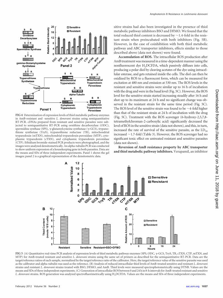
sitive strains had also been investigated in the presence of thiolmetabolic pathway inhibitors BSO and DFMO. We found that thetotal reduced thiol content is decreased by �1.4-fold in the resis-tant strain when preincubated with both inhibitors (Fig. 5B).However, in the case of coinhibition with both thiol metabolicpathway and ABC transporter inhibitors, effects similar to thosedescribed above (data not shown) were found.
Accumulation of ROS. The intracellular ROS production afterAmB treatment was measured in a time-dependent manner using thenonfluorescent dye H2DCFDA, which passively diffuses into cells,producing a polar diol by cleaving acetates of the dye using intracel-lular esterase, and gets retained inside the cells. The diol can then beoxidized by ROS to a fluorescent form, which can be measured forexcitation at 480 nm and emission at 530 nm. The ROS levels in theresistant and sensitive strains were similar up to 16 h of incubationwith the drug and were in the basal level (Fig. 5C). However, the ROSlevel for the sensitive strain started increasing steadily after 16 h andshot up to its maximum at 24 h and no significant change was ob-served in the resistant strain for the same time period (Fig. 5C).The ROS level of the sensitive strain was found to be �4-fold higherthan that of the resistant strain at 24 h of incubation with the drug(Fig. 5C). Treatment with the ROS scavenger (6-hydroxy-2,5,7,8-tetramethylchroman-2-carboxylic acid) significantly decreased thelevel of ROS in the sensitive strain (data not shown), and this, in turn,increased the rate of survival of the sensitive parasite, as the LD50
increased �1.7-fold (Table 3). However, the ROS scavenger had nosignificant toxic effect on untreated resistant and sensitive parasites(data not shown).
Reversion of AmB resistance property by ABC transporterand thiol metabolic pathway inhibitors. Verapamil, an inhibitor
FIG 4 Determination of expression levels of thiol metabolic pathway enzymesin AmB-resistant and -sensitive L. donovani strains using semiquantitativeRT-PCR. cDNAs prepared from resistant and sensitive parasites were sub-jected to semiquantitative RT-PCR using ornithine decarboxylase (ODC),spermidine synthase (SPS), �-glutamylcysteine synthetase (�-GCS), trypano-thione synthetase (TryS), trypanothione reductase (TR), mitochondrialtryparedoxin (mTXN), mitochondrial tryparedoxin peroxidase (MTP), cyto-plasmic tryparedoxin (cTXN), and cytoplasmic tryparedoxin peroxidase(CTP). Ethidium bromide-stained PCR products were photographed, and theimages were analyzed densitometrically. An alpha-tubulin PCR was conductedto show uniform expression of a housekeeping gene in both parasites. Data arethe means and SDs of three independent experiments. Panel 1 shows the gelimages; panel 2 is a graphical representation of the densitometric data.
FIG 5 (A) Quantitative real-time PCR analysis of expression levels of thiol metabolic pathway enzymes (SPS, ODC, �-GCS, TryS, TR, cTXN, CTP, mTXN, andMTP) for AmB-treated resistant and sensitive L. donovani strains using the same set of primers as described for the semiquantitative RT-PCR. Data are thetarget/reference ratios of each sample, normalized by the target/reference ratio of the calibrator. Here, the target/reference value of the sensitive parasite was usedas the calibrator and alpha-tubulin was used as the reference. (B) Analysis of reduced intracellular thiol levels of AmB-treated sensitive and resistant L. donovanistrains and resistant L. donovani strains treated with BSO, DFMO, and AmB. Thiol levels were measured spectrophotometrically using DTNB. Values are themeans and SDs of three independent experiments. (C) Generation of intracellular ROS between 0 and 24 h in 8-h intervals for AmB-treated resistant and sensitiveL. donovani strains. ROS generation was analyzed spectrofluorimetrically using H2DCFDA. Values are the means and SDs of three independent experiments.
Amphotericin B Resistance in Leishmania donovani
February 2012 Volume 56 Number 2 aac.asm.org 1037
on June 11, 2018 by guesthttp://aac.asm
.org/D
ownloaded from
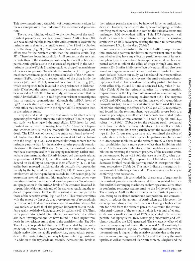
of MDR1, had no significant toxic effect on the untreated sensitiveand resistant parasites (data not shown). Preincubation with ve-rapamil demonstrated partial reversion of the resistant property,as the LD50 of AmB for the resistant strain decreased �1.9-fold(Table 3). However, the LD50 of the sensitive strain was not alteredby preincubation with verapamil (the LD50 decreased �1.01-fold). BSO and DFMO are inhibitors of �-GCS and ODC, respec-tively, which are important rate-limiting enzymes of the thiol met-abolic pathway. It has been demonstrated that BSO and DFMOhave no significant toxic effect on the untreated sensitive and re-sistant parasites (data not shown). Preincubation with BSO andDFMO demonstrated a partial reversion of the resistant property,as the LD50 of AmB for the resistant strain decreased �1.8-fold(Table 3). However, the LD50 of the sensitive strain did not changesignificantly by preincubation with the same inhibitors (the LD50
decreased �1-fold). Coinhibition with both thiol metabolic path-way and ABC transporter inhibitors demonstrated a much largerdecrease in the LD50 value (�2.4-fold) for the resistant strain thandid inhibition with either ABC transport inhibitors or thiol met-abolic pathway inhibitors (Table 3), whereas the decrease in theLD50 value was insignificant for the sensitive strain (�1.04-fold)(Table 3). Therefore, coinhibition has a much more potent effecton the partial strain reversion.
DISCUSSION
In this study, an attempt was made for the first time to understandthe mechanism of resistance developed by clinical isolates ofLeishmania donovani against AmB. The clinical isolates obtainedfrom the VL patients were axenic, as they were selected in NNNblood agar medium and characterized by kDNA amplification(Fig. 1). The resistant or sensitive nature of the clinical isolates wasconfirmed by both in vitro and ex vivo drug sensitivity assays (Ta-ble 1). The resistant isolates were stable even after being exposed tothe drug. In addition, the resistant line did not show cross-resistance to paromomycin, hexadecylphosphocholine, or so-dium stibogluconate, but a low level of resistance to sodium anti-
mony gluconate (SAG) was observed (data not shown). Theseresults suggest that the resistance to AmB may be related to thechemical structure of the drug.
Sterols of Leishmania species have been previously studied, andcommon characteristics in biosynthetic pathways have beenfound between Leishmania species and fungi (22, 23). It has earlierbeen reported that in a laboratory-derived AmB-resistant Leish-mania promastigote, ergosterol is replaced by a precursor,cholesta-5,7,24-trien-3�-ol (38). This probably results from a de-fect in C-24 transmethylation due to a loss of function of SCMT(12). According to Pourshafie et al. (48), SCMT has two tran-scripts, SCMT A and SCMT B. SCMT A is absent and SCMT B ishighly expressed in AmB-resistant L. donovani. However, SCMT Bhas no spliced leader sequence and translation may be prevented,which could be the cause of the absence of sterol transmethylation(48). Our findings also demonstrated that SCMT A is absent andSCMT B is overexpressed in the AmB-resistant parasites com-pared to levels in sensitive parasites (Fig. 2C and D) and henceindicate the presence of an altered sterol (with a defect in C-24transmethylation) in the resistant strain (12). To confirm our hy-pothesis, we analyzed the sterol compositions of both the AmB-resistant and -sensitive parasites through a standard GC/MS tech-nique, and the mass spectra for TMS ether derivatives (see thesupplemental material) were used to identify the structure of thesterols. Four different sterols have been identified on the basis ofthe fragment ions of the mass spectra (for the pattern of fragmen-tation, see references 21, 22, 30, 37, and 38). We found that ergos-terol was the major sterol present in the sensitive strain, while inAmB-resistant promastigotes, the major sterol was cholesta-5,7,24-trien-3�-ol (Table 2) and no ergosterol was detected (Table2). AmB-resistant Leishmania species are therefore defective inC-24 transmethylation of C-27 sterols. It has been reported (usinglecithin liposome as a biometric model) that interaction betweenAmB and the membrane needs the ordered state (low fluidity) ofthe membrane (27). Membrane fluidity is dependent on the steroland phospholipid composition (3, 13). According to Mbongo etal. (38), fatty acid composition is not significantly affected by re-sistance, and the predominant effect on membrane fluidity couldbe ascribed to cholesta-5,7,24-trien-3�-ol. It has been reportedthat fluorescence anisotropy is inversely proportional with themembrane fluidity (29). Fluorimetric analysis demonstratedlower anisotropy in the motion of the diphenylhexatriene fluores-cent probe, which indicated a higher fluidity of the membrane forthe resistant strain than for the sensitive strain (Fig. 2A). It wasalso demonstrated earlier that cholesterol decreased the order ofdipalmitoyl-lecithin liposomes by reducing interactions betweenhydrocarbon chains of phospholipids (27). In AmB-resistant pro-mastigote membranes, cholesta-5,7,24-trien-3�-ol could act sim-ilarly and make the membrane less ordered. These observationssuggest that the high membrane fluidity and less-ordered state ofthe membranes of AmB-resistant parasites are mainly due to thepresence of cholesta-5,7,24-trien-3�-ol instead of ergosterol.Therefore, altered sterol composition and increased membranefluidity of the resistant strain probably make its membrane lessordered and, in turn, decrease binding affinity of AmB to themembrane.
Sokol-Anderson et al. (52) had reported earlier that sensitivityto AmB was measured as a loss of K�i after exposure to AmB infungi. We have also found that after AmB treatment, K�i contentis lower in the sensitive strain than in the resistant strain (Fig. 2B).
TABLE 3 Analysis of partial reversion of the resistant strain using ABCtransporter and thiol metabolic pathway inhibitors and the ROSscavenger (6-hydroxy-2,5,7,8-tetramethylchroman-2-carboxylic acid)
Experimental set [strain � AmB �scavenger or inhibitor(s)]
LD50
(�g/ml)a
Fold changefrom LD50
of straintreated withonly AmBb
Resistant strain � AmB 0.837Sensitive strain � AmB 0.125Sensitive strain � AmB � ROS scavenger 0.22 �1.7Resistant strain � AmB � verapamil 0.43 �1.9Resistant strain � AmB � BSO � DFMO 0.46 �1.8Resistant strain � AmB � BSO � DFMO �
verapamil0.35 �2.4
Sensitive strain � AmB � verapamil 0.122 �1.01Sensitive strain � AmB � BSO � DFMO 0.124 �1.0Sensitive strain � AmB � BSO � DFMO �
verapamil0.120 �1.04
a LD50 of AmB in presence and absence of different inhibitors has been calculated froman in vitro drug sensitivity assay as described in Materials and Methods.b Fold increase (in the presence of ROS scavenger) or decrease (in the presence ofdifferent inhibitors) from the LD50 value of the sensitive and resistant strains treatedwith only AmB.
Purkait et al.
1038 aac.asm.org Antimicrobial Agents and Chemotherapy
on June 11, 2018 by guesthttp://aac.asm
.org/D
ownloaded from
This lower membrane permeability of the monovalent cations forthe resistant parasites may lead toward less membrane depolariza-tion.
The reduced binding of AmB to the membrane of the AmB-resistant parasites can also lead toward lower AmB uptake (38).We have found that the intracellular AmB content is lower in theresistant strain than in the sensitive strain after 8 h of incubationwith the drug (Fig. 3C). We have also observed a higher AmBefflux rate for the resistant strain than the sensitive strain (Fig.3D). Therefore, lower intracellular AmB content in the resistantparasite than in the sensitive parasite may be a result of both im-paired AmB uptake due to the absence of ergosterol in the AmB-resistant parasite (Table 2) and upregulated drug efflux machineryof the resistant parasite. To validate the involvement of drug effluxmachinery, we investigated the expression levels of the ABC trans-porters (PgPA, involved in sequestration of the drug inside thevesicles [45], and MDR1, involved in efflux of the drug [25])which are reported to be involved in drug resistance in leishman-iasis (47) in both the resistant and sensitive strains and which maybe involved in AmB efflux. In our study, we have observed that themRNA level of MDR1 is �3-fold higher in resistant promastigotesthan in sensitive promastigotes, although the mRNA levels ofPgPA in each strain are similar (Fig. 3A and B). Therefore, theAmB efflux may correlate with the higher MDR1 expression levelin the resistant strain.
Lamy-Freund et al. reported that AmB could affect cells bygenerating free radicals after auto-oxidizing itself (32). In the pres-ent study, we investigated the intracellular ROS levels for bothresistant and sensitive promastigotes after AmB treatment to pre-dict whether ROS is the key molecule for AmB-mediated celldeath. The ROS level of the sensitive strain was found to be �3-fold higher than that of the resistant strain at 24 h of incubationwith the drug (Fig. 5C). Lower intracellular AmB content for theresistant parasite than for the sensitive parasite probably contrib-utes toward this lower ROS level. Moreover, the resistant parasitemay have overexpressed ROS scavenging machinery. Earlier stud-ies have demonstrated that if AmB-induced cell damage is relatedto generation of ROS (41), the cell’s resistance to damage mightdepend on its ability to decompose them efficiently (4, 7). It hadearlier been reported that kinetoplastids detoxify hydroperoxidesmainly by the tryparedoxin pathway (39, 43). To investigate theinvolvement of the tryparedoxin cascade in ROS scavenging, theexpression levels of different thiol metabolic pathway genes wereinvestigated in both resistant and sensitive parasites. We observedan upregulation in the mRNA levels of the enzymes involved intrypanothione biosynthesis and of the enzymes regulating the re-duced trypanothione level in the resistant strain compared tothose of the sensitive strain (Fig. 4 and 5A). This result correlateswith the report by Lin et al. that overexpression of tryparedoxinperoxidase is linked with resistance against oxidative stress (36).Low-molecular-mass thiol also plays an important role in the de-fense against oxidative damage caused by oxidants (42, 47). Thus,in the present study, total intracellular thiol content (reduced) hasalso been investigated and we have found �2-fold-higher thiollevels in the resistant strain than in the sensitive strain (Fig. 5B).Therefore, it is predicted that the ROS generated by the auto-oxidation of AmB may be decomposed by the end product of ahighly active thiol metabolic pathway, i.e., tryparedoxin peroxi-dase in the resistant strain, and may help in conferring resistance.In addition to the tryparedoxin cascade, increased thiol levels in
the resistant parasite may also be involved in better antioxidantdefense. However, the sensitive strain, devoid of upregulated de-toxifying machinery, is unable to combat the oxidative stress andundergoes ROS-dependent killing. This ROS-dependent celldeath can again be confirmed by pretreatment of the sensitivestrain with an ROS scavenger, causing the strain to demonstratean increased LD50 for the drug (Table 3).
We have also demonstrated the effect of ABC transporter andthiol metabolic pathway inhibitors on the resistant strain to findout whether they have any effect on partial reversion of its resis-tant phenotype to a sensitive phenotype. Verapamil had been re-ported earlier to inhibit the efflux of drugs through ABC trans-porters (20). Similar inhibitory effects of verapamil had also beenfound in Trypanosoma and laboratory-derived resistant L. don-ovani isolates (43). In our study, we have found that verapamil (aninhibitor of MDR1) partially reverses the AmB resistance pheno-type, a result which has been demonstrated by partial inhibition ofAmB efflux (Fig. 3C and D) and a decreased LD50 value (�1.9-fold) (Table 3) for the resistant parasites. In trypanosomatids,trypanothione is the key molecule involved in maintaining theoptimal intracellular reducing environment (18). The enzymes�-GCS and ODC catalyze the rate-limiting step of trypanothionebiosynthesis (47). In our present study, we have used BSO andDFMO for inhibiting the effects of �-GCS and ODC, respectively,and found that the resistant phenotype partially reverts back to asensitive phenotype, a result which has been demonstrated by de-creased intracellular thiol content (�1.4-fold) (Fig. 5B) and LD50
values (�1.8-fold) (Table 3) for the resistant strain pretreatedwith thiol metabolic pathway inhibitors. This result correlateswith the report that BSO can partially revert the resistant pheno-type (1, 24). In our study, we have also examined the effect ofcoinhibition with both thiol metabolic pathway inhibitors andABC transporter inhibitors on the resistant strain. We have foundthat coinhibition has a more potent effect than inhibition witheither ABC transporter inhibitors or thiol metabolic pathway in-hibitors in reversing the resistant property of the resistant strain,as demonstrated by an �2.4-fold decrease in the LD50 value dur-ing coinhibition (Table 3), compared to �1.8-fold and �1.9-folddecreases for thiol metabolic pathway and ABC transporter inhib-itors, respectively (Table 3). This may indicate a synergistic in-volvement of both drug efflux and ROS scavenging machinery inconferring AmB resistance.
Taken together, it is concluded that the absence of ergosterol inthe resistant parasite’s membranes and the upregulated AmB ef-flux and ROS scavenging machinery are having a cumulative effectin conferring resistance against AmB to the Leishmania parasite.The affinity of AmB for the membrane in the resistant parasite isless, owing to its altered membrane composition, and concomi-tantly, it reduces the amount of AmB taken up. Moreover, theoverexpressed drug efflux machinery is allowing a higher effluxrate for AmB from the resistant parasite. As a result, the intracel-lular AmB content of the resistant strain is lower and, after auto-oxidation, a smaller amount of ROS is generated. The resistantparasite has upregulated ROS scavenging machinery and effi-ciently detoxifies the ROS generated. The synergistic effect of allthese pathways may lead toward the decreased AmB sensitivity forthe resistant parasite (Fig. 6). In contrast, the AmB sensitivity tothe membrane is higher in the sensitive parasite due to the pres-ence of membrane ergosterol (Table 2), and that is why the AmBuptake, as well as the intracellular AmB content, is higher and the
Amphotericin B Resistance in Leishmania donovani
February 2012 Volume 56 Number 2 aac.asm.org 1039
on June 11, 2018 by guesthttp://aac.asm
.org/D
ownloaded from
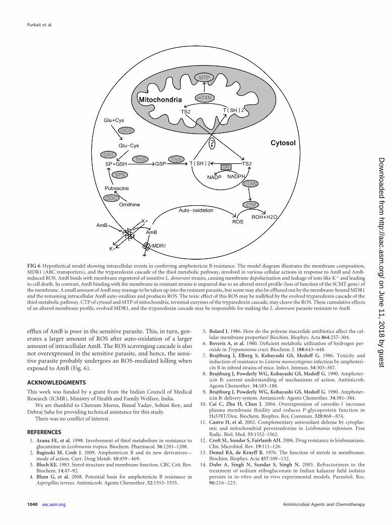
efflux of AmB is poor in the sensitive parasite. This, in turn, gen-erates a larger amount of ROS after auto-oxidation of a largeramount of intracellular AmB. The ROS scavenging cascade is alsonot overexpressed in the sensitive parasite, and hence, the sensi-tive parasite probably undergoes an ROS-mediated killing whenexposed to AmB (Fig. 6).
ACKNOWLEDGMENTS
This work was funded by a grant from the Indian Council of MedicalResearch (ICMR), Ministry of Health and Family Welfare, India.
We are thankful to Chetram Meena, Binod Yadav, Sohini Roy, andDebraj Saha for providing technical assistance for this study.
There was no conflict of interest.
REFERENCES1. Arana FE, et al. 1998. Involvement of thiol metabolism in resistance to
glucantime in Leishmania tropica. Biochem. Pharmacol. 56:1201–1208.2. Baginski M, Czub J. 2009. Amphotericin B and its new derivatives—
mode of action. Curr. Drug Metab. 10:459 – 469.3. Bloch KE. 1983. Sterol structure and membrane function. CRC Crit. Rev.
Biochem. 14:47–92.4. Blum G, et al. 2008. Potential basis for amphotericin B resistance in
Aspergillus terreus. Antimicrob. Agents Chemother. 52:1553–1555.
5. Bolard J. 1986. How do the polyene macrolide antibiotics affect the cel-lular membrane properties? Biochim. Biophys. Acta 864:257–304.
6. Boveris A, et al. 1980. Deficient metabolic utilization of hydrogen per-oxide in Trypanosoma cruzi. Biochem. J. 188:643– 648.
7. Brajtburg J, Elberg S, Kobayashi GS, Medoff G. 1986. Toxicity andinduction of resistance to Listeria monocytogenes infection by amphoteri-cin B in inbred strains of mice. Infect. Immun. 54:303–307.
8. Brajtburg J, Powderly WG, Kobayashi GS, Medoff G. 1990. Amphoter-icin B: current understanding of mechanisms of action. Antimicrob.Agents Chemother. 34:183–188.
9. Brajtburg J, Powderly WG, Kobayashi GS, Medoff G. 1990. Amphoter-icin B: delivery system. Antimicrob. Agents Chemother. 34:381–384.
10. Cai C, Zhu H, Chen J. 2004. Overexpression of caveolin-1 increasesplasma membrane fluidity and reduces P-glycoprotein function inHs578T/Dox. Biochem. Biophys. Res. Commun. 320:868 – 874.
11. Castro H, et al. 2002. Complementary antioxidant defense by cytoplas-mic and mitochondrial peroxiredoxins in Leishmania infantum. FreeRadic. Biol. Med. 33:1552–1562.
12. Croft SL, Sundar S, Fairlamb AH. 2006. Drug resistance in leishmaniasis.Clin. Microbiol. Rev. 19:111–126.
13. Demel RA, de Kruyff B. 1976. The function of sterols in membranes.Biochim. Biophys. Acta 457:109 –132.
14. Dube A, Singh N, Sundar S, Singh N. 2005. Refractoriness to thetreatment of sodium stibogluconate in Indian kalaazar field isolatespersists in in-vitro and in-vivo experimental models. Parasitol. Res.96:216 –223.
FIG 6 Hypothetical model showing intracellular events in conferring amphotericin B resistance. The model diagram illustrates the membrane composition,MDR1 (ABC transporters), and the tryparedoxin cascade of the thiol metabolic pathway, involved in various cellular actions in response to AmB and AmB-induced ROS. AmB binds with membrane ergosterol of sensitive L. donovani strains, causing membrane depolarization and leakage of ions like K� and leadingto cell death. In contrast, AmB binding with the membrane in resistant strains is impaired due to an altered sterol profile (loss of function of the SCMT gene) ofthe membrane. A small amount of AmB may manage to be taken up into the resistant parasite, but some may also be effluxed out by the membrane-bound MDR1and the remaining intracellular AmB auto-oxidizes and produces ROS. The toxic effect of this ROS may be nullified by the evolved tryparedoxin cascade of thethiol metabolic pathway. CTP of cytosol and MTP of mitochondria, terminal enzymes of the tryparedoxin cascade, may cleave the ROS. These cumulative effectsof an altered membrane profile, evolved MDR1, and the tryparedoxin cascade may be responsible for making the L. donovani parasite resistant to AmB.
Purkait et al.
1040 aac.asm.org Antimicrobial Agents and Chemotherapy
on June 11, 2018 by guesthttp://aac.asm
.org/D
ownloaded from
15. Dutcher JD, Gold W, Pagano JF, Vandepatte J. September 1959. Am-photericin B, its production and its salts. US patent 2,908,611.
16. Ellman GL. 1959. Tissue sulfhydryl groups. Arch. Biochem. Biophys. 82:70 –77.
17. Fairlamb AH, Cerami A. 1992. Metabolism and functions of trypano-thione in the Kinetoplastida. Annu. Rev. Microbiol. 46:695–729.
18. Fairlamb AH, Blackburn P, Ulrich P, Chait BT, Cerami A. 1985.Trypanothione: a novel bis (glutathionyl) spermidine cofactor for gluta-thione reductase in trypanosomatids. Science 227:1485–1487.
19. Flohé L, Hecht HJ, Steinert P. 1999. Glutathione and trypanothione inparasitic hydroperoxide metabolism. Free Radic. Biol. Med. 27:966 –984.
20. Fojo A, Akiyama S, Gottesman MM, Pastan I. 1985. Reduced drugaccumulation in multiply drug-resistant human KB carcinoma cell lines.Cancer Res. 45:3002–3007.
21. Galli G, Maroni S. 1967. Mass spectrometric investigations of some un-saturated sterols biosynthetically related to cholesterol. Steroids 10:189 –197.
22. Goad LJ, Holtz GG, Jr, Beach DH. 1984. Sterols of Leishmania species.Implications for biosynthesis. Mol. Biochem. Parasitol. 10:161–170.
23. Goad LJ, Holtz GG, Jr, Beach DH. 1985. Sterols of ketoconazole inhib-ited Leishmania mexicana mexicana promastigotes. Mol. Biochem. Para-sitol. 15:257–279.
24. Grondin K, Haimeur A, Mukhopadhyay R, Rosen BP, Ouellette M.1997. Co-amplification of the gamma-glutamylcysteine synthetase genegsh1 and of the ABC transporter gene pgpA in arsenite-resistant Leishma-nia tarentolae. EMBO J. 16:3057–3065.
25. Henderson DM, et al. 1992. Multidrug resistance in Leishmania donovaniis conferred by amplification of a gene homologous to the mammalianmdr1 gene. Mol. Cell. Biol. 12:2855–2865.
26. Higgins CF. 1992. ABC transporters: from microorganisms to man.Annu. Rev. Cell Biol. 8:67–113.
27. HsuChen C-C, Feingold DS. 1973. Polyene antibiotic action on lecithinliposomes: effect of cholesterol and fatty acyl chains. Biochem. Biophys.Res. Commun. 51:972–978.
28. Jha TK, Giri YN, Singh TK, Jha S. 1995. Use of amphotericin B indrug-resistant cases of visceral leishmaniasis in north Bihar, India. Am. J.Trop. Med. Hyg. 52:536 –538.
29. Kawato S, Kinoshita K, Ikegami A. 1977. Dynamic structure of lipidbilayers studied by nanosecond fluorescence techniques. Biochemistry 16:2319 –2324.
30. Knights BA. 1967. Identification of plant sterol using combined GLC/mass spectrometry. J. Gas Chromatogr. 5:273–282.
31. Krauth-Siegel RL, Bauer H, Schirmer RH. 2005. Dithiol proteins asguardians of the intracellular redox milieu in parasites: old and new drugtargets in trypanosomes and malaria-causing plasmodia. Angew. Chem.Int. Ed. Engl. 44:690 –715.
32. Lamy-Freund MT, Ferreira VFN, Schreier S. 1985. Mechanism of inac-tivation of the polyene antibiotic amphotericin B: evidence for radicalformation in the process of autooxidation. J. Antibiot. (Tokyo) 38:753–757.
33. Lemke A, Kiderlen AF, Kayser O. 2005. Amphotericin B. Appl. Micro-biol. Biotechnol. 68:151–162.
34. Lentz BR. 1993. Use of fluorescent probes to monitor order and motionswithin liposome bilayers. Chem. Phys. Lipids 64:99 –116.
35. Lentz BR, Barenholz Y, Thompson TE. 1976. Fluorescence depolariza-tion studies of phase transitions and fluidity in phospholipids bilayers. 1.Single component phosphatidylcholine liposomes. Biochemistry 15:4521– 4528.
36. Lin YC, Hsu JY, Chiang SC, Lee ST. 2005. Distinct overexpression ofcytosolic and mitochondrial tryparedoxin peroxidases results in preferen-tial detoxification of different oxidants in arsenite-resistant Leishmaniaamazonensis with and without DNA amplification. Mol. Biochem. Para-sitol. 142:66 –75.
37. Massey IJ, Djerassi C. 1979. Structural and stereochemical applications ofmass spectrometry in the marine sterol field. Synthesis and electron im-pact induced mass spectral fragmentation of D24 and D24 (28)-3�-hydroxy-D5-sterols. J. Org. Chem. 44:2448 –2456.
38. Mbongo N, Loiseau PM, Billion MA, Robert-Gero M. 1998. Mechanismof amphotericin B resistance in Leishmania donovani promastigotes. An-timicrob. Agents Chemother. 42:352–357.
39. McGonigle S, Dalton JP, James ER. 1998. Peroxidoxins: a new antioxi-dant family. Parasitol. Today 14:139 –145.
40. Mendez S, Nell M, Alunda JM. 1996. Leishmania infantum: infection ofmacrophages in vitro with promastigotes. Int. J. Parasitol. 26:619 – 622.
41. Moreira W, Leprohon P, Ouellette M. 2011. Tolerance to drug-inducedcell death favours the acquisition of multidrug resistance in Leishmania.Cell Death Dis. doi:10.1038/cddis.2011.83.
42. Mukhopadhyay R, et al. 1996. Trypanothione overproduction and resis-tance to antimonials and arsenicals in Leishmania. Proc. Natl. Acad. Sci.U. S. A. 93:10383–10387.
43. Neal RA, Van BJ, McCoy NG, Iwobi M. 1989. Reversal of drug resistancein Trypanosoma cruzi and Leishmania donovani by verapamil. Trans. R.Soc. Trop. Med. Hyg. 83:197–198.
44. Nogoceke E, Gommel DU, Kiess M, Kalisz HM, Flohe L. 1997. A uniquecascade of oxidoreductases catalyses trypanothione mediated peroxidemetabolism in Crithidia fasciculate. Biol. Chem. 378:827– 836.
45. Ouellette M, et al. 1998. ABC transporters in Leishmania and their role indrug resistance. Drug Resist. Updat. 1:43– 48.
46. Pahissa A. 1997. Amphotericin B. Lipid complex versus liposomes.Which, why, when? Enferm. Infecc. Microbiol. Clin. 15:1–3.
47. Perez-Victoria JM, Parodi-Talice A, Torres C, Gamarro F, Castanys S.2001. ABC transporters in the protozoan parasite Leishmania. Int. Micro-biol. 4:159 –166.
48. Pourshafie M, et al. 2004. Cloning of S-adenosyl-L-methionine:C-24-�-sterolmethyltransferase (ERG6) from Leishmania donovani and character-ization of mRNAs in wild-type and amphotericin B-resistant promasti-gotes. Antimicrob. Agents Chemother. 48:2409 –2414.
49. Ritmeijer K, Dejenie A, Assefa Y. 2006. A comparison of miltefosine andsodium stibogluconate for treatment of visceral leishmaniasis in an Ethi-opian population with high prevalence of HIV infection. Clin. Infect. Dis.43:357–364.
50. Seifert K, Croft SL. 2006. In vitro and in vivo interactions between milte-fosine and other antileishmanial drugs. Antimicrob. Agents Chemother.50:73–79.
51. Sinha PK, et al. 2006. Pre- & post-treatment evaluation of immunologicalfeatures in Indian visceral leishmaniasis (VL) patients with HIV co-infection. Indian J. Med. Res. 123:197–202.
52. Sokol-Anderson M, et al. 1988. Role of cell defense against oxidativedamage in the resistance of Candida albicans to the killing effect of am-photericin B. Antimicrob. Agents Chemother. 32:702–705.
53. Srivastava P, Prajapati VK, Rai M, Sundar S. 2011. Unusual case ofresistance to amphotericin B in visceral leishmaniasis in a region in Indiawhere leishmaniasis is not endemic. J. Clin. Microbiol. 49:3088 –3091.
54. Sundar S, Jha TK, Thakur CP, Sinha PK, Bhattacharya SK. 2007.Injectable paromomycin for visceral leishmaniasis in India. N. Engl. J.Med. 356:2571–2581.
55. Tetaud E, et al. 2001. Molecular characterisation of mitochondrial andcytosolic trypanothione-dependent tryparedoxin peroxidases in Trypano-soma brucei. Mol. Biochem. Parasitol. 116:171–183.
56. Urbina JA, Cohen BE, Perozo E, Cornivelli L. 1987. Spin-labeled am-photericin B: synthesis, characterization, biological and spectroscopicproperties. Biochim. Biophys. Acta 897:467– 473.
57. World Health Organization. 1998. The World Health Report. WHO,Geneva, Switzerland. http://www.who.int/whr/1998/en/whr98_en.pdf.
Amphotericin B Resistance in Leishmania donovani
February 2012 Volume 56 Number 2 aac.asm.org 1041
on June 11, 2018 by guesthttp://aac.asm
.org/D
ownloaded from