mcd24, a glycoprotein transiently expressed by neurons, is
TRANSCRIPT

The Journal of Neuroscience, April 15, 1996, 16(8):2624-2634
mCD24, a Glycoprotein Transiently Expressed by Neurons, Is an Inhibitor of Neurite Outgrowth
Dertyck Shewan,’ Viviane Calaora,z Peter Nielsen,3 James Cohen,’ Genevibve Rougon,* and Her-v6 Moreau*
‘Department of Developmental Neurobiology, United Medical and Dental Schools, Guy’s Campus, London SE1 9RT, United Kingdom, *Laboratoire de G&&tique et Physiologie du D&eloppement, CNRS UMR 9943, Facult6 des Sciences de Luminy, 13288 Marseille Cedex 9, France, and 3Max Planck lnstitut ftir Immunbiologie, 07800 Freiburg, Germany
In the immune system, mCD24, the mouse homolog of the human glycosyl phosphatidylinositol-anchored glycoprotein CD24, may play a role in cell adhesion. In the nervous system, the function of mCD24 has not been determined, but its tran- sient expression by neurons suggests that it may be involved in axon growth in development. Here we show that retinal gan- glion cells (RGCs) and dorsal root ganglion (DRG) neurons express mCD24 in the developing but not adult mouse in viva and in DRG neurons of the injured adult peripheral nervous system (PNS). In vitro, mCD24 was expressed by immature neurons and by a subpopulation of adult DRG neurons. To analyze the possible function of mCD24 in the nervous system, we prepared rat C6 glioma cells stably transfected or retrovi- rally infected with mCD24 cDNA. The cells did not exhibit changes in their adhesive properties or cell division rate after
transfection or infection. When mCD24-expressing cells were used as monolayer substrates for culturing RGCs and DRG neurons, neurite outgrowth was inhibited, depending on neu- ronal age and on the relative levels of mCD24 in the monolayer. This inhibition, however, was not dependent on the expression of mCD24 by the neurons themselves, because DRG neurons of a mouse deleted of the mCD24 gene showed the same response. These results show that mCD24 interacts in a het- erophilic manner with a developmentally regulated molecule expressed by neurons, and they suggest that in viva, mCD24 may inhibit the further extension or collateral branching of axons in late embryonic development.
Key words: cell surface; CD24; axon-growth inhibition; neu- rons; in vitro; mouse; rat
mCD24, the mouse homolog of human CD24, initially defined by use of monoclonal antibodies (mAbs) as heat-stable antigen (Springer et al., 1978; Symington and Hakomori, 1984) and also known as ~31 (Nedelec et al., 1992) or nectadrin (Kadmon et al., 1992), has attracted much interest because of its transient expres- sion in development on cells of the hematopoietic lineage (Hardy et al., 1991), neurons (Rougon et al., 1991; Nedelec et al., 1992) and some epithelial cells (Shirasawa et al., 1993). mCD24 is a glycoprotein with a peptide core of only 30 amino acids (Kay et al., 1991; Wenger et al., 1991), anchored to the membrane via a glycosyl phosphatidylinositol (GPI) group (Pierres et al., 1987). Its molecular weight varies between different cell types. In Western blots of lymphoid cells, mCD24 appears as a broad band of 40-68 kDa (Alterman et al., 1990) whereas in embryonic brain it ranges between 28 and 35 kDa (Rougon et al., 1991). Its amino acid sequence, together with biochemical analyses, suggest that it is highly glycosylated and heterogeneous with respect to both its N-linked and O-linked carbohydrates (Alterman et al., 1990; Kay et al., 1991; Nedelec et al., 1992).
Received Nov. 27, 1995; accepted Jan. 4, 1996.
This work was sunoorted bv Grant CT930326 from the EEC (Concerted Action in Biotechnology) (G:R., J.C.): grants from the Medical Research Council (J.C.), Association Francaise contre les Myopathies (G.R.), Ligue Dtpartemental des Bouches du Rhone de Lutte contre le Cancer (H.M.), and a European Molecular Biology Organization short-term fellowship (D.S.). We thank Dr. J. Major (Wash- ington University School of Medicine, St. Louis, MO) for providing us with the retroviral vectors, and Kevin Fitzpatrick and Sarah Smith for photographic assistance.
Correspondence should be addressed to Derryck Shewan, Department of Devel- opmental Neurobiology, United Medical and Dental Schools, Guy’s Campus, Lon- don SE1 9RT, UK. --
Copyright 0 1996 Society for Neuroscience 0270.64741961162624.ll$U5.00/0
In the immune system, CD24 may be involved in cell adhesion (Kadmon et al., 1992). Although it lacks a transmembrane do- main, it may be able to transduce signals, because Stefanova et al. (1991) reported in B cells a protein kinase activity coprecipitated with CD24; incubation of B cells with mAbs to CD24 results in the tyrosine phosphorylation of several cytoplasmic proteins. Thus, much evidence exists for possible mechanisms of action of CD24 in the immune system, but its role in the nervous system remains largely unexplored.
In mouse brain, mCD24 is expressed between embryonic day 12 and postnatal day 5 (Nedelec et al., 1992); this concurs with immunolabeling data on developing rat cerebellum (Kuchler et al., 1989) and mouse brain (Caladra et al., in press) showing that mCD24 is expressed transiently on postmitotic migrating neuroblasts and in developing axonal tracts. This strategic timing of expression prompted us to study the potential influ- ence of mCD24 on axon outgrowth. We prepared a stable cell line and isolated individual clones of C6 rat glioma cells ex- pressing different levels of mCD24 on their surface. These cells did not exhibit changes in their division rate or adhesive properties induced by low or high levels of expression of mCD24. When used as monolayer substrata for PNS and CNS neurons, however, neurite outgrowth was inhibited substan- tially compared with the exuberant growth observed on un- transfected parental C6 cells. This effect was dependent on both neuronal age and mCD24 levels of expression on the monolayer. The inhibition could not be accounted for by a homophilic interaction of mCD24, because neonatal neurons isolated from a mCD24-deficient mouse were also inhibited. We therefore propose that in the nervous system, mCD24

Shewan et al. . mCD24 Inhibition of Neurite-Outgrowth In Vitro Is Age-Dependent J. Neurosci., April 15, 1996, 76(8):2624-2634 2625
modulates neurite outgrowth via a heterophilic interaction with
a developmentally regulated neuronal receptor.
MATERIALS AND METHODS Construction of mCD24 expression vectors. For transfection, the pRC/ CMV vector (Invitrogen, San Diego, CA) that contains the bacterial neomycin (neo) resistance gene under the control of the SV40 promoter was chosen. The vector was cut in the polylinker at the unique site HindIII. A full-length mCD24 cDNA was excised from pSEX62.2 vector as a BamHI-BarnHI fragment. Both pRC/CMV and mCD24 cDNA were made blunt with T4 DNA polymerase and ligated.
The retroviral vector used in this study was prepared with different vectors provided by Dr. J. Major (Washington University School of Medicine, St. Louis, MO). Brieflv. this vector exuressed the uostream gene (mCD24) from the Rous -sarcoma virus iong-terminal * repeat, whereas the downstream gene (p-gal) was expressed from the encepha- lomyocarditis virus internal ribosome entry site (Ghattas et al., 1991). cDNA coding for mCD24 was inserted in the sense orientation using the 1704 vector BumHI unique site.
C6 transfection and infection, The rat C6 glioma cell line (ATCC CCL107) was cultured in DMEM (Gibco, Cergy Pontoise, France) con- taining 10% fetal calf serum (DMEM-FCS). Fifty micrograms of pRC/ CMV plasmid were transfected by electroporation into 5 X 10h cells suspende’d in 0.8 ml of DMEM-FCS in a 0.4 cm3 cuvette (Eurogentec, Seraing, Belgium) and pulsed with 1800 PF at 250 V. Stable transfectants were selected in G418 (800 Fg/ml of active drug) and cloned by limiting dilution. The isolated clones were probed for surface expression of the mCD24 glycoprotein by fluorescence-activated cell sorter (FACS) anal- ysis using the rat anti-mCD24 mAb clone 194-563 (Rougon et al., 1991).
To produce retroviral infectious particles, the viral producer cell line Psi 2 was transfected using electroporation as above, and viral particles were produced as described by Price et al. (1987). Forty-eight hours after infection, C6 cells were sorted with a Becton Dickinson (Labotechnia, Marseille, France) cell sorter, and the 100% mCD24-positive population that was collected was named A6.
Cell division rate. Individual wells of a 96-well tissue culture plate were inoculated with 0.2 ml of medium containing concentrations of cells ranging from lo3 to lo4 cells/well and incubated for 24-72 hr. 3-(4,5- dimethylthiazol 2-yl)-2,5-diphenyl tetrazolium bromide (Tetrazolium MTT, Sigma, Saint Quentin Fallavier, France) was prepared as a solution of 4 mg/ml in DMEM without phenol red, stored in the dark at 4°C and filter-sterilized. Before use it was diluted 1:4 in DMEM without phenol red, and 0.1 ml was added to each well. The plates were covered with foil and incubated at 37°C for an additional 3 hr. Subsequently, the superna- tants were removed by inverting the plates, and 0.1 ml of propanol was added to extract and solubilize the formazan. After vigorous shaking, the plates were transferred to a microplate reader, and the optical density of each well was measured using a 550 nm wavelength (Miller et al., 1991). Cell division rate was expressed as the ratio of the variation in the total number of cells within 24 hr.
Cell aggregation assay. Subconfluent monolayers of transfected or pa- rental cells were detached from the culture dishes in 10 ml of PBS/l mM EDTA (Gibco) (10 min at room temperature) and dissociated into single cells by gentle pipetting with a Pasteur pipette. After an additional wash in EDTA, the cells were resuspended in complete medium at 2 X 10h cells/ml. For quantification of aggregate formation, they were transferred to 15 ml polystyrene tubes (Falcon, Labotechnia, Marseille, France) that had been coated with FCS for 1 hr at 37°C. The test was performed by incubating 2 ml aliquots of the cell suspension at 37°C in a 7.5% CO, atmosphere without agitation. Aliquots were withdrawn at 15 min inter- vals after mixing by gentle inversion, and the remaining single cells were counted in a hematocytometer (Gennarini et al., 1991).
Preparation of dissociated retinal cells. Retinae from embryonic and neonatal Wistar rats were collected in calcium- and magnesium-free Earle’s balanced salt solution (CMF) (Gibco, Glasgow, UK), transferred to 5 ml papain (Lorne Laboratories, Twyford, UK) (10 U/ml) in a HEPES-buffered salt solution containing cysteine (Leifer et al., 1984) and incubated at 37°C for 15 min. The tissue was then transferred to 0.5 ml CMF/DNase (Type I, 50 pg/ml; Sigma, Poole, UK) and triturated to give a single-cell suspension by several passes through a small-bore pipette tip. The mixed retinal cell suspension from E14-18 rats was plated at a concentration of 100,000 cells/ml.
Retinal ganglion cells (RGCs) were purified from E20-P4 retinal cell suspensions by immunopanning for Thy-l-positive cells using mAb 0x7, as described by Shewan et al. (1995).
Preparation of dissociated dorsal root ganglion (DRG) neurons. Dorsal root ganglia were dissected out from embryonic, postnatal, and adult Wistar rats or C57BL/6 mice, collected in Ham’s F-12 medium (Gibco), and trimmed closely to remove the attached roots. Embryonic and neo- natal DRG tissue was enzymatically treated, and cells were dissociated as described for retinal tissue (see above). The dissociated cells were then diluted to a concentration of 5000/ml in modified Bottenstein and Sato’s tissue culture medium containing 2% fetal calf serum (BSF2) in F-12 supplemented with nerve growth factor (NGF) (100 @ml).
Adult ganglia were incubated at 37°C for 3 hr in 0.125% collagenase (Sigma) in F12, as described by Lindsay (1988) transferred to papain solution, and incubated for an additional 15 min, as described above, washed in F12, and triturated in 1 ml of fresh BSF2. The cell suspension was spun through a 2 ml cushion of 15% bovine serum albumin (fatty acid-free; Sigma) in F-12 to remove myelin debris, and the pelleted cells were resuspended in BSF2 containing NGF (100 @ml) to a cell concen- tration of 2OOO/ml.
Neuronal cultures on C6 cell monolayers. C6 rat glioma cells were passaged using 0.125% trypsin (Sigma) in EDTA (Sigma), collected in an equal volume of conditioned medium, and spun at 200 X g. The pellet was resuspended in conditioned medium, and cells were plated in DMEM- FCS. When the different C6 clones formed monolayers, 0.25 ml of neuronal cell suspensions was pipetted into each well and cultured for a period of 18-24 hr before fixation and immunostaining.
Antibodies. Rabbit anti-GAP43 antiserum from Graham Wilkin (Im- perial College, London) was used as a marker for cultured neurons at a dilution of 1:lOOO (Bedi et al., 1992). Where mixed retinal cultures were used, we confirmed by double immunolabeling that Thy-l-positive RGCs were the only GAP43-positive cells present (data not shown). To test that the underlying transfectediinfected C6 cells expressed mCD24, the anti- GAP43 antibodies were combined in double-immunostaining with a rat mAb to mCD24 (H194-563), at a dilution of 1:lOO.
Immunohistochemistry. For simple mCD24 immunolabeling, cells grown overnight on poly-L-lysine-treated coverslips were incubated with the rat antibody specific to mCD24 and revealed with fluorescein rabbit anti-rat IgG fragments (Immunotech, Marseille, France). Cells were then fixed with cold 5% acetic acid/95% ethanol for 1 min. Retina and DRG sections were fixed with 4% paraformaldehyde, and the immunolabeling was carried out as described above.
Cultures to be labeled solely with neuronal markers were fixed at room temperature for at least 10 min in 2% paraformaldehyde in PBS and permeabilized in 100% methanol at -20°C for 3 min. The labtek slides were incubated first at room temperature for 2 hr (or overnight at 4°C) in the primary antibodies, and then antibody-binding was revealed with the Texas red-biotin-streptavidin system (Amersham, Buckinghamshire, UK).
For mCD24/GAP43 double-immunolabeling, cultures were para- formaldehyde-fixed as above and then first incubated in the anti-mCD24 mAb, revealed by the Texas red-biotin-streptavidin system (Amersham). Cells were then permeabilized with 100% methanol at -20°C and incu- bated with GAP43 antiserum revealed by an anti-rabbit fluorescein- conjugated antibody.
All slides were mounted in glycerol containing diazabicyclo[2,2,2]octane (Sigma) to prevent fading of fluorescence and were examined with a Zeiss Axiophot microscope equipped with epifluorescence.
Cell counts. Counts were made under fluorescence optics of all GAP43- positive neurons attached to the underlying C6 cell monolayers and of those with neurites of more than three cell-body diameters (cbd) in length. The proportion of neurite-bearing cells was expressed as a per- centage of the total number of attached cells in duplicate cultures, averaged for at least three separate preparations of each neuronal type + SEM, statistically evaluated by the Student’s two-tailed t test.
Measurement of total neurite outgrowthlneuron by PI DRG neurons of homozygous and heterozygous mCD24 mutant mice. Cultures of dissoci- ated DRG cells from homozygous and heterozygous mCD24 mutant mice were grown on different monolayers for 18 hr in BSF2. Neurite lengths were quantified after anti-GAP43 immunolabeling of the neurons with a Zeiss Axiophot microscope equipped with epifluorescence and a COHU 4700 camera. Quantification was made using a custom macro written for Optimas 4.1 software package (Bioscan, Washington, D.C.) running on a PC 486DX66 computer with a PC Vision plus frame grabber (Imaging Technology, France). Neurite-length curves were generated in Stat-View (Microsoft) and plotted in Cricket-Graph (Microsoft) as described (Chang et al., 1987). Only neurites that emerged from an isolated neuron and that did not contact other neurites or cells were considered. Total

2626 J. Neurosci., April 15, 1996, 16(8):2624-2634 Shewan et al. l mCD24 Inhibition of Neurite-Outgrowth In Vitro Is Age-Dependent
Figure I. Flow cytometric analysis of parental C6 (A), mCD24-transfected A4 (B), and E3 (C) or infected A6 (D) rat glioma cell lines. A suspension of EDTA-treated cells was labeled with the mAb 193-563 specific to mCD24 (see Materials and Methods) and analyzed with an FACS-Scan (Bec- ton Dickinson), and the number of cells was plot- ted as a function of fluorescence intensity. A6 cells (D) expressing a fluorescence intensity higher than the threshold (dashed line) were sorted to give a 100% mCD24-positive population.
neurite lengths were quantified as the total extent of the arborization from the cell soma to the growth cone, including all branches.
RESULTS
Expression of mCD24 by C6 cells A construct encompassing the entire mCD24 cDNA coding se- quence was cloned into the pRC/CMV plasmid downstream of the strong cytomegalovirus promotor. C6 cells were chosen for trans- fection on the following basis. (1) They do not normally express mCD24, (2) they readily express GPI-anchored molecules at their cell surface, (3) they have a glial phenotype, a cell type normally contacted by neurons, and (4) they rapidly form confluent monolayers.
Of the 21 G418-resistant, independent transfectants, two (A4 and E3) expressed high levels of the mCD24 glycoprotein at the cell surface. FACS analysis indicated that the average level of expression on E3 cells was approximately twice that of A4 (Fig. lB,C); however, these clones showed a high tendency to lose mCD24 expression after several passages. To overcome this dif- ficulty and to make sure that the observations made on individual clones were indeed attributable to mCD24 expression, we pre- pared a retrovirally infected cell line named A6. This cell line was stable after several passages and consistently exhibited the highest level of expression (Fig. 1D).
To assess that all cells were expressing mCD24, A4 and E3 clones and the A6 cell line were grown on coverslips and labeled by immunofluorescence with the anti-mCD24 mAb. Although the intensity of immunolabeling varied between cells of a given clone, as already indicated by the broadness of the peak obtained by FACS, most of the cells were positive, as shown by the subcon- fluent monolayer in Figure 2A. The presence of a GPI anchor for mCD24 in transfected cells was verified by incubating cells on a coverslip with 0.05 U/ml phosphatidylinositol phospholipase C (PI-PLC). This treatment resulted in the disappearance of fluo- rescence intensity after labeling of the cells (data not shown). To test whether the transfection/infection of the C6 cells with mCD24 cDNA affected the cell surface expression of other molecules, we
immunolabeled A6 and parental C6 cells with an antibody against the neural cell adhesion molecule, NCAM, a member of the immunoglobulin superfamily. Figure 2C,D shows that the expres- sion of NCAM does not change after cell transfection. The ex- pression of matrix molecules, such as laminin and fibronectin, is also unaffected by the transfection/infection process (data not shown).
Cell division rate of mCD24-transfected C6 cells Because mCD24 is normally expressed only on postmitotic neu- roblasts, we tested the effect of ectopic expression by C6 cells on their cell division rate. Cell division rates of parental and mCD24 cDNA-transfected C6 cells were measured using a calorimetric assay with MTT after plating different concentrations of cells (see Materials and Methods). The small differences in cell division rates of C6 parental cells (mean 2.8 5 0.21 SEM; ratio of total cell number at 24 hr over total at time 0), and the A4 and E3 mCD24-transfected clones (2.5 i 0.22 and 2.5 + 0.26, respec- tively) were not statistically significant.
Influence of mCD24 on C6 cell aggregation On the basis of experiments using mCD24 purified from lym- phocytes and coated on latex beads, mCD24 glycoprotein has been implicated in homophilic cell-cell adhesion (Kadmon et al., 1992). We therefore investigated whether expression of mCD24 conferred new adhesive properties on the transfected cells by comparison with the parental C6 cell line. Single-cell suspensions were prepared by trituration in the presence of EDTA, a treatment that leaves cell surface mCD24 intact; cells were resuspended in complete medium and incubated for vary- ing times in bacteriological tubes that do not allow cell attach- ment. Under these conditions, the parental cell line was able to aggregate slowly (50% in 60 min) (Fig. 2). This aggregation was abolished completely in the presence of EDTA. Transfectants A4 and E3 displayed similar kinetics of cell-cell aggregation compared with that of parental C6 cells (Fig. 3) which was also EDTA-sensitive.

Shewan et al. l mCD24 Inhibition of Neunte-Outgrowth In V&o Is Age-Dependent J. Neurosci., April 15, 1996, 76(8):2624-2634 2627
Figure 2. Fluorescent photomicrographs showing the expression of mCD24 (A) after retroviral infection of C6 rat glioma cells that normally do not express mCD24 (B) with the cDNA for mCD24. The expression of other cell surface molecules, including NCAM, is not affected by the infection process, because both infected (C) and control (0) cells are equally immunopositive. A, B, 194.563; C, D, NCAM. Scale bar, 40 ym.
Expression of mCD24 by RGCs and DRG neurons in vivo and in vitro To examine the in viva expression of mCD24, prefixed cryosec- tions of mouse brain and spinal cord with attached DRGs taken at various developmental ages, from early embryonic to adult, were immunostained with anti-mCD24 antibody. Within the retina, the RGCs and optic fiber layers were strongly positive by E12.5, as was the optic nerve (Fig. U), at a time when axons have begun to exit the eye (Silver, 1984). Expression remained strong on RGC axons throughout the embryonic optic nerve pathway until the neonatal period, when immunolabeling intensity diminished to reach negligible levels by P15 (data not shown). In adult mice, the optic axons were completely mCD24-negative (Fig. 4B). DRGs were CD2Cpositive as early as E13, and expression was retained at El6 (Fig. 4C). The glycoprotein was downregulated from P2 to negligible levels at PlO (Fig. 40).
Dissociated cultures of mouse retina and DRG neurons grown on merosin were tested for mCD24 expression. More than 90% of both RGCs and DRG neurons were immunopositive for mCD24 at embryonic and neonatal stages (Fig. 54-D). Thus, GAP43- positive RGCs from El7 mice express mCD24 (Fig. 5A,B, small awows) from their soma to the tips of their growth cones. Other GAP43-negative retinal cells were also labeled with mCD24 (large arrows). A similar expression pattern was observed on DRG neurons, because most of the GAP43-positive neurons of El7 mice express mCD24 over their entire surface (Fig. 5C,D).
Inhibitory effect of mCD24 on neurite outgrowth in vitro The growth responses, revealed by GAP43 immunolabeling, of different ages of rat RGCs and DRG neurons cultured on C6 rat glioma cell monolayers are shown in Figure 6. Early embryonic RGCs (E14) extended lengthy processes on the parental, mCD24- negative C6 cells (Fig. 6A). No significant difference could be observed when these RGCs were grown over the mCD24- expressing E3 cell monolayer (Fig. 6B). In contrast, neonatal RGCs that grew lengthy neurites on parental cells (Fig. 6C) were substantially inhibited on E3 monolayers (Fig. 60). Thus, neurite outgrowth by RGCs seemed to be inhibited by mCD24 only at later stages of development.
In common with RGCs, early embryonic DRG neurons regen- erated extensively, not only on parental C6 monolayers (Fig. 6E)
but also over E3 monolayers (Fig. 6F). In the case of neonatal and adult DRG neurons, however, neurite outgrowth was substantially reduced on the mCD24-expressing cell lines (Fig. 6G-J).
Figure 7 shows the magnitude of inhibition for different ages of rat RGCs and DRG neurons. This reveals clearly that for both classes of neurons, neurite outgrowth by the early embryonic cells was undiminished on CD24-expressing C6 cells. In contrast, compared with the extent of outgrowth on control C6 cells, there was striking inhibition of neurite outgrowth by neonatal RGCs (75%) and DRG neurons (62%) and by adult DRG neu- rons (57%). In Figure 7, the data were pooled for the different

2628 J. Neurosci., April 15, 1996, 76(8):2624-2634 Shewan et al. l mCD24 Inhibition of Neurite-Outgrowth In vitro Is Age-Dependent
Figure 3. Aggregation kinetics of mCD24 transfectants and control C6 cells (see Materials and Methods). The results are expressed as percentage of single cells at each time point compared with the number of single cells at time 0. Values shown are the mean 2 SEM for four independent experiments. Cell vi- ability, as judged by trypan blue exclu- sion, remained >90% throughout the experiment.
8 - A4
-.--+-- E3
CD24-expressing lines; however, when the response to individual cell lines was analyzed separately in the case of neonatal RGCs (Fig. 7A, inset), the extent of inhibition of neurite outgrowth was shown to be proportional to the levels of CD24 expression (i.e., A4 < E3 < A6).
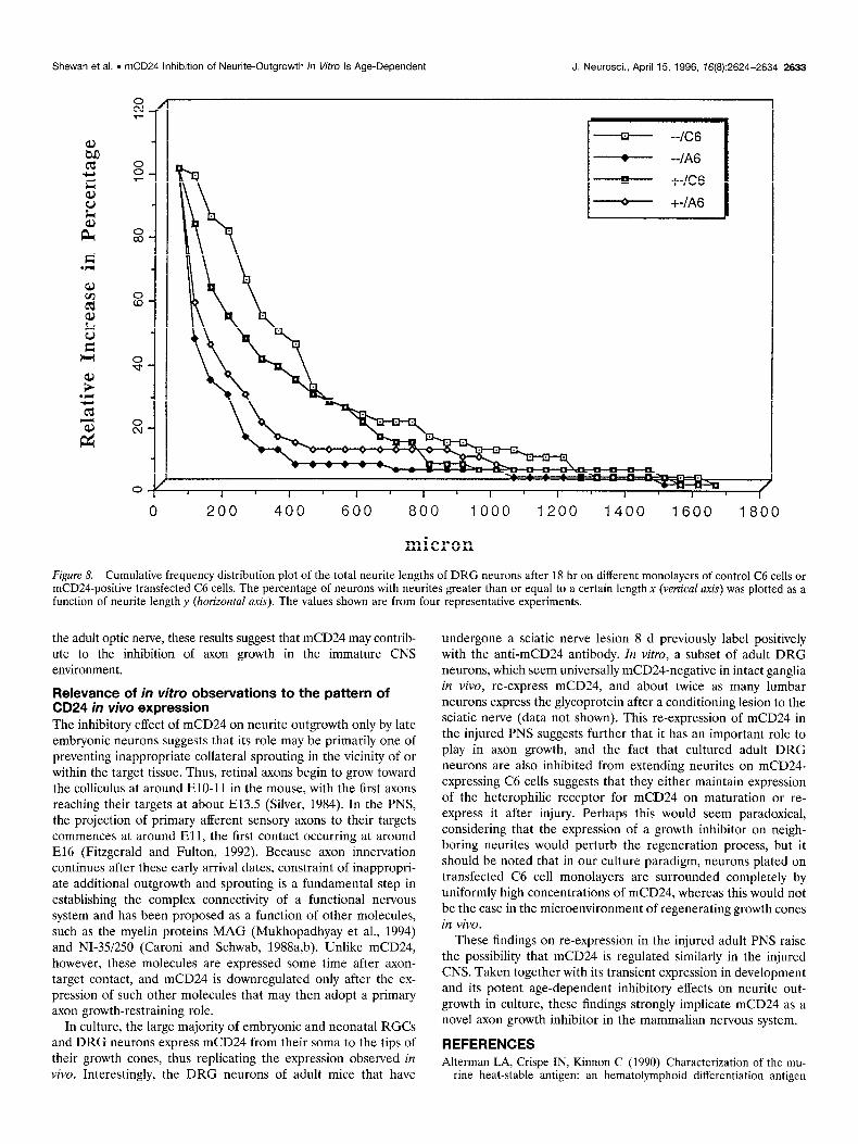
Effect of mCD24 on neurite outgrowth by mCD24-negative neurons The above experiments imply clearly that expression of mCD24 by neurons is of itself insufficient to evoke an inhibitory response and that a heterophilic receptor may be involved. To test this possi- bility, cultures were prepared from neurons obtained from a mouse homozygous for the mCD24 gene deletion (Wenger et al., 1995). All the data presented here were obtained with DRG neurons taken at Pl, a stage in the rat at which consistent inhibition of DRG neurite outgrowth was observed (see above). Four independent experiments were made in parallel with DRG from either homozygous (-/-) or heterozygous (2) pups of the same litter. Analyses were made after 18 hr of culture and GAP-43 immunolabeling, and the average total neurite output per neuron (i.e., the total length of neurites emanating from each cell body) was estimated by image analysis. When the responses of mCD24-negative and -positive neurons cultured on monolayers of C6 or A6 cells were compared, no differences were detected in their behavior (Fig. 8). Nevertheless, the total neurite output per neuron was reduced substantially, regardless of CD24 expression, when grown on mCD24-expressing cells. Thus, Figure 8 shows that for neurons grown on A6 monolayers, 50% of the neurite output has a mean value of -3.50 pm on control C6 cells com-
50 100 150
time (min.)
pared with only 150 pm on mCD24-expressing A6 cells, an inhi- bition of >60%.
DISCUSSION An increasing number of molecules have been implicated in the control of axon growth and guidance in development by their ability to inhibit or repel neurite outgrowth in vitro (for review, see Keynes and Cook, 1995). In pathfinding, axon growth inhibition is probably at least as important as the promotion or support of axon extension, because inhibitory boundaries enable axons to reach appropriate destinations and to know when to stop. In accordance with such an important role, candidate inhibitory molecules show great variety in their patterns of expression and structure and include extracellular matrix glycoproteins such as tenascin and janusin (Bartsch et al., 1992; Wintergerst et al., 1993) and some proteoglycans (Snow et al., 1990, 1991); components of myelin such as myelin-associated glycoprotein (MAG) (Mukhopadhyay et al., 1994) and NI-351250 (Caroni and Schwab, 1988a,b); soluble factors including the netrins (Kennedy et al., 1994; Sefarini et al., 1994) and collapsin (Luo et al., 1993), a member of the conserved semaphorin family (Kolodkin et al., 1993); and GPI-anchored molecules such as ELF-l (Cheng et al., 1995), RAGS (Drescher et al., 1995), and Thy-l (Tiveron et al., 1992).
In the present in vitro studies, we have examined several cellular interactions and possible functions of the developmentally regu- lated GPI-anchored glycoprotein, mCD24. In cells of the immune system, mCD24 has been ascribed a role in cell adhesion (Kad- mon et al., 1992; Hahne et al., 1994). Although it lacks a trans- membrane domain, mCD24 nevertheless may be able to trans-

Shewan et al. . mCD24 Inhibition of Neurite-Outgrowth In Vitro Is Age-Dependent J. Neurosci., April 15, 1996, 16(6):2624-2634 2629
Figure 4. Expression of mCD24 in the mouse optic nerve projection pathway and DRG. A, B, Horizontal sections of head were stained with anti-mCD24 antibodies at E12.5 (A) and P30 (B). Immunoreactivity of both neural retina and optic nerve was observed in the embryo and disappeared in the young adult. Note that the lens appeared to be mCD24-positive, but this tissue was also labeled in controls, indicating a nonspecific reaction. C, D, Sagittal section of DRG stained with anti-mCD24 antibodies at E16.5 (C), and section of dissected P30 DRG (0). In the embryo, DRG neurons were positive for mCD24 expression and immunonegative in the adult. In lumbar DRG of an adult mouse that had undergone sciatic nerve lesion 8 d before removal, GAP43-positive neurons (for example, nrmw~ in E) also express mCD24 (complementary arrows in F). E, GAP43; A-D, F, 194.563. Re, Retina; Le, lens; On, optic nerve; Ve, vertebrae; Dg, dorsal root ganglion. Scale bars: 100 km in A-C, 1 mm in D.
duce signals, because it coprecipitates with a protein kinase activity in B cells (Stefanova et al., 1991), and incubation of B cells with mAbs to CD24 results in the tyrosine phosphorylation of several cytoplasmic proteins. Hitherto, the role of mCD24 in the nervous system has been largely unexplored. The possibility that it may be a novel neural cell adhesion molecule prompted us to test
its effects on neurite growth by both CNS and PNS neurons in vitro. Interestingly, our results show that when stably expressed by the C6 rat glioma cell line and used as a monolayer substrate for cultured neurons, mCD24 inhibits neurite outgrowth in a concentration-dependent manner. This inhibitory response is de- pendent on the age of the neurons and does not operate via a

2630 J. Neurosci., April 15, 1996, 16(8):2624-2634 Shewan et al. l mCD24 Inhibition of Neurite-Outgrowth In Vitro Is Age-Dependent
Figure 5. Photomicrographs showing mCD24 expression by immature mouse retinal cells and DRG neurons in vitro. Double-immunolabeling with GAP43 and mCD24 antibodies revealed that RGCs in El7 retinal cultures expressed mCD24 from the soma to the tips of the growth cones (A, B, small arrows). Some El7 retinal cells that expressed mCD24 were GAP43-negative (large UYXVV~). Double-immunolabeled El7 DRG neurons also exhibited mCD24 expression in their entirety, from soma to growth cones (C, D: n, neuronal cells body). A, C, 193.563; B, D, GAP43. Scale bar, 25 km.
homophilic mechanism, because neurite outgrowth by immature mCD24-positive neurons and neurons isolated from a mCD24- deleted mouse were similarly affected.
Characterization of mCD24-expressing glial cells We have developed a range of C6 rat glioma cells expressing the GPI-anchored mCD24 glycoprotein. As judged by quantitative immunofluorescence analysis, the transfected clones A4 and E3 and the cell-sorted retrovirally infected A6 line all express mCD24 at the cell surface but at different levels; thus, A4 < E3 < A6. The absence of specific homophilic adhesive properties conferred on the transfected cells by mCD24 contrasts with data reported by Kadmon et al. (1992). Thus, mCD24 may have different roles in the immune system, where it is heavily glycosylated, and in the brain, where glycosylation is less extensive.
Inhibition of neurite outgrowth by mCD24 is age- dependent and mediated by a heterophilic receptor Neonatal RGCs and neonatal and adult DRG neurons were substantially inhibited from extending processes over monolayers of mCD24-expressing C6 cells. None of the available anti-mCD24 antibodies were able to disinhibit this effect. This was not surpris- ing, however, because all of these antibodies are known to be directed against a dominant epitope composed of the GPI anchor and an arginine residue in the C-terminal region of the peptide (Rougon et al., 1991; Nedelec et al., 1992). Nevertheless, other lines of evidence exist in favor of a direct effect of mCD24 in
mediating the inhibition of neurite outgrowth. First, inhibition was obtained with one infected and two different transfected clones expressing mCD24. Second, inhibition was more pro- nounced as the level of mCD24 expression by the underlying C6 cells increased. Third, as judged by immunolabeling, expression of other cell surface proteins such as NCAM was not perturbed in the presence of mCD24.
Unlike their more mature counterparts, early embryonic RGCs and DRG neurons failed to respond to mCD24, regardless of the level expressed on the underlying monolayer. This raises an impor- tant question concerning the mode of interaction of neurons with mCD24. Thus, the age-dependent response exhibited by neurons expressing comparable levels of surface mCD24 suggests that a homophilic interaction of neuronal mCD24 is not involved in the inhibition of neurite outgrowth. This implicates a heterophilic mode of interaction of mCD24 expressed by the monolayer with an un- known neuronal receptor. This conclusion is supported further by the finding that neurite outgrowth by DRG neurons from mice homozy- gous for mCD24 gene deletion was also inhibited when these neu- rons were plated on monolayers of mCD24-expressing C6 cells. The nature of this putative heterophilic receptor is unknown. Recent studies on mCD24 suggest P-selectin may be a ligandireceptor in immune tissue (Sammar et al., 1994). In addition, mCD24 (nectadrin) interacts with the Ll glycoprotein on a neuronal cell line and activates the signal transduction pathway of Ll (Kadmon et al., 1995). Such studies suggest that mCD24 may have other functions in

Shewan et al. . mCD24 Inhibition of Neunte-Outgrowth In Vitro Is Age-Dependent J. Neurosci., April 15, 1996, 16(8):2624-2634 2631
Figure 6. Fluorescent photomicrographs showing the neurite outgrowth responses of early embryonic, neonatal, and adult neurons cultured on monolayers of parental and mCD24 cDNA-transfectediinfected C6 cells. El4 RGCs extended long neurites on both C6 cells (A) and E3 cells (I?). Pl RGCs, however, were substantially inhibited from extending neurites on E3 cells (0) when compared with their growth on C6 cells (C). Similarly, El4 DRG neurons grew extensively on both C6 cells (E) and E3 cells (F), whereas neonatal and adult DRG neurons, which regenerated long neurites on C6 cells (G and I, respectively), were markedly inhibited when cultured on A4 cells (H) and A6 cells (I), respectively. A-I, GAP43 antiserum; J, GAP43 mAb. Scale bar: 80 pm in A, C, D, F-J; 40 pm in B; 160 pm in E.

2632 J. Neurosci., April 15, 1996, 16(8):2624-2634 Shewan et al. l mCD24 Inhibition of Neurite-Outgrowth In Vitro Is Age-Dependent
O/
--k T
B 70
60
%
4 50 .E
x
';: 40 3
.E
a
: 30 .= s
; $ 20 E
s
10
0
E14-18
N A4 E3 A6
El41S Neurond Age Neonate
q C6N I-1 El trans T
T
Neonate Adull
Neurond Age
1 T
Figure 7. Histogram showing the neurite outgrowth responses of rat RGCs (A) and DRG neurons (B) of various developmental ages on mCD24negative and -positive C6 cells. The abscissa shows the age of the neurons, whereas the ordinate represents the percentage of neurons that extended neurites of at least 3 cbd in length. Data for the various transfectediinfected cell lines were pooled. E14-18 RGCs grew equally well on mCD24-negative and -positive cells (A), but neonatal RGC neurite outgrowth was inhibited by 75% on transfected cells when compared with growth on C6 cells. The incidence of growth inhibition increased with an increasing level of mCD24 expression (A4 < E3 < A6) on the transfected cells (A, inset). E14-17 DRG neurons also grew extensively on both C6 and mCD24-positive cells (B), but neurite outgrowth by neonatal and adult DRG neurons cultured on mCD24-positive cells was inhibited by 62 and 57%, respectively, when compared with their outgrowth on C6 cells. Open bars symbolize untransfected C6 monolayers; hatched bars represent monolayers of transfected, mCD24-positive cells.
addition to one as an axon growth inhibitor. Thus, the expression of mCD24 by early embryonic neurons that fail to be inhibited by mCD24 may be attributable to functions of the glycoprotein yet to be defined. These findings resemble our earlier work, which demon-
strated a sharp neuronal age-dependent inhibitory effect on neurite outgrowth by both DRG neurons and RGCs grown on unmyelinated neonatal (EWPl) optic nerve cryosections (Shewan et al., 1993, 1995). Because mCD24 is strongly expressed in the immature but not
Shewan et al. . mCD24 Inhibition of Neurite-Outgrowth In Vitro Is Age-Dependent J. Neurosci., April 15, 1996, 16(8):2624-2634 2633
0 200 400 600 800 1000 1200 1400 1600 1800
micron Figure 8. Cumulative frequency distribution plot of the total neurite lengths of DRG neurons after 18 hr on different monolayers of control C6 cells or mCD24-positive transfected C6 cells. The percentage of neurons with neurites greater than or equal to a certain length x (vertical auis) was plotted as a function of neurite lengthy (horizontal wis). The values shown are from four representative experiments.
the adult optic nerve, these results suggest that mCD24 may contrib- ute to the inhibition of axon growth in the immature CNS environment.
Relevance of in vitro observations to the pattern of CD24 in vivo expression The inhibitory effect of mCD24 on neurite outgrowth only by late embryonic neurons suggests that its role may be primarily one of preventing inappropriate collateral sprouting in the vicinity of or within the target tissue. Thus, retinal axons begin to grow toward the colliculus at around ElO-11 in the mouse, with the first axons reaching their targets at about E13.5 (Silver, 1984). In the PNS, the projection of primary afferent sensory axons to their targets commences at around Ell, the first contact occurring at around El6 (Fitzgerald and F&on, 1992). Because axon innervation continues after these early arrival dates, constraint of inappropri- ate additional outgrowth and sprouting is a fundamental step in establishing the complex connectivity of a functional nervous system and has been proposed as a function of other molecules, such as the myelin proteins MAG (Mukhopadhyay et al., 1994) and NI-3512.50 (Caroni and Schwab, 1988a,b). Unlike mCD24, however, these molecules are expressed some time after axon- target contact, and mCD24 is downregulated only after the ex- pression of such other molecules that may then adopt a primary axon growth-restraining role.
In culture, the large majority of embryonic and neonatal RGCs and DRG neurons express mCD24 from their soma to the tips of their growth cones, thus replicating the expression observed in viva. Interestingly, the DRG neurons of adult mice that have
undergone a sciatic nerve lesion 8 d previously label positively with the anti-mCD24 antibody. In vitro, a subset of adult DRG neurons, which seem universally mCD24-negative in intact ganglia in viva, re-express mCD24, and about twice as many lumbar neurons express the glycoprotein after a conditioning lesion to the sciatic nerve (data not shown). This re-expression of mCD24 in the injured PNS suggests further that it has an important role to play in axon growth, and the fact that cultured adult DRG neurons are also inhibited from extending neurites on mCD24- expressing C6 cells suggests that they either maintain expression of the heterophilic receptor for mCD24 on maturation or re- express it after injury. Perhaps this would seem paradoxical, considering that the expression of a growth inhibitor on neigh- boring neurites would perturb the regeneration process, but it should be noted that in our culture paradigm, neurons plated on transfected C6 cell monolayers are surrounded completely by uniformly high concentrations of mCD24, whereas this would not be the case in the microenvironment of regenerating growth cones in vivo.
These findings on re-expression in the injured adult PNS raise the possibility that mCD24 is regulated similarly in the injured CNS. Taken together with its transient expression in development and its potent age-dependent inhibitory effects on neurite out- growth in culture, these findings strongly implicate mCD24 as a novel axon growth inhibitor in the mammalian nervous system.
REFERENCES Alterman LA, Crispe IN, Kinnon C (1990) Characterization of the mu-
rine heat-stable antigen: an hematolymphoid differentiation antigen

2634 J. Neurosci., April 15, 1996, 76(8):2624-2634 Shewan et al. l mCD24 Inhibition of Neurite-Outgrowth In Vitro is Age-Dependent
defined by the Jlld, Ml/69 and B2A2 antibodies. Em J Immunol 20:1597-1602.
Bartsch U, Bartsch S, Dorries U, and Schachner M (1992) Immunohis- tological localization of tenascin in the developing and lesioned adult mouse optic nerve. Eur J Neurosci 4:338-352.
Bedi KS, Winter J, Berry M, Cohen J (1992) Adult rat dorsal root ganglion neurons extend neurites on predegenerated but not on normal peripheral nerves in vitro. Eur J Neurosci 4:193-200.
Caladra V, Chazal G, Nielsen PJ, Rougon G, Moreau H (1996) mCD24 Expression in the developing mouse brain and in zones of secondaty nemogenesis in the adult; Neuroscience, in press.
Caroni P. Schwab ME (1988a) Two membrane orotein fractions from rat central myelin with inhibit&v properties for neurite growth and fibro- blast spreading. J Cell Biol 106:1281-1288.
Caroni P. Schwab ME (1988b) Antibodv against mvelin-associated inhib- itors of neurite growth neutralizes nonper”missive substrate properties of CNS white matter. Neuron 185-96.
Chang S, Ratjen FG, Raper JA (1987) Extension of neurites on axons is impaired by antibodies against specific neural cell surface glycoproteins. J Cell Biol 104:355-362.
Cheng H-J, Nakamoto M, Bergemann AD, Flanagan JG (1995) Comple- mentary gradients in expression and binding of ELF-l and Mek4 in development of the topographic retinotectal projection map. Cell 82:371-381.
Drescher U, Kremoser C, Handwerker C, Loschinger J, Noda M, Bon- hoefer F (1995) In vitro guidance of retinal ganglion cell axons by RAGS, a 25 kDa tectal protein related to ligands for Eph receptor tyrosine kinases. Cell 82:359-370.
Fitzgerald M, Fulton BP (1992) The physiological properties of develop- ing sensory neurons. In: Sensory neurons: diversity, development and plasticity (Scott S, ed), pp 287-306. New York: Oxford UP.
Gennarini G, Durbec P, Boned A, Rougon G, Goridis C (1991) Trans- fected cell surface protein mediates intercellular adhesion and pro- motes neurite outgrowth. Neuron 6:595-606.
Ghattas IR, Sanes JR. Maiors JE (1991) The enceohalomvocarditis virus internal ribosome entry” site allows ethcient coexpression of two genes from a recombinant provirus in cultured cells and in embryos. Mol Cell Biol 11:5848-5859.
Hahne M, Wenger RH, Vestweber D, Nielsen PJ (1994) The heat-stable antigen can alter very late antigen 4-mediated adhesion. J Exp Med 179:1391-1395.
Hardy RR, Carmack CE, Shinton SA, Kemp JD, Hayakawa K (1991) Resolution and characterization of pro-B and pre-pro-B cell stages in normal mouse bone marrow. J ExpMed 173:1213-1225. -
Kadmon G. Eckert M. Sammar M, Schachner M. Altevoet P (1992) Nectadrin, the heat-stable antigen; is a cell adhesion mol&le. J Cell Biol 118:1245-1258.
Kadmon G, von Bohlen und Halbach F, Horstkorte R, Eckert M, Altevogt P, Schachner M (1995) Evidence for cis interaction and cooperative signalling by the heat-stable antigen nectadrin (murine CD24) and the cell adhesion molecule Ll in neurons. Eur J Neurosci 7:993-1004.
Kay R, Rosten PM, Humphries RK (1991) CD24, a signal transducer mod- ulating B cell activation responses, is a very short peptide with a glycosyl phosphatidylinositol membrane anchor. J Immunol 145:1412-1416.
Kennedy TE, Serafini T, de la Torre JR, Tessier-Lavigne M (1994) Netrins are diffusible chemotropic factors for commissural axons in the embryonic spinal cord. Cell 78425-435.
Keynes RJ, Cook GMW (1995) Repulsive and inhibitory signals. Curr Opin Neurobiol 5:75-82.
Kolodkin AL, Matthes DJ, Goodman CS (1993) The semuphorin genes encode a family of transmembrane and secreted growth cone guidance molecules. Cell 75:1389-1399.
Kuchler S, Rougon G, Marschal P, Lehmann S, Reeber A, Vincendon G, Zanetta JP (1989) Location of a transiently expressed glycoprotein in developing cerebellum delineating its possible ontogenic roles. Neuro- science 33:111-124.
Leifer D, Lipton SA, Barnstaple CJ, Masland SH (1984) Monoclonal antibody to Thy-l enhances regeneration of processes by rat retinal ganglion cells in culture. Science 232:303-306.
Lindsay RM (1988) Nerve growth factors (NGF, BDNF) enhance axonal regeneration but are not required for survival of adult sensory neurons. J Neurosci 8:2394-2405.
Luo Y, Raible D, Raper JA (1993) A protein that induces the collapse and paralysis of neuronal growth cones. Cell 75:217-227.
Miller RR, McDevitt CA (1991) A quantitative microwell assay for chon- drocvte cell adhesion. Anal Biochem 192:380-383.
Mukhdpadhyay G, Doherty P, Walsh FS, Cracker PR, Filbin MT (1994) A novel role for myelin-associated glycoprotein as an inhibitor of axonal regeneration. Neuron 13:757-767.
Ntdelec J, Pierres M, Moreau H, Barbet J, Naquet P, Faivre-Sarrailh C, Rougon G (1992) Isolation and characterization of a novel glycosyl phosphatidylinositol-anchored glycoconjugate expressed by developing neurons. Eur J Biochem 203:433-440.
Pierres M, Naquet P, Barbet J, Marcheto S, Marics I, Devaux C, Barad M, Hyman R, Rougon G (1987) Evidence that the hematopoietic cell subset marker JllD is attached to a glycosyl phosphatidylinositol mem- brane anchor. Eur J Immunol 17:1781-1785.
Price J, Turner D, Cepko C (1987) Lineage analysis in the vertebrate nervous system by retrovirus-mediated gene transfer. Proc Nat1 Acad Sci USA 84:156-160.
Rougon G, Alterman LA, Dennis K, Guo XJ, Kinnon C (1991) The murine heat-stable antigen: a differentiation antigen expressed in both the hema- tolymphoid and neural cell lineages. Eur J Immunol 21:1397-1402.
Sammar M, Aigner S, Hubbe M, Schirrmacher V, Schachner M, Vestwe- ber D, Altevogt P (1994) Heat-stable antigen (CD24) as ligand for mouse P-selectin. Int Immunol 6:1027-1036.
Serafini T, Kennedy TE, Galko MJ, Mirzayan C, Jesscll TM, Tessier- Lavigne M (1994) The netrins define a family of axon outgrowth- promoting proteins homologous to C. elegans UNC-6. Cell 78:409-424.
Shewan D, Berry M, Bedi K, Cohen J (1993) Embryonic optic nerve tissue fails to support neurite outgrowth by central and peripheral neurons in vitro. Eur J Neurosci 5809-817.
Shewan D, Berry M, Cohen J (1995) Extensive regeneration in vitro by early embryonic neurons on immature and adult CNS tissue. J Neurosci 15:2057-2062.
Shirasawa T, Akashi T, Sakamoto K, Takahashi H, Maruyama N, Hiro- kawa K (1993) Gene expression of CD24 core peptide molecule in developing brain and developing non-neural tissues. Dev Dyn 198:1-13.
Silver J (1984) Studies on the factors that govern directionality of axon growth in the embryonic optic nerve and at the chiasm of mice. J Comp Neurol 223:238-251.
Snow DM, Steindler DA, Silver J (1990) Molecular and cellular charac- terization of the glial roof plate of the spinal cord and optic tectum: a possible role for a proteoglycan in the development of an axon barrier. Dev Biol 138359-376.
Snow DM, Watanabe M, Letourneau PC, Silver J (1991) A chondroitin sulfate proteoglycan may influence the direction of retinal ganglion cell outgrowth. Development 113:1473-1486.
Sperry RW (1943) Visuomotor coordination in the newt (Triturus viride- scens) after regeneration of the optic nerve. J Comp Neural 79:33-55.
Springer T, Galfrt G, Secher D, Milstein C (1978) Monoclonal xeno- genie antibodies to murine cell surface antigens: identification of novel leukocytes differentiation antigens. Eur J Immunol 8:539-551.
Stefanova I, Horejsi V, Ansotegui IJ, Knapp W, Stockinger H (1991) GPI-anchored cell-surface molecules complexed to protein tyrosine kinases. Science 254:1016-1019.
Symington FW, Hakomori SI (1984) Hematopoietic subpopulations ex- press cross-reactive, lineage-specific molecules detected by monoclonal antibody. Mol Immunol 21:507-514.
Wenger RH, Ayane M, Bose R, Kohler G, Nielsen PJ (1991) The genes for a mouse hematopoietic differentiation marker called the heat-stable antigen. Eur J Immunol 21:1039-1046.
Wenger RH, Kopf M, Nitschke L, Lamers MC, Kolher G, Nielsen PJ (1995) B-cell maturation in chimaeric mice deficient for the heat stable antigen (HSAimouse CD24). Transgenic Res 4:173-183.
Wintergerst ES, Fuss B, Bartsch U (1993) Localization of janusin mRNA in the central nervous system of the developing and adult mouse. Eur J Neurosci 5:299-310.